Observational study designs Alka M. Kanaya, MD Professor of Medicine, Epidemiology & Biostatistics...
52
Observational study designs Alka M. Kanaya, MD Professor of Medicine, Epidemiology & Biostatistics University of California, San Francisco
-
Upload
aleesha-watkins -
Category
Documents
-
view
215 -
download
0
Transcript of Observational study designs Alka M. Kanaya, MD Professor of Medicine, Epidemiology & Biostatistics...

Observational study designs
Alka M. Kanaya, MDProfessor of Medicine, Epidemiology & Biostatistics
University of California, San Francisco

Objectives
To understand the difference between descriptive and analytic observational studies
To identify the strengths and weakness of different designs and apply different study designs to the same research question
To recognize types of study designs in the literature

Descriptive vs. Analytic
Risk factors
Descriptive Questions
What proportion of patients in the DGIM have diabetes?
What is the average age of patients with diabetes in the DGIM?
Diabetes
Analytic QuestionsIs race/ethnicity associated with diabetes among DGIM patients?
Is excessive consumption of sugar-sweetened beverages associated with diabetes among DGIM patients?

Analytic Studies Attempt to establish a causal link between
a predictor/risk factor and an outcome.
You are doing an analytic study if you have any of the following words in your research question: causes, leads to, compared with, more likely
than, associated with, related to, similar to, correlated with, greater than, less than
Predictor(risk factor)
Outcome(disease)

Hierarchy of Study Types??
Descriptive•Case report•Case series•Survey
Analytic
Observational•Cross sectional•Case-control•Cohort studies
Experimental•Randomized controlled trials
Strength of evidence for causality between a risk factor and outcome
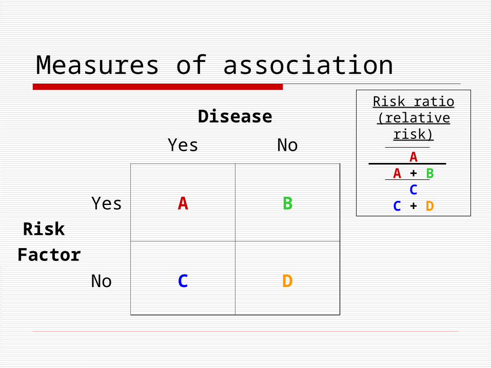
Measures of association
Disease
Yes No
Risk Facto
r
Yes A B
No C D
Risk ratio (relative risk)
AA + B
CC + D

Key elements of study design
Timing of the study
Timing of variable occurrence and
measurement
How the subjects will be sampled
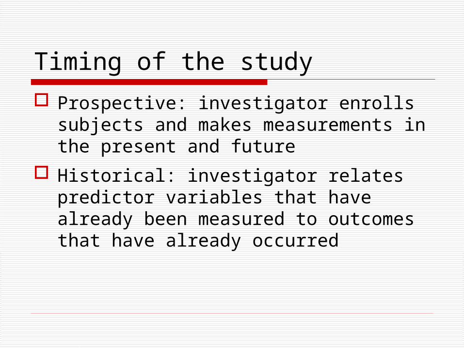
Timing of the study
Prospective: investigator enrolls subjects and makes measurements in the present and future
Historical: investigator relates predictor variables that have already been measured to outcomes that have already occurred

Prospective studies
Control over subject selection and variable measurements
Have to wait for outcomes to occur Take longer More expensive

Historical studies
Less control over subject selection and variable measurements
Outcomes have already occurred Done sooner Less expensive

Timing of measurements
Longitudinal: measurements in subjects made at more than one time
Cross-sectional: predictor and outcome measured at the same time

Research Question
Among patients seen in General Medicine Clinic, who is
at risk for type 2 diabetes?

Great idea, but how do you get started….
Observations in clinical practice
Moving from descriptive to analytic studies
What is feasible?

Study Design #1 Cross-sectional study
National Health and Nutrition Exam Survey (NHANES)
Outcome: “have you been told by a doctor that you have diabetes?”
Multiple possible predictors (demographic, behavioral/lifestyle, other risk factors)
Research question: Is depression associated with diabetes risk?
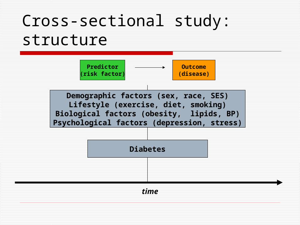
Cross-sectional study: structure
time
Diabetes
Demographic factors (sex, race, SES)Lifestyle (exercise, diet, smoking)
Biological factors (obesity, lipids, BP)Psychological factors (depression, stress)
Predictor(risk factor)
Outcome(disease)
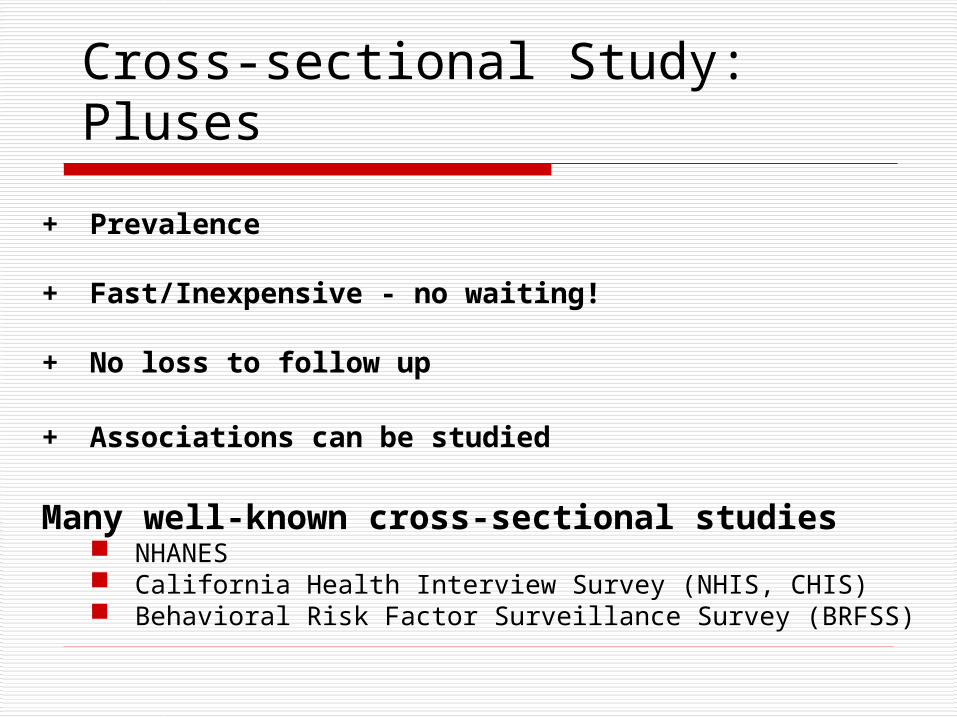
Cross-sectional Study: Pluses
+ Prevalence
+ Fast/Inexpensive - no waiting!
+ No loss to follow up
+ Associations can be studied
Many well-known cross-sectional studies NHANES California Health Interview Survey (NHIS, CHIS) Behavioral Risk Factor Surveillance Survey (BRFSS)

Cross-sectional study: minuses
time
- Cannot determine causality
Diabetes
Depression

Cross-sectional study: minuses
- Cannot study rare outcomes
- Cannot determine incidence
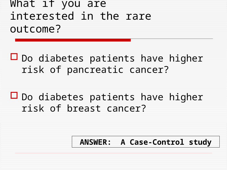
What if you are interested in the rare outcome?
Do diabetes patients have higher risk of pancreatic cancer?
Do diabetes patients have higher risk of breast cancer?
ANSWER: A Case-Control study

Study Design #2 A case-control study
Cases: Patients with pancreatic cancer General medicine vs. oncology UCSF vs. community practice
Controls: Patients without pancreatic cancer Who are the appropriate controls?
Potential predictors: based on questionnaire demographic, behavioral, co-morbid risk factors
Research question: Is type 2 diabetes associated with pancreatic cancer?
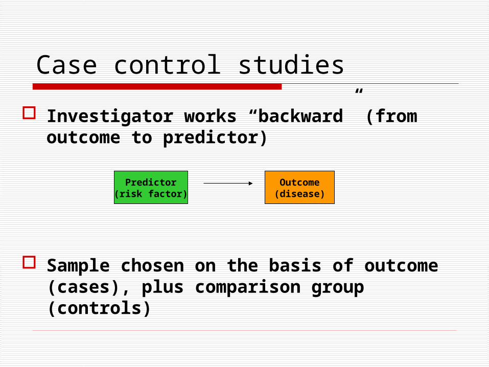
Case control studies
Investigator works “backward” (from outcome to predictor)
Sample chosen on the basis of outcome (cases), plus comparison group (controls)
Predictor(risk factor)
Outcome(disease)
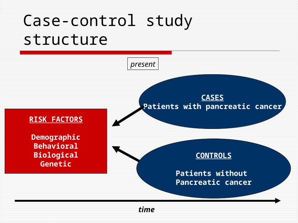
Case-control study structure
time
CASESPatients with pancreatic cancer
CONTROLS
Patients without Pancreatic cancer
RISK FACTORS
DemographicBehavioralBiologicalGenetic
present

Case control studies
Cannot yield estimates of incidence or prevalence of disease in the population (why?)
Odds Ratio is statistics
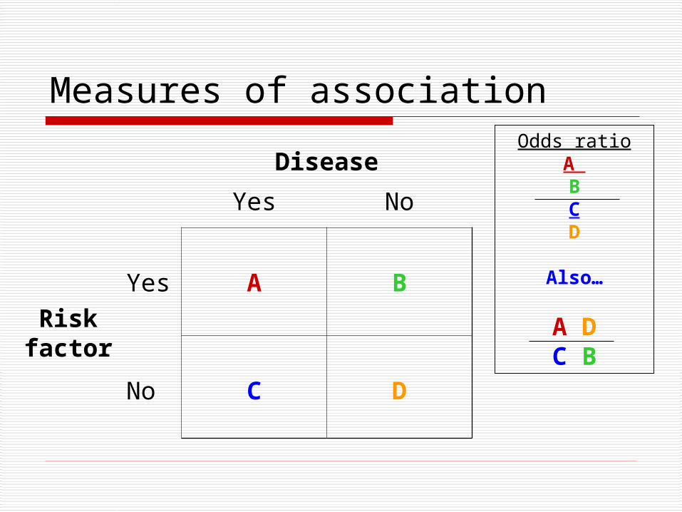
Measures of association
Disease
Yes No
Risk factor
Yes A B
No C D
Odds ratioA BCD
Also…
A DC B
Case-control Study: pluses
+ Rare outcome/Long latent period
+ Inexpensive and efficient: may be only feasible option
+ Establishes association (Odds ratio)
+ Useful for generating hypotheses (multiple risk factors can be explored)

Case-control study-minuses
- Causality still difficult to establish
- Selection bias (appropriate controls)- Caffeine and Pancreatic cancer in the GI clinic
- Recall bias: sampling (retrospective)- Abortion and risk of breast cancer in Sweden

Case-control study--minuses
Incidence New cases of an outcome that occur Can measure in a cohort study
Prevalence Cases that exist at a given time Can measure in a cross-sectional study
Neither can be measured in a case-control study

Case-control - “the house red” Rely tampons and toxic shock syndrome:
High rates of toxic shock syndrome in menstruating women
Suspected OCPs or meds for PMS
Cases: 180 women with TSS in 6 geographic areas
Controls: 180 female friends of these patients and 180 females in the same telephone code
Tampon associated with TSS (OR = 29!)
Super absorbency associated with TSS (OR 1.34 per gm increase in absorbency)
Led to “RELY” brand tampons being taken off the market.

Where are we?
Preliminary results from our cross-sectional and case-control study suggest that black race, hypertension, and chronic kidney disease are associated with premature heart failure.
What’s missing? - strengthening evidence for a causal link between risk factors and heart failure.
Use results from our previous studies to apply for funding for a prospective cohort study!

Study design #3 Prospective cohort study
DISTANCE study: 64,211 Kaiser diabetes registry patients Age >18 years Followed for 10 years (1996-2006)
Primary predictor: race/ethnicity (8 groups) Outcomes: Incident MI, CHF, stroke, ESRD,
LEA

Elements of a cohort study
Selection of sample from population Measures predictor variables in sample Follow population for period of time Measure outcome variable
Famous cohort studies Framingham Nurses’ Health Study Physicians’ Health Study
Predictor(risk factor)
Outcome(disease)

time
The present The future
MI, CHF, stroke, ESRD, LEA
Everyone else
Prospective cohort study structure

What we found…
•Lots of heterogeneity among the Asian American groups
•Patterns differ greatly for macrovascular and microvascular outcomes
Kanaya, Diabetes Care, 2011

Strengths of cohort studies Know that predictor variable was present
before outcome variable occurred (some evidence of causality)
Directly measure incidence of a disease outcome
Can study multiple outcomes of a single exposure (RR is measure of association)

Weaknesses of cohort studies Expensive and inefficient for studying rare
outcomes HERS vs. WHI
Often need long follow-up period or a very large population CARDIA
Loss to follow-up can affect validity of findings Framingham

Other types of cohort studies
Retrospective cohort Identification of cohort, measurement of
predictor variables, follow-up and measurement of outcomes have all occurred in the past
Much less costly than prospective cohorts Investigator has minimal control over study
design

Studies of Medical Tests
Causality often irrelevant. Not enough to show that test result
is associated with disease status or outcome*.
Need to estimate parameters (e.g., sensitivity and specificity) describing test performance.
*Although if it isn’t, you can stop.

Studies of Diagnostic Test Accuracy for Prevalent Disease
Predictor = Test ResultOutcome = Disease status as
determined by Gold Standard
Designs:
•Case-control (sample separately from disease positive and disease negative groups)
•Cross-sectional (sample from the whole population of interest)
•Double-cohort-like sampling (sample separately from test-positive and test-negative groups)

Studies of Dx Tests
Importance of Sampling Scheme
If sampling separately from Disease+ and Disease– groups (case-control sampling), cannot calculate prevalence, positive predictive value, or negative predictive value.

Dx Test: Case-Control Sampling
Disease +Sampled
Separately
Disease –Sampled
Separately
Test +a
True Positives
bFalse Positives
Test -c
False Negatives
dTrue Negatives
Totala + c
Total With Disease
b + dTotal Without
Disease
Sensitivity = a/(a + c) Specificity = d/(b + d)

Dx Test: Cross-sectional Sampling
PPV = a/(a + b)
NPV = d/(c + d)
Prevalence = (a + c)/N
Disease + Disease - Total
Test +
aTrue
Positives
bFalse
Positives
a + bTotal
Positives
Test -
cFalse
Negatives
dTrue
Negatives
c + dTotal
Negatives
Total a + cTotal With
Disease
b + dTotal
WithoutDisease
a + b + c + d
Total N

Studies of Prognostic Tests for Incident Outcomes
Predictor = Test ResultDevelopment of outcome or time to
development of outcome.
Design: Cohort study

Hierarchy of Study Types??
Descriptive•Case report•Case series•Survey
Analytic
Observational•Cross sectional•Case-control•Cohort studies
Experimental•Randomized controlled trials
Strength of evidence for causality between a risk factor and outcome
A study type of every budget, purpose and research question

NAME THAT STUDY DESIGN…
Abstracts from the New England Journal of Medicine

Plasma Natriuretic Peptide Levels and the Risk of Cardiovascular Events and Death
Thomas J. Wang, M.D., Martin G. Larson, Sc.D., Daniel Levy, M.D., Emelia J. Benjamin, M.D., Eric P. Leip, M.S., Torbjorn Omland, M.D., Philip A. Wolf, M.D., and Ramachandran S. Vasan,
M.D.Background The natriuretic peptides are counterregulatory hormones involved in volume homeostasis and
cardiovascular remodeling. The prognostic significance of plasma natriuretic peptide levels in apparently asymptomatic persons has not been established.
Methods We prospectively studied 3346 persons without heart failure. Using proportional-hazards regression, we examined the relations of plasma B-type natriuretic peptide and N-terminal pro–atrial natriuretic peptide to the risk of death from any cause, a first major cardiovascular event, heart failure, atrial fibrillation, stroke or transient ischemic attack, and coronary heart disease.
Results During a mean follow-up of 5.2 years, 119 participants died and 79 had a first cardiovascular event. After adjustment for cardiovascular risk factors, each increment of 1 SD in log B-type natriuretic peptide levels was associated with a 27 percent increase in the risk of death (P=0.009), a 28 percent increase in the risk of a first cardiovascular event (P=0.03), a 77 percent increase in the risk of heart failure (P<0.001), a 66 percent
increase in the risk of atrial fibrillation (P<0.001), and a 53 percent increase in the risk of stroke or transient ischemic attack (P=0.002). Peptide levels were not significantly associated with the risk of coronary heart disease events. B-type natriuretic peptide values above the 80th percentile (20.0 pg per milliliter for men and 23.3 pg per milliliter for women) were associated with multivariable-adjusted hazard ratios of 1.62 for death
(P=0.02), 1.76 for a first major cardiovascular event (P=0.03), 1.91 for atrial fibrillation (P=0.02), 1.99 for stroke or transient ischemic attack (P=0.02), and 3.07 for heart failure (P=0.002). Similar results were obtained for N-terminal pro–atrial natriuretic peptide.
Conclusions In this community-based sample, plasma natriuretic peptide levels predicted the risk of death and cardiovascular events after adjustment for traditional risk factors. Excess risk was apparent at natriuretic peptide levels well below current thresholds used to diagnose heart failure. N Eng J Med 2004; 350:655-663.

Needlestick Injuries among Surgeons in TrainingMartin A. Makary, M.D., M.P.H., Ali Al-Attar, M.D., Ph.D., Christine G. Holzmueller, B.A., J.
Bryan Sexton, Ph.D., Dora Syin, B.S., Marta M. Gilson, Ph.D., Mark S. Sulkowski, M.D., and Peter J. Pronovost, M.D., Ph.D
Background Surgeons in training are at high risk for needlestick injuries. The reporting of such injuries is a critical step in initiating early prophylaxis or treatment. Methods We surveyed surgeons in training at 17 medical centers about previous needlestick injuries. Survey items inquired about whether the most recent injury was reported to an employee health service or involved a "high-risk" patient (i.e., one with a history of infection with human immunodeficiency virus, hepatitis B or hepatitis C, or injection-drug use); we also asked about the perceived cause of the injury and the surrounding circumstances.
Results The overall response rate was 95%. Of 699 respondents, 582 (83%) had had a needlestick injury during training; the mean number of needlestick injuries during residency increased according to the postgraduate year (PGY): PGY-1, 1.5 injuries; PGY-2, 3.7; PGY-3, 4.1; PGY-4, 5.3; and PGY-5, 7.7. By their final year of training, 99% of residents had had a needlestick injury; for 53%, the injury had involved a high-risk patient. Of the most recent injuries, 297 of 578 (51%) were not reported to an employee health service, and 15 of 91 of those involving high-risk patients (16%) were not reported. Lack of time was the most common reason given for not reporting such injuries among 126 of 297 respondents (42%). If someone other than the respondent knew about an unreported injury, that person was most frequently the attending physician (51%) and least frequently a "significant other" (13%).
Conclusions Needlestick injuries are common among surgeons in training and are often not reported. Improved prevention and reporting strategies are needed to increase occupational safety for surgical providers (N Eng J Med 2007; 356:2693-2699).

First-Trimester Use of Selective Serotonin-Reuptake Inhibitors and the Risk of Birth Defects
Carol Louik, Sc.D., Angela E. Lin, M.D., Martha M. Werler, Sc.D., Sonia Hernández-Díaz, M.D., Sc.D., and Allen A. Mitchell, M.D.
Background: The risk of birth defects after antenatal exposure to selective serotonin-reuptake inhibitors (SSRIs) remains controversial.
Methods: We assessed associations between first-trimester maternal use of SSRIs and the risk of birth defects among 9849 infants with and 5860 infants without birth defects participating in the Slone Epidemiology Center Birth Defects Study.
Results: In analyses of defects previously associated with SSRI use (involving 42 comparisons), overall use of SSRIs was not associated with significantly increased risks of craniosynostosis (115 subjects, 2 exposed to SSRIs; odds ratio, 0.8; 95% confidence interval [CI], 0.2 to 3.5), omphalocele (127 subjects, 3 exposed; odds ratio, 1.4; 95% CI, 0.4 to 4.5), or heart defects overall (3724 subjects, 100 exposed; odds ratio, 1.2; 95% CI, 0.9 to 1.6). Analyses of the associations between individual SSRIs and specific defects showed significant associations between the use of sertraline and omphalocele (odds ratio, 5.7; 95% CI, 1.6 to 20.7; 3 exposed subjects) and septal defects (odds ratio, 2.0; 95% CI, 1.2 to 4.0; 13 exposed subjects) and between the use of paroxetine and right ventricular outflow tract obstruction defects (odds ratio, 3.3; 95% CI, 1.3 to 8.8; 6 exposed subjects). The risks were not appreciably or significantly increased for other defects or other SSRIs or non-SSRI antidepressants. Exploratory analyses involving 66 comparisons showed possible associations of paroxetine and sertraline with other specific defects.
Conclusions: Our findings do not show that there are significantly increased risks of craniosynostosis, omphalocele, or heart defects associated with SSRI use overall. They suggest that individual SSRIs may confer increased risks for some specific defects, but it should be recognized that the specific defects implicated are rare and the absolute risks are small. (N Eng J Med 2007;356:2675-83)

THE ROLE OF BLACK AND HISPANIC PHYSICIANS IN PROVIDING HEALTH CARE FOR UNDERSERVED POPULATIONS
MIRIAM KOMAROMY, M.D., KEVIN GRUMBACH, M.D., MICHAEL DRAKE, M.D., KAREN VRANIZAN, M.A., NICOLE LURIE, M.D., M.S.P.H., DENNIS KEANE, M.P.H., AND ANDREW B. BINDMAN, M.D.
Background: Patients who are members of minority groups may be more likely than others to consult physicians of the same race or ethnic group, but little is known about the relation between patients’ race or ethnic group and the supply of physicians or the likelihood that minority-group physicians will care for poor or black and Hispanic patients.
Methods: We analyzed data on physicians’ practice locations and the racial and ethnic makeup and socioeconomic status of communities in California in 1990. We also surveyed 718 primary care physicians from 51 California communities in 1993 to examine the relation between the physicians’ race or ethnic group and the characteristics of the patients they served.
Results: Communities with high proportions of black and Hispanic residents were four times as likely as others to have a shortage of physicians, regardless of community income. Black physicians practiced in areas where the percentage of black residents was nearly five times as high, on average, as in areas where other physicians practiced. Hispanic physicians practiced in areas where the percentage of Hispanic residents was twice as high as in areas where other physicians practiced. After we controlled for the racial and ethnic makeup of the community, black physicians cared for significantly more black patients (absolute difference, 25 percentage points; P <0.001) and Hispanic physicians for significantly more Hispanic patients (absolute difference, 21 percentage points; P<0.001) than did other physicians. Black physicians cared for more patients covered by Medicaid (P<0.001) and Hispanic physicians for more uninsured patients (P=0.03) than did other physicians.
Conclusions:Black and Hispanic physicians have a unique and important role in caring for poor, black, and Hispanic patients in California. Dismantling affirmative action programs, as is currently proposed, may threaten health care for both poor people and members of minoritygroups. (N Engl J Med 1996;334:1305-10.)

Effect of Cigar Smoking on the Risk of Cardiovascular Disease, Chronic Obstructive Pulmonary Disease, and Cancer in Men
Carlos Iribarren, M.D., M.P.H., Ph.D., Irene S. Tekawa, M.A., Stephen Sidney, M.D., M.P.H., and Gary D. Friedman, M.D.
Background The sale of cigars in the United States has been increasing since 1993. Cigar smoking is a known risk factor for certain cancers and for chronic obstructive pulmonary disease (COPD). However, unlike the relation between cigarette smoking and cardiovascular disease, the association between cigar smoking and cardiovascular disease has not been clearly established.
Methods We performed a cohort study among 17,774 men 30 to 85 years of age at base line (from 1964 through 1973) who were enrolled in the Kaiser Permanente health plan and who reported that they had never smoked cigarettes and did not currently smoke a pipe. Those who smoked cigars (1546 men) and those who did not (16,228) were followed from 1971 through the end of 1995 for a first hospitalization for or death from a major cardiovascular disease or COPD, and through the end of 1996 for a diagnosis of cancer.
Results In multivariate analyses, cigar smokers, as compared with nonsmokers, were at higher risk for coronary heart disease (relative risk, 1.27; 95 percent confidence interval, 1.12 to 1.45), COPD (relative risk, 1.45; 95 percent confidence interval, 1.10 to 1.91), and cancers of the upper aerodigestive tract
(relative risk, 2.02; 95 percent confidence interval, 1.01 to 4.06) and lung (relative risk, 2.14; 95 percent confidence interval, 1.12 to 4.11), with evidence of dose–response effects. There appeared to be a synergistic relation between cigar smoking and alcohol consumption with respect to the risk of oropharyngeal cancers and cancers of the upper aerodigestive tract.
Conclusions Independently of other risk factors, regular cigar smoking can increase the risk of coronary heart disease, COPD, and cancers of the upper aerodigestive tract and lung. (N Eng J Med 1999 340:1773-1780)

CLINICAL AND NEURORADIOGRAPHIC MANIFESTATIONS OF EASTERN EQUINE ENCEPHALITIS
ROBERT L. DERESIEWICZ, M.D., SCOTT J. THALER, M.D., LIANGGE HSU, M.D., AND AMIR A. ZAMANI, M.D.
Background: Eastern equine encephalitis occurs principally along the east and Gulf coasts of the United States. Recognition of the neuroradiographic manifestations of eastern equine encephalitis could hasten the diagnosis of the illness and speed the response to index cases.
Methods: We reviewed all cases of eastern equine encephalitis reported in the United States between 1988 and 1994. The records of 36 patients were studied, along with 57 computed tomographic (CT) scans and 23 magnetic resonance imaging (MRI) scan from 33 patients.
Results: The mortality rate was 36 percent, and 35 percent of the survivors were moderately or severely disabled. Neuroradiographic abnormalities were common and best visualized by MRI. Among the patients for whom MRI scans were available, the results were abnormal for all eight comatose patients as well as for all three noncomatose patients who subsequently became comatose. The CT results were abnormal in 21 of 32 patients with readable scans. The abnormal findings included focal lesions in the basal ganglia (found in 71 percent of patients on MRI and in 56 percent on CT), thalami (found in 71 percent on MRI and in 25 percent on CT), and brain stem (found in 43 percent on MRI and in 9 percent on CT). Cortical lesions, meningeal enhancement, and periventricular white-matter changes were less common. The presence of large radiographic lesions did not predict a poor outcome, but either high cerebrospinal fluid white-cell counts or severe hyponatremia did.
Conclusions: Eastern equine encephalitis produces focal radiographic signs. The characteristic early involvement of the basal ganglia and thalami distinguishes this illness from herpes simplex encephalitis. MRI is a sensitive technique to identify the characteristic early radiographic manifestations of this viral encephalitis. (N Engl J Med 1997;336:1867-74.)

Helicobacter pylori Infection and Gastric LymphomaJulie Parsonnet, Svein Hansen, Larissa Rodriguez, Arnold B. Gelb, Roger A. Warnke, Egil
Jellum, Norman Orentreich, Joseph H. Vogelman, and Gary D. Friedman
Background Helicobacter pylori infection is a risk factor for gastric adenocarcinoma. We examined whether this infection is also a risk factor for primary gastric non-Hodgkin's lymphoma.
Methods This __________________________ involved two large cohorts (230,593 participants). Serum had been collected from cohort members and stored, and all subjects were followed for cancer. Thirty-three patients with gastric non-Hodgkin's lymphoma were identified, and each was matched to four controls according to cohort, age, sex, and date of serum collection. For comparison, 31 patients with nongastric non-Hodgkin's lymphoma from one of the cohorts were evaluated, each of whom had been previously matched to 2 controls. Pathological reports and specimens were reviewed to confirm the histologic type of the tumor. Serum samples from all subjects were tested for H. pylori IgG by an
enzyme-linked immunosorbent assay.
Results Thirty-three cases of gastric non-Hodgkin's lymphoma occurred a median of 14 years after serum collection. Patients with gastric lymphoma were significantly more likely than matched controls to have evidence of previous H. pylori infection (matched odds ratio, 6.3; 95 percent confidence interval, 2.0 to 19.9). The results were similar in both cohorts. Among the 31 patients with nongastric lymphoma, a median of six years had elapsed between serum collection and the development of disease. No
association was found between nongastric non-Hodgkin's lymphoma and previous H. pylori infection (matched odds ratio, 1.2; 95 percent confidence interval, 0.5 to 3.0).
Conclusions Non-Hodgkin's lymphoma affecting the stomach, but not other sites, is associated with previous H. pylori infection. A causative role for the organism is plausible, but remains unproved. (N Eng J Med 1994; 330:1267-1271).

Adherence to a Mediterranean Diet and Survival in a Greek Population
Antonia Trichopoulou, M.D., Tina Costacou, Ph.D., Christina Bamia, Ph.D., and Dimitrios Trichopoulos, M.D.
Background Adherence to a Mediterranean diet may improve longevity, but relevant data are limited.
Methods We conducted a _______________________________ involving 22,043 adults in Greece who completed an extensive, validated, food-frequency questionnaire at base line. Adherence to the traditional Mediterranean diet was assessed by a 10-point Mediterranean-diet scale that incorporated the salient characteristics of this diet (range of scores, 0 to 9, with higher scores indicating greater adherence). We used proportional-hazards regression to assess the relation between adherence to the Mediterranean diet and total mortality, as well as mortality due to coronary heart disease and mortality due to cancer, with adjustment for age, sex, body-mass index, physical-activity level, and other potential confounders.
Results During a median of 44 months of follow-up, there were 275 deaths. A higher degree of adherence to the Mediterranean diet was associated with a reduction in total mortality (adjusted
hazard ratio for death associated with a two-point increment in the Mediterranean-diet score, 0.75 [95 percent confidence interval, 0.64 to 0.87]). An inverse association with greater adherence to this diet was evident for both death due to coronary heart disease (adjusted hazard ratio, 0.67 [95 percent confidence interval, 0.47 to 0.94]) and death due to cancer (adjusted hazard ratio, 0.76 [95 percent confidence interval, 0.59 to 0.98]). Associations between individual food groups contributing to the Mediterranean-diet score and total mortality were generally not significant.
Conclusions Greater adherence to the traditional Mediterranean diet is associated with a significant reduction in total mortality. (N Eng J Med 2003; 348:2599-2608)