Nutrigenomics and Selenium: Gene Expression Patterns ... · ANRV350-NU28-09 ARI 10 July 2008 14:15...
28
Nutrigenomics and Selenium: Gene Expression Patterns, Physiological Targets, and Genetics John Hesketh Institute for Cell and Molecular Biosciences, The Medical School, Newcastle University, Newcastle-upon-Tyne, United Kingdom, NE1 4HH; email: [email protected] Annu. Rev. Nutr. 2008.28:157–77 The Annual Review of Nutrition is online at nutr.annualreviews.org This article’s doi: 10.1146/annurev.nutr.28.061807.155446 Copyright c 2008 by Annual Reviews. All rights reserved 0199-9885/08/0821-0157$20.00 Key Words selenoproteins, SNP, transcriptomics, nutrient-gene interactions Abstract Dietary selenium intake is regarded as an important factor in determin- ing optimal health and susceptibility to disease. Therefore, it is critical to understand the interaction between selenium intake and molecular events at the genetic and cellular level. This article addresses two facets of this interaction. The first facet is how genomics is contributing to understanding the molecular mechanisms by which selenium affects cell function through selenoproteins and downstream targets of Se supply in other metabolic pathways. The contribution of transgenic animals in this field is emphasized, and the more recent studies using transcrip- tomics are discussed. The second facet is the extent to which single nucleotide polymorphisms (SNPs) in genes encoding selenoproteins and components of the selenoprotein synthetic machinery affect indi- vidual dietary requirements for optimal health. The state of knowledge of known functional SNPs in selenoprotein genes is presented, and a strategy for future studies is discussed. 157 Click here for quick links to Annual Reviews content online, including: • Other articles in this volume • Top cited articles • Top downloaded articles • Our comprehensive search Further ANNUAL REVIEWS Annu. Rev. Nutr. 2008.28:157-177. Downloaded from www.annualreviews.org by Universidade Federal de Sao Paulo on 04/17/12. For personal use only.
-
Upload
hoangtuyen -
Category
Documents
-
view
214 -
download
0
Transcript of Nutrigenomics and Selenium: Gene Expression Patterns ... · ANRV350-NU28-09 ARI 10 July 2008 14:15...

ANRV350-NU28-09 ARI 10 July 2008 14:15
Nutrigenomics and Selenium:Gene Expression Patterns,Physiological Targets,and GeneticsJohn HeskethInstitute for Cell and Molecular Biosciences, The Medical School, Newcastle University,Newcastle-upon-Tyne, United Kingdom, NE1 4HH; email: [email protected]
Annu. Rev. Nutr. 2008. 28:157–77
The Annual Review of Nutrition is online atnutr.annualreviews.org
This article’s doi:10.1146/annurev.nutr.28.061807.155446
Copyright c© 2008 by Annual Reviews.All rights reserved
0199-9885/08/0821-0157$20.00
Key Words
selenoproteins, SNP, transcriptomics, nutrient-gene interactions
AbstractDietary selenium intake is regarded as an important factor in determin-ing optimal health and susceptibility to disease. Therefore, it is criticalto understand the interaction between selenium intake and molecularevents at the genetic and cellular level. This article addresses two facetsof this interaction. The first facet is how genomics is contributing tounderstanding the molecular mechanisms by which selenium affects cellfunction through selenoproteins and downstream targets of Se supplyin other metabolic pathways. The contribution of transgenic animalsin this field is emphasized, and the more recent studies using transcrip-tomics are discussed. The second facet is the extent to which singlenucleotide polymorphisms (SNPs) in genes encoding selenoproteinsand components of the selenoprotein synthetic machinery affect indi-vidual dietary requirements for optimal health. The state of knowledgeof known functional SNPs in selenoprotein genes is presented, and astrategy for future studies is discussed.
157
Click here for quick links to Annual Reviews content online, including:
• Other articles in this volume• Top cited articles• Top downloaded articles• Our comprehensive search
FurtherANNUALREVIEWS
Ann
u. R
ev. N
utr.
200
8.28
:157
-177
. Dow
nloa
ded
from
ww
w.a
nnua
lrev
iew
s.or
gby
Uni
vers
idad
e Fe
dera
l de
Sao
Paul
o on
04/
17/1
2. F
or p
erso
nal u
se o
nly.

ANRV350-NU28-09 ARI 10 July 2008 14:15
SNP: single-nucleotidepolymorphism
Selenome: thebiochemical unit thatdetermines much ofthe functional activityof Se; this includesproteins involved insynthesis ofselenocysteine tRNA,in incorporation ofselenocysteine duringtranslation, and intransport of Se in theblood
Contents
INTRODUCTION . . . . . . . . . . . . . . . . . . 158SELENOPROTEINS . . . . . . . . . . . . . . . . 159REGULATION OF THE PATTERN
OF SELENOPROTEINEXPRESSION . . . . . . . . . . . . . . . . . . . . 161
DOWNSTREAM TARGETSOF Se . . . . . . . . . . . . . . . . . . . . . . . . . . . . . 162
NUTRIGENETICS OF SELENIUMAND SELENOPROTEINSGENES . . . . . . . . . . . . . . . . . . . . . . . . . . . 164
CONCLUSIONS AND FUTUREPERSPECTIVES . . . . . . . . . . . . . . . . . 169The Physiological Effects
of Suboptimal Se Intake . . . . . . . . . 169Gene-Se Interactions and
Disease Susceptibility . . . . . . . . . . . 170
INTRODUCTION
Nutrigenomics is the study of nutrient-geneinteractions and as such impinges on nutri-tion in two major ways. First, it allows theinvestigation of how genetic factors influencenutritional requirements. Traditionally, nutri-tion has dealt with requirements for popu-lations, but the potential availability of ge-netic information from the Human GenomeProject and subsequent single-nucleotide poly-morphism (SNP) consortia projects is provid-ing a basis for dissecting the extent to whichgenetic factors influence inter-individual vari-ability in nutritional requirements. Nutrige-nomics is bringing in a new era of researchin which inter-individual variation is used toinvestigate nutritional mechanisms. Nutrige-nomics also has implications for the deliveryof nutritional advice and has led to much en-thusiasm for so-called personalized nutrition:the tailoring of nutrition requirements to indi-viduals or population subgroups based on ge-netic variations, gender, and life stage (62, 76).Second, nutrigenomics provides the tools (tran-scriptomics, proteomics, and metabolomics) toexamine how alterations in nutritional status af-fect not simply one or two parameters but rather
a whole range of biochemical pathways (19,41, 104). Such approaches, in conjunction withgene knock-down and knock-out technologies,allow nutritionists to explore at the functional,mechanistic level how nutrients affect gene ex-pression and cell function. Such knowledge isessential to complement data from interventiontrials and epidemiology to develop better nutri-tional advice.
The aim of this article is to review thepresent state of knowledge about how in-take of the micronutrient selenium (Se) affectsgene expression and how genetic factors influ-ence Se metabolism and thus potentially nutri-tional requirements for Se and optimal health(Figure 1). Low Se intake has been linkedwith a number of diseases, including colon andprostate cancer (86, 99), and thus the effectsof Se at the molecular level and interaction ofsuboptimal Se intake with genetic factors arepotentially important in increasing our under-standing of the links between Se intake and sus-ceptibility to disease. Se is incorporated into∼25 selenoproteins in humans (52, 63), andthese proteins and their genes, as well as thefactors involved in their synthesis, provide a fo-cus for both mechanistic and genetic studies.This article addresses how genomics is con-tributing to studies of this biochemical unit thatdetermines much of the functional activity ofSe (herewith called the selenome) and then dis-cusses how it is being used to identify down-stream targets of Se supply in other metabolicpathways.
Selenium (Se) was first shown to be essen-tial for human health in the 1970s when Keshandisease, an endemic cardiomyopathy in parts ofChina, was shown to be caused by a combina-tion of severe Se deficiency and viral infection(see 9, 10). Since then, it has been suggestedthat Se plays a role in a number of physio-logical and pathological processes: in immunefunction, viral suppression (10, 20, 93), malefertility, thyroid function, and as an anticanceragent (86, 99). Human dietary Se intake variesthroughout the world, and the very low intakes(<10 μg/day) that result in severe symptomssuch as Keshan disease are rare. However,
158 Hesketh
Ann
u. R
ev. N
utr.
200
8.28
:157
-177
. Dow
nloa
ded
from
ww
w.a
nnua
lrev
iew
s.or
gby
Uni
vers
idad
e Fe
dera
l de
Sao
Paul
o on
04/
17/1
2. F
or p
erso
nal u
se o
nly.

ANRV350-NU28-09 ARI 10 July 2008 14:15
suboptimal Se intake is more common. Forexample, in the United Kingdom and otherparts of Europe, Se intake is estimated at about30 μg/day, well below the current reference nu-trient intake of 75 and 60 μg/day for men andwomen, respectively. Low Se intake has beenlinked to cancer susceptibility, and Se supple-mentation to above 200 μg/day has been re-ported to reduce cancer mortality (28). Thus,although Se intakes in many parts of the worldare not low enough to cause overt deficiency,they may not be sufficient for optimal health.For example, a human supplementation trial hasshown that an additional daily intake of 100 μgSe as sodium selenite above U.K. levels leads tochanges in viral handling (20), which suggeststhat Se intake is suboptimal for response to vi-ral infection. Recently, two prospective stud-ies of aging populations have demonstrated anassociation between Se status, as assessed byplasma Se, and subsequent cancer mortality (2,85). Two large Se supplementation trials (theSelenium and Vitamin E Cancer PreventionTrial and the Phase III Randomized Evalua-tion of Convection Enhanced Delivery of IL13-PE38QQR with Survival Endpoint trial) arenow in progress to assess the extent to which Sesupplementation has health benefits in cancerprevention. In addition, suboptimal Se intakemay affect susceptibility or outcome of otherclinical conditions, such as thyroid diseases andAIDS (10, 11, 86, 99).
SELENOPROTEINS
The physiological functions of Se are thoughtto result from its existence in a number ofselenoproteins in which Se is present as theamino-acid selenocysteine (Sec). Se was firstshown to be an essential component of glu-tathione peroxidase and subsequently has beenfound (or predicted to be found) in 25 mam-malian selenoproteins (52, 63, 94). Sec is incor-porated into the amino acid sequence of seleno-proteins during translation, being coded for bya UGA codon in the coding region of the mes-senger ribonucleic acid (mRNA) (52). In mostmRNAs, UGA codes for the stopping of trans-
Sec: selenocysteine
SECIS:selenocysteineinsertion sequence
3′UTR:3′untranslated region
GPx: glutathioneperoxidase
lation, and this recoding of the UGA codonto incorporate Sec requires a specific stem-loop structure (Sec-insertion sequence, SECIS)within the mRNA; in bacteria, this occurs closeto the UGA codon, but in eukaryotic mRNAsit is found at some distance from the UGAwithin the 3′untranslated region (3′UTR), asillustrated in Figure 2. The presence of oneor more UGA-encoded Sec combined with apredicted SECIS structure is common to all se-lenoprotein mRNAs.
The absolute requirement for both a UGAcodon and the SECIS for selenoprotein syn-thesis has provided the basis for bioinformaticsearches of genome data in order to predictnovel selenoproteins (52). Recently, such ap-proaches have made a major contribution to ourknowledge of selenoproteins; based on thesepredictions, a number of novel mammalian se-lenoproteins have been identified, and in somecases purified and assigned functional charac-teristics (35, 45, 52). In the majority of cases sofar studied, the Sec is present at the active siteof an enzyme with oxido-reductase activity.
The best characterized selenoproteins arethe glutathione peroxidases (GPxs), the thiore-doxin reductases (TRs) and deiodinases (IDIs),and selenoprotein P (SEPP). For example, fiveglutathione peroxidases are selenoproteins (18,52): cytosolic glutathione peroxidase GPx1,gastrointestinal glutathione peroxidase GPx2,plasma glutathione peroxidase GPx3, phos-pholipid hydroperoxide glutathione peroxidaseGPx4 and GPx6 (identified in silico). GPx1and GPx2 have antioxidant functions, protect-ing cells from oxidative stress; knock-out micelacking both GPx1 and 2 are more suscepti-ble to an oxidative challenge (27). Responsesof transgenic mice lacking or overexpressingGPx1 have suggested novel roles for GPx1 inrelation to both reactive oxygen species andreactive nitrogen species as well as a link toinsulin-mediated effects (64). GPx4 appears tohave a complex range of functions in protectionfrom oxidative stress, lipoxygenase metabolism,and sperm function (17, 82). TR1, 2, and 3 rep-resent a second family of selenoproteins withredox functions: reduction of ribonucleotides
www.annualreviews.org • Nutrigenomics and Selenium 159
Ann
u. R
ev. N
utr.
200
8.28
:157
-177
. Dow
nloa
ded
from
ww
w.a
nnua
lrev
iew
s.or
gby
Uni
vers
idad
e Fe
dera
l de
Sao
Paul
o on
04/
17/1
2. F
or p
erso
nal u
se o
nly.

ANRV350-NU28-09 ARI 10 July 2008 14:15
SEPP:selenoprotein P
to deoxyribonucleotides, maintenance of redoxstate, and regulation of transcription factor ac-tivity (8). The distinct cellular defense roles ofGPx1, 2, and 4 reflect distinct regulatory effectson redox sensing and redox regulation of tran-scription factors (18). The iodothyronine deio-dinases (IDII, IDIII, and IDIIII) are a familyof selenoproteins involved in thyroid hormonemetabolism (11).
SEPP is an extracellular protein that isunique in containing up to 10 Secys residues,9 of which reside in a Sec-rich C-terminal do-main. SEPP accounts for more than 50% of thetotal plasma Se content, and the generation oftransgenic mice lacking SEPP has shown thatthe protein has a key function in delivery ofSe to extrahepatic tissues. It has also been sug-gested to have a second function in antioxidantdefense (21, 22, 87–89). This transport functionappears to be due to both liver-derived SEPPand locally synthesized protein (87). Transgenicmice with the Sec-rich region after the serineat codon 239 of SEPP deleted have a similar,but less severe, phenotype to that of the full-SEPP knock-out mice with low brain and testisSe content (53), indicating the importance ofthe Sec-rich region in transport of Se to thebrain and testis.
Less-comprehensively studied and more-novel selenoproteins include selenoproteins H,L, N, S, and W (SelH, Sel L, SelN, SelS, andSelW) and the 15 kDa selenoprotein. Severalof these proteins appear to be members of anovel redox protein family. SelW was suggestedto have an antioxidant function because over-expression in CHO and H1299 cells resultedin lower sensitivity to an oxidative challengefrom hydrogen peroxide (60), and structuralstudies have recently revealed a thioredoxin-like fold with a CxxSec redox fold (1). SelH is aredox-sensing DNA-binding protein (79), andtogether with SelW, SelL, SelM, SelT, and the15 kDa selenoprotein, it seems to be a memberof a family of selenoproteins that contain sucha thioredoxin-like redox fold (35, 45, 92). SelShas recently been found to be a membrane pro-tein of the endoplasmic reticulum that is possi-bly involved in redox balance and protein fold-
ing (49). SelN and SelM are also present in theendoplasmic reticulum where SelM appears tohave a redox function (45).
As illustrated schematically in Figure 2, Seincorporation requires not only an in-frameUGA codon and a SECIS structure but also aspecific selenocysteyl-tRNA (tRNA-Sec). ThetRNA-Sec contains both a highly modifiedadenosine (N6-isopentyladenosine) at position37 within the anticodon loop and a methy-lated ribose grouping on the uridine at posi-tion 34 (Um34). Sec is synthesized directly onthe tRNA from selenide, ATP, and seryl-tRNA.Synthesis of this tRNA-Sec requires both aselenophosphate synthetase to synthesize se-lenophosphate from selenide and a selenocys-teine synthetase to convert selenophosphateto the tRNA-Sec (102). Two selenophosphatesynthetases have been identified by homologyto the bacterial enzyme, one of which is a se-lenoprotein itself (SPS2) and one that is not aselenoprotein (SPS1). RNA interference and invitro studies indicate SPS2, but not SPS1, is es-sential for selenoprotein synthesis (101, 102).In addition, a number of specific RNA-bindingproteins are required as part of the machinerynecessary for UGA recoding and Sec incorpo-ration. The SECIS-binding protein 2 (SBP2) isessential for selenoprotein synthesis: It binds tothe SECIS structure within the 3′UTR and is alimiting factor in selenoprotein synthesis (29).SBP2 itself binds to a specific elongation factor,EF-Sec, that is also essential for Sec incorpora-tion (43) and competes for SECIS binding withribosomal protein L30 (26).
It is now clear that the overall pathway fromdietary Se to a functional selenoprotein thusconsists of several key interrelated steps, illus-trated schematically in Figure 3: (a) synthesisof tRNA-Sec from selenide; (b) transport of Sefrom liver to the target tissues; and (c) SECIS-dependent incorporation of Sec into active se-lenoproteins. The enzyme SPS2, the seleno-protein SEPP, 3′UTR sequences and proteinsof the Sec incorporation machinery all form a“unit” that coordinates selenoprotein synthesis.The pattern of selenoproteins and their down-stream targets are the functional outcome of
160 Hesketh
Ann
u. R
ev. N
utr.
200
8.28
:157
-177
. Dow
nloa
ded
from
ww
w.a
nnua
lrev
iew
s.or
gby
Uni
vers
idad
e Fe
dera
l de
Sao
Paul
o on
04/
17/1
2. F
or p
erso
nal u
se o
nly.

ANRV350-NU28-09 ARI 10 July 2008 14:15
dietary Se intake. Potentially both nutrition (Seintake) and genetic factors (SNPs in the se-lenome) can influence this pattern, as illustratedschematically in Figure 4.
REGULATION OF THE PATTERNOF SELENOPROTEINEXPRESSION
A large body of evidence shows that low Se in-take in animals or modulation of cell cultureSe content influence both the level of activityof several selenoproteins and in some cases alsocause changes in mRNA levels for selenopro-tein genes. Thus, for example, in Se-deficientrats there is a dramatic fall in GPx1 activityand mRNA levels in liver and other tissues (12,13, 42, 65). Effects on other selenoproteins arealso observed in the liver, and there are fallsin activity of both GPx4 and IDI (13), but toa lesser extent than was observed for GPx1.Similarly, although falls in both thioredoxin re-ductase isoenzymes were observed in the liver,TR1 was more sensitive to Se depletion thanwas TR2 (33). In colonic cell lines, Se depletionleads to dramatic falls in GPx1 expression, butnot GPx2 expression (75, 100). Thus, seleno-protein synthesis is sensitive to the availabilityof Se, but not all selenoproteins are affected tothe same extent. Therefore, a hierarchy of ef-fects exists such that synthesis of some proteinsis maintained more than that of others (12, 13,67, 75, 100).
The 3′UTRs of the different selenoproteinmRNAs, central to Secys incorporation in eu-karyotes (52), appear to play a role in determin-ing the hierarchy. Selenoprotein mRNAs showconsiderable variation in 3′UTR length and inthe position of the Sec-encoding UGA withinthe coding region and, as a result, in the num-ber of nucleotides between UGA and SECIS(66, 71). Thus, SECIS-based Secys incorpora-tion functions over a range of UGA-SECIS dis-tances (200–1700 nt with a minimum distanceof 50–111 nt) and it has proved possible to pro-duce chimeric gene constructs with various cod-ing region–UTR combinations that are activein driving UGA read-through or synthesis of an
active selenoprotein reporter. Such gene con-structs express transcripts in which the 3′UTRfrom a selenoprotein is linked either to a UGA-containing coding region such as the seleno-protein deiodinase (15), to two reporters sepa-rated by an in-frame UGA (57, 100), or to partof a glutathione peroxidas coding region (upto and including the UGA) joined to luciferase(33). Data from such studies indicate that the3′UTR can be influential in determining theresponse to Se. Investigators using the deiodi-nase reporter found that synthesis of active en-zyme by transfected hepatoma cells was affectedless by Se depletion when the reporter waslinked to the GPx4 3′UTR than when linkedto the GPx1 3′UTR (15). In addition, read-through at a UGA between B-galactosidaseand luciferase reporters linked to GPx2 3′UTRwas less sensitive to Se depletion than whenthe reporters were linked to the GPx1 3′UTR(75, 100).
The precise mechanism behind such effectsis not known, but it is likely to be relatedto the ability of the different transcripts tobind the proteins necessary to form the Secincorporation complex. Indeed, fibroblasts de-rived from individuals who carry a mutationin SBP2 show differential down-regulation ofselenoprotein expression, with a greater effecton iodothyronine deiodinase than GPx1 (39).However, factors other than the 3′UTR seemto be involved because in the case of TR1and TR2, reporter transcripts with the two3′UTRs failed to demonstrate any differencesin read-through, although there were differ-ences in response to Se deficiency in vivo (33).One possible additional factor is methylationof Um34, a step in maturation of the tRNA-Sec (25). Mutation of tRNA-Sec at codon 37inhibits modification of Um34, and transgenicmice carrying this mutation show a protein- andtissue-specific lowering of selenoprotein syn-thesis (25, 74), which suggests that the extentof Um34 modification influences Sec incorpo-ration into some selenoproteins more than intoothers: GPx1, GPx3, and SelT are more sen-sitive to the mutation than are TR1 and GPx4(25).
www.annualreviews.org • Nutrigenomics and Selenium 161
Ann
u. R
ev. N
utr.
200
8.28
:157
-177
. Dow
nloa
ded
from
ww
w.a
nnua
lrev
iew
s.or
gby
Uni
vers
idad
e Fe
dera
l de
Sao
Paul
o on
04/
17/1
2. F
or p
erso
nal u
se o
nly.

ANRV350-NU28-09 ARI 10 July 2008 14:15
As illustrated in Figure 5 an important fea-ture of the hierarchy in selenoprotein synthesisis the tissue specificity. For example, in the Se-deficient rat, IDI activity falls by >90% in theliver but increases in the thyroid, expression ofthe TR1 is affected more in the liver than kid-ney, and GPx4 activity falls in the heart and liverbut not in the thyroid (15, 33). Thus, determi-nation of the selenoprotein hierarchy is com-plex, involving differences in both 3′UTR se-quences, the RNA-binding protein SBP2, andpotential other factors such as modification ofUm34, some of which must be tissue specific.Notably, the effects of tRNA-Sec modificationon selenoprotein synthesis appear to be tis-sue specific (25, 59). Although it is now wellsubstantiated that a tissue-specific hierarchyexists in selenoprotein synthesis, our knowl-edge is limited to only a few selenoproteins,and much less—or in some cases, nothing—isknown about the responses of the other seleno-proteins, such as SelS, X, H, W, relative to ef-fects on the GPx, TR, and IDI families. Becausethe overall physiological and functional effectsof changes in Se intake effects will be deter-mined by the modification of the pattern of se-lenoprotein expression, it is important to obtaina clear picture of how a much wider range, orpreferably all, of the selenoproteins respond toaltered Se intake.
In theory, genomic techniques such as tran-scriptomics and proteomics offer the potentialto assess how the overall pattern of selenopro-tein expression changes in response to seleniumsupply. Since Se is incorporated into the seleno-proteins during translation, it is arguable to howgreat an extent transcriptomics, which measuremRNA levels, can be used to assess expressionpatterns of the selenoproteins. Such techniqueswill not pick up differences in protein expres-sion regulated by mRNA translation, but theywill be able to show changes in expression thatreflect altered mRNA stability or promoter ac-tivity. However, the use of focused gene arrayscovering all the selenoprotein genes to investi-gate the effects of Se depletion on the intestinalepithelial cell line Caco-2 has proved useful inshowing that SelW mRNA levels were partic-
ularly sensitive to lower Se supply (77) and inconfirming that GPx2 expression in these cellswas unchanged by Se depletion, whereas GPx1expression was highly sensitive; this effect onSelW was confirmed in the rat colon. In thiscase, the gene array approach was able to iden-tify SelW expression as being potentially highlysensitive to nutritional modulation by Se, atleast in the colonic epithelium. This may be dueto the instability of SelW mRNA when Se sup-ply is low (50), as found also for GPx1 mRNA(14, 64).
Transgenic mouse models are proving usefulin investigating the selenoprotein hierarchy. Asmentioned above, generation of mice lackingmodification of tRNA-Sec has clearly demon-strated a protein- and tissue-specific hierarchyin selenoprotein synthesis (25, 59, 74). Investi-gators using reverse transcription and polymer-ization chain reaction to measure transcript lev-els for 26 selenoprotein or Se-associated genesin a range of mouse tissues have shown dif-ferential effects of SEPP gene knock-out be-tween different selenoproteins and different tis-sues (54). The use of proteomics to examine thepattern of selenoproteins rather than their tran-script levels could provide more informationabout the response of the selenoproteome tonutritional modulation. The potential of suchan approach is illustrated by the recent obser-vations, using proteomics, that GPx1 and TR1levels are regulated by dietary fatty acid intake(6).
DOWNSTREAM TARGETS OF Se
In order to fully understand the physiologi-cal effects of altered Se intake, it is impor-tant to assess not only the effects of Se nu-trition on selenoproteins but also the effectson changes secondary to altered selenopro-tein function (downstream targets) that con-tribute the full physiological consequences ofmarginally low Se intake or Se supplementa-tion. DNA microarrays that allow assessmentof either the genome-wide or the pathway-specific pattern of gene expression provide toolsto identify such targets.
162 Hesketh
Ann
u. R
ev. N
utr.
200
8.28
:157
-177
. Dow
nloa
ded
from
ww
w.a
nnua
lrev
iew
s.or
gby
Uni
vers
idad
e Fe
dera
l de
Sao
Paul
o on
04/
17/1
2. F
or p
erso
nal u
se o
nly.

ANRV350-NU28-09 ARI 10 July 2008 14:15
To date, gene-array studies of Se in a nu-tritional context have been limited in number.However, several studies have been carried outin transformed cell lines, for example, prostatecell lines treated with either methylseleninicacid (37, 38, 105) or Se-methionine (105). Aftertreatment with methylseleninic acid, 951 geneswere reported to change in expression level.Particularly significant changes were seen inclusters of cell cycle regulated genes as well as ingenes that exhibit altered expression associatedwith changes in cell proliferation, androgen-regulated genes including the androgen recep-tor, and genes encoding phase 2 detoxifica-tion enzymes. Following supplementation ofcell culture medium of these prostate cancercells with selenomethionine, a larger numberof genes showed evidence of altered expres-sion, but again there were significant changesin clusters of genes associated with cell cycleregulation and induced cell cycle arrest, and an-drogen signaling. Thus, there was similarity inthe pathways affected by administration of ei-ther selenomethionine or methylseleninic acid.In another study, expression of 154 genes wasfound to change in both rat and human prostatecancer cell lines (90), and data mining identi-fied genes encoding IGF binding protein 3 andretinoid receptor alpha as showing the largestchanges.
In a study of methylseleninic acid treatmentof breast cancer cell lines using cell cycle andapoptosis targeted arrays, major changes in ex-pression of 30 genes were seen in cell cyclecheckpoint controllers, regulators of apoptosis,and signaling pathways (37). Array analysis ofthe HTC116 colonic cell line after supplemen-tation of the culture medium with selenome-thionine led to changes in expression of 50 cellcycle and apoptosis genes; similarly, treatmentof acute promyelocytic leukemia-derived NB4cells with 10 μM sodium selenite led to changesin the expression profile of apoptosis and cellcycle–related genes (24). Thus, consistent dataare emerging from gene array studies of a va-riety of transformed cell lines following treat-ment with high concentrations (10–30 μM) ofmethylseleninic acid, selenomethionine, or se-
lenite; in all cases, there is evidence for changesin genes involved in cell cycle regulation. Thisis consistent with known cell growth inhibi-tion that results from treatment of cells withmethylseleninic acid and selenomethionine atmicromolar concentrations (collated in 40). Itis not clear either to what extent these effectsare the result of pharmacological effects on tu-mor cells or whether the effects are secondaryto changes in cell growth; they may be of lit-tle relevance to the effects of marginally low Seintake or Se supplementation in humans.
Data on effects of nutritional modulation ofpatterns of global gene expression by Se in vivoare sparse. In the rat, Se deficiency has beenshown to lead to changes in expression of genesencoding enzymes of xenobiotic metabolism(46) in the liver. Muscle global gene expres-sion has been assessed in mice fed either anSe-depleted or control diet for three genera-tions (55). The major changes were increasesin expression of prostaglandin E2 receptor, theT-cell receptor beta, and the T-cell transcrip-tion factor Tcf-7, as well as a decrease in theVav2 oncogene. Selenomethionine supplemen-tation for 10 months to patients with esophagealdysplasia failed to reveal any more changesin esophageal biopsy gene expression than ex-pected by chance despite the chemopreventiontrial reporting beneficial effects (61). Thus, mi-croarray studies to date have not provided muchinsight into downstream targets of marginallysuboptimal Se intake or Se supplementationin humans to a level associated with improvedhealth outcomes. However, recently a study hasbeen carried out in which lymphocyte gene ex-pression was assessed by microarray analysis inhealthy volunteers following Se supplementa-tion (78). The European volunteers were sup-plemented with 100 μg/day sodium selenite forsix weeks, a supplementation regime that is suf-ficient to raise plasma Se from approximately1.15 to 1.38 μmol/l and to raise plasma SEPP toa level comparable to that in the United States(69). Not surprisingly, such a small modificationof Se intake was associated with relatively sub-tle changes in gene expression. However, 250genes were found to show consistent changes in
www.annualreviews.org • Nutrigenomics and Selenium 163
Ann
u. R
ev. N
utr.
200
8.28
:157
-177
. Dow
nloa
ded
from
ww
w.a
nnua
lrev
iew
s.or
gby
Uni
vers
idad
e Fe
dera
l de
Sao
Paul
o on
04/
17/1
2. F
or p
erso
nal u
se o
nly.

ANRV350-NU28-09 ARI 10 July 2008 14:15
expression, and pathway analysis allowed iden-tification of an up-regulation of genes associ-ated with protein biosynthesis. These changesare consistent with an up-regulation of seleno-protein synthesis or increased lymphocyte ac-tivity following Se supplementation, but theirfurther significance remains to be explored. Im-portantly, this study shows the potential of tran-scriptomics, combined with pathway analysisof the data, to describe the overall effects ofchanges of Se intake at suboptimal rather thansevere deficiency levels. Pathway analysis hasbeen reported to be critical for identificationof expression pattern changes following nutri-tional interventions (96).
In summary, studies on cancer cell lineshave identified cell cycle genes as downstreamtargets of high Se concentrations in cell cul-ture. However, it remains to be seen whetherthese genes are targets of nutritionally rele-vant changes in Se intake in non-neoplastictissue. Investigators who have combined tran-scriptomics with pathway analysis have beenable to detect changes in patterns of lympho-cyte gene expression in humans following smallchanges in dietary intake, including Se supple-mentation (78, 96). Information on expressionchanges in other tissues is needed, and one wayto obtain this may be to carry out comparisonsof changes in gene expression patterns acrossspecies: human supplementation versus animaland cell-line studies and comparison within an-imals/cells of responses of different tissues tonutritionally relevant levels of Se.
NUTRIGENETICS OF SELENIUMAND SELENOPROTEINS GENES
Se, through the selenoproteins, has a range ofimportant biochemical functions, and geneticvariation in components of the selenome there-fore would be expected to have some pheno-typic consequences. Mutations in two genesrelated to Se metabolism, namely selenopro-tein N and SBP2, have been found to give riseto disease. First, a missense mutation in SBP2has been found cause a defective SECIS-drivenSecys incorporation characterized by defective
thyroid function (39). Second, mutations in se-lenoprotein N gene within the predicted SECISregion of the 3′UTR lead to a congenital mus-cular dystrophy (5, 72). This mutation is associ-ated with lower levels of both mRNA and pro-tein and gives rise to lower binding of SBP2 tothe SECIS. Both demonstrate the importanceof SBP2-SECIS interactions in selenoproteinsynthesis. However, these mutations are rare,and their phenotype is independent of Se in-take. In contrast, when considering the role ofdietary Se and genetic factors in determinationof susceptibility to multifactorial disease, themajor questions are whether there are commongenetic variations in the selenome that havefunctional consequences and whether they havephenotypic effects alone or only in combinationwith dietary factors (see Figure 4).
As with all genes, selenoprotein and Se-related genes contain stable allelic variationsat single nucleotides (SNP). Sequencing datashow that the human genome contains a hugenumber of such allelic variations within genesequences, with SNPs occurring every 100–300bases throughout the genome. By definition, inSNPs the minor allele occurs in at least 1%of the population; it has been estimated thatthere are approximately 3 million SNPs. Thisis a huge source of subtle genetic variation, andcrucially the allelic variants of these SNPs oc-cur at frequencies that are stable in the pop-ulation and alone do not necessarily give riseto any major phenotype. In order to affect thephenotype, diet, environmental factors, or evenadditional genetic variations may need to comeinto play. Furthermore, much of this variationmay not have functional consequences becausethe SNPs are in an intron, because they donot cause any amino acid change, or becausethe amino acid change has essentially no effecton function. Therefore, when considering Seand selenoprotein genes, the first challenge isto identify SNPs that alone or in conjunctionwith other SNPs or nutritional factors affectmetabolism and selenoprotein function.
An SNP may cause an amino acid changethat alters protein function or it may occur ina gene regulatory region so that it alters the
164 Hesketh
Ann
u. R
ev. N
utr.
200
8.28
:157
-177
. Dow
nloa
ded
from
ww
w.a
nnua
lrev
iew
s.or
gby
Uni
vers
idad
e Fe
dera
l de
Sao
Paul
o on
04/
17/1
2. F
or p
erso
nal u
se o
nly.

ANRV350-NU28-09 ARI 10 July 2008 14:15
regulation of expression of the gene. The generegulatory region could be the promoter. Sincethe SECIS region within the 3′UTR is vital forSe incorporation, SNPs in the 3′UTR also havethe potential to alter Sec incorporation and se-lenoprotein expression and are of particular in-terest in the selenoprotein genes. Selenopro-tein expression and activity, and thus functionalSe metabolism, could be influenced by SNPswithin the coding region or 3′UTR of seleno-protein genes, the coding region of proteins in-volved in the Se incorporation mechanism (e.g.,SBP2, EF-Sec), or in tRNA-Sec and its modifi-cation, or Se transport. Thus, research on SNPsin relation to Se intake, Se metabolism, and dis-ease should focus on the whole selenome, andindeed several SNPs in regulatory regions havebeen described.
Selenoprotein synthesis and functional ac-tivity will result from the combined influencesof the genetic information in the genes en-coding the selenome and dietary Se intake(Figure 4). SNPs in these genes could poten-tially influence the Se intake required to achievea particular level of functional activity and thusindividual requirements for Se. Any functionaleffect of a single nucleotide change is likely to berelatively small, and therefore detectable phys-iological changes are likely to be due to a com-bination of different SNP(s) in selenoprotein-related gene(s), possibly also in combinationwith dietary Se intake. When Se intake is sub-optimal, such an interaction may give rise to dif-ferences in the ability of selenoproteins to func-tion at optimal capacity; thus, selenoproteinfunction in protection of cells from oxidativestress, thyroid hormone, Se metabolism, fer-tility, or inflammation could be compromised.This could be relevant in parts of the world (e.g.,Europe, China, Zaire) where Se intake is low(86). To date, the number of SNPs identified inselenoprotein genes and shown to be functionalis limited (see Table 1). This information haslargely accrued through studies of single SNPs;very little information is available on the com-bined effects of SNPs in multiple genes codingfor different components of the Sec incorpora-tion machinery and selenoproteins.
The glutathione peroxidases are thought tofunction, at least in part, as protective antioxi-dant enzymes that react with damaging oxida-tive free radicals (17, 18, 94). Thus, SNPs inthe genes encoding the glutathione peroxidasescould potentially influence antioxidant defense.A T/C variation in the protein-coding regionof the GPx1 gene (rs1050450) was first identi-fied by loss of heterozygosity and bioinformat-ics (73) and was confirmed in a Scandinavianpopulation (47). The C variant encodes Pro atcodon 198, and the alternative T variant causesan amino acid change to Leu. Both variants havebeen reported in Afro-Caribbean, Caucasian,and Japanese populations (47, 51, 56, 83), and arecent gene-screening study has confirmed theSNP in these ethnic groups (48). The alterna-tive homozygous T occurs at a frequency ofonly 7%–11% in healthy Caucasians (47) butat higher frequency (∼15%) in healthy Afro-Caribbeans (48, 56, 83). The amino acid changecaused by this SNP has been shown to lead tofunctional changes, with the Leu variant of theprotein having lower activity in transfected celllysates. No data are available on whether thisSNP affects either GPx1 function in vivo orthe Se intake required to maintain GPx1 activ-ity, but several studies suggest an association ofthe Leu variant of this SNP with disease sus-ceptibility. The Leu allele has been reportedto show increased association with lung, breast,and bladder cancer (56, 58, 83). The associationwith higher risk of breast cancer has been con-firmed in some further studies (84) but not inothers (30). Of course, the disease consequencesof this SNP might be evident only when com-bined with other factors such as other SNPs ordiet, and in this regard it is interesting to notethat the impact of the Leu allele on suscep-tibility to either bladder cancer (58) or breastcancer (31) has been reported to be influencedby a second SNP—one within the gene thatcodes for another antioxidant defense protein,manganese superoxide dismutase (58). Further-more, a stronger association between alcoholintake and smoking with lung cancer risk hasbeen reported in the homozygous Leu carriersthan in carriers of the other variants (81).
www.annualreviews.org • Nutrigenomics and Selenium 165
Ann
u. R
ev. N
utr.
200
8.28
:157
-177
. Dow
nloa
ded
from
ww
w.a
nnua
lrev
iew
s.or
gby
Uni
vers
idad
e Fe
dera
l de
Sao
Paul
o on
04/
17/1
2. F
or p
erso
nal u
se o
nly.
ANRV350-NU28-09 ARI 10 July 2008 14:15
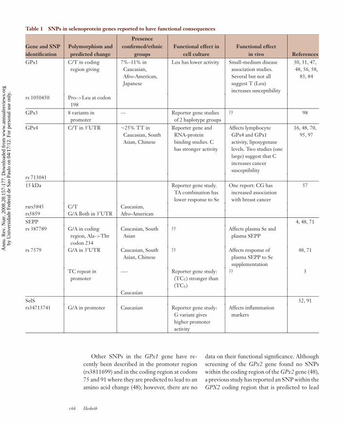
Table 1 SNPs in selenoprotein genes reported to have functional consequences
Gene and SNPidentification
Polymorphism andpredicted change
Presenceconfirmed/ethnic
groupsFunctional effect in
cell cultureFunctional effect
in vivo ReferencesGPx1 C/T in coding
region giving7%–11% inCaucasian,Afro-American,Japanese
Leu has lower activity Small-medium diseaseassociation studies.Several but not allsuggest T (Leu)increases susceptibility
30, 31, 47,48, 56, 58,
83, 84
rs 1050450 Pro->Leu at codon198
GPx3 8 variants inpromoter
— Reporter gene studiesof 2 haplotype groups
?? 98
GPx4 C/T in 3′UTR ∼25% TT inCaucasian, SouthAsian, Chinese
Reporter gene andRNA-proteinbinding studies. Chas stronger activity
Affects lymphocyteGPx4 and GPx1activity, lipoxygenaselevels. Two studies (onelarge) suggest that Cincreases cancersusceptibility
16, 48, 70,95, 97
rs 71304115 kDa Reporter gene study.
TA combination haslower response to Se
One report: CG hasincreased associationwith breast cancer
57
rsrs5845 C/T Caucasian,rs5859 G/A Both in 3′UTR Afro-AmericanSEPP 4, 48, 71rs 387789 G/A in coding
region, Ala->Thrcodon 234
Caucasian, SouthAsian
?? Affects plasma Se andplasma SEPP
rs 7579 G/A in 3′UTR Caucasian, SouthAsian, Chinese
?? Affects response ofplasma SEPP to Sesupplementation
48, 71
TC repeat inpromoter
—- Reporter gene study:(TC5) stronger than(TC3)
?? 3
CaucasianSelS 32, 91rs34713741 G/A in promoter Caucasian Reporter gene study:
G variant giveshigher promoteractivity
Affects inflammationmarkers
Other SNPs in the GPx1 gene have re-cently been described in the promoter region(rs3811699) and in the coding region at codons75 and 91 where they are predicted to lead to anamino acid change (48); however, there are no
data on their functional significance. Althoughscreening of the GPx2 gene found no SNPswithin the coding region of the GPx2 gene (48),a previous study has reported an SNP within theGPX2 coding region that is predicted to lead
166 Hesketh
Ann
u. R
ev. N
utr.
200
8.28
:157
-177
. Dow
nloa
ded
from
ww
w.a
nnua
lrev
iew
s.or
gby
Uni
vers
idad
e Fe
dera
l de
Sao
Paul
o on
04/
17/1
2. F
or p
erso
nal u
se o
nly.

ANRV350-NU28-09 ARI 10 July 2008 14:15
to a Pro-to-Leu change at codon 103 (4). A re-cent study of 123 individuals has identified up toeight strongly linked variants in the promoterregion of the GPx3 gene that fell into two hap-lotype groups (98); reporter studies indicateda lower activity in the haplotype that was alsoover-represented in children and young adultswith arterial ischemic stroke.
GPx4 has a role in the sperm midpiece andtherefore attempts have been made to relateseveral genetic variants to male infertility orsperm viability (34, 68); however, no clear re-lationship has been found. An SNP within theregion of the GPx4 gene corresponding to the3′UTR at position 718 (rs713041) is found inCaucasians, and both allelic variants occur atcommon frequencies (48, 97). A second vari-ant at position 738 was reported as a rare vari-ant in individuals of African American/Africanheritage (48) and was not found in Caucasians(97). In contrast, several lines of evidence sug-gest that rs713041 has functional significance.First, homozygous CC individuals were foundto differ from the TT homozygotes in the levelof lymphocyte 5′ lipoxygenase metabolites (97).Subsequently, reporter gene studies were car-ried out in which the two variants of the 3′UTRwere linked to the iodothyronine deiodinasecoding region and transfected into Caco-2 cells(16); under both Se-adequate and Se-deficientconditions, the C variant produced a higherlevel of deiodinase reporter activity. Further-more, RNA-protein binding studies in vitro us-ing electromobility shift assays indicate that thesingle-nucleotide change from T to C leadsto altered protein binding (70), and computerprediction suggests that this may be the resultof altered RNA structure within the 3′UTR(16). Recently, a human supplementation trialshowed that this T/C variation in the GPx43′UTR led to differences in responses of GPx4,GPx1, and GPx3 protein expression or activ-ity in response to Se supplementation or with-drawal (70). In vitro studies indicate that tran-scripts containing the T/C allelic variants differin their ability to form RNA-protein complexes:The C variant competes better with GPx1 tran-scripts than does the T variant. These in vivo
and in vitro data are compatible with this SNPaltering the position of GPx4 in the hierarchyof selenoprotein synthesis. Interestingly, in twoassociation studies the T variant has been re-ported to be associated with a lower risk of ul-cerative colitis (80) and colon cancer (16). Inaddition, a large association study in the UnitedKingdom has reported a link between genotypeat this SNP and susceptibility to breast can-cer (95). Thus, several sources of data suggestthat rs713041, the SNP at position 718 in the3′UTR of GPx4, has functional consequencesand is linked to changes in glutathione perox-idase 1 and 4 expression as well as possibly in-flammatory changes and cancer susceptibility.Further work is required to confirm its relationto disease and to explore whether its influenceis compounded by other SNPs or nutritional Sestatus.
Thirteen SNPs have been described inthe selenoprotein S gene, and of these, one(rs34713741) has been reported to be func-tionally associated with alterations in markersof inflammation such as tumor necrosis factor-alpha and interleukin-1 beta (32). This func-tional SNP is a G-to-A variation at position 105in the promoter, and functional assays show itmodulates response to stress agents that affectthe endoplasmic reticulum. However, a recentdisease association study failed to link the SNPto risk of ulcerative colitis (91).
Two polymorphic variants, a C/T substitu-tion at position 811 (rs5845) and a G/A at posi-tion 1125 (rs5859), have been identified in theregion of the Sep15 gene that corresponds to the3′UTR of the mRNA (57). The SNP at position1125 is located close to the SECIS structure.Assessment of read-through at a UGA codonusing a reporter gene assay indicated that thecombination of C811 and T1125 made SECISfunction more sensitive to Se supply, which sug-gests that these two SNPs would affect Se incor-poration during Sep15 synthesis. Furthermore,malignant mesothelioma cells expressing the Avariant at position 1125 were less responsive togrowth inhibition by increased Se supply (7).There is also one report of an association of thecombined variants with breast cancer (57).
www.annualreviews.org • Nutrigenomics and Selenium 167
Ann
u. R
ev. N
utr.
200
8.28
:157
-177
. Dow
nloa
ded
from
ww
w.a
nnua
lrev
iew
s.or
gby
Uni
vers
idad
e Fe
dera
l de
Sao
Paul
o on
04/
17/1
2. F
or p
erso
nal u
se o
nly.

ANRV350-NU28-09 ARI 10 July 2008 14:15
Strong evidence now exists to implicateSEPP in having a central and critical role inthe transport of Se in the bloodstream. Thisextraordinary protein, which contains multipleSec and has two SECIS in its mRNA (illus-trated schematically in Figure 6), is found inplasma, where it contributes the bulk of func-tionally available Se. The central role of SePPin Se transport makes it likely that variants inthe SePP gene could have significant functionalconsequences. A functionally relevant SNP hasbeen identified in the promoter region of theSePP gene (3): this is a variant in a TC re-peat sequence that reduces basal promoter ac-tivity in reporter gene constructs expressed inhepatoma cells. Screening for transcript het-erozygosity by denaturing high-performanceliquid chromatography has identified two vari-ants within the coding region and 3′UTR ofthe SePP gene (69): both G-to-A variations,one resulting in an alanine-to-threonine aminoacid change at codon 234 (rs3877899) and theother close to the SECIS within the 3′UTR atposition 25191 (rs7579). The predicted Ala toThr change at codon 234 is close to the be-ginning of the Sec-rich region in the protein(Figure 6). The frequencies of the allelic vari-ants in rs3877899 in a cohort of Caucasianswere 46% for the GG, 47% for the heterozy-gotes, and 7% for the rare AA homozygous al-lele. Interestingly, the SNP was not detectedin a population of Chinese ethnic origin. Onehomozygote (GG) and the GA heterozygote forthe 25191 g/a also occur at high frequency (47%and 46%, respectively) with a rare (7%) AA ho-mozygote, but in this case the frequency wassimilar in Caucasian, South Asian, and Chinesegroups (69). Thus, within one gene we see oneSNP with different allele frequencies in differ-ent ethnic groups and one with similar frequen-cies. This illustrates the importance of con-sidering ethnicity in studies of nutrient-geneinteractions.
Data from a human Se supplementation trialin which prospectively genotyped individualswere supplemented with Se provided evidencethat both rs3877899 and rs7579 influenced a
number of biomarkers of Se status (69). PlasmaSe at baseline depended on both these SNPs aswell as body mass index (BMI), baseline plasmaSePP levels depended on rs24731, and plasmaSePP postsupplementation depended on thers25191 SNP. Both SNPs also had effects onplasma and lymphocyte glutathione peroxidaseactivities. Thus, both these variants appear tohave functional consequences, but the mecha-nistic basis of each needs further investigation.The SNP within the 3′UTR (G/A at nt 25191)occurs at positions close to, but not within, theregion predicted from the two SECIS struc-tures. However, unlike for the rs713041 in theGPx4 gene, no data are available from in vitroexperiments to show whether it affects func-tionality of the 3′UTR in reporter or protein-binding assays. Initial studies suggest that nei-ther the TC promoter polymorphism nor theAla-Thr SNP show altered allele frequencies incolon cancer patients (3, 4).
It is clear from the above-mentioned studiesthat a number of SNPs in selenoprotein genesdemonstrate functional differences between theallelic variants. These have been mostly iden-tified in studies of individual SNPs in singlegenes. However, when assessing the impact ofsuch variants on Se metabolism, it is impor-tant to take a wider view and consider twocritical aspects. First, as when considering ge-netic variants in genes involved in any nutrientmetabolism and function, one should take intoaccount variation in all the metabolic eventsin which the nutrient is involved (see sidebarThe Effect of Genetic Variation on NutrientMetabolism) (Figure 7). Thus, in the case ofSe, since there is a known hierarchy and compe-tition in the use of Se for selenoprotein synthe-sis (12, 13, 66, 75), changes in synthesis of oneselenoprotein owing to an SNP may alter an-other selenoprotein. Second, it is important toconsider the effect of genetic variation over thewhole functional pathway because the conse-quences of one SNP may be magnified or coun-terbalanced by variants in other genes; this netpathway effect is likely to determine the overallphysiological interaction between genetics and
168 Hesketh
Ann
u. R
ev. N
utr.
200
8.28
:157
-177
. Dow
nloa
ded
from
ww
w.a
nnua
lrev
iew
s.or
gby
Uni
vers
idad
e Fe
dera
l de
Sao
Paul
o on
04/
17/1
2. F
or p
erso
nal u
se o
nly.

ANRV350-NU28-09 ARI 10 July 2008 14:15
nutrition. For example, an SNP in the SBP2gene might affect Se incorporation but withoutconsequences unless combined with an SNP inthe 3′UTR of the GPx4 gene; or the effect ofthe SNP in GPx1 might be magnified by anSNP in the SePP gene that affects Se deliv-ery to target tissues. However, to date the re-search on nutrient-gene interactions in relationto Se metabolism has been limited to studies ofindividual SNPs in relation to measurementsof physiological parameters, so as yet such ef-fects of multiple SNPs are largely unknown. In-terestingly, one haplotype study reported thatthe association of the Leu allele for the GPx1Leu198Ala SNP with bladder cancer is influ-enced by an SNP within the manganese super-oxide dismutase gene that codes for another an-tioxidant defense protein (58). Similarly, a re-cent nested case-control study of breast can-cer showed increased disease risk in individu-als who carried both the Ala16Ala genotype forthe manganese dismutase and the Leu198Leugenotype for GPx1 at codon 198 (33). Thesedata illustrate that it is important to considercombined effects of multiple genetic variants(see Figure 6) and that when considering suchcombined effects, it is necessary to take intoaccount a wide view of the functional pathwayin which the nutrient and its associated genesfunction. The functional pathway would be dif-ferent depending on the tissue and functionaloutcome under consideration.
When one considers Se intake and geneticfactors, the picture that is emerging is a com-plex one of multiple, possibly interacting, func-tional SNPs, the influence of which (in publichealth terms) may be modulated by ethnicity,gender, BMI, lifestyle, and dietary factors. Forexample the influence of two SNPs in SePP(Ala234 Thr and 25191 g/a) on plasma Se areboth modulated by BMI (69), whereas the ef-fect of rs713041 in GPx4 on lymphocyte GPx4levels following withdrawal of Se supplementa-tion was observed in females but not males (70).Data on several SNPs in selenoprotein geneshighlight differences in frequency distributionin populations of different ethnic origins (48,57, 69).
THE EFFECT OF GENETIC VARIATION ONNUTRIENT METABOLISM
Approaches to analyzing how multiple genetic variations con-tribute to nutrient requirements are also relevant to folate acidintake, where there is a question of linking data from SNP as-sociation studies and biomarkers of status with regard to B vi-tamin intake, folate metabolism, and disease susceptibility (seeHaggarty 2007 in Related Resources). This problem has dis-tinct similarities to the question of how variation in selenopro-tein genes and other genes associated with Se metabolism af-fect Se requirements. In both cases, in order to understand howgenetic variation affects nutrient metabolism, it is necessary toconsider data from several sources: mechanistic studies givingknowledge of nutrient function, SNP-disease association studies,and nutritional intervention and biomarker studies on genotypedindividuals. One investigator (see Haggarty 2007 in Related Re-sources) advocates combining such data in a “causal-pathway”approach to assess how multiple genetic variants contribute tooverall nutrient-gene interactions. This approach considers howgenetic variation across a broad functional nutrient-related path-way influences metabolism and is potentially applicable to a num-ber of nutrient-gene interactions, including those discussed inthis review in relation to selenium.
CONCLUSIONS AND FUTUREPERSPECTIVES
The Physiological Effectsof Suboptimal Se Intake
Transcriptomic approaches have provided in-teresting data on the responses of certain se-lenoprotein genes, for example, SelW, to mod-ulation of Se supply, but in general, the data arelimited by the major regulation of selenopro-tein synthesis at the level of translation. Pro-teomic methods, perhaps in combination withinductively coupled plasma mass spectroscopyto detect selenoproteins, need to be developed.In contrast, gene array studies have identifieddownstream targets of Se. However, to datethese have been largely limited to cell culturestudies with high concentrations of Se that in-duce apoptosis. Further studies are needed inwhich nutritionally relevant concentrations ofSe are used in cell culture, animal, and human
www.annualreviews.org • Nutrigenomics and Selenium 169
Ann
u. R
ev. N
utr.
200
8.28
:157
-177
. Dow
nloa
ded
from
ww
w.a
nnua
lrev
iew
s.or
gby
Uni
vers
idad
e Fe
dera
l de
Sao
Paul
o on
04/
17/1
2. F
or p
erso
nal u
se o
nly.

ANRV350-NU28-09 ARI 10 July 2008 14:15
studies. Recent studies of lymphocyte gene ex-pression patterns in humans show that pathwayanalysis is very useful in allowing detection ofsubtle changes in gene expression following nu-tritional interventions (41, 69, 96).
Metabolomics has not been used to anysignificant extent to explore the downstreamtargets of Se intake. However, studies in cellculture indicate that metabolomics combinedwith transcriptomics may allow annotation ofmetabolic networks by gene regulation andgene regulatory networks by metabolites. Thisapproach was used to identify changes inmetabolic pathways in A549 lung carcinomacells in response to 0.2–0.5 ppm sodium se-lenite (44), at which concentrations there isgrowth inhibition and induction of apoptosis.This combined approach does detect differ-ences between treatment with selenite and se-lenomethionine. Such an approach, combinedwith inductively coupled plasma mass spec-troscopy to detect Se species, may prove use-ful in identifying regulated pathways in humansthat respond to Se and in dissecting the differ-ent metabolic responses to different selenium-containing compounds. Interestingly, studies of75Se metabolism in transgenic mice express-ing a mutated tRNA-Sec have allowed explo-ration of the relative roles of selenoproteins andlow-molecular-weight selenocompounds in thecolon (59).
In general, the development of transgenicmice with selenoprotein genes knocked out ormodified has proved highly useful not only inelucidating mechanisms of Sec incorporationand selenoprotein function but also in provid-ing more details of the selenoprotein hierarchy(25, 27, 53, 59, 64). Further investigation ofsuch models (for example, in conjunction withmicroarray analysis) is likely to assist in defin-ing the downstream targets of altered seleno-protein function. In addition, further breedingof such mice with other transgenic animals withincreased cancer susceptibility, as is done to ex-amine prostate cancer (36), may provide noveltools to investigate the links between seleno-proteins and disease. Likewise, the recent re-port of a technique to combine knock-down and
knock-in (103) may prove useful in assessing se-lenoprotein function and downstream targets.Se function may also be explored using trans-genic mice in which non-selenoprotein geneshave been modified. For example, Se feedingexperiments in mice lacking apolipoprotein Ereceptor 2 show that this receptor is requiredfor maintenance of brain Se (23).
Gene-Se Interactions andDisease Susceptibility
Several SNPs have been identified in the glu-tathione peroxidases and other selenoproteingenes, some within the coding region and oth-ers within regulatory regions that correspond tothe promoter or the 3′UTRs. There is evidencethat some of these are functional and that theymay be associated with susceptibility to a rangeof diseases (see Table 1). Our level of knowl-edge is limited, however: SNPs with apparentfunctional consequences have been identified inonly a few selenoprotein genes but not in oth-ers, and we have essentially no picture of howthese SNPs combine together, and with Se in-take, to influence Se metabolism and cell phys-iology. A major challenge is to obtain a viewof how genetic variation in the selenome as awhole, combined with Se intake, influences se-lenoprotein function, its downstream targets,and susceptibility to disease (see Figure 7).
One approach to this challenge would beto use modern genotyping technology to carryout large-scale human studies measuring manySNPs (even genome-wide scans) in combina-tion with measures of Se status and dietary andlifestyle characterization. To date, nutritionaldata in such studies have usually been sparse,and to adopt this approach would require ob-taining detailed biochemical and dietary data inlarge population cohorts. A second limitation tothis approach is that in general it does not test,but rather generates, a hypothesis. An advan-tage is that it does not depend on our existingknowledge or preconceptions.
An alternative is to use our knowledge ofSe metabolism to focus on the selenome genesand genes encoding proteins that function in
170 Hesketh
Ann
u. R
ev. N
utr.
200
8.28
:157
-177
. Dow
nloa
ded
from
ww
w.a
nnua
lrev
iew
s.or
gby
Uni
vers
idad
e Fe
dera
l de
Sao
Paul
o on
04/
17/1
2. F
or p
erso
nal u
se o
nly.

ANRV350-NU28-09 ARI 10 July 2008 14:15
Se-related metabolic pathways (see Figures 3,4, and 7). In this way, the number of SNPs tobe tested is limited, so a lower number of indi-viduals participate in the study while the prob-lems of loss of statistical power are minimized.Such an approach has several advantages. It al-lows hypothesis testing; lowering the numberof individuals in the study improves the qual-ity of nutritional and phenotypic data; multipleeffects of different SNPs from genes within apathway may magnify effects; and one can as-sess the effects of genetic variation in a path-way as a whole. This approach, focused on onebiochemical “unit,” would screen the selenomegenes for SNPs, identify the SNPs in thesegenes that are critical in terms of their func-tional effects, and then define how these inter-act with each other and with nutritional factorssuch as Se intake to influence physiological pro-cesses, optimal health status, and susceptibilityto disease. It would require a demonstration ofSNP functionality at the molecular (protein orRNA), cellular, and physiological levels (prefer-ably at more than one) for each individual SNPto build up the full list of SNPs that affectSe-related cell processes. Finally, case-controlassociation studies would be needed to defineif the individual SNPs and/or combinations ofSNPs influence disease risk.
Until now, very few intervention studieshave been carried out to assess the function-ality of SNPs in vivo. There is a need toassess SNPs in human intervention studies us-ing prospectively genotyped cohorts and ap-propriate biomarkers in order to show causalityin the relationship between Se, SNPs, and thebiomarkers of health status. There is also a needto carry out further disease association studiesinvolving analysis of larger, repeat cohorts andto combine genotyping with studies of nutri-
tional intake, or status, to assess the importanceon nutrient-gene interactions in determiningsusceptibility. A wide range of SNPs should bestudied, initially based on a selenome approachto include genes encoding products involved inSe incorporation mechanisms and Se transport.Since effects of individual SNPs in isolation areexpected to be relatively small, it will be neces-sary to assess how these individual genetic fac-tors fit into the complex picture: How do thegroups of SNPs interact with each other or withother factors such as Se intake, ethnicity, andgender to contribute to biomarkers of healthand nutrient requirements? Finally, mathemat-ical modeling is needed to define which SNPsare the major determinants of individual re-sponses to dietary Se and consequently indi-vidual Se requirements, and thus the extent oftheir role, in conjunction with Se intake, in de-termination of risk of diseases such as prostateand colon cancer. Our ultimate aim should beto define the extent of the contribution fromthese different SNPs and other factors. Thereare major challenges to develop statistical ap-proaches needed both to analyze the data fromlarge human studies on multiple SNPs andnutritional factors likely to influence the bi-ological outcomes and then to model mathe-matically the multiple effects in terms of thedifferent factors that influence Se metabolismand optimal health or disease. However, ris-ing to these challenges is worthwhile becausethe associations between suboptimal Se statusand disease in large population groups world-wide and in animal models identify Se intakeas a potential public health concern. Nutrige-nomics, both in terms of mechanistic studiesand detailed SNP studies, provides exciting ap-proaches with which to tackle this importantissue.
SUMMARY POINTS
1. Both dietary Se intake and genetic factors can potentially influence the pattern of seleno-protein synthesis.
2. There is a hierarchy in the response of selenoprotein synthesis to Se supply. The hier-archy determines the pattern of selenoprotein expression and functional effects of Se. It is
www.annualreviews.org • Nutrigenomics and Selenium 171
Ann
u. R
ev. N
utr.
200
8.28
:157
-177
. Dow
nloa
ded
from
ww
w.a
nnua
lrev
iew
s.or
gby
Uni
vers
idad
e Fe
dera
l de
Sao
Paul
o on
04/
17/1
2. F
or p
erso
nal u
se o
nly.

ANRV350-NU28-09 ARI 10 July 2008 14:15
important to determine how the pattern of synthesis of all selenoproteins is affected bySe supply and genetic factors.
3. Conversion of Se into functioning selenoproteins requires synthesis of selenocysteinetRNA, incorporation of selenocysteine during translation by a mechanism involving the3′UTR and specific RNA-binding proteins, and transport of Se in the blood by SEPP.SNPs in genes that code for proteins that are involved in these processes may influenceselenoprotein synthesis and nutrient requirements.
4. Functional SNPs have been identified in some selenoprotein genes, and some have beenlinked to disease susceptibility in small association studies. However, our knowledge islimited, and much further work is needed to identify and assess the effects of SNPs thatinfluence Se metabolism, nutritional requirements for Se, and disease susceptibility.
5. It is important to consider how multiple SNPs interact to affect Se metabolism.
6. Transcriptomic studies (gene microarrays) on transformed cells in culture have shownrelatively high concentrations of Se to alter expression of genes involved in apoptosis andcell cycle control.
7. Combining transcriptomics and pathway analysis may allow identification of novel targetsof dietary Se, even in human studies.
8. Proteomic and metabolomic approaches to Se metabolism should be explored.
FUTURE ISSUES
1. What are the molecular mechanisms behind tissue-specific hierarchy in selenoproteinsynthesis?
2. What downstream (i.e., nonselenoprotein) pathways are affected by dietary Se intake,and what can this tell us about the physiological effects of Se?
3. Can transcriptomics, proteomics, or metabolomics provide novel, improved biomarkersof Se status?
4. To what extent does genetic variation in Se metabolism genes affect which SNPs havethe most influence on nutritional requirements for Se and susceptibility to multifactorialdiseases?
ACKNOWLEDGMENTS
Work in the author’s laboratory has been supported by the Food Standards Agency, Biologicaland Biotechnology Research Council, and World Cancer Research Fund. The author thanks thecoworkers in his laboratory who have contributed to various projects and discussion, especiallyGiovanna Bermano, Catherine Meplan, Vasilieos Pagmantidis, and Stephane Villette.
DISCLOSURE STATEMENT
The author is not aware of any biases that might be perceived as affecting the objectivity of thisreview.
172 Hesketh
Ann
u. R
ev. N
utr.
200
8.28
:157
-177
. Dow
nloa
ded
from
ww
w.a
nnua
lrev
iew
s.or
gby
Uni
vers
idad
e Fe
dera
l de
Sao
Paul
o on
04/
17/1
2. F
or p
erso
nal u
se o
nly.

ANRV350-NU28-09 ARI 10 July 2008 14:15
LITERATURE CITED
1. Aachmann FL, Fomenko DE, Soragni A, Gladyshev VN, Dikiy A. 2007. Structural analysis of seleno-protein W and NMR analysis of its interaction with 14-3-3 proteins. J. Biol. Chem. 282:37036–44
2. Akbaraly NT, Arnaud J, Hininger-Favier I, Gourlet V, Roussel AM, et al. 2005. Selenium and mortalityin the elderly: results from the EVA study. Clin. Chem. 51:2117–23
3. Al-Taie OH, Seufert J, Mork H, Treis H, Mentrup B, et al. 2002. A complex DNA-repeat structurewithin the selenoprotein P promoter contains a functionally relevant polymorphism and is geneticallyunstable under conditions of mismatch repair deficiency. Eur. J. Hum. Genet. 10:499–504
4. Al-Taie OH, Uceyler N, Eubner U, Jakob F, Mork H, et al. 2004. Expression profiling and geneticalterations of the selenoproteins GI-GPx and SePP in colorectal carcinogenesis. Nutr. Cancer 48:6–14
5. Allamand V, Richard P, Lescure A, Ledeuil C, Desjardin D, et al. 2006. A single homozygous pointmutation in a 3′untranslated region motif of selenoprotein N mRNA causes SEPN1-related myopathy.EMBO Rep. 7:450–54
6. Arbones-Mainar JM, Ross K, Rucklidge GJ, Reid M, Duncan G, et al. 2007. Extra virgin olive oilsincrease hepatic fat accumulation and hepatic antioxidant protein levels in APOE(-/-) mice. J. ProteomeRes. 6:4041–54
7. Apostolou S, Klein JO, Mitsuuchi Y, Shetler JN, Poulikakos PI, et al. 2004. Growth inhibition andinduction of apoptosis in mesothelioma cells by selenium and dependence on selenoprotein SEP15genotype. Oncogene 23:5032–40
8. Arner ES, Holmgren A. 2006. The thioredoxin system in cancer. Semin. Cancer Biol. 16:420–269. Beck MA, Esworthy RS, Ho YS, Chu FF. 1998. Glutathione peroxidase protects mice from viral-induced
myocarditis. FASEB J. 12:1143–4910. Beck MA, Levander OA, Handy J. 2003. Selenium deficiency and viral infection. J. Nutr. 133:1463–67S11. Beckett GJ, Arthur JR. 2005. Selenium and endocrine systems. J. Endocrinol. 184:455–6512. Behne D, Hilmert H, Scheid S, Gessner H, Elger W. 1988. Evidence for specific selenium target tissues
and new biologically important selenoproteins. Biochim. Biophys. Acta 966:12–2113. Bermano G, Nicol F, Dyer JA, Sunde RA, Beckett GJ, et al. 1995. Tissue-specific regulation of sele-
noenzyme gene expression during selenium deficiency in rats. Biochem. J. 311:425–3014. Bermano G, Arthur JR, Hesketh JE. 1996. Selective control of cytosolic glutathione peroxidase and
phospholipid hydroperoxide glutathione peroxidase mRNA stability by selenium supply. FEBS Lett.387:157–60
15. Bermano G, Arthur JR, Hesketh JE. 2006. Role of the 3′ untranslated region in the regulation of cy-tosolic glutathione peroxidase and phospholipid-hydroperoxide glutathione peroxidase gene expressionby selenium supply. Biochem. J. 320:891–95
16. Single pointmutation in 3′UTR (butnot within the SECIS)alters ability of 3′UTRto supportselenoprotein synthesis.
16. Bermano G, Pagmanditis V, Holloway N, Kadri S, Mowat NAG, et al. 2007. Evidence that apolymorphism within the 3′UTR of glutathione peroxidase 4 is functional and is associated withsusceptibility to colorectal cancer. Genes Nutr. 2:227–32
17. Brigelius-Flohe R. 1999. Tissue-specific functions of individual glutathione peroxidases. Free Radic. Biol.Med. 27:951–65
18. Brigelius-Flohe R. 2006. Glutathione peroxidases and redox-regulated transcription factors. Biol. Chem.387:1329–35
19. Brigelius-Flohe R, Joost H-G, eds. 2006. Nutritional Genomics. Weinheim, Germ.: Wiley-VCH Verlag20. Broome CS, McArdle F, Kyle JA, Andrews F, Lowe NM, et al. 2004. An increase in selenium intake
improves immune function and poliovirus handling in adults with marginal selenium status. Am. J. Clin.Nutr. 80:154–62
21. Burk RF, Hill KE. 2005. Selenoprotein P: an extracellular protein with unique physical characteristicsand a role in selenium homeostasis. Annu. Rev. Nutr. 25:215–35
22. Burk RF, Hill KE, Motley AK. 2003. Selenoprotein metabolism and function: evidence for more thanone function for selenoprotein P. J. Nutr. 133:1517–20S
23. Burk RF, Hill KE, Olson GE, Weeber EJ, Motley AK, et al. 2007. Deletion of apolipoprotein E receptor-2in mice lowers brain selenium and causes severe neurological dysfunction and death when a low-seleniumdiet is fed. J. Neurosci. 27:6207–11
www.annualreviews.org • Nutrigenomics and Selenium 173
Ann
u. R
ev. N
utr.
200
8.28
:157
-177
. Dow
nloa
ded
from
ww
w.a
nnua
lrev
iew
s.or
gby
Uni
vers
idad
e Fe
dera
l de
Sao
Paul
o on
04/
17/1
2. F
or p
erso
nal u
se o
nly.

ANRV350-NU28-09 ARI 10 July 2008 14:15
24. Cao TM, Hua FY, Xu CM, Han BS, Dong H, et al. 2006. Distinct effects of different concentrations ofsodium selenite on apoptosis, cell cycle, and gene expression profile in acute promyelocytic leukemia-derived NB4 cells. Ann. Hematol. 85:434–42
25. tRNA methylationinfluencesselenoprotein hierarchy.
25. Carlson BA, Moustafa ME, Sengupta A, Schweizer U, Shrimali R, et al. 2007. Selective restora-tion of the selenoprotein population in a mouse hepatocyte selenoproteinless background withdifferent mutant selenocysteine tRNAs lacking Um34. J. Biol. Chem. 282:32591–602
26. Chavatte L, Brown BA, Driscoll DM. 2005. Ribosomal protein L30 is a component of the UGA-selenocysteine recoding machinery in eukaryotes. Nat. Struct. Mol. Biol. 12:408–16
27. Chu FF, Esworthy RS, Chu PG, Longmate JA, Huycke MM, et al. 2004. Bacteria-induced intestinalcancer in mice with disrupted Gpx1 and Gpx2 genes. Cancer Res. 64:962–68
28. Clark LC, Combs GF Jr, Turnbull BW, Slate EH, Chalker DK, et al. 1996. Effects of selenium supple-mentation for cancer prevention in patients with carcinoma of the skin. A randomized controlled trial.Nutritional Prevention of Cancer Study Group. JAMA 276:1957–63
29. Copeland PR, Stepanik VA, Driscoll DM. 2001. Insight into mammalian selenocysteine insertion: domainstructure and ribosome binding properties of Sec insertion sequence binding protein 2. Mol. Cell Biol.21:1491–98
30. Cox DG, Hankinson SE, Kraft P, Hunter DJ. 2004. No association between GPX1 Pro198Leu andbreast cancer risk. Cancer Epidemiol. Biomarkers Prev. 13:1821–22
31. Cox DG, Tamimi RM, Hunter DJ. 2006. Gene × gene interaction between MnSOD and GPX-1 andbreast cancer risk: a nested case-control study. BMC Cancer 6:217
32. Curran JE, Jowett JB, Elliott KS, Gao Y, Gluschenko K, et al. 2005. Genetic variation in selenoproteinS influences inflammatory response. Nat. Genet. 37:1234–41
33. Crosley LK, Meplan C, Nicol F, Rundlof AK, Arner ES, et al. 2007. Differential regulation of expressionof cytosolic and mitochondrial thioredoxin reductase in rat liver and kidney. Arch. Biochem. Biophys.459:178–88
34. Diaconu M, Tangat Y, Bohm D, Kuhn H, Michelmann HW, et al. 2006. Failure of phospholipid hy-droperoxide glutathione peroxidase expression in oligoasthenozoospermia and mutations in the PHGPxgene. Andrologia 38:152–57
35. Structural analysisshows a new family ofselenoproteins.
35. Dikiy A, Novoselov SV, Fomenko DE, Sengupta A, Carlson BA, et al. 2007. SelT, SelW, SelH,and Rdx12: genomics and molecular insights into the functions of selenoproteins of a novelthioredoxin-like family. Biochemistry 46:6871–82
36. Diwadkar-Navsariwala V, Prins GS, Swanson SM, Birch LA, Ray VH, et al. 2006. Selenoprotein defi-ciency accelerates prostate carcinogenesis in a transgenic model. Proc. Natl. Acad. Sci. USA 103:8179–84
37. Dong Y, Ganther HE, Stewart C, Ip C. 2002. Identification of molecular targets associated with selenium-induced growth inhibition in human breast cells using cDNA microarrays. Cancer Res. 62:708–14
38. Dong Y, Zhang H, Hawthorn L, Ganther HE, Ip C. 2003. Delineation of the molecular basis forselenium-induced growth arrest in human prostate cancer cells by oligonucleotide array. Cancer Res.63:52–59
39. Mutations in SBP2cause dysfunction ofthyroid metabolism.
39. Dumitrescu AM, Liao XH, Abdullah MS, Lado-Abeal J, Majed FA, et al. 2005. Mutations inSECISBP2 result in abnormal thyroid hormone metabolism. Nat. Genet. 37:1247–52
40. El-Bayoumy K, Sinha R. 2005. Molecular chemoprevention by selenium: a genomic approach. Mut. Res.591:224–36
41. Elliott R, Pico C, Dommels Y, Wybranska I, Hesketh J, Keijer J. 2007. Nutrigenomic approaches forbenefit-risk analysis of foods and food components: defining markers of health. Br. J. Nutr. 98:1095–100
42. Evenson JK, Wheeler AD, Blake SM, Sunde RA. 2004. Selenoprotein mRNA is expressed in blood atlevels comparable to major tissues in rats. J. Nutr. 134:2640–45
43. Fagegaltier D, Hubert N, Yamada K, Mizutani T, Carbon P, et al. 2000. Characterization of mSelB, anovel mammalian elongation factor for selenoprotein translation. EMBO J. 19:4796–805
44. Fan TW, Higashi RM, Lane AN. 2006. Integrating metabolomics and transcriptomics for probing Seanticancer mechanisms. Drug Metab. Rev. 38:707–32
45. Ferguson AD, Labunskyy VM, Fomenko DE, Arac D, Chelliah Y, et al. 2006. NMR structures of theselenoproteins Sep15 and SelM reveal redox activity of a new thioredoxin-like family. J. Biol. Chem.281:3536–43
174 Hesketh
Ann
u. R
ev. N
utr.
200
8.28
:157
-177
. Dow
nloa
ded
from
ww
w.a
nnua
lrev
iew
s.or
gby
Uni
vers
idad
e Fe
dera
l de
Sao
Paul
o on
04/
17/1
2. F
or p
erso
nal u
se o
nly.

ANRV350-NU28-09 ARI 10 July 2008 14:15
46. Fischer A, Pallauf J, Gohil K, Weber SU, Packer L, et al. 2001. Effect of selenium and vitamin E deficiencyon differential gene expression in rat liver. Biochem. Biophys. Res. Commun. 285:470–75
47. Demonstratesfunctional SNP incoding region of GPx1.
47. Forsberg L, de Faire U, Marklund SL, Andersson PM, Stegmayr B, Morgenstern R. 2000. Phe-notype determination of a common Pro-Leu polymorphism in human glutathione peroxidase 1.Blood Cells Mol. Dis. 26:423–26
48. Foster CB, Aswath K, Chanock SJ, McKay HF, Peters U. 2006. Polymorphism analysis of six selenopro-tein genes: support for a selective sweep at the glutathione peroxidase 1 locus (3p21) in Asian populations.BMC Genet. 7:56
49. Gao Y, Feng HC, Walder K, Bolton K, Sunderland T, et al. 2004. Regulation of the selenoprotein SelSby glucose deprivation and endoplasmic reticulum stress—SelS is a novel glucose-regulated protein.FEBS Lett. 563:185–90
50. Gu QP, Ream W, Whanger PD. 2002. Selenoprotein W gene regulation by selenium in L8 cells.Biometals 15:411–20
51. Hamanishi T, Furuta H, Kato H, Doi A, Tamai M, et al. 2004. Functional variants in the glutathioneperoxidase-1 (GPx-1) gene are associated with increased intima-media thickness of carotid arteries andrisk of macrovascular diseases in Japanese type 2 diabetic patients. Diabetes 53:2455–60
52. Hatfield DL, Gladyshev VN. 2002. How selenium has altered our understanding of the genetic code.Mol. Cell Biol. 22:3565–76
53. Transgenic micedemonstrateimportance ofselenoprotein P,particularly theC-terminal region, toSe delivery.
53. Hill KE, Zhou J, Austin LM, Motley AK, Ham AJ, et al. 2007. The selenium-rich C-terminaldomain of mouse selenoprotein P is necessary for the supply of selenium to brain and testis butnot for the maintenance of whole body selenium. J. Biol. Chem. 282:10972–80
54. Hoffmann PR, Hoge SC, Li PA, Hoffmann FW, Hashimoto AC, et al. 2007. The selenoproteomeexhibits widely varying, tissue-specific dependence on selenoprotein P for selenium supply. Nucleic AcidsRes. 35:3963–73
55. Hooven LA, Butler J, Ream LW, Whanger PD. 2006. Microarray analysis of selenium-depleted andselenium-supplemented mice. Biol. Trace Elem. Res. 109:173–79
56. Hu YJ, Diamond AM. 2003. Role of glutathione peroxidase 1 in breast cancer: loss of heterozygosityand allelic differences in the response to selenium. Cancer Res. 63:3347–51
57. First to demonstratethat SNPs in 3′UTR of aselenoprotein gene canbe functional.
57. Hu YJ, Korotkov KV, Mehta R, Hatfield DL, Rotimi CN, et al. 2001. Distribution and functionalconsequences of nucleotide polymorphisms in the 3′-untranslated region of the human Sep15gene. Cancer Res. 61:2307–10
58. Ichimura Y, Habuchi T, Tsuchiya N, Wang L, Oyama C, et al. 2004. Increased risk of bladder cancerassociated with a glutathione peroxidase 1 codon 198 variant. J. Urol. 172:728–32
59. Irons R, Carlson BA, Hatfield DL, Davis CD. 2006. Both selenoproteins and low molecular weightselenocompounds reduce colon cancer risk in mice with genetically impaired selenoprotein expression.J. Nutr. 136:1311–17
60. Jeong D, Kim TS, Chung YW, Lee BJ, Kim IY. 2002. Selenoprotein W is a glutathione-dependentantioxidant in vivo. FEBS Lett. 517:225–28
61. Joshi N, Johnson LL, Wei WQ, Abnet CC, Dong ZW, et al. 2006. Selenomethionine treatment doesnot alter gene expression in normal squamous esophageal mucosa in a high-risk Chinese population.Cancer Epidemiol. Biomarkers Prev. 15:1046–47
62. Joost HG, Gibney MJ, Cashman KD, Gorman U, Hesketh JE, et al. 2007. Personalised nutrition: statusand perspectives. Br. J. Nutr. 98:26–31
63. Kryukov GV, Castellano S, Novoselov SV, Lobanov AV, Zehtab O, et al. 2003. Characterization ofmammalian selenoproteomes. Science 300:1439–43
64. Lei XG, Cheng WH, McClung JP. 2007. Metabolic regulation and function of glutathione peroxidase-1.Annu. Rev. Nutr. 27:41–61
65. Lei XG, Evenson JK, Thompson KM, Sunde RA. 1995. Glutathione peroxidase and phospholipid hy-droperoxide glutathione peroxidase are differentially regulated in rats by dietary selenium. J. Nutr.125:1438–46
66. Low SC, Berry MJ. 1996. Knowing when not to stop: selenocysteine incorporation in eukaryotes. TrendsBiochem. Sci. 21:203–8
www.annualreviews.org • Nutrigenomics and Selenium 175
Ann
u. R
ev. N
utr.
200
8.28
:157
-177
. Dow
nloa
ded
from
ww
w.a
nnua
lrev
iew
s.or
gby
Uni
vers
idad
e Fe
dera
l de
Sao
Paul
o on
04/
17/1
2. F
or p
erso
nal u
se o
nly.

ANRV350-NU28-09 ARI 10 July 2008 14:15
67. Low SC, Grundner-Culemann E, Harney JW, Berry MJ. 2000. SECIS-SBP2 interactions dictate se-lenocysteine incorporation efficiency and selenoprotein hierarchy. EMBO J. 19:6882–90
68. Maiorino M, Bosello V, Ursini F, Foresta C, Garolla A, et al. 2003. Genetic variations of gpx-4 and maleinfertility in humans. Biol. Reprod. 68:1134–41
69. Two SNPs in SEPP
shown to be functionalin prospectivelygenotyped volunteers.
69. Meplan C, Crosley LK, Nicol F, Beckett GJ, Howie AF, et al. 2007. Genetic polymorphisms inthe human selenoprotein P gene determine the response of selenoprotein markers to seleniumsupplementation in a gender-specific manner (the SELGEN study). FASEB J. 21:3063–74
70. Meplan C, Crosley LK, Nicol F, Horgan G, Mathers JC, et al. 2008. Functional effects of a commonsingle nucleotide polymorphism (GPX4c718t) in the glutathione peroxidase 4 gene: interaction withgender. Am. J. Clin. Nutr. 87:1019–27
71. Meplan C, Pagmantidis V, Hesketh JE. 2006. Advances in selenoprotein expression—patterns and in-dividual variations. In Nutritional Genomics, ed. R Brigelius-Flohe, H-G Joost, pp 132–58. Weinheim,Germ.: Wiley-VCH Verlag
72. Moghadaszadeh B, Petit N, Jaillard C, Brockington M, Roy SQ, et al. 2001. Mutations in SEPN1 causecongenital muscular dystrophy with spinal rigidity and restrictive respiratory syndrome. Nat. Genet.29:17–18
73. Moscow JA, Morrow CS, He R, Mullenbach GT, Cowan KH. 1992. Structure and function of the 5′-flanking sequence of the human cytosolic selenium-dependent glutathione peroxidase gene (hgpx1). J.Biol. Chem. 267:5949–58
74. Moustafa ME, Carlson BA, El-Saadani MA, Kryukov GV, Sun QA, et al. 2001. Selective inhibi-tion of selenocysteine tRNA maturation and selenoprotein synthesis in transgenic mice expressingisopentenyladenosine-deficient selenocysteine tRNA. Mol. Cell Biol. 21:3840–52
75. Muller C, Wingler K, Brigelius-Flohe R. 2003. 3′UTRs of glutathione peroxidases differentially affectselenium-dependent mRNA stability and selenocysteine incorporation efficiency. Biol. Chem. 384:11–18
76. Ordovas JM. 2006. Nutrigenetics, plasma lipids, and cardiovascular risk. J. Am. Diet. Assoc. 106:1074–8177. Pagmantidis V, Bermano G, Villette S, Broom I, Arthur J, Hesketh J. 2005. Effects of Se-depletion on
glutathione peroxidase and selenoprotein W gene expression in the colon. FEBS Lett. 579:792–96
78. Microarray studyshowing subtle effectsof a nutritionallyrelevant Sesupplementation inhumans.
78. Pagmantidis V, Meplan C, van Schothorst EM, Keijer J, Hesketh J. 2008. Supplementation ofhealthy volunteers with nutritionally relevant levels of selenium increases expression of lympho-cyte protein biosynthesis genes. Am. J. Clin. Nutr. 87:181–89
79. Panee J, Stoytcheva ZR, Liu W, Berry MJ. 2007. Selenoprotein H is a redox-sensing high mobilitygroup family DNA-binding protein that up-regulates genes involved in glutathione synthesis and phaseII detoxification. J. Biol. Chem. 282:23759–65
80. Qatatsheh A, Seal CJ, Jowet SL, Welfare MR, Hesketh JE. 2005. Patients with ulcerative colitis showan altered frequency distribution of a single nucleotide polymorphism (SNP) in the gene encodingphospholipid hydroperoxide glutathione peroxidise (GPx4). Proc. Nutr. Soc. 64:20A (Abstr.)
81. Raaschou-Nielsen O, Sørensen M, Hansen RD, Frederiksen K, Tjønneland A, et al. 2007. GPX1Pro198Leu polymorphism, interactions with smoking and alcohol consumption, and risk for lung cancer.Cancer Lett. 247:293–300
82. Ran Q, Liang H, Gu M, Qi W, Walter CA, et al. 2004. Transgenic mice overexpressing glutathioneperoxidase 4 are protected against oxidative stress-induced apoptosis. J. Biol. Chem. 279:55137–46
83. Ratnasinghe D, Tangrea JA, Andersen MR, Barrett MJ, Virtamo J, et al. 2000. Glutathione peroxidasecodon 198 polymorphism variant increases lung cancer risk. Cancer Res. 60:6381–83
84. Ravn-Haren G, Olsen A, Tjønneland A, Dragsted LO, Nexø BA, et al. 2006. Associations between GPX1Pro198Leu polymorphism, erythrocyte GPX activity, alcohol consumption and breast cancer risk in aprospective cohort study. Carcinogenesis 27:820–25
85. Ray AL, Semba RD, Walston J, Ferrucci L, Cappola AR, et al. 2006. Low serum selenium and totalcarotenoids predict mortality among older women living in the community: the Women’s Health andAging Studies. J. Nutr. 136:172–76
86. Rayman MP. 2002. The argument for increasing selenium intake. Proc. Nutr. Soc. 61:203–1587. Renko K, Werner M, Renner-Muller I, Cooper TG, Yeung CH, et al. 2008. Hepatic selenoprotein
P (Sepp) expression restores selenium transport and prevents infertility and motor-incoordination inSepp-knockout mice. Biochem. J. 409:741–49
176 Hesketh
Ann
u. R
ev. N
utr.
200
8.28
:157
-177
. Dow
nloa
ded
from
ww
w.a
nnua
lrev
iew
s.or
gby
Uni
vers
idad
e Fe
dera
l de
Sao
Paul
o on
04/
17/1
2. F
or p
erso
nal u
se o
nly.

ANRV350-NU28-09 ARI 10 July 2008 14:15
88. Saito Y, Sato N, Hirashima M, Takebe G, Nagasawa S, et al. 2004. Domain structure of bi-functionalselenoprotein P. Biochem. J. 381:841–46
89. Saito Y, Takahashi K. 2002. Characterization of selenoprotein P as a selenium supply protein. Eur. J.Biochem. 269:5746–51
90. Schlicht M, Matysiak B, Brodzeller T, Wen X, Liu H, et al. 2004. Cross-species global and subset geneexpression profiling identifies genes involved in prostate cancer response to selenium. BMC Genomics5:58
91. Seiderer J, Dambacher J, Kuhnlein B, Pfennig S, Konrad A, et al. 2007. The role of the selenoprotein S(SELS) gene -105G>A promoter polymorphism in inflammatory bowel disease and regulation of SELSgene expression in intestinal inflammation. Tissue Antigens 70:238–46
92. Shchedrina VA, Novoselov SV, Malinouski MY, Gladyshev VN. 2007. Identification and characteriza-tion of a selenoprotein family containing a diselenide bond in a redox motif. Proc. Natl. Acad. Sci. USA104:13919–24
93. Sheridan PA, Zhong N, Carlson BA, Perella CM, Hatfield DL, et al. 2007. Decreased selenoproteinexpression alters the immune response during influenza virus infection in mice. J. Nutr. 137:1466–71
94. Stadtman TC. 1996. Selenocysteine. Annu. Rev. Biochem. 65:83–10095. Udler M, Maia AT, Cebrian A, Brown C, Greenberg D, et al. 2007. Common germline genetic variation
in antioxidant defense genes and survival after diagnosis of breast cancer. J. Clin. Oncol. 25:3015–2396. van Erk MJ, Blom WA, van Ommen B, Hendriks HF. 2006. High-protein and high-carbohydrate break-
fasts differentially change the transcriptome of human blood cells. Am. J. Clin. Nutr. 84:1233–4197. Villette S, Kyle JA, Brown KM, Pickard K, Milne JS, et al. 2002. A novel single nucleotide polymorphism
in the 3′ untranslated region of human glutathione peroxidase 4 influences lipoxygenase metabolism. BloodCells Mol. Dis. 29:174–78
98. Voetsch B, Jin RC, Bierl C, Benke KS, Kenet G, et al. 2007. Promoter polymorphisms in the plasmaglutathione peroxidase (GPx-3) gene: a novel risk factor for arterial ischemic stroke among young adultsand children. Stroke 38:41–49
99. Whanger PD. 2004. Selenium and its relationship to cancer: an update dagger. Br. J. Nutr. 91:11–28100. Wingler K, Bocher M, Flohe L, Kollmus H, Brigelius-Flohe R. 1999. mRNA stability and selenocys-
teine insertion sequence efficiency rank gastrointestinal glutathione peroxidase high in the hierarchy ofselenoproteins. Eur. J. Biochem. 259:149–57
101. Xu XM, Carlson BA, Irons R, Mix H, Zhong N, et al. 2007. Selenophosphate synthetase 2 is essentialfor selenoprotein biosynthesis. Biochem. J. 404:115–20
102. Xu XM, Carlson BA, Mix H, Zhang Y, Saira K, et al. 2007. Biosynthesis of selenocysteine on its tRNAin eukaryotes. PLoS Biol. 5:e4
103. Yoo MH, Xu XM, Turanov AA, Carlson BA, Gladyshev VN, et al. 2007. A new strategy for assessingselenoprotein function: siRNA knockdown/knock-in targeting the 3′-UTR. RNA 13:921–29
104. Zeisel SH. 2007. Nutrigenomics and metabolomics will change clinical nutrition and public healthpractice: insights from studies on dietary requirements for choline. Am. J. Clin. Nutr. 86:542–48
105. Shows the ability ofmicroarray studies topick up changes in geneexpression intransformed cells inresponse to Se in cellculture.
105. Zhao H, Whitfield ML, Xu T, Botstein D, Brooks JD. 2004. Diverse effects of methylseleninicacid on the transcriptional program of human prostate cancer cells. Mol. Biol. Cell 15:506–19
RELATED RESOURCES
Bleys J, Navas-Acien A, Guallar E. 2008. Serum selenium levels and all-cause, cancer, and cardio-vascular mortality among US adults. Arch. Intern. Med. 168:404–10. Suggests that the optimalSe intake is one that gives a serum Se of ∼130 ng/ml
Haggarty P. 2007. B-vitamins, genotype and disease causality. Proc. Nutr. Soc. 66:539–47. Discussesapproaches to study of nutrient-gene interactions
Jablonska E, Gromadzinska J, Sobala W, Reszka E, Wasowicz W. 2008. Lung cancer risk associatedwith selenium status is modified in smoking individuals by Sep15 polymorphism. Eur. J. Nutr.47:47–54. Suggests that in smokers, SNPs in the selenoprotein 15 gene influence lung cancerrisk
www.annualreviews.org • Nutrigenomics and Selenium 177
Ann
u. R
ev. N
utr.
200
8.28
:157
-177
. Dow
nloa
ded
from
ww
w.a
nnua
lrev
iew
s.or
gby
Uni
vers
idad
e Fe
dera
l de
Sao
Paul
o on
04/
17/1
2. F
or p
erso
nal u
se o
nly.

www.annualreviews.org ● Nutrigenomics and Selenium C-1
Transcriptomics, proteomics, Metabolomics, Gene knock-down/out
New functional biomarkers and understanding mechanisms of Se action
Identification of SNPs in Se metabolic pathways
Se requirements: Se intake and susceptibility to disease
Functional SNPS: genetic factors thatinfluence Se metabolic pathways
Downstreamtargets
Selenoproteins
Figure 1
The contribution of genomics to understanding the nutritional science of selenium (Se). Two aspects areemphasized: (a) the genetics of selenoprotein and other genes relating to Se metabolism, and (b) the poten-tial of functional genomic approaches to elucidate novel aspects of physiological targets of altered Se sup-ply. SNPs, single-nucleotide polymorphisms.
Hesketh.qxd 06/13/2008 08:15 PM Page C-1
Ann
u. R
ev. N
utr.
200
8.28
:157
-177
. Dow
nloa
ded
from
ww
w.a
nnua
lrev
iew
s.or
gby
Uni
vers
idad
e Fe
dera
l de
Sao
Paul
o on
04/
17/1
2. F
or p
erso
nal u
se o
nly.

C-2 Hesketh
UGA STOP
Selenoprotein mRNA
3’UTR
SelenocysteineInsertion
Sequence(SECIS)
SBP2
SecystRNA
L30
EFSec
Active Selenoprotein
Figure 2Selenocysteine incorporation. The scheme illustrates the key factors in Sec incorporation: (a) the in-frame UGA, (b) Sec-tRNA, (c) the selenocysteine insertion sequence (SECIS) element in the 3' untrans-lated region (UTR), and (d ) RNA-binding proteins SECIS-binding protein 2, EF-Sec, and ribosomalprotein L30. RNA, ribonucleic acid; mRNA, messenger RNA; tRNA, transfer RNA.
Hesketh.qxd 06/13/2008 08:15 PM Page C-2
Ann
u. R
ev. N
utr.
200
8.28
:157
-177
. Dow
nloa
ded
from
ww
w.a
nnua
lrev
iew
s.or
gby
Uni
vers
idad
e Fe
dera
l de
Sao
Paul
o on
04/
17/1
2. F
or p
erso
nal u
se o
nly.
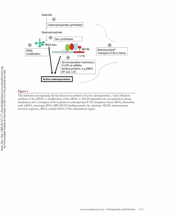
www.annualreviews.org ● Nutrigenomics and Selenium C-3
Figure 3
The selenome encompassing the key features in synthesis of active selenoproteins. 1 and 2 illustratesynthesis of Sec-tRNA; 3, modification of Sec-tRNA; 4, SECIS-dependent Sec incorporation duringtranslation; and 5, transport of Se in plasma as selenoprotein P. EF, elongation factor; RNA, ribonucleicacid; mRNA, messenger RNA; SBP, SECIS-binding protein; Se, selenium; SECIS, selenocysteineinsertion sequence; tRNA, transfer RNA; UTR, untranslated region.
Selenide
SECIS
3’UTRUGA
tRNA-Sec
Selenophosphate synthetase
Selenophosphate
Active selenoproteins
Sec synthetase
Se incorporation machinery:3’UTR of mRNAsbinding proteins, e.g.SBP2,EF-sec, L30
•1
•2
•3
SelenoproteinPTransport of Se in blood
•4
•5
tRNAmodification
Hesketh.qxd 06/13/2008 08:15 PM Page C-3
Ann
u. R
ev. N
utr.
200
8.28
:157
-177
. Dow
nloa
ded
from
ww
w.a
nnua
lrev
iew
s.or
gby
Uni
vers
idad
e Fe
dera
l de
Sao
Paul
o on
04/
17/1
2. F
or p
erso
nal u
se o
nly.

C-4 Hesketh
SNPs
Selenophosphate synthetase
Sec synthetase
Se incorporation machinery:3’UTR of mRNAsbinding proteins, e.g.SBP2,EF-sec, L30
SelenoproteinPTransport of Se in blood
SELENOME
Pattern of active
selenoproteins
Se supply
Expression of Downstream targets
Figure 4
Single-nucleotide protein (SNP)-diet interactions determine response to dietary Se. The scheme illus-trates how theoretically selenoprotein activity and physiological effects on downstream targets of Se arecaused by an interaction between dietary Se supply and genetic variation (SNPs) in genes encodingcomponents of the selenome. EF, elongation factor; mRNA, messenger ribonucleic acid; SBP, SECIS-binding protein; Se, selenium; Sec, selenocysteine; SECIS, selenocysteine insertion sequence;UTR, untranslated region.
Figure 5
The hierarchy in selenoprotein synthesis. The sensitivity of expression to lower selenium (Se) supply isshown for four different cell types/tissues based on data from various studies on rats (colon, liver, thy-roid), human cells in culture (colon, liver), and human supplementation trials (lymphocytes). Sensitivityis illustrated schematically by the thickness of the arrows. Note that some selenoproteins are more sen-sitive than are others to Se depletion and that this sensitivity is tissue dependent. GPx, glutathione per-oxidase; IDI, deiodinase; SelW, selenoprotein W.
Se
IDI
GPx1
GPx4
Liver
IDI
GPx4
GPx1
Thyroid
GPx2
GPx1
GPx4
Colon
SelW
Lymphocytes
GPx1
GPx4
Hesketh.qxd 06/13/2008 08:15 PM Page C-4
Ann
u. R
ev. N
utr.
200
8.28
:157
-177
. Dow
nloa
ded
from
ww
w.a
nnua
lrev
iew
s.or
gby
Uni
vers
idad
e Fe
dera
l de
Sao
Paul
o on
04/
17/1
2. F
or p
erso
nal u
se o
nly.

www.annualreviews.org ● Nutrigenomics and Selenium C-5
Figure 6
Single-nucleotide polymorphisms in the human selenoprotein P (SEPP) gene. The exons of the SEPPgene are illustrated schematically together with the Sec-rich C-terminal region of the coding regionand the two SECIS in the 3'UTR. The positions of three known genetic variations in the SEPP geneare highlighted by arrows. Sec are denoted by stars. Sec, selenocysteine; SECIS, selenocysteine inser-tion sequence; UTR, untranslated region.
Sec-rich C-terminal coding region
rs3877899:Ala/Thrat codon 234
rs: 7579G/A in 3/UTR
TC repeat polymorphismin promoter
2 SECIS
Figure 7
The theoretical impact of multiple single-nucleotide polymorphisms (SNPs) in the genes encodingcomponents of the selenome and selenium (Se)-related pathways. SNPs can occur in genes encodingdifferent steps in the selenome (e.g., steps 1–5 in Figure 4) as well as in associated pathways leading todownstream targets. Two aspects should be considered when assessing the interaction between theseSNPs and dietary Se supply and their contribution to determining individual Se requirements for opti-mal health. First, genetic variation across this overall metabolic unit (schematically illustrated by theblue-bordered box) may theoretically influence Se requirements. Second, the different SNPs in theindividual steps and individual genes within the selenome, as well as dietary Se, may make quantitative-ly different contributions to the determination of Se requirements (illustrated by the pie chart).
A B C Selenoproteins•1 •2
•3
•4
•5
Contribution of genetic variationin steps 1-5
to disease susceptibilityor Se requirementsfor optimal health
Functionaleffects andDownstreamtargets
•1
•4•3
•2
•5
Dietary Se and SNPs in other genes
Overall genetic variationin Se-related genes
Hesketh.qxd 06/13/2008 08:15 PM Page C-5
Ann
u. R
ev. N
utr.
200
8.28
:157
-177
. Dow
nloa
ded
from
ww
w.a
nnua
lrev
iew
s.or
gby
Uni
vers
idad
e Fe
dera
l de
Sao
Paul
o on
04/
17/1
2. F
or p
erso
nal u
se o
nly.

AR350-FM ARI 22 June 2008 12:55
Annual Review ofNutrition
Volume 28, 2008Contents
Translating Nutrition Science into Policy as Witness and ActorIrwin H. Rosenberg � � � � � � � � � � � � � � � � � � � � � � � � � � � � � � � � � � � � � � � � � � � � � � � � � � � � � � � � � � � � � � � � � � � � � � � � � � � � � 1
The Efficiency of Cellular Energy Transduction and Its Implicationsfor ObesityMary-Ellen Harper, Katherine Green, and Martin D. Brand � � � � � � � � � � � � � � � � � � � � � � � � � � � � �13
Sugar Absorption in the Intestine: The Role of GLUT2George L. Kellett, Edith Brot-Laroche, Oliver J. Mace, and Armelle Leturque � � � � � � � � � � �35
Cystic Fibrosis and Nutrition: Linking Phospholipids and EssentialFatty Acids with Thiol MetabolismSheila M. Innis and A. George F. Davidson � � � � � � � � � � � � � � � � � � � � � � � � � � � � � � � � � � � � � � � � � � � � � � � � �55
The Emerging Functions and Mechanisms of Mammalian FattyAcid–Binding ProteinsJudith Storch and Betina Corsico � � � � � � � � � � � � � � � � � � � � � � � � � � � � � � � � � � � � � � � � � � � � � � � � � � � � � � � � � � � � �73
Where Does Fetal and Embryonic Cholesterol Originateand What Does It Do?Laura A. Woollett � � � � � � � � � � � � � � � � � � � � � � � � � � � � � � � � � � � � � � � � � � � � � � � � � � � � � � � � � � � � � � � � � � � � � � � � � � � � �97
Nicotinic Acid, Nicotinamide, and Nicotinamide Riboside:A Molecular Evaluation of NAD+ Precursor Vitaminsin Human NutritionKatrina L. Bogan and Charles Brenner � � � � � � � � � � � � � � � � � � � � � � � � � � � � � � � � � � � � � � � � � � � � � � � � � � � � 115
Dietary Protein and Bone Health: Roles of Amino Acid–SensingReceptors in the Control of Calcium Metabolism and BoneHomeostasisA.D. Conigrave, E.M. Brown, and R. Rizzoli � � � � � � � � � � � � � � � � � � � � � � � � � � � � � � � � � � � � � � � � � � � � � 131
Nutrigenomics and Selenium: Gene Expression Patterns, PhysiologicalTargets, and GeneticsJohn Hesketh � � � � � � � � � � � � � � � � � � � � � � � � � � � � � � � � � � � � � � � � � � � � � � � � � � � � � � � � � � � � � � � � � � � � � � � � � � � � � � � � � 157
Regulation of Intestinal Calcium TransportRamesh C. Khanal and Ilka Nemere � � � � � � � � � � � � � � � � � � � � � � � � � � � � � � � � � � � � � � � � � � � � � � � � � � � � � � � 179
Systemic Iron Homeostasis and the Iron-ResponsiveElement/Iron-Regulatory Protein (IRE/IRP) Regulatory NetworkMartina U. Muckenthaler, Bruno Galy, and Matthias W. Hentze � � � � � � � � � � � � � � � � � � � � � � 197
v
Ann
u. R
ev. N
utr.
200
8.28
:157
-177
. Dow
nloa
ded
from
ww
w.a
nnua
lrev
iew
s.or
gby
Uni
vers
idad
e Fe
dera
l de
Sao
Paul
o on
04/
17/1
2. F
or p
erso
nal u
se o
nly.

AR350-FM ARI 22 June 2008 12:55
Eukaryotic-Microbiota Crosstalk: Potential Mechanisms for HealthBenefits of Prebiotics and ProbioticsNorman G. Hord � � � � � � � � � � � � � � � � � � � � � � � � � � � � � � � � � � � � � � � � � � � � � � � � � � � � � � � � � � � � � � � � � � � � � � � � � � � � 215
Insulin Signaling in the Pancreatic β-CellIngo B. Leibiger, Barbara Leibiger, and Per-Olof Berggren � � � � � � � � � � � � � � � � � � � � � � � � � � � � � � � 233
Malonyl-CoA, a Key Signaling Molecule in Mammalian CellsDavid Saggerson � � � � � � � � � � � � � � � � � � � � � � � � � � � � � � � � � � � � � � � � � � � � � � � � � � � � � � � � � � � � � � � � � � � � � � � � � � � � � 253
Methionine Metabolism and Liver DiseaseJose M. Mato, M. Luz Martınez-Chantar, and Shelly C. Lu � � � � � � � � � � � � � � � � � � � � � � � � � � � � 273
Regulation of Food Intake Through Hypothalamic SignalingNetworks Involving mTORStephen C. Woods, Randy J. Seeley, and Daniela Cota � � � � � � � � � � � � � � � � � � � � � � � � � � � � � � � � � � � � 295
Nutrition and MutagenesisLynnette R. Ferguson and Martin Philpott � � � � � � � � � � � � � � � � � � � � � � � � � � � � � � � � � � � � � � � � � � � � � � � � 313
Complex Genetics of Obesity in Mouse ModelsDaniel Pomp, Derrick Nehrenberg, and Daria Estrada-Smith � � � � � � � � � � � � � � � � � � � � � � � � � � 331
Dietary Manipulation of Histone Structure and FunctionBarbara Delage and Roderick H. Dashwood � � � � � � � � � � � � � � � � � � � � � � � � � � � � � � � � � � � � � � � � � � � � � � � 347
Nutritional Implications of Genetic Taste Variation: The Role ofPROP Sensitivity and Other Taste ReceptorsBeverley J. Tepper � � � � � � � � � � � � � � � � � � � � � � � � � � � � � � � � � � � � � � � � � � � � � � � � � � � � � � � � � � � � � � � � � � � � � � � � � � � 367
Protein and Amino Acid Metabolism in the Human NewbornSatish C. Kalhan and Dennis M. Bier � � � � � � � � � � � � � � � � � � � � � � � � � � � � � � � � � � � � � � � � � � � � � � � � � � � � � 389
Achieving a Healthy Weight Gain During PregnancyChristine M. Olson � � � � � � � � � � � � � � � � � � � � � � � � � � � � � � � � � � � � � � � � � � � � � � � � � � � � � � � � � � � � � � � � � � � � � � � � � � 411
Age-Related Changes in Nutrient Utilization by Companion AnimalsGeorge C. Fahey Jr., Kathleen A. Barry, and Kelly S. Swanson � � � � � � � � � � � � � � � � � � � � � � � � � � 425
Bioethical Considerations for Human NutrigenomicsManuela M. Bergmann, Ulf Gorman, and John C. Mathers � � � � � � � � � � � � � � � � � � � � � � � � � � � � 447
Indexes
Cumulative Index of Contributing Authors, Volumes 24–28 � � � � � � � � � � � � � � � � � � � � � � � � � � � 469
Cumulative Index of Chapter Titles, Volumes 24–28 � � � � � � � � � � � � � � � � � � � � � � � � � � � � � � � � � � � � 472
Errata
An online log of corrections to Annual Review of Nutrition articles may be found athttp://nutr.annualreviews.org/errata.shtml
vi Contents
Ann
u. R
ev. N
utr.
200
8.28
:157
-177
. Dow
nloa
ded
from
ww
w.a
nnua
lrev
iew
s.or
gby
Uni
vers
idad
e Fe
dera
l de
Sao
Paul
o on
04/
17/1
2. F
or p
erso
nal u
se o
nly.