Non-Specific Defenses The first line against disease.
25
Non-Specific Non-Specific Defenses Defenses The first line against The first line against disease disease
-
Upload
elmer-alexander -
Category
Documents
-
view
217 -
download
1
Transcript of Non-Specific Defenses The first line against disease.

Non-Specific Non-Specific DefensesDefenses
The first line against diseaseThe first line against disease
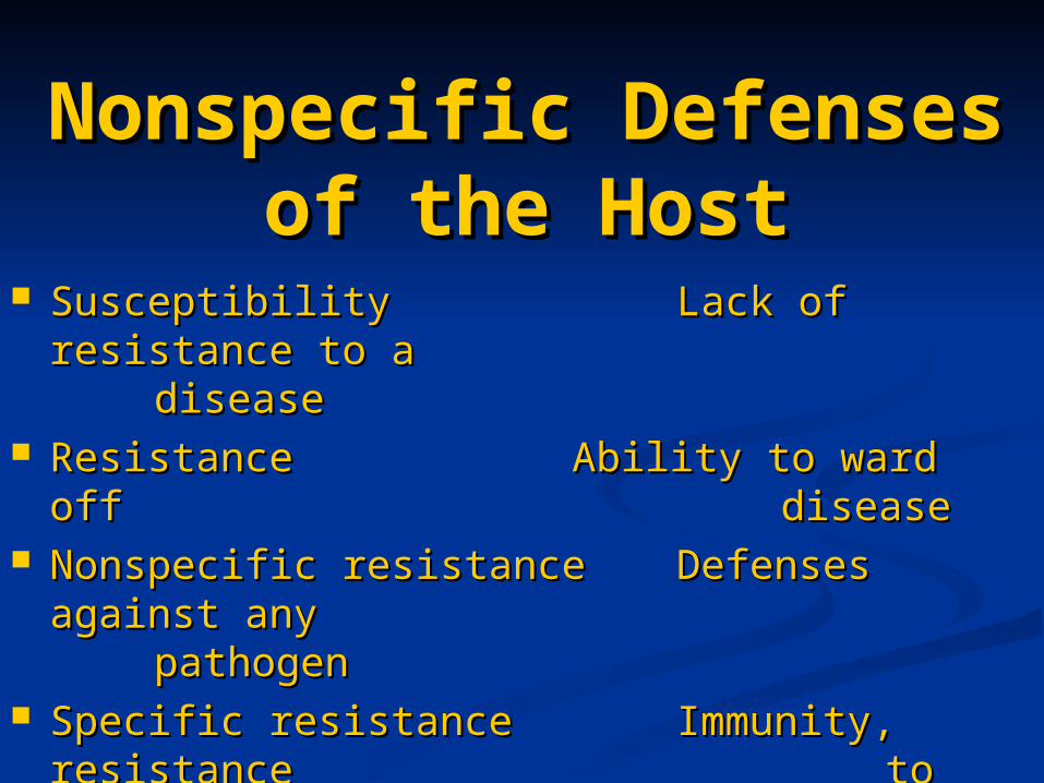
Nonspecific Defenses of the Nonspecific Defenses of the HostHost
SusceptibilitySusceptibility Lack of resistance Lack of resistance to a to a disease disease
Resistance Resistance Ability to ward off Ability to ward off diseasedisease
Nonspecific resistanceNonspecific resistance Defenses against Defenses against any any pathogenpathogen
Specific resistanceSpecific resistance Immunity, Immunity, resistance resistance to a to a specific pathogenspecific pathogen

Host DefensesHost Defenses
Figure 16.1

SkinSkin Epidermis consists of tightly packed cells with Keratin, a Epidermis consists of tightly packed cells with Keratin, a
protective proteinprotective protein Mucous membranesMucous membranes Ciliary escalatorCiliary escalator
Microbes trapped in mucus are transported away from Microbes trapped in mucus are transported away from the lungsthe lungs
Lacrimal apparatusLacrimal apparatus Washes eyeWashes eye
SalivaSaliva Washes microbes offWashes microbes off
UrineUrine Flows outFlows out
Vaginal secretionsVaginal secretions Flow outFlow out
Mechanical FactorsMechanical Factors
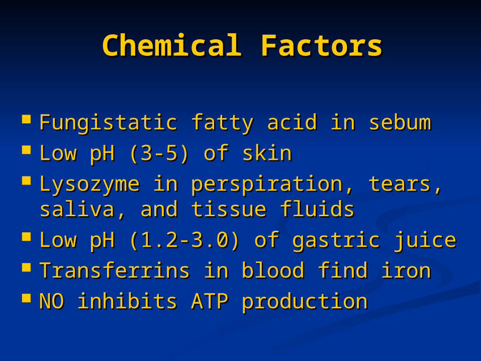
Fungistatic fatty acid in sebumFungistatic fatty acid in sebum Low pH (3-5) of skinLow pH (3-5) of skin Lysozyme in perspiration, tears, saliva, Lysozyme in perspiration, tears, saliva,
and tissue fluidsand tissue fluids Low pH (1.2-3.0) of gastric juiceLow pH (1.2-3.0) of gastric juice Transferrins in blood find ironTransferrins in blood find iron NO inhibits ATP productionNO inhibits ATP production
Chemical FactorsChemical Factors

Microbial antagonism/competitive Microbial antagonism/competitive exclusionexclusion Normal microbiota compete with Normal microbiota compete with
pathogens.pathogens.
Normal MicrobiotaNormal Microbiota

Formed Elements In Formed Elements In BloodBlood
Table 16.1

Percentage of each type of white cell in Percentage of each type of white cell in a sample of 100 white blood cellsa sample of 100 white blood cells
Differential White Cell Differential White Cell CountCount
NeutrophilsNeutrophils 60-70%60-70%BasophilsBasophils 0.5-1%0.5-1%
EosinophilsEosinophils 2-4%2-4%
MonocytesMonocytes 3-8%3-8%
LymphocytesLymphocytes 20-25%20-25%

Neutrophils: PhagocyticNeutrophils: Phagocytic Basophils: Produce histamineBasophils: Produce histamine Eosinophils: Toxic to parasites, some Eosinophils: Toxic to parasites, some
phagocytosisphagocytosis Monocytes: Phagocytic as mature Monocytes: Phagocytic as mature
macrophagesmacrophages Fixed macrophages in lungs, liver, bronchiFixed macrophages in lungs, liver, bronchi Wandering macrophages roam tissuesWandering macrophages roam tissues Lymphocytes: Involved in specific immunityLymphocytes: Involved in specific immunity
White Blood CellsWhite Blood Cells

PhagocytosisPhagocytosis
Figure 16.8a

Microbial Evasion of Microbial Evasion of PhagocytosisPhagocytosis
• • Inhibit adherence: M Inhibit adherence: M protein, capsulesprotein, capsules
Streptococcus pyogenes, S. Streptococcus pyogenes, S. pneumoniaepneumoniae
• • Kill phagocytes: Kill phagocytes: LeukocidinsLeukocidins
Staphylococcus aureusStaphylococcus aureus
• • Lyse phagocytes: Lyse phagocytes: Membrane attack Membrane attack complexcomplex
ListeriamonocytogenesListeriamonocytogenes
• • Escape phagosomeEscape phagosome ShigellaShigella
• • Prevent phagosome-Prevent phagosome-lysosome fusionlysosome fusion
HIVHIV
• • Survive in Survive in phagolysosomephagolysosome
Coxiella burnettiCoxiella burnetti

RednessRedness PainPain HeatHeat Swelling (edema)Swelling (edema) Acute-phase proteins activated Acute-phase proteins activated
(complement, cytokine, kinins)(complement, cytokine, kinins) Vasodilation (histamine, kinins, Vasodilation (histamine, kinins,
prostaglandins, leukotrienes)prostaglandins, leukotrienes) Margination and emigration of WBCsMargination and emigration of WBCs Tissue repairTissue repair
InflammationInflammation

Chemicals Released by Chemicals Released by Damaged CellsDamaged Cells
• • HistamineHistamine Vasodilation, increased Vasodilation, increased permeability of blood vesselspermeability of blood vessels
• • KininsKinins Vasodilation, increased Vasodilation, increased permeability of blood vesselspermeability of blood vessels
• • ProstaglandinsProstaglandins Intensity histamine and kinin Intensity histamine and kinin effecteffect
• • LeukotrienesLeukotrienes Increased permeability of blood Increased permeability of blood vessels, phagocytic attachmentvessels, phagocytic attachment

InflammationInflammation
Figure 16.9a, b

InflammationInflammation
Figure 16.9c, d

Hypothalamus normally set at 37°CHypothalamus normally set at 37°C Gram-negative endotoxin cause Gram-negative endotoxin cause
phagocytes to release interleukin 1phagocytes to release interleukin 1 Hypothalamus releases prostaglandins Hypothalamus releases prostaglandins
that reset the hypothalamus to a high that reset the hypothalamus to a high temperaturetemperature
Body increases rate of metabolism and Body increases rate of metabolism and shivering to raise temperatureshivering to raise temperature
When IL-1 is eliminated, body When IL-1 is eliminated, body temperature falls. (Crisis)temperature falls. (Crisis)
Fever: Abnormally High Fever: Abnormally High Body TemperatureBody Temperature
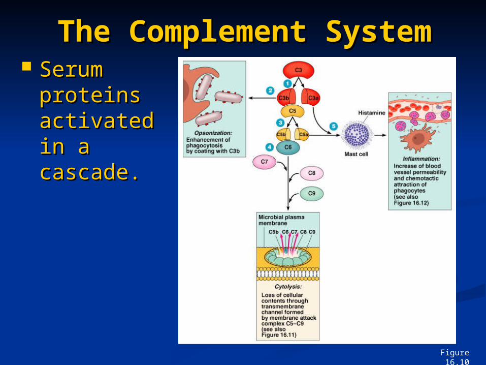
Serum Serum proteins proteins activated activated in a in a cascade. cascade.
The Complement The Complement SystemSystem
Figure 16.10

Effects of Complement Effects of Complement ActivationActivation
Opsonization or Opsonization or immune immune adherence: adherence: enhanced enhanced phagocytosisphagocytosis
Membrane attack Membrane attack complex: cytolysis complex: cytolysis
Attract phagocytesAttract phagocytes
Figure 16.11

Effects of Complement Effects of Complement ActivationActivation
Figure 16.12

Classical PathwayClassical Pathway
Figure 16.13

Alternative PathwayAlternative Pathway
Figure 16.14

Lectin PathwayLectin Pathway
Figure 16.15

Some bacteria evade Some bacteria evade complementcomplement
Capsules prevent C activationCapsules prevent C activation Surface lipid-carbohydrates prevent Surface lipid-carbohydrates prevent
MAC formationMAC formation Enzymatic digestion of C5aEnzymatic digestion of C5a

Alpha IFN & Beta IFNAlpha IFN & Beta IFN Cause cells to produce antiviral Cause cells to produce antiviral
proteins that inhibit viral replicationproteins that inhibit viral replication Gamma IFNGamma IFN
Causes neutrophils and macrophages Causes neutrophils and macrophages to phagocytize bacteriato phagocytize bacteria
Interferons (IFNs)Interferons (IFNs)

Interferons (IFNs)Interferons (IFNs)
Figure 16.16
1
2
3
4
5
Viral RNA from an infecting virus enters the cell.
The infecting virus replicates into new viruses.
The infecting virus also induces the host cell to produce interferon on RNA (IFN-mRNA), which is translated into alpha and beta interferons.
Interferons released by the virus-infected host cell bind to plasma membrane or nuclear membrane receptors on uninfected neighboring host cells, inducing them to synthesize antiviral proteins (AVPs). These include oligoadenylate synthetase, and protein kinase.
New viruses released by the virus-infected host cell infect neighboring host cells. 6 AVPs degrade viral
m-RNA and inhibit protein synthesis and thus interfere with viral replication.