Non-neoplastic Conditions in the Mediastinumeposterkiosk.com/wcti17/ePosters/EEE-02-18-Kim.pdf ·...
35
Non-neoplastic Conditions in the Mediastinum Mi Young Kim, MD, PhD Hyun Jung Koo, MD Jae Woo Song, MD, PhD Department of Radiology and Research Institute of Radiology Asan Medical Center, University of Ulsan College of Medicine, South Korea 2017 WCTI
Transcript of Non-neoplastic Conditions in the Mediastinumeposterkiosk.com/wcti17/ePosters/EEE-02-18-Kim.pdf ·...

Non-neoplastic Conditions
in the MediastinumMi Young Kim, MD, PhDHyun Jung Koo, MDJae Woo Song, MD, PhD
Department of Radiology and Research Institute of RadiologyAsan Medical Center, University of Ulsan College of Medicine, South Korea
2017 WCTI

Disclosures
None.1. Acute mediastinitis
2. Mesenchymal lesion
3. Vascular lesion
4. LN disease, other than tumor
5. Mediastinal cysts
6. Others
Contents
ThymolipomaHemangiomaLymphangioma
Perforation or rupture of esophagusDescending necrotizing mediastinitisDirect extension by adjacent infectionsFibrosing mediastinitisPerforation or rupture of airwaysPost surgical complication-mc

Acute MediastinitisPerforation of esophagus, fish bone (red arrow), UI 24cm
68/F
Abscess pocketGas bubbles Esophageal wall thickeningIncreased attenuation of mediastinal fat

Acute Mediastinitis
Iatrogenic rupture of esophagus during S-B tube insertion65/M
Extraluminal gas, pneumomediastinumSingle or multiple mediastinal abscesses with fluid collection

Distal esophageal rupture with acute mediastinitis
Pleural effusionPericardial effusionpneumomediastinum
Acute Mediastinitis
39/M C.C. vomiting

Acute Mediastinitis
55/M Esophageal dissection and perforation
Mucosal flap with submucosal distribution of gas or contrast, the classic double-barreled appearanceOccur posterior to the true lumen of the esophagus

Descending necrotizing mediastinitis and retropharyngeal abscess caused by K. pneumoniae
Multiple retropharyngeal and mediastinal abscesses
Increased soft tissue density and obliteration of normal fat planes, neck and
mediastinum
Acute Mediastinitis
56/M C.C. fever

Mediastinitis after cardiac surgery, S. aureus on pus culture
Retrosternal complicated fluid collection
Air bubbles
Fistula Focal osteomyelitis, nonunion of bone
Acute Mediastinitis
56/M S/P MV repair

Mediastinitis
20/M Trauma Post traumatic bronchial rupture
Pneumomediastinum
Contour deformity of airway or airway narrowing
Atelectasis or aspiration pneumonia

Mediastinitis
20/M C.C. dyspnea Fibrosing mediastinitis
Diffuse mediastinal soft tissue attenuationHilar or mediastinal massObstruction or narrowing of a pulmonary arteryObstruction or narrowing of SVCTracheobronchial narrowing or irregularities
CalcificationMultiple collateral veinsEnlarged left superior intercostals vein ( )2010-11-11
2001-11-15

2010-01-15
Steroid Tx.
Fibrosing mediastinitis, companion case
Soft tissue, ( mediastinum ), biopsy: Sclerosing inflammation
2009-10-17
Mediastinitis
51/M C.C. dyspnea

17/M
Predominant fat attenuation and intermingled soft tissue attenuationConnection between tumor and the thymusWell circumscribed consisting of mature adipose tissue with interspersed islands of thymic tissueIncidentally found in young adultsFat suppression or chemical shift MR imaging technique may be helpful
Thymolipoma
17/M1. Acute mediastinitis
2. Mesenchymal lesion
3. Vascular lesion
4. LN disease, other than tumor
5. Mediastinal cysts
6. Others
Contents
ThymolipomaHemangiomaLymphangioma

Hemangioma
Occur in the first four decades of life (75 %)Arise in the anterior mediastinum (68 %). Smoothly outlinedContain punctate calcification; phleboliths
35/M

Lymphangioma
Well-circumscribed lesion of low (or water) attenuation molding to the mediastinal contours and enveloping the great vessels3 types, unilocular (most common), cavernous, and intermediate types
47/F

Iatrogenic Left Innominate Vein Injury
Hematoma in anterior mediastinum
47/F1. Acute mediastinitis
2. Mesenchymal lesion
3. Vascular lesion
4. LN disease, other than tumor
5. Mediastinal cysts
6. Others
Contents
Dilatation of mediastinal veins, pulmonary artery, and bronchial artery Vascular injuryAcute aortic disease

Iatrogenic aortic injury
Focal contrast extravasation, active bleeding.Pericardial effusionDiffuse high attenuation at mediastinum, hematoma
73/F

Aneurysms of bronchial arteries
60/M
Tortuous and engorged left bronchial arteriesThrombosed aneurysm of right bronchial artery ( )Collateral vessels Ill-defined mediastinitis
Right bronchial artery: Hypertrophic and tortuous appearance.Left bronchial artery: Hypertrophic, tortuous appearance and aneurysm at the orifice of left bronchial artery. embolization

47/F
Sarcoidosis
Bilateral hilar lymph node enlargement is the most common finding, followed by interstitial lung disease. Sarcoidosis is a multisystem disorder that is characterized by noncaseous epithelioid cell granulomas. RadioGraphics 2010
47/F1. Acute mediastinitis
2. Mesenchymal lesion
3. Vascular lesion
4. LN disease, other than tumor
5. Mediastinal cysts
6. Others
Contents
Sarcoidosis TuberculosisCastleman’s disease

Tuberculous lymphadenopathy
Young adultPreponderance of involvement of the right paratracheal and subcarinal lymph nodes. Nodes larger than 2 cm in diameter invariably show central areas of relative low density and peripheral rim enhancement after injection of contrast medium
Radiology 1987
47/F

Nodo-Esophago-bronchial fistula caused by TB lymphadenitis36/M

Castleman's disease, Hyaline vascular type
AJR 2004
A solitary, well-circumscribed mediastinal mass Strong enhancementThe hyaline-vascular type (90%, unicentric form)
22/F

Disseminated Castleman’s disease manifests with diffuse mediastinal lymphadenopathy Plasma cell type (10%, multicentric form, systemic)
Castleman's disease, Plasma cell type59/F

Mediastinal cysts, Common findings
• Smooth and sharply marginated masses• Water density (about 50%) or homogeneous soft tissue density
(50%) on CT• Do not enhance after IV administration of contrast• Variable pattern on T1-weighted sequence• Hyperintense on T2-weighted sequence regardless of the
nature of the cyst content
60/F Thymic cyst, ant. mediastinum 39/M Pericardial cyst, Rt cardiophrenic angle
1. Acute mediastinitis
2. Mesenchymal lesion
3. Vascular lesion
4. LN disease, other than tumor
5. Mediastinal cysts
6. Others
Contents
Bronchogenic cystThymic cystPericardial cystForgut duplication cystThoracic duct cystMediastinal pancreatic pseudocyst

59/M Mediastinal,subcarinal,
complicated
57/M Lung
38/M Mediastinal, paratracheal
Bronchogenic cyst
54/F posterior mediastinum
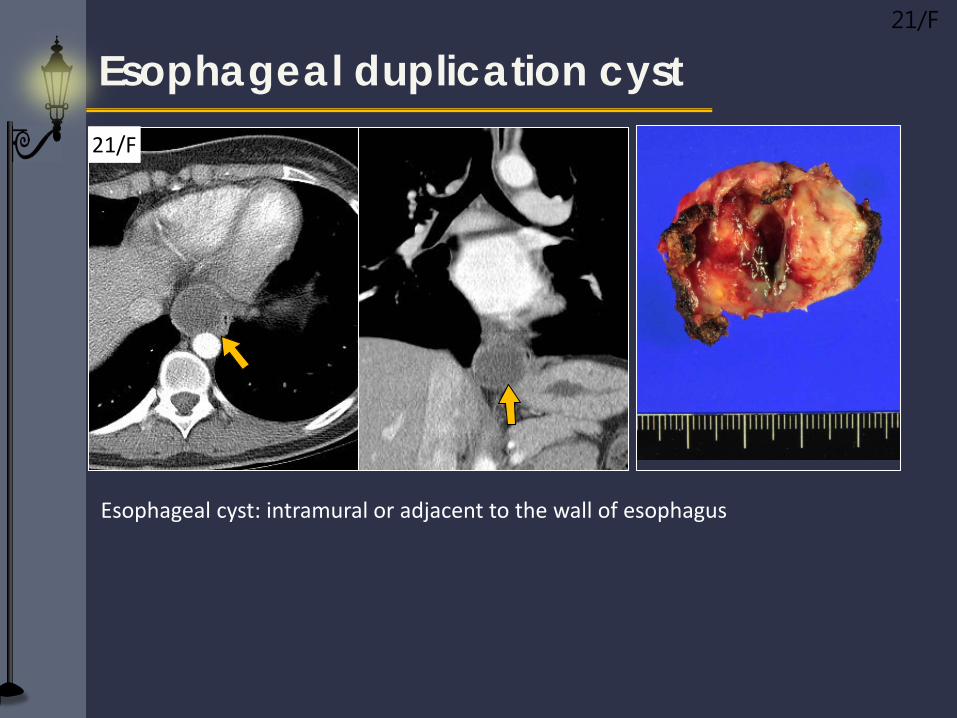
Esophageal duplication cyst21/F
Esophageal cyst: intramural or adjacent to the wall of esophagus
21/F

Thoracic duct cyst Rare cystic lesion in the mediastinum. Weakness in the thoracic duct wall allows formation of a cyst.
Chyle in the fluid of the cystSymptomless or compression of surrounding structures
58/FThoracic duct cyst and dilatation47/M

Mediastinal extension of pseudocyst can occur through anatomical openings of diaphragm.The posterior mediastinum is the most common location of the mediastinal pseudocysts through esophageal and aortic hiatus.
Mediastinal pancreatic pseudocyst44/M, chronic pancreatitis

Intrathoracic goiter
Continuity with the cervical gland Focal calcificationArise in the posterolateral portion of the thyroidgland and descend inferiorly to the posteriormediastinum
47/F
Intrathoracic goiterDiverticulumMediastinal lipomatosisExtramedullary hematopoiesis
1. Acute mediastinitis
2. Mesenchymal lesion
3. Vascular lesion
4. LN disease, other than tumor
5. Mediastinal cysts
6. Others
Contents

Intrathoracic goiterPre enhanced Post enhanced
T1WI
Enhanced T1WI
T2WI
Sharp borders (>90%) High attenuation on unenhanced CT (>100 HU)After IV contrast administration, thyroid tissue exhibits early and prolonged enhancementContinuity with the cervical gland
45/F
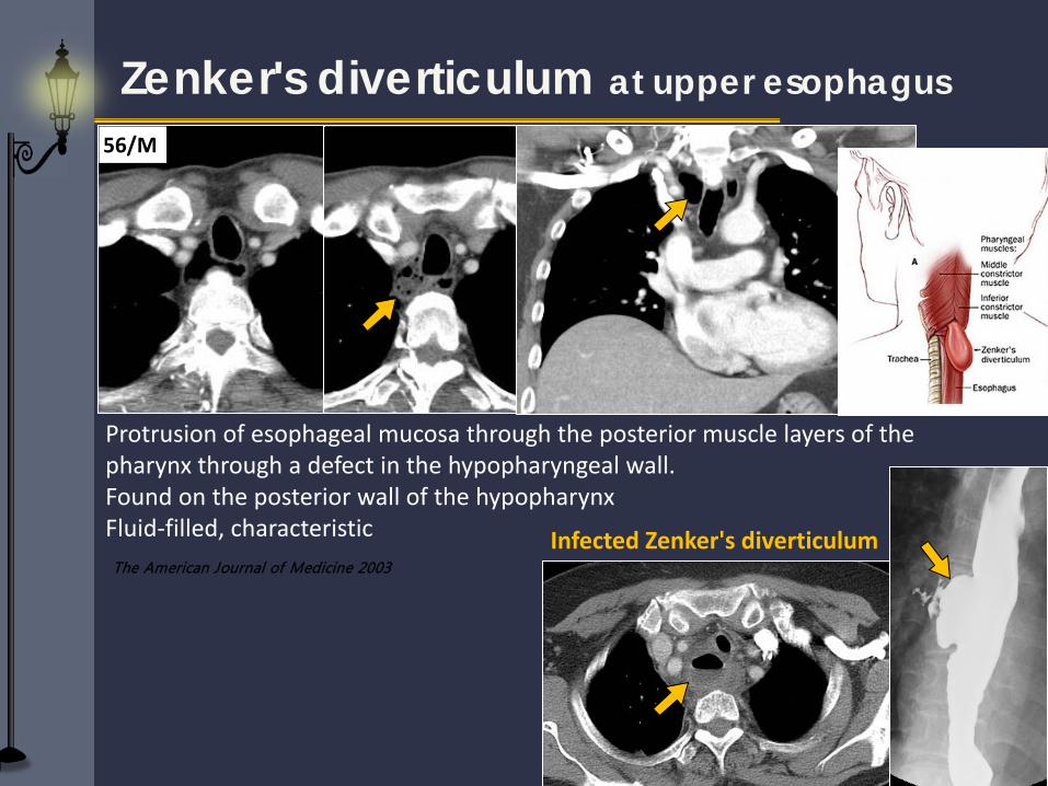
Protrusion of esophageal mucosa through the posterior muscle layers of the pharynx through a defect in the hypopharyngeal wall.Found on the posterior wall of the hypopharynxFluid-filled, characteristicThe American Journal of Medicine 2003
Zenker's diverticulum at upper esophagus
Infected Zenker's diverticulum
56/M

72/M, UI 37cmdistal esophageal diverticulum
Esophageal diverticulum
68/M, UI 30cmmiddle esophageal diverticulum

26/F Grave’s disease
Thymic lymphoid hyperplasia
Thymic lymphoid (follicular) hyperplasiaIncreased number of lymphoid follicles, seen in immune disorders including systemic lupus erythematosus, rheumatoid arthritis, scleroderma, thyrotoxicosis, and Grave’s disease Commonly associated with myasthenia gravis
2008-08-22 2009-03-02 f/u

60/M S/P left nephrectomy d/t RCC
Soft tissue, ( mediastinum ), excision: Low grade lipogenic tumor, favor lipoma
Mediastinal lipomatosis

61/M AML, Anemia
Extramedulary hematopoiesisWell-circumscribed, smooth, soft tissue attenuation masses, usually at multiple levels in a paraspinal location without erosion or pressure changes on the adjacent ribs or vertebral bodies
Hypertrophy of the medullary cavity of the ribsRare cause of posterior mediastinal masses and is usually seen in patients with severe, long-standing anemia
Summary
• CT is most often used in the assessment of mediastinal non-neoplastic diseases, with MR imaging usually being used as an adjunct to CT
• An awareness of the CT findings associated with the spectrum of mediastinal non-neoplastic diseases facilitates the accurate and prompt diagnosis
• The compartments of the mediastinum may help narrow the differential diagnosis.
Thank you for your attention.
Contact: [email protected]