A survey on computational methods for enhancer and enhancer ...
NF-Y Antagonizes Renin Enhancer Function by BlockingStimulatory Transcription Factors
Qi Shi, Kenneth W. Gross, Curt D. Sigmund
Abstract—We previously reported that the promoter proximal portion of the mouse renin enhancer contains a binding sitefor NF-Y (Ea) that overlaps with a positive regulatory element (Eb). In the context of the renin enhancer, NF-Y acts tooppose enhancer activity. We tested the hypothesis that NF-Y acts as a negative regulator by physically blocking thebinding of transcription factors to element-b (Eb). Increasing the spacing between the NF-Y binding site (Ea) and Ebby 2, 5, or 10 nucleotides increased activity of the enhancer to the same extent as mutations abolishing NF-Y binding.The increase in transcription caused by increasing the spacing between Ea and Eb was not due to a shift of NF-Y froma negative regulator to a positive regulator because there was no loss of activity when Ea was also mutated.Oligonucleotides containing the normal or increased spacing mutants still allowed the binding of both NF-Y to Ea andtranscription factors to Eb. In fact, we present evidence that both NF-Y and the Eb-binding factor(s) can each bindtogether on the same oligonucleotide containing either a 5- or 10-bp spacing between Ea and Eb. Our data stronglysuggest that the mechanism by which NF-Y opposes renin enhancer activity is to sterically block the binding of factorsto Eb. (Hypertension. 2001;38:332-336.)
Key Words: transcriptionn renin-angiotensin systemn activator n repressor
The renin-angiotensin system is an important regulator ofarterial pressure and electrolyte homeostasis. The level
of renin transcription, processing, and secretion dictates theeventual level of angiotensin II, because the angiotensinogencleavage step is thought to be rate limiting. Knowledge of themolecular mechanisms defining renin gene regulation remainincomplete but has been aided recently by the identificationof an enhancer of transcription located upstream of the mouserenin gene.1 This enhancer (mE) can markedly induce tran-scription of renin promoter reporter constructs when trans-fected into As4.1 cells, a renin-expressing tumor cell lineisolated from the kidney.2 mE consists of a 242-bp sequencelocated from22866 to22625 in the 59-flanking region ofmouseRen-1c. A homologous sequence with transcriptionalenhancer activity also exists upstream of the human reningene, although its position is much further from 59 (approx-imately 212 kb).3,4
Recently, we reported that a 40-bp promoter proximalregion of mE (called m40) was required for maximalenhancer activity.3 Structural and functional dissection ofm40 revealed that this region harbored 2cis-acting ele-ments: Ea, a negative regulatory element, and Eb, apositive regulatory element. Both of them specificallyinteracted with different nuclear proteins derived fromAs4.1 cells. NF-Y was identified as the Ea-binding factor
by electrophoretic mobility supershift assay. NF-Y is aubiquitous heterotrimeric transcription factor composed of3 subunits, NF-YA, NF-YB, and NF-YC.5,6 NF-YB andNF-YC first form a heterodimer, and then NF-YA interactswith the dimerized NF-YB/NF-YC to form the functionalheterotrimer. The DNA binding and transactivation activ-ity is unique to the heterotrimer.7 The NF-Y binding site isoften called a CCAAT box but can function in the invertedorientation.8 The overall consensus sequence is CT
GCTC
G-
CATTGGCT
TCG (CTCTGAGTGGCTG in Ren-1c), but less
conserved core recognition motifs have been identified inother genes.3,8,9
Although NF-Y is a ubiquitous transcriptional activator, itcan also function as a transcriptional repressor.9–13 Thenegative regulatory activity of NF-Y is generally due tooverlap in the binding site between NF-Y and anotherstimulatory transcription factor. In the insulin gene, thebinding sites for NF-Y and CREB overlap, and NF-Yantagonizes the transcriptional induction caused by cAMP.13
In the renin enhancer, Ea, the NF-Y binding site, overlapswith Eb, the binding site for retinoic acid receptor and otherunknown transcriptional activator(s).14 We therefore testedthe hypothesis that the negative regulatory activity of NF-Y isexerted by sterically blocking the binding of stimulatoryfactors to Eb.
Received January 29, 2001; first decision February 21, 2001; revision accepted February 23, 2001.From the Departments of Internal Medicine and Physiology and Biophysics, University of Iowa, College of Medicine (Q.S., C.D.S.), Iowa City; and
Department of Molecular and Cellular Biology, Roswell Park Cancer Institute (K.W.G.), Buffalo, NY.Correspondence to Curt D. Sigmund, PhD, Chair, Molecular Biology Interdisciplinary Program, Director, Transgenic and Gene Targeting Facility,
Department of Internal Medicine and Physiology & Biophysics, 2191 Medical Laboratory, University of Iowa, College of Medicine, Iowa City, IA 52242.E-mail [email protected]
© 2001 American Heart Association, Inc.
Hypertensionis available at http://www.hypertensionaha.org
332
by guest on June 16, 2018http://hyper.ahajournals.org/
Dow
nloaded from
Methods
PlasmidsThe luciferase (LUC) reporter vectors 2.6 kLUC, mE2.6 kLUC, andmEma2.6 kLUC were described previously.3 mE represents the242-bp mouseRenenhancer; 2.6 k represents a 2.6-kb 59-flankingsequence of mouseRen lacking mE; and mE2.6 k is a 2866-bpsequence with anSphI site separating mE from m2.6 k. Site-directedmutagenesis was performed by use of the GeneEditor in vitroSite-Directed Mutagenesis System (Promega). The sequence of allmutants was confirmed by DNA sequencing and restriction digestionanalysis. The 2-base insertion mutant mE(b2a)2.6 kLUC, 5-baseinsertion mutant mE(b5a)2.6 kLUC, and 10-base insertion mutantmE(b10a)2.6 kLUC were generated with the following oligonucle-otides: GTACTCTGACCTCTCTGAGTGGCTGG, GGCTG-TACTCTGACCTAGGCTCTGAGTGGCTGG, and CTCTGAC-CTAGGCGATGCTCTGAGTGGCTG (inserted bases are italicized),respectively. The 2-, 5-, and 10-base insertion mutants in mE(b2ma)2.6 kLUC, mE(b5ma)2.6 kLUC, and mE(b10ma)2.6 kLUC weregenerated with these oligonucleotides: GGCTGTACTCTGAC-CTCTCTTCGCTGCTGG, GCTGTACTCTGACCTAGGCTCT-TCGCTGCTGG, and CTCTGACCTAGGCGATGCTCTTCGCT-GCTG (ma mutation indicated by italics).
Cell Culture and Transient TransfectionCell culture and transient transfection of As4.1 cells (ATCCCRL2193) were previously described.3 In brief, As4.1 cells werecultured in reduced-serum OptiMEM supplemented with 2% fetalbovine serum, 1 mg/mL Albumax-II (GIBCO-BRL), penicillin (100U/mL), and streptomycin (100 mg/mL) for 2 days. The cells(2.53107) were transfected with equal-molar amounts of plasmidDNA by electroporation balancing with pUC19. RSV-LUC was usedas a positive control, and 0.1mg of CMV-bGal was cotransfected asan internal control. Cells were harvested and assayed for luciferaseand b-galactosidase activity 48 hours later.3,15 Luciferase activitywas normalized tob-galactosidase activity and then calculated as apercentage of RSV promoter activity. All activity assays wereperformed in duplicate, the average of 2 readings being 1 data point.
Electrophoretic Mobility Shift andSupershift AssayPreparations of the nuclear extract from As4.1 cells and probes forelectrophoretic mobility shift assay (EMSA) were previously de-scribed.3 The parent probe sequence is gatcTGTACTCTGACCTCT-GAGTGGCTGGTTGTG (top strand shown), where the 59-GATCoverhangs at each end of the annealed double-stranded oligonucle-otides were filled with [a-32P]dATP (NEN) and 3 other coldnucleotides using Klenow DNA polymerase. The mutant probeswere generated by inserting CT (b2a), AGGCT (b5a), or AGGC-GATGCT (b10a) between theitalicized TC. Each binding reactioncontained 0.02 pmol labeled probe ('60 000 dpm), 6mg nuclearextract, 1 mg poly[d(I-C)] (Boehringer Mannheim), and bindingbuffer with the final concentration of (in mmol/L) Tris-HCl (pH 7.5)10, EDTA 1, DTT 1, MgCl2 1, and KCl 60, as well as 5% glycerolin a total volume of 20mL. Cold competitor oligos were preincu-bated with nuclear extract and binding buffer for 15 minutes on icebefore the addition of probes. Binding reactions were incubated withprobe on ice for 15 minutes, followed by electrophoresis on 5%nondenaturing polyacrylamide gels. For supershift, the indicatedamount of NF-YA or NF-YB antiserum (gift of Dr R. Mantovani,Milan, Italy) was added after the initial incubation of probe, nuclearextract, and binding buffer, and the mixture was left on ice for 30minutes before electrophoresis.3
Statistical AnalysisAll data are presented as mean6SEM. Multiple comparisons wereanalyzed by 1-way ANOVA with SigmaStat (SPSS Scientific).Single comparisons were performed by use of at test.
ResultsThe purpose of this study was to determine the molecularmechanism by which NF-Y antagonizes mE. On the basis ofprevious studies of the mechanism of NF-Y–mediated tran-scriptional repression and the overlap between Ea and Eb inmE, we hypothesized that NF-Y binding to Ea may physicallydisrupt or prevent the binding of Eb-binding factor(s) to Eb.Our strategy was to alter the spacing between Ea and Eb toremove the overlap with the rationale that if transcriptionalrepression works via steric hindrance (NF-Y blocking Eb-binding protein to an overlapping site), then removing theoverlap between Ea and Eb should relieve transcriptionalrepression even in the presence of NF-Y.
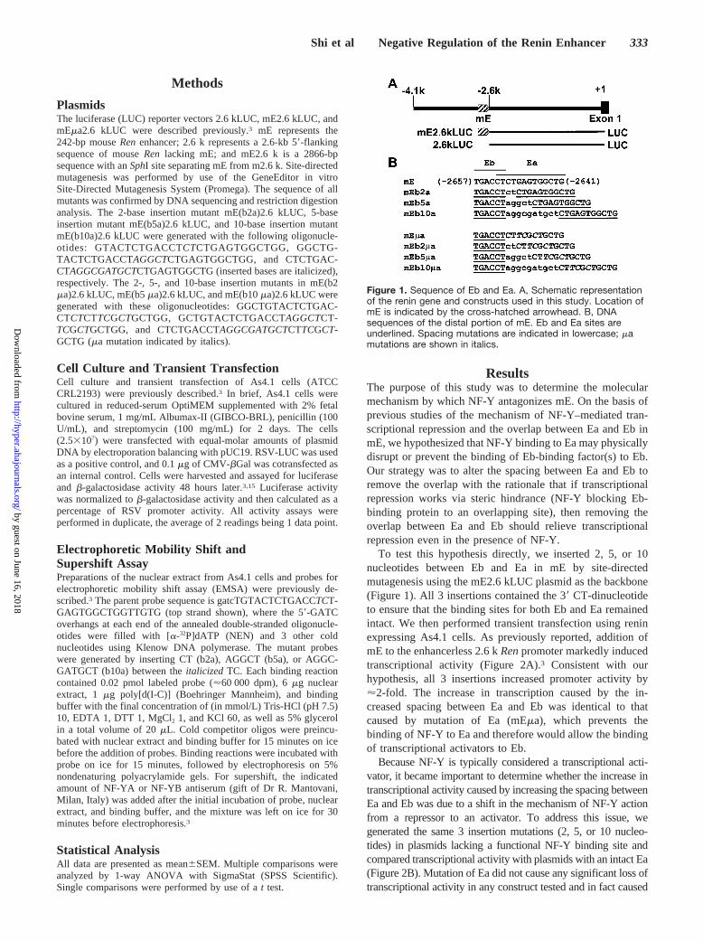
To test this hypothesis directly, we inserted 2, 5, or 10nucleotides between Eb and Ea in mE by site-directedmutagenesis using the mE2.6 kLUC plasmid as the backbone(Figure 1). All 3 insertions contained the 39 CT-dinucleotideto ensure that the binding sites for both Eb and Ea remainedintact. We then performed transient transfection using reninexpressing As4.1 cells. As previously reported, addition ofmE to the enhancerless 2.6 kRenpromoter markedly inducedtranscriptional activity (Figure 2A).3 Consistent with ourhypothesis, all 3 insertions increased promoter activity by'2-fold. The increase in transcription caused by the in-creased spacing between Ea and Eb was identical to thatcaused by mutation of Ea (mEma), which prevents thebinding of NF-Y to Ea and therefore would allow the bindingof transcriptional activators to Eb.
Because NF-Y is typically considered a transcriptional acti-vator, it became important to determine whether the increase intranscriptional activity caused by increasing the spacing betweenEa and Eb was due to a shift in the mechanism of NF-Y actionfrom a repressor to an activator. To address this issue, wegenerated the same 3 insertion mutations (2, 5, or 10 nucleo-tides) in plasmids lacking a functional NF-Y binding site andcompared transcriptional activity with plasmids with an intact Ea(Figure 2B). Mutation of Ea did not cause any significant loss oftranscriptional activity in any construct tested and in fact caused
Figure 1. Sequence of Eb and Ea. A, Schematic representationof the renin gene and constructs used in this study. Location ofmE is indicated by the cross-hatched arrowhead. B, DNAsequences of the distal portion of mE. Eb and Ea sites areunderlined. Spacing mutations are indicated in lowercase; mamutations are shown in italics.
Shi et al Negative Regulation of the Renin Enhancer 333
by guest on June 16, 2018http://hyper.ahajournals.org/
Dow
nloaded from

a significant increase in the b2a mutant. The increase intranscriptional activity caused by mutation of Ea in the b2aconstruct suggests that the 2-bp insertion may have only incom-pletely removed the overlap between the 2 sites. This finding isconfirmed below by EMSA. However, the fact that mutation ofthe NF-Y binding site did not result in a loss of transcriptionalactivity suggests that the increased transcriptional activity ob-served in the b2a, b5a, and b10a mutants is not due totranscriptional activation by NF-Y.
We previously reported that a double-stranded oligonucle-otide mX30 (Figure 3A) that contained Ea and Eb formed 2major DNA-protein complexes (a and b) with nuclear extractsfrom As4.1 cells.3 To verify that both NF-Y and Eb-bindingprotein can still form a complex on the insertion mutants, weindividually labeled double-stranded oligonucleotides con-taining the b2a, b5a, or b10a insertions and tested their
binding activity using EMSA. Strong complex-a formationwas evident on all 3 insertions (Figure 3B). Although strongcomplex-b formation was observed on the wild-type (WT)and b2a insertions, we noted a reproducible drop in theintensity of complex-b using b5a and b10a as a probe.Nevertheless, that b2a, b5a, and b10a still effectively boundto complex-a and complex-b is demonstrated by their effec-tiveness as competitors even when competing against the WToligonucleotide. Moreover, competition analysis using vary-ing levels of competitor DNAs revealed that there was noobservable change in the affinity of NF-Y for Ea in the b2a,b5a, and b10a mutants, because each was equally effective incompeting for binding to the WT probe (Figure 4). Signifi-cantly, the observation that antiserum against either the A orB subunit of NF-Y supershifted complex-a in all 3 mutantsconfirmed the interaction between NF-Y and Ea in b2a, b5a,and b10a (Figure 5).
The increase in transcriptional activity and the EMSA dataabove suggest that increasing the spacing between Ea and Ebmay allow both factors to simultaneously bind to the en-
Figure 2. Transfection analysis. Luciferase reporter vectors weretransiently transfected into As4.1 cells. A, Comparison of tran-scriptional activity of insertion mutants (cross-hatched bar) toWT (open bars) and mEma (closed bars) is shown. *P,0.05 vsmE; NS indicates P.0.05 vs mEma (n57). B, Comparison of theactivity of insertion mutants with intact Ea (open bars) to theactivity of insertion mutants with ma (closed bars). *P50.01; NSindicates P.0.05 (n57).
Figure 3. Nuclear protein binding to Ea and Eb. A, Sequence ofthe mX30 oligonucleotide and insertion variants used for EMSA.Top strand only is shown. Ea is overlined and Eb in underlined.Arrowhead indicates the location of b2a, b5a, and b10 inser-tions. All oligonucleotides have the same 59 and 39 termini. B,Binding activity of the insertion mutants in EMSA. Labeledprobes were incubated with 6 mg of the nuclear extract fromAs4.1 cells and 2.0 pmol of the indicated cold competitors(Comp; 100-fold molar excess). a indicates complex-a; b,complex-b; WT, WT probe; W, WT competitor; 2, b2a competi-tor; 5, b5a competitor; 10, b10a competitor; and -, no competi-tor. Complex-a represents the protein binding to Ea (NF-Y);complex-b, the protein(s) binding to Eb (RXR or an RXRpartner).
Figure 4. Affinity of Ea for mutant templates. The affinity for Eaand Eb on the insertion mutations was determined by competi-tion using WT and the indicated insertion mutants. The WT tem-plate in Figure 3 was used as a probe and incubated with 6 mgof As4.1 cell nuclear extract. Ramps indicate increasing molarexcess (10-, 30-, and 90-fold) of the indicated competitor. aindicates complex-a (NF-Y); b, complex-b; WT, WT competitor;0, no nuclear extract; and -, no competitors.
Figure 5. NF-Y binds to Ea. Labeled probes (as indicated) wereincubated with 6 mg of the nuclear extracts from As4.1 cells for15 minutes. Antibody against NF-YA subunit (3 mg) or 4 mg anti-body against NF-YB subunit was added into the reactions asindicated and incubated for 30 minutes. a indicates complex-a(NF-Y); b, complex-b; s, NF-Y supershift; W, WT probe; 2, b2aprobe; 5, b5a probe; and 10, b10a probe.
334 Hypertension September 2001
by guest on June 16, 2018http://hyper.ahajournals.org/
Dow
nloaded from

hancer. Simultaneous binding could also potentially explainthe loss of complex-b on b5a and b10a in Figure 3. To testthis hypothesis, we examined whether the insertion mutationscould permit simultaneous binding of both complexes (a andb) to 1 probe molecule. In normal EMSA, a limited amount ofnuclear proteins and an excess of oligonucleotide probe areused. Under these conditions, it is unlikely that 1 probemolecule will bind 2 proteins unless a strong cooperativeinteraction exists. To increase the probability of 2 complexesassembling on 1 probe, we raised the amount of nuclearextract from 6 to 15mg per reaction and reduced the amountof labeled probes from 0.02 to 0.002 pmol per reaction. Weanticipated that probes carrying 2 protein complexes wouldmigrate more slowly than probes carrying a single complexand will be visualized as a further retarded band on anondenaturing gel. Interestingly, a “superretarded” band wasevident on b5a and b10a (Figure 6A). That this superretardedcomplex results from the binding of both NF-Y and Eb-binding protein is suggested by competition analysis. Asexpected, WT mX30, containing both Ea and Eb, efficientlycompetes for complex-a, complex-b, and complex-ab. MutantmX30 ma, which lacks Ea, efficiently competes forcomplex-b on WT and b2a and competes for complex-ab onb5a and b10a, suggesting that complex-ab does indeedcontain the Eb-binding protein. Similarly, mutant mX30mb,which lacks Eb, competes for complex-a on all 4 oligonucle-otides and competes for complex-ab on b5a and b10a,suggesting the presence of NF-Y in complex-ab. This isfurther supported by the observation that competition withmX30 mb restores the formation of complex-b on b5a andb10a, presumably because excess binding of NF-Y on themX30 mb competitor prevents the formation of the ab doublecomplex and frees the Eb-binding protein to complex alonewith Eb. Antiserum to subunit A of NF-Y causes a supershiftof complex-a and prevents the formation of complex-abwithout causing any obvious additional supershift product(Figure 6B). Interestingly, under some experimental condi-tions, NF-Y antiserum prevents complex formation, whereas
under other conditions, the antiserum causes a clear andabundant supershift to appear.16,17
Taken together, these data strongly suggest that NF-Ymediates its antagonistic activity by preventing the binding ofEb-binding protein(s) to Eb. Increasing the spacing did notcause NF-Y to act as a positive factor and did not affect theability of NF-Y to bind to the modified enhancers. In fact,increasing the spacing by 5 and 10 nucleotides provided anopportunity for both NF-Y and Eb-binding protein to simul-taneously bind to mX30.
DiscussionThe ability of the renin gene enhancer to stimulate a 100-foldincrease in transcriptional activity of theRenpromoter makesit an important candidate inRen gene regulation.1 Wepreviously identified 2 transcription factor binding sites thatmechanistically oppose each other.3 We hypothesized that thebinding of NF-Y to Ea acts as a negative regulator because itblocks the binding of transcription factors to Eb. Changingthe spacing between Ea and Eb has the same effect asmutating Ea, suggesting that this hypothesis is correct. Thesedata are supported by the observations that NF-Y can stillbind to Ea even when the spacing between Ea and Eb isincreased and that mutations of Ea in the b5a and b10amutants does not inhibit transcription of theRenpromoter.
NF-Y is generally considered a ubiquitous transcriptionalactivator, although the exact mechanisms through whichNF-Y regulates transcription is not totally clear. Sequencesurveys have revealed that most stimulatory NF-Y bindingsites are located from280 through 2100 in TATA-containing promoters or very close to the transcription startsite in TATA-less promoters.8,18 Other studies of NF-Ycontaining promoters have demonstrated that the positivefunction of the NF-Y site also depends on other adjacentcis-acting elements.19,20 In mouse Ren, Ea is located farupstream of the promoter (about22.6 kb) and does not havethe previously reported adjacent cis-acting elements thatcooperated with positive NF-Y sites in the other genes.
The negative regulatory activity of NF-Y has been reportedin several genes.9–13 Mutation of an NF-Y binding siteoverlapping with a C/EBP site upstream of the humanapolipoprotein A-I promoter increases transcription.9 Overlapof NF-Y and a CRE in the promoter proximal region of the ratinsulin I gene attenuates transcriptional stimulation bycAMP.9,13 Although Ea does not match a perfect consensusNF-Y site (It has a single-base substitution in its corerecognition motif), the same single-base substitution alsoexists in human apolipoprotein A-I gene, which is alsorepressed by NF-Y.9
Under normal conditions, activation of the enhancer mayinvolve a competition between NF-Y and the factors bindingto Eb. It is likely that both the amount of NF-Y andEb-binding protein and their relative binding affinity may bedeterminants of this competition. EMSA experiments repro-ducibly suggest that the level of NF-Y exceeds the level ofEb-binding protein under baseline conditions. It is possiblethat specific conditions exist that change the balance betweenthese proteins and therefore favor binding to Eb. It is knownthat the level or affinity of NF-Y can be regulated by the
Figure 6. Dual complex formation on b5a and b10a. Analysis ofdouble complexes formed on single probes in EMSA. A,Labeled probes (0.002 pmol per lane) as indicated were incu-bated with 15 mg of the nuclear extracts from As4.1 cells for 15minutes. Then, 1.0 pmol of the indicated cold competitors (500-fold molar excess) was added and incubated for another 15minutes. B, Probes were incubated as above and then incu-bated either with or without NF-YA antiserum. a indicatescomplex-a; b, complex-b; ab, double complex a1b; as,complex-a supershift; W, WT probe; 2, b2a probe; 5, b5a probe;and 10, b10a probe.
Shi et al Negative Regulation of the Renin Enhancer 335
by guest on June 16, 2018http://hyper.ahajournals.org/
Dow
nloaded from

redox state of the cell, intracellular calcium concentration,cellular differentiation, and the presence of serum supplemen-tation.16,21–23 Exactly what physiological cues regulate thelevel or affinity of the Eb-binding protein will have to wait forthe unequivocal identification of that factor(s) (see below).
It is now becoming clear that the mechanism by which therenin enhancer regulates renin transcription will be quitecomplicated because, in addition to the elements describedabove, additional elements upstream of Eb have recently beenshown to be required for maximal enhancer activity (Figure7). One element, termed Ec, which located 10 bp upstream ofEb, is a direct repeat of the Eb TGACCT motif. TGACCTdirect repeats have been reported to bind members of thenuclear hormone receptor superfamily of transcription fac-tors.24 We have recently demonstrated that Eb is a half-sitefor retinoic acid receptor (RAR). Eb and Ec together can bindRARa and RXRa and form a functional RAR element.14 Inthe studies described herein, Ec was intact and the distancebetween Ec and Eb remained unaltered. In addition, anelement called Ed, which lies further upstream of Ec and Eb,has homology to a CRE and may thus bind members of theCREB/ATF-1 family of transcription factors (T.A. Black, etal, unpublished observation, 2000). It is likely that theinterplay between transcription factors binding to these sitesmay be required because the CREB/ATF-1 and RAR/RXRpathways share a similar coactivator in p300/CBP.25,26 Otheryet-unidentified regulatory elements may also be present intheRenenhancer, and other transcription factors may bind toEd, Ec, Eb, and Ea. In the end, it is likely that ubiquitouspositive and negative regulatory factors will be found tocooperate to regulate expression of renin through theenhancer.
AcknowledgmentsFunds in support of this work were obtained from the NIH (HL-48058, HL-61446, and HL-55006 to C.D.S. and HL-48459 toK.W.G.). We acknowledge the outstanding technical assistance ofDeborah Davis and Xiaoji Zhang. DNA sequencing was performedat the University of Iowa DNA Core Facility.
References1. Petrovic N, Black TA, Fabian JR, Kane CM, Jones CA, Loudon JA,
Abonia JP, Sigmund CD, Gross KW. Role of proximal promoter elementsin regulation of renin gene transcription.J Biol Chem. 1996;271:22499–22505.
2. Sigmund CD, Okuyama K, Ingelfinger J, Jones CA, Mullins JJ, Kim U,Kane-Haas C, Wu C, Kenney L, Rustum Y, Dzau V, Gross KV. Isolationand characterization of renin expressing cell lines from transgenic micecontaining a renin promoter viral oncogene fusion construct.J Biol Chem.1990;265:19916–19922.
3. Shi Q, Black TA, Gross KW, Sigmund CD. Species-specific differencesin positive, and negative regulatory elements in the renin gene enhancer.Circ Res. 1999;85:479–488.
4. Yan Y, Jones CA, Sigmund CD, Gross KW, Catanzaro DF. Conservedenhancer elements in human and mouse renin genes have different tran-scriptional effects in As4.1 cells.Circ Res. 1997;81:558–566.
5. Bellorini M, Lee DK, Dantonel JC, Zemzoumi K, Roeder RG,Tora L,Mantovani R. CCAAT binding NF-Y-TBP interactions: NF-YB andNF-YC require short domains adjacent to their histone fold motifs forassociation with TBP basic residues.Nucleic Acids Res. 1997;25:2174–2181.
6. Maity SN, de Crombrugghe B. Role of the CCAAT-binding proteinCBF/NF-Y in transcription.Trends Biochem Sci. 1998;23:174–178.
7. Sinha S, Kim IS, Sohn KY, de Crombrugghe B, Maity SN. Three classesof mutations in the A subunit of the CCAAT-binding factor CBFdelineate functional domains involved in the three-step assembly of theCBF-DNA complex.Mol Cell Biol. 1996;16:328–337.
8. Mantovani R. A survey of 178 NF-Y binding CCAAT boxes.NucleicAcids Res. 1998;26:1135–1143.
9. Papazafiri P, Ogami K, Ramji DP, Nicosia A, Monaci P, Cladaras C,Zannis VI. Promoter elements and factors involved in hepatic tran-scription of the human ApoA-I gene positive and negative regulators bindto overlapping sites.J Biol Chem. 1991;266:5790–5797.
10. Boucher PD, Piechocki MP, Hines RN. Partial characterization of thehuman CYP1A1 negatively acting transcription factor and mutationalanalysis of its cognate DNA recognition sequence.Mol Cell Biol. 1995;15:5144–5151.
11. Moriuchi H, Moriuchi M, Cohen JI. The varicella-zoster virusimmediate-early 62 promoter contains a negative regulatory element thatbinds transcriptional factor NF-Y.Virology. 1995;214:256–258.
12. Kelly D, Kim SJ, Rizzino A. Transcriptional activation of the type IItransforming growth factor-b receptor gene upon differentiation ofembryonal carcinoma cells.J Biol Chem. 1998;273:21115–21124.
13. Eggers A, Siemann G, Blume R, Knepel W. Gene-specific transcriptionalactivity of the insulin cAMP-responsive element is conferred by NF-Y incombination with cAMP response element-binding protein.J Biol Chem.1998;273:18499–18508.
14. Shi Q, Gross KW, Sigmund CD. Retinoic acid-mediated activation of themouse renin enhancer.J Biol Chem. 2001;276:3597–3603.
15. Ying L, Morris BJ, Sigmund CD. Transactivation of the human reninpromoter by the cyclic AMP/protein kinase A pathway is mediated byboth CREB-dependent and CREB-independent mechanisms in Calu-6cells.J Biol Chem. 1997;272:2412–2420.
16. Marziali G, Perrotti E, Ilari R, Testa U, Coccia EM, Battistini A. Tran-scriptional regulation of the ferritin heavy-chain gene: the activity of theCCAAT binding factor NF-Y is modulated in heme-treated Friendleukemia cells and during monocyte-to-macrophage differentiation.MolCell Biol. 1997;17:1387–1395.
17. Lu CC, Yen TS. Activation of the hepatitis B virus S promoter bytranscription factor NF-Y via a CCAAT element.Virology. 1996;225:387–394.
18. Bucher P. Weight matrix descriptions of four eukaryotic RNA poly-merase II promoter elements derived from 502 unrelated promotersequences.J Mol Biol. 1990;212:563–578.
19. Linhoff MW, Wright KL, Ting JP. CCAAT-binding factor NF-Y andRFX are required for in vivo assembly of a nucleoprotein complex thatspans 250 base pairs: the invariant chain promoter as a model.Mol CellBiol. 1997;17:4589–4596.
20. Jackson SM, Ericsson J, Mantovani R, Edwards PA. Synergistic acti-vation of transcription by nuclear factor Y and sterol regulatory elementbinding protein.J Lipid Res. 1998;39:767–776.
21. Nakshatri H, Bhat-Nakshatri P, Currie RA. Subunit association and DNAbinding activity of the heterotrimeric transcription factor NF-Y is reg-ulated by cellular redox.J Biol Chem. 1996;271:28784–28791.
22. Chang ZF, Liu CJ. Human thymidine kinase CCAAT-binding protein isNF-Y, whose A subunit expression is serum-dependent in human IMR-90diploid fibroblasts.J Biol Chem. 1994;269:17893–17898.
23. Roy B, Lee AS. Transduction of calcium stress through interaction of thehuman transcription factor CBF with the proximal CCAAT regulatoryelement of the grp78/BiP promoter.Mol Cell Biol. 1995;15:2263–2274.
24. Glass CK. Differential recognition of target genes by nuclear receptormonomers, dimers, and heterodimers.Endocr Rev. 1994;15:391–407.
25. Glass CK, Rose DW, Rosenfeld MG. Nuclear receptor coactivators.Current Opinion in Cell Biology. 1997;9:222–232.
26. De Cesare D, Fimia GM, Sassone-Corsi P. Signaling routes to CREM andCREB: plasticity in transcriptional activation.Trends Biochem Sci. 1999;24:281–285.
Figure 7. Sequence of the promoter proximal portion of mE.The cis-acting elements in the promoter proximal portion of mEand specific mutations are shown. The locations of Ed, Ec, Eb,and Ea are underlined, and the consensus sequences for theircognate factors are indicated.
336 Hypertension September 2001
by guest on June 16, 2018http://hyper.ahajournals.org/
Dow
nloaded from
Qi Shi, Kenneth W. Gross and Curt D. SigmundFactors
NF-Y Antagonizes Renin Enhancer Function by Blocking Stimulatory Transcription
Print ISSN: 0194-911X. Online ISSN: 1524-4563 Copyright © 2001 American Heart Association, Inc. All rights reserved.
is published by the American Heart Association, 7272 Greenville Avenue, Dallas, TX 75231Hypertension doi: 10.1161/01.HYP.38.3.332
2001;38:332-336Hypertension.
http://hyper.ahajournals.org/content/38/3/332World Wide Web at:
The online version of this article, along with updated information and services, is located on the
http://hyper.ahajournals.org//subscriptions/
is online at: Hypertension Information about subscribing to Subscriptions:
http://www.lww.com/reprints Information about reprints can be found online at: Reprints:
document. Permissions and Rights Question and Answer this process is available in the
click Request Permissions in the middle column of the Web page under Services. Further information aboutOffice. Once the online version of the published article for which permission is being requested is located,
can be obtained via RightsLink, a service of the Copyright Clearance Center, not the EditorialHypertensionin Requests for permissions to reproduce figures, tables, or portions of articles originally publishedPermissions:
by guest on June 16, 2018http://hyper.ahajournals.org/
Dow
nloaded from