SIRT3 overexpression antagonizes high glucose accelerated ... · SIRT3 overexpression antagonizes...
17
SIRT3 overexpression antagonizes high glucose accelerated cellular senescence in human diploid fibroblasts via the SIRT3–FOXO1 signaling pathway Bin Zhang & Shaoyuan Cui & Xueyuan Bai & Li Zhuo & Xuefeng Sun & Quan Hong & Bo Fu & Jianzhong Wang & Xiangmei Chen & Guangyan Cai Received: 12 April 2012 / Accepted: 18 February 2013 / Published online: 14 March 2013 # The Author(s) 2013. This article is published with open access at Springerlink.com Abstract Sirtuin 3 (SIRT3) is one of the seven mamma- lian sirtuins, which are homologs of the yeast Sir2 gene. SIRT3 is the only sirtuin reported to be associated with human life span. Many recent studies have indicated that SIRT3 levels are elevated by exercise and caloric restric- tion, but whether SIRT3 influences cell senescence under stressed conditions in human diploid fibroblasts has not been established. Our data showed that expression of AGE (2013) 35:2237–2253 DOI 10.1007/s11357-013-9520-4 Bin Zhang and Shaoyuan Cui, these authors contributed equally to this work. B. Zhang : S. Cui : X. Bai : X. Sun : Q. Hong : B. Fu : J. Wang : X. Chen (*) : G. Cai (*) State Key Laboratory of Kidney Diseases, Department of Nephrology, Chinese PLA General Hospital and Military Medical Postgraduate College, Beijing, 100853, China e-mail: [email protected] e-mail: [email protected] B. Zhang e-mail: [email protected] S. Cui e-mail: [email protected] X. Bai e-mail: [email protected] X. Sun e-mail: [email protected] Q. Hong e-mail: [email protected] B. Fu e-mail: [email protected] J. Wang e-mail: [email protected] B. Zhang : X. Chen School of Medicine, Nankai University, Tianjin 300071, China B. Zhang Department of Nephrology, The Second Affiliated Hospital, Chongqing Medical University, Chongqing 400010, China L. Zhuo Department of Nephrology, China - Japan Friendship Hospital, Beijing 100029, China e-mail: [email protected]
Transcript of SIRT3 overexpression antagonizes high glucose accelerated ... · SIRT3 overexpression antagonizes...

SIRT3 overexpression antagonizes high glucose acceleratedcellular senescence in human diploid fibroblasts viathe SIRT3–FOXO1 signaling pathway
Bin Zhang & Shaoyuan Cui & Xueyuan Bai &Li Zhuo & Xuefeng Sun & Quan Hong & Bo Fu &
Jianzhong Wang & Xiangmei Chen & Guangyan Cai
Received: 12 April 2012 /Accepted: 18 February 2013 /Published online: 14 March 2013# The Author(s) 2013. This article is published with open access at Springerlink.com
Abstract Sirtuin 3 (SIRT3) is one of the seven mamma-lian sirtuins, which are homologs of the yeast Sir2 gene.SIRT3 is the only sirtuin reported to be associated withhuman life span. Many recent studies have indicated that
SIRT3 levels are elevated by exercise and caloric restric-tion, but whether SIRT3 influences cell senescence understressed conditions in human diploid fibroblasts has notbeen established. Our data showed that expression of
AGE (2013) 35:2237–2253DOI 10.1007/s11357-013-9520-4
Bin Zhang and Shaoyuan Cui, these authors contributed equallyto this work.
B. Zhang : S. Cui :X. Bai :X. Sun :Q. Hong :B. Fu :J. Wang :X. Chen (*) :G. Cai (*)State Key Laboratory of Kidney Diseases,Department of Nephrology, Chinese PLA General Hospitaland Military Medical Postgraduate College,Beijing, 100853, Chinae-mail: [email protected]: [email protected]
B. Zhange-mail: [email protected]
S. Cuie-mail: [email protected]
X. Baie-mail: [email protected]
X. Sune-mail: [email protected]
Q. Honge-mail: [email protected]
B. Fue-mail: [email protected]
J. Wange-mail: [email protected]
B. Zhang :X. ChenSchool of Medicine, Nankai University,Tianjin 300071, China
B. ZhangDepartment of Nephrology,The Second Affiliated Hospital,Chongqing Medical University,Chongqing 400010, China
L. ZhuoDepartment of Nephrology,China - Japan Friendship Hospital,Beijing 100029, Chinae-mail: [email protected]

SIRT3 is elevated in human diploid fibroblasts under lowglucose (3.3 mM glucose) growth conditions and de-creased under high glucose (25 mM glucose) growthconditions. We have demonstrated that SIRT3 interactswith forkhead box protein O1 (FOXO1). High glucoselevels also increased aging phenotypes and FOXO1 acet-ylation level. We have demonstrated that overexpressionof SIRT3 under high glucose conditions reduces FOXO1acetylation, suggesting that deacetylation of FOXO1 bySIRT3elevates theexpressionof theFOXO1targetgenes,catalase, andmanganese superoxidedismutase (MnSOD)while decreasing senescence phenotypes. We studied theeffectsofSIRT3proteinknockdownbyshRNAunder lowglucose conditions. The data showed that shRNA-SIRT3accelerated senescence phenotypes and acetylation ofFOXO1; the expression level of catalase and MnSODdecreased compared with the control group. As a conse-quence, SIRT3 antagonized cellular senescence with thecharacteristic features of delayed SA-β-gal staining,senescence-associated heterochromatin foci (SAHF) for-mation, and p16INK4A expression. These results demon-strate for the first time that SIRT3 overexpressionantagonizes high glucose-induced cellular senescence inhuman diploid fibroblasts via the SIRT3–FOXO1 signal-ing pathway.
Keywords SIRT3 . FOXO1 . Human diploidfibroblasts . High glucose . Cellular senescence
Introduction
The sirtuins (or Sir2-like proteins) are a conserved fam-ily of NAD+-dependent protein deacetylases whichoverexpression has been reported to extend life span inyeast, worms, and flies (Kenyon 2010). Accumulatingevidence suggests that reversible protein acetylation,which was historically considered to be limited to his-tone proteins, may be a major regulatory mechanismthat controls functions of nonhistone proteins. Sevenmammalian homologs belonging to the family of Sir2proteins (Sirt 1–7) have been identified. The challengenow is to explain how these proteins communicate toregulate cross talk between aging and onset and pro-gression of age-related disorders (de Oliveira et al.2010). SIRT1 and SIRT6 are localized to the nucleus;SIRT7 is localized to the nucleolus; sirtuin 3 (SIRT3),SIRT4, and SIRT5 are localized to the mitochondria;and SIRT2 is localized to the cytoplasm. They regulate a
wide range of intracellular processes (Haigis andGuarente 2006). Among them, SIRT3 is unique becauseit is the only analog which increased expression hasbeen found to be associated with extended life span inhumans (Rose et al. 2003; Bellizzi et al. 2005).
SIRT3 is localized predominantly in mitochondriaand is considered to be a mitochondrial stress sensorthat can modulate activity of several mitochondrialproteins involved in metabolism and oxidative stressregulatory pathways (Ahn et al. 2008; Scher et al.2007; Onyango et al. 2002; Shi et al. 2005; Yang etal. 2010; Kong et al. 2010). Several lines of evidencelink SIRT3 to metabolism: caloric restriction activatesSIRT3 expression in both white and brown adiposetissues. In addition, cold exposure upregulates SIRT3expression in brown fat, whereas elevated climatictemperature reduces its expression (Shi et al. 2005).Ang II downregulates SIRT3 mRNA, and this effect isinhibited by an AT1 antagonist in cultured tubularepithelial cells (Benigni et al. 2009). However, therelationship between SIRT3 and cell senescence isyet unclear.
A conserved insulinlike signaling pathway exists ininvertebrates and vertebrates. The constituents of thispathway act as second messengers, conveying signalinginformation from the insulin receptor (termed DAF-2 inCaenorhabditis elegans and inR in Drosophilamelanogaster) to key downstream effectors includingthe forkhead transcription factors (FOXO) (DAF-16 inC. elegans and dFOXO in Drosophila) (Murphy 2006).The FOXO family in mammals consists of theevolutionally highly conserved forkhead transcriptionfactors forkhead box protein O1 (FOXO1), FOXO3a,FOXO4, and FOXO6, which are mammalian orthologsof DAF-16 in C. elegans. DAF-16 is required for lifespan extension in C. elegans (Huang and Tindall 2007;Zanella et al. 2010; Yamaza et al. 2010; Giannakou et al.2008). A recent series of studies has demonstrated thatFOXO factors play important roles in inducing variousdownstream target genes, including regulators of metab-olism, cell cycle, DNA repair, cell survival or apoptosis,and oxidative stress response (Furukawa-Hibi et al.2005; Sedding 2008). The activity of FOXO factors isregulated by a sophisticated signaling network that in-tegrates information from PI3K/Akt and stress-inducedsignaling pathways resulting in a specific pattern ofpost-translational modifications. The multiple post-transcriptional events regulated the activity of FOXOproteins including phosphorylation, ubiquitylation, and
2238 AGE (2013) 35:2237–2253

acetylation at three different levels: subcellular localiza-tion, stability, and transcriptional activity.
A series of recent studies in mammalian cells haveindicated that the reversible acetylation of FOXO pro-teins by nuclear coactivators and corepressors providesanother layer of regulation of nuclear FOXO transcrip-tional factors. Acetylation of FOXO proteins byacetylases such as CBP and p300 increases in responseto oxidative stress (Brunet et al. 2004; Frescas et al.2005; Kitamura et al. 2005). Acetyl-FOXO proteinsaccumulate in the nucleus and cytoplasm associate withPml bodies, which hinder their activity (Kitamura et al.2005). A recent study showed that the human Sir2ortholog SIRT1 binds and deacetylates FOXO proteinsat lysine residues that are acetylated by CBP/p300(Daitoku et al. 2004). However, the effect of FOXOacetylation–deacetylation on the transcription of targetgenes is not quite so straightforward. We consideredthat acetylation could interfere with FOXO bindingto target DNA and thereby prevent FOXO-mediatedtranscription. A recent study demonstrated thatSIRT3 blocked cardiac hypertrophy by activating theFOXO3a-dependent, antioxidant-encoding genes, cata-lase and manganese superoxide dismutase (MnSOD),thereby decreasing cellular levels of ROS (Sundaresanet al. 2009).
In this study, we aimed to determine the relationshipbetween SIRT3 and the senescence ofWI-38 cells underconditions of low glucose (40 % below the normalserum glucose; lower concentration glucose, 3.3 mM),normal serum glucose (normal concentration glucose,5.6 mM), and high serum glucose (high concentrationglucose, 25 mM). Our results indicated that SIRT3 issignificantly overexpressed inWI-38 cells under the lowglucose condition compared with the other conditions.We have demonstrated that SIRT3 interacts withFOXO1. Furthermore, we found that nuclear-acetylated FOXO1 is significantly increased in WI-38cells under the high glucose vs. other conditions.Moreover, our data demonstrated that enhanced SIRT3expression under high serum glucose activates theFOXO1-dependent antioxidant-encoding genes, cata-lase and MnSOD, and then antagonizes cellular senes-cence with the characteristic features of delayed SA-β-gal staining, senescence-associated heterochromatin fo-ci (SAHF) formation, and p16INK4A expression. Finally,we considered that SIRT3 delays the progression ofhuman diploid fibroblast senescence via the SIRT3–FOXO1 signaling pathway.
Material and methods
Materials
WI-38 cells (human embryonic lung diploid fibro-blasts) and HEK 293 T cells (human embryonic kid-ney 293 T cells) were originally obtained from theAmerican Type Culture Collection (Manassas, VA,USA). Dulbecco's Modified Eagle's Medium (11966-025, 31600-034, 12100-046), fetal calf serum (FBS),and MitoTracker Green FM (M-7514) were purchasedfrom Invitrogen (Carlsbad, CA, USA). TOMM20antibody (ab56783), MnSOD antibody (ab86087),TATA-binding protein antibody (TBP ab818),FOXO1 antibody (ab70382), and p16INK4A antibody(ab54210) were purchased from Abcam PLC(Cambridge, MA, USA). Catalase antibody (sc-50508), SIRT3 antibody (sc-99143, sc-365175),FOXO1 antibody (sc-9809, sc-67140), and Ac-FOXO1 antibody (sc-49437) were purchased fromSanta Cruz Biotechnology, Inc. (Santa Cruz, CA,USA). β-Actin antibody (A-1978) was purchasedfrom Sigma-Aldrich (St. Louis, MO, USA). SIRT3antibody (5490 s), Myc antibody (2278 s), FLAGantibody (2908 s), and normal rabbit IgG (2729) werepurchased from Cell Signaling Technology, Inc.(Beverly, MA, USA). Anti-rabbit (A0208) or anti-mouse (A0216) IgG, horseradish peroxidase-linkedspecies-specific antibodies, Cy3-labeled goat anti-rabbit IgG (H+L) (A0516), fluorescein isothiocyanate(FITC)-labeled goat anti-rabbit IgG (H+L) (A0562),Cy3-labeled goat anti-mouse IgG (H+L) (A0521), and4',6-diamidino-2-phenylindole (DAPI) (C1002) werepurchased from Beyotime (Beijing, China). FITC-conjugated AffiniPure Rabbit Anti-Goat IgG (H+L)(ZF-0314) was purchased from ZSGB-Bio (Beijing,China).
pCMV6-AC-green fluorescent protein (GFP)tagged-ORF clone of Homo sapiens SIRT3 protein(pCMV6-AC-SIRT3-GFP, RG217770) and pCMV6-Myc-DDK-tagged ORF clone of H. sapiens forkheadbox protein O1 (FOXO1) protein (pCMV6-FOXO1-Myc-DDK, RC200477) were purchased from OriGene(Rockville, MD, USA). pCMV6-AC-GFP vector was akind gift from Chi-Dug Kang (Pusan NationalUniversity). pGPU6-GFP-Neo-SIRT3-shRNA (A03390)and pGPU6-GFP-Neo-sh negative control (NC)(A03394) were purchased from Shanghai GenePharma(Shanghai, China). Nuclear–cytosol extraction kit
AGE (2013) 35:2237–2253 2239
(#P1200) was purchased from Applygen TechnologiesInc. (Beijing, China). Enhanced chemiluminescence de-tection reagents were purchased from ApplygenTechnologies Inc. (Beijing, China). jetPRIME™ trans-fection reagent was purchased from Polyplus-transfection Inc. (Illkirch, France). SIRT3 fluorimetricdrug discovery kit (AK557) was purchased from BMLInc. (Farmingdale, NY, USA). Coulter counter Z1 wasfrom Beckman Coulter (Miami, FL, USA). All otherchemicals were obtained from Sigma-Aldrich (St.Louis, MO, USA).
Cell culture
WI-38 cells and HEK 293 T cells were cultured indulbecco's modified eagle medium (DMEM)supplemented with 10 % FBS, 2 mM glutamine, 100U/ml penicillin, and 100 μg/ml streptomycin glucoseconcentration in this medium was 5.6 mM. Cells wereincubated at 37 °C in room air with 5 % supplementalCO2 until reaching confluency. For experiments, cellswere grown in 25-cm2 flasks at an initial seeding densityof 1×10⁴/cm2 unless stated. Cells were passaged beforereaching 70–80 % confluence. Viable cells were countedat each passage by trypan blue staining using a Coultercounter Z1 and population doublings (PDs) was deter-mined as current PDs=last PDs+log2 (collected cellnumber / seeded cell number) (Borradaile and Pickering2009). Beginning with 20 PDs, WI-38 cells were pas-saged under three different growth conditions as follows:(1) low glucose, 40 % below the normal glucose level(lower concentration glucose, 3.3 mM, LG); (2) normalglucose (normal concentration glucose 5.6 mM, NG);and (3) high glucose (high concentration glucose,25 mM, HG) media, respectively.
Coimmunoprecipitation
WI-38 cells were seeded at 75-cm2 cell culture flasks.Harvested cells no less than 5×10⁷/ml with triton lysisbuffer (20 mM Tris–HCl, pH7.5, 0.12 M NaCl, 10 %glycerol, 1 % Triton X-100, 2 mM ethylene diaminetetraacetic acid (EDTA), 1 mM phenylmethanesulfonylfluoride (PMSF), 10 μg/mL aprotinin, 1 mM Na3VO4).Lysed cells were centrifuged for 15 min at 12,000 rpm.The supernatant was precleared and incubated withFOXO1 antibodies overnight at 4 °C with gentle rotationand then protein A/G PLUS-agarose beads (Santa CruzBiotechnology, Inc.), incubated for 1 h at 4 °C were
added. Sepharose beads were washed three times withhigh salt lysis buffer (0.5 M NaCl, 1 mM PMSF) andonce with low salt lysis buffer (0.12 M NaCl, 1 mMPMSF). Beads were resuspended in 2× sodium salt(SDS) loading buffer, heated to 100 °C for 10 min, andcentrifuged for 1 min at 5,000 rpm. Supernatants werecollected and then separated on a sodium dodecyl sulfatepolyacrylamide gel electrophoresis (SDS-PAGE)followed by western blotting.
HEK 293 T cells were seeded at six-well plate.Transfection of expression for each SIRT3 each wellwas used for the NC and cotransfection of expressionfor SIRT3 and each FOXO1 well was used for thepositive control. Lysed cells were centrifuged. The super-natant was incubated with Myc-tagged mAb-conjugatedsepharose beads (Cell Signaling Technology, Inc.), thebound proteins were eluted and then separated on a SDS-PAGE followed by western blotting.
Stable transfection of human diploid fibroblasts
pCMV6-AC-GFP vector, pCMV6-AC-SIRT3-GFP,pGPU6-GFP-Neo-shNC, and pGPU6-GFP-Neo-SIRT3-shRNAwere transfected into young WI-38 cellsat 20 PDs with jetPRIME™ transfection reagent,according to the manufacturer's instructions. After48 h, the cells were selected by G418 (300 μg/ml).Colonies of stable transformationswere isolated 2weekslater and propagated in complete medium (colonies ofpCMV6-AC-GFP vector and pCMV6-AC-SIRT3-GFPwere cultured under high glucose media; colonies ofpGPU6-GFP-Neo-shNC and pGPU6-GFP-Neo-SIRT3-shRNAwere cultured under low glucose media,containing 50 μg/mlG418). The resulting transforma-tions were termed pVector, pSIRT3, sh-NC, and sh-SIRT3, respectively.
Measurement of population doublings
Clones of transfected cells were obtained with G418sustained selection. The PD number of a clone grownto about 106 cells is about 20. Therefore, the actual PDnumber of the transformants should be increased by 20PDs when they are compared with the untransfectedcells (Duan et al. 2001; Liu et al. 2010). When thecells stopped dividing and remained impossible to befurther passaged for 2 weeks, they were regarded asgrowth arrested. Conclusions were confirmed by twoindependent transduction tests.
2240 AGE (2013) 35:2237–2253

Senescence-associated β-galactosidase staining
Assays were performed in standard plastic six-welldishes (Corning) containing subconfluent cultures of ap-proximately 60,000 cells/well. Cells were washed thricetimes in phosphate buffered saline (PBS), fixed for10 min (room temperature) in 2 % formaldehyde/0.2 %glutaraldehyde, washed again in PBS, and incubated for12–16 h at 37 °C (without CO2) with 2 ml freshsenescence-associated β-gal (SA-β-gal) stain solution(1 mg/ml X-gal, 40 mM citric acid/sodium phosphate,5 mM potassium ferrocyanide, 5 mM potassium ferrocy-anide, 150 mM Nacl, and 2 mM Mgcl2 [pH6.0]).Thereafter, cells were washed in PBS, and the numberof β-galactosidase activity-positive cells (blue staining)was counted under a microscope.
Senescence-associated heterochromatin foci analysis
SAHF formation is a novel specific biomarker of cellularsenescence because marked focal heterochromatin existsin aged cells. To determine SAHF formation, cells werecultured directly on glass cover slips and then fixed with4 % paraformaldehyde. After washing with PBS, cellswere permeabilized with 0.2 % Triton X-100/PBS for10 min. DNA was visualized by DAPI (1 μg/ml) for1minatroomtemperatureandthenwashedwithPBSthreetimes. Cover slips were mounted in a 90 % glycerol PBSsolution. Cover slipswere examined under amicroscopy.
Preparation of whole-cell protein lysates and westernblot analysis
WI-38 cells were homogenized by sonication and high-speed centrifugation. Cell lysate supernatant was assayedfor total protein content and subjected to western blotanalysis as described by Zhuo et al. (2009). Briefly, atotal of 60 μg protein was separated by 6–15 % SDS-PAGE and then transferred to a membrane that was thenblocked with 2.5 % casein solution, probed with a pri-mary antibody at 4 °C or 28 °C overnight, and afterwardincubated with a horseradish peroxidase-conjugated sec-ondary antibody. The protein bands were visualized byenhanced chemiluminescence detection reagents.
Preparation of nuclear and cytosolic protein lysates
The cells were washed twice with ice-cold PBS buffer.Cytosol extraction buffer A was added. The lysate was
then incubated on ice for 15 min, and 5 % cytosolextraction buffer B was added to the suspension, whichwas then centrifuged at 12,000 ×g at 4 °C for 5 min.Supernatant was collected for cytosolic protein lysate.The remaining cell pellet was resuspended in nuclearextraction buffer. The suspension was incubated on icefor 30 min and then centrifuged at 12,000 ×g for 5 minat 4 °C. The supernatant was collected for nuclearprotein lysate. Nuclear and cytosolic protein concentra-tions were determined with a bicinchoninic acid (BCA)protein assay. Both nuclear and cytosolic lysates wereused for subsequent western blotting experiments (de-scribed above).
In vitro Sirt3 deacetylation activity assay
Deacetylation was measured using the Fluor-de-Lys kit.The acetylated lysine residue was coupled to anaminomethylcoumarin moiety. The peptide wasdeacetylated by Sirt3, followed by the addition of aproteolytic developer that released the fluorescentaminomethylcoumarin. Briefly, enzyme preparationswere incubated with 3 mM NAD+ for 45 min at 37 °Cfollowed by incubation in developer for 15min at 25 °C.Fluorescence was measured by excitation at 360 nm andemission at 460 nm and enzymatic activity wasexpressed in relative fluorescence units. Assays wereperformed in triplicate.
Immunofluorescence microscopy
To study the distribution and relative amounts of SIRT3,we performed immunofluorescent staining for SIRT3 inWI-38 cells using a routine procedure. Briefly, cellswere incubated with 400 nM MitoTracker Green FMfor 40 min at 37 °C in PBS. The unbound MitoTrackerwas removed by washing the cells three times with PBSfor 15 min each. The cells were fixed in 4 % parafor-maldehyde for 30 min, washed in PBS and then perme-abilized with 0.2 % Triton X-100 for 15 min, blockedwith 1 % bovine serum albumin in PBS for 1 h, andincubated overnight with SIRT3 antibody (1:50) orTOMM20 antibody (1:100) overnight at +4 °C,FOXO1 antibody (1:50) at 4 °C in a moist chamber.After washing with PBS three times, the cells wereincubated for 1.5 h with Cy3- or FITC-conjugatedanti-IgG secondary antibody (1:400 or 1:50). Afterwashing with PBS three times, nuclear staining wascarried out with DAPI (1 μg/ml) for 1 min in PBS,
AGE (2013) 35:2237–2253 2241

and the cells were then washed with PBS three times.All microscopy and imaging analyses were done in thedigital confocal microscopy core facility of theUniversity of Chicago.
Statistical analysis
All data analyses were performed with SPSS ver. 17.0(SPSS Inc., Chicago, IL, USA) software values areexpressed as mean±SD. Comparisons among groupswere conducted with analysis of variance (ANOVA).In all analyses, P<0.05 denoted the presence of a statis-tically significant difference. All experiments were re-peated at least three times.
Results
Population doublings of the WI-38 cells under threedifferent growth conditions
We monitored the cellular proliferation and PDs duringthe entire cellular life span under LG, NG, and HGconditions. The WI-38 cells in the LG group displayedan increased life span (59 PDs), while the cells in theNG group showed a normal life span (52 PDs), but thecells in the HG group showed a shorter life span (45PDs). Experiments were terminated when cells stoppedproliferating and underwent replicative senescence. Asindicated in Fig. 1, the life span of WI-38 cells grown inLG medium was extended by an additional 3 weeksaccompanied by an additional seven PDs comparedwith the NG group. However, the life span of WI-38cells grown in HG medium was decreased by 2 weeksaccompanied by seven PDs compared with the NGgroup. In a culture cellular system, this study establishedthat LG may provide an excellent in vitro model forcaloric restriction (CR) studies. LG can induce longevityin WI-38 cells, but HG accelerated WI-38 cellularsenescence.
Senescence phenotypes in WI-38 cells under threedifferent growth conditions
The expression of neutral β-galactosidase activity hasbeen demonstrated as a characteristic biomarker forsenescent cells in culture and in vivo (Dimri et al.1995). It has been reported that yeast cells grown on0.5 % glucose medium exhibited a longer life span
than cells grown on 2 % glucose medium (Lin et al.2000). We discovered that the percentages of SA-β-gal positive cells were decreased significantly in theLG group compared with the NG and HG groups(Fig. 2a and b). These results suggest that low glucosereduces cellular replicative senescence in vitro.
The accumulation of SAHF is another specific bio-marker of senescent cells (Narita et al. 2003). Asshown in Fig. 4, senescence induced by HG conditionswas characterized by pronounced punctuated DNA foci,which were visualized by DAPI staining. Moreover, highglucose leads to cell hypertrophy and nuclear volumeincrease.
The protein levels of p16INK4A (another biomarkerof cell senescence) (Lauri et al. 2001) expression inthe HG group were significantly higher than in the LGand NG groups (Fig. 2c and d). We found that lowglucose decreased p16INK4A expression and thus pro-moted proliferation and longevity in WI-38 cells.
Differential expression of SIRT3 in WI-38 cellsunder three different growth conditions and SIRT3interaction with FOXO1
It was reported that in mice, SIRT3 protein levels in theliver were decreased by a long-term high-fat diet (Bao etal. 2010) and that SIRT3 expression in white and brownfat was elevated by caloric restriction (Shi et al. 2005).We decided to investigate whether low glucose affectsSIRT3 expression in human diploid fibroblasts in vitro.After culturing WI-38 cells from 20 to 35 PDs in lowglucose, normal glucose, and high glucose media,SIRT3 protein levels increased significantly in the LGgroup compared with the NG and HG groups (Fig. 3aand b). Meanwhile, SIRT3 is the enzyme that catalyzesNAD+-dependent protein deacetylation. Our experi-mental data showed that the levels of SIRT3 activitywere decreased in the HG group compared with the LGand NG groups (Fig. 3e). To obtain additional evidencefor these findings, we carried out confocal microscopicanalysis of cultured WI-38 cells stained for endogenousSIRT3. Mitochondria and nuclei were localized byMitoTracker or TOMM20 (green) and DAPI (blue)staining, respectively. We found that the endogenousSIRT3 was highly expressed in the mitochondria andcytoplasm (Fig. 4a and d), FOXO1 was expressed in thecytoplasm and nuclei (Fig. 4b and e), and the SIRT3staining strongly overlapped with that of FOXO1 in thecytoplasm (Fig. 4c). These results indicated a hypothesis
2242 AGE (2013) 35:2237–2253

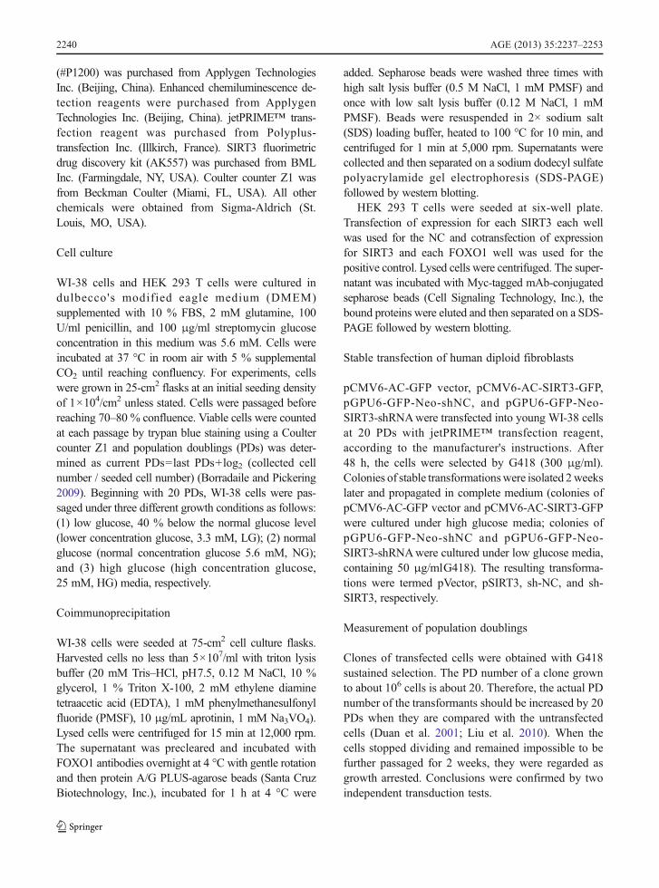
that there is a possible interaction between the SIRT3protein and FOXO1 protein. SIRT3 may deacetylateFOXO1 in the cytoplasm. Furthermore, we carried outCo-IPs between SIRT3 and FOXO1. As is shown inFig. 5, we could confirm that SIRT3 was binding withFOXO1. Overall, these data have demonstrated thatSIRT3 protein physically interacts with FOXO1. Inaddition, the fluorescence intensity of SIRT3 andFOXO1 showed a marked increase in WI-38 cells inthe LG group compared with the NG and HG groups.These results suggest that low glucose activates SIRT3
a n d
FOXO1 expression in vitro. These results may be attrib-uted to SIRT3, while being closely related to low glu-cose delaying cellular senescence.
FOXO1 transactivation activity in WI-38 cellsunder three different growth conditions
The FOXO transcription factors play a central role inregulating stress response (Sedding 2008; Furukawa-Hibi et al. 2005), and FOXO factors activate the expres-sion of its target genes, such as catalase and MnSOD. In
Fig. 2 Changes ofsenescence-associated fea-tures in WI-38 cells underthree different growth con-ditions. a WI-38 cells (35PDs) grown under LG, NG,and HG incubation condi-tions were stained for SA-β-gal, which shows blue pre-cipitation in the cytoplasmin senescent cells. b Thepercentage of SA-β-galstained positive cells. Foreach group, 100 to 200 cellswere counted. c, d Westernblot analysis of p16INK4A inthe three groups. Compari-son among groups wasconducted with ANOVA.*P<0.05 vs. LG group,#P<0.05 vs. NG group. Datarepresent three experimentswith similar results. a 100×
Fig. 1 Low glucose extends lifespan of WI-38 cells. PD curves ofWI-38 cells in LG, NG, and HG growth conditions. Viable cellswere counted at each passage by trypan blue staining using a
Coulter counter Z1 and PDs were determined as current PDs=lastPDs+log2 (collected cell number / seeded cell number). Each graphdepicts the averaged results from three longevity assessments
AGE (2013) 35:2237–2253 2243

mammalian cells, deacetylation of FOXO factors is ageneral mechanism that activates FOXO transcriptionalactivity (Daitoku et al. 2004; Brunet et al. 2004). To studythe effect of FOXO acetylation state on target genetranscription, we tested the protein levels of nuclearFOXO1, and acetylated FOXO1 (Ac-FOXO1) and itstarget genes. As shown in Fig. 3c and d, the protein levelsof nuclear total FOXO1 expression in the LG group weresignificantly higher than those in cells exposed to NGand HG conditions, but nuclear Ac-FOXO1 expressionin the HG group was significantly higher than in the NGand HG groups. Nuclear deacetylated FOXO1 proteinexpression in the HG group was significantly lower thanthose in the other groups. We concluded that high serumglucose activates acetylation of FOXO1 and inhibitsFOXO1 activity. In addition, catalase and MnSOD pro-tein expressions of the LG group were significantlyhigher than in the other groups (Fig. 3a and b).
SIRT3 overexpression upregulates FOXO1-targetedgene expression through its deacetylase activity
As SIRT3 is an NAD+-dependant deacetylase, we inves-tigated whether SIRT3 affects FOXO1 acetylation. Forthis purpose, pSIRT3 andpVectorwere stably transfectedinto theWI-38cellsat20PDs.Aclonewasgrowntoabout40 PDs under high glucose media. Western blot analysiswas performed from pSIRT3 cells and revealed approxi-mately 2.3-fold and 2.5-fold increases of total SIRT3protein, respectively, compared with control and vectorgroups (Fig. 6a and b). SIRT3 overexpression keepsFOXO1 acetylation to a very low level (Fig. 6c and d).However, the protein levels of nuclear total FOXO1 ex-pression of the three groups did not show a statisticallysignificant difference (Fig. 6c and d). These results impli-cate SIRT3 as a positive cofactor for FOXO1-dependenttransactivation through its deacetylase activity. At the
Fig. 3 Expression of SIRT3, catalase, MnSOD, nuclearFOXO1, and nuclear Ac-FOXO1 in WI-38 cells under threedifferent growth conditions. a Western blot analysis of catalase,MnSOD, and SIRT3 in the LG group, NG group, and HG group.b Catalase, MnSOD, and SIRT3 were compared using ANOVA.*P<0.05 vs. LG group, #P<0.05 vs. NG group. c Western blotanalysis of nuclear FOXO1 and nuclear Ac-FOXO1 in the LGgroup, NG group, and HG group. d Nuclear FOXO1 and
nuclear Ac-FOXO1 were compared with ANOVA. *P<0.05vs. LG group, ▲P>0.05 vs. LG group, #P<0.05 vs. NG group.e The deacetylase activity of Sirt3 in the LG group, NG group,and HG group. *P<0.05 vs. LG group, #P<0.05 vs. NG group.AFU arbitrary fluorescence units. Equal loading was confirmedby reprobing the blots for β-actin (cytosolic) or TATA-bindingprotein (TBP) (nuclear). Data represent three experiments withsimilar results
2244 AGE (2013) 35:2237–2253

same time, toverify thedirect effectofSIRT3onFOXO1-targeted geneexpression including catalase andMnSOD,we examined the expression of target genes. The resultsindicated that expression levels were increased in thepSIRT3 group compared with control and vector groups(Fig. 6a and b). Together, these results indicate that thedeacetylase activity of SIRT3 actuallymediates FOXO1-mediated transcription in mammalian cells. Meanwhile,our experimental data showed that the levels of SIRT3deacetylase activity were greatly increased in the SIRT3group compared with the control and vector groups(Fig. 6e).
Overexpression of SIRT3 blocks highglucose-induced premature senescence
Next, we examined whether SIRT3 overexpressioncan protect WI-38 cells from high glucose-inducedpremature senescence. As shown previously(Zhang et al. 2006), high serum glucose inducespremature senescence (Figs. 1 and 2). Our resultsindicated that the overexpression of SIRT3 signif-icantly reduced the number of cells positive forSA-β-gal and inhibited SAHF formation comparedwith empty vector (Fig. 7a, b, c and d).
Fig. 4 Confocal microscopic images of SIRT3, mitochondria,and FOXO1 in WI-38 cells (35 PDs). a and d SIRT3 is localizedin the mitochondria and cytoplasm of WI-38 cells. Representa-tive confocal microscopic images of WI-38 cells are stainedwith anti-SIRT3 antibody (red). MitoTracker, anti-TOMM20antibody (green), and DAPI (blue) staining were utilized forlocalization of mitochondria and nuclei, respectively. The im-ages were merged for analysis of colocalization (yellow). b ande FOXO1 is localized in the cytoplasm and nuclei of WI-38cells. Representative confocal microscopic images of WI-38
cells are stained with anti-FOXO1 antibody (red). MitoTracker,anti-TOMM20 antibody (green), and DAPI (blue) staining wasutilized for localization of mitochondria and nuclei, respectively.The images were merged for analysis of colocalization (yellow).c SIRT3 colocalizes with FOXO1. Representative confocal mi-croscopic images of WI-38 cells are stained with anti-SIRT3antibody (red) and FOXO1 antibody (green). DAPI (blue)staining was utilized for localization of nuclei. The images weremerged for analysis of colocalization (yellow). ×1,200
AGE (2013) 35:2237–2253 2245
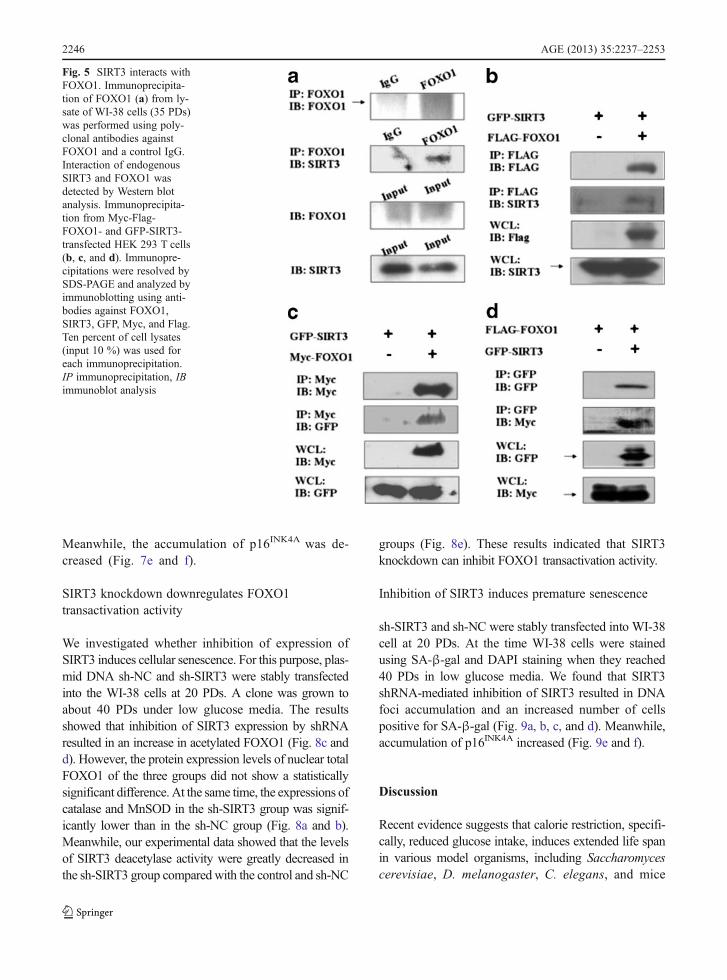
Meanwhile, the accumulation of p16INK4A was de-creased (Fig. 7e and f).
SIRT3 knockdown downregulates FOXO1transactivation activity
We investigated whether inhibition of expression ofSIRT3 induces cellular senescence. For this purpose, plas-mid DNA sh-NC and sh-SIRT3 were stably transfectedinto the WI-38 cells at 20 PDs. A clone was grown toabout 40 PDs under low glucose media. The resultsshowed that inhibition of SIRT3 expression by shRNAresulted in an increase in acetylated FOXO1 (Fig. 8c andd). However, the protein expression levels of nuclear totalFOXO1 of the three groups did not show a statisticallysignificant difference. At the same time, the expressions ofcatalase and MnSOD in the sh-SIRT3 group was signif-icantly lower than in the sh-NC group (Fig. 8a and b).Meanwhile, our experimental data showed that the levelsof SIRT3 deacetylase activity were greatly decreased inthe sh-SIRT3 group compared with the control and sh-NC
groups (Fig. 8e). These results indicated that SIRT3knockdown can inhibit FOXO1 transactivation activity.
Inhibition of SIRT3 induces premature senescence
sh-SIRT3 and sh-NC were stably transfected into WI-38cell at 20 PDs. At the time WI-38 cells were stainedusing SA-β-gal and DAPI staining when they reached40 PDs in low glucose media. We found that SIRT3shRNA-mediated inhibition of SIRT3 resulted in DNAfoci accumulation and an increased number of cellspositive for SA-β-gal (Fig. 9a, b, c, and d). Meanwhile,accumulation of p16INK4A increased (Fig. 9e and f).
Discussion
Recent evidence suggests that calorie restriction, specifi-cally, reduced glucose intake, induces extended life spanin various model organisms, including Saccharomycescerevisiae, D. melanogaster, C. elegans, and mice
Fig. 5 SIRT3 interacts withFOXO1. Immunoprecipita-tion of FOXO1 (a) from ly-sate of WI-38 cells (35 PDs)was performed using poly-clonal antibodies againstFOXO1 and a control IgG.Interaction of endogenousSIRT3 and FOXO1 wasdetected by Western blotanalysis. Immunoprecipita-tion from Myc-Flag-FOXO1- and GFP-SIRT3-transfected HEK 293 T cells(b, c, and d). Immunopre-cipitations were resolved bySDS-PAGE and analyzed byimmunoblotting using anti-bodies against FOXO1,SIRT3, GFP, Myc, and Flag.Ten percent of cell lysates(input 10 %) was used foreach immunoprecipitation.IP immunoprecipitation, IBimmunoblot analysis
2246 AGE (2013) 35:2237–2253

(Ristow and Zarse 2010; Lopez-Lluch et al. 2008; Kassiand Papavassiliou 2008; Judge and Leeuwenburgh 2007;Hunt et al. 2006; Jazwinski 2005). Meanwhile, it is wellknown that overexpression of Sir2 orthologs extendslife span in a wide range of lower eukaryotes (Lin etal. 2000; Kenyon 2010; Rahat et al. 2011; Frankel etal. 2011). In this study, first, we found that theprotein expression of SIRT3 increased under LGconditions and decreased under HG conditions inWI-38 cells. These findings inspired the assumptionthat SIRT3 has a role in regulating cellular senes-cence in WI-38 cells and is a potential pharmacologicaltarget to treat the major diseases of senescence. Second,we found that FOXO1 is acetylated by high serumglucose, and total FOXO1 expression is decreased.Third, SIRT3 binds to FOXO1 in the cytoplasm. Wetried to elucidate the mechanisms of how SIRT3 andFOXO1 communicate in cellular senescence in humandiploid fibroblasts.
The SIRT3 gene lies at the telomeric terminal onthe 11p15.5 chromosome (Rose et al. 2003). Schwer et
al. (2002) assayed endogenous SIRT3 in the mito-chondrial fraction of human embryonic kidney cellsand found that it had specific NAD-dependentdeacetylase activity. Mitochondrial import of SIRT3was dependent on an N-terminal amphipathic alphahelix rich in basic residues. A study showed thatSIRT3 exists in two forms: in humans, a full-lengthprotein of 44 kDa and a processed polypeptide lacking142 amino acids at its N-terminus. They discoveredthat SIRT3 not only localizes to the mitochondria butalso to the nucleus (Scher et al. 2007). SIRT3 wasproteolytically processed in the mitochondrial matrixto a 28-kDa product, and this processing wasreconstituted in vitro with recombinant mitochondrialmatrix processing peptidase (MPP). Mutation analysisshowed that SIRT3 was cleaved between arginines 99and 100. The unprocessed form of SIRT3 was enzy-matically inactive and became fully activated in vitroafter cleavage by MPP (Schwer et al. 2002). A recentstudy showed that CR reduces oxidative DNA damagein multiple tissues and prevents age-related hearing
Fig. 6 SIRT3-dependent suppression of cellular senescencewas through the FOXO1 pathway. a Stable transfected WI-38cells containing pVector or pSIRT3 and an untransfected controlgroup were lysed and prepared for Western blot analysis byusing specific antibodies against SIRT3, catalase, MnSOD,and β-actin. b Expression of the proteins SIRT3, catalase, andMnSOD was compared using ANOVA. *P>0.05 vs. controlgroup, #P<0.05 vs. vector group. c Western blot analysis of
nuclear FOXO1 and nuclear Ac-FOXO1 in the control group,vector group, and SIRT3 group, respectively. d Nuclear FOXO1and nuclear Ac-FOXO1 were compared using ANOVA.*P>0.05 vs. control group, ▲P>0.05 vs. vector group,#P<0.05 vs. vector group. e The deacetylase activity of SIRT3in the control group, vector group, and SIRT3 group. *P>0.05vs. control group, #P<0.05 vs. vector group. Data representthree experiments with similar results
AGE (2013) 35:2237–2253 2247
loss in wild type mice but fails to modify these phe-notypes in mice lacking the mitochondrial deacetylaseSIRT3 (Someya et al. 2010). Two studies demonstrat-ed that SIRT3 deacetylates mitochondrial 3-hydroxy-3-methylglutaryl CoA synthase 2, regulates ketonebody production, and reduces cellular ROS levelsdependent on superoxide dismutase 2 (Qiu et al.2010; Shimazu et al.2010). We found that SIRT3 washighly expressed in the mitochondria and cytoplasm.The SIRT3 of cytoplasm came from the mitochondriain the process of cellular senescence in WI-38 cell. TheSIRT3 strongly overlapped with that of FOXO1 inthe cytoplasm, SIRT3 deacetylated FOXO1 and thenFOXO1 from the cytoplasm into the nucleus upregulatedits transcriptional activity. We observed changes inFOXO1 and Ac-FOXO1 by overexpression and sup-pression by the expression of SIRT3. Our results
indicated that overexpression of SIRT3 results in de-creased levels of nuclear Ac-FOXO1 protein, andshRNA-SIRT3 increased the expression levels of nuclearAc-FOXO1 protein. Transcriptional activity of FOXO1has been known to be mainly regulated by post-translational modifications of phosphorylation-dependent nuclear exclusion and deacetylation-dependent nuclear retention. In spite of Sirt3overexpression-mediated deacetylation, there is no dif-ference in total nuclear FOXO levels in Fig. 6.Furthermore, Sirt3 shRNA was shown to increase acet-ylated FOXO1 without a difference in total nuclearlevels (Fig. 8). These results suggest that there are dif-ferent types of regulation for nuclear FOXO1 levelsother than Sirt3-mediated regulation in the setting of highglucose levels. These results need to be explored infurther experiments.
Fig. 7 Changes ofsenescence-associated fea-tures in WI-38 cells were in-duced by enforced expressionof SIRT3. a Stable transfectedWI-38 cells expressing emptyvector or SIRT3 and theuntransfected control groupunder HG conditions. WI-38cells cultured for 40 PDs werestained for SA-β-gal, whichshowed blue precipitation inthe cytoplasm of senescentcells. b The percentage of SA-β-gal stained positive cells.For each group, *P>0.05 vs.control group, #P<0.05 vs.vector group. c Senescence-associated heterochromatinfoci (SAHF), another classicalmarker of senescence, wereevident in WI-38 cells. DNAfoci accumulate was visual-ized by DAPI staining in WI-38 cells. d Enlarged images ofDAPI staining are shown inthe lower panels. eWesternblot analysis of p16INK4A inthe three groups. f Compari-son among groups wasconducted using ANOVA.*P>0.05 vs. control group,#P<0.05 vs. vector group.Data represent three experi-ments with similar results.a×100, c×1,200, d×3,600×
2248 AGE (2013) 35:2237–2253

In a clinical study, it was reported that older people(59–76 years) with a sedentary lifestyle had about 40 %reduced SIRT3 levels compared with younger people(18–30 years), and after endurance exercise, the healthbenefits in older people were accompanied by elevatedlevels of SIRT3 (Lanza et al. 2008). In two studies,SIRT3 is a longevity factor (Bellizzi et al. 2005; Roseet al. 2003), and it is the only member of the sirtuinfamily which increased expression has been linked to anextended life span of humans. Increased expression ofSIRT3 due to polymorphism in the SIRT3 promoter wasfound to be associated with an extended life span inhumans, thus implicating a role of SIRT3 in the geneticcontrol of human aging.
In this study, our data, for the first time, demonstratedthat SIRT3 interacts with and deacetylates a FOXO
transcription factor, FOXO1. Deacetylation of FOXO1by SIRT3 results in the stimulation of the expression ofthe FOXO target genes, catalase and MnSOD. Resultsfrom both SIRT3 overexpression and knockdown ex-periments suggest that FOXO deacetylation by SIRT3activates FOXO transactivation activity. These findingsare in agreement with a recent report that deacetylationof FOXO allows ubiquitination and subsequent increaseof transcriptional activity of FOXO factors (Sundaresanet al. 2009).
The most important and novel finding of thisstudy is that enforced SIRT3 expression in WI-38cells under high glucose conditions prevented cellularpremature senescence. Meanwhile, knockdown ofSIRT3 by shRNA under low glucose induced cellularpremature senescence.
Fig. 8 SIRT3 knockdown downregulates FOXO1 transactivationactivity. a Stable transfected WI-38 cells containing sh-NC or sh-SIRT3 and an untransfected control groupwere lysed and preparedfor western blot analysis. b Expression of proteins SIRT3,p16INK4A, catalase, and MnSOD was compared with ANOVA.*P>0.05 vs. control group, #P<0.05 vs. sh-NC group. c Westernblot analysis of nuclear FOXO1 and nuclear Ac-FOXO1 in the
control group, sh-NC group, and sh-SIRT3 group. d NuclearFOXO1 and nuclear Ac-FOXO1 were compared using ANOVA.*P>0.05 vs. control group, ▲P>0.05 vs. sh-NC group, #P<0.05vs. sh-NC group. eThe deacetylase activity of SIRT3 in the controlgroup, sh-NC group, and sh-SIRT3 group. *P>0.05 vs. controlgroup, #P<0.05 vs. sh-NC group. Data represent threeexperiments with similar results
AGE (2013) 35:2237–2253 2249

Caloric restriction is the only well-characterizedcondition that can extend the life span in organismsranging from yeast to mammals (Kenyon 2010). Li etal. found that glucose restriction (GR) can inhibitcellular senescence and significantly extend cellularlife span compared with cells receiving normal glu-cose (NG) in the culture medium (Li and Tollefsbol2011). Previously, many studies have demonstratedthat CR increases the expression of SIRT1 and thatthe life span extension associated with CR is depen-dent on SIRT1 expression (Canto and Auwerx 2009;Saunders and Verdin 2009; Gillum et al. 2010).Meanwhile, many studies found that FOXO gene var-iants have also been linked to longevity in severalcohorts. It is striking that FOXO variants are consis-tently associated with longevity (Anselmi et al. 2009;Flachsbart et al. 2009; Li et al. 2009; Lunetta et al.2007; Willcox et al. 2008).
Fig. 10 Schematic model for a mechanism of SIRT3overexpression antagonizes high glucose accelerated cellularsenescence in human diploid fibroblasts via the SIRT3–FOXO1signaling pathway
Fig. 9 Changes insenescence-associated fea-tures in WI-38 cells wereinduced by inhibited ex-pression of SIRT3. a Stabletransfected WI-38 cells ex-pressing shRNA-NC orshRNA-SIRT3 and theuntransfected control groupunder LG conditions. WI-38cells cultured for 40 PDswere stained for SA-β-gal. bThe percentage of SA-β-gal-stained positive cells.For each group, *P>0.05 vs.control group, #P<0.05 vs.shRNA-NC group. c Analy-sis of SAHF formationshown in WI-38 cells. DNAfoci accumulation was visu-alized by DAPI staining inWI-38 cells. d Enlarged im-ages of DAPI staining areshown in the lower panels. eWestern blot analysis ofp16INK4A in the threegroups. f Comparisonamong groups wasconducted using ANOVA.*P>0.05 vs. control group,#P<0.05 vs. sh-NC group.Data represent three experi-ments with similar results.a×100, c×1,200, d×3,600
2250 AGE (2013) 35:2237–2253

This model indicated SIRT3 may pose a central rolein cellular senescence through directly modulatingFOXO1 signaling under the circumstance of high glu-cose (Fig. 10). In summary, our data, for the first time,suggested roles of SIRT3 and FOXO1 in the relation-ship with cellular premature senescence in humandiploid fibroblasts: SIRT3 overexpression antagonizeshigh glucose-accelerated cellular senescence in humandiploid fibroblasts via the SIRT3–FOXO1 signalingpathway.
Acknowledgments We thank Chi-Dug Kang for the gift ofpCMV6-AC-GFP vector. This work was supported by the Na-tional Basic Research program of China (973 Program)(2007CB507400), and a grant from the National Natural Sci-ence Foundation of China (30771014, 81070267).
Open Access This article is distributed under the terms of theCreative Commons Attribution License which permits any use,distribution, and reproduction in any medium, provided theoriginal author(s) and the source are credited.
References
Ahn BH, Kim HS, Song S, Lee IH, Liu J, Vassilopoulos A,Deng CX, Finkel T (2008) A role for the mitochondrialdeacetylase Sirt3 in regulating energy homeostasis. ProcNatl Acad Sci USA 105(38):14447–14452. doi:10.1073/pnas.0803790105
Anselmi CV, Malovini A, Roncarati R, Novelli V, Villa F,Condorelli G, Bellazzi R, Puca AA (2009) Association ofthe FOXO3A locus with extreme longevity in a southernItalian centenarian study. Rejuvenation Res 12(2):95–104.doi:10.1089/rej.2008.0827
Bao J, Scott I, Lu Z, Pang L, Dimond CC, Gius D, Sack MN(2010) SIRT3 is regulated by nutrient excess and modulateshepatic susceptibility to lipotoxicity. Free Radic Biol Med 49(7):1230–1237. doi:10.1016/j.freeradbiomed.2010.07.009
Bellizzi D, Rose G, Cavalcante P, Covello G, Dato S, De RangoF, Greco V, Maggiolini M, Feraco E, Mari V, Franceschi C,Passarino G, De Benedictis G (2005) A novel VNTRenhancer within the SIRT3 gene, a human homologue ofSIR2, is associated with survival at oldest ages. Genomics85(2):258–263. doi:10.1016/j.ygeno.2004.11.003
Benigni A, Corna D, Zoja C, Sonzogni A, Latini R, Salio M,Conti S, Rottoli D, Longaretti L, Cassis P, Morigi M,Coffman TM, Remuzzi G (2009) Disruption of the AngII type 1 receptor promotes longevity in mice. J Clin Invest119(3):524–530. doi:10.1172/JCI36703
Borradaile NM, Pickering JG (2009) Nicotinamidephosphoribosyltransferase imparts human endothelial cellswith extended replicative lifespan and enhanced angiogen-ic capacity in a high glucose environment. Aging Cell8(2):100–112. doi:10.1111/j.1474-9726.2009.00453.x
Brunet A, Sweeney LB, Sturgill JF, Chua KF, Greer PL, Lin Y,TranH, Ross SE,MostoslavskyR, Cohen HY, Hu LS, Cheng
HL, Jedrychowski MP, Gygi SP, Sinclair DA, Alt FW,Greenberg ME (2004) Stress-dependent regulation of FOXOtranscription factors by the SIRT1 deacetylase. Science 303(5666):2011–2015. doi:10.1126/science.1094637
Canto C, Auwerx J (2009) Caloric restriction, SIRT1 and lon-gevity. Trends Endocrinol Metab 20(7):325–331.doi:10.1016/j.tem.2009.03.008
Daitoku H, Hatta M, Matsuzaki H, Aratani S, Ohshima T,Miyagishi M, Nakajima T, Fukamizu A (2004) Silent infor-mation regulator 2 potentiates Foxo1-mediated transcriptionthrough its deacetylase activity. Proc Natl Acad Sci USA 101(27):10042–10047. doi:10.1073/pnas.0400593101
de Oliveira RM, Pais TF, Outeiro TF (2010) Sirtuins: commontargets in aging and in neurodegeneration. Curr Drug Tar-gets 11(10):1270–1280, doi:BSP/CDT/E-Pub/00134
Dimri GP, Lee X, Basile G, Acosta M, Scott G, Roskelley C,Medrano EE, Linskens M, Rubelj I, Pereira-Smith O et al(1995) A biomarker that identifies senescent human cells inculture and in aging skin in vivo. Proc Natl Acad Sci USA92(20):9363–9367
Duan J, Zhang Z, Tong T (2001) Senescence delay of humandiploid fibroblast induced by anti-sense p16INK4a expres-sion. J Biol Chem 276(51):48325–48331. doi:10.1074/jbc.M104814200
Flachsbart F, Caliebe A, Kleindorp R, Blanche H, von Eller-Eberstein H, Nikolaus S, Schreiber S, Nebel A (2009)Association of FOXO3A variation with human longevityconfirmed in German centenarians. Proc Natl Acad SciUSA 106(8):2700–2705. doi:10.1073/pnas.0809594106
Frankel S, Ziafazeli T, Rogina B (2011) dSir2 and longevity inDrosophila. Exp Gerontol 46(5):391–396. doi:10.1016/j.exger.2010.08.007
Frescas D, Valenti L, Accili D (2005) Nuclear trapping of theforkhead transcription factor FoxO1 via Sirt-dependentdeacetylation promotes expression of glucogenetic genes.J Biol Chem 280(21):20589–20595. doi:10.1074/jbc.M412357200
Furukawa-Hibi Y, Kobayashi Y, Chen C, Motoyama N (2005)FOXO transcription factors in cell-cycle regulation and theresponse to oxidative stress. Antioxid Redox Signal 7(5–6):752–760. doi:10.1089/ars.2005.7.752
Giannakou ME, Goss M, Partridge L (2008) Role of dFOXO inlifespan extension by dietary restriction in Drosophilamelanogaster: not required, but its activity modulates theresponse. Aging Cell 7(2):187–198. doi:10.1111/j.1474-9726.2007.00362.x
Gillum MP, Erion DM, Shulman GI (2010) Sirtuin-1 regulationof mammalian metabolism. Trends Mol Med. doi:S1471-4914(10)00140-1 [pii] 10.1016/j.molmed.2010.09.005
Haigis MC, Guarente LP (2006) Mammalian sirtuins—emerg-ing roles in physiology, aging, and calorie restriction.Genes Dev 20(21):2913–2921. doi:10.1101/gad.1467506
Huang H, Tindall DJ (2007) Dynamic FoxO transcription factors.J Cell Sci 120(Pt 15):2479–2487. doi:10.1242/jcs.001222
Hunt ND, Hyun DH, Allard JS, Minor RK, Mattson MP, IngramDK, de Cabo R (2006) Bioenergetics of aging and calorierestriction. Ageing Res Rev 5(2):125–143. doi:10.1016/j.arr.2006.03.006
Jazwinski SM (2005) Yeast longevity and aging—the mitochon-drial connection. Mech Ageing Dev 126(2):243–248.doi:10.1016/j.mad.2004.08.016
AGE (2013) 35:2237–2253 2251
Judge S, Leeuwenburgh C (2007) Cardiac mitochondrial bioener-getics, oxidative stress, and aging. Am J Physiol Cell Physiol292(6):C1983–1992. doi:10.1152/ajpcell.00285.2006
Kassi E, Papavassiliou AG (2008) Could glucose be a proagingfactor? J Cell Mol Med 12(4):1194–1198. doi:10.1111/j.1582-4934.2008.00329.x
Kenyon CJ (2010) The genetics of ageing. Nature 464(7288):504–512. doi:10.1038/nature08980
Kitamura YI, Kitamura T, Kruse JP, Raum JC, Stein R, Gu W,Accili D (2005) FoxO1 protects against pancreatic beta cellfailure through NeuroD and MafA induction. Cell Metab 2(3):153–163. doi:10.1016/j.cmet.2005.08.004
Kong X, Wang R, Xue Y, Liu X, Zhang H, Chen Y, Fang F,Chang Y, Deb S (2010) Sirtuin 3, a new target of PGC-1α,plays an important role in the suppression of ROS andmitochondrial biogenesis. PLoS One 5(7):e11707.doi:10.1371/journal.pone.0011707
Lanza IR, Short DK, Short KR, Raghavakaimal S, Basu R,Joyner MJ, McConnell JP, Nair KS (2008) Enduranceexercise as a countermeasure for aging. Diabetes 57(11):2933–2942. doi:10.2337/db08-0349
Lauri SE, Delany C, VR JC, Bortolotto ZA, Ornstein PL JTRI,Collingridge GL (2001) Synaptic activation of a presynap-tic kainate receptor facilitates AMPA receptor-mediatedsynaptic transmission at hippocampal mossy fibre synap-ses. Neuropharmacology 41(8):907–915
Li Y, Tollefsbol TO (2011) p16(INK4a) suppression by glucoserestriction contributes to human cellular lifespan extensionthrough SIRT1-mediated epigenetic and genetic mechanisms.PLoS One 6(2):e17421. doi:10.1371/journal.pone.0017421
Li Y, Wang WJ, Cao H, Lu J, Wu C, Hu FY, Guo J, Zhao L,Yang F, Zhang YX, Li W, Zheng GY, Cui H, Chen X, ZhuZ, He H, Dong B, Mo X, Zeng Y, Tian X (2009) Geneticassociation of FOXO1A and FOXO3Awith longevity traitin Han Chinese populations. Hum Mol Genet 18(24):4897–4904. doi:10.1093/hmg/ddp459
Lin SJ, Defossez PA, Guarente L (2000) Requirement of NAD andSIR2 for life-span extension by calorie restriction in Saccha-romyces cerevisiae. Science 289(5487):2126–2128, doi:8813
Liu W, Hong Q, Bai XY, Fu B, Xie Y, Zhang X, Li J, Shi S, LvY, Sun X, Chen X (2010) High-affinity Na(+)-dependentdicarboxylate cotransporter promotes cellular senescenceby inhibiting SIRT1. Mech Ageing Dev 131(10):601–613. doi:10.1016/j.mad.2010.08.006
Lopez-Lluch G, Irusta PM, Navas P, de Cabo R (2008) Mito-chondrial biogenesis and healthy aging. Exp Gerontol 43(9):813–819. doi:10.1016/j.exger.2008.06.014
Lunetta KL, D'Agostino RB Sr, Karasik D, Benjamin EJ, Guo CY,Govindaraju R, Kiel DP, Kelly-Hayes M, Massaro JM,Pencina MJ, Seshadri S, Murabito JM (2007) Genetic corre-lates of longevity and selected age-related phenotypes: agenome-wide association study in the Framingham Study.BMCMed Genet 8(Suppl 1):S13. doi:10.1186/1471-2350-8-S1-S13
Murphy CT (2006) The search for DAF-16/FOXO transcrip-tional targets: approaches and discoveries. Exp Gerontol 41(10):910–921. doi:10.1016/j.exger.2006.06.040
Narita M, Nunez S, Heard E, Lin AW, Hearn SA, Spector DL,Hannon GJ, Lowe SW (2003) Rb-mediated heterochroma-tin formation and silencing of E2F target genes duringcellular senescence. Cell 113(6):703–716
Onyango P, Celic I, McCaffery JM, Boeke JD, Feinberg AP(2002) SIRT3, a human SIR2 homologue, is an NAD-dependent deacetylase localized to mitochondria. Proc NatlAcad Sci USA 99(21):13653–13658. doi:10.1073/pnas.222538099
Qiu X, Brown K, Hirschey MD, Verdin E, Chen D (2010)Calorie restriction reduces oxidative stress by SIRT3-mediated SOD2 activation. Cell Metab 12(6):662–667.doi:10.1016/j.cmet.2010.11.015
Rahat O,MaozN, CohenHY (2011)Multiple pathways regulatingthe calorie restriction response in yeast. J Gerontol A Biol SciMed Sci 66(2):163–169. doi:10.1093/gerona/glq165
Ristow M, Zarse K (2010) How increased oxidative stress pro-motes longevity and metabolic health: the concept of mi-tochondrial hormesis (mitohormesis). Exp Gerontol 45(6):410–418. doi:10.1016/j.exger.2010.03.014
Rose G, Dato S, Altomare K, Bellizzi D, Garasto S, Greco V,Passarino G, Feraco E, Mari V, Barbi C, BonaFe M,Franceschi C, Tan Q, Boiko S, Yashin AI, De BenedictisG (2003) Variability of the SIRT3 gene, human silentinformation regulator Sir2 homologue, and survivorshipin the elderly. Exp Gerontol 38(10):1065–1070
Saunders LR, Verdin E (2009) Cell biology. Stress response andaging. Science 323(5917):1021–1022. doi:10.1126/science.1170007
Scher MB, Vaquero A, Reinberg D (2007) SirT3 is a nuclearNAD+-dependent histone deacetylase that translocates tothe mitochondria upon cellular stress. Genes Dev 21(8):920–928. doi:10.1101/gad.1527307
Schwer B, North BJ, Frye RA, Ott M, Verdin E (2002) Thehuman silent information regulator (Sir)2 homologuehSIRT3 is a mitochondrial nicotinamide adeninedinucleotide-dependent deacetylase. J Cell Biol 158(4):647–657. doi:10.1083/jcb.200205057
Sedding DG (2008) FoxO transcription factors in oxidativestress response and ageing—a new fork on the way tolongevity? Biol Chem 389(3):279–283. doi:10.1515/BC.2008.033
Shi T, Wang F, Stieren E, Tong Q (2005) SIRT3, a mitochon-drial sirtuin deacetylase, regulates mitochondrial functionand thermogenesis in brown adipocytes. J Biol Chem 280(14):13560–13567. doi:10.1074/jbc.M414670200
Shimazu T, Hirschey MD, Hua L, Dittenhafer-Reed KE, SchwerB, Lombard DB, Li Y, Bunkenborg J, Alt FW, Denu JM,Jacobson MP, Verdin E (2010) SIRT3 deacetylates mito-chondrial 3-hydroxy-3-methylglutaryl CoA synthase 2 andregulates ketone body production. Cell Metab 12(6):654–661. doi:10.1016/j.cmet.2010.11.003
Someya S, Yu W, Hallows WC, Xu J, Vann JM, LeeuwenburghC, Tanokura M, Denu JM, Prolla TA (2010) Sirt3 mediatesreduction of oxidative damage and prevention of age-related hearing loss under caloric restriction. Cell 143(5):802–812. doi:10.1016/j.cell.2010.10.002
Sundaresan NR, Gupta M, Kim G, Rajamohan SB, Isbatan A,Gupta MP (2009) Sirt3 blocks the cardiac hypertrophicresponse by augmenting Foxo3a-dependent antioxidantdefense mechanisms in mice. J Clin Invest 119(9):2758–2771. doi:10.1172/JCI39162
Willcox BJ, Donlon TA, He Q, Chen R, Grove JS, Yano K,Masaki KH, Willcox DC, Rodriguez B, Curb JD (2008)FOXO3A genotype is strongly associated with human
2252 AGE (2013) 35:2237–2253

longevity. Proc Natl Acad Sci USA 105(37):13987–13992,10.1073/pnas.0801030105
Yamaza H, Komatsu T, Wakita S, Kijogi C, Park S, Hayashi H,Chiba T, Mori R, Furuyama T, Mori N, Shimokawa I (2010)FoxO1 is involved in the antineoplastic effect of calorierestriction. Aging Cell 9((3):372–382. doi:10.1111/j.1474-9726.2010.00563.x
Yang Y, Cimen H, Han MJ, Shi T, Deng JH, Koc H, PalaciosOM, Montier L, Bai Y, Tong Q, Koc EC (2010) NAD+-dependent deacetylase SIRT3 regulates mitochondrial pro-tein synthesis by deacetylation of the ribosomal proteinMRPL10. J Biol Chem 285(10):7417–7429, 10.1074/jbc.M109.053421
Zanella F, Link W, Carnero A (2010) Understanding FOXO,new views on old transcription factors. Curr Cancer DrugTargets 10 (2):135-146. doi:EPub-Abstract-CCDT-02 [pii]
Zhang X, Chen X, Wu D, Liu W, Wang J, Feng Z, Cai G, Fu B,Hong Q, Du J (2006) Downregulation of connexin 43expression by high glucose induces senescence in glomerularmesangial cells. J Am Soc Nephrol 17(6):1532–1542.doi:10.1681/ASN.2005070776
Zhuo L, Cai G, Liu F, Fu B, Liu W, Hong Q, Ma Q, Peng Y,Wang J, Chen X (2009) Expression and mechanism ofmammalian target of rapamycin in age-related renalcell senescence and organ aging. Mech Ageing Dev130(10):700–708. doi:10.1016/j.mad.2009.08.005
AGE (2013) 35:2237–2253 2253