New Hampshire Prehospital Rapid Sequence Intubation ... · New Hampshire Prehospital Rapid Sequence...
16
New Hampshire Prehospital Rapid Sequence Intubation Administrative Packet 2019 New Hampshire Bureau of EMS 33 Hazen Drive Concord, NH 03305 (603) 271-456
Transcript of New Hampshire Prehospital Rapid Sequence Intubation ... · New Hampshire Prehospital Rapid Sequence...
New Hampshire Prehospital
Rapid Sequence Intubation
Administrative Packet 2019
New Hampshire Bureau of EMS
33 Hazen Drive
Concord, NH 03305
(603) 271-456
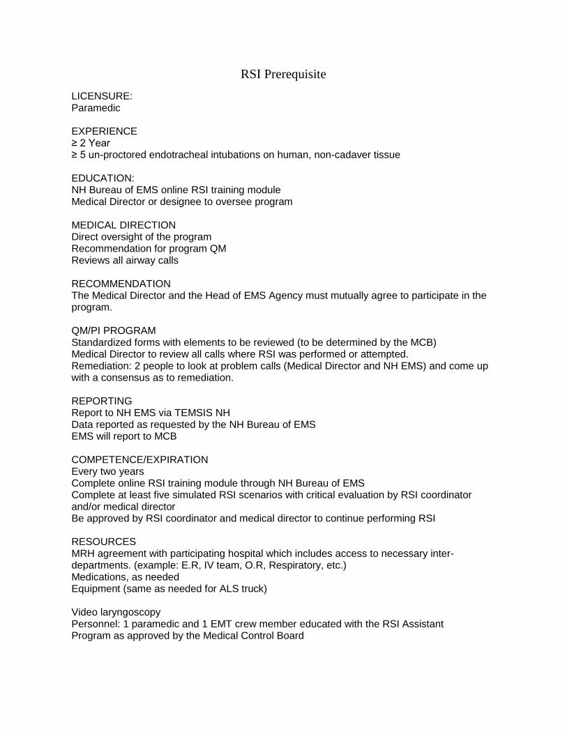
RSI Prerequisite
LICENSURE: Paramedic
EXPERIENCE ≥ 2 Year ≥ 5 un-proctored endotracheal intubations on human, non-cadaver tissue
EDUCATION: NH Bureau of EMS online RSI training module Medical Director or designee to oversee program
MEDICAL DIRECTION Direct oversight of the program Recommendation for program QM Reviews all airway calls
RECOMMENDATION The Medical Director and the Head of EMS Agency must mutually agree to participate in the program.
QM/PI PROGRAM Standardized forms with elements to be reviewed (to be determined by the MCB) Medical Director to review all calls where RSI was performed or attempted. Remediation: 2 people to look at problem calls (Medical Director and NH EMS) and come up with a consensus as to remediation.
REPORTING Report to NH EMS via TEMSIS NH Data reported as requested by the NH Bureau of EMS EMS will report to MCB
COMPETENCE/EXPIRATION Every two years Complete online RSI training module through NH Bureau of EMS Complete at least five simulated RSI scenarios with critical evaluation by RSI coordinator and/or medical director Be approved by RSI coordinator and medical director to continue performing RSI
RESOURCES MRH agreement with participating hospital which includes access to necessary inter- departments. (example: E.R, IV team, O.R, Respiratory, etc.) Medications, as needed Equipment (same as needed for ALS truck) Video laryngoscopy Personnel: 1 paramedic and 1 EMT crew member educated with the RSI Assistant Program as approved by the Medical Control Board
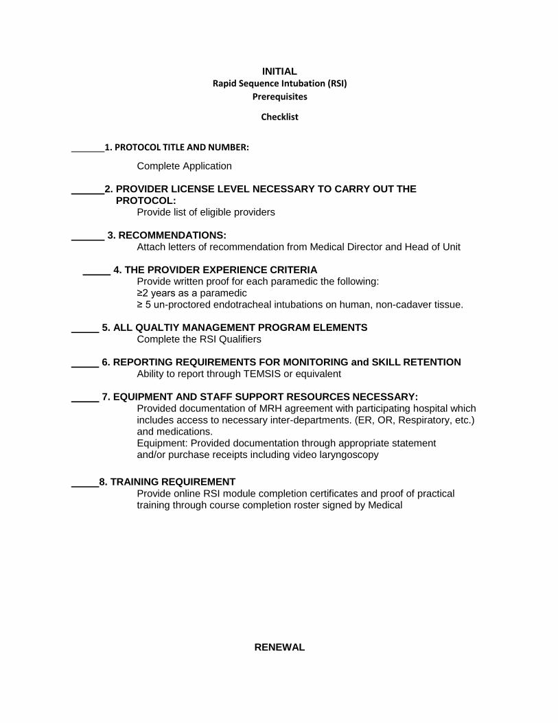
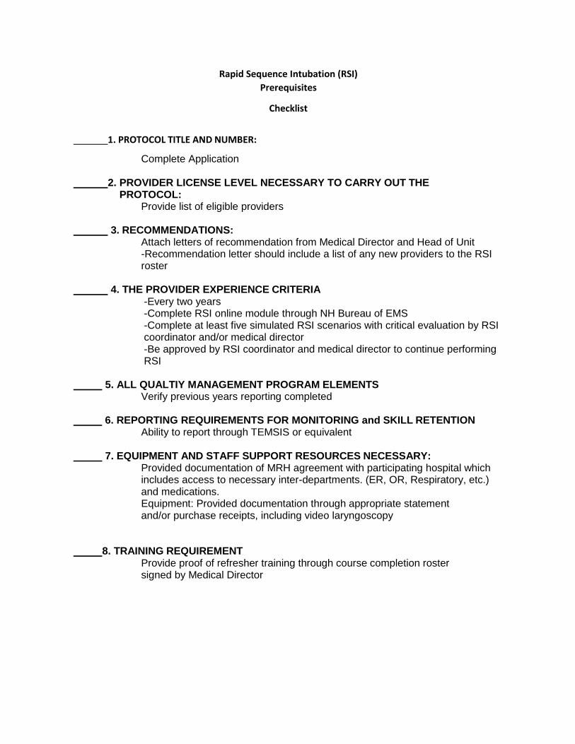
INITIAL Rapid Sequence Intubation (RSI)
Prerequisites
Checklist
1. PROTOCOL TITLE AND NUMBER:
Complete Application
2. PROVIDER LICENSE LEVEL NECESSARY TO CARRY OUT THE PROTOCOL:
Provide list of eligible providers
3. RECOMMENDATIONS: Attach letters of recommendation from Medical Director and Head of Unit
4. THE PROVIDER EXPERIENCE CRITERIA Provide written proof for each paramedic the following: ≥2 years as a paramedic ≥ 5 un-proctored endotracheal intubations on human, non-cadaver tissue.
5. ALL QUALTIY MANAGEMENT PROGRAM ELEMENTS Complete the RSI Qualifiers
6. REPORTING REQUIREMENTS FOR MONITORING and SKILL RETENTION Ability to report through TEMSIS or equivalent
7. EQUIPMENT AND STAFF SUPPORT RESOURCES NECESSARY: Provided documentation of MRH agreement with participating hospital which includes access to necessary inter-departments. (ER, OR, Respiratory, etc.) and medications. Equipment: Provided documentation through appropriate statement and/or purchase receipts including video laryngoscopy
8. TRAINING REQUIREMENT Provide online RSI module completion certificates and proof of practical training through course completion roster signed by Medical
RENEWAL
Rapid Sequence Intubation (RSI)
Prerequisites
Checklist
1. PROTOCOL TITLE AND NUMBER:
Complete Application
2. PROVIDER LICENSE LEVEL NECESSARY TO CARRY OUT THE PROTOCOL:
Provide list of eligible providers
3. RECOMMENDATIONS: Attach letters of recommendation from Medical Director and Head of Unit -Recommendation letter should include a list of any new providers to the RSI roster
4. THE PROVIDER EXPERIENCE CRITERIA -Every two years -Complete RSI online module through NH Bureau of EMS -Complete at least five simulated RSI scenarios with critical evaluation by RSI coordinator and/or medical director -Be approved by RSI coordinator and medical director to continue performing RSI
5. ALL QUALTIY MANAGEMENT PROGRAM ELEMENTS Verify previous years reporting completed
6. REPORTING REQUIREMENTS FOR MONITORING and SKILL RETENTION Ability to report through TEMSIS or equivalent
7. EQUIPMENT AND STAFF SUPPORT RESOURCES NECESSARY: Provided documentation of MRH agreement with participating hospital which includes access to necessary inter-departments. (ER, OR, Respiratory, etc.) and medications. Equipment: Provided documentation through appropriate statement and/or purchase receipts, including video laryngoscopy
8. TRAINING REQUIREMENT Provide proof of refresher training through course completion roster signed by Medical Director
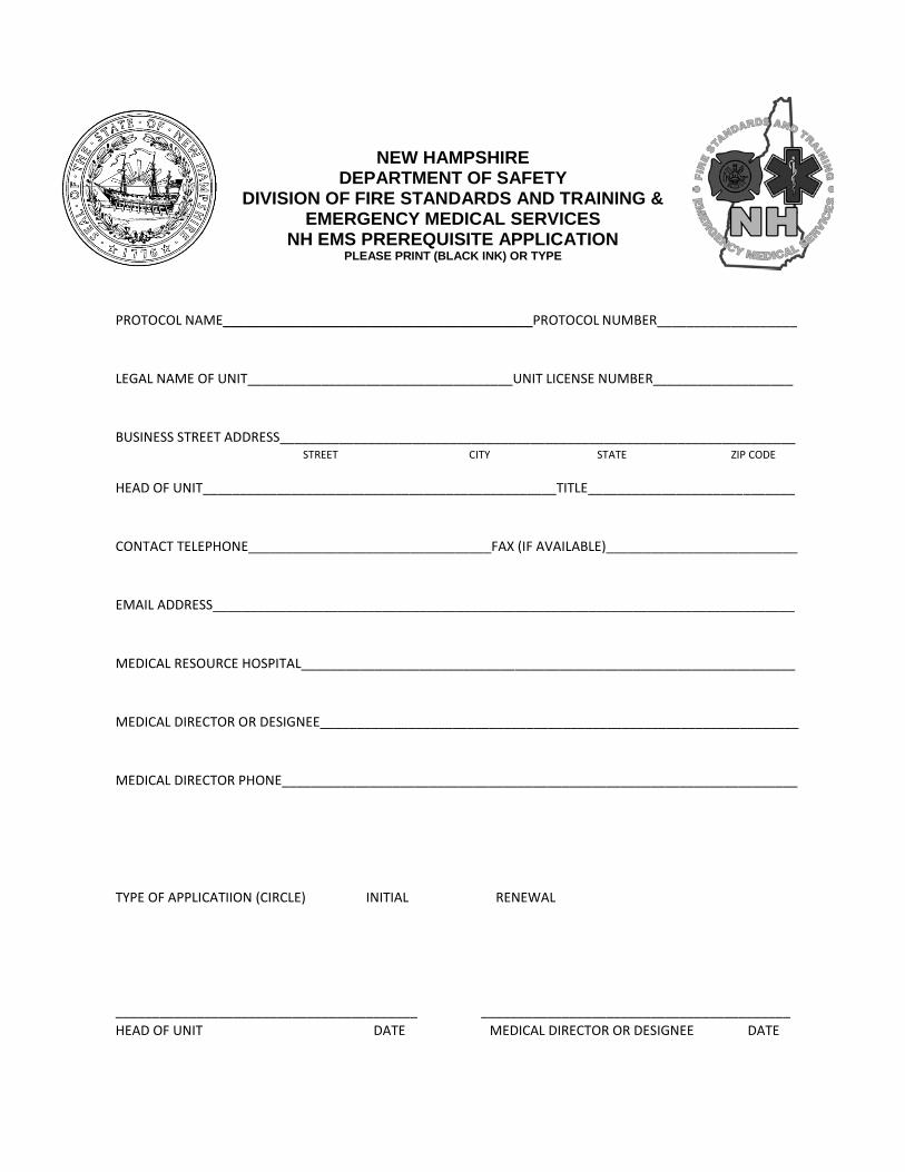
NEW HAMPSHIRE DEPARTMENT OF SAFETY
DIVISION OF FIRE STANDARDS AND TRAINING & EMERGENCY MEDICAL SERVICES
NH EMS PREREQUISITE APPLICATION PLEASE PRINT (BLACK INK) OR TYPE
PROTOCOL NAME__________________________________________PROTOCOL NUMBER___________________
LEGAL NAME OF UNIT____________________________________UNIT LICENSE NUMBER___________________
BUSINESS STREET ADDRESS______________________________________________________________________ STREET CITY STATE ZIP CODE
HEAD OF UNIT________________________________________________TITLE____________________________
CONTACT TELEPHONE_________________________________FAX (IF AVAILABLE)__________________________
EMAIL ADDRESS_______________________________________________________________________________
MEDICAL RESOURCE HOSPITAL___________________________________________________________________
MEDICAL DIRECTOR OR DESIGNEE_________________________________________________________________
MEDICAL DIRECTOR PHONE______________________________________________________________________
TYPE OF APPLICATIION (CIRCLE) INITIAL RENEWAL
_________________________________________ __________________________________________
HEAD OF UNIT DATE MEDICAL DIRECTOR OR DESIGNEE DATE
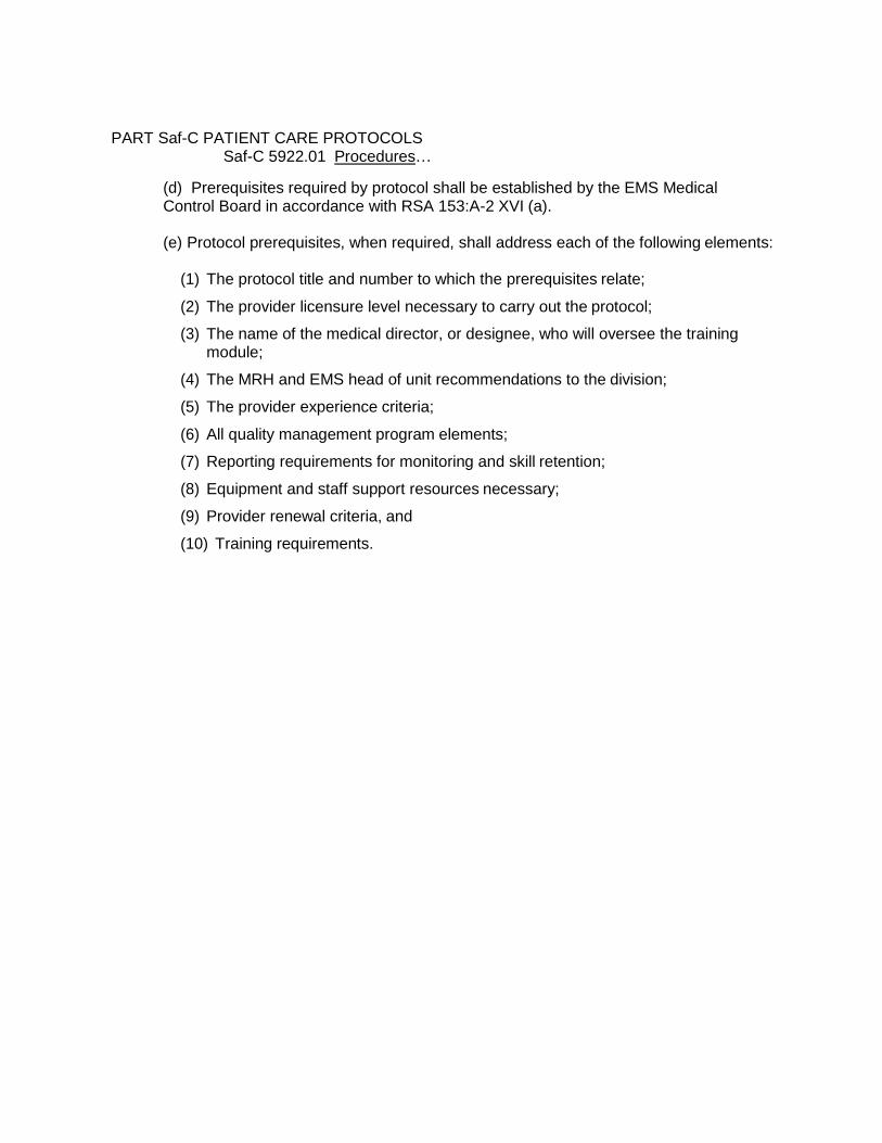
PART Saf-C PATIENT CARE PROTOCOLS Saf-C 5922.01 Procedures…
(d) Prerequisites required by protocol shall be established by the EMS Medical Control Board in accordance with RSA 153:A-2 XVI (a).
(e) Protocol prerequisites, when required, shall address each of the following elements:
(1) The protocol title and number to which the prerequisites relate;
(2) The provider licensure level necessary to carry out the protocol;
(3) The name of the medical director, or designee, who will oversee the training module;
(4) The MRH and EMS head of unit recommendations to the division;
(5) The provider experience criteria;
(6) All quality management program elements;
(7) Reporting requirements for monitoring and skill retention;
(8) Equipment and staff support resources necessary;
(9) Provider renewal criteria, and
(10) Training requirements.
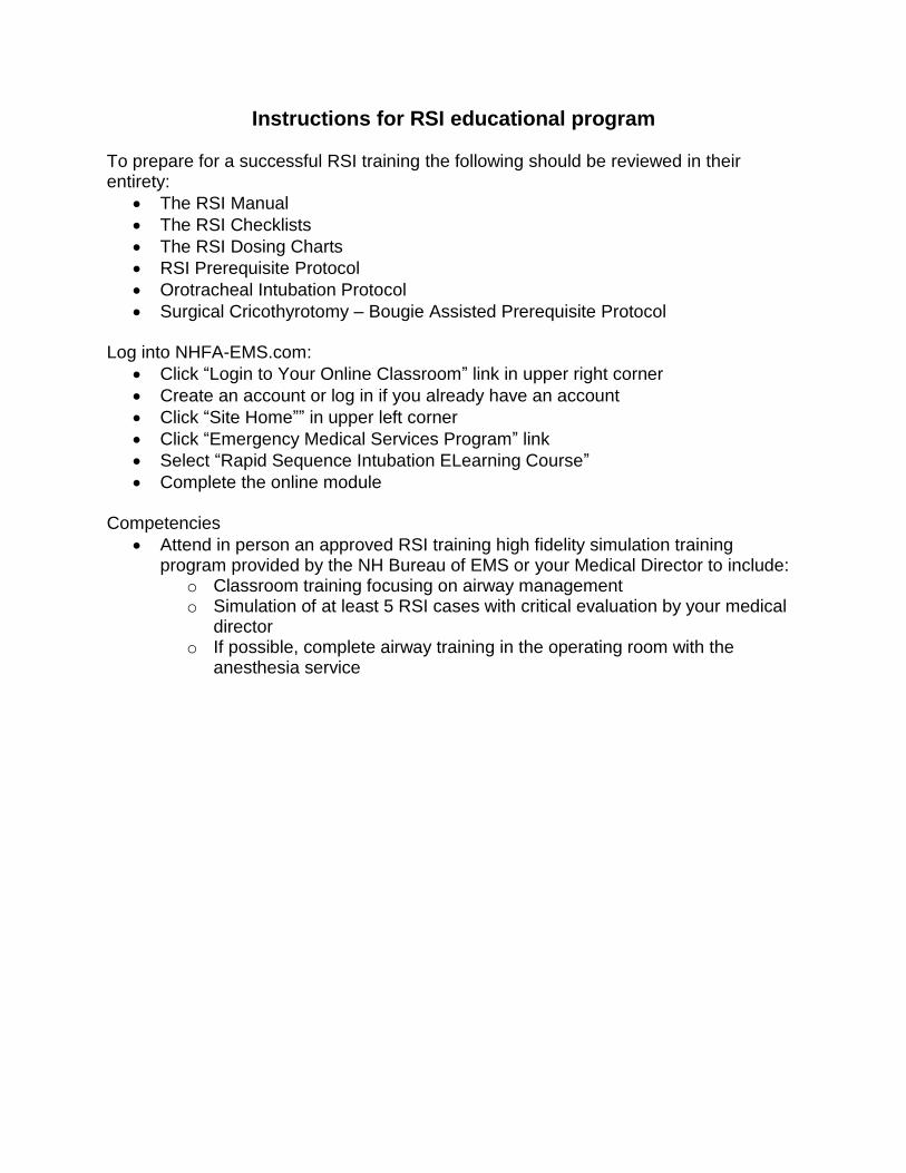
Instructions for RSI educational program To prepare for a successful RSI training the following should be reviewed in their entirety:
The RSI Manual
The RSI Checklists
The RSI Dosing Charts
RSI Prerequisite Protocol
Orotracheal Intubation Protocol
Surgical Cricothyrotomy – Bougie Assisted Prerequisite Protocol Log into NHFA-EMS.com:
Click “Login to Your Online Classroom” link in upper right corner
Create an account or log in if you already have an account
Click “Site Home”” in upper left corner
Click “Emergency Medical Services Program” link
Select “Rapid Sequence Intubation ELearning Course”
Complete the online module Competencies
Attend in person an approved RSI training high fidelity simulation training program provided by the NH Bureau of EMS or your Medical Director to include:
o Classroom training focusing on airway management o Simulation of at least 5 RSI cases with critical evaluation by your medical
director o If possible, complete airway training in the operating room with the
anesthesia service
Critical Criteria
Does not utilize RSI checklist
Fails to address airway/breathing/circulation concerns in a timely manner
Does not properly preoxygenate the patient
Does not address critical hemodynamic compromise prior to intubation
Does not properly prepare equipment prior to intubation Fails to secure the airway
Does not properly verify tube placement or fails to recognize improper placement
Administers incorrect medications or wrong doses
Evaluator determines candidate did not provide treatment in a safe or adequate manner
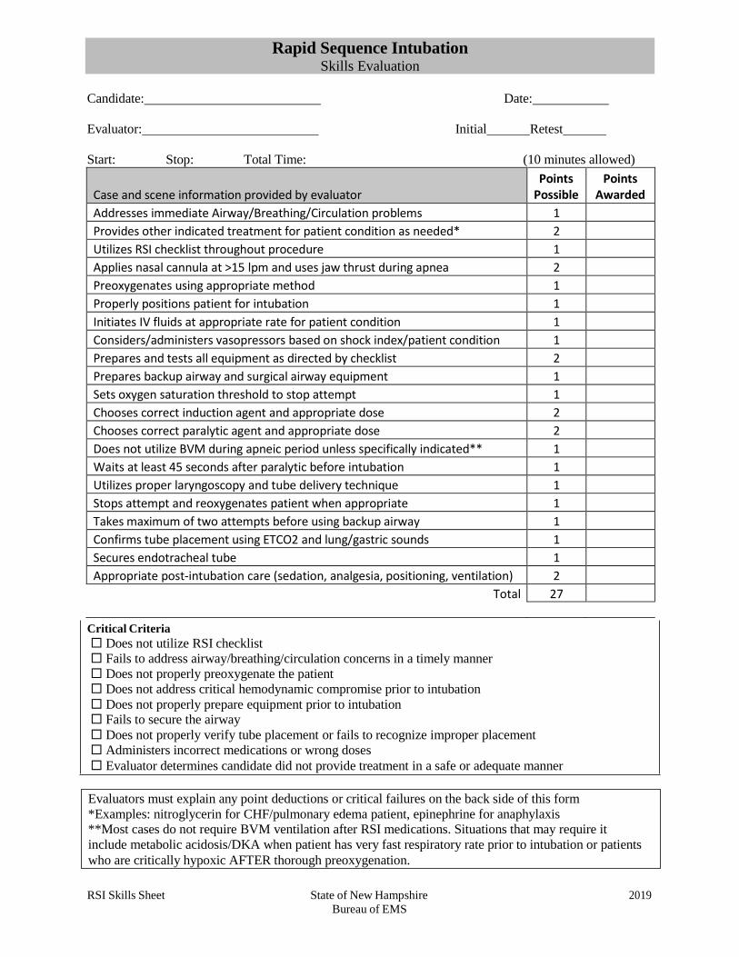
Candidate: Date:
Evaluator: Initial Retest
Start: Stop: Total Time: (10 minutes allowed)
Case and scene information provided by evaluator
Points Possible
Points Awarded
Addresses immediate Airway/Breathing/Circulation problems 1
Provides other indicated treatment for patient condition as needed* 2
Utilizes RSI checklist throughout procedure 1
Applies nasal cannula at >15 lpm and uses jaw thrust during apnea 2
Preoxygenates using appropriate method 1
Properly positions patient for intubation 1
Initiates IV fluids at appropriate rate for patient condition 1
Considers/administers vasopressors based on shock index/patient condition 1
Prepares and tests all equipment as directed by checklist 2
Prepares backup airway and surgical airway equipment 1
Sets oxygen saturation threshold to stop attempt 1
Chooses correct induction agent and appropriate dose 2
Chooses correct paralytic agent and appropriate dose 2
Does not utilize BVM during apneic period unless specifically indicated** 1
Waits at least 45 seconds after paralytic before intubation 1
Utilizes proper laryngoscopy and tube delivery technique 1
Stops attempt and reoxygenates patient when appropriate 1
Takes maximum of two attempts before using backup airway 1
Confirms tube placement using ETCO2 and lung/gastric sounds 1
Secures endotracheal tube 1
Appropriate post-intubation care (sedation, analgesia, positioning, ventilation) 2
Total 27
RSI Skills Sheet State of New Hampshire 2019
Bureau of EMS
Rapid Sequence Intubation Skills Evaluation
Evaluators must explain any point deductions or critical failures on the back side of this form
*Examples: nitroglycerin for CHF/pulmonary edema patient, epinephrine for anaphylaxis **Most cases do not require BVM ventilation after RSI medications. Situations that may require it
include metabolic acidosis/DKA when patient has very fast respiratory rate prior to intubation or patients
who are critically hypoxic AFTER thorough preoxygenation.

2013
Rapid Sequence Intubation (RSI)
P
This procedure is only to be used by paramedics who are trained and credentialed
to perform RSI. This protocol provides a brief outline of the scope of the RSI
paramedic but is not comprehensive of the entire RSI procedure. For full RSI
guidelines refer to the 2017 New Hampshire Prehospital RSI Manual. The
guidelines in this manual are part of the RSI protocol and are incorporated in this
protocol by reference.
PARAMEDIC - PREREQUISITES REQUIRED - ADULT ONLY
SUCCINYLCHOLINE CONTRAINDICATIONS:
· Extensive recent burns or crush injuries > 24 hours old.
· Known or suspected hyperkalemia.
· History of malignant hyperthermia.
Pre
req
uis
ite P
roto
co
l 7.4
The New Hampshire Bureau of EMS has taken extreme caution to ensure all information is accurate and in accordance with professional standards in effect at the time of publication. These protocols, policies, or procedures MAY NOT BE altered or modified. 2017
7.4
Each RSI procedure must be performed in a
controlled fashion and must involve careful planning
and preparation. RSI requires at least one RSI
credentialed paramedic and one credentialed RSI
assistant or non-RSI paramedic. Intubation must be
performed by the most appropriate provider as
determined by the RSI paramedic leading the call.
After intubation, the RSI paramedic must remain with
the patient at all times unless there are extenuating
circumstances (mass casualty, etc.) and ensure that
adequate staff remain.
RSI may only be performed on adults (i.e., patients
who are taller than a length based resuscitation tape).
Medications
The correct medication regimen should be chosen on
a case-by-case basis by the RSI paramedic and care
team. Medication options are listed here:
IBW = Ideal Body Weight (refer to chart)
ABW = Actual Body Weight
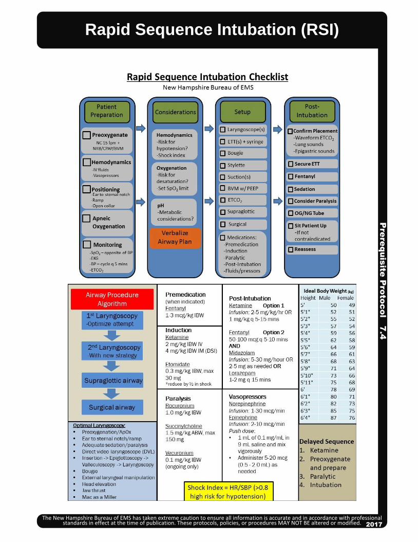
Premedication (if indicated)
· Fentanyl 1 - 3 mcg/kg IBW IV at least three
minutes prior to inductionInduction
· Ketamine 2 mg/kg IBW IV or 4 mg/kg IBW IM (only if performing Delayed
Sequence Intubation)
o For elderly, shock, or risk of hypotension: 1 mg/kg IBW IV or 2 mg/kg
IBW IM
OR
· Etomidate 0.3 mg/kg IBW IV, maximum single dose 30 mg
o For elderly, shock, or risk of hypotension: 0.15 mg/kg IBW IV
Paralysis
· Rocuronium 1 mg/kg IBW IV
OR
· Succinylcholine 1.5 mg/kg ABW IV, maximum 150 mg
Protocol Continues
Pre
req
uis
ite P
roto
co
l 7.4
Rapid Sequence Intubation (RSI)
The New Hampshire Bureau of EMS has taken extreme caution to ensure all information is accurate and in accordance with professional standards in effect at the time of publication. These protocols, policies, or procedures MAY NOT BE altered or modified.
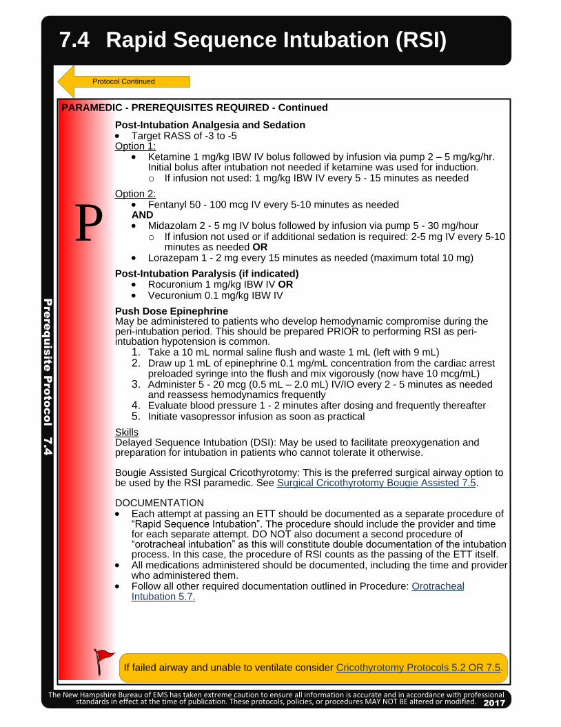
Post-Intubation Analgesia and Sedation· Target RASS of -3 to -5 Option 1:
· Ketamine 1 mg/kg IBW IV bolus followed by infusion via pump 2 – 5 mg/kg/hr. Initial bolus after intubation not needed if ketamine was used for induction.o If infusion not used: 1 mg/kg IBW IV every 5 - 15 minutes as needed
Option 2:· Fentanyl 50 - 100 mcg IV every 5-10 minutes as neededAND· Midazolam 2 - 5 mg IV bolus followed by infusion via pump 5 - 30 mg/hour
o If infusion not used or if additional sedation is required: 2-5 mg IV every 5-10 minutes as needed OR
· Lorazepam 1 - 2 mg every 15 minutes as needed (maximum total 10 mg)
Post-Intubation Paralysis (if indicated)· Rocuronium 1 mg/kg IBW IV OR· Vecuronium 0.1 mg/kg IBW IV
Push Dose EpinephrineMay be administered to patients who develop hemodynamic compromise during the peri-intubation period. This should be prepared PRIOR to performing RSI as peri-intubation hypotension is common.
1. Take a 10 mL normal saline flush and waste 1 mL (left with 9 mL)2. Draw up 1 mL of epinephrine 0.1 mg/mL concentration from the cardiac arrest
preloaded syringe into the flush and mix vigorously (now have 10 mcg/mL)3. Administer 5 - 20 mcg (0.5 mL – 2.0 mL) IV/IO every 2 - 5 minutes as needed
and reassess hemodynamics frequently4. Evaluate blood pressure 1 - 2 minutes after dosing and frequently thereafter5. Initiate vasopressor infusion as soon as practical
SkillsDelayed Sequence Intubation (DSI): May be used to facilitate preoxygenation and preparation for intubation in patients who cannot tolerate it otherwise.
Bougie Assisted Surgical Cricothyrotomy: This is the preferred surgical airway option to be used by the RSI paramedic. See Surgical Cricothyrotomy Bougie Assisted 7.5.
DOCUMENTATION· Each attempt at passing an ETT should be documented as a separate procedure of
“Rapid Sequence Intubation”. The procedure should include the provider and time for each separate attempt. DO NOT also document a second procedure of “orotracheal intubation” as this will constitute double documentation of the intubation process. In this case, the procedure of RSI counts as the passing of the ETT itself.
· All medications administered should be documented, including the time and provider who administered them.
· Follow all other required documentation outlined in Procedure: Orotracheal Intubation 5.7.
P
Protocol Continued
PARAMEDIC - PREREQUISITES REQUIRED - Continued
2017
7.4
If failed airway and unable to ventilate consider Cricothyrotomy Protocols 5.2 OR 7.5.
Rapid Sequence Intubation (RSI)P
rere
qu
isite
Pro
toc
ol 7
.4
The New Hampshire Bureau of EMS has taken extreme caution to ensure all information is accurate and in accordance with professional standards in effect at the time of publication. These protocols, policies, or procedures MAY NOT BE altered or modified. 2017
Airw
ay P
roc
ed
ure
5.7
2013
2013
P
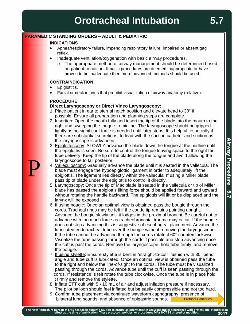
INDICATIONS
· Apnea/respiratory failure, impending respiratory failure, impaired or absent gag reflex.
· Inadequate ventilation/oxygenation with basic airway procedures.
o The appropriate method of airway management should be determined based on patient condition. If basic procedures are deemed inappropriate or have proven to be inadequate then more advanced methods should be used.
CONTRAINDICATION
· Epiglottitis.
· Facial or neck injuries that prohibit visualization of airway anatomy (relative).
PROCEDUREDirect Laryngoscopy or Direct Video Laryngoscopy:1. Place patient in ear to sternal notch position and elevate head to 30° if possible. Ensure all preparation and planning steps are complete.2. Insertion: Open the mouth fully and insert the tip of the blade into the mouth to the right and sweeping the tongue to midline. The laryngoscope should be gripped lightly as no significant force is needed until later steps. It is helpful, especially if there are substantial secretions, to lead with the suction catheter and suction as the laryngoscope is advanced.3. Epiglottoscopy: SLOWLY advance the blade down the tongue at the midline until the epiglottis is seen. Be sure to control the tongue leaving space to the right for tube delivery. Keep the tip of the blade along the tongue and avoid allowing the laryngoscope to fall posterior.4. Valleculoscopy: Gradually advance the blade until it is seated in the vallecula. The blade must engage the hypoepiglottic ligament in order to adequately lift the epiglottis. The ligament lies directly within the vallecula. If using a Miller blade pass tip of blade under the epiglottis to control it directly.5. Laryngoscopy: Once the tip of Mac blade is seated in the vallecula or tip of Miller blade has passed the epiglottis lifting force should be applied forward and upward without rotating the handle backward. The epiglottis will lift or be displaced and the larynx will be exposed6. If using bougie: Once an optimal view is obtained pass the bougie through the cords. Tracheal rings may be felt if the coude tip remains pointing upright. Advance the bougie slowly until it lodges in the proximal bronchi. Be careful not to advance with too much force as tracheobronchial trauma may occur. If the bougie does not stop advancing this is suggestive of esophageal placement. Advance the lubricated endotracheal tube over the bougie without removing the laryngoscope. If the tube cannot be advanced through the cords rotate it 60° counterclockwise. Visualize the tube passing through the cords if possible and stop advancing once the cuff is past the cords. Remove the laryngoscope, hold tube firmly, and remove the bougie.7. If using stylette: Ensure stylette is bent in “straight-to-cuff” fashion with 30° bend angle and tube cuff is lubricated. Once an optimal view is obtained pass the tube to the right and below the line-of-sight to the cords. The tube must be visualized passing through the cords. Advance tube until the cuff is seen passing through the cords. If resistance is felt rotate the tube clockwise. Once the tube is in place hold it firmly and remove the stylette.8. Inflate ETT cuff with 5 - 10 mL of air and adjust inflation pressure if necessary. The pilot balloon should feel inflated but be easily compressible and not too hard.9. Confirm tube placement via continued waveform capnography, presence of bilateral lung sounds, and absence of epigastric sounds.
PARAMEDIC STANDING ORDERS – ADULT & PEDIATRIC
Orotracheal Intubation
The New Hampshire Bureau of EMS has taken extreme caution to ensure all information is accurate and in accordance with professional standards in effect at the time of publication. These protocols, policies, or procedures MAY NOT BE altered or modified. 2017
Protocol Continues
5.7
Airw
ay P
roc
ed
ure
5.7
Orotracheal Intubation
The New Hampshire Bureau of EMS has taken extreme caution to ensure all information is accurate and in accordance with professional standards in effect at the time of publication. These protocols, policies, or procedures MAY NOT BE altered or modified. 2017
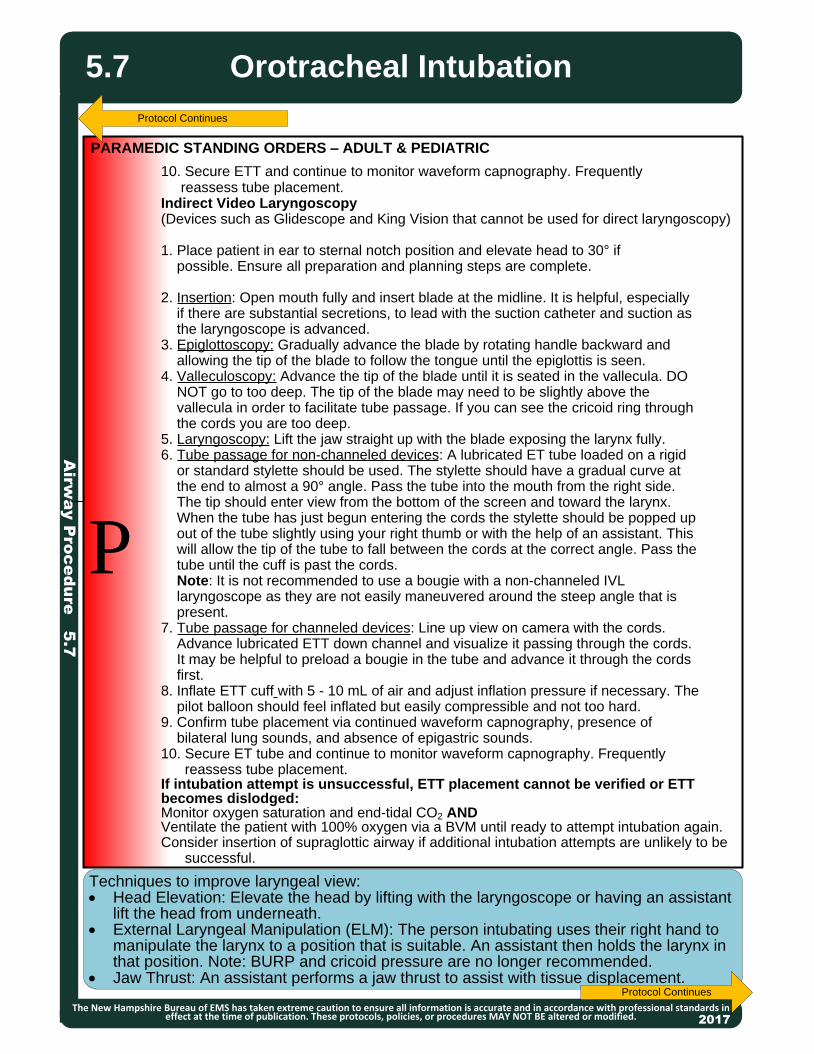
10. Secure ETT and continue to monitor waveform capnography. Frequently reassess tube placement.Indirect Video Laryngoscopy(Devices such as Glidescope and King Vision that cannot be used for direct laryngoscopy)
1. Place patient in ear to sternal notch position and elevate head to 30° if possible. Ensure all preparation and planning steps are complete.
2. Insertion: Open mouth fully and insert blade at the midline. It is helpful, especially if there are substantial secretions, to lead with the suction catheter and suction as the laryngoscope is advanced.3. Epiglottoscopy: Gradually advance the blade by rotating handle backward and allowing the tip of the blade to follow the tongue until the epiglottis is seen. 4. Valleculoscopy: Advance the tip of the blade until it is seated in the vallecula. DO NOT go to too deep. The tip of the blade may need to be slightly above the vallecula in order to facilitate tube passage. If you can see the cricoid ring through the cords you are too deep.5. Laryngoscopy: Lift the jaw straight up with the blade exposing the larynx fully. 6. Tube passage for non-channeled devices: A lubricated ET tube loaded on a rigid or standard stylette should be used. The stylette should have a gradual curve at the end to almost a 90° angle. Pass the tube into the mouth from the right side. The tip should enter view from the bottom of the screen and toward the larynx. When the tube has just begun entering the cords the stylette should be popped up out of the tube slightly using your right thumb or with the help of an assistant. This will allow the tip of the tube to fall between the cords at the correct angle. Pass the tube until the cuff is past the cords. Note: It is not recommended to use a bougie with a non-channeled IVL laryngoscope as they are not easily maneuvered around the steep angle that is present. 7. Tube passage for channeled devices: Line up view on camera with the cords. Advance lubricated ETT down channel and visualize it passing through the cords. It may be helpful to preload a bougie in the tube and advance it through the cords first.8. Inflate ETT cuff with 5 - 10 mL of air and adjust inflation pressure if necessary. The pilot balloon should feel inflated but easily compressible and not too hard.9. Confirm tube placement via continued waveform capnography, presence of bilateral lung sounds, and absence of epigastric sounds. 10. Secure ET tube and continue to monitor waveform capnography. Frequently reassess tube placement.If intubation attempt is unsuccessful, ETT placement cannot be verified or ETTbecomes dislodged: Monitor oxygen saturation and end-tidal CO2 ANDVentilate the patient with 100% oxygen via a BVM until ready to attempt intubation again. Consider insertion of supraglottic airway if additional intubation attempts are unlikely to be
successful.
5.7
P
Techniques to improve laryngeal view:· Head Elevation: Elevate the head by lifting with the laryngoscope or having an assistant
lift the head from underneath.· External Laryngeal Manipulation (ELM): The person intubating uses their right hand to
manipulate the larynx to a position that is suitable. An assistant then holds the larynx in that position. Note: BURP and cricoid pressure are no longer recommended.
· Jaw Thrust: An assistant performs a jaw thrust to assist with tissue displacement.
PARAMEDIC STANDING ORDERS – ADULT & PEDIATRIC
Protocol Continues
Protocol Continues
Airw
ay P
roc
ed
ure
5.7
Orotracheal Intubation
The New Hampshire Bureau of EMS has taken extreme caution to ensure all information is accurate and in accordance with professional standards in effect at the time of publication. These protocols, policies, or procedures MAY NOT BE altered or modified. 2017
5.7
If continued intubation attempts are unsuccessful (maximum of 3 attempts per patient) consider Cricothyrotomy. See Cricothyrotomy Procedure 5.2 OR 7.5.
POST TUBE PLACEMENT CARE – ADULTOption 1:· Ketamine 1 mg/kg ideal body weight (IBW) IV every 5 – 15 minutes, as needed.
Option 2:· Fentanyl 50 - 100 mcg IV every 5-10 minutes, as needed.AND· Midazolam 2 - 5 mg IV every 5 – 10 minutes as needed OR· Lorazepam 1 - 2 mg every 15 minutes as needed (maximum total 10 mg)
POST TUBE PLACEMENT CARE – PEDIATRICOption 1:· Ketamine 1 mg/kg IV every 5 - 15 minutes, as needed.
Option 2:· Fentanyl 2 - 3 mcg/kg IV every 5 - 10 minutes as needed.
AND· Midazolam 0.1 mg/kg IV (maximum single dose 2.5 mg) every 5 - 10 minutes as
needed OR· Lorazepam 0.1 mg/kg IV (maximum single dose 2 mg) every 15 minutes as needed
(maximum total 10 mg)
DocumentationDocument each attempt as a separate procedure so it can be time stamped in theePCR. An attempt is defined as placement of the blade into the patient’smouth. For each attempt, document the time, provider, placement success, preoxygenation, airway grade, ETT size, placement depth, placement landmark (e.g. cm
at the patient’s teeth), and confirmation of tube placement including chest rise, bilateralequal breath sounds, absence of epigastric sounds and capnography readings.
PARAMEDIC STANDING ORDERS – ADULT & PEDIATRIC
P
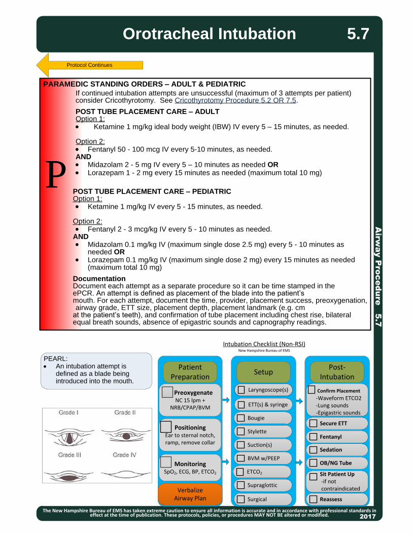
PEARL:· An intubation attempt is
defined as a blade being introduced into the mouth.
Protocol Continues
PatientPreparation
Post-Intubation
Setup
PreoxygenateNC 15 lpm +
NRB/CPAP/BVM
PositioningEar to sternal notch, ramp, remove collar
MonitoringSpO2, ECG, BP, ETCO2
VerbalizeAirway Plan Surgical
Stylette
Bougie
ETT(s) & syringe
Laryngoscope(s)
Suction(s)
BVM w/PEEP
ETCO2
Supraglottic
Secure ETT
Fentanyl
Sedation
OB/NG Tube
Sit Patient Up -if not contraindicated
Reassess
Confirm Placement
-Waveform ETCO2 -Lung sounds -Epigastric sounds
Intubation Checklist (Non-RSI)New Hampshire Bureau of EMS
2013
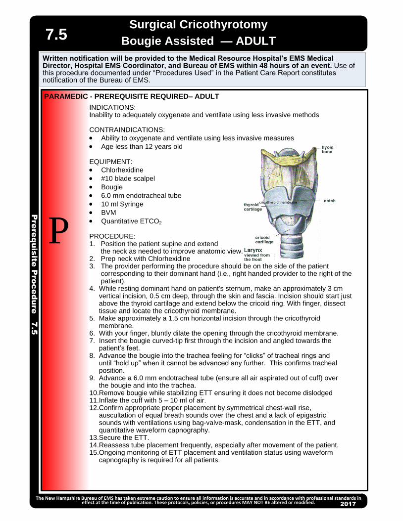
INDICATIONS:Inability to adequately oxygenate and ventilate using less invasive methods
CONTRAINDICATIONS:
· Ability to oxygenate and ventilate using less invasive measures
· Age less than 12 years old
EQUIPMENT:
· Chlorhexidine
· #10 blade scalpel
· Bougie
· 6.0 mm endotracheal tube
· 10 ml Syringe
· BVM
· Quantitative ETCO2
PROCEDURE:1. Position the patient supine and extend the neck as needed to improve anatomic view.2. Prep neck with Chlorhexidine 3. The provider performing the procedure should be on the side of the patient corresponding to their dominant hand (i.e., right handed provider to the right of the patient).4. While resting dominant hand on patient's sternum, make an approximately 3 cm vertical incision, 0.5 cm deep, through the skin and fascia. Incision should start just above the thyroid cartilage and extend below the cricoid ring. With finger, dissect tissue and locate the cricothyroid membrane.5. Make approximately a 1.5 cm horizontal incision through the cricothyroid membrane.6. With your finger, bluntly dilate the opening through the cricothyroid membrane.7. Insert the bougie curved-tip first through the incision and angled towards the patient’s feet.8. Advance the bougie into the trachea feeling for “clicks” of tracheal rings and until “hold up” when it cannot be advanced any further. This confirms tracheal position.9. Advance a 6.0 mm endotracheal tube (ensure all air aspirated out of cuff) over the bougie and into the trachea.10.Remove bougie while stabilizing ETT ensuring it does not become dislodged11.Inflate the cuff with 5 – 10 ml of air.12.Confirm appropriate proper placement by symmetrical chest-wall rise, auscultation of equal breath sounds over the chest and a lack of epigastric sounds with ventilations using bag-valve-mask, condensation in the ETT, and quantitative waveform capnography.13.Secure the ETT.14.Reassess tube placement frequently, especially after movement of the patient.15.Ongoing monitoring of ETT placement and ventilation status using waveform capnography is required for all patients.
PARAMEDIC - PREREQUISITE REQUIRED– ADULT
Pre
req
uis
ite P
roc
ed
ure
7.5
Surgical Cricothyrotomy
Bougie Assisted — ADULT
The New Hampshire Bureau of EMS has taken extreme caution to ensure all information is accurate and in accordance with professional standards in effect at the time of publication. These protocols, policies, or procedures MAY NOT BE altered or modified. 2017
7.5
P cricothyroid membrane
Written notification will be provided to the Medical Resource Hospital’s EMS Medical Director, Hospital EMS Coordinator, and Bureau of EMS within 48 hours of an event. Use of this procedure documented under “Procedures Used” in the Patient Care Report constitutes notification of the Bureau of EMS.