Neuroscience Review
112
-
Upload
drsaeed-shafi -
Category
Health & Medicine
-
view
205 -
download
4
Transcript of Neuroscience Review


Ap
plie
d A
na
tom
y o
f B
rain
&
Sp
ina
l C
ord
Pro
f. S
ae
ed
Sh
afi
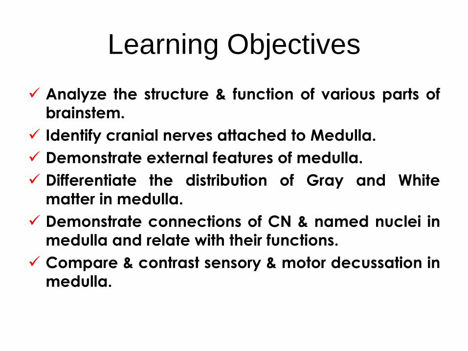
Learning Objectives
Analyze the structure & function of various parts of
brainstem.
Identify cranial nerves attached to Medulla.
Demonstrate external features of medulla.
Differentiate the distribution of Gray and White
matter in medulla.
Demonstrate connections of CN & named nuclei in
medulla and relate with their functions.
Compare & contrast sensory & motor decussation in
medulla.

Identify Pyramid, Olive, Faciculus
gracilus, Faciculus cuneatus on
external surface of medulla.
Identify Vagal, Hypoglosssal &
Vestibular triangles; Obex, area
postrema and Facial Colliculus in
floor of 4th ventricle.
LOs continued…………….

White matter of spinal cord Descending Tracts
Cortico Spinal
Tectospinal
Rubrospinal
Reticulospinal
Vestibulospinal
Hypothalamospinal
Ascending Tracts
Dorsal Column(Fasciculus Gracilus& Cuneatus)
Spinothalamic (Anterior & Lateral)
Spinocerebellar (Anterior & Posterior)
Spinovestibular
Spinotectal
Spinoreticular
Spino-olivary


Neu
ron
al C
ircu
its o
f D
ors
al
Gre
y H
orn
Neu
ro
nal C
ircu
itry o
f V
en
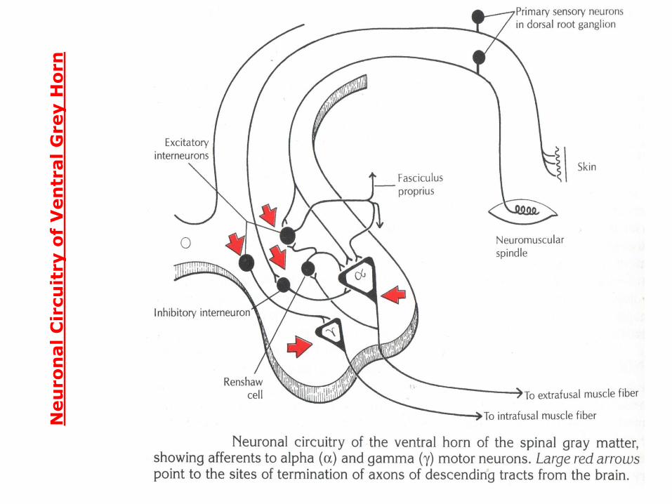
tral G
rey H
orn


BRAIN VESICLES
• PROSENCEPHALON
– Diencephalon
– Telencephalon
• MESENCEPHALON
• RHOMBENCEPHALON
– Metencephalon
– Myelencephalon
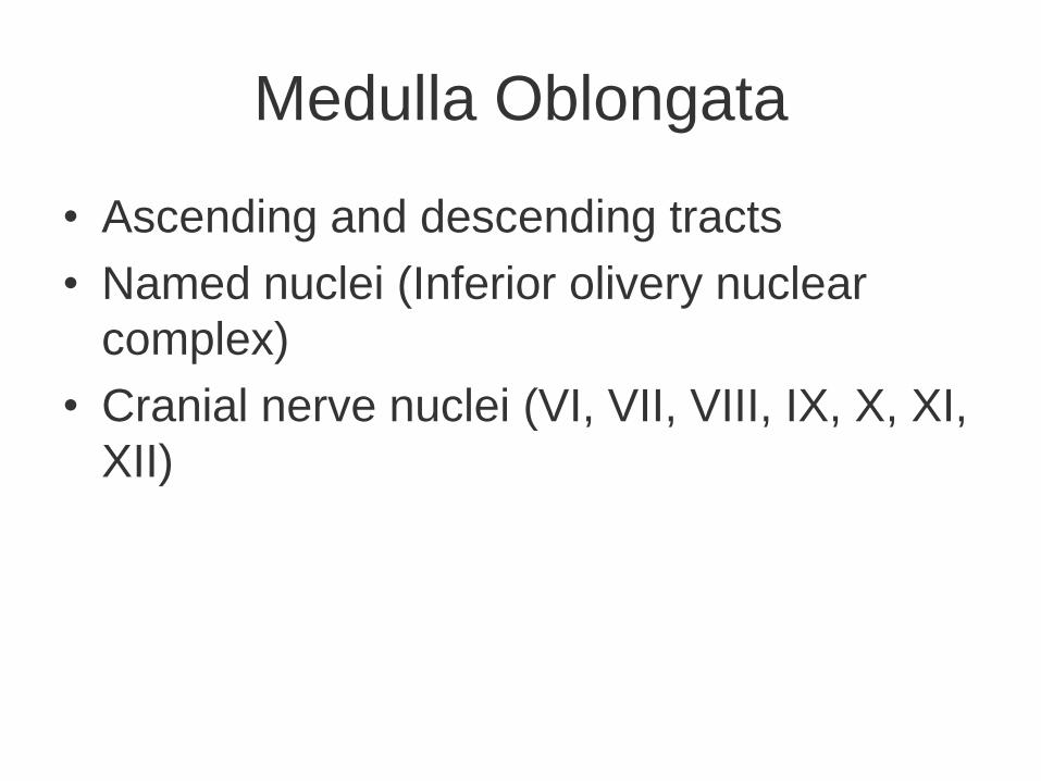
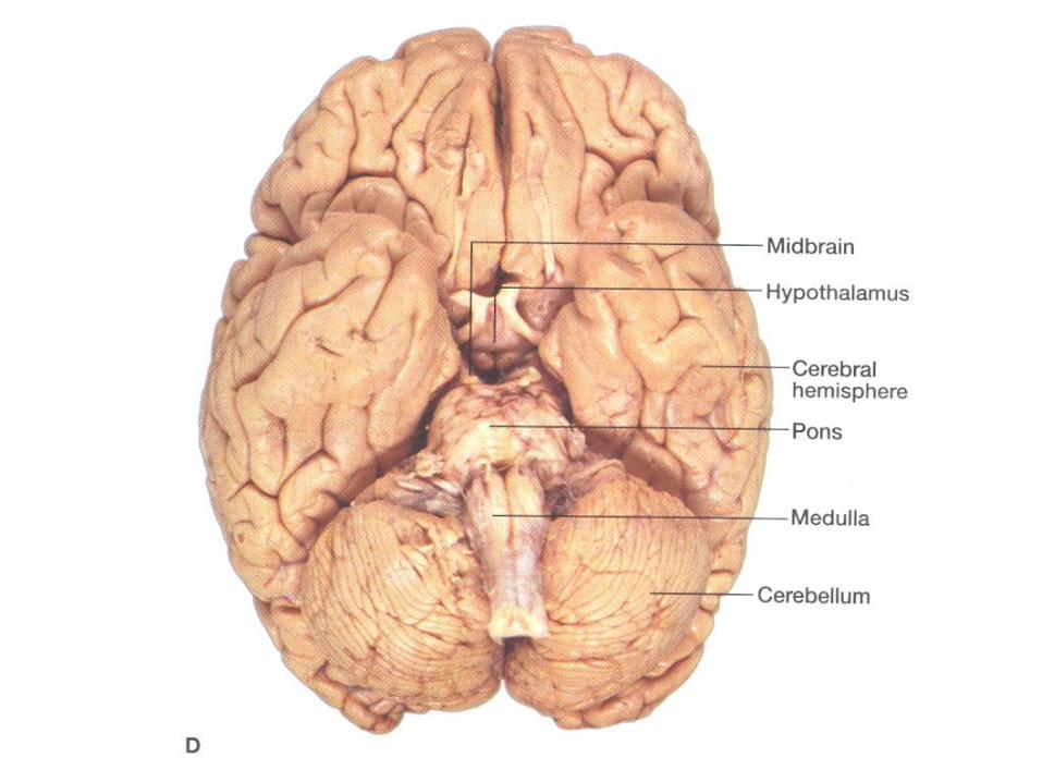
Medulla Oblongata
• Ascending and descending tracts
• Named nuclei (Inferior olivery nuclear
complex)
• Cranial nerve nuclei (VI, VII, VIII, IX, X, XI,
XII)


Pons
• Basal portion (Bridges between ipsilateral
cerebral hemisphere and contralateral
cerebellar hemisphere)
• Dorsal portion
– Ascending and descending tracts
– Cranial nerve nuclei (V, VI, VII, VIII)
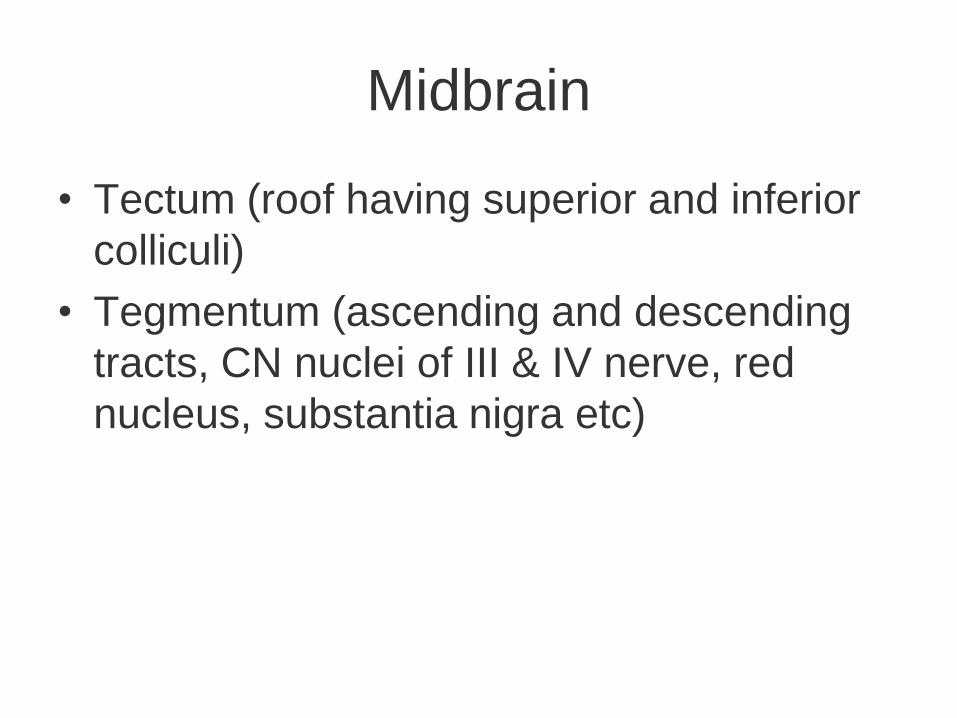
Midbrain
• Tectum (roof having superior and inferior
colliculi)
• Tegmentum (ascending and descending
tracts, CN nuclei of III & IV nerve, red
nucleus, substantia nigra etc)

Diencephalon
• Constitute central core of cerebrum
– Thalamus
– Hypothalamus
– Subthalamus
– Epithalamus
– Metathalamus

Telencephalon
• The most well developed part of CNS
– Cortex (the peripheral gray matter)
– Medulla (central white matter)
• Projection fibers
• Association fibers
• Commissural fibers
– Corpus striatum (deep seated basal nuclei)


Morphology of Medulla Oblongata
• Medulla is 3 cm long
• Upper limit clearly marked by lower border
of basal pons
• Inferior limit at rootlets of 1st cervical spinal
nerve


Dorsal surface of Medulla
On dorsal surface junction of medulla and pons is
marked by a line passing through inferior edge
of middle cerebellar peduncles (ICP)
– Closed part of medulla Fasciculus gracilis
Fasciculus cuneatus
– Open part of medulla Floor of 4th ventricle

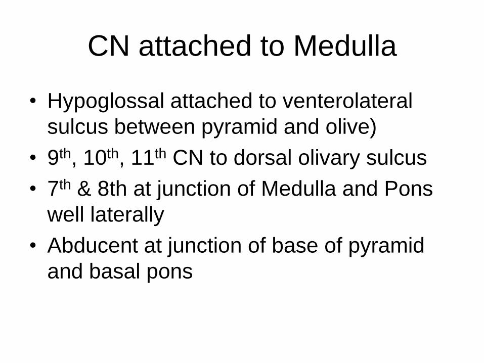
CN attached to Medulla
• Hypoglossal attached to venterolateral
sulcus between pyramid and olive)
• 9th, 10th, 11th CN to dorsal olivary sulcus
• 7th & 8th at junction of Medulla and Pons
well laterally
• Abducent at junction of base of pyramid
and basal pons

Cranial nerve nuclei
Medulla –Hypoglossal nucleus, Nucleus
ambiguous, DMN of Vagus, Vestibular
nuclear complex, Auditory nuclei, Spinal
trigeminal nucleus, Tractus solitarius nucleus
Pons – Abducent nerve nucleus, facial nucleus,
trigeminal nuclei (Chief sensory nucleus,
motor nucleus)
Midbrain – 3rd & 4th nerve nuclei,
mesencephalic nucleus of trigeminal

Named Nuclei in Brain stem
Inferior olivary nuclear complex
Pontine nuclei
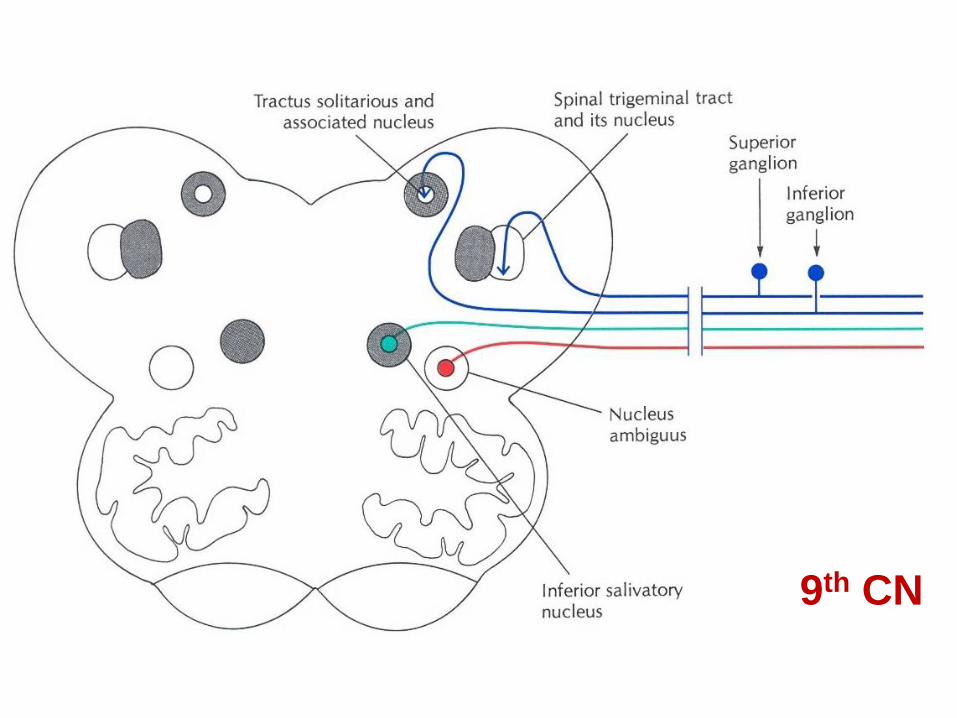
Inferior Salivatory Nucleus
Superior Salivatory Nucleus
Red nucleus of midbrain
Substantia nigra
Reticular formation nucli

Brainstem Ascending tracts• Dosral Column Tracts / Posterior Column Tracts
– Fasciculus Gracilis
Medial Lemniscus
– Fasciculus Cuneatus
• Spinocerebellar Tracts
• Spinotectal Tracts
• Spinothalamic Tracts Spinal Lemniscus
• Spinoreticular
• Lateral Lemniscus
• Trigeminal lemniscus
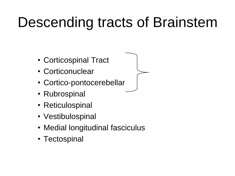
Descending tracts of Brainstem
• Corticospinal Tract
• Corticonuclear
• Cortico-pontocerebellar
• Rubrospinal
• Reticulospinal
• Vestibulospinal
• Medial longitudinal fasciculus
• Tectospinal

Medullary Nuclei
1. Inferior olivary nuclear complex (inferior olivary
nucleus, medial accessory olivary nucleus,
dorsal accessory olivary nucleus)
– Afferents by
• Central tegmental tract from corpus striatum & red nucleus
• Cortico-olivary fibers
• Spino-olivary fibers
– Efferent by
• Olivo-cerebellar fibers through ICP
• Olivo-spinal

Medullary Nuclei
2. Gracile Nuclus
3. Cuneate Nucleus
4. Arcuate nucleus
5. Lateral cuneate nucleus
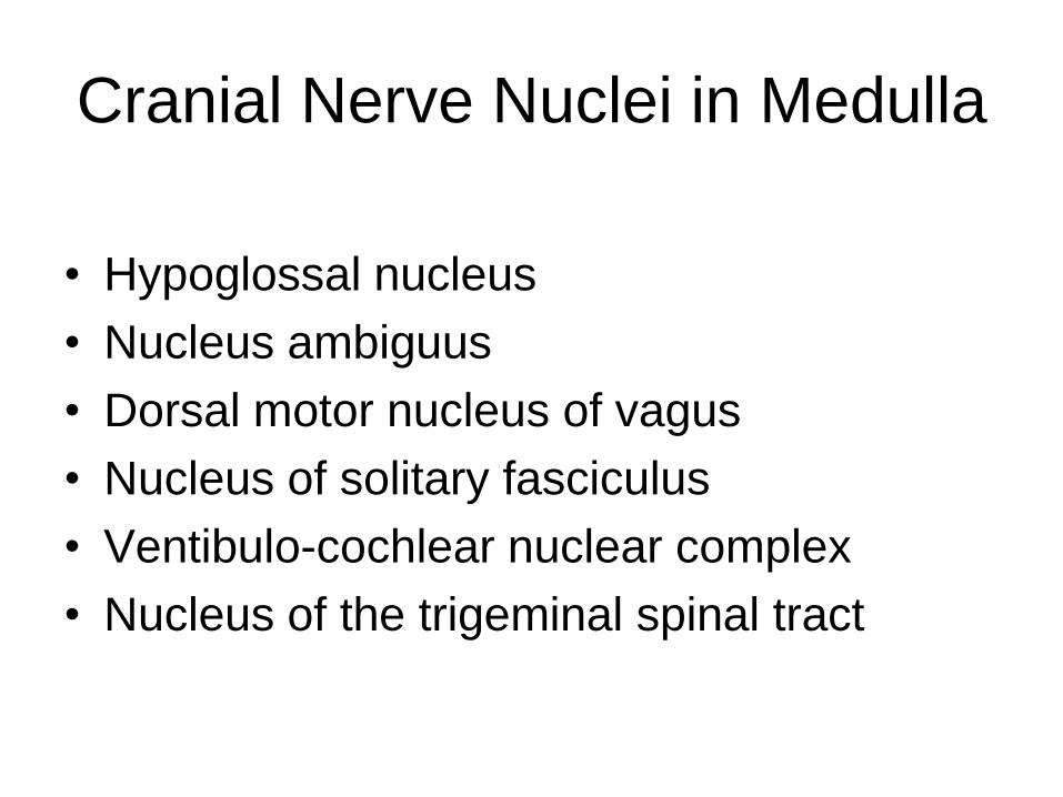
Cranial Nerve Nuclei in Medulla
• Hypoglossal nucleus
• Nucleus ambiguus
• Dorsal motor nucleus of vagus
• Nucleus of solitary fasciculus
• Ventibulo-cochlear nuclear complex
• Nucleus of the trigeminal spinal tract
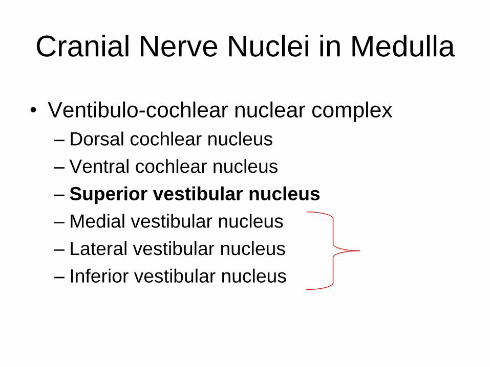
Cranial Nerve Nuclei in Medulla
• Ventibulo-cochlear nuclear complex
– Dorsal cochlear nucleus
– Ventral cochlear nucleus
– Superior vestibular nucleus
– Medial vestibular nucleus
– Lateral vestibular nucleus
– Inferior vestibular nucleus

NA


PONS
• Basal Pons
• Tegmentum
• Only cranial nerve attached to pons is
trigeminal

Internal structure
• Pontine nuclei
• Cranial nerves Nuclei (5th, 6th, 7th)
&
• Fiber tracts• ML, SL, LL
• Corticopontine fibers
• Pontocerebellar fibers
• Corticospinal fibers


• Motor nucleus
• Chief sensory nucleus
• Mesencephalic nucleus
• Spinal nucleus
5thCN Nuclei in Pons

• Superior salivary nucleus lies along side the facial nucleus.
• It is parasympathetic in function.
• It provides axons which pass out in nervous intermedius part of facial nerve and reach to pterygopalatine and Submandibular ganglia.
• Its lower part is called inferior salivatory nucleus. Its fibers joins to the glossopharyngeal nerve to reach to otic ganglion.

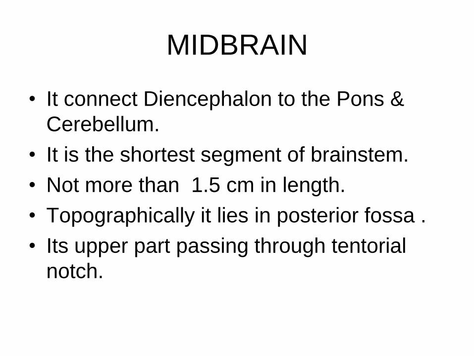
MIDBRAIN
• It connect Diencephalon to the Pons &
Cerebellum.
• It is the shortest segment of brainstem.
• Not more than 1.5 cm in length.
• Topographically it lies in posterior fossa .
• Its upper part passing through tentorial
notch.


Midbrain
Crus cerebri
Substancia Nigra Cerebral peduncle
Tegmentum
Tectum is the roof of midbrain

• 3rd & 4th cranial nerves leave midbrain.
• Occulomotor nerve leaves through the
medial surface of crus on the ventral
surface.
• Trochlear nerve leaves the dorsal surface
of midbrain just below the inferior colliculi.

Internal structure
• Each crus contains corticospinal , cortico
nuclear and corticopontine fibers.


4th CN

Cerebellar Peduncles
• Inferior cerebellar peduncle– Posterior spinal cerebellar tracts
– Olivocerebellar tract
– Vestibulo-cerebellar tract
– Cuneocerebellar tract
– Cerebelovestibular
• Middle cerebellar peduncle – Pontocerebellar
• Superior cerebellar peduncle – Anterior spinocerebellar
– Dentatorubrocerebellar
– Cerebellospinal fibers


COMPONENTS OF
CRANIAL&SPINAL NERVES• Afferent components
– Special somatic afferent• Optic nerve
• Cochlear
• Vestibular
– General Somatic Afferent• Dorsal gray horn of spinal cord
• Graycile and nuclei
• Special Visceral afferent
• Taste
• Olfaction
• General Visceral afferent (for visceral reflexes)
• Dorsal grayhorn / nucelus and tractus solitarius
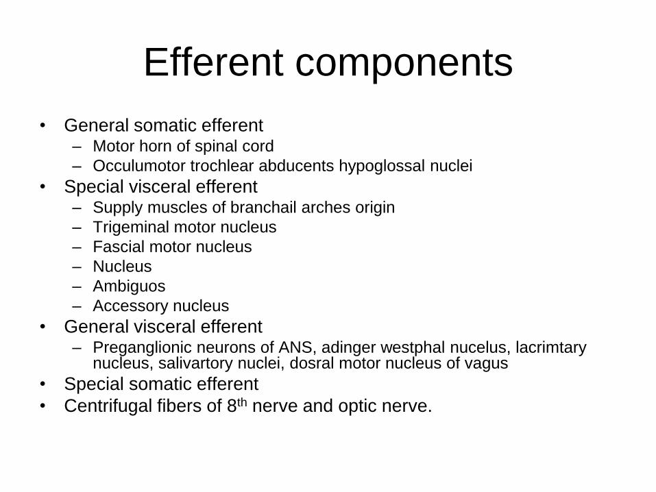
Efferent components
• General somatic efferent– Motor horn of spinal cord
– Occulumotor trochlear abducents hypoglossal nuclei
• Special visceral efferent– Supply muscles of branchail arches origin
– Trigeminal motor nucleus
– Fascial motor nucleus
– Nucleus
– Ambiguos
– Accessory nucleus
• General visceral efferent– Preganglionic neurons of ANS, adinger westphal nucelus, lacrimtary
nucleus, salivartory nuclei, dosral motor nucleus of vagus
• Special somatic efferent
• Centrifugal fibers of 8th nerve and optic nerve.

Nervous System
Central Peripheral
Somatic
Autonomic
Somatic
Autonomic
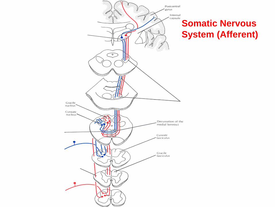
Somatic Nervous
System (Afferent)

Somatic
Nervous
System
(Eff.)

RESPONSE OF SNS & ANS
• SOMATIC NERVOUS SYSTEM
– Voluntary
– Focused, precise and specific function /
response
• AUTONOMIC NERVOUS SYSTEM
– Mass discharge in sympathetic nervous
system (Fight or flight response )
– Discrete localized response by
parasympathetic

PARASYMPATHETIC
ANS

ANS-Parasympathetic
CN III
CN VII
CN IX
Vagus

FUNCTIONS OF PARASYMPATHETIC
• Decreased HR & force of contraction
• Vasodilator
• Secretomotor and relax sphincters in GIT
• Genital erectile tissue engorged
• Emptying of urinary bladder
• Action is discrete and localized because at
synapsis divergence is less and Ach is
rapidly deactivated Acetylcholinesterase.

Sympathetic ANS


Distribution of Sympathetic nerves
• White rami communicants - preganglionic
• Somatic branches through grey rami
communicants to all spinal nerves
supplying BV, sweat glands & erector pili.
• Vascular branches spread along BV.
• Visceral branches through Splanchnic
nerves carry preganglionic fibers to
prevertebral ganglia.

3rd CN

3rd CN
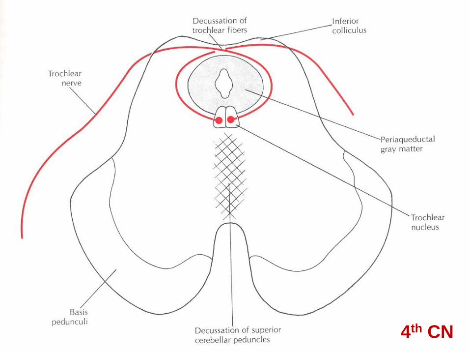
4th CN

6th CN

MLF


5th CN
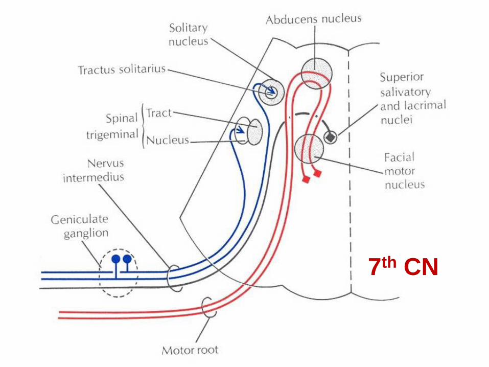
7th CN

7th CN

Ta
ste
Pa
thw
ay
9th CN

10th CN

11
thC
N

12th CN


Boundaries between
• Midbrain and diencephalon
– Posterior commissure Mammillary bodies)
• Diencephalon and Telencephalon
– (Interventricular foramen Optic Chiasma)


Components of Diencephalon
• Thalamus
• Hypothalamus
• Subthalamus
• Epithalamus
• Metathalamus

Morphology of thalamus
• Largest sensory relay station to cortex
• 3 x 1.5 x 1.5 cm
• Medial, Lateral, Dorsal &Ventral Surfaces
• Relation to 3rd ventricle & internal capsule
• Divisions into nuclei
• External medullary lamina
• Internal medullary lamina


Medial Geniculate nucleus
• AFF: Inferior Colliculus / inferior branchium
• EFF: Auditory radiations to Temporal lobe

Lateral geniculate nucleus
• AFF: Retina / OT / opposite visual filed
• EFF: Geniculocalcarine tract

VPN
• AFF: ML / SL / TL
• EFF:
– Sensory cortex (3,1,2,5,7)
– Medial nucleus
– Nonspecific nuclei

VA & VL nucleus
• AFF:
– Dentate N.
– Substantia nigra
– Corpus striatum
• EFF:
– Motor cortex (4,6)
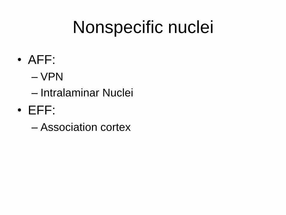
Nonspecific nuclei
• AFF:
– VPN
– Intralaminar Nuclei
• EFF:
– Association cortex
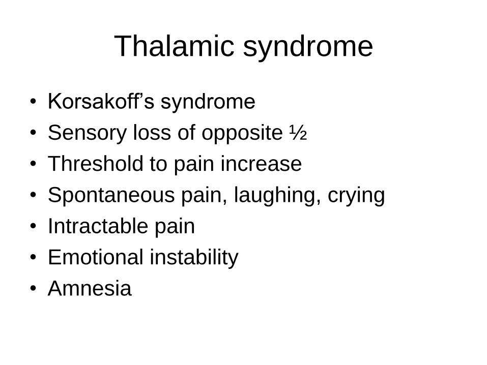
Thalamic syndrome
• Korsakoff’s syndrome
• Sensory loss of opposite ½
• Threshold to pain increase
• Spontaneous pain, laughing, crying
• Intractable pain
• Emotional instability
• Amnesia


Subthalamus
• Subthalamic N.
• Fiber tracts – dentatothalamic SL, ML, TL,
Lenticular fasciculus, Ansa lenticularis
• Substantia nigra
• Zona incenta (Red nucleus)
• Hemiballisnus ?

Epithalamus
• Pineal gland
– Biological clock + circadian rhythm
– Neuroendocrine tranducer
– Epiphysis cerebri
• Habenular nucleus
– AFF:• Stria medullaris thalami, Stria terminalis
– EFF: • Habenulo interpeduncular fasciculus
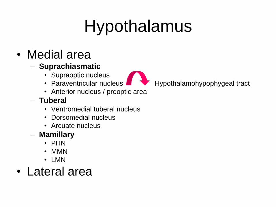
Hypothalamus
• Medial area – Suprachiasmatic
• Supraoptic nucleus
• Paraventricular nucleus Hypothalamohypophygeal tract
• Anterior nucleus / preoptic area
– Tuberal • Ventromedial tuberal nucleus
• Dorsomedial nucleus
• Arcuate nucleus
– Mamillary • PHN
• MMN
• LMN
• Lateral area


103

104

105
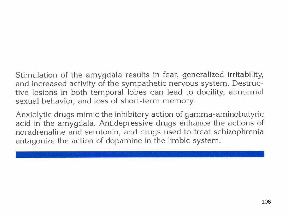
106


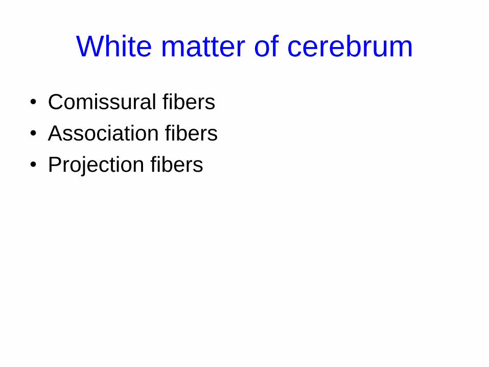
White matter of cerebrum
• Comissural fibers
• Association fibers
• Projection fibers