Neuroscience and Behavior. Types of Neurons SensoryMotor Interneurons.
59
Neuroscience and Behavior
-
date post
21-Dec-2015 -
Category
Documents
-
view
228 -
download
4
Transcript of Neuroscience and Behavior. Types of Neurons SensoryMotor Interneurons.
Neuroscience and Behavior

Types of Neurons
Types of Neurons
Sensory Motor Interneurons

SpinalCord
BrainSensoryNeuron
Sensory Neurons• INPUT From sensory organs to the brain and spinal
cord
Drawing shows a sensory neuron

SpinalCord
BrainSensoryNeuron
MotorNeuron
Motor Neurons
• OUTPUT From the brain and spinal cord, to the muscles and glands

SpinalCord
BrainSensoryNeuron
MotorNeuron
Interneurons
• Interneurons carry information between other neurons only found in the brain and spinal cord

Parts of a Neuron

The Cell Body
– round, centrally located structure
– contains DNA
– controls protein manufacturing
– directs metabolism
– no role in neural signaling
Contains the cell’s nucleus
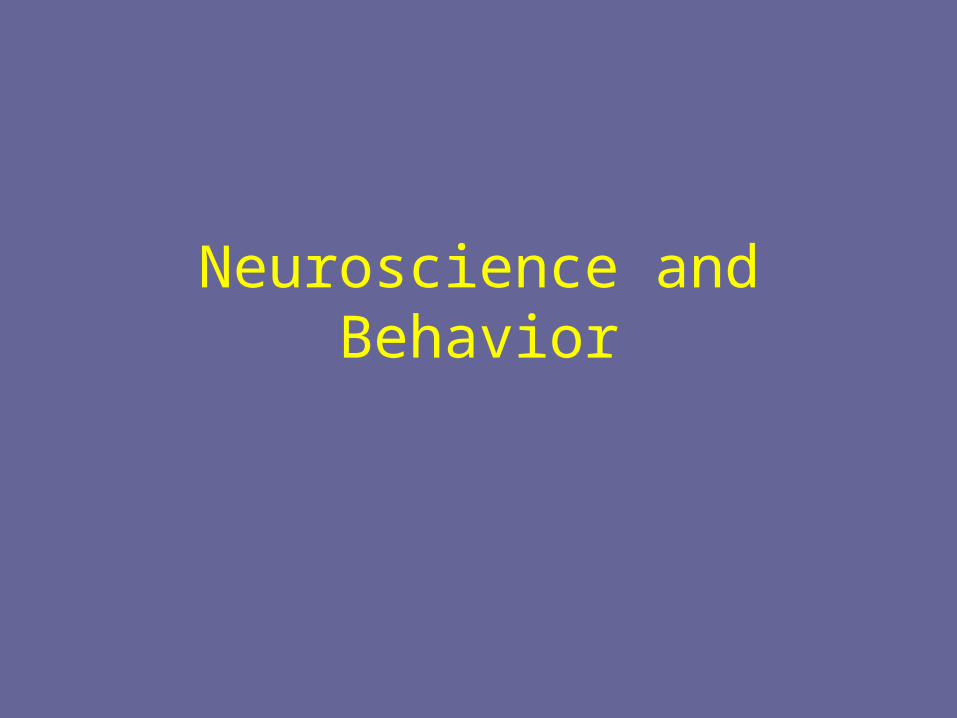
Dendrites
• Information collectors
• Receive inputs from neighboring neurons
• Inputs may number in thousands
• If enough inputs, the cell’s AXON may generate an output

Axon
• The cell’s output structure
• One axon per cell, 2 distinct parts– tube-like structure – branches at end that connect to dendrites
of other cells
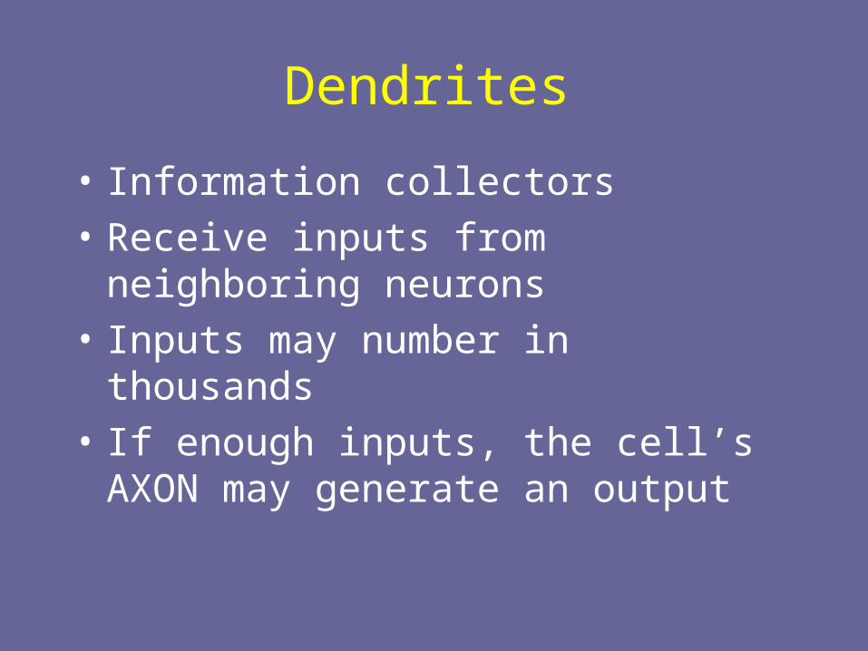
Myelin Sheath
• White fatty casing on axon • Acts as an electrical insulator • Not present on all cells• When present, increases the speed of neural signals
down the axon

How Neurons Communicate
• Neurons communicate by means of an electrical signal called the Action Potential
• Action Potentials are based on movements of ions between the outside and inside of the cell
• When an Action Potential occurs, a molecular message is sent to neighboring neurons

All or None Law
• This is the principle that a neuron is either sufficiently stimulated and an action potential occurs; or the neuron is not sufficiently stimulated and the action potential does not occur.
• In other words, the neuron can’t fire just a little bit--it either sends a message or it doesn’t

Action Potential Within a Neuron

Resting Potential
• At rest, the inside of the cell is at -70 microvolts• With inputs to dendrites, the inside becomes more positive • If resting potential rises above the sensory threshold, an action potential
starts to travel from cell body down the axon• Figure shows resting axon being approached by an action potential

Repolarization follows
• After the action potential passes, the inside of the axon returns to a negative voltage
• This is called repolarization

Neuron to Neuron
• Axons branch out and end near dendrites of neighboring cells
• Axon terminals are the tips of the axon’s branches
• A gap separates the axon terminals from dendrites
• Gap is called the synapse or synaptic gap


Neurotransmitter Release
• Action Potential causes vesicle to open
• Neurotransmitter released into synapse• Locks onto receptor molecule in postsynaptic membrane
• Reuptake occurs when, after sending a message, the neurotransmitters are taken back into the presynaptic neuron

Excitatory and Inhibitory Messages
• Excitatory message—increases the likelihood that the postsynaptic neuron will activate
• Inhibitory message—decreases the likelihood that the postsynaptic neuron will activate

Locks and Keys
• Neurotransmitter molecules have specific shapes
• When NT binds to receptor, ions enter
• Receptor molecules have binding sites

Types of Neurotransmitters• Acetylcholine• Serotonin• Norepinephrine• Dopamine• Endorphins• GABA

Acetylcholine (Ach)
• Found in neuromuscular junctions
• Involved in muscle movements
• Involved in learning and memory

Disruption of Acetylcholine Functioning
• Curare—blocks ACh receptors– paralysis results
• Nerve gases and Black Widow spider venom; too much ACh leads to severe muscle spasms and possible death

Disruptions in ACh Functioning
• Cigarettes—nicotine works on ACh receptors– can artificially stimulate skeletal
muscles, leading to slight trembling movements

Alzheimer’s Disease
• Deterioration of memory, reasoning, and language skills
• Symptoms may be due to loss of ACh neurons

Serotonin
• Involved in sleep
• Involved in depression– Prozac works by keeping serotonin in
the synapse longer, giving it more time to exert an effect

Norepinephrine
• Arousal• “Fight or flight” response• Depression and stress

Dopamine
• Involved in movement, attention, and learning
• Dopamine imbalance also involved in schizophrenia
• Loss of dopamine-producing neurons is cause of Parkinson’s disease

Parkinson’s Disease
• Results from loss of dopamine-producing neurons in the substantia nigra
• Symptoms include– difficulty starting and stopping voluntary movements
– tremors at rest
– stooped posture
– rigidity
– poor balance

Parkinson’s Disease
• Treatments– L-dopa– transplants of fetal dopamine-producing
substantia nigra cells– adrenal gland transplants– electrical stimulation of the thalamus has
been used to stop tremors

Endorphins
• Control pain and pleasure
• Released in response to pain
• Morphine and codeine work on endorphin receptors; involved in healing effects of acupuncture
• Runner’s high— feeling of pleasure after a long run is due to heavy endorphin release

GABA
• Inhibition of brain activity
• Implicated in anxiety disorders
• Huntington’s disease involves loss of neurons in striatum that utilize GABA– Symptoms:
• jerky involuntary movements
• mental deterioration

Summary
• Neuron structure• Action potentials• Synapse• Neurotransmitters• Receptors and ions

Parts of the Nervous System
• Central Nervous System (CNS)– Brain and spinal cord
• Peripheral Nervous System (PNS)– Carries messages to and from CNS


Sympathetic and parasympathetic divisions of the nervous system

Endocrine System
• Pituitary gland—attached to the base of the brain; hormones affect the function of other glands
• Adrenal glands—hormones involved in human stress response
• Gonads—hormones regulate sexual characteristics and reproductive processes--testes in males, ovaries in females

Brain• Developing Brain• Brainstem
– Hindbrain– Midbrain
• Forebrain– Limbic system– Cortex

Developing Brain
•Neural tube—beginning of nervous system develops at 2 weeks after conception
•Neurogenesis—development of new neurons

Hindbrain
• cerebellum
• medulla
• reticular formation
• pons

Cerebellum
• Coordinated, rapid voluntary movements– e.g., playing the piano,
kicking, throwing, etc.
• Lesions to cerebellum– jerky, exaggerated
movements
– difficulty walking
– loss of balance
– shaking hands

Medulla
• Breathing• Heart rate• Digestion• Other vital reflexes
– swallowing
– coughing
– vomiting
– sneezing

Reticular Formation
• Network of neurons in the brainstem (and thalamus)
• Sleep and arousal• Attention
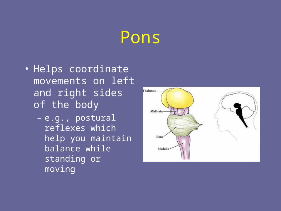
Pons
• Helps coordinate movements on left and right sides of the body– e.g., postural reflexes
which help you maintain balance while standing or moving

Midbrain
• Smallest region of the brain• Involved in processing auditory and visual
sensory signals• Contains the substantia nigra where there is
a large concentration of dopamine producing neurons
• Implicated in movement disorders such as Parkinson’s disease

Forebrain Structures
• Cortex
• Limbic System


Lobes of the Cortex
• Frontal lobe—largest lobe, produces voluntary muscle movements, involved in thinking, planning, emotional control
• Temporal lobe—primary receiving area for auditory information
• Occipital lobe—primary receiving area for visual information
• Parietal lobe—processes somatic information


The Limbic System
• Thalamus
• Hypothalamus
• Amygdala
• Hippocampus

Thalamus
• Relay station in brain• Processes most
information to and from higher brain centers

Hypothalamus• Contains nuclei involved in a variety
of behaviors– sexual behavior
– hunger, thirst
– sleep
– water and salt balance
– body temperature regulation
– circadian rhythms
– role in hormone secretion

Hypothalamus and Hormones
Hypothalamus releases hormones or releasing factors which in turn cause pituitary gland to release its hormones

Amygdala and Emotion
• Identify emotion from facial expressions
Amygdala damage makes this task difficult
(click on picture to advance photos)

Hippocampus
• Large “sea-horse” shaped structure imbedded in the temporal lobes
• Plays a role in the formation of new memories

Cortical Specialization
• Localization—notion that different functions are located in different areas of the brain
• Lateralization—notion that different functions are processed primarily on one side of the brain or the other
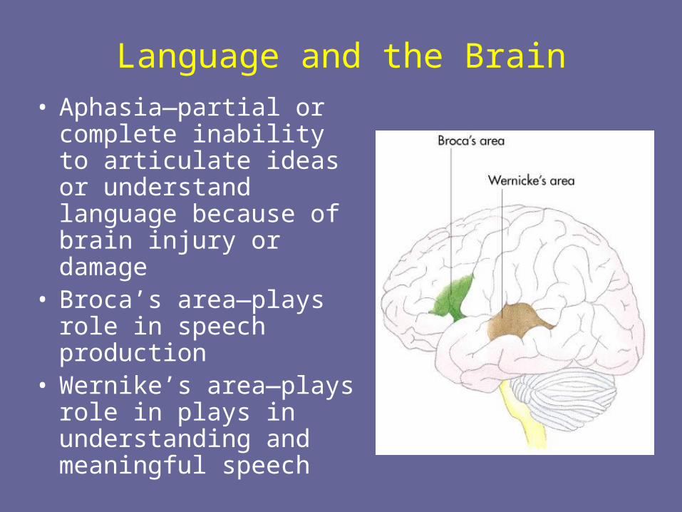
Language and the Brain
• Aphasia—partial or complete inability to articulate ideas or understand language because of brain injury or damage
• Broca’s area—plays role in speech production
• Wernike’s area—plays role in plays in understanding and meaningful speech
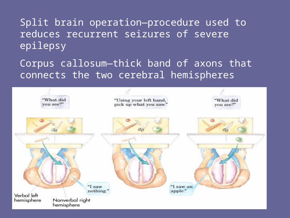
Split brain operation—procedure used to reduces recurrent seizures of severe epilepsy
Corpus callosum—thick band of axons that connects the two cerebral hemispheres