Neuronal Serotonin Release Triggers the Heat Shock...
12
Current Biology 25, 1–12, January 19, 2015 ª2015 Elsevier Ltd All rights reserved http://dx.doi.org/10.1016/j.cub.2014.11.040 Article Neuronal Serotonin Release Triggers the Heat Shock Response in C. elegans in the Absence of Temperature Increase Marcus C. Tatum, 1 Felicia K. Ooi, 1 Madhusudana Rao Chikka, 1 Laetitia Chauve, 2 Luis A. Martinez-Velazquez, 3 Harry W.M. Steinbusch, 4 Richard I. Morimoto, 2 and Veena Prahlad 1, * 1 Department of Biology, Aging Mind and Brain Initiative, University of Iowa, 338 Biology Building East, 210 Iowa Avenue, Iowa City, IA 52242-1324, USA 2 Department of Molecular Biosciences, Rice Institute for Biomedical Sciences, Northwestern University, 2205 Tech Drive, Hogan 2-100, Evanston, IL 60208-3500, USA 3 Program in Cellular Neuroscience, Neurodegeneration, and Repair, Department of Cell Biology, Yale University School of Medicine, 333 Cedar Street, P.O. Box 208002, New Haven, CT 06520-8002, USA 4 Department Translational Neuroscience, Faculty of Health, Medicine and Life Sciences, Maastricht University Medical Center, Universiteitssingel 40, Room 2.578, P.O. Box 616, 6200 MD Maastricht, the Netherlands Summary Background: Cellular mechanisms aimed at repairing protein damage and maintaining homeostasis, widely understood to be triggered by the damage itself, have recently been shown to be under cell nonautonomous control in the metazoan C. elegans. The heat shock response (HSR) is one such conserved mechanism, activated by cells upon exposure to proteotoxic conditions such as heat. Previously, we had shown that this conserved cytoprotective response is regu- lated by the thermosensory neuronal circuitry of C. elegans. Here, we investigate the mechanisms and physiological rele- vance of neuronal control. Results: By combining optogenetic methods with live visuali- zation of the dynamics of the heat shock transcription factor (HSF1), we show that excitation of the AFD thermosensory neurons is sufficient to activate HSF1 in another cell, even in the absence of temperature increase. Excitation of the AFD thermosensory neurons enhances serotonin release. Serotonin release elicited by direct optogenetic stimulation of serotonergic neurons activates HSF1 and upregulates molecu- lar chaperones through the metabotropic serotonin receptor SER-1. Consequently, excitation of serotonergic neurons alone can suppress protein misfolding in C. elegans peripheral tissue. Conclusions: These studies imply that thermosensory activity coupled to serotonergic signaling is sufficient to activate the protective HSR prior to frank proteotoxic damage. The ability of neurosensory release of serotonin to control cellular stress responses and activate HSF1 has powerful implications for the treatment of protein conformation diseases. Introduction Serotonin (5-hydroxytryptamine, 5-HT) is implicated in the stress response of all animals, and its enhanced release modulates physiological and metabolic adaptation to adverse conditions [1, 2]. Anticipation of danger can trigger serotonin release [3]. In addition, experimentally manipulating serotonin signaling in mammalian models can increase stress hormone production and elicit aversive behaviors, even in the absence of the actual stressor [1, 2]. Thus, although it is unclear whether enhanced release of serotonin is specific to particular stressors or whether it is a nonspecific response associated with increased arousal, serotonergic signaling allows organ- isms to rapidly adjust their physiology, metabolism, and behavior in expectation of imminent danger. Exposure to unfavorable environments can also cause macromolecular damage [4, 5]. However, cellular mechanisms aimed at repairing damage and maintaining homeostasis are widely understood to be triggered by the damage itself, either through damage to the cell that activates the response [4, 6–8] or, as has been suggested by recent experiments, through the activation of a stress response in other ‘‘sending’’ cells of an organism that are subject to proteotoxic conditions [9]. Indeed, there is little evidence to date for the preemptive acti- vation of cellular stress responses prior to macromolecular damage. One key homeostatic mechanism by which cells pro- tect themselves against protein damage is through the heat shock response (HSR) and activation of the highly conserved transcription factor, heat shock factor 1 (HSF1) [6, 7, 10]. HSF1 upregulates heat shock protein (HSP) genes that act as molecular chaperones to maintain protein conformation under stress, refold misfolded proteins, and target irrevers- ibly damaged proteins for degradation [6, 7, 10]. Thus, HSF1 activation suppresses protein misfolding and toxicity in numerous animal models of protein conformational diseases, such as Alzheimer’s, Parkinson’s, and Huntington’s diseases [6, 10, 11]. We have previously shown that in C. elegans, the HSR is regulated cell nonautonomously by the animals’ AFD thermo- sensory neuronal circuitry [12, 13]. In addition, other groups have also shown nonautonomous regulation of HSF1 and other cellular stress responses by the nervous system [9, 12–16]. Thermosensory neurons in C. elegans are exquisitely sensitive and detect changes as small as 0.05 C above ambient temperature [17]; hence, they can arguably be excited by temperature increments well below those that cause cellular damage. Therefore, a central question that arises from the observation that thermosensory neurons control the HSR of peripheral tissue is whether the nervous system plays an instructive role in activating the HSR upon sensing temper- ature increase, prior to macromolecular damage, or whether it plays a more general, permissive role in allowing the stress response to be triggered by macromolecular damage. An instructive role involving signaling pathways from neuronal cells to peripheral tissue could activate protective mecha- nisms against macromolecular damage in anticipation of its actual occurrence. This would not only be adaptive for organ- isms but, if identified, could suggest powerful strategies to counteract diseases of protein conformation. Here, we tested whether excitation of the AFD thermo- sensory neuronal circuitry was sufficient to activate the HSR in distant tissues, even in the absence of heat, through *Correspondence: [email protected] Please cite this article in press as: Tatum et al., Neuronal Serotonin Release Triggers the Heat Shock Response in C. elegans in the Absence of Temperature Increase, Current Biology (2015), http://dx.doi.org/10.1016/j.cub.2014.11.040
Transcript of Neuronal Serotonin Release Triggers the Heat Shock...
Please cite this article in press as: Tatum et al., Neuronal Serotonin Release Triggers the Heat Shock Response in C. elegans in theAbsence of Temperature Increase, Current Biology (2015), http://dx.doi.org/10.1016/j.cub.2014.11.040
Neuronal Serotonin Release
Current Biology 25, 1–12, January 19, 2015 ª2015 Elsevier Ltd All rights reserved http://dx.doi.org/10.1016/j.cub.2014.11.040
ArticleTriggers
the Heat Shock Response in C. elegansin the Absence of Temperature Increase
Marcus C. Tatum,1 Felicia K. Ooi,1
Madhusudana Rao Chikka,1 Laetitia Chauve,2
Luis A. Martinez-Velazquez,3 Harry W.M. Steinbusch,4
Richard I. Morimoto,2 and Veena Prahlad1,*1Department of Biology, Aging Mind and Brain Initiative,University of Iowa, 338 Biology Building East, 210 IowaAvenue, Iowa City, IA 52242-1324, USA2Department of Molecular Biosciences, Rice Institute forBiomedical Sciences, Northwestern University, 2205 TechDrive, Hogan 2-100, Evanston, IL 60208-3500, USA3Program in Cellular Neuroscience, Neurodegeneration, andRepair, Department of Cell Biology, Yale University School ofMedicine, 333 Cedar Street, P.O. Box 208002, New Haven,CT 06520-8002, USA4Department Translational Neuroscience, Faculty of Health,Medicine and Life Sciences, Maastricht University MedicalCenter, Universiteitssingel 40, Room 2.578, P.O. Box 616,6200 MD Maastricht, the Netherlands
Summary
Background: Cellular mechanisms aimed at repairing proteindamage and maintaining homeostasis, widely understood tobe triggered by the damage itself, have recently been shownto be under cell nonautonomous control in the metazoanC. elegans. The heat shock response (HSR) is one suchconserved mechanism, activated by cells upon exposure toproteotoxic conditions such as heat. Previously, we hadshown that this conserved cytoprotective response is regu-lated by the thermosensory neuronal circuitry of C. elegans.Here, we investigate the mechanisms and physiological rele-vance of neuronal control.Results: By combining optogenetic methods with live visuali-zation of the dynamics of the heat shock transcription factor(HSF1), we show that excitation of the AFD thermosensoryneurons is sufficient to activate HSF1 in another cell, evenin the absence of temperature increase. Excitation of theAFD thermosensory neurons enhances serotonin release.Serotonin release elicited by direct optogenetic stimulation ofserotonergic neurons activates HSF1 and upregulates molecu-lar chaperones through the metabotropic serotonin receptorSER-1. Consequently, excitation of serotonergic neurons alonecan suppress proteinmisfolding inC. elegansperipheral tissue.Conclusions: These studies imply that thermosensory activitycoupled to serotonergic signaling is sufficient to activate theprotective HSR prior to frank proteotoxic damage. The abilityof neurosensory release of serotonin to control cellular stressresponses and activate HSF1 has powerful implications for thetreatment of protein conformation diseases.
Introduction
Serotonin (5-hydroxytryptamine, 5-HT) is implicated in thestress response of all animals, and its enhanced release
*Correspondence: [email protected]
modulates physiological and metabolic adaptation to adverseconditions [1, 2]. Anticipation of danger can trigger serotoninrelease [3]. In addition, experimentally manipulating serotoninsignaling in mammalian models can increase stress hormoneproduction and elicit aversive behaviors, even in the absenceof the actual stressor [1, 2]. Thus, although it is unclear whetherenhanced release of serotonin is specific to particularstressors or whether it is a nonspecific response associatedwith increased arousal, serotonergic signaling allows organ-isms to rapidly adjust their physiology, metabolism, andbehavior in expectation of imminent danger.Exposure to unfavorable environments can also cause
macromolecular damage [4, 5]. However, cellular mechanismsaimed at repairing damage and maintaining homeostasis arewidely understood to be triggered by the damage itself, eitherthrough damage to the cell that activates the response [4, 6–8]or, as has been suggested by recent experiments, throughthe activation of a stress response in other ‘‘sending’’ cells ofan organism that are subject to proteotoxic conditions [9].Indeed, there is little evidence to date for the preemptive acti-vation of cellular stress responses prior to macromoleculardamage. One key homeostatic mechanism by which cells pro-tect themselves against protein damage is through the heatshock response (HSR) and activation of the highly conservedtranscription factor, heat shock factor 1 (HSF1) [6, 7, 10].HSF1 upregulates heat shock protein (HSP) genes that actas molecular chaperones to maintain protein conformationunder stress, refold misfolded proteins, and target irrevers-ibly damaged proteins for degradation [6, 7, 10]. Thus, HSF1activation suppresses protein misfolding and toxicity innumerous animal models of protein conformational diseases,such as Alzheimer’s, Parkinson’s, and Huntington’s diseases[6, 10, 11].We have previously shown that in C. elegans, the HSR is
regulated cell nonautonomously by the animals’ AFD thermo-sensory neuronal circuitry [12, 13]. In addition, other groupshave also shown nonautonomous regulation of HSF1 andother cellular stress responses by the nervous system [9,12–16]. Thermosensory neurons in C. elegans are exquisitelysensitive and detect changes as small as 0.05�C aboveambient temperature [17]; hence, they can arguably be excitedby temperature increments well below those that causecellular damage. Therefore, a central question that arisesfrom the observation that thermosensory neurons control theHSR of peripheral tissue is whether the nervous system playsan instructive role in activating the HSR upon sensing temper-ature increase, prior to macromolecular damage, or whetherit plays a more general, permissive role in allowing the stressresponse to be triggered by macromolecular damage. Aninstructive role involving signaling pathways from neuronalcells to peripheral tissue could activate protective mecha-nisms against macromolecular damage in anticipation of itsactual occurrence. This would not only be adaptive for organ-isms but, if identified, could suggest powerful strategies tocounteract diseases of protein conformation.Here, we tested whether excitation of the AFD thermo-
sensory neuronal circuitry was sufficient to activate the HSRin distant tissues, even in the absence of heat, through
2
Please cite this article in press as: Tatum et al., Neuronal Serotonin Release Triggers the Heat Shock Response in C. elegans in theAbsence of Temperature Increase, Current Biology (2015), http://dx.doi.org/10.1016/j.cub.2014.11.040
serotonergic signaling. To do this, we used optogenetics toexcite specific neurons combined with live imaging of HSF1dynamics in noninnervated gonad nuclei of live, intact animals.We found that excitation of thermosensory and serotonergicneurons alone can activate the HSR in other cells throughregulated serotonin release and protect these cells from pro-tein aggregation.
Results
Optogenetic Excitation of C. elegans AFD Thermosensory
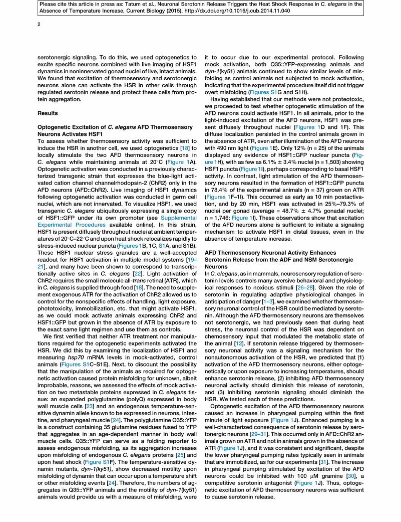
Neurons Activates HSF1To assess whether thermosensory activity was sufficient toinduce the HSR in another cell, we used optogenetics [18] tolocally stimulate the two AFD thermosensory neurons inC. elegans while maintaining animals at 20�C (Figure 1A).Optogenetic activation was conducted in a previously charac-terized transgenic strain that expresses the blue-light acti-vated cation channel channelrhodopsin-2 (ChR2) only in theAFD neurons (AFD::ChR2). Live imaging of HSF1 dynamicsfollowing optogenetic activation was conducted in germ cellnuclei, which are not innervated. To visualize HSF1, we usedtransgenic C. elegans ubiquitously expressing a single copyof HSF1::GFP under its own promoter (see SupplementalExperimental Procedures available online). In this strain,HSF1 is present diffusely throughout nuclei at ambient temper-atures of 20�C–22�Cand upon heat shock relocalizes rapidly tostress-induced nuclear puncta (Figures 1B, 1C, S1A, and S1B).These HSF1 nuclear stress granules are a well-acceptedreadout for HSF1 activation in multiple model systems [19–21], and many have been shown to correspond to transcrip-tionally active sites in C. elegans [22]. Light activation ofChR2 requires the small molecule all-trans retinal (ATR), whichinC. elegans is supplied through food [18]. The need to supple-ment exogenous ATR for the activation of ChR2 allowed us tocontrol for the nonspecific effects of handling, light exposure,phototoxicity, immobilization, etc. that might activate HSF1,as we could mock activate animals expressing ChR2 andHSF1::GFP but grown in the absence of ATR by exposure tothe exact same light regimen and use them as controls.
We first verified that neither ATR treatment nor manipula-tions required for the optogenetic experiments activated theHSR. We did this by examining the localization of HSF1 andmeasuring hsp70 mRNA levels in mock-activated, controlanimals (Figures S1C–S1E). Next, to discount the possibilitythat the manipulation of the animals as required for optoge-netic activation caused protein misfolding for unknown, albeitimprobable, reasons, we assessed the effects of mock activa-tion on two metastable proteins expressed in C. elegans tis-sue: an expanded polyglutamine (polyQ) expressed in bodywall muscle cells [23] and an endogenous temperature-sen-sitive dynamin allele known to be expressed in neurons, intes-tine, and pharyngeal muscle [24]. The polyglutamine Q35::YFPis a construct containing 35 glutamine residues fused to YFPthat aggregates in an age-dependent manner in body wallmuscle cells. Q35::YFP can serve as a folding reporter toassess endogenous misfolding, as its aggregation increasesupon misfolding of endogenous C. elegans proteins [25] andupon heat shock (Figure S1F). The temperature-sensitive dy-namin mutants, dyn-1(ky51), show decreased motility uponmisfolding of dynamin that can occur upon a temperature shiftor other misfolding events [24]. Therefore, the numbers of ag-gregates in Q35::YFP animals and the motility of dyn-1(ky51)animals would provide us with a measure of misfolding, were
it to occur due to our experimental protocol. Followingmock activation, both Q35::YFP-expressing animals anddyn-1(ky51) animals continued to show similar levels of mis-folding as control animals not subjected to mock activation,indicating that the experimental procedure itself did not triggerovert misfolding (Figures S1G and S1H).Having established that our methods were not proteotoxic,
we proceeded to test whether optogenetic stimulation of theAFD neurons could activate HSF1. In all animals, prior to thelight-induced excitation of the AFD neurons, HSF1 was pre-sent diffusely throughout nuclei (Figures 1D and 1F). Thisdiffuse localization persisted in the control animals grown inthe absence of ATR, even after illumination of the AFD neuronswith 490 nm light (Figure 1E). Only 12% (n = 25) of the animalsdisplayed any evidence of HSF1::GFP nuclear puncta (Fig-ure 1H), with as few as 6.1%6 3.4% nuclei (n = 1,503) showingHSF1 puncta (Figure 1I), perhaps corresponding to basal HSF1activity. In contrast, light stimulation of the AFD thermosen-sory neurons resulted in the formation of HSF1::GFP punctain 78.4% of the experimental animals (n = 37) grown on ATR(Figures 1F–1I). This occurred as early as 10 min postactiva-tion, and by 20 min, HSF1 was activated in 25%–79.3% ofnuclei per gonad (average = 48.7% 6 4.7% gonadal nuclei;n = 1,746; Figure 1I). These observations show that excitationof the AFD neurons alone is sufficient to initiate a signalingmechanism to activate HSF1 in distal tissues, even in theabsence of temperature increase.
AFD Thermosensory Neuronal Activity Enhances
Serotonin Release from the ADF and NSM SerotonergicNeurons
InC. elegans, as inmammals, neurosensory regulation of sero-tonin levels controls many aversive behavioral and physiolog-ical responses to noxious stimuli [26–28]. Given the role ofserotonin in regulating adaptive physiological changes inanticipation of danger [1–3], we examinedwhether thermosen-sory neuronal control of the HSR could bemediated by seroto-nin. Although the AFD thermosensory neurons are themselvesnot serotonergic, we had previously seen that during heatstress, the neuronal control of the HSR was dependent onchemosensory input that modulated the metabolic state ofthe animal [12]. If serotonin release triggered by thermosen-sory neuronal activity was a signaling mechanism for thenonautonomous activation of the HSR, we predicted that (1)activation of the AFD thermosensory neurons, either optoge-netically or upon exposure to increasing temperatures, shouldenhance serotonin release, (2) inhibiting AFD thermosensoryneuronal activity should diminish this release of serotonin,and (3) inhibiting serotonin signaling should diminish theHSR. We tested each of these predictions.Optogenetic excitation of the AFD thermosensory neurons
caused an increase in pharyngeal pumping within the firstminute of light exposure (Figure 1J). Enhanced pumping is awell-characterized consequence of serotonin release by sero-tonergic neurons [29–31]. This occurred only in AFD::ChR2 an-imals grown onATR and not in animals grown in the absence ofATR (Figure 1J), and it was consistent and significant, despitethe lower pharyngeal pumping rates typically seen in animalsthat are immobilized, as for our experiments [31]. The increasein pharyngeal pumping stimulated by excitation of the AFDneurons could be inhibited with 100 mM gramine [30], acompetitive serotonin antagonist (Figure 1J). Thus, optoge-netic excitation of AFD thermosensory neurons was sufficientto cause serotonin release.

A
B C
D E
F G
H
I
J
Figure 1. Optogenetic Stimulation of AFD Thermosensory Neurons in C. elegans Activates HSF1 and Elicits Serotonin Release in the Absence of Stress
(A) Experimental setup: localized illumination of AFD neurons in individual C. elegans expressing ChR2 in AFD neurons (the scale bar represents 50 mm).
Wavelength = 490 nm. Experimental animals were grown in the presence of all-trans retinal (+ATR) and control animals in the absence of all-trans retinal
(2ATR). HSF1::GFP was monitored in germ cell nuclei.
(B andC) HSF1::GFP localization at 20�C (B) and following heat shock at 34�C for 10min (C) (arrows indicate HSF1 nuclear stress granules/puncta). The scale
bar represents 5 mm.
(D and E) HSF1::GFP in control (2ATR) animals at 0 min (D) and 20 min (E) after the start of optogenetic stimulation. The scale bar represents 5 mm.
(F and G) HSF1::GFP in experimental (+ATR) animals at 0 min (F) and 20 min (G) after the start of optogenetic stimulation. The scale bar represents 5 mm.
(H) Percentage of control (2ATR) and experimental (+ATR) animals showing HSF1 nuclear puncta after stimulation of AFD neurons. n = 25–37 animals.
(I) Percentage of nuclei with HSF1 puncta in control (2ATR) and experimental (+ATR) animals following optogenetic stimulation. n = 25–37 animals; 1,503–
1,746 nuclei. Values indicate mean 6 SEM, t test; **p < 0.01.
(J) Pharyngeal pumping rates in control (2ATR) and experimental (+ATR) animals following optogenetic stimulation and pharyngeal pumping in (+ATR)
experimental animals treated with 100 mM gramine and optogenetically stimulated. n = 22–70 animals. Values indicate mean 6 SEM, t test; **p < 0.01.
See also Figure S1.
3
Please cite this article in press as: Tatum et al., Neuronal Serotonin Release Triggers the Heat Shock Response in C. elegans in theAbsence of Temperature Increase, Current Biology (2015), http://dx.doi.org/10.1016/j.cub.2014.11.040

4
Please cite this article in press as: Tatum et al., Neuronal Serotonin Release Triggers the Heat Shock Response in C. elegans in theAbsence of Temperature Increase, Current Biology (2015), http://dx.doi.org/10.1016/j.cub.2014.11.040
Serotonin was also released in an AFD-dependent mannerupon increases in temperature. Animals were subjected toa controlled increase in temperature of 1�C/min (Figure S2B),a rate known to activate the AFD thermosensory neurons[17, 32], and pharyngeal pumping was measured [29]. At20�C–22�C, wild-type animals exhibited a pumping rate of204 6 24 pumps per minute, similar to what has been previ-ously reported (Figures S2A and S2C) [29]. As early as 5 minupon temperature increase to 26.7�C, there was a dramatic in-crease in pumping rates to an average rate of 3046 50 (Figures2A, S2A, and S2C), representing an approximately 50% in-crease in the number of pumps per minute. This increasewas transient but occurred in all animals (Figures S2A andS2C). Animals deficient in serotonin synthesis because of mu-tations in the only C. elegans tryptophan hydroxylase gene,tph-1 [33], did not increase their pumping rates upon temper-ature increase, confirming that the temperature-dependentincreases in pumping occurred as a consequence of seroto-nergic signaling (Figures 2A, S2A, and S2D). Disrupting thefunction of the AFD thermosensory neurons by mutations inthe guanylyl cyclase genes gcy-8, gcy-18, and gcy-23 [32,34] greatly diminished the increase in pharyngeal pumpingupon temperature upshift (Figures 2A, S2A, S2F, and S2G),confirming the role of the AFD neurons in eliciting tempera-ture-dependent serotonin release. On the other hand, bacteriathat serve as food for C. elegans and are known to modulateserotonin release [28, 29] were not responsible for the temper-ature-enhanced pumping as animals grown on dead bacteriashowed a comparable increase (from 167 6 20, 20�C to 2656 39, 26.7�C; Figures 2A, S2A, and S2E). These data showthat serotonin was released in response to sensed increasesin temperature in an AFD-dependent manner.
We confirmed that serotonin was indeed released by visual-izing endogenous serotonin localization in animals at 20�C andupon increasing temperature. Specifically, we examined ani-mals exposed to the temperature ramp rate of 1�C/min for 5and 10 min (26.7�C and 28.9�C, where 26.7�C corresponds tothe peak of pharyngeal pumping) and upon heat shock at34�C for 15 min. In wild-type C. elegans raised at 20�C, anti-bodies against serotonin stained cell bodies of a pair of neuro-secretory motor neurons (NSMs), a pair of ADF chemosensoryneurons (Figure 2D), the HSN neurons that innervate vulvalmuscle cells, and, less consistently, a single RIH neuron anda pair of AIM interneurons [28, 29, 35]. When animals wereexposed to increases in temperature, serotonin stainingexpanded into areas outside serotonergic neuronal cell bodies(Figures 2E, 2F, and 2H). This was evidenced by immunolocal-izing serotonin in animals expressing GFP under the tph-1 pro-moter (Ptph-1::GFP), where GFP marked the boundaries ofserotonergic neuronal cell bodies. In these animals, followingheat shock, serotonin staining could be detected outsidethe confines of GFP (Figures 2B and 2C). We examinedwhether changes in tph-1 expression itself could account forthe change in localization of serotonin with increased temper-atures [27, 28], as tph-1 can be expressed in other tissues,such as pharyngeal muscle cells of the procorpus and meta-corpus during embryogenesis [36]. However, neither Ptph-1::GFP localization (Figures 3A and 3B) nor GFP mRNA levelschanged following heat shock (Figure 3C). The altered localiza-tion of serotonin following heat shock was transient, as manyanimals showed control immunolocalization patterns by 1 hrfollowing recovery at 20�C after exposure to 34�C for 15 min(Figures 2F, 2G, 2H, and 2M). We confirmed that the immuno-localization pattern was specific for serotonin by using tph-
1(mg280) II mutant animals that do not make serotonin (Fig-ures 2I and 2J). In addition, a loss-of-function mutation inunc-31 that disrupts dense core vesicle-dependent neurose-cretion required for serotonin secretion [35] diminished sero-tonin relocalization upon heat shock (88% versus 33%animalsshowed localization outside serotonergic cell bodies; Figures2K, 2L, and 2N).We then investigated which of the serotonergic neurons
were responsible for the temperature-dependent serotoninrelease by genetically ablating specific serotonergic neurons.The cell bodies of the NSM neurons are buried in cavities in-side the pharyngeal muscle cells, whereas the ADF neuronalcell bodies lie just outside the pharynx, and both were possiblecandidates to contribute to the observed serotonin stainingpattern [36, 37]. Genetic ablation of the NSM and ADF neuronsonly, achieved by directing the expression of a split caspase tothese cells (Figures 3D–3K; [38]), resulted in the loss of seroto-nin relocalization following heat shock (Figure 3F and 3G). Aswith temperature-enhanced pharyngeal pumping, the heatshock-induced immunolocalization of serotonin outside theneuronal cell bodies required AFD thermosensory neuronalfunction: only 27%of the gcy-8 thermosensorymutant animalscompared with 88% of wild-type animals showed serotoninrelease following exposure to increased temperature (Figures3L–3N). These studies together support the conclusion that, aswith optogenetic activation, temperature-induced AFD ther-mosensory activity also elicits the release of serotonin. Thisoccurs from serotonergic NSM and/or ADF neurons.
Serotonin Is Necessary for the HSRWe next tested whether serotonin was necessary for heatshock-dependent induction of hsp70mRNA. Animals deficientin serotonin signaling andwild-type animals with intact seroto-nin signaling systems were subjected to a brief heat shock,corresponding to the regimen that induced the relocalizationof serotonin as described above. hsp70 mRNA accumulationwas then quantified using quantitative RT-PCR (qRT-PCR).Both the tph-1mutants (Figure 4A) and animals lacking seroto-nergic ADF and NSM neurons (Figure 4B) showed significantdecreases in hsp70 mRNA accumulation upon heat shockcompared to wild-type animals. In C. elegans, four metabo-tropic G protein-coupled receptors (GPCRs), SER-1, SER-4,SER-5 and SER-7, and one serotonin-gated chloride channel,MOD-1, are known to bind serotonin [39, 40]. Among these re-ceptors, loss-of-functionmutations in SER-1 decreased hsp70upregulation upon heat shock in a manner similar to the loss oftph-1 (Figures 3C and S3), suggesting that the serotonin signalmay be transmitted through this ortholog of the mammalianmetabotropic 5-HT2 receptor to regulate the HSR. These ex-periments collectively indicate that serotonergic signaling isnecessary for hsp70 mRNA expression upon heat shock andcould therefore serve as a signaling mechanism for HSRactivation.
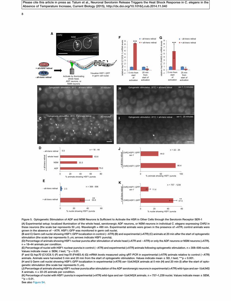
Optogenetic Excitation of Serotonergic ADF and NSMNeurons Activates the HSR
If serotonin release triggered by thermosensory activity wasindeed a signal to nonautonomously activate HSF1 in othercells, we would predict that stimulating serotonin releasedirectly from the NSM and ADF neurons in wild-type animalswithout exciting thermosensory neurons should be sufficientto activate HSF1 in distant tissues. To test this, we againused optogenetics [18] to stimulate only the NSM and ADFneurons while maintaining animals at 20�C (Figure 5A). This

A
B C
D E
F G
K L
JI
M N
H
Figure 2. Serotonin Is Released upon Temperature Increase
(A) The change in mean pumping rates when temperature increases from ambient (20�C–22�C) to 26.7�C in the following animals: wild-type animals under
normal growth conditions, tph-1(mg280) II, wild-type animals transferred to dead bacteria, gcy-8(oy44) IV, and gcy-23(nj37) gcy-8(oy44) gcy-18(nj38) IV. n = 9
for wild-type on dead bacteria; n = 20–25 for all others. Values indicate mean 6 SEM, t test; ***p < 0.001.
(B and C) Collapsed confocal z stack image showing serotonin staining inmgIs42 animals expressing GFP in serotonergic neurons under a tph-1 promoter.
Animals were fixed immediately following a 15 min exposure to 34�C.(B) Ptph-1::GFP marks serotonergic neuronal cell bodies (arrowhead), present within areas confined by dashed lines.
(C) Serotonin staining is detected both within the neuronal cell bodies and in areas outside the dashed lines (arrow).
(D–G) Serotonin immunolocalization in wild-type animals at 20�C (n = 119) (D), exposed for 5 min to temperature ramp rate of 1�C/min (corresponding to
26.7�C; n = 96) (E), exposed to 34�C for 15 min (n = 119) (F), and exposed to 34�C for 15 min and recovered at 20�C for 1 hr (n = 7) (G).
(H) Percentage of wild-type animals, scored blinded (see Experimental Procedures), showing serotonin localization within or outside neuronal cell bodies at
20�C, 26.7�C, 28.9�C, and 34�C.(I–L) Serotonin immunolocalization in tph-1(mg280) II at 20�C (n = 15) (I), tph-1(mg280) II at 34�C for 15 min (n = 9) (J), unc-31(e928) IV; C12C8.1p::mCherry at
20�C (n = 13) (K), and unc-31(e928) IV; C12C8.1p::mCherry at 34�C for 15 min (n = 15) (L). Arrowheads indicate cell bodies; arrows indicate serotonin local-
ization outside cell bodies.
(M) Percentage of wild-type animals exposed to 34�C for 15 min and recovered at 20�C for 1 hr scored blinded for serotonin localization.
(N) Percentage of unc-31; C12C8.1p::mCherry animals exposed to 34�C for 15 min scored blinded for serotonin localization compared to wild-type.
See also Figure S2.
5
Please cite this article in press as: Tatum et al., Neuronal Serotonin Release Triggers the Heat Shock Response in C. elegans in theAbsence of Temperature Increase, Current Biology (2015), http://dx.doi.org/10.1016/j.cub.2014.11.040

A B
D
H I J K
L M N
E F G
C
Figure 3. Serotonin Is Released from Serotonergic ADF and NSM Neurons upon Temperature Increase in an AFD-Dependent Manner
(A and B) Localization of Ptph-1::GFP at 20�C (A) and following exposure to 34�C for 15 min (B).
(C) GFP mRNA levels measured by qRT-PCR in Ptph-1::GFP animals before and immediately after 15 min heat exposure (34�C). Values indicate
mean 6 SEM.
(D and E) The expression of a split caspase under a minimal tph-1 promoter ablates the ADF and NSM neurons in olaEx75-expressing animals; Ptph-1::GFP
localization in serotonergic neurons in heads of wild-type animals (D) and olaEx75-expressing animals (E).
(F and G) Serotonin immunolocalization in olaEx75-expressing animals at 20�C (n = 11) (F) and immediately following a 15 min exposure to 34�C (n = 15) (G).
(H and I) Ptph-1::GFP expression in wild-type (H) and olaEx75-expressing (I) animals showing the presence of HSN neurons.
(J andK) Serotonin immunolocalization in HSN neurons is preserved in olaEx75-expressing animals at both 20�C (n = 11) (J) and following 15min exposure to
34�C (n = 15) (K).
(L and M) Serotonin immunolocalization in gcy-8(oy44) IV animals at 20�C (n = 31) (L) and following 15 min exposure to 34�C (n = 11) (M).
(N) Percentage of gcy-8(oy44) animals scored for serotonin localization (20�C and 34�C, 15 min). Arrowheads indicate cell bodies.
6
Please cite this article in press as: Tatum et al., Neuronal Serotonin Release Triggers the Heat Shock Response in C. elegans in theAbsence of Temperature Increase, Current Biology (2015), http://dx.doi.org/10.1016/j.cub.2014.11.040
was done by locally illuminating the NSM and ADF neurons in apreviously characterized transgenic strain lite-1(ce314) X;ljIs102, which expresses ChR2 only in these neurons [40]. Weconfirmed that the optogenetic activation of these seroto-nergic neurons caused the relocalization of serotonin intoareas outside serotonergic neuronal cell bodies, as observedupon heat shock (Figures S4A–S4C). We also confirmed that,as previously reported [40], optogenetic excitation of seroto-nergic neurons decreased locomotory rates (number of body
bends) and increased pharyngeal pumping rates [29, 30] (Fig-ures S4D and S4E). As described above, in all animals, prior tothe light-induced excitation of the NSM and ADF neurons,HSF1 was present diffusely throughout nuclei (Figure S1C).This diffuse localization persisted in the control mock-acti-vated animals grown in the absence of ATR (Figure 5B), andnone (n = 22) of these animals displayed the relocalization ofHSF1::GFP to nuclear stress granules/puncta (Figures 5Dand 5E). In contrast, as seen upon excitation of the AFD
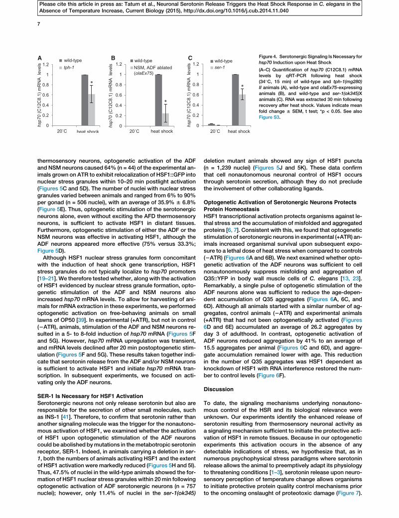
A B C Figure 4. Serotonergic Signaling Is Necessary for
hsp70 Induction upon Heat Shock
(A–C) Quantification of hsp70 (C12C8.1) mRNA
levels by qRT-PCR following heat shock
(34�C, 15 min) of wild-type and tph-1(mg280)
II animals (A), wild-type and olaEx75-expressing
animals (B), and wild-type and ser-1(ok345)X
animals (C). RNA was extracted 30 min following
recovery after heat shock. Values indicate mean
fold change 6 SEM, t test; *p < 0.05. See also
Figure S3.
7
Please cite this article in press as: Tatum et al., Neuronal Serotonin Release Triggers the Heat Shock Response in C. elegans in theAbsence of Temperature Increase, Current Biology (2015), http://dx.doi.org/10.1016/j.cub.2014.11.040
thermosensory neurons, optogenetic activation of the ADFand NSM neurons caused 64% (n = 44) of the experimental an-imals grown on ATR to exhibit relocalization of HSF1::GFP intonuclear stress granules within 10–20 min postlight activation(Figures 5C and 5D). The number of nuclei with nuclear stressgranules varied between animals and ranged from 6% to 90%per gonad (n = 506 nuclei), with an average of 35.9% 6 6.8%(Figure 5E). Thus, optogenetic stimulation of the serotonergicneurons alone, even without exciting the AFD thermosensoryneurons, is sufficient to activate HSF1 in distant tissues.Furthermore, optogenetic stimulation of either the ADF or theNSM neurons was effective in activating HSF1, although theADF neurons appeared more effective (75% versus 33.3%;Figure 5D).
Although HSF1 nuclear stress granules form concomitantwith the induction of heat shock gene transcription, HSF1stress granules do not typically localize to hsp70 promoters[19–21]. We therefore tested whether, along with the activationof HSF1 evidenced by nuclear stress granule formation, opto-genetic stimulation of the ADF and NSM neurons alsoincreased hsp70 mRNA levels. To allow for harvesting of ani-mals for mRNA extraction in these experiments, we performedoptogenetic activation on free-behaving animals on smalllawns of OP50 [39]. In experimental (+ATR), but not in control(2ATR), animals, stimulation of the ADF and NSM neurons re-sulted in a 5- to 8-fold induction of hsp70 mRNA (Figures 5Fand 5G). However, hsp70 mRNA upregulation was transient,and mRNA levels declined after 20 min postoptogenetic stim-ulation (Figures 5F and 5G). These results taken together indi-cate that serotonin release from the ADF and/or NSM neuronsis sufficient to activate HSF1 and initiate hsp70 mRNA tran-scription. In subsequent experiments, we focused on acti-vating only the ADF neurons.
SER-1 Is Necessary for HSF1 Activation
Serotonergic neurons not only release serotonin but also areresponsible for the secretion of other small molecules, suchas INS-1 [41]. Therefore, to confirm that serotonin rather thananother signaling molecule was the trigger for the nonautono-mous activation of HSF1, we examined whether the activationof HSF1 upon optogenetic stimulation of the ADF neuronscould be abolished bymutations in themetabotropic serotoninreceptor, SER-1. Indeed, in animals carrying a deletion in ser-1, both the numbers of animals activating HSF1 and the extentof HSF1 activation were markedly reduced (Figures 5H and 5I).Thus, 47.5% of nuclei in the wild-type animals showed the for-mation of HSF1 nuclear stress granules within 20min followingoptogenetic activation of ADF serotonergic neurons (n = 757nuclei); however, only 11.4% of nuclei in the ser-1(ok345)
deletion mutant animals showed any sign of HSF1 puncta(n = 1,239 nuclei) (Figures 5J and 5K). These data confirmthat cell nonautonomous neuronal control of HSF1 occursthrough serotonin secretion, although they do not precludethe involvement of other collaborating ligands.
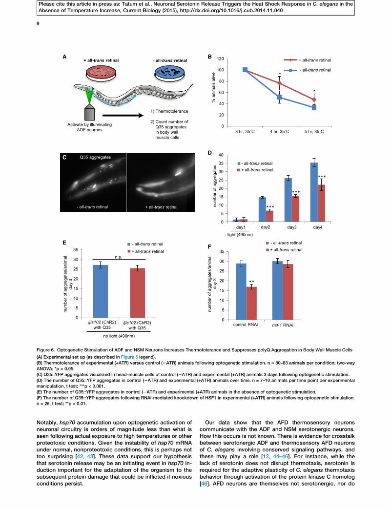
Optogenetic Activation of Serotonergic Neurons ProtectsProtein Homeostasis
HSF1 transcriptional activation protects organisms against le-thal stress and the accumulation of misfolded and aggregatedproteins [6, 7]. Consistent with this, we found that optogeneticstimulation of serotonergic neurons in experimental (+ATR) an-imals increased organismal survival upon subsequent expo-sure to a lethal dose of heat stress when compared to controls(2ATR) (Figures 6A and 6B). We next examined whether opto-genetic activation of the ADF neurons was sufficient to cellnonautonomously suppress misfolding and aggregation ofQ35::YFP in body wall muscle cells of C. elegans [13, 23].Remarkably, a single pulse of optogenetic stimulation of theADF neurons alone was sufficient to reduce the age-depen-dent accumulation of Q35 aggregates (Figures 6A, 6C, and6D). Although all animals started with a similar number of ag-gregates, control animals (2ATR) and experimental animals(+ATR) that had not been optogenetically activated (Figures6D and 6E) accumulated an average of 26.2 aggregates byday 3 of adulthood. In contrast, optogenetic activation ofADF neurons reduced aggregation by 41% to an average of15.5 aggregates per animal (Figures 6C and 6D), and aggre-gate accumulation remained lower with age. This reductionin the number of Q35 aggregates was HSF1 dependent asknockdown of HSF1 with RNA interference restored the num-ber to control levels (Figure 6F).
Discussion
To date, the signaling mechanisms underlying nonautono-mous control of the HSR and its biological relevance wereunknown. Our experiments identify the enhanced release ofserotonin resulting from thermosensory neuronal activity asa signaling mechanism sufficient to initiate the protective acti-vation of HSF1 in remote tissues. Because in our optogeneticexperiments this activation occurs in the absence of anydetectable indications of stress, we hypothesize that, as innumerous psychophysical stress paradigms where serotoninrelease allows the animal to preemptively adapt its physiologyto threatening conditions [1–3], serotonin release upon neuro-sensory perception of temperature change allows organismsto initiate protective protein quality control mechanisms priorto the oncoming onslaught of proteotoxic damage (Figure 7).
AF G
B
C
H
I
D
E
J
K
Figure 5. Optogenetic Stimulation of ADF and NSM Neurons Is Sufficient to Activate the HSR in Other Cells through the Serotonin Receptor SER-1
(A) Experimental setup: localized illumination of the whole head, serotonergic ADF neurons, or NSM neurons in individual C. elegans expressing ChR2 in
these neurons (the scale bar represents 50 mm). Wavelength = 490 nm. Experimental animals were grown in the presence of +ATR; control animals were
grown in the absence of 2ATR. HSF1::GFP was monitored in germ cell nuclei.
(B and C) Germ cell nuclei showing HSF1::GFP localization in control (2ATR) (B) and experimental (+ATR) (C) animals at 20 min after the start of optogenetic
stimulation (the scale bar represents 5 mm; arrows indicate HSF1 puncta).
(D) Percentage of animals showing HSF1 nuclear puncta after stimulation of whole head (+ATR and2ATR) or only the ADF neurons or NSM neurons (+ATR).
n = 18–44 animals per condition.
(E) Percentage of nuclei with HSF1 nuclear puncta in control (2ATR) and experimental (+ATR) animals following optogenetic stimulation. n = 308–506 nuclei.
Values indicate mean 6 SEM, t test; **p < 0.01.
(F and G) hsp70 (C12C8.1) (F) and hsp70 (F44E5.4) (G) mRNA levels measured using qRT-PCR in experimental (+ATR) animals relative to control (2ATR)
animals. Animals were harvested 5 min and 20 min from the start of optogenetic stimulation. Values indicate mean 6 SD, t test; ***p < 0.001.
(H and I) Germ cell nuclei showing HSF1::GFP localization in experimental (+ATR) ser-1(ok345)X animals at 0 min (H) and 20 min (I) after the start of opto-
genetic stimulation (the scale bar represents 5 mm).
(J) Percentage of animals showing HSF1 nuclear puncta after stimulation of the ADF serotonergic neurons in experimental (+ATR)wild-type and ser-1(ok345)
X animals. n = 22–25 animals per condition.
(K) Percentage of nuclei with HSF1 puncta in experimental (+ATR) wild-type and ser-1(ok345)X animals. n = 757–1,239 nuclei. Values indicate mean6 SEM,
**p < 0.01.
See also Figure S4.
8
Please cite this article in press as: Tatum et al., Neuronal Serotonin Release Triggers the Heat Shock Response in C. elegans in theAbsence of Temperature Increase, Current Biology (2015), http://dx.doi.org/10.1016/j.cub.2014.11.040
A B
D
EF
C
Figure 6. Optogenetic Stimulation of ADF and NSM Neurons Increases Thermotolerance and Suppresses polyQ Aggregation in Body Wall Muscle Cells
(A) Experimental set up (as described in Figure 5 legend).
(B) Thermotolerance of experimental (+ATR) versus control (2ATR) animals following optogenetic stimulation. n = 80–83 animals per condition; two-way
ANOVA; *p < 0.05.
(C) Q35::YFP aggregates visualized in head-muscle cells of control (2ATR) and experimental (+ATR) animals 3 days following optogenetic stimulation.
(D) The number of Q35::YFP aggregates in control (2ATR) and experimental (+ATR) animals over time. n = 7–10 animals per time point per experimental
manipulation, t test; ***p < 0.001.
(E) The number of Q35::YFP aggregates in control (2ATR) and experimental (+ATR) animals in the absence of optogenetic stimulation.
(F) The number of Q35::YFP aggregates following RNAi-mediated knockdown of HSF1 in experimental (+ATR) animals following optogenetic stimulation.
n = 26, t test; **p < 0.01.
9
Please cite this article in press as: Tatum et al., Neuronal Serotonin Release Triggers the Heat Shock Response in C. elegans in theAbsence of Temperature Increase, Current Biology (2015), http://dx.doi.org/10.1016/j.cub.2014.11.040
Notably, hsp70 accumulation upon optogenetic activation ofneuronal circuitry is orders of magnitude less than what isseen following actual exposure to high temperatures or otherproteotoxic conditions. Given the instability of hsp70 mRNAunder normal, nonproteotoxic conditions, this is perhaps nottoo surprising [42, 43]. These data support our hypothesisthat serotonin release may be an initiating event in hsp70 in-duction important for the adaptation of the organism to thesubsequent protein damage that could be inflicted if noxiousconditions persist.
Our data show that the AFD thermosensory neuronscommunicate with the ADF and NSM serotonergic neurons.How this occurs is not known. There is evidence for crosstalkbetween serotonergic ADF and thermosensory AFD neuronsof C. elegans involving conserved signaling pathways, andthese may play a role [12, 44–46]. For instance, while thelack of serotonin does not disrupt thermotaxis, serotonin isrequired for the adaptive plasticity of C. elegans thermotaxisbehavior through activation of the protein kinase C homolog[46]. AFD neurons are themselves not serotonergic, nor do

A
Figure 7. Model for HSF1 Activation upon Neurosensory Release of
Serotonin
The HSR of a cell can be activated in C. elegans in two distinct phases. An
initial preemptive neurosensory phase involves the excitation of AFD ther-
mosensory neurons that typically respond to innocuous temperature shifts.
This elicits serotonin release, directly or indirectly activates HSF1 to tran-
siently increase hsp70 mRNA levels, and occurs in the absence of protein
misfolding. Subsequently, prolonged exposure to heat can cause protein
misfolding, and the resulting damage induces HSF1-dependent hsp70 tran-
scription as has been well described. The role of serotonin in this latter
phase is not known.
10
Please cite this article in press as: Tatum et al., Neuronal Serotonin Release Triggers the Heat Shock Response in C. elegans in theAbsence of Temperature Increase, Current Biology (2015), http://dx.doi.org/10.1016/j.cub.2014.11.040
they synapse directly onto serotonergic neurons. Thus, AFDneurons could act through downstream interneurons, suchas the AIY, to communicate with serotonergic neurons viasynaptic mechanisms or electrical/chemical coupling acrossgap junctions. Alternatively, the excitation of AFD neuronsmay cause local paracrine release of neuroendocrine factors,which act on the serotonergic neurons to elicit serotoninrelease. These mechanisms are not mutually exclusive andremain to be investigated. We also do not know where sero-tonin is localized following its enhanced release or whetherits effects on germ cell nuclei are direct or indirect. TheNSM neurons have sensory endings in the pharyngeal lumenand are well poised to communicate with the rest of theworm’s body via secretion into the pseudocoelomic fluid[37, 39], and SER-1 is expressed in many tissues, includingpharyngeal muscle, neuronal processes in the nerve ring,neurons in the tail, ventral cord motor neurons in vulval mus-cle, uterine cells, and perhaps in the posterior intestine [47,48], where it could play a role in the secondary transmissionof the stress signal to germ cell nuclei. In addition, the AIMand RIH neurons express the serotonin reuptake channelMOD-5 and are known to uptake extrasynaptic serotonin[35]; however, since deletion of mod-5 does not cause adecrease in the HSR, this pathway may not be integral tothe signaling mechanism.
An intriguing possibility is that enhanced serotonin releasecould act as a node for the nonautonomous regulation of HSF1in response to multiple noxious environmental stimuli. Seroto-nergic signaling is involved in the innate immune responseto pathogens and response to hypoxia [27, 28]. It would be ofinterest to see whether activation of the sensory neuronsinvolved in pathogen recognition or oxygen tension alone canalso preemptively activate HSF1 through serotonin release.The decrease in polyQ aggregation following stimulation of
serotonergic neurons is also striking. The most dramaticdecrease occurs within the first 2 days following optogeneticstimulation, presumably due to the transient activation ofHSF1. However, aggregation continues to occur at apparentlylower rates. At the molecular level, we do not know whetherthis corresponds to decreasing the pool of oligomers,increasing degradation rates, or both. Although technicallychallenging in our current assays, it would be interesting todetermine whether a more-patterned regimen of optogeneticstimulation could completely prevent exacerbation of aggre-gation altogether. Serotonergic signaling pathways are highlyconserved betweenC. elegans andmammals, suggesting thatthe nonautonomous regulation of protein homeostasis seenhere may also be conserved. Indeed, serotonin is integral tothermoregulation in mammals [49, 50], and serotonergic syn-drome, a life-threatening human condition associated withexcessive serotonergic activity, causes fever, a stimulus forHSF1 activation [51, 52]. Indeed, modulating serotonin levelsby administration of SSRIs can provide enhanced protectionagainst neurodegeneration in vertebrate models of proteinconformational diseases [53]. Conversely, dysregulation of se-rotonin signaling alters aging rates in C. elegans and is associ-ated with Alzheimer’s disease and ALS [53, 54]. In summary, ifprotein quality control mechanisms in multicellular organismscan be activated cell nonautonomously and preemptivelythrough the regulation of sensory neuronal modalities and se-rotonin release as shown here, another treatment of neurode-generative diseases could involvemodulation of neurosensorysystems: a method that can be used alone or in combinationwith small molecules to effectively target protein misfolding.
Experimental Procedures
Detailed experimental procedures are described in Supplemental Experi-
mental Procedures.
Growth Conditions
All strains were grown and maintained at 20�C. Extreme care was taken
to maintain ambient temperature in the room very close to 20�C. Two trans-
genic strains were generated for these experiments: (1) a strain, AM1061,
that expressed a hsf-1 minigene fused to GFP integrated into the ttTi5605
II locus and (2) a strain, DCR186, that expressed the olaEx75 [Ptph-1::
caspase-3(p12)::nz; Ptph-1::cz::caspase-3(p17);Punc-122::mCherry] trans-
gene, which induced the caspase-mediated ablation of NSM and ADF neu-
rons. Strains expressing ChR2 in the AFD thermosensory neurons and ADF
and NSM serotonergic neurons were kind gifts from Paul Sternberg and
Michael R. Koelle, respectively. The remaining strains were obtained from
the Caenorhabditis Genetics Center (CGC).
Measuring Pumping Rates
The pumping rate of each wormwas recorded under a MZ10F stereomicro-
scope (Leica) at 83 magnification or under the Axio Observer A1 inverted
microscope (Zeiss) at 403 magnification, as needed.
Serotonin Whole-Worm Immunostaining Protocol
Anti-serotonin staining was performed following a modified protocol devel-
oped by the Loer laboratory (http://home.sandiego.edu/wcloer/loerlab/
anti5htshort.html). Serotonin localization patterns were scored blinded
11
Please cite this article in press as: Tatum et al., Neuronal Serotonin Release Triggers the Heat Shock Response in C. elegans in theAbsence of Temperature Increase, Current Biology (2015), http://dx.doi.org/10.1016/j.cub.2014.11.040
and quantified using Ptph-1::GFP expression to define serotonergic
neuronal cell bodies. Specifically, collapsed confocal z stack images were
taken for each worm to visualize both GFP (Ptph-1::GFP) and serotonin.
Colocalization of serotonin and GFP expression was seen within neuronal
cell bodies, but only anti-serotonin staining was observed outside the
neuronal cell bodies. Each worm was divided into the following three
areas: area 1 (from metacorpus to buccal cavity), area 2 (serotonergic
neuronal cell bodies), and area 3 (frompharyngeal terminal bulb toward pos-
terior body). Images were classified in a binary manner, based on the
following criteria: (1) location (presence of staining in area 1); (2) continuity
of staining in area 1; (3) intensity higher than that of background. For crite-
rions (1) and (3), the background fluorescence in area 1 was quantified as
mean pixel intensity. Animals with continuous staining in area 1, which
had an average intensity of 150 mean pixel intensity over background,
were scored as positive for staining outside serotonergic neuronal cell
bodies.
Heat Shock Protocol
The animals were subjected to temperature increase in two ways: (1) heat
shock on a Peltier stage (PeCon GmbH Temp Controller) and (2) heat shock
in a water bath.
RNA Extraction and qRT-PCR
RNA extraction was conducted according to previously published methods
[1]. The relative amounts of hsp mRNA were determined using the delta
delta CT (ddCT) method. Actin mRNA was used as an internal control. All
relative changes of hsp mRNA were normalized to that of wild-type heat-
shocked animals, except where otherwise noted. Technical duplicates or
triplicates were used to obtain CT values. Experiments were repeated at
least three times. Values shown indicate mean 6 SEM. All quantitative
PCR (qPCR) reactions were assessed by melt-curve analysis performed
at the end of the reaction and included no reverse transcriptase (no-RT)
controls.
Optogenetic Experiments
Optogenetic activation was conducted according to previously published
methods as per the requirements of the experiment. The main concerns
were to minimize stress from the methodology itself and to obtain enough
sample material. Thus, for HSF1 activation and Q35 protection assays, op-
togenetic stimulation was conducted on immobilized animals under the
Axio Observer A1 inverted microscope (Zeiss) using Andor iQ 2.9.1 soft-
ware (Andor) and X-Cite XLED1 1.1.0.2 software (Lumen Dynamics) to con-
trol the light source. For hsp70 mRNA measurements and thermotolerance
assays, free-behaving ijIs102; lite-1(ce314) animals were activated at 6.33
magnification using a MZ10 F microscope with an EL6000 light source
(Leica). Optogenetic serotonin release following light stimulation was
confirmed by measuring pharyngeal pumping rates or body bends per
minute.
Thermotolerance Experiments
All experiments following exposure to lethal heat stress were scored
blinded.
Statistical Analysis
Where applicable, significance was tested using Student’s t tests or, for
the thermotolerance assay, a two-way ANOVA. p values are indicated
as follows: *p < 0.05, **p < 0.01, ***p < 0.001. A Fisher’s exact test in
conjunction with a post hoc power analysis was used for determining
the appropriateness of our smaller sample sizes to support our
conclusions.
Supplemental Information
Supplemental Information includes Supplemental Experimental Procedures
and four figures and can be foundwith this article online at http://dx.doi.org/
10.1016/j.cub.2014.11.040.
Author Contributions
M.C.T, M.R.C., F.K.O., and V.P. designed and performed the experi-
ments and wrote the manuscript. L.C. and R.I.M. made and contributed
the AM1061 strain. L.A.M.-V. made the DCR186 strain. H.W.M.S. contrib-
uted the anti-serotonin antibodies. All authors commented on the
manuscript.
Acknowledgments
We would like to acknowledge the members of the V.P. laboratory, Jessica
Nelson, JoshWeiner, Sarit Smolikove, Tali Gidalevitz, Anat Ben-Zvi, Michael
Petrascheck, Sandra Encalada, and Daniel Colon-Ramos for helpful
comments. The DCR186 strain was a kind gift from Daniel Colon-Ramos.
Nematode strains were obtained from the CGC, funded by the NIH Office
of Research Infrastructure Programs (P40 OD010440). L.A.M.-V. was
supported by the Diversity Supplement to NS057931 (Colon-Ramos/
L.A.M.-V.). L.C. was funded by the Fulbright Franco-American Commission.
R.I.M. was funded by grants from the NIH (NIGMS, NIA, NINDS), the Ellison
Medical Foundation, and the Daniel F. and Ada L. Rice Foundation. V.P. was
funded by the Ellison Medical Foundation (AG-NS-1056-13).
Received: July 23, 2014
Revised: October 13, 2014
Accepted: November 17, 2014
Published: December 31, 2014
References
1. Chaouloff, F., Berton, O., andMormede, P. (1999). Serotonin and stress.
Neuropsychopharmacology Suppl. 21, 28S–32S.
2. Joels, M., and Baram, T.Z. (2009). The neuro-symphony of stress. Nat.
Rev. Neurosci. 10, 459–466.
3. Rozeske, R.R., Evans, A.K., Frank, M.G., Watkins, L.R., Lowry, C.A., and
Maier, S.F. (2011). Uncontrollable, but not controllable, stress desensi-
tizes 5-HT1A receptors in the dorsal raphe nucleus. J. Neurosci. 31,
14107–14115.
4. Bukau, B. (1993). Regulation of the Escherichia coli heat-shock
response. Mol. Microbiol. 9, 671–680.
5. Somero, G.N. (1995). Proteins and temperature. Annu. Rev. Physiol. 57,
43–68.
6. Akerfelt, M., Morimoto, R.I., and Sistonen, L. (2010). Heat shock factors:
integrators of cell stress, development and lifespan. Nat. Rev. Mol. Cell
Biol. 11, 545–555.
7. Lindquist, S. (1986). The heat-shock response. Annu. Rev. Biochem. 55,
1151–1191.
8. Vabulas, R.M., Raychaudhuri, S., Hayer-Hartl, M., and Hartl, F.U. (2010).
Protein folding in the cytoplasm and the heat shock response. Cold
Spring Harb. Perspect. Biol. 2, a004390.
9. van Oosten-Hawle, P., and Morimoto, R.I. (2014). Transcellular chap-
erone signaling: an organismal strategy for integrated cell stress re-
sponses. J. Exp. Biol. 217, 129–136.
10. Morimoto, R.I. (1998). Regulation of the heat shock transcriptional
response: cross talk between a family of heat shock factors, molecular
chaperones, and negative regulators. Genes Dev. 12, 3788–3796.
11. Bonini, N.M. (2002). Chaperoning brain degeneration. Proc. Natl. Acad.
Sci. USA 99 (Suppl 4 ), 16407–16411.
12. Prahlad, V., Cornelius, T., and Morimoto, R.I. (2008). Regulation of the
cellular heat shock response in Caenorhabditis elegans by thermosen-
sory neurons. Science 320, 811–814.
13. Prahlad, V., and Morimoto, R.I. (2011). Neuronal circuitry regulates the
response of Caenorhabditis elegans to misfolded proteins. Proc. Natl.
Acad. Sci. USA 108, 14204–14209.
14. Kumsta, C., Ching, T.T., Nishimura, M., Davis, A.E., Gelino, S., Catan,
H.H., Yu, X., Chu, C.C., Ong, B., Panowski, S.H., et al. (2014). Integrin-
linked kinase modulates longevity and thermotolerance in C. elegans
through neuronal control of HSF-1. Aging Cell 13, 419–430.
15. Maman, M., Carvalhal Marques, F., Volovik, Y., Dubnikov, T., Bejerano-
Sagie, M., and Cohen, E. (2013). A neuronal GPCR is critical for the in-
duction of the heat shock response in the nematode C. elegans.
J. Neurosci. 33, 6102–6111.
16. Taylor, R.C., and Dillin, A. (2013). XBP-1 is a cell-nonautonomous regu-
lator of stress resistance and longevity. Cell 153, 1435–1447.
17. Clark, D.A., Biron, D., Sengupta, P., and Samuel, A.D.T. (2006). The AFD
sensory neurons encode multiple functions underlying thermotactic
behavior in Caenorhabditis elegans. J. Neurosci. 26, 7444–7451.
18. Husson, S.J., Gottschalk, A., and Leifer, A.M. (2013). Optogenetic
manipulation of neural activity in C. elegans: from synapse to circuits
and behaviour. Biol. Cell 105, 235–250.
19. Sandqvist, A., and Sistonen, L. (2004). Nuclear stress granules: the
awakening of a sleeping beauty? J. Cell Biol. 164, 15–17.

12
Please cite this article in press as: Tatum et al., Neuronal Serotonin Release Triggers the Heat Shock Response in C. elegans in theAbsence of Temperature Increase, Current Biology (2015), http://dx.doi.org/10.1016/j.cub.2014.11.040
20. Jolly, C., Usson, Y., and Morimoto, R.I. (1999). Rapid and reversible re-
localization of heat shock factor 1within seconds to nuclear stress gran-
ules. Proc. Natl. Acad. Sci. USA 96, 6769–6774.
21. Sarge, K.D., Murphy, S.P., and Morimoto, R.I. (1993). Activation of heat
shock gene transcription by heat shock factor 1 involves oligomeriza-
tion, acquisition of DNA-binding activity, and nuclear localization and
can occur in the absence of stress. Mol. Cell. Biol. 13, 1392–1407.
22. Morton, E.A., and Lamitina, T. (2013). Caenorhabditis elegans HSF-1 is
an essential nuclear protein that forms stress granule-like structures
following heat shock. Aging Cell 12, 112–120.
23. Morley, J.F., Brignull, H.R., Weyers, J.J., and Morimoto, R.I. (2002). The
threshold for polyglutamine-expansion protein aggregation and cellular
toxicity is dynamic and influenced by aging in Caenorhabditis elegans.
Proc. Natl. Acad. Sci. USA 99, 10417–10422.
24. Clark, S.G., Shurland, D.L., Meyerowitz, E.M., Bargmann, C.I., and van
der Bliek, A.M. (1997). A dynamin GTPase mutation causes a rapid
and reversible temperature-inducible locomotion defect in C. elegans.
Proc. Natl. Acad. Sci. USA 94, 10438–10443.
25. Gidalevitz, T., Ben-Zvi, A., Ho, K.H., Brignull, H.R., and Morimoto, R.I.
(2006). Progressive disruption of cellular protein folding in models of
polyglutamine diseases. Science 311, 1471–1474.
26. Chang, A.J., Chronis, N., Karow, D.S., Marletta, M.A., and Bargmann,
C.I. (2006). A distributed chemosensory circuit for oxygen preference
in C. elegans. PLoS Biol. 4, e274.
27. Pocock, R., and Hobert, O. (2010). Hypoxia activates a latent circuit
for processing gustatory information in C. elegans. Nat. Neurosci. 13,
610–614.
28. Zhang, Y., Lu, H., andBargmann, C.I. (2005). Pathogenic bacteria induce
aversive olfactory learning in Caenorhabditis elegans. Nature 438,
179–184.
29. Song, B.M., Faumont, S., Lockery, S., and Avery, L. (2013). Recognition
of familiar food activates feeding via an endocrine serotonin signal in
Caenorhabditis elegans. Elife 2, e00329.
30. Avery, L., and Horvitz, H.R. (1990). Effects of starvation and neuroactive
drugs on feeding in Caenorhabditis elegans. J. Exp. Zool. 253, 263–270.
31. Keane, J., and Avery, L. (2003). Mechanosensory inputs influence
Caenorhabditis elegans pharyngeal activity via ivermectin sensitivity
genes. Genetics 164, 153–162.
32. Wasserman, S.M., Beverly, M., Bell, H.W., and Sengupta, P. (2011).
Regulation of response properties and operating range of the AFD ther-
mosensory neurons by cGMP signaling. Curr. Biol. 21, 353–362.
33. Sze, J.Y., Victor, M., Loer, C., Shi, Y., and Ruvkun, G. (2000). Food and
metabolic signalling defects in a Caenorhabditis elegans serotonin-syn-
thesis mutant. Nature 403, 560–564.
34. Inada, H., Ito, H., Satterlee, J., Sengupta, P., Matsumoto, K., and Mori, I.
(2006). Identification of guanylyl cyclases that function in thermosen-
sory neurons of Caenorhabditis elegans. Genetics 172, 2239–2252.
35. Jafari, G., Xie, Y., Kullyev, A., Liang, B., and Sze, J.Y. (2011). Regulation
of extrasynaptic 5-HT by serotonin reuptake transporter function in
5-HT-absorbing neurons underscores adaptation behavior in
Caenorhabditis elegans. J. Neurosci. 31, 8948–8957.
36. Axang, C., Rauthan, M., Hall, D.H., and Pilon, M. (2008). Developmental
genetics of the C. elegans pharyngeal neurons NSML and NSMR. BMC
Dev. Biol. 8, 38.
37. Albertson, D.G., and Thomson, J.N. (1976). The pharynx of
Caenorhabditis elegans. Philos. Trans. R. Soc. Lond. B Biol. Sci. 275,
299–325.
38. Sze, J.Y., Zhang, S., Li, J., and Ruvkun, G. (2002). The C. elegans POU-
domain transcription factor UNC-86 regulates the tph-1 tryptophan hy-
droxylase gene and neurite outgrowth in specific serotonergic neurons.
Development 129, 3901–3911.
39. Chase, D.L., and Koelle, M.R. (2007). Biogenic amine neurotransmitters
in C. elegans. WormBook, 1–15.
40. Gurel, G., Gustafson, M.A., Pepper, J.S., Horvitz, H.R., and Koelle, M.R.
(2012). Receptors and other signaling proteins required for serotonin
control of locomotion in Caenorhabditis elegans. Genetics 192, 1359–
1371.
41. Harris, G., Korchnak, A., Summers, P., Hapiak, V., Law,W.J., Stein, A.M.,
Komuniecki, P., and Komuniecki, R. (2011). Dissecting the serotonergic
food signal stimulating sensory-mediated aversive behavior in C. ele-
gans. PLoS ONE 6, e21897.
42. Petersen, R., and Lindquist, S. (1988). The Drosophila hsp70message is
rapidly degraded at normal temperatures and stabilized by heat shock.
Gene 72, 161–168.
43. Petersen, R.B., and Lindquist, S. (1989). Regulation of HSP70 synthesis
by messenger RNA degradation. Cell Regul. 1, 135–149.
44. Adachi, R., Osada, H., and Shingai, R. (2008). Phase-dependent prefer-
ence of thermosensation and chemosensation during simultaneous
presentation assay in Caenorhabditis elegans. BMC Neurosci. 9, 106.
45. Ailion, M., and Thomas, J.H. (2000). Dauer formation induced by high
temperatures in Caenorhabditis elegans. Genetics 156, 1047–1067.
46. Li, Y., Zhao, Y., Huang, X., Lin, X., Guo, Y., Wang, D., Li, C., andWang, D.
(2013). Serotonin control of thermotaxis memory behavior in nematode
Caenorhabditis elegans. PLoS ONE 8, e77779.
47. Xiao, H., Hapiak, V.M., Smith, K.A., Lin, L., Hobson, R.J., Plenefisch, J.,
and Komuniecki, R. (2006). SER-1, a Caenorhabditis elegans 5-HT2-like
receptor, and amulti-PDZ domain containing protein (MPZ-1) interact in
vulval muscle to facilitate serotonin-stimulated egg-laying. Dev. Biol.
298, 379–391.
48. Dempsey, C.M., Mackenzie, S.M., Gargus, A., Blanco, G., and Sze, J.Y.
(2005). Serotonin (5HT), fluoxetine, imipramine and dopamine target
distinct 5HT receptor signaling to modulate Caenorhabditis elegans
egg-laying behavior. Genetics 169, 1425–1436.
49. Azmitia, E.C. (2001). Modern views on an ancient chemical: serotonin ef-
fects on cell proliferation, maturation, and apoptosis. Brain Res. Bull.
56, 413–424.
50. Feldberg, W., and Myers, R.D. (1963). A new concept of temperature
regulation by amines in the hypothalamus. Nature 200, 1325.
51. Brown, I.R., and Rush, S.J. (1996). In vivo activation of neural heat shock
transcription factor HSF1 by a physiologically relevant increase in body
temperature. J. Neurosci. Res. 44, 52–57.
52. Iqbal, M.M., Basil, M.J., Kaplan, J., and Iqbal, M.T. (2012). Overview of
serotonin syndrome. Ann. Clin. Psychiatry 24, 310–318.
53. Mattson, M.P., Maudsley, S., and Martin, B. (2004). A neural signaling
triumvirate that influences ageing and age-related disease: insulin/
IGF-1, BDNF and serotonin. Ageing Res. Rev. 3, 445–464.
54. Ye, X., Linton, J.M., Schork, N.J., Buck, L.B., and Petrascheck, M.
(2014). A pharmacological network for lifespan extension in
Caenorhabditis elegans. Aging Cell 13, 206–215.


![Selective serotonin reuptake inhibitors [SSRIs] for stroke recoveryclok.uclan.ac.uk/6814/19/17551 - Selective serotonin reuptake... · Hackett, Maree (2012) Selective serotonin reuptake](https://static.fdocuments.net/doc/165x107/5f9c1bce9667ca02083a93ee/selective-serotonin-reuptake-inhibitors-ssris-for-stroke-selective-serotonin.jpg)