![BENIN FY2020 Annual Work Plan Act W… · SOP Standard Operating Procedure STH Soil-Transmitted Helminths STTA Short-Term Technical Assistance TAP Trachoma Action Plan ... [DNSP])](https://static.fdocuments.net/doc/165x107/6076656e961a952145630380/benin-fy2020-annual-work-plan-act-w-sop-standard-operating-procedure-sth-soil-transmitted.jpg)
NEGLECTED TROPICAL DISEASES AND THEIR CONTROL IN · dracunculiasis (Guinea worm), lymphatic...
74
NEGLECTED TROPICAL DISEASES AND THEIR CONTROL IN SOUTHERN SUDAN SITUATION ANALYSIS, INTERVENTION OPTIONS APPRAISAL AND GAP ANALYSIS February 2008 Ministry of Health, Government of Southern Sudan
Transcript of NEGLECTED TROPICAL DISEASES AND THEIR CONTROL IN · dracunculiasis (Guinea worm), lymphatic...
NEGLECTED TROPICAL DISEASES
AND THEIR CONTROL IN
SOUTHERN SUDAN
SITUATION ANALYSIS, INTERVENTION OPTIONS
APPRAISAL AND GAP ANALYSIS
February 2008
Ministry of Health, Government of Southern Sudan

NEGLECTED TROPICAL DISEASES IN SOUTHERN SUDAN, FEBRUARY 2008
FOREWORD
With the return of peace and stability, we, the people of Southern Sudan face great challenges in ensuring adequate health care for all and in controlling the enormous burden of disease. At the same time, reconstruction provides tremendous opportunities for us to improve on efficiency and equity of service delivery and to implement novel approaches to disease control. The real test in the post-conflict period is how to devote our efforts to address these needs and to join the international community in its war against poverty and deprivation.
We know that Neglected Tropical Diseases (NTDs), a diverse group ranging from onchocerciasis to Buruli ulcer, contribute greatly to the ill health of our people. An accurate description of the associated burden, current and potential interventions, and the resources needed to implement effective NTD control, is a prerequisite for managing expectations and development in post-conflict Southern Sudan. Until now, we only knew we had a mountain to climb. This document, produced by the Ministry of Health and its partners, gives us for the first time a map of this mountain and possible paths to climb it. Some of the data provided in this publication are appalling, other merely confirm what we already suspected. The need for more data is apparent, to generate a better understanding of where to focus our efforts and to monitor the impact of interventions over time. Ongoing improvements in the health management information system and targeted surveys will generate it.
In total, this document identifies 12 NTDs as endemic to Southern Sudan; probably the largest number in any given area in Africa or elsewhere in the world. For some diseases, such as onchocerciasis, guinea worm and trachoma, control programmes have been established with assistance from international partners, such as the African Programme for Onchocerciasis Control. For others, such as lymphatic filariasis, we are still lacking the necessary baseline data to formulate our intervention strategy. All NTDs will need more support, either to start control or to scale-up existing interventions. With the ongoing reconstruction of the health system, issues such as integrated strategies for NTD control will need to be explored. For visceral leishmaniasis and human African trypanosomiasis, integration into multi-functional health care delivery is in progress, while integrated mass drug administration to control lymphatic filariasis, onchocerciasis, schistosomiasis, soil-transmitted helminths and trachoma will be started in selected states as of 2008. These new undertakings will generate valuable evidence on cost and effectiveness, which we will harness to inform future NTD policies and strategies.
Now that we have all the available information on NTD in Southern Sudan compiled in this document, we will use it to strengthen links among all existing partners and to identify new ones, willing to accompany us along the path of effective and sustainable control of these debilitating and deadly diseases. Armed with credible and well-presented information and analysis, we will close the gap created by decades of war and civil unrest and make Southern Sudan a leader in NTD control. With new resource being made available by the Government of Southern Sudan and health sector donors, the Ministry of Health has an unprecedented opportunity to control, and in some cases, eliminate the burden caused by NTDs. Through partnership and innovation we will maximize this opportunity in our efforts to meet the Millennium Development Goals.
i
NEGLECTED TROPICAL DISEASES IN SOUTHERN SUDAN, FEBRUARY 2008
RECOMMENDED CITATION
MoH GoSS. 2008. Neglected Tropical Disease in Southern Sudan: Situation Analysis, Gap Analysis and Intervention Options Appraisal. Ministry of Health, Government of Southern Sudan.
CONTRIBUTIONS
Affiliation Name Contributions Contact Details
John Rumunu Contextual and health systems inputs
[email protected] [email protected]
Samson Baba Onchocerciasis, Nodding Disease
Lasu Hickson Leprosy, Buruli ulcer [email protected] Samuel Makoy Dracunculiasis [email protected] Lucia Kur Trachoma [email protected]
MoH GoSS
Apollo S. O. Meru HAT [email protected] Malaria Consortium Jan Kolaczinski Editor, intervention
options, gap analysis [email protected]
Malaria Consortium (Consultant) & WHO
Michaleen Richer Preparation of first draft
[email protected]@ofda.gov
LSHTM Simon Brooker Editor, intervention options, gap analysis
Jose Antonio Ruiz HAT, STH, LF schistosomiasis, VL
Ireneaus Sindani Leprosy, Buruli ulcer [email protected] Steven Becknell Dracunculiasis,
Trachoma [email protected]
Paul Emerson Trachoma [email protected] Gideon Gatpan Trachoma [email protected]
The Carter Center (TCC)
Jeremiah Ngondi Trachoma [email protected] Karinya Lewis Trachoma,
Onchocerciasis [email protected]
Sture Nyholm Trachoma [email protected] Adrian Hopkins Onchocerciasis [email protected]
Christoffel Blindenmission (CBM)
Fasil Chane Onchocerciasis [email protected]
ACKNOWLEDGEMENTS
This document is the result of a meeting between MoH GOSS, the World Bank, Malaria Consortium, WHO and Management Sciences for Health in Juba during May 2007 where the need for a detailed analysis of NTDs in Southern Sudan was identified. The work was subsequently led by Malaria Consortium Africa [http://www.malariaconsortium.org] and funded by COMDIS, a Research Programme Consortium coordinated by the Nuffield Centre for International Health and Development, University of Leeds, with funds from the Department for International Development, UK.
We wish to thank all the individuals and organizations that have contributed to the control of NTDs in Southern Sudan over the last decades. Without their dedication, many lives would have been lost and many people would not have been cured from disabilities such as blinding trachoma. Long may the fruitful collaboration between the multitude of implementing partners last.
Dr John Paquale Rumunu Director General for Preventive Medicine Ministry of Health, Government of Southern Sudan
ii
NEGLECTED TROPICAL DISEASES IN SOUTHERN SUDAN, FEBRUARY 2008
EXECUTIVE SUMMARY
Background: Neglected tropical diseases (NTDs) are a group of 13 infections caused by parasitic worms, protozoa or bacteria. They strike the world’s poorest people, living in remote and rural areas of low-income countries in Sub-Saharan Africa, Asia and the Americas, causing life-long disability, disfigurement, reduced economic productivity and social stigma. When expressed in disability-adjusted life years (DALYs), NTDs account for approximately one-quarter of the global disease burden from HIV/AIDS and for almost the same burden as from malaria.
Over the least years, international advocacy has drawn attention to the global NTD burden and to the fact that substantial improvements can be readily achieved at relatively low cost. Targeting this group of diseases is therefore widely promoted as a means to reaching some of the Millennium Development Goals (MDGs). As many of the NTD occur in the same geographical areas and, in some cases, can be treated with the same drug, there is potential for integration of control activities, both within this group of diseases as well as with other interventions.
After more than 20 years of war and little disease control, Southern Sudan is thought to be among the countries with the highest NTD burden in the world. However, most of the attention and funding has been dedicated to the control of HIV/AIDS, tuberculosis and malaria and to the control of outbreak prone diseases such as cholera and meningococcal meningitis. To be able to reach the MDGs, control of NTDs will need to be given a much higher priority by the Ministry of Health (MoH) of the Government of Southern Sudan (GoSS) and by health sector donors. In recognition of this, the MoH-GoSS has included the control of NTDs among the health sector priorities and has started discussion with key partners on how to proceed further. The present document was developed to facilitate this process. It aims at providing the essential background information on NTDs and their control in Southern Sudan, suggests ways in which interventions could be initiated or improved on and identifies the existing generic gaps. Estimation of specific financial gaps will required further information generate through prevalence surveys and intervention experience. Methods: The information presented in this document is based on documents and presentations provided by the MoH-GoSS, WHO Southern Sudan and existing key partners in the control of NTDs, such as The Carter Centre (TCC) and the Christoffel Blindenmission (CBM). These documents were supplemented by information collected through a systematic literature search of the electronic online database PubMed (US National Library of Medicine, Bethesda, USA) using combinations of the keywords: Sudan, control, epidemiology, onchocerciasis, schistosomiasis, helminths, lymphatic filariasis, trachoma, leprosy, buruli ulcer, guinea worm, dracunculiasis. Further searches were conducted by accessing the WHO website (http://www.who.int/) and by using the web-based search engine GOOGLE (http://www.google.com). Additional non-peer reviewed and unpublished literature was examined for information related to the subject. Desk-based work was accompanied by e-mail exchanges between key contributors to discuss and clarify specific issues and to agree on which recommendations are the most relevant. Findings: The NTDs of major public health and socio-economic importance in Southern Sudan are visceral leishmaniasis (VL), human African trypanosomiasis (HAT), onchocerciasis, dracunculiasis (Guinea worm), lymphatic filariasis (LF), schistosomiasis, trachoma, and soil-transmitted helminths (STH). Data on buruli ulcer is insufficient to assess its distribution and importance. Control of these diseases is the responsibility of the Directorate of Preventive Health, MoH-GoSS. Control programmes exist for onchocerciasis, trachoma and dracunculiasis; focal persons for some of the other disease have been identified by the MoH-GoSS. Detailed data on the geographic distribution and associated burden is lacking for most NTDs in Southern Sudan. For VL, HAT and onchocerciasis the geographic distribution has been largely established, but detailed data on the associated burden is lacking. For LF, trachoma, schistosomiasis and STH further prevalence surveys need to be undertaken to fully determine the geographic scope of
iii
NEGLECTED TROPICAL DISEASES IN SOUTHERN SUDAN, FEBRUARY 2008
disease. Ongoing surveillance is required for all NTDs. Present efforts by the MoH-GoSS to strengthen disease surveillance at central and state level will improve our understanding of NTD epidemiology and distribution. Current shortcomings in ongoing NTD control are largely attributable to lack of human resources and funding, in particular at state level. Conclusion: Strong political commitment and a dynamic environment of health sector reconstruction in Southern Sudan present unique opportunities for scaling-up of NTD control. A number of successful control programmes are ongoing, providing useful lessons on the operational challenges in this context and potential to expand the range of interventions delivered through existing community-based networks. However, such integration can only be achieved if sufficient additional support is provided to ensure that existing interventions and systems are not overloaded. To avoid such overload, new approaches should be piloted in selected sites to generate the evidence required to scale them up. USAID funding for implementation of integrated mass drug administration (MDA) in two states of Southern Sudan, recently awarded to Malaria Consortium by RTI International, will make an important contribution to strengthening of the evidence-base for this approach.
While continued and expanded MDA will be an important component of NTD control, complementary interventions will need to receive sufficient attention and funding, to ensure delivery of comprehensive NTD control. An example of this is trachoma control, where azithromycin distribution only constitutes one component of the SAFE strategy. For diseases such as HAT and VL where treatment is too toxic, complicated or lengthy to be delivered at community level, resources are urgently required to strengthen their control as a component of multi-functional health care delivery and to target complementary interventions, such as long-lasting insecticide-treated nets, to communities at risk.
iv
NEGLECTED TROPICAL DISEASES IN SOUTHERN SUDAN, FEBRUARY 2008
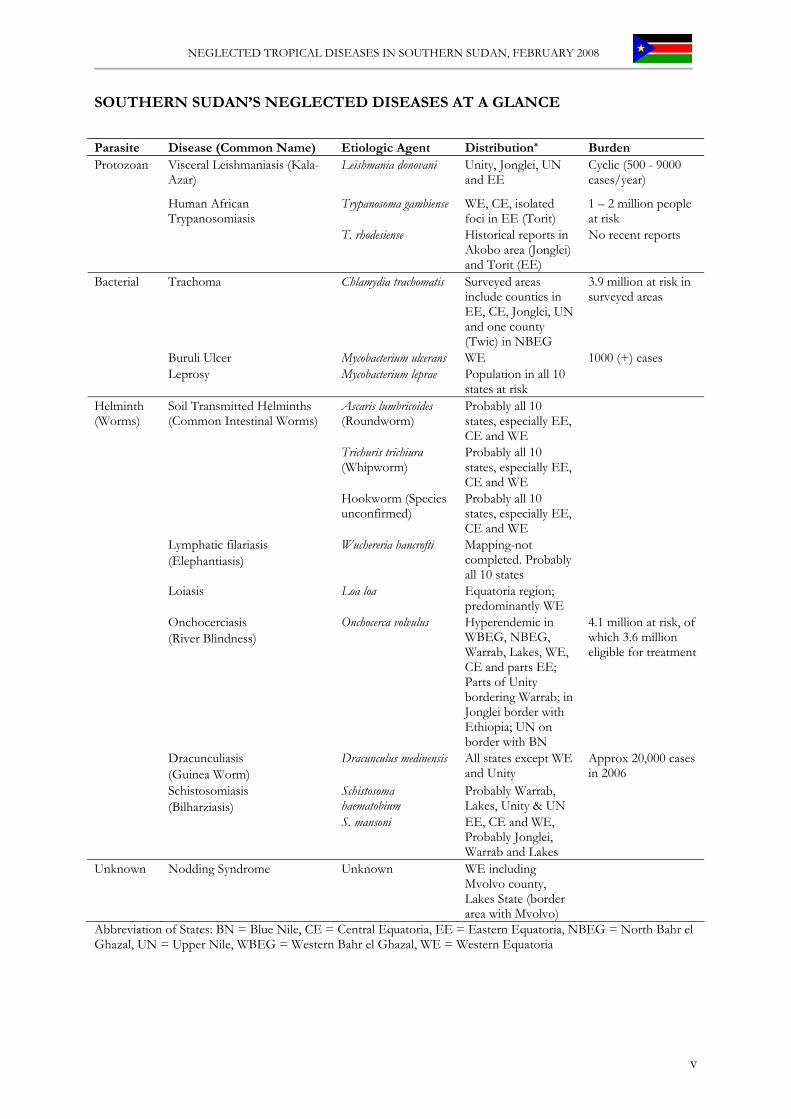
SOUTHERN SUDAN’S NEGLECTED DISEASES AT A GLANCE
Parasite Disease (Common Name) Etiologic Agent Distribution* Burden
Visceral Leishmaniasis (Kala-Azar)
Leishmania donovani Unity, Jonglei, UN and EE
Cyclic (500 - 9000 cases/year)
Trypanosoma gambiense WE, CE, isolated foci in EE (Torit)
1 – 2 million people at risk
Protozoan
Human African Trypanosomiasis
T. rhodesiense Historical reports in Akobo area (Jonglei) and Torit (EE)
No recent reports
Trachoma Chlamydia trachomatis Surveyed areas include counties in EE, CE, Jonglei, UN and one county (Twic) in NBEG
3.9 million at risk in surveyed areas
Buruli Ulcer Mycobacterium ulcerans WE 1000 (+) cases
Bacterial
Leprosy Mycobacterium leprae Population in all 10 states at risk
Ascaris lumbricoides (Roundworm)
Probably all 10 states, especially EE, CE and WE
Trichuris trichiura (Whipworm)
Probably all 10 states, especially EE, CE and WE
Soil Transmitted Helminths (Common Intestinal Worms)
Hookworm (Species unconfirmed)
Probably all 10 states, especially EE, CE and WE
Lymphatic filariasis (Elephantiasis)
Wuchereria bancrofti Mapping-not completed. Probably all 10 states
Loiasis Loa loa Equatoria region; predominantly WE
Onchocerciasis (River Blindness)
Onchocerca volvulus Hyperendemic in WBEG, NBEG, Warrab, Lakes, WE, CE and parts EE; Parts of Unity bordering Warrab; in Jonglei border with Ethiopia; UN on border with BN
4.1 million at risk, of which 3.6 million eligible for treatment
Dracunculiasis (Guinea Worm)
Dracunculus medinensis All states except WE and Unity
Approx 20,000 cases in 2006
Schistosoma haematobium
Probably Warrab, Lakes, Unity & UN
Helminth (Worms)
Schistosomiasis (Bilharziasis)
S. mansoni EE, CE and WE, Probably Jonglei, Warrab and Lakes
Unknown Nodding Syndrome Unknown WE including Mvolvo county, Lakes State (border area with Mvolvo)
Abbreviation of States: BN = Blue Nile, CE = Central Equatoria, EE = Eastern Equatoria, NBEG = North Bahr el Ghazal, UN = Upper Nile, WBEG = Western Bahr el Ghazal, WE = Western Equatoria
v
NEGLECTED TROPICAL DISEASES IN SOUTHERN SUDAN, FEBRUARY 2008
ACRONYMS
APOC - African Programme for Onchocerciasis Control CAR - Central African Republic CBM - Christoffel Blindenmission CDD - Community Drug Distributor CDTI - Community-Directed Treatment with Ivermectin CO - Corneal Opacity COSV - Coordinating Committee of the Organization for Voluntary Services CPA - Comprehensive Peace Agreement DALY - Disability-Adjusted Life Year DAT - Direct Agglutination Test DEC - Diethylcarbamazine DRC - Democratic Republic of Congo EMRO - East Mediterranean Regional Office (WHO) FAO - Food and Agriculture Organisation FMoH - Federal Ministry of Health (Government of Sudan, Khartoum) GIS - Geographical Information Systems GoSS - Government of Southern Sudan HAT - Human African Trypanosomiasis HNI - Health Net International ICRC - International Committee of the Red Cross ICT - Immuno-Chromatographic Test IDP - Internally Displaced Person IEC - Information, Education, Communication IMC - International Medical Corps (NGO) IMRF - International Medical Relief Fund (NGO) ITI - International Trachoma Initiative ITN - Insecticide-Treated Net KEMRI - Kenya Medical Research Institute LSHTM - London School of Hygiene & Tropical Medicine LF - Lymphatic Filariasis MC - Malaria Consortium MDA - Mass Drug Administration MDG - Millennium Development Goal MDP - Mectizan Donation Program MDT - Multi Drug Therapy MSF - Médecins Sans Frontières MoH-GoSS - Ministry of Health, Government of Southern Sudan NGO - Non-Governmental Organization NOTF - National Onchocerciasis Task Force NTD - Neglected Tropical Disease OCP - Onchocerciasis Control Programme PCR - Polymerase chain reaction PELF - Programme for Elimination of Lymphatic Filariasis PHC - Primary Health Care Centre RAPLOA - Rapid Assessment Procedure for Loa loa RDT - Rapid Diagnostic Test REMO - Rapid Epidemiological Mapping of Onchocerciasis SAE - Serious Adverse Event SMC - Sudan Medical Care
vi
NEGLECTED TROPICAL DISEASES IN SOUTHERN SUDAN, FEBRUARY 2008
SPLM - Sudanese People’s Liberation Movement SSG - Sodium Stibogluconate (Pentavalent antimonial drug) SSGWEP - Southern Sudan Guinea Worm Eradication Programme SSOCP - Southern Sudan Onchocerciasis Control Programme SSOTF - Southern Sudan Onchocerciasis Task Force STH - Soil-Transmitted Helminths TB - Tuberculosis TCC - The Carter Center TI - Trachomatous Inflammation Intense TS - Conjunctival Scarring of Trachoma TT - Trachomatous Trichiasis UNDP - United Nations Development Programme UNICEF - United Nations Children’s Fund USAID - US Agency for International Development VL - Visceral Leishmaniasis WHA - World Health Assembly WHO - World Health Organization ZOA - Zuid Oost Azie Refugee Care (NGO)
vii
NEGLECTED TROPICAL DISEASES IN SOUTHERN SUDAN, FEBRUARY 2008
TABLE OF CONTENTS
FOREWORD........................................................................................................................ I RECOMMENDED CITATION ....................................................................................... II CONTRIBUTIONS ........................................................................................................... II ACKNOWLEDGEMENTS................................................................................................ II EXECUTIVE SUMMARY ................................................................................................III SOUTHERN SUDAN’S NEGLECTED DISEASES AT A GLANCE..............................V ACRONYMS...................................................................................................................... VI 1. INTRODUCTION ........................................................................................................... 1
1.1 SOUTHERN SUDAN AND NEGLECTED TROPICAL DISEASES .......................................................1 1.2 THE NEED FOR A SITUATION ANALYSIS..........................................................................................2
2. HEALTH STATUS AND HEALTH CARE DELIVERY IN SOUTHERN SUDAN ..2 3. THE NEGLECTED TROPICAL DISEASES OF SOUTHERN SUDAN....................5
3.1 LEISHMANIASIS...................................................................................................................................5 3.1.1 Background....................................................................................................................................5 3.1.2 Epidemiology and control in Southern Sudan ..................................................................................6 3.1.3 Challenges ......................................................................................................................................9
3.2 HUMAN AFRICAN TRYPANOSOMIASIS ..........................................................................................10 3.2.1 Background..................................................................................................................................10 3.2.2 Epidemiology and control in Southern Sudan ................................................................................11 3.3.3 Challenges ....................................................................................................................................13
3.3 SOIL TRANSMITTED HELMINTHS (STHS).....................................................................................14 3.3.1 Background..................................................................................................................................14 3.2.2 Epidemiology and control in Southern Sudan ................................................................................14 3.3.3 Challenges ....................................................................................................................................16
3.4 SCHISTOSOMIASIS.............................................................................................................................17 3.4.1 Background..................................................................................................................................17 3.4.2 Epidemiology and control in Southern Sudan ................................................................................17 3.4.3 Challenges ....................................................................................................................................18
3.5 DRACUNCULIASIS (GUINEA WORM) .............................................................................................19 3.5.1 Background..................................................................................................................................19 3.5.2 Epidemiology and control in Southern Sudan ................................................................................20 3.5.3 Challenges ....................................................................................................................................22
3.6 LYMPHATIC FILARIASIS ...................................................................................................................23 3.6.1 Background..................................................................................................................................23 3.6.2 Epidemiology and control in Southern Sudan ................................................................................24 3.6.3 Challenges ....................................................................................................................................25
3.7 LOIASIS ..............................................................................................................................................26 3.7.1 Background..................................................................................................................................26 3.7.2 Epidemiology and control in Southern Sudan ................................................................................27 3.7.3 Challenges ....................................................................................................................................28
3.8 ONCHOCERCIASIS ............................................................................................................................29 3.8.1 Background..................................................................................................................................29 3.8.2 Epidemiology and control in Southern Sudan ................................................................................30 3.8.3 Challenges ....................................................................................................................................32
viii
NEGLECTED TROPICAL DISEASES IN SOUTHERN SUDAN, FEBRUARY 2008
3.9 TRACHOMA .......................................................................................................................................34
3.9.1 Background..................................................................................................................................34 3.9.2 Epidemiology and control in Southern Sudan ................................................................................35 3.9.3 Challenges for the national control programme ...............................................................................37 3.9.4 Future of Trachoma Control in Southern Sudan ...........................................................................38
3.10 LEPROSY..........................................................................................................................................39 3.10.1 Background................................................................................................................................39 3.10.2 Epidemiology and control in Southern Sudan ..............................................................................40 3.10.3 Challenges ..................................................................................................................................40
3.11 BURULI ULCER ...............................................................................................................................42 3.11.1 Background................................................................................................................................42 3.11.2 Epidemiology and control in Southern Sudan ..............................................................................43 3.11.3 Challenges ..................................................................................................................................44
3.12 NODDING DISEASE/SYNDROME ................................................................................................45 4. INTERVENTION OPTIONS ......................................................................................47
4.1 DISEASE-SPECIFIC INTERVENTIONS..............................................................................................49 4.2 INTEGRATED MASS DRUG ADMINISTRATION (MDA) .................................................................50 4.3 MULTIFUNCTIONAL HEALTH-CARE DELIVERY............................................................................52
5. GAP ANALYSIS..............................................................................................................53 6. REFERENCES...............................................................................................................55
ix

NEGLECTED TROPICAL DISEASES IN SOUTHERN SUDAN, FEBRUARY 2008
1. INTRODUCTION
1.1 Southern Sudan and Neglected Tropical Diseases
A series of internal conflicts have raged in Southern Sudan since independence in 1956. The civil war ended through the signing of a Comprehensive Peace Agreement (CPA) on January 9, 2005. This led to an interim constitution and a regional government. The Government of Southern Sudan (GoSS) has committed itself to improving the health of its citizens, including reducing the disease burden of major tropical diseases. In addition to malaria, HIV/AIDS and tuberculosis, Southern Sudan is affected by a high burden of so-called Neglected Tropical Diseases (NTDs), most of which are readily preventable and/or treatable. The ones that have been reported in Southern Sudan include the following:
• Visceral leishmaniasis (VL, also called kala-azar), • Human African trypanosomiasis (HAT) • Trachoma • Soil-transmitted helminth infections (STH: hookworm, ascariasis and trichuriasis) • Lymphatic filariasis (LF) • Onchocerciasis • Schistosomiasis (Schistosoma haematobium and S. mansoni). • Dracunculiasis (guinea worm) • Leprosy • Buruli ulcer
Some of these diseases, such as VL and HAT, are major causes of death in endemic areas.
For the others, the disease burden arises mainly from chronic disability and morbidity. Afflicted populations are largely poor and marginalized with limited access to health care, and there is a need therefore for sustainable and effective intervention strategies to combat the human suffering caused by NTDs.
International advocacy has suggested an integrated approach for the control of a number of NTDs (WHO, 2006; Hotez et al., 2007). This approach focuses on mass drug administration (MDA) by community drug distributors (CDDs) and includes the following elements:
• Albendazole or mebendazole to treat STH • Praziquantel to treat schistosomiasis • Ivermectin to treat onchocerciasis • Ivermectin plus albendazole to treat LF • Azythromycin (Zithromax) or tetracycline eye ointment to treat trachoma
By contrast, for VL and HAT case detection and health facility-based treatment remains the only intervention option (Chappuis et al. 2007).
As part of its drive for better health, the GoSS has identified the control of NTDs as a priority. The Ministry of Health (MoH) GoSS, assisted by the World Bank, is managing a multi-donor trust fund (MDTF), whereby each dollar provided by humanitarian donors through the MDTF will be matched by a contribution of two US$ by the GoSS. The health sector development programme supported by these funds totals US$ 60 million in the present ‘Phase 1’ and US$ 225 million over the three-year project period. Some of these funds will be channelled into the control of NTDs.
1
NEGLECTED TROPICAL DISEASES IN SOUTHERN SUDAN, FEBRUARY 2008
1.2 The need for a situation analysis
Effective planning of all aspects of NTD control in Southern Sudan relies upon up-to-date, accurate data concerning the disease burden caused by NTDs, as well as technical information on available intervention options. This information can facilitate the most appropriate use of MDTF and other funds for NTD control. Previous descriptions of NTDs and their control are rare and often scattered among the literature. For this reason, it was deemed necessary to conduct a situation analysis of NTDs and their control in Southern Sudan.
Similar situation analyses have already been undertaken in Uganda (Kolaczinski et al., 2007) and Ethiopia (Tadesse et al. 2008). The specific aims of the present situation analysis, led by the Malaria Consortium on the behalf of the MoH-GoSS, World Bank and World Health Organization (WHO), include the following: • To compile information on the prevalence and burden of different NTDs in Southern Sudan • To identify priority geographical areas for different NTD intervention strategies • To review experience to date of implementation of NTD control • To outline potential for improved or new NTD intervention strategies • To identify gaps between present and proposed NTD control 2. HEALTH STATUS AND HEALTH CARE DELIVERY IN SOUTHERN SUDAN
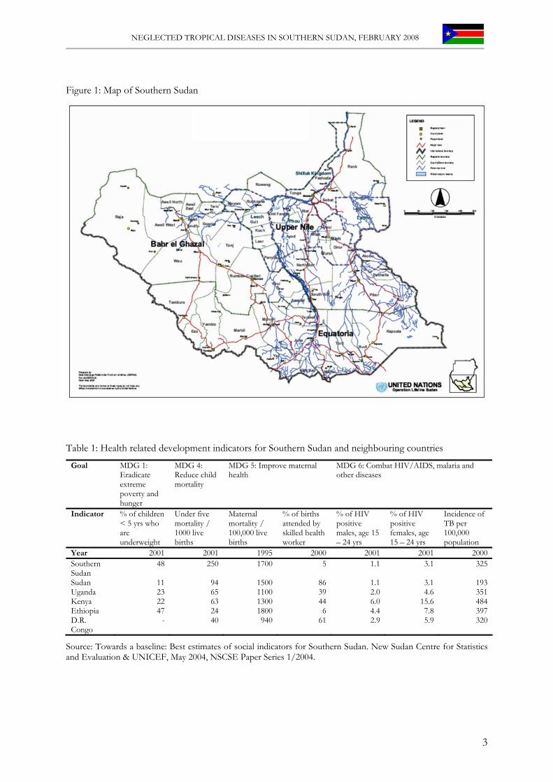
National administrative boundaries of Southern Sudan are derived for purposes of local government, resource allocation and population censuses (Figure 1). Southern Sudan lies within the Nile basin and shares borders with four countries (Ethiopia, Kenya, Uganda and the Democratic Republic of Congo (DRC). It covers an area of 640,000 km2 and is divided into a three-tiered system forming the state (n=10) at the first level; county at the second level (n=53); payam (>200) at the third level. No exact census data are available but in 2003 the population was estimated to be approximately 8 million, based on data from National Immunization Days (NIDs). This population is expected to grow by as much as 4.5 million by 2010, resulting from the return of refugees and internally displaced persons (IDP) and from the high natural population growth of about 3% (NSCSE, 2004).
The population is the youngest in the world, with an estimated 21% of persons aged less than 5 years old and 49% below the age of 15. Only 1.6% of the population are above the age of 65. Live expectancy at birth is 42 years. Infant and under five mortality is high at 150 death/1000 live births and 250/1,000 live births, respectively. Under five mortality makes up 57% of the total deaths (NSCSE, 2004). Data on health related indicators to measure progress towards the Millennium Development Goals are shown in table 1. Though somewhat out of date, these data indicate the poor overall health of the Southern Sudanese population.
2
NEGLECTED TROPICAL DISEASES IN SOUTHERN SUDAN, FEBRUARY 2008
Figure 1: Map of Southern Sudan
Table 1: Health related development indicators for Southern Sudan and neighbouring countries
Goal MDG 1: Eradicate extreme poverty and hunger
MDG 4: Reduce child mortality
MDG 5: Improve maternal health
MDG 6: Combat HIV/AIDS, malaria and other diseases
Indicator % of children < 5 yrs who are underweight
Under five mortality / 1000 live births
Maternal mortality / 100,000 live births
% of births attended by skilled health worker
% of HIV positive males, age 15 – 24 yrs
% of HIV positive females, age 15 – 24 yrs
Incidence of TB per 100,000 population
Year 2001 2001 1995 2000 2001 2001 2000Southern Sudan
48 250 1700 5 1.1 3.1 325
Sudan 11 94 1500 86 1.1 3.1 193Uganda 23 65 1100 39 2.0 4.6 351Kenya 22 63 1300 44 6.0 15.6 484Ethiopia 47 24 1800 6 4.4 7.8 397D.R. Congo
- 40 940 61 2.9 5.9 320
Source: Towards a baseline: Best estimates of social indicators for Southern Sudan. New Sudan Centre for Statistics and Evaluation & UNICEF, May 2004, NSCSE Paper Series 1/2004.
3

NEGLECTED TROPICAL DISEASES IN SOUTHERN SUDAN, FEBRUARY 2008
The health service administration and infrastructure is still developing and experiences severe financial and human resource constraints. In turn, access to healthcare is extremely limited (Figure 2). With the long-term goal of ensuring access of the majority of the population to basic health care, the Health Sector Recovery Strategy has set the following targets for 2010: one hospital per 300,000 people, one Primary Health Care Centre (PHC) for 50,000 people, and one PHC unit for 15,000 people. These targets represent a substantial expansion of the present health care infrastructure, corresponding to increases of 100% for the number of hospitals, of 133% for PHC centres and of 45% for PHC units.
With limited capacity, the MoH-GoSS at central, state and county level is in great need for management and human resource capacity strengthening, as well as commodity support. In this context, non-governmental (NGO) service providers continue to play a key role in the delivery of health care and have the potential to play a major role in building the capacity of MoH-GoSS at state and, particularly, county levels. Consequently, there must be an early emphasis on building simple and robust systems to facilitate health service delivery strategies to achieve rapid impact. It is crucial that any sustained approach to health systems development and strengthening is implemented through supporting the capacity development of MoH-GoSS at state and county levels in partnership with non-state service providers who can maintain and improve service delivery. It is within this current and planned system of health care delivery that the GoSS plans to reduce the disease burden due to NTDs.
Figure 2: Health facility access in Southern Sudan
4

NEGLECTED TROPICAL DISEASES IN SOUTHERN SUDAN, FEBRUARY 2008
3. THE NEGLECTED TROPICAL DISEASES OF SOUTHERN SUDAN
Health data collected by the MoH-GoSS, such as hospital admissions, can be used as a starting point to illustrate the burden of NTDs. However, because of a weak health surveillance infrastructure and the fact that populations affected are poor and isolated, these data are likely to be a gross underestimate. A more accurate picture can be provided by population-based surveys. The following section reviews the available information on the nature and disease burden of each of the major NTDs. Also reviewed are past and current control strategies tackling these diseases in Southern Sudan. 3.1 Leishmaniasis
Authors: Richer M, Ruiz JA, Kolaczinski JH & Booker S
3.1.1 Background The parasite and its life-cycle: The leishmaniases are a group of diseases caused by over 17 species of the protozoan Leishmania parasite. Infection is transmitted by the bite of phlebotomine sandflies and results in cutaneous, mucosal or visceral manifestations. Disease burden: After malaria and lymphatic filariasis, the leishmaniases are the third most important vector-borne disease, responsible for an estimated 500,000 new cases per year, 51000 deaths annually, and 2.1 million DALYs (WHO 2004a). These figures are thought to be an underestimate however, as only 40 of 88 endemic countries consider leishmaniasis a reportable disease (Croft et al. 2003). Geographical distribution: Much of the disease burden due to the leishmaniases in Africa is due to visceral leishmaniasis (VL) and concentrated in eastern Africa. Here, VL is caused by the parasite Leishmania donovani and is endemic in remote regions of Uganda, North and Southern Sudan, Ethiopia, Kenya and Somalia (Reithinger et al. 2007). Clinical features: VL is characterised by fever, splenomegaly, and cachexia (wasting and weakness). Up to 95% of untreated cases eventually die due to organ failure, anaemia or secondary infections (Desjeux 1996, Chappuis et al. 2007). Diagnosis and control options: Classically the diagnosis of VL is confirmed by demonstration of the parasite. Intracellular leishmania can be identified from aspirates of the spleen, bone marrow, lymph node or liver. Serological techniques based on the enzyme-linked immunosorbent assay, the direct agglutination test (DAT) and the rK39-based rapid diagnostic tests (RDT) have been developed for field use; unfortunately there are some concerns regarding the sensitivity and specificity of the rK-39 RDTs in East Africa (Boelaert et al. 2008). PCR is still not easily used in the field (Chappuis et al. 2007).
Efficient case management is the key to limit morbidity and to prevent mortality, and is also a measure to control the reservoir and transmission. First-line treatment by most agencies working on VL in eastern Africa still relies on antimonial drugs: sodium stibogluconate (SSG) or meglumine antimoniate (Glucantime). These rather toxic compounds need to be administered at as a single daily dose of 20mg/kg bodyweight for 30 days. To reduce the length of treatment and prevent the selection of antimonial drug resistance a short course (17 day) combination of SSG and paromomycin is being tested in a multi-country phase III trial in Ethiopia, Kenya and north Sudan by the Drugs for Neglected Diseases Initiative (DNDI, www.dndi.org). Treatment data on the use of this short course in Southern Sudan, which was recently published by Médecins Sans Frontières (MSF), shows better rates of survival and cure relative to the 30 day SSG monotherapy (Melaku et al. 2007). Miltefosine, which is already being used for VL treatment in India, has not undergone efficacy trials in Africa. Amphotericin B is recommended as second-line treatment, meaning it should be used for relapse cases, pregnant women and for patients who cannot tolerate (i.e. intractable vomiting, pancreatitis) or do not respond to antimony compounds.
5
NEGLECTED TROPICAL DISEASES IN SOUTHERN SUDAN, FEBRUARY 2008
In addition to treatment, vector control should be implemented where feasible. To set up an
effective control strategy for VL is a challenge in endemic areas, as these are largely in the poorest countries of the world, in remote places and/or in complex settings (e.g. civil war in Somalia). Personal protection by use of insecticide-treated nets (ITNs) is effective in foci where sandflies bite at night (Ritmeijer et al. 2007). One major limitation has been the cost of regular re-impregnation of the nets, which has been overcome through development of nets with long-lasting insecticide impregnation (LLINs) (Hill et al. 2006). Vaccines are being investigated, but none is yet ready for use (Chappuis et al. 2007).
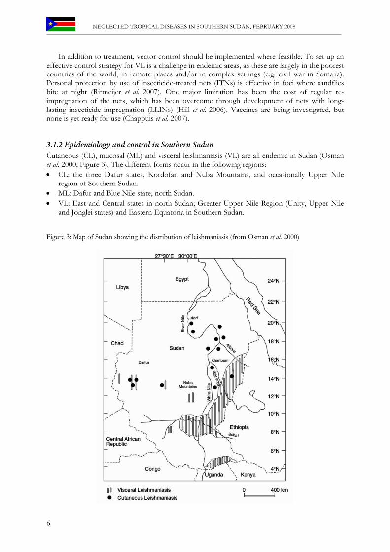
3.1.2 Epidemiology and control in Southern Sudan Cutaneous (CL), mucosal (ML) and visceral leishmaniasis (VL) are all endemic in Sudan (Osman et al. 2000; Figure 3). The different forms occur in the following regions: • CL: the three Dafur states, Kordofan and Nuba Mountains, and occasionally Upper Nile
region of Southern Sudan. • ML: Dafur and Blue Nile state, north Sudan. • VL: East and Central states in north Sudan; Greater Upper Nile Region (Unity, Upper Nile
and Jonglei states) and Eastern Equatoria in Southern Sudan. Figure 3: Map of Sudan showing the distribution of leishmaniasis (from Osman et al. 2000)
6

NEGLECTED TROPICAL DISEASES IN SOUTHERN SUDAN, FEBRUARY 2008
In the focus of the Greater Upper Nile region, the sandfly vector, Phlebotomus orientalis, lives in forests of Acacia seyal and Balanites aegyptiaca (Quate 1964, Ashford et al. 1992), and is the same vector that transmits VL in North Sudan. The main tribes in the area are the semi-nomadic pastoralists Dinka, Nuer and Shilluk. Males of these tribes move with their cattle to find appropriate grazing sites during the dry season.
Although infection occasionally occurs in animals (Mukhtar et al. 2000, Dereure et al. 2000, Dereure et al. 2003), transmission is thought to be human-human, i.e. anthroponotic. Transmission of VL is perennial, with seasonal peaks occurring during or shortly after the rains (i.e. from April to June), leading to a peak in clinical cases in the dry season, which usually lasts from November to January. The clinical disease associated with VL in this focus is typically fulminant (i.e. sudden and severe) and usually leads to rapid death.
Inter-year epidemics of VL are a common occurrence. The first recorded epidemic occurred in 1940 in the Dinka tribe living around Melut town and across the River Nile in the Shilluk tribe living in the Kaka-town area. Melut and Kaka are about 150 km north of Malakal town on the Nile in Upper Nile State. Further epidemics occurred 1960-61 in the Khor-Flous area, and in 1984 and 1994 in Unity State, in an estimated population of 300,000 people. The entire population was susceptible because the disease had never been reported in this location and the resultant epidemic caused approximately 100,000 deaths over a ten-year period (Seaman et al. 1996). Epidemics of VL are due to multiple factors including famine, malnutrition, mass migration, civil disturbance, poor economic conditions and impaired immunity of the hosts (e.g. due to HIV infection) (Reithinger et al. 2007).
Available epidemiological data indicate that epidemics in Southern Sudan occur every 7-10 years (Figure 4), although the complexity of factors involved makes temporal predictions difficult. Passive case detection data collected by WHO over the last 18 years indicate a low case-load (Figure 4) though these data are likely to be a gross underestimate since large proportion of cases not present to health facilities (Kolaczinski et al. 2008). Epidemiological modelling of VL data from Upper Nile suggests that only 55% of cases reported to health facilities between October 1998 and May 2002, and that 91% of VL deaths were undetected (Collin et al. 2006). The dynamics presented Figure 4 also suggest a current inter-epidemic stage, but that cases may rise dramatically in coming years.
Figure 4: Total annual number of VL cases (primary, relapse and PKDL) reported by health care providers in Southern Sudan to WHO from 1989 to 2006. Data was reported by MSF (Dutch Section) for all the years shown, by MSF-France for 2005 and 2006 and by some, but not all NGOs, from 1999 onwards.
0
1000
2000
3000
4000
5000
6000
7000
8000
9000
89 90 91 92 93 94 95 96 97 98 99 00 01 02 03 04 05 06
Year
7
NEGLECTED TROPICAL DISEASES IN SOUTHERN SUDAN, FEBRUARY 2008
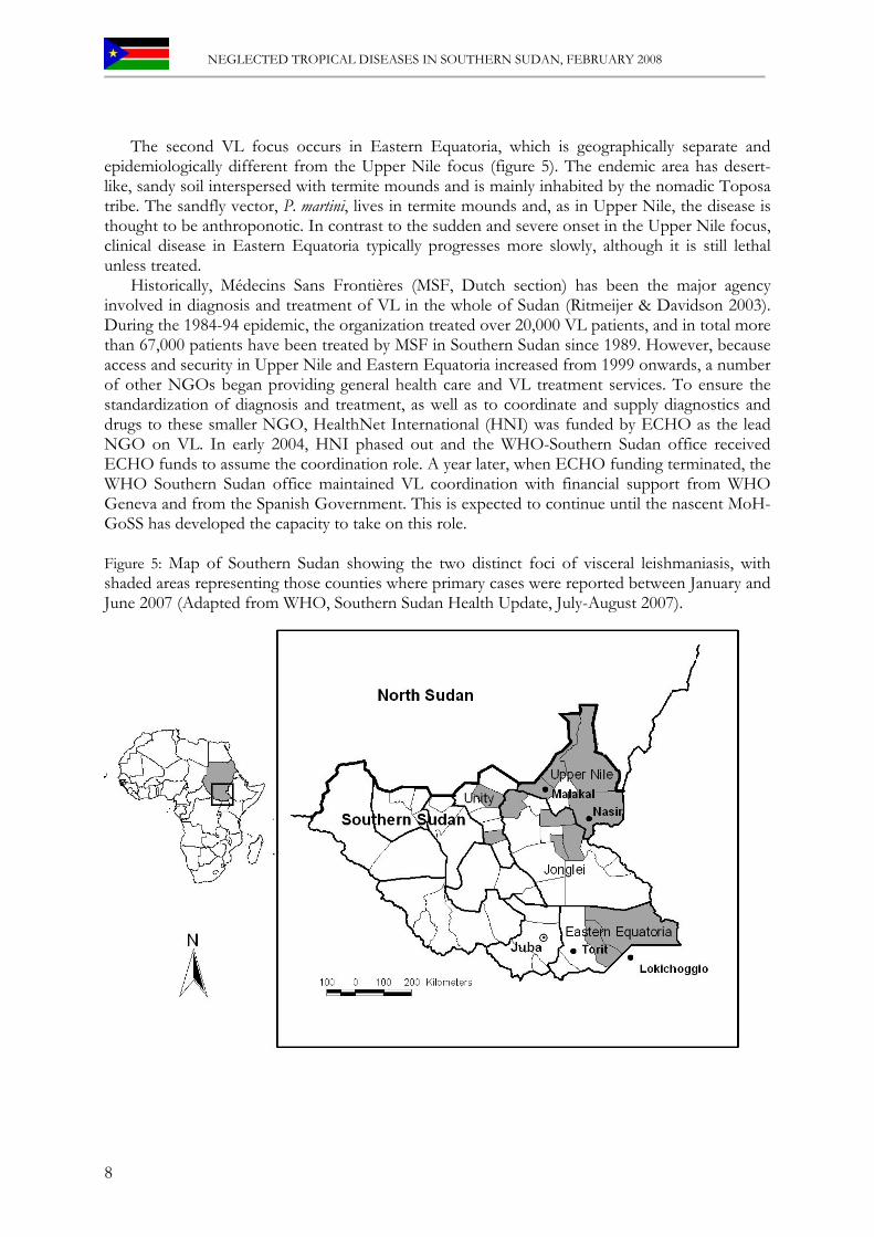
The second VL focus occurs in Eastern Equatoria, which is geographically separate and epidemiologically different from the Upper Nile focus (figure 5). The endemic area has desert-like, sandy soil interspersed with termite mounds and is mainly inhabited by the nomadic Toposa tribe. The sandfly vector, P. martini, lives in termite mounds and, as in Upper Nile, the disease is thought to be anthroponotic. In contrast to the sudden and severe onset in the Upper Nile focus, clinical disease in Eastern Equatoria typically progresses more slowly, although it is still lethal unless treated.
Historically, Médecins Sans Frontières (MSF, Dutch section) has been the major agency involved in diagnosis and treatment of VL in the whole of Sudan (Ritmeijer & Davidson 2003). During the 1984-94 epidemic, the organization treated over 20,000 VL patients, and in total more than 67,000 patients have been treated by MSF in Southern Sudan since 1989. However, because access and security in Upper Nile and Eastern Equatoria increased from 1999 onwards, a number of other NGOs began providing general health care and VL treatment services. To ensure the standardization of diagnosis and treatment, as well as to coordinate and supply diagnostics and drugs to these smaller NGO, HealthNet International (HNI) was funded by ECHO as the lead NGO on VL. In early 2004, HNI phased out and the WHO-Southern Sudan office received ECHO funds to assume the coordination role. A year later, when ECHO funding terminated, the WHO Southern Sudan office maintained VL coordination with financial support from WHO Geneva and from the Spanish Government. This is expected to continue until the nascent MoH-GoSS has developed the capacity to take on this role. Figure 5: Map of Southern Sudan showing the two distinct foci of visceral leishmaniasis, with shaded areas representing those counties where primary cases were reported between January and June 2007 (Adapted from WHO, Southern Sudan Health Update, July-August 2007).
8

NEGLECTED TROPICAL DISEASES IN SOUTHERN SUDAN, FEBRUARY 2008
A VL control strategy, including guidelines on diagnosis, treatment and prevention, has not yet been finalised. However, standardization of treatment procedures has come a long way. The WHO-Southern Sudan office in cooperation with the previous Secretariat of Health, MSF (Dutch Section) and other experts drafted diagnostic and treatment guidelines, which have been utilized by NGOs since 2005 and remain in use. A workshop was conducted in October 2007 to develop these into a formal document, which was endorsed by the MoH-GoSS in January 2008.
Based on the guidelines, the WHO Southern Susan office has provided training on VL diagnosis and treatment. Recommended treatment of primary VL is 30 days of SSG monotherapy. The drug is supplied through Pharmaciens Sans Frontières (PSF) once VL infection has been confirmed by DAT (see below). The exception to this are health facilities supported by MSF (Dutch section), where a short course of combination therapy (SSG plus paromomycin) was introduced as of 2002. In contrast to other facilities, MSF also provided second-line treatment with ambisome. Though not yet officially endorsed, there is evidence that combination therapy of SSG/paromomycin is more effective than SSG monotherapy. In addition it offers of the advantage that it halves the patients’ required hospital stay, thus reducing overcrowding and the risk of nosocomial outbreaks of infectious diseases associated with overcrowding (Melaku et al. 2007).
VL diagnosis largely depends on DAT, relying on a reference laboratory in Lokichoggio supported by the MSF (Dutch section). Blood samples on filter paper are sent from the various treatment centres to the laboratory for testing. The need for samples to be sent to Lokichoggio and for results to be sent back to health facilities can introduce treatment delays and additional costs. To speed-up diagnostic procedures, some health care providers have started to conduct DAT at their own facilities, such as IMRF, MedAir and Tearfund. Fortunately, use of a rK39-based RDT is becoming more widespread, meaning that diagnosis can be extended to periphery. After the October 2007 workshop, the new diagnostic protocols endorsed by MoH-GoSS will allow SSG for treatment to be provided by PSF based on the results of RDTs (Kolaczinski et al. 2008).
3.1.3 Challenges 1. Guidelines for VL diagnosis and treatment have been finalised and now need to be widely
implemented. 2. The sensitivity and specificity of rK39 RDTs in the two foci remains to be confirmed 3. Short-course combination therapy needs to be introduced once sufficient evidence on its
efficacy has been generated by DNDI 4. MoH-GoSS needs to develop a VL control programme for Southern Sudan and provide
support to the states with the major VL burden. 5. MoH-GoSS needs to develop the national referral laboratory at the Juba level to become the
Southern Sudan referral DAT laboratory instead of relying on MSF (Dutch section) facilities in Lokichoggio for this service. Laboratory diagnosis at referral centres also needs strengthening.
6. The VL endemic states need to urgently increase their capacity on VL case-management and prevention.
9
NEGLECTED TROPICAL DISEASES IN SOUTHERN SUDAN, FEBRUARY 2008
3.2 Human African Trypanosomiasis
Authors: Ruiz JA, Richer M & Meru A
3.2.1 Background Human African Trypanosomiasis (HAT), also known as sleeping sickness, is a severe disease that is fatal if left untreated. The parasite and its life cycle: HAT is caused by protozoan parasites of the genus Trypanosoma that are transmitted between infected humans and animals by tsetse flies (Glossina spp.) and enter the blood stream during blood feeding. Two sub-species of Trypanosoma cause HAT, Trypanosoma brucei rhodesiense and T. b. gambiense (Fèvre et al. 2006). Disease burden: HAT occurs in both epidemic and endemic patterns across more than 200 foci throughout sub-Saharan Africa. In 2006 WHO estimates put the number of cases at 50,000 to 70,000 (www.who.int/mediacentre/factsheets/fs259/en/). The extrapolated estimates are somewhat imprecise, since less than 10% of the population at risk of HAT (about 60 million people) is under surveillance (Barrett et al. 2003). In terms of DALYs lost, HAT ranks third among parasitic diseases, behind malaria and lymphatic filariasis and ahead of leishmaniasis, schistosomiasis and onchocerciasis. Geographical distribution: T. b. rhodesiense occurs mainly in east and southern Africa, while T. b. gambiense mainly occurs in west and central Africa. Antelopes and cattle are the main reservoirs, but wild carnivores, such as hyenas and lions, can also serve as reservoirs for T. b. rhodiense (zoonosis). Humans are the main reservoir for T. b. gambiense (anthroponosis), though domestic animals such as pigs, sheep and dogs, can also host the parasite. In animals, many other species of Trypanosoma are known to cause trypanosomiasis, also called Nagana. Clinical features: Once inside the human host, trypanosomes multiply and invade most tissues. Infection leads to malaise, lassitude and irregular fevers. Early symptoms, including fever and enlarged lymph glands and spleen, are more severe and acute in T.b. rhodesiense infections. An early sign, a primary chancre at the site of the tsetse bite, is followed by a range of symptoms including fever, enlargement of the cervical lymph nodes (posterior cervical adenopathy), headache, anaemia, joint pains, swollen tissues. Advanced symptoms include neurological and, psychiatric disorders. After the parasites invade the central nervous system, mental deterioration begins, leading to coma and death. T.b rhodesiense infection is usually acute, causing severe symptoms and death within a few days or weeks. T.b. gambiense infection tends to progress more slowly (over several years) and is less severe although still lethal unless treated. Diagnosis and control options: Control of T. b. gambiense involves active case-finding and prompt treatment. Screening of the population is usually done with a card-agglutination test. For T. b. rhodesiense, passive case-finding based on clinical algorithms is recommended, because diagnostic tools are not readily available. Treatment of infected people especially with advanced disease has always been difficult and expensive. Few effective drugs are available and specialised administration of drugs requires long period of hospital care (Legros et al. 2002). In addition, reduction of tsetse fly numbers can play a significant role, especially against the rhodesiense form of the disease. In the past, this has involved extensive clearance of bush to destroy tsetse fly breeding and resting sites, and widespread application of insecticides. More recently, efficient traps and screens have been developed that can keep tsetse populations at low levels. However, this method has proven difficult to sustain for various reasons, including physical degradation, damage, theft and lack of education in use of the traps (Kuzoe et al. 2005).
10
NEGLECTED TROPICAL DISEASES IN SOUTHERN SUDAN, FEBRUARY 2008
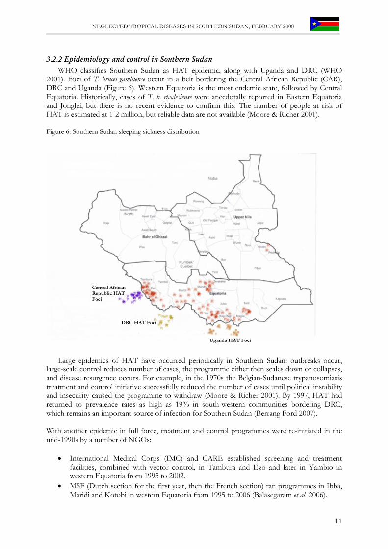
3.2.2 Epidemiology and control in Southern Sudan WHO classifies Southern Sudan as HAT epidemic, along with Uganda and DRC (WHO
2001). Foci of T. brucei gambiense occur in a belt bordering the Central African Republic (CAR), DRC and Uganda (Figure 6). Western Equatoria is the most endemic state, followed by Central Equatoria. Historically, cases of T. b. rhodesiense were anecdotally reported in Eastern Equatoria and Jonglei, but there is no recent evidence to confirm this. The number of people at risk of HAT is estimated at 1-2 million, but reliable data are not available (Moore & Richer 2001). Figure 6: Southern Sudan sleeping sickness distribution
Central African Republic HAT Foci
DRC HAT Foci
Uganda HAT Foci
Large epidemics of HAT have occurred periodically in Southern Sudan: outbreaks occur, large-scale control reduces number of cases, the programme either then scales down or collapses, and disease resurgence occurs. For example, in the 1970s the Belgian-Sudanese trypanosomiasis treatment and control initiative successfully reduced the number of cases until political instability and insecurity caused the programme to withdraw (Moore & Richer 2001). By 1997, HAT had returned to prevalence rates as high as 19% in south-western communities bordering DRC, which remains an important source of infection for Southern Sudan (Berrang Ford 2007). With another epidemic in full force, treatment and control programmes were re-initiated in the mid-1990s by a number of NGOs:
• International Medical Corps (IMC) and CARE established screening and treatment facilities, combined with vector control, in Tambura and Ezo and later in Yambio in western Equatoria from 1995 to 2002.
• MSF (Dutch section for the first year, then the French section) ran programmes in Ibba, Maridi and Kotobi in western Equatoria from 1995 to 2006 (Balasegaram et al. 2006).
11
NEGLECTED TROPICAL DISEASES IN SOUTHERN SUDAN, FEBRUARY 2008
• MSF (Swiss section) in Kajo-Kegi County (Kiri Hospital) from June 2000 to 2006. • Malteser initiated treatment in Yei in March 2002. • WHO established an emergency intervention in 2004 in Tambura and Ezo and MSF
(Spanish section) took over from 2005 to 2006. • Merlin established a programme in Nimule, Magwi County, Eastern Equatoria, in 2005. • Lui hospital supported by WHO initiated treatment for stage 2 patients in October 2007
covering a gap existing in the county since April 2006.
As HAT was brought under control, funds were again getting difficult to access and NGOs were forced to withdraw their support. In the absence of a government control programme, HAT started to re-emergence. As before, NGOs intervened to get the resurgence under control.
Current HAT control is still organized vertically and implemented largely by international NGOs. These are often the only organizations that have the resources to operate specialized hospitals, and are able to procure supportive drugs, supplies and equipment for the intensive interventions required to control focal epidemics. The MoH-GoSS only supports treatment at Juba Teaching Hospital, but where older drugs and treatment regimes – melarsoprol – continue to be used as first-line for stage 2 patients, due to lack of human resources to be trained on the use of eflornithine, which is used in all other stage 2 treatment centres in Southern Sudan. There is an urgent need to upgrade treatment protocols at Juba Teaching Hospital.
WHO provides anti-trypanosome drugs free of charge for all NGO and MoH treatment centres thanks to an agreement with the manufacturer. The current HAT treatment centres are listed in Table 2., Table 2: Current HAT treatment centres in Southern Sudan, by location and supporting agency
Facility type Location Supporting agencyStage 1 and 2 treatment* Tambura, Western Equatoria IMC
“ Lui hospital Diocese of Lui“ Yambio, Western Equatoria MSF (Spanish section)“ Yei, Central Equatoria Malteser“ Kajo-Keji, Central Equatoria IMC“ Nimule, Eastern Equatoria Merlin“ Juba, Central Equatoria MoH-GoSS
Stage 1 treatment only Maridi. Western Equatoria Aktion Afrika Hilfe“ Source Yubu, W. Equatoria MoH“ Ezo, Western Equatoria World Vision International
* Stage 1 and 2 refer to early or non-meningoencephalitic stage and late or meningoencephalitic stage, respectively
Coordination of activities of different implementing partners was the initiative of NGOs
during the war in Sudan, in the absence of a functioning MoH-GoSS, and started in 2003 in Uganda. Partners met annually to standardize protocols, share lessons and improve coordination including neighbouring countries. In 2004, WHO facilitated the participation of the Secretariat of Health (the precursor to MoH-GoSS) and became actively involved in the organization and support of the meeting. By 2006, the meeting was for the first time held in Juba and sponsored by the WHO with representatives from neighbouring countries (Uganda, DRC and CAR). Recommendations for both diagnostic and treatment protocols were agreed upon by all implementing NGOs, which will assist the MoH-GoSS in formulating standardized guidelines for all HAT treatment facilities. Since mid 2007, monthly coordination meetings led by MoH-GoSS have been held in Juba focussing to Southern Sudan specific issues. Southern Sudan has reported
12
NEGLECTED TROPICAL DISEASES IN SOUTHERN SUDAN, FEBRUARY 2008
42 stage 1 and 166 stage 2 sleeping sickness new cases and 9,632 people screened from January to October 2007. The reporting rate for the same period has been 37 %.
A number of challenges are experienced. First, treatment data from different partners have so far not been collated into a single database and the reporting needs to be improved. Second, there is a need for active case-finding in selected areas where prevalence is estimated to be still above 1%.
To help support data collation and programme planning activities, WHO is now assisting the HAT coordinator at MoH-GoSS. A HAT control strategy is currently being formulated. The DG for Preventive Health also recently participated in a HAT meeting at WHO-Geneva for all HAT endemic African countries. This provided background and discussions with other endemic country leaders to facilitate formulation of a MoH-GoSS control strategy. This strategy will need to pay special attention to the monitoring of drug efficacy and the management of failures.
3.3.3 Challenges 1. GoSS needs to establish a HAT control programme to ensure that control measures are in
place in endemic foci and that local capacity is built to sustain interventions over time. 2. The HAT unit at Juba Teaching Hospital needs to be upgraded to the use of newer treatment
regimens. 3. Activities of different implementing partners need to be better coordinated, including
reporting at national level, establishment and maintenance of a centralised database and formulation of a regional control strategy.
13

NEGLECTED TROPICAL DISEASES IN SOUTHERN SUDAN, FEBRUARY 2008
3.3 Soil Transmitted Helminths (STHs)
Authors: Richer M, Ruiz JA, Brooker S & Kolaczinski JH
3.3.1 Background The parasite and its life cycle: STHs are also known as common intestinal worms. In terms of public health, the most important species are: roundworms (Ascaris lumbricoides), hookworms (Ancylostoma duodenale and Necator americanus), and whipworms (Trichuris trichiura). A person infected with STH has parasite eggs in their faeces. In areas where there is no latrine system, the soil and water around the community become contaminated with faeces containing worm eggs. In the soil, the eggs mature over two to four weeks, depending on the type of worm and environmental conditions, and then infect humans by being ingested or by penetrating the skin (hookworms only). Disease burden: Globally, it is estimated that over a billion people living in the tropics and sub-tropics are infected with STHs. Although the largest numbers of infections occur in Asia, the greatest burden of disease occurs in Africa since the morbidity caused by STHs is related to the intensity of infection and host nutrition, and infections are most intense and nutrition is often inadequate in Africa. Geographical distribution: STHs are widely distributed throughout the tropics and subtropics and are particularly prevalent throughout much of sub-Saharan Africa, as well as in South China, the Pacific and Southeast Asia. Clinical features: The symptoms of infections are non-specific and only become evident when the infection is particularly intense. Non-specific symptoms include nausea, tiredness, abdominal pain, loss of appetite and, in children, a cough or wheeze. Chronic and intense STH infections can contribute to malnutrition and iron-deficiency anaemia, and also can adversely affect physical and mental growth in childhood (Bethony et al., 2006). Diagnosis and Control options: Current efforts to control STH infection, as well as schistosomiasis, focus on the school-age population. The cornerstone of control is population-based chemotherapy, especially targeting schoolchildren. School-age children are the natural targets for treatment, and school-based treatment delivery programmes offer major cost advantages, because of the use of the existing school infrastructure and the fact that schoolchildren are accessible through schools. There are four drugs to treat STH infections: Albendazole and mebendazole are particularly attractive because they are easy to administer. Pyrantel pamoate and levamisole are alternatives for treatment of hookworm and ascaris infections (WHO 2005); the former is not effective for treatment of trichuriasis and they are administered by bodyweight. As a general strategy, WHO recommends that in areas where STH prevalence is ≥ 50%, treatment is provided twice yearly; in areas where prevalence is between 20 – 49%, annual treatment is provided; in areas with prevalence < 20%, drugs are made available at the health facility (WHO 2002a).
3.2.2 Epidemiology and control in Southern Sudan Data collated by UNICEF from health partners operating in Southern Sudan during the war
consistently indicated that 8-10% of all outpatient visits were for treatment of intestinal worms. Population-based estimates of STH infection prevalence in Southern Sudan are limited, however. Data collected by the Federal MoH (Khartoum) in the 1990s show that STH were prevalent in the Southern Sudan, especially in Central and Eastern Equatoria (Table 3).
14
NEGLECTED TROPICAL DISEASES IN SOUTHERN SUDAN, FEBRUARY 2008
Table 3: Prevalence of helminth infection in Southern Sudan
Location Sample Size
% infected Source
Hookworm Ascaris Trichuris S. mansoni S. hematobium Central Equatoria (Juba), 1992
241 36.0% 1.2% 0.8% FMoH Khartoum
Eastern Equatoria, 1998
275 13.1% 0 1.8% 2.2% Magambo et al. 1998
Western Equatoria (Lui), 2002
200 51.5% 0
Upper Nile (Nyal), 2002
200 70.0% 73.0%
Deganello et al. 2007
Western Equatoria (Lui), 2007
200 5% 0.5% 2% 50.5% - WHO Southern Sudan
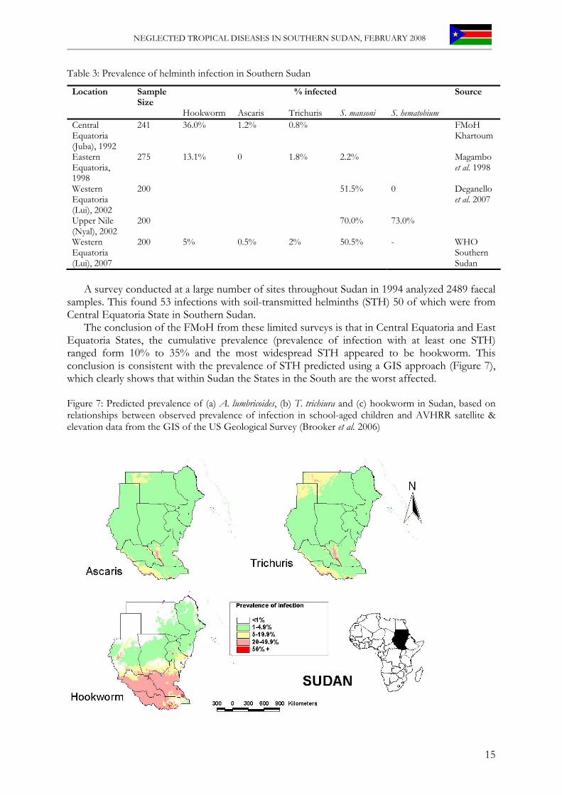
A survey conducted at a large number of sites throughout Sudan in 1994 analyzed 2489 faecal
samples. This found 53 infections with soil-transmitted helminths (STH) 50 of which were from Central Equatoria State in Southern Sudan.
The conclusion of the FMoH from these limited surveys is that in Central Equatoria and East Equatoria States, the cumulative prevalence (prevalence of infection with at least one STH) ranged form 10% to 35% and the most widespread STH appeared to be hookworm. This conclusion is consistent with the prevalence of STH predicted using a GIS approach (Figure 7), which clearly shows that within Sudan the States in the South are the worst affected. Figure 7: Predicted prevalence of (a) A. lumbricoides, (b) T. trichiura and (c) hookworm in Sudan, based on relationships between observed prevalence of infection in school-aged children and AVHRR satellite & elevation data from the GIS of the US Geological Survey (Brooker et al. 2006)
15
NEGLECTED TROPICAL DISEASES IN SOUTHERN SUDAN, FEBRUARY 2008
Until recently, treatment for STH with albendazole was only available at health facilities. In
2006, MDA providing albendazole distribution to 1-5 year olds was piloted as a component of NIDs in all ten states. This is meant to provide an interim solution for STH control until MDA through a school-based programme becomes a viable option. Training for the polio vaccinators on albendazole distribution was carried out between September and December 2006. 87% (1,908,445 children) of the targeted 1-5 year old children received the medication. A total of 2.5 million doses were distributed by vaccinators during the campaign. Some of these doses were used to treat siblings above 5 years of age. A second round of albendazole MDA alongside NIDs was conducted in December 2007. Once school structures have been rebuilt and attendance rates have increased, the MoH-GoSS is planning to implement routine MDA for STH through schools, as is common practice in the region (Kabatereine et al. 2006).
3.3.3 Challenges 1. Investigate alternative options for MDA, ideally as an integrated component of other drug
distribution campaigns and/or through permanent distribution channels such as schools, once the required infrastructure has been constructed.
2. Ensure regular supplies of albendazole, as this drug is generally not donated by the manufacturer. An exception exists for areas where lymphatic filariasis is endemic (see below), though even in these areas not all age groups that qualify for STH treatment would be covered.
16

NEGLECTED TROPICAL DISEASES IN SOUTHERN SUDAN, FEBRUARY 2008
3.4 Schistosomiasis
Authors: Richer M, Ruiz JA, Brooker S & Kolaczinski JH
3.4.1 Background The parasite and its life cycle: Human schistosomiasis, a water-borne disease, is mainly caused by two species of blood flukes (called schistosomes): Schistosoma mansoni causing intestinal schistosomiasis and S. haematobium causing urinary schistosomiasis. The schistosomes require a molluscan (i.e. snail) intermediate host in which to undergo development. Freshwater snails from four different genera form an essential component in the life cycle of the four major schistosome species that are responsible for human schistosomiasis. This ties transmission of the disease to places where people and snails come together at the same water habitat. Hence, schistosomiasis tends to be commonly found in rural communities where contact with freshwater bodies is a routine and inevitable occurrence. Disease burden: Among human parasitic diseases, schistosomiasis, also called bilharziasis, ranks second behind malaria in terms of socio-economic and public health importance in Africa. The disease is endemic in 74 developing countries, infecting more than 200 million people in rural agricultural and peri-urban areas. Of these, 20 million suffer severe consequences from the disease and 120 million are symptomatic. In many areas, schistosomiasis infects a large proportion of children under 14 years. An estimated 500-600 million people worldwide are at risk from the disease. Geographical distribution: S. haematobium occurs mainly in Africa and also in the Middle East, while S. mansoni occurs throughout Africa and in parts of South America. Within endemic areas, the precise distribution of infection is highly focal. Clinical features: Disease is caused primarily by schistosome eggs that are deposited by adult worms in the blood vessels surrounding the bladder or intestines, depending on the specific species. S. haematobium causes bladder wall pathology, leading to ulcer formation, hematuria, and dysuria. Granulomatous changes and ulcers of the bladder wall and ureter can lead to bladder obstruction, secondary urinary tract infections and subsequent bladder calcification, renal failure, lesions of the female and male genital tracts, and hydronephrosis. The morbidity commonly associated with S. mansoni infection includes lesions of the liver, portal vein, and spleen, leading to periportal fibrosis, portal hypertension, hepatosplenomegaly, and ascites. Schistosomiasis also causes chronic growth faltering and can contribute to anaemia. Diagnosis and Control options: Diagnosis is typically made by finding the characteristic spined eggs in urine (S. haematobium) or stool (S. mansoni). Schistosomiasis control aims to reduce the amount of disease, rather than to halt transmission entirely. The main strategy for controlling morbidity due to schistosomiasis is based on chemotherapy using praziquantel. Even though re-infection may occur after treatment, the risk of developing severe organ pathology is diminished and even reversed in young children.
3.4.2 Epidemiology and control in Southern Sudan Sudan was one of the first African countries where schistosomiasis control was attempted.
Egyptian labourers who went to Sudan to dig the canals for the Gezira irrigation scheme were screened and treated with antimony potassium tartrate. Despite this, the prevalence of schistosomiasis gradually increased among the local farmers; first infections were caused by Schistosoma haematobium, followed by S. mansoni. Despite intermittent efforts at control, schistosomiasis continues to be a major public health problem in Sudan, with an estimated 5 million people infected.
A comprehensive review of schistosomiasis in Sudan was published in 1987 (WHO 1987) using historical data to depict the distribution of schistosomiasis throughout the country. This indicates that south of the 9th degree latitude S. mansoni is very common whereas the largest
17
NEGLECTED TROPICAL DISEASES IN SOUTHERN SUDAN, FEBRUARY 2008
endemic area of S. haematobium is to be found between the 9th and 16th degree latitudes. This includes Unity and Upper Nile States of Southern Sudan. Hospital data from 1949 indicated a prevalence of S. mansoni of 44.3% in the Eastern, Central and Western Equatoria as well as Jonglei state, while prevalence in Bahr el Ghazal was 1-5%.
From 2002 to 2004 the WHO Southern Sudan office carried out 3 surveys, all of which were consistent with the findings of the historical data. In 2002, 73% and 70% of 200 school-aged children in Nyal (Unity State) were found to be infected with S. haematobium or S. mansoni, respectively. During the same year, 52.5% of 200 school-aged children in Lui (Western Equatoria) were found to be infected with S. mansoni, whereas none were infected with S. haematobium. In 2004, no infection with S. haematobium could be found in samples from 75 school-aged children in Tambura (Western Equatoria). In 2007, 50.5% of school-aged children in Lui were still found to be infected with S. mansoni. Though it is apparent that schistosomiasis is a problem in Southern Sudan, the exact geographical distribution and present burden is unknown.
For schistosomiasis, treatment was only available to symptomatic patients at health facilities. Based on the results of the 2002 study in Nyal (see above), the NGO Coordination Committee for Voluntary Services (COSV) began MDA with praziquantel (provided by WHO) through a school-based programme. Drugs were distributed every six months for two years until the drug supply terminated and distribution had to be discontinued. WHO subsequently surveyed the area in which COSV had been operating and found a continuing high prevalence of S. haematobium and S. mansoni. This indicates that small, isolated projects have limited impact; successful control will require a coordinated approach regularly targeting all of the endemic foci. In 2006, WHO developed a questionnaire on the presence of haematuria and distributed this to NGO partners for screening of S. haematobium in schools. The NGO World Relief distributed the questionnaire in schools in Abienenom (Unity State) where 687 school-aged children from 10 schools were screened and 26% reported gross haematuria. Parasitological studies in the area were scheduled for mid 2007 to determine baseline intensity of infection, but had to be postponed repeatedly. Depending on survey results, praziquantel distribution will be implemented through the schools in this area.
3.4.3 Challenges 1. The lack of schools and low school attendance has limited the possibility for MDA of
praziquantel to school-aged children. 2. In the interim, alternative distribution mechanisms, such as campaigns or integrated MDA,
will need to be used. 3. Praziquantel is not available from drug donation programmes. The cost of purchasing,
shipping and distributing it thus needs to be calculated and budgeted for.
18

NEGLECTED TROPICAL DISEASES IN SOUTHERN SUDAN, FEBRUARY 2008
3.5 Dracunculiasis (Guinea Worm)
Authors: Mackoy S & Becknell S
3.5.1 Background The parasite and its life cycle: Dracunculiasis is caused by the parasitic filarial worm Dracunculus medinensis, the largest of all filarial worms affecting man (Cairncross et al. 2002, Greenaway 2004). The parasite migrates through the victim's subcutaneous tissues. The mature female worm eventually moves to the surface of the skin (of the feet in 90% of cases), causing formation of a painful blister, which bursts and exposes the anterior end of the worm. Infected persons try to relieve the burning sensation cause at the site of the blister by cooling it in a local water sources. This induces a contraction of the female worm causing the sudden expulsion of hundreds of thousands of first stage larvae into the water. They move actively in the water and can live for a few days. For further development, they need to be ingested by a suitable species of predatory copepod (water flea). Inside the copepod larvae develop into the infective third stage within about two weeks. When a person drinks contaminated water from ponds or shallow open wells, larvae are released in the stomach and migrate through the intestinal wall. After approximately four month, adult male and female worms mate. The male then become encapsulated and dies in the tissues while the female move down the muscle planes. After about one year of the infection, the female worm with the uterus filled with larvae, emerges usually from the feet, repeating the life cycle. Disease burden: Dracunculiasis used to be a formidable public health problem, mainly in terms of morbidity, incapacity and suffering of those affected. About 50% of cases suffer from secondary infections and become severely incapacitated (e.g. Smith et al. 1989). The disease is still found among the poorest rural communities in areas without safe water supplies in sub-Saharan Africa especially in Southern Sudan, where 85% of all cases in the world are reported (Muller 2005, Ruiz-Tiben & Hopkins 2006). In 1986 there were approximately 3.5 million cases in 20 countries of the world, an estimated 3.32 million of which were in Africa (Muller 2005). Since 1989 when intense international eradication efforts were initiated the number of cases has been reduced to 25,195 in 2006 (CDC 2007). From January to May 2007, 4,460 cases were reported. This was a reduction of more than 50% when compared to the same reporting period in 2006. The goal as set at the 39th World Health Assembly in 1986 is to eradicate the disease by the end of 2009. Geographical distribution: As of May 2007, only nine countries in the world continue to have active indigenous cases of dracunculiasis: Burkina Faso, Côte d’Ivoire, Ethiopia, Ghana, Mali, Niger, Nigeria, Togo and Sudan (CDC 2007). The other eleven countries with disease in 1989 have either been certified free of Guinea worm or are in the pre-certification stage. Four of the above endemic countries (Burkina Faso, Côte d’Ivoire, Ethiopia and Togo) reported no indigenous cases during January to May 2007. In 2006, about 98% of all dracunculiasis cases worldwide were reported from Ghana and Sudan (CDC 2007). Clinical features: The parasite migrates through the victim's subcutaneous tissues causing pain, especially when it occurs or dies in a joint. When the worm emerges it provokes an intensely painful blister, which can be accompanied by fever, nausea and vomiting possibly symptoms of an allergic reaction. It usually takes about one-month for the worm to be slowly extracted from the wound (by winding it out on a stick), during which the track of the worm may become secondarily infected. Worms may brake during extraction, causing severe immune reaction as part of the worm dies within the person. Female worms sometimes burst in the tissues, resulting in a pus-filled abscess and severe cellulitis (Cairncross et al. 2002). Control options: A number of interventions, ideally deployed in combination, can control Guinea worm. These are: i) provision of a safe water supply, ii) filtration of drinking water to remove copepods, iii) searching for active cases and proper management of them, iv) ensuring that patients avoid contact with open water sources, and v) killing or removing copepods in
19
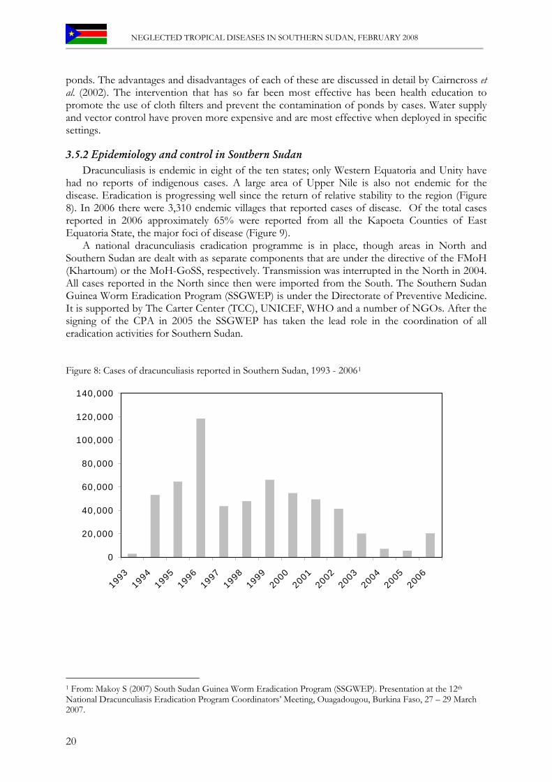
NEGLECTED TROPICAL DISEASES IN SOUTHERN SUDAN, FEBRUARY 2008
ponds. The advantages and disadvantages of each of these are discussed in detail by Cairncross et al. (2002). The intervention that has so far been most effective has been health education to promote the use of cloth filters and prevent the contamination of ponds by cases. Water supply and vector control have proven more expensive and are most effective when deployed in specific settings.
3.5.2 Epidemiology and control in Southern Sudan Dracunculiasis is endemic in eight of the ten states; only Western Equatoria and Unity have
had no reports of indigenous cases. A large area of Upper Nile is also not endemic for the disease. Eradication is progressing well since the return of relative stability to the region (Figure 8). In 2006 there were 3,310 endemic villages that reported cases of disease. Of the total cases reported in 2006 approximately 65% were reported from all the Kapoeta Counties of East Equatoria State, the major foci of disease (Figure 9).
A national dracunculiasis eradication programme is in place, though areas in North and Southern Sudan are dealt with as separate components that are under the directive of the FMoH (Khartoum) or the MoH-GoSS, respectively. Transmission was interrupted in the North in 2004. All cases reported in the North since then were imported from the South. The Southern Sudan Guinea Worm Eradication Program (SSGWEP) is under the Directorate of Preventive Medicine. It is supported by The Carter Center (TCC), UNICEF, WHO and a number of NGOs. After the signing of the CPA in 2005 the SSGWEP has taken the lead role in the coordination of all eradication activities for Southern Sudan. Figure 8: Cases of dracunculiasis reported in Southern Sudan, 1993 - 20061
0
20,000
40,000
60,000
80,000
100,000
120,000
140,000
1993
1994
1995
1996
1997
1998
1999
2000
2001
2002
2003
2004
2005
2006
1 From: Makoy S (2007) South Sudan Guinea Worm Eradication Program (SSGWEP). Presentation at the 12th National Dracunculiasis Eradication Program Coordinators’ Meeting, Ouagadougou, Burkina Faso, 27 – 29 March 2007.
20
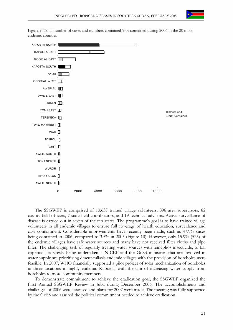
NEGLECTED TROPICAL DISEASES IN SOUTHERN SUDAN, FEBRUARY 2008
Figure 9: Total number of cases and numbers contained/not contained during 2006 in the 20 most endemic counties
0 2000 4000 6000 8000 10000
AWEIL NORTH
KHORFULUS
WUROR
TONJ NORTH
AWEIL SOUTH
TORIT
NYIROL
WAU
TWIC MAYARDIT
TEREKEKA
TONJ EAST
DUKEN
AWEIL EAST
AWERIAL
GOGRIAL WEST
AYOD
KAPOETA SOUTH
GOGRIAL EAST
KAPOETA EAST
KAPOETA NORTH
ContainedNot Contained
The SSGWEP is comprised of 13,637 trained village volunteers, 896 area supervisors, 82 county field officers, 7 state field coordinators, and 19 technical advisors. Active surveillance of disease is carried out in seven of the ten states. The programme’s goal is to have trained village volunteers in all endemic villages to ensure full coverage of health education, surveillance and case containment. Considerable improvements have recently been made, such as 47.9% cases being contained in 2006, compared to 3.5% in 2005 (Figure 10). However, only 15.9% (525) of the endemic villages have safe water sources and many have not received filter cloths and pipe filter. The challenging task of regularly treating water sources with temephos insecticide, to kill copepods, is slowly being undertaken. UNICEF and the GoSS ministries that are involved in water supply are prioritizing dracunculiasis endemic villages with the provision of boreholes were feasible. In 2007, WHO financially supported a pilot project of solar mechanization of boreholes in three locations in highly endemic Kapoeta, with the aim of increasing water supply from boreholes to more community members.
To demonstrate commitment to achieve the eradication goal, the SSGWEP organized the First Annual SSGWEP Review in Juba during December 2006. The accomplishments and challenges of 2006 were assessed and plans for 2007 were made. The meeting was fully supported by the GoSS and assured the political commitment needed to achieve eradication.
21
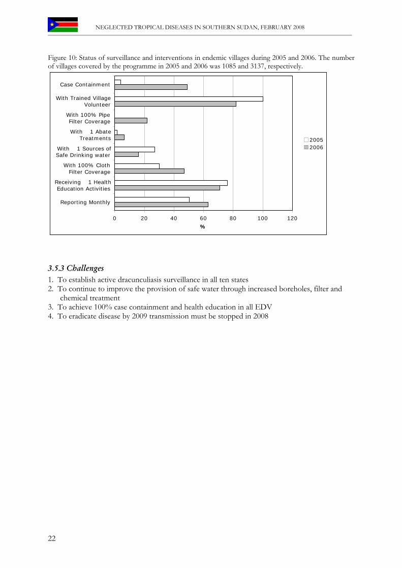
NEGLECTED TROPICAL DISEASES IN SOUTHERN SUDAN, FEBRUARY 2008
Figure 10: Status of surveillance and interventions in endemic villages during 2005 and 2006. The number of villages covered by the programme in 2005 and 2006 was 1085 and 3137, respectively.
0 20 40 60 80 100 120
Reporting Monthly
Receiving �1 HealthEducation Activities
With 100% ClothFilter Coverage
With �1 Sources ofSafe Drinking water
With �1 AbateTreatments
With 100% PipeFilter Coverage
With Trained VillageVolunteer
Case Containment
%
20052006
3.5.3 Challenges 1. To establish active dracunculiasis surveillance in all ten states 2. To continue to improve the provision of safe water through increased boreholes, filter and
chemical treatment 3. To achieve 100% case containment and health education in all EDV 4. To eradicate disease by 2009 transmission must be stopped in 2008
22
NEGLECTED TROPICAL DISEASES IN SOUTHERN SUDAN, FEBRUARY 2008
3.6 Lymphatic Filariasis
Authors: Ruiz JA & Richer M
3.6.1 Background Lymphatic filariasis (LF), more commonly known as elephantiasis, can manifest itself as a painful and profoundly disfiguring disease. The parasite and its life cycle: LF is caused by infection with mosquito-borne, parasitic worm of the genera Wuchereria and Brugia. Bancroftian filariasis, caused by Wuchereria bancrofti, is mainly transmitted by Culex quinquefasciatus and by some species of Anopheles and Aedes (Remme et al. 2006). Infective larvae are transmitted to humans during blood feeding by infected mosquitoes. The parasites are deposited in the vicinity of the skin puncture wound, from where they penetrate the skin and migrate to the lymphatic vessels. Over a period of 6 - 12 months, they develop into adult worms that cause damage and dilatation of the lymphatic vessels. The filariae live for several years in the human host. During this period they produce millions of young stages of microfilariae that circulate in the peripheral blood and are ingested by mosquitoes when these bite infected humans. The larval forms further develop inside the mosquito before becoming infectious to man. Disease burden: LF puts at risk more than a billion people in more than 80 countries. Over 120 million are estimated to be affected by it, of which over 40 million are seriously incapacitated and disfigured. Recent estimates indicate that more than 50 million people in sub-Saharan Africa are affected, accounting for 37% of the global burden (Michael & Bundy 1997). Geographical distribution: One-third of the people infected with the disease live in India, one third in Africa and most of the remainder in South Asia, the Pacific and the Americas. Clinical features: While LF is usually acquired in childhood, its visible manifestations occur in adults where they lead to temporary and permanent disability. As such, the disease has a major social and economic impact on endemic countries. LF is now recognized as a major source of morbidity and physical disability (Ramaiah et al. 1997) and has been ranked as the second major cause of long-term disability after mental illness (WHO 1999). Filariae lodge in the lymphatic system where they cause inflammation, dilatation and lymphatic system failure. They are responsible for a variety of clinical manifestations, including lymphoedema of the limbs, genital disease (hydrocele, chylocele and swelling of the scrotum and penis) and acute, recurrent secondary bacterial infections known as "acute attacks". The vast majority of infected people are asymptomatic, but virtually all of them have sub clinical lymphatic damage and as many as 40% have renal involvement. Control options: The strategy of the Global Programme to Eliminate Lymphatic Filariasis (PELF) has two components: firstly to interrupt transmission and secondly to alleviate the suffering of affected individuals [Lymphatic Filariasis Elimination Strategy (http://www.filariasis.org/resources/elimination_strategy.htm)]. To interrupt transmission, endemic districts must be identified and MDA be implemented to treat the entire at-risk population. In most countries this will be based on once-yearly administration of single doses of two drugs given together: albendazole plus either diethylcarbamazine (DEC) or ivermectin, the latter in areas where either onchocerciasis or loiasis may also be endemic (WHO 2000a,b). This yearly single-dose treatment must be carried out for 4-6 years. To alleviate the suffering caused by the disease, community education is used to raise awareness in affected patients. This promotes the benefits of intensive local hygiene and the possible improvement, both in the damage that has already occurred and in preventing the debilitating and painful acute episodes of inflammation. In addition to MDA, vector control should be carried out where this is feasible (WHO 2002b, Sunish et al. 2007). Control or elimination of Culex breeding sites in polluted water is possible by improving sanitation systems and hygiene in general. Where such improvements are not possible, the emphasis should be on the prevention of mosquito bites by means of self-protection.
23

NEGLECTED TROPICAL DISEASES IN SOUTHERN SUDAN, FEBRUARY 2008
3.6.2 Epidemiology and control in Southern Sudan
Historically, LF is known to be endemic in both North and Southern Sudan (Kirk 1957, Satti & Abdel Nur 1974). In 2003, the FMoH (Khartoum) started LF mapping in the North (El Setouhy & Ramzy 2003) and included some sites in Southern Sudan that were under the control of the FMoH. At the yearly LF control meeting sponsored by WHO EMRO in February 2007, the FMoH reported that nine northern states had completed the mapping exercise. The following prevalences were found: Khartoum State 4.5%, Kassala 5.4%, Gedaref 4.8%, Sennar 1%, El Gezira 26%, White Nile 3.3%, Blue Nile 27%, North Kordofan 41%, South Kordofan 6.6%. The three Darfur States could not be surveyed due to continued civil unrest. The three northern states of Red Sea, Nile and Northern were scheduled for surveys in 2007. The status of West Kordofan was not clear at the meeting, as discussions regarding boundaries are ongoing. The FMoH planed to pilot MDA using ivermectin plus albendazole in some states in 2007.
Information and data on LF in the ten states of Southern Sudan is more scarce. Anecdotal information indicates that the disease may be endemic in all of them. LF prevalence surveys, using Immuno-Chromatographic Test (ICT) for diagnosis were planned by WHO for 2006, but resources were diverted from this due to outbreaks of cholera and meningitis. Nevertheless, all available data were reviewed, a survey questionnaire on the occurrence of LF symptoms was distributed to health NGOs working throughout Southern Sudan and the responses received were analyzed. ICT data were available from two sources: i) the surveys carried out by the FMoH (Khartoum) in specific locations in the Southern Sudan, and ii) various screenings programmes conducted by NGOs. Areas surveyed are shown in Figure 11.
The existing data indicate that LF is hyperendemic in four states (Upper Nile, Western Equatoria, Central Equatoria and parts of East Equatoria). Questionaire results show that clinical manifestations occur in Jonglei, Lakes and Warrab. No information is as yet available from the remaining three states (Northern Bahr el Ghazal, Western Bahr el Ghazal and Unity). Figure 11: Areas surveyed for lymphatic filariasis (based on data collated by WHO Juba)
24
NEGLECTED TROPICAL DISEASES IN SOUTHERN SUDAN, FEBRUARY 2008
3.6.3 Challenges 1. Convenient implementation units for an LF elimination programme in Southern Sudan need
to be determined. Implementation units could be, but don’t need to be, counties. Considering two or more counties as one implementation unit may be more appropriate and practical, at least in some states.
2. Mapping of LF, using ICTs and the Lot Quality Assurance Sampling (LQAS) method, needs to be completed for all implementation units where there is no data. However, provided that a comprehensive mapping plan is presented to the Mectizan Donation Program (MDP), not all implementation units need to be mapped before ivermectin and albendazole will be donated for distribution in already mapped implementation units. A gradual expansion of the programme, ideally from the north (e.g. Upper Nile, Northern Bahr-el-Ghazal) towards the L. loa co-endemic areas in the South seems advisable, to ensure that implementation is started soon while the distribution of L. loa and areas of co-endemicity with W. bancrofti are clearly delineated.
3. A national strategy for the LF elimination needs to be formulated and presented to the MDP, for Southern Sudan to become eligible for drugs. This strategy will need to address the issue of intervening in areas where LF is co-endemic with L. loa. The MDP does not provide drugs for these areas, because the risk of Serious Adverse Events (SAE) is considered to outweigh the benefits of treatment. For areas where L. loa is endemic, a strategy aiming at high and sustained coverage with LLINs should thus be presented to the MDP, to complement the MDA approach implemented elsewhere.
4. To clearly determine which areas are co-endemic, further assessments for L. loa are required (see section 3.7)
5. The submission of an application for drugs to eliminate LF to the MDP needs to be combined with the ongoing annual submission of an application by the Southern Sudan Onchocerciasis Task Force (SSOTF) for ivermectin donations to control onchocerciasis. Good coordination between partners involved in LF elimination and the SSOTF will thus be essential.
6. MDP experience from other countries has shown that, in areas where LF and onchocerciasis are co-endemic, ivermectin and albendazole tend to be distributed by the LF elimination programme, because the implementation units are larger and encompass those for onchocerciasis. This will require further discussion with the SSOTF, as does the concept of integrated MDA (see section 4.2).
7. As part of an LF elimination programme, sentinel sites will need to be established to monitor progress.
25
NEGLECTED TROPICAL DISEASES IN SOUTHERN SUDAN, FEBRUARY 2008
3.7 Loiasis
Authors: Richer M & Kolaczinski J
3.7.1 Background Loiasis is a skin and eye disease that occurs in a minority of individuals infected with Loa loa; however the major public health concern arises from SAEs following treatment with ivermectin. The parasite and its life cycle: Loiasis is caused by the filarial nematode Loa loa, which is transmitted to humans by day-biting Chrysops flies (Boussinesq 2006). Two species are vectors, C. silacea and C. dimidiata. Once inside the body the infective larvae take about a year to develop into a mature adult. During this period the parasite lives and moves around the fascial layers of the skin. In periods of growth and development L. loa makes frequent excursions through the subdermal connective tissues where it is often noticed by the host. Once they reach maturity the adults mate and produce sheathed microfilariae. These closely resemble the microfilariae of W. bancrofti, but in stained films they assume a stiff angular attitude. The microfilariae are diurnally periodic in synchrony with their vector. Inside a fly they undergo two stages of development into infective larvae, which takes about ten days. Infective larvae can then be transmitted to humans. Disease burden: In some areas of Africa loiasis constitutes the second or third most common reason for medical consultation (Pinder 1988). However, the disease is less wide-spread than onchocerciasis or LF, and has been inadequately studied. Information on the burden of disease is thus not available. Because of the risk of SAEs to ivermectin in areas that are potentially endemic for L. loa, several treatment programmes for onchocerciasis have come to a standstill. This has re-awakened the interest of parasitologists and clinicians in this parasite, which will hopefully lead to a better understanding of its epidemiology and the loiasis burden over the coming years. Geographical distribution: L. loa is restricted to Africa, stretching from south–eastern Benin to Southern Sudan and Uganda, and from a latitude of 10°N to, perhaps, Zambia in the south (Boussinesq & Gardon 1997). It is often regarded as a parasite of forest regions, but very high prevalence rates of human infection have been reported from savannah areas where forest galleries seem favourable to the Chrysops vectors. A spatial model based on environmental factors has been developed to predict the prevalence of human L. loa infection in the area covered by the parasite’s distribution (Thomson et al. 2004). Such prevalence can also be evaluated in the field, using a rapid assessment method (RAPLOA) that is based on a history of ‘eye worm’ (TDR 2001, Takougang et al. 2002). This procedure can be used in combination with assessments for onchocerciasis (Wanji et al. 2005). Clinical features: Clinical signs may occur as soon as five months post-infection (Churchill et al. 1996), but the clinical prepatency may last up to 13 years (Thomas et al. 1970). Most of the pathological problems observed in people infected with L. loa are connected to periods when the migrating adult worms appear near the surface of the skin. The worms often appear around the eye (‘eye worm’), where they can be easily seen and extracted before they damage the conjunctiva. Migrating worms can also cause characteristic calabar swellings in the arms and legs. These are localized infections where the adult worm is secreting toxins. The swelling is an immune reaction caused when the worms residing in the subcutaneous tissue are injured by applying force to the skin. The insides of the worm are then exposed to the immune system, and a powerful reaction ensues. Recurrent swelling can lead to the formation of cyst-like enlargements of the connective tissues around the tendon sheaths. These swellings can be extremely painful when moved. Dying worms can also cause chronic abscesses followed by granulomatous reactions and fibrosis. Control options: Several drugs are active against the microfilariae of L. loa, but only DEC appears to have a macrofilaricidal effect on the parasite. The definitive cure of loiasis therefore relies on DEC, but great precautions need be taken to prevent SAEs that may occur following the DEC treatment of patients with high microfilarial loads (Boussinesq 2006). Within several days of treatment, DEC brings about a destruction of the microfilariae in the liver and a decrease
26
NEGLECTED TROPICAL DISEASES IN SOUTHERN SUDAN, FEBRUARY 2008
in microfilaraemia to negligible levels. In addition, DEC has a significant macrofilaricidal effect on L. loa. However, in 20%–60% of cases some adult worms remain alive after a first course of DEC, and two or more treatments are required to obtain a definitive cure. Despite the use of gradually increasing doses, various adverse events (including itching, rashes, oedemas, headaches and fever) develop in about 50% of patients. Their intensity is generally related to initial microfilarial load.
Ivermectin has a marked microfilaricidal effect on L. loa. One month after a single dose of 50–200 mg/kg, microfilarial loads fall to <20% of their initial values and this suppression of microfilaraemias persists for at least one year. After repeated monthly treatments, microfilarial counts fall to negligible levels. The adverse reactions to treatment are usually mild but ivermectin, like DEC, can induce an encephalopathy in patients harbouring >30,000 L. loa microfilariae/ml.
When administered daily for 3–4 weeks, mebendazole can also reduce L. loa microfilaraemias but this effect is often transitory and varies widely between patients. As mentioned above, most patients that develop L. loa encephalopathy following treatment with DEC or ivermectin have high microfilaraemias (exceeding 30,000– 50,000 mff/ml) pre-treatment. Gardon et al. (1997) estimated that the threshold above which there is a risk of developing a prolonged functional impairment after ivermectin treatment is markedly lower — about 8000 mff/ml. There have been attempts to see whether adverse reactions to DEC treatment of loiasis can be avoided or significantly reduced by using very low doses in the initial treatments. Unfortunately, such a strategy only seems to delay, and not prevent, the problems, with SAE developing as soon as a dose of 50 or 100 mg is reached (Boulesteix & Carme 1986). It would therefore seem wise not to treat with DEC or ivermectin any individual who has >8000 L. loa microfilariae/ml. Instead, such cases should initially have their microfilaraemias reduced to <8000 mff/ml, either by a three-week course of albendazole or, when possible, with three sessions of apheresis (Boussinesq 2006). Once microfilaraemias have been brought down, each case should be given a single dose of ivermectin (150–200 mg/kg), to reduce the microfilaraemia further. Once microfilarial loads of <1000 microfilariae/ml have been obtained, DEC can be administered, to achieve a complete cure of the disease. The first course of DEC should last 3–4 weeks, beginning with very low doses (of 6.25 or 12.50 mg/day, in microfilaraemic patients) and then gradually increasing to 300– 400 mg/day. Several courses of DEC, at intervals of 2–3 weeks, may be required to kill all the adult worms. Albendazole may be useful if repeated DEC treatments are ineffective.
3.7.2 Epidemiology and control in Southern Sudan In the past, a number of studies on L. loa were conducted in Sudan (Woodman & Bokhari
1941, Kirk 1953). To date it seems that the geographical distribution based on these data still applies. At the time, loiasis was found to occur between latitude 4º to 6º North, extending westwards into French Equatorial Africa and southwards into the Belgian Congo. It did not occur east of longitude 30º East and was not reported in Uganda. In Southern Sudan, this region corresponds to the present day Western Equatoria. In the 1950s about 20% of the population was infected with L. loa. The limited data collected over the last years indicate that prevalence remains high (APOC 2005). This is of major concern to the onchocerciasis control programme, because parts of Western Equatoria are co-endemic for loiasis and onchocerciasis, meaning that specific treatment procedures should be followed to avoid adverse reactions resulting from MDA with ivermectin. To assess the risk and provide recommendations, experts from the African Programme for Onchocerciasis Control (APOC) implemented a RAPLOA assessment in the Equatoria States of Southern Sudan in April 2005 (APOC 2005). Unfortunately, logistical and security constraints prevented the team from accessing many of areas that were suspected to be at risk. Further prevalence data are therefore needed to develop a map indicating high-risk areas (L. loa prevalence >20%) (Diggle et al. 2007), so that a modified ivermectin distribution protocol can be implemented in these. Clear delineation of the L. loa endemic area is also required as part
27
NEGLECTED TROPICAL DISEASES IN SOUTHERN SUDAN, FEBRUARY 2008
of planning for a national LF elimination programme, as L. loa endemic areas are generally excluded from ivermectin distribution and need to be targeted with alternative control measures, such as LLINs, to reduce transmission.
3.7.3 Challenges Areas co-endemic for L. loa, O. volvulus and/or W. bancrofti needs to be clearly identified to
allow targeted implementation of a modified onchocerciasis treatment protocol and formulation of an intervention strategy for areas where LF and L. loa are co-endemic (see Mectizan Expert Committee & Technical Consultative Committee, 2004) and recent update [Available from: http://www.mectizan.org/loarecs.asp]
28
NEGLECTED TROPICAL DISEASES IN SOUTHERN SUDAN, FEBRUARY 2008
3.8 Onchocerciasis
Authors: Baba S, Richer M, Lewis K, Chane F & Hopkins A
3.8.1 Background The parasite and its life cycle: Onchocerciasis is caused by infection with the filarial parasite Onchocerca volvulus. It is transmitted to humans through the bite of blackflies (Simulium spp.), which breed in fast-flowing streams and rivers in the inter-tropical zones. Living near these breeding sites increases the risk of blindness, hence the commonly known name ‘river blindness’ (Remme et al. 2006). Disease burden: Onchocerciasis is the world's second leading infectious cause of blindness. Prior to concerted control efforts, about 50% of men over the age of 40 years in some West African communities had been blinded by the disease. People therefore fled the fertile river valleys to settle in less productive upland country. In the 1970s, the resulting annual economic losses were estimated at US$ 30 million. According to recent estimates, 120 million people are at risk and 18 million are already infected. The disease is responsible for the loss of 1 million DALYs per year (WHO 2002c). Geographical distribution: The disease occurs in 35 countries. Of these, 28 are in tropical Africa, which is where 96% of the people at risk of onchocerciasis and 99% of those already infected live. Clinical features: Inside the human body, the adult female worm (macrofilaria) produces thousands of larvae (microfilariae) that migrate in the skin and the eye. The death of microfilariae is toxic to the skin and the eye, causing terrible itching and various eye manifestations (lesions). After repeated years of exposure, these lesions may lead to irreversible blindness and disfigurative skin diseases sometimes named leopard- or lizard-skin. Diagnosis and Control options: Because of the dramatic consequences of onchocerciasis in West Africa, WHO in 1974 launched the Onchocerciasis Control Programme (OCP) in collaboration with the World Bank, the United Nations Development Programme (UNDP) and the Food and Agriculture Organization (FAO). Vector control by treating the breeding sites with larvicides was the only available approach. The programme systematically expanded over its first few years to achieve full coverage of several river systems in seven countries. Nonetheless, even this ambitious start was not sufficient and the programme subsequently doubled in size to cover 11 countries. At this point the programme stretched over 1 200 000 km² to protect 30 million people. Vector control was the primary strategy in West Africa, and it was supplemented by drug distribution as of 1989-90. The OCP was officially closed in December 2002 after virtually stopping the transmission of the disease in all participating countries except Sierra Leone where operations were interrupted by a decade-long civil war. Based on the knowledge and experience gained by the OCP, the sponsoring agencies and NGOs launched a second programme in 1995, the African Programme for Onchocerciasis Control (APOC). This covered 19 more countries, the remainder of onchocerciasis endemic Africa. APOC is based on the distribution of Mectizan (Ivermectin), which was developed by Merck & Co. in the 1980s and is now donated for onchocerciasis control. Mectizan is distributed by communities themselves, trained and supported by the APOC partners; a strategy referred to as community-directed treatment with ivermectin (CDTI). It empowers local communities to fight river blindness in their own villages, relieving suffering and slowing transmission. In a few isolated foci in APOC areas, ground larviciding is used in addition to CDTI, with the aim of local vector eradication within a period of 1-2 years. After 8 years of operation, APOC had established 107 projects, which in 2006 treated 50 million people in 16 countries. The programme intends to increase this to treat 90 million people annually in 19 countries, protecting an at-risk population of 109 million, and to prevent 43,000 cases of blindness every year. The distribution network has been used extensively to deliver other interventions, such as vitamin A, integrated eye care, ivermectin and albendazole combined for LF control, praziquantel for schistosomiasis, and ITNs
29
NEGLECTED TROPICAL DISEASES IN SOUTHERN SUDAN, FEBRUARY 2008
for malaria prevention. Research is ongoing to measure the number and complexity of interventions that can be added on to this system without the CDDs being overwhelmed.
3.8.2 Epidemiology and control in Southern Sudan Sudan has been a member of APOC since its establishment in 1995. At the time, there were
an estimated 620,000 cases of onchocerciasis. The Southern Sudan Onchocerciasis Control Programme (SSOCP) was created in 1996, to coordinate the procurement and MDA of ivermectin by all NGO implementing partners. HNI assumed the role as lead organization for the SSOCP. With the emergence of a stronger Secretariat of Health in late 2000, the role of the SSOCP diminished and the SSOTF was created. This mechanism is in place to date and is equivalent to the National Onchocerciasis Task Force (NOTF) of North Sudan. In 2001, the SSOTF was recognized by APOC as the official partner in charge of onchocerciasis control activities in Southern Sudan, and in 2002 financial support commenced.
Onchocerciasis is endemic in many parts of Southern Sudan, though endemicity varies considerably between areas. In 2003, the distribution of onchocerciasis was estimated by the SSOTF, using the Rapid Epidemiological Mapping of Onchocerciasis (REMO) method. This has allowed classification of communities into three categories: priority areas requiring CDTI; areas not requiring treatment; and possible endemic areas that need further investigation (Figure 12). Based on the results of the REMO exercise the onchocerciasis control programme was divided into five project zones, which receive financial support from APOC.
All of the states of Southern Sudan are endemic for onchocerciasis, with the main endemic foci being located in Western Equatoria, Northern Bahr Al-Ghazal, and Western Bahr Al-Ghazal (figure 12). In these areas >80% of individuals in some villages have palpable nodules and blindness exceeds 12% (Mukhtar et al. 1998). Most onchocercal infection causes only a mild skin reaction, although microfilarial loads in the skin are high. These individuals are important epidemiologically, as they serve as a reservoir for transmission. Skin reactions include pruritic papular rashes that cover wide areas of the body, particularly the lower limbs. A major concern in the West Equatoria zone is the co-existence of Loa loa in specific areas, which can precipitate SAEs in those who are given ivermectin (see section 3.7).
MDA with ivermectin had already been initiated in 1995. At the time, HNI procured ivermectin for all NGO partners implementing MDA in hyper- and meso-endemic areas from the MDP. In 1995 the programme distributed 31,500 treatments, increasing to 437,773 treatments by 2003 (see figure 13). Following the ceasefire REMO was conducted in much of the Southern Sudan and five projects for onchocerciasis control were developed with APOC. These projects followed the more normal pattern in APOC countries with 75% of funding coming through the APOC / WHO system and 25% through NGOs who also give technical advice. HNI handed over the lead NGO role to CBM at this time, who then based their office at the Health Secretariat in Rumbek, and were responsible for procuring ivermectin and giving financial, logistical and technical support to the SSOTF, which is now an activity of the MoH-GoSS in partnership with CBM and local NGOs.
In 2006, the focus was on training and refreshing the different cadres who make up the programme from state to community level. The trainings were organized and facilitated by SSOTF and included training of 137 Trainers of Trainees (TOT), 33 County supervisors, Payam supervisors, 592 Health workers, 5526 CDDs and Community leaders. The GoSS has officially recognized onchocerciasis as one of the major NTDs, as it appeared in the "Presidential 200 Day Action Plan". The SSOTF co-ordination office is based in Eastern Bahr el Ghazal.
30
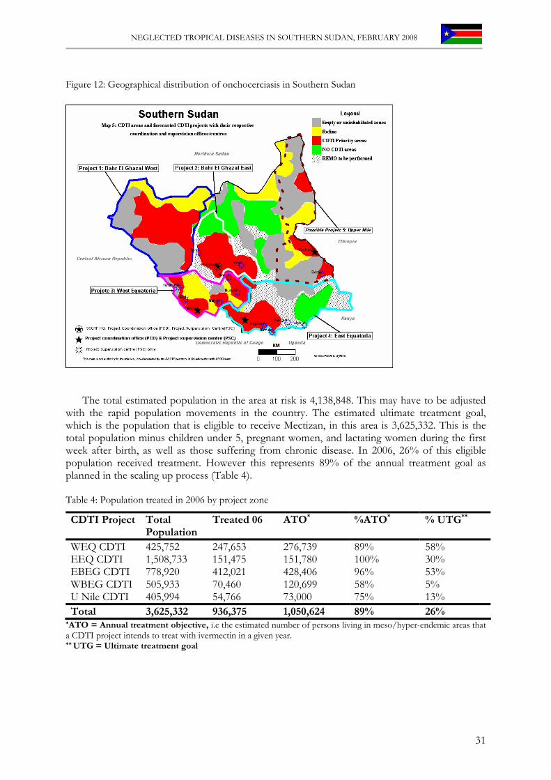
NEGLECTED TROPICAL DISEASES IN SOUTHERN SUDAN, FEBRUARY 2008
Figure 12: Geographical distribution of onchocerciasis in Southern Sudan
The total estimated population in the area at risk is 4,138,848. This may have to be adjusted with the rapid population movements in the country. The estimated ultimate treatment goal, which is the population that is eligible to receive Mectizan, in this area is 3,625,332. This is the total population minus children under 5, pregnant women, and lactating women during the first week after birth, as well as those suffering from chronic disease. In 2006, 26% of this eligible population received treatment. However this represents 89% of the annual treatment goal as planned in the scaling up process (Table 4). Table 4: Population treated in 2006 by project zone
CDTI Project Total Population
Treated 06 ATO* %ATO* % UTG**
WEQ CDTI 425,752 247,653 276,739 89% 58% EEQ CDTI 1,508,733 151,475 151,780 100% 30% EBEG CDTI 778,920 412,021 428,406 96% 53% WBEG CDTI 505,933 70,460 120,699 58% 5% U Nile CDTI 405,994 54,766 73,000 75% 13% Total 3,625,332 936,375 1,050,624 89% 26%
*ATO = Annual treatment objective, i.e the estimated number of persons living in meso/hyper-endemic areas that a CDTI project intends to treat with ivermectin in a given year. ** UTG = Ultimate treatment goal
31
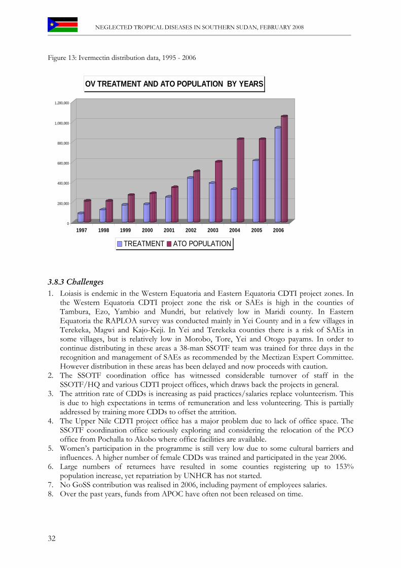
NEGLECTED TROPICAL DISEASES IN SOUTHERN SUDAN, FEBRUARY 2008
Figure 13: Ivermectin distribution data, 1995 - 2006
0
200,000
400,000
600,000
800,000
1,000,000
1,200,000
1997 1998 1999 2000 2001 2002 2003 2004 2005 2006
OV TREATMENT AND ATO POPULATION BY YEARS
TREATMENT ATO POPULATION
3.8.3 Challenges 1. Loiasis is endemic in the Western Equatoria and Eastern Equatoria CDTI project zones. In
the Western Equatoria CDTI project zone the risk or SAEs is high in the counties of Tambura, Ezo, Yambio and Mundri, but relatively low in Maridi county. In Eastern Equatoria the RAPLOA survey was conducted mainly in Yei County and in a few villages in Terekeka, Magwi and Kajo-Keji. In Yei and Terekeka counties there is a risk of SAEs in some villages, but is relatively low in Morobo, Tore, Yei and Otogo payams. In order to continue distributing in these areas a 38-man SSOTF team was trained for three days in the recognition and management of SAEs as recommended by the Mectizan Expert Committee. However distribution in these areas has been delayed and now proceeds with caution.
2. The SSOTF coordination office has witnessed considerable turnover of staff in the SSOTF/HQ and various CDTI project offices, which draws back the projects in general.
3. The attrition rate of CDDs is increasing as paid practices/salaries replace volunteerism. This is due to high expectations in terms of remuneration and less volunteering. This is partially addressed by training more CDDs to offset the attrition.
4. The Upper Nile CDTI project office has a major problem due to lack of office space. The SSOTF coordination office seriously exploring and considering the relocation of the PCO office from Pochalla to Akobo where office facilities are available.
5. Women’s participation in the programme is still very low due to some cultural barriers and influences. A higher number of female CDDs was trained and participated in the year 2006.
6. Large numbers of returnees have resulted in some counties registering up to 153% population increase, yet repatriation by UNHCR has not started.
7. No GoSS contribution was realised in 2006, including payment of employees salaries. 8. Over the past years, funds from APOC have often not been released on time.
32

NEGLECTED TROPICAL DISEASES IN SOUTHERN SUDAN, FEBRUARY 2008
Recommendations 1. Increase in therapeutic and geographic distribution of Mectizan 2. Solicit funds for CDTI activities from the GoSS, local NGOs and the private sector. 3. Empower affected communities to take ownership of their programmes. 4. Train more CDDs, CHWs and community supervisors to obtain a CDD to population ratio
of 1:150 5. Involve more females to obtain 50% female CDDs.
33
NEGLECTED TROPICAL DISEASES IN SOUTHERN SUDAN, FEBRUARY 2008
3.9 Trachoma
Authors: Richer M, Gatpan G, Lewis K, Kur L, Ngondi J, Emerson P, Becknell S & Nyholm S
3.9.1 Background Causative agent: Trachoma is caused by ocular infection with the obligate intracellular bacterium Chlamydia trachomatis. Ocular Chlamydia is spread through contact with eye discharge from the infected person (on towels, handkerchiefs, fingers, etc.) and through transmission by eye-seeking flies (Gambhir et al. 2007). The disease is associated with poor personal and environmental hygiene (in particular, limited access to water and sanitation), overcrowding and poor socioeconomic conditions. Disease burden: Trachoma is one of the oldest infectious diseases known to mankind and is the leading infectious cause of blindness, estimated to be responsible for 3.6% of blindness worldwide (Resnikoff et al. 2004). WHO estimates that there are 8 million people with trachoma who are already blind or are at high risk of developing blindness and 84 million are infected and in need of treatment. In areas where prevalence of active trachoma (WHO simplified grading system, grade TF) among children aged 1-9 years exceeds 10% and prevalence of trachomatous trichiasis (WHO simplified grading system, grade TT) in adults aged 15 years and above exceeds 1%, the disease is considered to be a serious public health problem. Geographical distribution: Today, trachoma is endemic in 56 countries mainly in poor rural areas, including pockets in Central and South America, many African countries and some countries in the Eastern Mediterranean. Trachoma is also still endemic in parts of several Asian countries and among the aboriginal communities in Australia. However, there is a lack of information from some major populations, which remains an important obstacle to trachoma control efforts (Polack et al. 2005). Clinical features: Ocular infection with C. trachomatis results in conjunctival inflammation that presents clinically as characteristic lymphoid follicles (trachomatous inflammation-follicular [TF]) and/ or papillary hypertrophy (trachomatous inflammation intense [TI]). Repeated infections result in conjunctival scarring of trachoma (TS). People with TS may progress to trachomatous trichiasis (TT) whereby the eyelashes turn inwards and touch the eyeball. TT leads to scarring of the cornea (corneal opacity, CO) and eventually irreversible blindness. Women are at greater risk (two to four fold) of developing TS, TT, CO and trachomatous blindness compared to men. Control options: In 1997, WHO established the Alliance for the Global Elimination of Blinding Trachoma by the year 2020 (GET 2020). WHO has endorsed the SAFE strategy for trachoma control, which comprises: eyelid Surgery for trichiasis; Antibiotics for active trachoma (Azithromycin and Tetracycline eye ointment); Facial cleanliness; and Environmental improvements including building pit latrines (to reduce the number of eye-seeking flies), providing water sources (for face washing) and keeping home compounds clean. The SAFE strategy aims at reducing infection and re-infection leading to lowering of risk of conjunctival inflammation, scarring and the sequelae of scarring, as well as surgical correction of trichiasis. The packaging of the components into a four-pronged community-based approach provides a comprehensive programme for trachoma elimination that is adaptable to many different situations and which can be implemented at the community level. Each component of the SAFE strategy uses appropriate and readily adaptable technologies. Trachoma control can therefore be integrated with broader health and development efforts, targeting poor and marginalised populations.
Trachoma has been eliminated as a blinding disease from several previously endemic countries and regions, both through significant improvements in the socioeconomic status of populations and through specific control efforts. Despite these successes, blinding trachoma continues to be an important public health problem in many developing countries of the world.
34

NEGLECTED TROPICAL DISEASES IN SOUTHERN SUDAN, FEBRUARY 2008
3.9.2 Epidemiology and control in Southern Sudan Trachoma has long been known to be prevalent in parts of Sudan (Majcuk 1966), but data on
the distribution and burden particularly in Southern Sudan continues to be limited. Three of the surveys conducted over the last decades (Salim et al. 1975, Tizazu & Mburu 1983, Mahmoud et al. 1994) have been of limited use for guiding current prevention of blindness programmes (Ngondi et al. 2005).
Unpublished, population-based trachoma surveys conducted by the FMoH (Khartoum) in 1999 found a high prevalence of trachoma in Southern Sudan. TCC has since supported further prevalence surveys in East and Central Equatoria States, Northern Bahr El Gazal, Jonglei State, and Upper Nile State (Amann 2002, Ngondi et al. 2005, King 2008). In all locations with available survey data the average prevalence of active trachoma (TF in children aged 1-9) was 47% (range 15%-87%), which is well above the 10% threshold recommended for control interventions (figure 14). Overall it has been estimated that 3.9 million people need antibiotic treatment and 206,000 people are in need of immediate trichiasis surgery (Ngondi et al. 2005).
Surveys conducted in 2005 in the district of Mankien found that 4% of people aged 5 years and above were blind. This is more than twice the level that would be expected, given what is known about the prevalence of blindness in other parts of rural Africa. The two most common causes of blindness and low vision were cataract and trachoma, each accounting for over one-third of cases (Ngondi et al. 2006a). In a specific trachoma survey in the same district it was found that the earliest stages of infection were very common, particularly in children aged 1 to 9, over half of whom had clinical signs of trachoma infection (Ngondi et al. 2006b). In adults, one in five had trichiasis caused by trachoma. These survey data demonstrate the way uncontrolled trachoma can ravage inaccessible and underserved communities. In common with other NTDs in Southern Sudan, there is an immediate need for a concerted effort to survey the entire region, provide resources, and deliver services to the marginalized and poverty stricken communities (Ngondi et al. 2007).
TCC and CBM started trachoma control projects in 2000. Initially, villages around Malakal were targeted, but this has since been extended to cover most of Eastern Equatoria and Jonglei states. Prevention activities, primarily focusing on health education and improvement in environmental hygiene are being complemented by drug distribution. Since the signing of the Comprehensive Peace Agreement, TCC and CBM have been working in close collaboration with the newly formed MoH-GoSS. Other NGO partners have included MedAir, Christian Mission Aid, Adventist Development Relief Agency, Sudan Medical Care, Tear Fund and ZOA Refugee Care. Azithromycin is donated by Pfizer to TCC and is distributed to the at-risk population annually. In 2001, during the first year of MDA, about 60,000 doses of azithromycin (Zithromax) were distributed, which increased to 84,096 doses in 2005, covering 34% of the population in the targeted areas (Table 5). Treatment with topical tetracycline reached 23,035 people, covering 57.6% of the individuals with active disease in the identified endemic areas.
35
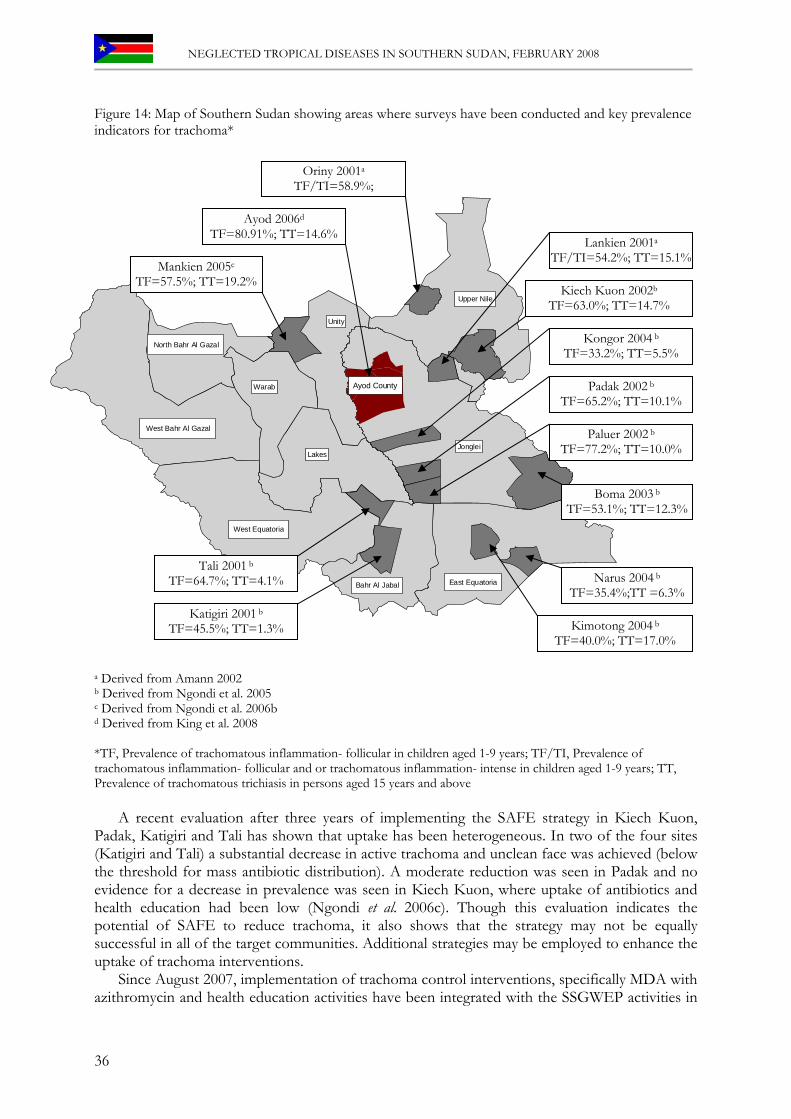
NEGLECTED TROPICAL DISEASES IN SOUTHERN SUDAN, FEBRUARY 2008
Figure 14: Map of Southern Sudan showing areas where surveys have been conducted and key prevalence indicators for trachoma*
West Bahr Al Gazal
North Bahr Al Gazal
Warab
Unity
Upper Nile
JongleiLakes
West Equatoria
Bahr Al Jabal East Equatoria
Ayod County
Oriny 2001a
TF/TI=58.9%;
Boma 2003 b
TF=53.1%; TT=12.3%
Lankien 2001a
TF/TI=54.2%; TT=15.1%
Kongor 2004 b
TF=33.2%; TT=5.5%
Padak 2002 b
TF=65.2%; TT=10.1%
Paluer 2002 b
TF=77.2%; TT=10.0%
Kiech Kuon 2002b
TF=63.0%; TT=14.7%
Mankien 2005c
TF=57.5%; TT=19.2%
Ayod 2006d
TF=80.91%; TT=14.6%
Tali 2001 b
TF=64.7%; TT=4.1%
Katigiri 2001 b
TF=45.5%; TT=1.3%
Narus 2004 b
TF=35.4%;TT =6.3%
Kimotong 2004 b
TF=40.0%; TT=17.0%
a Derived from Amann 2002 b Derived from Ngondi et al. 2005 c Derived from Ngondi et al. 2006b d Derived from King et al. 2008 *TF, Prevalence of trachomatous inflammation- follicular in children aged 1-9 years; TF/TI, Prevalence of trachomatous inflammation- follicular and or trachomatous inflammation- intense in children aged 1-9 years; TT, Prevalence of trachomatous trichiasis in persons aged 15 years and above
A recent evaluation after three years of implementing the SAFE strategy in Kiech Kuon, Padak, Katigiri and Tali has shown that uptake has been heterogeneous. In two of the four sites (Katigiri and Tali) a substantial decrease in active trachoma and unclean face was achieved (below the threshold for mass antibiotic distribution). A moderate reduction was seen in Padak and no evidence for a decrease in prevalence was seen in Kiech Kuon, where uptake of antibiotics and health education had been low (Ngondi et al. 2006c). Though this evaluation indicates the potential of SAFE to reduce trachoma, it also shows that the strategy may not be equally successful in all of the target communities. Additional strategies may be employed to enhance the uptake of trachoma interventions.
Since August 2007, implementation of trachoma control interventions, specifically MDA with azithromycin and health education activities have been integrated with the SSGWEP activities in
36
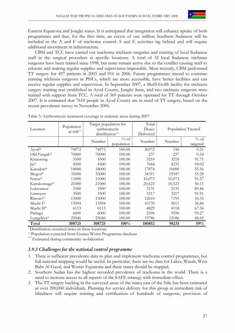
NEGLECTED TROPICAL DISEASES IN SOUTHERN SUDAN, FEBRUARY 2008
Eastern Equatoria and Jonglei states. It is anticipated that integration will enhance uptake of both programmes and that, for the first time, an excess of one million Southern Sudanese will be included in the A and F of trachoma control. S and E activities lag behind and will require additional investment in infrastructure.
CBM and TCC have carried out trachoma trichiasis surgeries and training of local Sudanese staff in the surgical procedure at specific locations. A total of 32 local Sudanese trichiasis surgeons have been trained since 1998, but none remain active due to the conflict causing staff to relocate and making regular supplies and supervision impossible. Most recently, CBM performed TT surgery for 497 patients in 2005 and 916 in 2006. Future programmes intend to continue training trichiasis surgeons in PHCs, which are more accessible, have better facilities and can receive regular supplies and supervision. In September 2007, a MoH-GoSS facility for trichiasis surgery training was established in Ayod County, Jonglei State, and two trichiasis surgeons were trained with support from TCC. A total of 369 patients were operated for TT through October 2007. It is estimated that 7610 people in Ayod County are in need of TT surgery, based on the recent prevalence survey in November 2006. Table 5: Azithromycin treatment coverage in endemic areas during 2007
Location Population at risk**
Target population for azithromycin distribution***
Total Doses
DeliveredPopulation Treated
Number % of population Number Number % of
targetedAyod* 74972 74972 100.00 26972 156 0.21Old Fangak* 70000 70000 100.00 237 237 0.34Kimotong 3500 3500 100.00 3210 3210 91.71Jie* 4500 4500 100.00 7644 4231 94.02Katodori* 18000 18000 100.00 17874 10000 55.56Mogos* 35000 35000 100.00 34311 19347 55.28Narus* 11000 11000 100.00 10,073 10,073 91.57Karukomuge* 21000 21000 100.00 20,623 10,523 50.11Lokwamor 3500 3500 100.00 3131 3131 89.46Lomeyen 3500 3500 100.00 3217 3217 91.91Riwoto* 13000 13000 100.00 12610 7195 55.35Machi I* 13594 13594 100.00 10739 5011 36.86Machi II* 6113 6113 100.00 4829 4118 67.36Paringa 6000 6000 100.00 5596 5596 93.27Longeleya* 25046 25046 100.00 19786 12186 48.65Total 308725 308725 100% 180852 98231 59%
* Distribution occurred twice in these locations
** Population extracted from Guinea Worm Programme database *** Estimated during community mobilization
3.9.3 Challenges for the national control programme 1. There is sufficient prevalence data to plan and implement trachoma control programmes, but
full national mapping would be useful. In particular, there are no data for Lakes, Warab, West Bahr Al Gazal, and Wester Equatoria and these states should be mapped.
2. Southern Sudan has the highest recorded prevalence of trachoma in the world. There is a need to increase access to all aspects of the SAFE strategy with immediate effect.
3. The TT surgery backlog in the surveyed areas of the states east of the Nile has been estimated at over 200,000 individuals. Planning for service delivery for this group at immediate risk of blindness will require training and certification of hundreds of surgeons, provision of
37
NEGLECTED TROPICAL DISEASES IN SOUTHERN SUDAN, FEBRUARY 2008
operating instruments and consumables, and provision of in-service support and supervision. Country-wide mapping will allow estimation of the national TT surgery backlog.
4. All localities surveyed in the last six years exceed the intervention threshold of 10% TF in children aged 1-9 years, to require the A, F and E components of SAFE. It is likely that the entire population of Southern Sudan is at risk of blinding trachoma. Trachoma control programmes will need to reach the communities with hygiene education, water and sanitation promotion and annual treatment with antibiotic.
5. National policy on MDA with azithromycin should be formalized to include all people living in trachoma endemic areas aged 6 months or older.
6. Most of the hyperendemic communities are inaccessible either by road or plane for between six to nine months of the year. Particularly in Upper Nile, the hyperendemic areas affect the Nilotic communities who are continuously on the move. Thus getting good coverage of azithromycin MDA is extremely difficult.
7. There is a dearth of water and sanitation facilities for enhancing the environment component of the SAFE strategy.
3.9.4 Future of Trachoma Control in Southern Sudan
Objectives include: 1. To conduct additional prevalence surveys, thus contributing to the mapping of trachoma in
Southern Sudan, and expand the existing programme of azithromycin distribution. 2. To increase access to water and sanitation facilities in hyperendemic communities. 3. To empower communities to improve hygiene and sanitation conditions among affected
populations. 4. To train health personnel in trichiasis surgical procedures and support them with logistics
support and supervision. 5. To establish a functional task force of trachoma control partners under the coordination of
the MoH-GoSS and implement standardized interventions, including the MDA policy, and monthly reporting formats.
38
NEGLECTED TROPICAL DISEASES IN SOUTHERN SUDAN, FEBRUARY 2008
3.10 Leprosy
Authors: Hickson L, Sindani I, Richer M & Kolaczinski J
3.10.1 Background The parasite and its life-cycle: Leprosy is a chronic infectious disease caused by Mycobacterium leprae, an acid-fast, rod-shaped bacillus (Remme et al. 2006). The exact mode of transmission of M. leprae is still not fully understood. Transmission is thought to occur only between humans, via nasal discharge and droplets from the respiratory tract of untreated patients with severe disease, although it may also occur via skin contact. The primary reservoir of infection is the human host. Untreated multibacillary leprosy patients are able to shed large amounts of M. leprae from the nose. Household and social contacts of such patients are at a higher risk of developing leprosy than the general population (van Beers et al. 1999). Disease burden: The burden of disease has been estimated at 192,000 DALYs and is concentrated in only a few countries. During 2003, 513,798 new cases were detected, of which more than 80 percent were in Brazil, India, Madagascar, Mozambique, Nepal, and Tanzania (WHO 2004b). India alone accounted for about 75% of the new cases. Case detection has remained remarkably stable over the past decade. Two difficulties affect the validity of DALY estimates for leprosy. The first is the lack of data, particularly on the burden of functional and psychosocial disability. The second is that the psychosocial consequences of leprosy often affect the whole family, not just the infected individual. People without any visible signs of leprosy may be stigmatized simply because they are known to be a leprosy patient. Even after completing treatment, people may remain stigmatized (Remme et al. 2006). Geographical distribution: Leprosy has been eliminated as a public health problem from 116 out of 122 endemic countries. Those few that remain are very close to eliminating the disease. However, pockets of high endemicity still remain in some areas of Angola, Brazil, Central African Republic, Democratic Republic of Congo, India, Madagascar, Mozambique, Nepal, and the United Republic of Tanzania. Clinical features: The disease mainly affects the skin, the peripheral nerves, mucosa of the upper respiratory tract and the eyes, but may affect the bones and internal organs. The skin signs of leprosy are relatively harmless, but complications of the disease may lead to severe consequences, such as blindness, infertility, disfigurement, and severe sensory and motor disability. Episodes of acute inflammation caused by hypersensitivity to bacterial antigens can be particularly severe. Patients can develop nerve damage without any obvious sign of these reactions, but after neuropathy has become irreversible, it may lead to secondary impairments, such as wounds, contractures, and shortening of digits. Many people experience psychosocial problems because of the visible impairment and activity limitations caused by leprosy (van Brakel 2000). Diagnosis and Control options: The objectives of leprosy control are to interrupt transmission, to cure patients, to prevent the development of associated deformities, and to rehabilitate those patients already afflicted with deformities. The strategy involves early case detection and the provision of adequate chemotherapy and comprehensive patient care (ILA 2002). Treatment relies on multi drug therapy (MDT) with dapsone, rifampin and clofazimine. Blister packs are provided free of charge by WHO.
39
NEGLECTED TROPICAL DISEASES IN SOUTHERN SUDAN, FEBRUARY 2008
3.10.2 Epidemiology and control in Southern Sudan Treatment of leprosy patients in Southern Sudan began in 1960 in greater Bahr el Ghazal.
Catholic missionaries established two leprosaria at Kuelkwac (near Wullu in Lakes State) and at Pagarau (Yirol County, Lakes State). With the expulsion of all religious organizations from the country in 1964 both of these facilities were destroyed. During the 1990s, leprosy control activities were re-initiated by a group of faith-based and other NGOs, with technical and commodity (drugs) support from WHO. As a result, the number of treatment centres increased from 12 to 29 between 2003 and 2005. At present, the majority of leprosy patients are being treated by the Catholic missionaries through the Diocese of Rumbek and by the Comboni Sisters working in the Tambura/Yambio Diocese, supported by the German Leprosy Relief Association (GLRA). The latter is a mobile outreach programme with trained Sudanese health workers visiting sites in Tambura, Yambio and Maridi Counties to diagnose new cases and distribute MDT. The programme operated by the Diocese of Rumbek and implemented by various religious congregations supports seven facilities for care and treatment of leprosy patients.
In November 2006, MoH-GoSS appointed a National TB/Leprosy/Buruli Programme Manager to coordinate activities, formulate policy and guidelines, with technical assistance from WHO. This appointment of the NTLP manager reflects the political commitment of the GoSS to leprosy control. GLRA is also reengaging itself in Southern Sudan to support the Leprosy and TB control programmes through the MoH-GoSS. Through concerted efforts, the leprosy control programme has achieved the following: • The expansion of leprosy (and TB) treatment as an integral part of primary health care
delivery • Improved early case-detection, and hence better prevention of disability • Increased case-management skills of health workers • Increased community awareness, through use of mobile IEC teams and health facility-based
health education • Establishment of socio-economic rehabilitation programmes in two areas • Revision of the reporting system
Although the exact prevalence of leprosy in Southern Sudan remains unclear, the available data indicate a declining trend. From 2003 to 2006 prevalence decreased from 3.9 to 2.3 cases per 10,000. Over the same duration, new cases detected also declined from 29.8 to 14.1 per 100,000. In 2006 a total of 1,060 new cases were reported. This decline in the prevalence and in the number of new cases reported can be attributed mainly to the improved case-management skills of health workers and to the updating of registers to remove those individuals that were cured, had defaulted or died. However, despite considerable improvements, MDT coverage remains low, at about 46%.
3.10.3 Challenges 1. Access to patients is increasing as areas that were previously not accessible due to insecurity
are now opening up. MDT needs to be expanded to these areas, require activities to raise community awareness and capacity building of health workers.
2. While MTD needs to be expanded to new areas, existing control areas require further support to increase staffing, provide training and improve on management. Further resources are also needed to increase community awareness, through advocacy and IEC activities, to promote self-reporting, reduce stigmatization and ensure rehabilitation and integration of cured patients and those with disabilities into the community.
40

NEGLECTED TROPICAL DISEASES IN SOUTHERN SUDAN, FEBRUARY 2008
3. The scaling up of leprosy control, in both existing and new areas, should be further integrated into the primary health care system. However, as the overall system is still weak, maintaining the balance between effective leprosy control and supporting the development of integrated service delivery is challenging.
4. Activities in areas that were under the control of the FMoH (Khartoum) need to be integrated into to operations governed by MoH-GoSS.
5. Regular monitoring and supervision is needed to ensure standardised case-management, recording and reporting in existing and new leprosy control areas.
41

NEGLECTED TROPICAL DISEASES IN SOUTHERN SUDAN, FEBRUARY 2008
3.11 Buruli Ulcer
Authors: Hickson L, Sindani I, Richer M & Kolaczinski J
3.11.1 Background The parasite and its life-cycle: Buruli ulcer is caused by Mycobacterium ulcerans and was first described in 1897 by Albert Cook in Uganda (Wansbrough-Jones & Phillips, 2006). In the 1950-60s many cases were reported from a county in Uganda called Buruli, hence the name (Clancey et al. 1962). The causative organism belongs to the family of bacteria that cause tuberculosis and leprosy, but it distinguishes itself from all other mycobacterial diseases by producing a potent toxin known as mycolactone. Mycolactone destroys cells in the subcutis, leading to development of large ulcers with underminded edges (Wansbrough-Jones & Phillips, 2006). Most patients are women and children who live in rural areas near rivers or wetlands. The incubation period of the disease is estimated to be three months. The exact mode of transmission remains enigmatic (v. d. Werf et al. 2005, Sizaire et al. 2006); however, it has recently been suggested that it may be transmitted by biting water bugs (Naucoridae cimicoides), meaning that it might be classified as a vector-borne disease (Marsollier et al. 2002). An alternative mode of transmission may involve penetrating skin injuries during fishing or farming activities that seed the micro-organism into subcutaneous tissues (Meyers et al. 1974). Disease burden: The burden of Buruli ulcer disease (i.e. cost, extensive surgery, long hospitalisation and development of debilitating sequelae) is largely due to late detection of the disease. Most patients seek treatment only when experiencing the advanced stages of the disease (Sizaire et al. 2006). Globally Buruli ulcer is the third most common mycobacterial infection after tuberculosis and leprosy. The disease causes high morbidity but has a low mortality rate. In Côte d'Ivoire, approximately 15,000 cases have been recorded since 1978 where up to 16 percent of the population in some villages are affected. In Benin, 4,000 cases have been recorded since 1989; in Ghana (6,000 recorded cases in a national survey in 1999) up to 22% of villagers are affected in some areas. The disease is hugely under-reported, with data collection mainly relying on passive case detection (WHO 2000c). Geographical distribution: Buruli ulcer is predominantly found in riverine areas with a humid, hot climate in the tropical and sub-tropical regions of Africa, Asia, Latin America and the Western Pacific. Currently the disease is prevalent in over 30 countries worldwide, though Africa is the most affected. Here Buruli ulcer emerged as an important cause of human suffering since 1980 and is most common in West Africa. All countries along the Gulf of Guinea are now affected. Clinical features: The disease often starts as a painless swelling in the skin and mainly occurs in the limbs. A nodule develops beneath the skin's surface teeming with mycobacteria. Unlike other mycobacteria, M. ulcerans produces a toxin, which destroys tissue and suppresses the immune system. Massive areas of skin and sometimes bone are destroyed causing gross deformities. When lesions heal, scarring may cause restricted movement of limbs and other permanent disabilities. One important feature of Buruli ulcer is the minimally painful nature of the disease, which may partly explain why those affected do not seek prompt treatment (v. d. Werf et al. 2005, Sizaire et al. 2006). Diagnosis and Control options: The clinical diagnosis of the ulcerative form is relatively straightforward, but more difficult for the nodule, plaque and oedematous forms. The differential diagnosis includes pyogenic abscess (i.e. bacterial infections that make pus or cause abscesses) in nodules, erysipelas (i.e. acute streptococcus bacterial infection of the dermis) in plaques, and cellulites in acute oedematous forms of the disease. In endemic areas, every suspicious lesion should be treated as an M. ulcerans infection until proven otherwise (Sizaire et al. 2006).
Tests for Buruli ulcer diagnosis include: i) direct smear examination with Ziehl-Neelsen staining to detect acid-fast bacilli from a swab or biopsy, ii) culture on Löwenstein-Jensen
42

NEGLECTED TROPICAL DISEASES IN SOUTHERN SUDAN, FEBRUARY 2008
medium, iii) histopathology, or iv) PCR. Smear examination in the presence of strong clinical suspicion is only about 40% sensitive. Culture is difficult, expensive, has a low sensitivity (up to 60%) and results are available only after 6-12 weeks. PCR allows quick detection and is sufficiently specific in patients’ samples (> 90%). In practice, however, diagnosis of Buruli ulcer is based on clinical aspects and rarely confirmed, because of limited access to laboratory services (Sizaire et al. 2006).
Until recently, surgery often involving extensive excision, with or without skin grafting, was the only available treatment. This is both costly and dangerous, leading to the loss of large amount of tissues/or permanent disability, and it does not prevent recurrence (v. d. Werf et al. 2005). Where surgical capacities are available, the cost is often far beyond the means of those severely affected (Asiedu & Etuaful 1998). Evidence on the role of antibiotics, such as the use of a combination of rifampicin and an aminoglycoside (streptomycin or amikacin), in the management of Buruli ulcer has been growing over the last years. Encouraging results have been obtained with the use of a combination of rifampicin and an aminoglycoside (streptomycin or amikacin) for the treatment of small early M. ulcerans lesions. These results have led to formulation of the recently released ‘Provisional guidelines for management of Buruli ulcer’, [http://www.who.int/buruli/information/antibiotics/en/index.html] recommending use of rifampicin plus streptomycin. To date, promotion of early detection and rapid treatment (by active case finding and health education) have achieved the best control (e.g. Evans et al. 2003). Inexpensive prevention strategies, such as wearing protective clothing when farming and the immediate cleansing of traumatic skin injuries might also help (e.g. Hospers et al. 2005).
3.11.2 Epidemiology and control in Southern Sudan During the 1990s, when the International Committee of the Red Cross (ICRC) reported four
cases of Buruli ulcer from Upper Nile and Bahr el Gazal. Before then the occurrence of the disease in Southern Sudan was unknown. From 2000 through 2006 an estimated 16,000 internally displaced people (IDPs) from the area around Raga were displaced to Mabia IDP camp in Tambura County. In July 2002, a suspected Buruli ulcer epidemic in the camp was reported by CARE International to WHO. From 25th to 26th July 2002, WHO, the Kenya Medical Research Institute (KEMRI) and CARE International carried out field investigation and collected specimens. Laboratory analysis and confirmation were conducted by KEMRI and the Institute of Tropical Medicine, Antwerp, Belgium. Though tests carried out at KEMRI showed that the 17 patients tested were infected with Mycobacterium species, M. ulcerans was only detected in two of the patients, using polymerase chain reaction (PCR). This was the first confirmed existence of Buruli ulcer in Southern Sudan. After the notification various agencies responded, including WHO, CARE International, Medair, Church Ecumenical Action in Sudan (CEAS) and the Catholic Church. A health facility was established in the camp to deal exclusively with the Buruli ulcer cases. From July 2002 to February 2004, a total of 1077 suspected Buruli ulcer cases were diagnosed in Mabia. At Yambio hospital 5 cases, all from Nzara, were diagnosed and treated. In Mabia the disease occurred predominantly in the IDPs and was most common among children (accounting for 60% of all cases), although it is known to affect all age groups. There appeared to be no sex difference between the affected patients.
In 2004, an advocacy meeting was convened in Nairobi (26-27 February) to improve awareness and strengthen surveillance and control of Buruli ulcer. One Sudanese surgeon was trained in Ghana on Buruli ulcer management and a national counterpart to the WHO focal point was appointed to coordinate all Buruli ulcer activities in Southern Sudan. Under this leadership, national and regional task forces were established and an investigation team was formed, which visited Mabia, Tambura, Nzara and Yambio counties to determine the scale of the problem in Western Equatoria. The disease was confirmed in all counties, and one case was reported from Nimule hospital (Eastern Equatoria). Suspected cases have since also been
43

NEGLECTED TROPICAL DISEASES IN SOUTHERN SUDAN, FEBRUARY 2008
reported from Upper Nile and Central Equatoria, but have not been confirmed to date. This indicates that other states of Southern Sudan may also be endemic for the disease, though based on current evidence the area around Nzara in Western Equatoria seems to be the epicentre.
During 2005, the number of new cases reported from Nzara increased from four in 2004 to 23 new cases and one recurrent case. No new cases were reported from Tambura after the IDPs returned to their original homes in Raga. A total of 27 health workers were trained on case detection and treatment in Nimule hospital (Eastern Equatoria). Drugs such as rifampicin and streptomycin and other supplies were purchased and distributed to Nzara and Yambio hospitals. Regular supervision and monitoring visits were carried out to support service providers in the field.
A further increase in Buruli ulcer burden was observed during 2006, when 36 new and four recurrent cases were diagnosed, mostly from Nzara. Activities to address diagnosis and control were further stepped up, focusing on strengthening of early case detection and IEC at community level, standardization of case-management and training of health workers. Regular field visits by the MoH-GoSS and WHO were conducted to provide the necessary support.
3.11.3 Challenges 1. With the return of peace to Southern Sudan, accessibility to remote areas is improving and it
is likely that more Buruli ulcer endemic areas will be discovered. For example, three suspected cases were recently reported from Bunagok (Lake State), but yet need to be confirmed. More resources are required to investigate reports of suspected cases and establish adequate laboratory facilities to allow confirmation.
2. As the awareness of Buruli ulcer among the community and health staff in Southern Sudan increases, more and more suspect cases are being reported. More training of health workers, school teachers and village health workers is needed to strengthen early diagnosis, and more resources are need to develop and disseminate appropriate IEC materials.
3. Further training and infrastructure development is needed to improve on case management of confirmed cases.
4. Standardize recording and reporting needs to be rolled out to all endemic areas
44

NEGLECTED TROPICAL DISEASES IN SOUTHERN SUDAN, FEBRUARY 2008
3.12 Nodding Disease/Syndrome
Authors: Richer M, Baba S & Kolaczinski J
A condition that communities referred to as “Nodding Disease” was first noted in 1990 or 1991 in Lui/Amadi villages in East Mundri County. In 1997, the condition was officially reported to WHO from Lui by Samaritan’s Purse, an NGO working in the area. After the first reports in 1997 the number of cases seen, especially in the Lui/Amadi region, seemed to increase until the situation stabilized after about three years. Although the epicenter seemed to be Lui/Amadi, isolated or small numbers of patients were also reported from Katigiri and Rokon (Juba County), Yambio (Yambio County), Morobo (Yei County), Bogori, Yeri, Mvolo (Mvolo County), Billing, Wulu, Kulu (Rumbek County), Kozi (Maridi County) and Kotobi (West Mundri County). WHO was requested to assess the situation in 2001.
The name “nodding disease” resulted from the first sign shown by affected children, which consists of an occasional, momentary, involuntary bobbing of the head especially when the child is given traditional food to eat or when awakening early in the morning. With time the nodding episodes become more frequent both in number and duration and children often injury themselves. The nodding episodes may progress until the child develops typical grand mal seizure episodes, psychomotor seizures and/or night terrors. Most children are considered normal by the parents when the nodding first appears, but as the disease progresses they are usually withdrawn from school. Over time most children deteriorate mentally, possibly related to uncontrolled seizures, and are known to die due to trauma resulting from the seizures. Treatment with anticonvulsants especially carbomazepine controls the seizures, but as the cause has not yet been identified there is no cure. Other manifestations of disease vary widely between individuals. Many children have typical findings of onchocerciasis such as nodules, skin disease and blindness and some children demonstrate growth retardation and fail to develop normal puberty.
The first assessment, in September 2001, was carried out by WHO-EWARN (Disease Early Warning and Response Network) in Lui/Amadi and confirmed the presence of the condition. In November 2001, HNI and the TCC collected samples for parasitological examination from children with and without nodding diseases in Lui and Amadi. It was established that nodding disease was not due to infection with T. brucei gambiense, W. bancrofti or L. loa. However, patients suffering from nodding disease consistently had more positive skin snips and higher microfilaria loads of O. volvulus when compared to children without the condition. This was consistent with earlier observations, for example from 1946, where British staff in Western Equatoria noted a high prevalence of seizure disorders in onchocerciasis endemic areas. Thus there seems to be an association between seizures and the presence of onchocerciasis.
In January 2002 further investigation by WHO ruled out the involvement of environmental pollutant, chemical agent or food toxins as a cause of nodding disease. However, as found a year earlier, a higher proportion of patients with nodding disease were infected with O. volvulus when compared to patients without the condition. In April 2002, a neurologist recruited by WHO preformed portable EEGs on 31 patients with nodding disease. All EEGs were abnormal, showing specific progressive epileptic encephalopathy.
In 2006, WHO was requested to re-assess the situation because affected communities were concerned that the disease was spreading. The reports received by WHO were unclear as to whether new cases of nodding disease were appearing or if existing cases were moving to new locations. Communities were fearing that affected children would spread the disease to other children. In response, WHO contacted Samaritans Purse in Lui/Amadi. The organization reported that new cases of nodding disease did continue to appear sporadically, but that there had been no marked increase in the number of new cases.
Based on the evidence to date, nodding disease is a seizure disorder characterised by abnormal EEG findings. The cause is unknown, but there seems to be an association with onchocerciasis. There is no known cure, but the use of anticonvulsants helps to control the
45
NEGLECTED TROPICAL DISEASES IN SOUTHERN SUDAN, FEBRUARY 2008
symptoms in some patients. Nodding disease has many similarities to a condition called Nakalanga or Kifafa, which has been reported from Uganda (Kipp et al. 1996) and Tanzania (Neuman et al. 1995), respectively. Both these conditions have been associated with onchocerciasis.
Further studies will be required to determine the pathophysiologic mechanism of disease. Meanwhile, regular access to anticonvulsant drugs needs to be expanded to improve on the quality of life and life expectancy of children affected by the condition.
46

NEGLECTED TROPICAL DISEASES IN SOUTHERN SUDAN, FEBRUARY 2008
47
4. INTERVENTION OPTIONS
The individual sections on the epidemiology and control of NTDs in Southern Sudan documented in this situation analysis, highlight the enormous NTD burden in the country. The multiple burden of different NTDs is probably the largest anywhere in Africa, with 12 NTDs endemic in the country. The full extent of the impact of NTDs on the population’s health becomes apparent when viewed in the context that out of 12 endemic diseases, only onchocerciasis, dracunculiasis and trachoma have so far benefited from sustained, large-scale control, supported by partners such as WHO, APOC, TCC, CBM and ITI. The MoH-GoSS has played a key role in this process by developing national control strategies and appointing focal persons. By contrast, the control of the other nine NTDs has either been intermittent or absent, and often lacks a cohesive strategy.
Experience from numerous settings provides promising evidence on how international partnerships can contribute to reducing the NTD burden. Dracunculiasis is on the verge of being eradicated globally (Ruiz-Tiben et al. 2006), as is the public health importance of trachoma in Morocco (Kumaresan & Mecaskey 2003, Levine et al. 2004) and of lymphatic filariasis in Egypt (Molyneux 2006) on Samoa (Ichimori & Crump 2005) and on Zanzibar (Mohamed et al. 2006). Significant reductions in the burden of other NTDs in selected foci have also been achieved, including onchocerciasis (Thylefors & Alleman 2006), STH and schistosomiasis (Kabatereine et al. 2006). Except for Guinea worm control, an important component of the interventions promoted by these global partnerships are donated drugs and their use in MDA. Globally, more than 300 million treatments for onchocerciasis have been administered to date (Boatin et al. 2006), Pfizer has partnered with ITI to donate azithromycin for trachoma elimination (Kumaresan 2005) and GlaxoSmithKline now donates albendazole to be added to ivermectin or diethylcarbamazine for treatment of LF.
To date, Southern Sudan has only benefited partially from international partnerships and the MDA approach, largely because resources were lacking to meet the prerequisites of becoming a member of global initiatives (e.g. Global Programme for Elimination of Lymphatic Filariasis, Schistosomiasis Control Initiative, Mebendazole Donation Initiative [for STHs]). The potential for expanding MDA to control or eliminate other NTDs, either through a disease specific programme or through an integrated approach is discussed in sections 4.1 and 4.2, respectively. Of the endemic NTDs in Southern Sudan, only trachoma, STHs, LF, onchocerciasis and schistosomiasis can be controlled through MDA (Table 6), whereas diseases like VL or HAT require more targeted interventions, because treatment is too toxic or too expensive to be administered to individuals other than those with confirmed infection. The control of these diseases needs to be largely addressed as part of multifunctional health care delivery, which is discussed in section 4.3.

NEGLECTED TROPICAL DISEASES IN SOUTHERN SUDAN, FEBRUARY 2008
48
Table 6: Major characteristics of NTDs endemic in Southern Sudan and gap analysis of intervention improvements Disease
Vulnerable Groups
Primary Interventions Currently Used Limitation of Current Intervention Potential Improvements Integrated MDA recommended?*
Visceral Leishmaniasis (Kala-Azar)
All ages Passive case detection at a few health facilities equipped to treat the disease; treatment with antimonials
Limited access to essential drugs; cost of drugs; emerging drug resistance; no officially endorsed treatment protocol or intervention strategy
Increase number and improve quality of treatment centres; deliver health education and LLINs to highly endemic communities; phase-in SSG/paromomycin combination therapy; ensure availability of second-line treatment
No
Human African Trypanosomiasis
All ages Passive case detection at a few health facilities; treatment with pentamidine, eflornithine and melarsoprol
Inadequate surveillance, limited number of treatment facilities and trained health workers
Active case detection; increase number of facilities and staff for HAT treatment; update treatment protocols at Juba Teaching Hospital; introduce and maintain vector control (tse-tse traps)
No
Trachoma Children, adults (especially women)
SAFE strategy: Trichiasis surgery, antibiotics for active trachoma, facial cleanliness and environmental improvements
Limited coverage and varying uptake of interventions by communities
Expand geographical coverage of SAFE; improved health education with the aim of increasing coverage within targeted areas
Yes
Buruli Ulcer Children Antibiotic treatment using, for example, rifampicin and aminoglycoside
Disease distribution not clearly established, limited access to treatment and surgery
Validate suspected endemic areas; expand access to treatment and surgery
No
Leprosy Adults Multidrug therapy, blisterpacks provided free of charge by WHO
Limited coverage (approx. 46% of Southern Sudan)
Expand coverage No
Ascariasis Trichuriasis School-age children
Hookworm Infection
School-age children, women of reproductive age
Single dose albendazole, distributed alongside NIDs
Limited coverage, both in terms of the number of doses delivered per year and the number of years over which MDA has been implemented to date
Expand frequency of drug administration to a maximum of 3 doses per year, every year, and expand the age range of the target population up to 15 year olds; introduce integrated MDA
Yes
Lymphatic Filariasis Children, adults None to date Lack of prevalence data; hence no application for free drugs submitted to MDP; no budget to cover drug distribution costs; no palliative care; Co-endemicity of L.loa in Equatoria region
Map disease distribution and prevalence; Further mapping of L. loa distribution to delineate areas of co-endemicity; formulate national control strategy, start drug distribution in some endemic states and gradually scale-up, combine with large-scale distribution of LLINs for vector control; establish palliative care
Yes
Loiasis Children, adults2 No specific interventions, though ivermectin distribution in onchocerciasis coendemic areas will reduce disease burden (and may cause SAEs)
Boundaries of loiasis endemic area not clearly delineated
Complete the RAPLOA assessment started in 2005; formulate guidelines for LF co-endemic areas according to latest recommendations of the Mectizan Expert Committee
Yes, provided precautionary measures are put in place*
Onchocerciasis Adults Single annual dose of ivermectin to entire communities
Incomplete coverage; high attrition rate of CDDs
Further expand therapeutic and geographical coverage of annual ivermectin treatment; continue training of CDDs particularly focusing on females to offset the attrition
Yes
Dracunculiasis All ages Water filtration, provision of safe water, treatment of water sources, health education, case containment, surveillance
Incomplete coverage Expand coverage of interventions and further strengthen surveillance No
Schistosomiasis School-age children, women of reproductive age
None to date Lack of prevalence data and intervention strategy
Map distribution and prevalence; formulate national control strategy, procure praziquantel and distribute according to strategy
Yes
Nodding Disease School-age children Administration of anticonvulsant drugs to some patients
Pathophysiologic mechanism poorly understood, hence no treatment. Limited access to anticonvulsant drugs
Expand access to regular anticonvulsant therapy, carry out further studies on pathophysiologic mechanism and disease distribution
No
2 Garcia et al. 1995, Pion et al. 2005, Pion et al. 2006 * According to WHO (2006). This manual recommends specific cautionary measures for onchocerciasis MDA in loiasis endemic areas. MDA for LF is not recommended, but other interventions, such as LLIN for vector control, should be scaled-up as part of a comprehensive national LF elimination plan.

NEGLECTED TROPICAL DISEASES IN SOUTHERN SUDAN, FEBRUARY 2008
4.1 Disease-specific interventions
In the emergency and post-emergency environment of Southern Sudan a number of successful NTD control programmes were launched, targeting onchocerciasis, dracunculiasis and, more recently, trachoma. The onchocerciasis and dracunculiasis control programmes provide good examples of disease-specific interventions, where new structures were established to deliver the necessary interventions to populations at risk. As demonstrated by both programmes, the impact of focusing on the control or elimination of a specific disease can be dramatic and well justified, particularly in an environment of insecurity and resource constraints. Important considerations prior to the establishment of disease-specific programmes are whether the goal is time-bound elimination or provision of continuous control (e.g. see table 7) and whether resources are sufficient to achieve the goal. If elimination is not an option, then integration with the delivery of other interventions may be more cost-effective in the medium to long-term.
In the current post-conflict environment, where new opportunities for control or elimination and new funding sources are opening up, the continuation of disease-specific programmes and the establishment of new ones require careful consideration. As shown by the dracunculiasis control programme, effective control leading up to elimination may free up resources that can be redeployed elsewhere. The MoH-GoSS and TCC have thus started to deliver interventions for trachoma (azithromycin) and malaria (LLINs; in collaboration with Malaria Consortium) through the existing village volunteer network. Similar opportunities for integration may also exist in the onchocerciasis control programme, with its extensive network of CDDs. It is therefore recommended that: • No new disease-specific programmes are established unless it can been shown that such
approach provides the only viable solution and that resources are available to reach the set goal.
• The scope of using existing disease-specific programmes (Guinea worm and onchocerciasis) to address other NTDs is further explored.
• Due attention is paid to the need for additional resources if existing structures, such as the CDD network, are seemingly suitable for the delivery of other interventions. Overloading established systems by ‘integrating’ additional activities in the absence of sufficient additional support, such as training, monitoring and supervision, needs to be avoided.
• Taking account of above points, clear strategies are formulated for the control or elimination of all NTDs in Southern Sudan. Though the strategies needs to be disease-specific, it should be clear how interventions will be delivered in the context of other health sector developments, be it targeted campaigns, community-based structures or multifunctional health care delivery. Development of a national strategy for elimination of LF is particularly pressing, as it is required to obtain free drugs (ivermectin and albendazole) from the MDP.
• Because of the dynamic situation, in particular health sector and other infrastructure reconstruction, NTD control strategies need to be regularly revisited to ensure that new opportunities for the delivery of interventions are incorporated. For example, as the network of schools increases, regular school-based de-worming should become a reality.
49

NEGLECTED TROPICAL DISEASES IN SOUTHERN SUDAN, FEBRUARY 2008
4.2 Integrated mass drug administration (MDA)
The use of MDA plus other interventions is presently recommended for seven NTDs – ascariasis, trichuriasis, hookworm infection, schistosomiasis, lymphatic filariasis, trachoma and onchocerciasis (WHO 2006). Though considerable progress has been made in their control, it is feared that existing financial resources and global political commitment are not sufficient to reach the goals of elimination or control by 2020 at the latest (Table 7), as set by the World Health Assembly. In 2006, WHO therefore developed a new strategy of ‘integrated NTD control’, in order to enhance the coverage and impact of existing programmes. Rather than delivering single interventions through single delivery systems, WHO now recommends that a range of delivery systems are used to provide MDA simultaneously for a number of NTDs. In certain situations integration with other existing control programme, for example the national malaria control programme, may also be an option (e.g. Molyneux & Nantulya 2004, Blackburn et al. 2006).
The rationale for proposed integration is that for those diseases that currently benefit from control, the existing programmes are mostly vertical and often work in parallel. Only four drugs – albendazole, ivermectin, praziquantel and azithromycin – are used to control the above seven NTDs (table 7). As they can exhibit geographical overlap in some settings, it is thought that a single structure, such as CDTI, could be readily used to deliver more than one treatment. Where the structures are already in place this would, in theory, only slightly increase costs when a component is added, or reduce costs if two structures were merged, while considerably expanding coverage (Molyneux & Nantulya 2004). Potential cost savings of 26 – 47% have been estimated for integrated MDA when compared to vertical programmes (Brady et al. 2006). Table 7: Programme goals and intervention strategies for diseases controlled through MDA (adapted from Brady et al. 2006)
Disease Goal Ages targeted
Drug regimen
Frequency Delivery Strategy
Trachoma Elimination of blinding trachoma as a public health problem by 2020
6 months to 80 years
Azythromycin 1 x annually MDA for five years
Soil-Transmitted Helminths (ascariasis, trichuriasis, hookworm)
Regular treatment of 75% of at-risk school-age population by 2010
6–15 years Albendazole/ mebendazole
2 x annually Health days and school health programmes
Lymphatic filariasis
Elimination as a public health problem by 2020
5 – 80 years Ivermectin + albendazole
1 x annually MDA for five years
Onchocerciasis Establishment of community-based sustainable yearly treatment in areas with moderate/high intensity by 2010
5 – 80 years Ivermectin 1 x annually MDA via community directed treatment
Schistosomiasis Regular treatment of 75% of at-risk school-age population by 2010
6–15 years, plus other high risk groups
Praziquantel 1 x annually MDA in high risk areas plus school health programmes
Although the concept of an integrated approach is logistically and economically appealing,
experience at the country level is surprisingly limited and a number of aspects need careful evaluation before MDA is rolled-out at national scale (Kolaczinski et al. 2007). Delivery structures and target geographical areas often differ, as well as the recommended frequency of drug administration. For example, STH treatment is recommended for school-age children every 6-12
50
NEGLECTED TROPICAL DISEASES IN SOUTHERN SUDAN, FEBRUARY 2008
months, whereas treatment for onchocerciasis and LF is recommended for the whole community on an annual basis, though the frequency and number of rounds of ivermectin treatment required to interrupt transmission of LF or onchocerciasis remain unknown (Remme et al. 2006).
Integration of trachoma control into MDA is particularly challenging since at least one week has to pass between the administration of azithromycin and that of other anthelminthic drugs, due to lack of drug safety data (WHO 2006). Moreover, drug treatment is only one component of the SAFE strategy for trachoma control. The integrated delivery of azithromycin and tetracycline into MDA programmes should not lead to the neglect of the other SAFE components. A better understanding of the opportunities and challenges of integrated MDA in the context of Southern Sudan will be generated over the coming years. USAID has recently awarded funding through RTI International to the Malaria Consortium, to implement this approach in two states (Upper Nile and Northern Bahr-el-Gazal).
Apart from integrating chemotherapy, there is considerable potential for integrated vector control for some NTDs. In large parts of Africa the same mosquito species transmit both LF and malaria. Increasing the coverage with LLINs as part of malaria control efforts is thus likely to impact on LF vector densities and transmission (Pedersen & Mukoko 2002, Brockarie et al. 2002), although the effectiveness of this approach will need further investigation (WHO 2002b). Use of LLINs is also likely to provide personal protection against the crepuscular or night-biting sandfly vectors of VL (Ritmeijer et al. 2007). Successful implementation of integrated approaches in Southern Sudan will require flexibility and innovation to meet the challenges of a country in flux and reconstruction. It is recommended that:
• The optimal drug delivery strategy is identified. These may include:
o Integrated community-based MDA for STH, LF and onchocerciasis using albendazole and ivermectin. Azithromycin for trachoma may be delivered at least one week before or after distribution of anthelmintics.
o School-based delivery of albendazole/mebendazole and praziquantel for STH and
schistosomiasis control, respectively, once reconstruction of school infrastructure has progressed further and school attendance has increased.
• The suitable timings of annual and biannual treatment should be explored. • The potential of distributing a combination of LLINs and other interventions, such as MDA,
through community-based campaigns should be investigated.
51
NEGLECTED TROPICAL DISEASES IN SOUTHERN SUDAN, FEBRUARY 2008
4.3 Multifunctional health-care delivery
As mentioned above, case-management for a number of NTDs has to be provided at health facilities, because of the technical requirements for diagnosis and/or treatment. At present, facility-based health care delivery continues to rely on considerable support from NGOs, because the number and availability of skilled Southern Sudanese medical staff is limited and there are no reliable logistics systems to ensure continuous supplies of diagnostics and drugs. Where NGOs have been the key provider of diagnosis and treatment for a specific NTD, for example the way that VL has benefited from the presence of MSF in parts of northern Jonglei and Upper Nile, there is a danger that the departure of the NGO results in a gap in case-management.
Experience from other countries has shown that the present, transitional environment, of Southern Sudan can easily result in such gaps, because emergency NGOs are gradually phasing out, while more development orientated organizations may be slow to respond and the MoH has often little capacity to absorb the activities that have been ‘handed over’ (e.g. Tulloch et al. 2003). In the interim, while health sector reconstruction is ongoing, it is therefore suggested that:
• NGOs seek closer communication with the MoH-GoSS, to ensure that staff at central and
state level are aware of plans for phasing-out and of funding constraints experienced. • Based on this information, resources from funding mechanisms such as the Common
Humanitarian Fund or the MDTF are allocated to priority areas to prevent gaps. • Mechanisms for gradual transition from NGO providers to the MoH-GoSS are explored and
funded. For example, secondment of a limited number of technical staff by NGOs or other agencies to key referral facilities may be an option to continue case-management by Southern Sudanese nurses according to best-practice, while the pool of national medical staff is being expanded and the drug supply chain is being strengthened.
• While the health information system is being developed, improved reporting by health care providers to the relevant organization (for many NTDs this currently continues to be WHO) will provide better data that can be used to target resources to areas most at need and for advocacy to ensure that adequate resources are made available now and in future to procure the required diagnostic and drug supplies.
52
NEGLECTED TROPICAL DISEASES IN SOUTHERN SUDAN, FEBRUARY 2008
5. GAP ANALYSIS
As indicated, many of the responses to NTDs in Southern Sudan have to date been piecemeal, small-scale, and largely supported/delivered by NGOs. Strong political support and international funding are essential to addressing such shortcomings in the future and to ensure that existing investments and levels of control can be maintained or scaled-up as the international presence gradually declines. The following general elements need to be undertaken in order to inform an effective strategy for NTD control in Southern Sudan: • Improve coordination and planning. Whereas the control programmes for onchocerciasis,
dracunculiasis and trachoma benefit from good coordination amongst partners, the control of other NTDs could benefit from a MoH-GoSS led coordination mechanism. Establishment of a NTD task force may be an option, to avoid setting-up of numerous new mechanisms that would be hard to administer. Unless spare capacity can be identified in the MoH-GoSS, administrative support to organize meetings and report outcomes may, in the interim, need to be provided by one of the NTD partners.
• Define objectives and outcomes. The government and its partners should identify key objectives
and establish outcomes that can be easily monitored. A suggested objective would be ‘to reduce the burden of NTD in Southern Sudan’ with the key outcome indicator being intervention coverage.
• Better define the prevalence and distribution of NTDs in order to guide control efforts. Sufficient funds
need to be made available to conduct prevalence surveys to confirm at least the distribution of schistosomiasis, STHs, LF and loiasis. The results could be used to estimate their burden and form the basis for formulating control or elimination strategies.
• Mobilise resources. Funding is needed to deliver donated drugs, or to procure and deliver them
for those diseases where donations are not available. Resource constraints are already experienced by the existing programmes and would also apply to new ones. For most programmes, the country is expected to finance at least part of drug distribution, monitoring and evaluation. It may therefore be more cost-effective to pool the resources of programmes that use MDA and deliver drugs at the same time and through a single structure.
• Scale-up other interventions such as the use of LLINs to control the vector of lymphatic filariasis and visceral
leishmaniasis. The current distribution of LLINs for malaria control may also reduce transmission of LF and VL. In areas where LF and L. loa are co-endemic, LLINs may be the only intervention option for LF. As better data on the distribution of LF and VL becomes available, targeting of LLINs for malaria control should take account of areas of co-endemicity.
• Generate and share knowledge. It is important to draw upon international and regional best
practice on the opportunities and challenges of implementing effective NTD control. It is also important to undertake analytical work to ensure scientifically sound approaches are developed and implemented cost-effectively. Evidence is required on the feasibility, health impact and cost-effectiveness of integrated NTD control. Information is also needed on the safety and efficacy of co-administration of different drug treatments in Southern Sudan.
• Strengthening local health systems. To ensure long-term sustainability, it is vital that NTD control
is integrated into local health systems, where feasible. There is therefore a need to strengthen
53
NEGLECTED TROPICAL DISEASES IN SOUTHERN SUDAN, FEBRUARY 2008
health care systems in Southern Sudan, including their capacity for diagnosis, treatment, case management and surveillance (WHO, 2007).
• Building capacity. Effective delivery of NTD control relies on appropriately trained staff. There
is a need to provide support, technical guidance and training to relevant health personnel.
In addition to these generic gaps, specific financial resources are required to control or eliminate the endemic NTDs. However, it is beyond the scope of the present analysis to estimate these costs, because information on the distribution and burden of many NTDs is insufficient, and because implementation costs for existing NTD control programmes vary considerably between service providers, depending on factors such as the scale of operation and target population. Also, large parts of the population are as yet not reached by NTD control programmes because they live in areas that are extremely hard to reach and only accessible for a few months each year. The ongoing escalation of costs, due to the large international presence, adds to the complexity of cost estimation in Southern Sudan. Extrapolation of intervention costs from other areas of Southern Sudan or from neighbouring countries, may therefore not be accurate. Furthermore, integrated MDA has not been implemented in Southern Sudan, but may have the potential to reduce costs when compared to disease-specific interventions.
The lack of cost data is thus a gap that needs to be addressed by investing resources into surveys (to establish disease distribution and prevalence) and collection of cost (and effectiveness) data on existing and new approaches, such as integrated MDA.
54
NEGLECTED TROPICAL DISEASES IN SOUTHERN SUDAN, FEBRUARY 2008
6. REFERENCES
Amann J (2002) Thesis: Trachoma in Upper Nile Region, Southern Sudan. Atlanta, Georgia USA: Emory University. APOC (2005) Rapid assessment of loiasis and onchocerciasis in Equatoria region of Southern Sudan. Mission Report, African Programme for Onchocerciasis Control, 4 – 24 April 2005 Ashford, R. et al. (1992) Epidemic visceral leishmaniasis in South Sudan: Identity and systematic positions of the parasites from patients and vectors. Trans. R. Soc. Trop. Med. Hyg. 86, 379-380 Asiedu, K. & Etuaful, S. (1998) Socioeconomic implications of Buruli ulcer in Ghana: a three-year review. Am. J. Trop. Med. Hyg 59, 1015–1022 Balasegaram, M. et al. (2006) Examples of tropical disease control in the humanitarian medical programmes of MSF and Merlin. Trans. R. Soc. Trop. Med. Hyg. 100, 327-334 Barrett, M. et al. (2003) The trypanosomiasis. Lancet 362, 1469-1480 Berrang Ford, L. (2007) Civil conflict and sleeping sickness in Africa in general and Uganda in particular. Confl. Health 1, 6 Bethony, J. et al. (2006) Soil-transmitted helminth infections: ascariasis, trichuriasis, and hookworm. Lancet 367, 1521-1532 Blackburn, B. et al. (2006) Successful integration of insecticide treated bed net distribution with mass drug administration in Central Nigeria. Am. J. Trop. Med. Hyg. 75, 650–655 Boatin, B. et al. (2006) Control of onchocerciasis. Adv. Parasitol. 61, 349-394 Boulesteix, G. & Carme, B. (1986) Encéphalite au cours du traitement de la filariose à Loa loa par la diéthylcarbamazine. Bull. Soc. Pathol. Exot., 79, 649–654 Boussinesq, M. (2006) Loiasis. Ann. Trop. Med. Parasitol. 100, 715-31 Boussinesq, M. & Gardon, J. (1997) Prevalences of Loa loa microfilaraemia throughout the area endemic for the infection. Ann. Trop. Med. Parasitol. 91, 573–589 Brady et al. (2006) Projected benefits from integrating NTD programs in sub-Saharan Africa. Trends Parasitol. 22, 285–291 Bockarie, M. et al. (2002) Impact of untreated bednets on prevalence of Wuchereria bancrofti transmitted by Anopheles farauti in Papua New Guinea. Med. Vet. Entomol. 16, 116-119. Boelaert, M. et al. (2008) Diagnostic tests for kala-azar: a multi-centre study of the freeze-dried DAT, rK39 strip test and KAtex in East Africa and the Indian subcontinent. Trans R Soc Trop Med Hyg. 102,32-40. Brooker, S, et al. (2006) Global epidemiology, ecology and control of soil-transmitted helminths infections. Adv. Parasitol. 62, 221-261
55

NEGLECTED TROPICAL DISEASES IN SOUTHERN SUDAN, FEBRUARY 2008
Burton, M. et al. (2005) Clamydia trachomatis infection after mass antibiotic treatment of a trachoma-endemic Gambian community: a longitudinal study. Lancet 365, 1321-1328 Cairncross, S. et al. (2002) Dracunculiasis (Guinea worm disease) and the eradication initiative. Clin. Microbiol. Rev. 15, 223-246 CDC (2007) Progress toward global eradication of dracunculiasis, January 2005-May 2007. MMWR Morb. Mortal Wkly. Rep. 56, 813-817 Chappuis, F. et al. (2007) Visceral leishmaniasis: what are the needs for diagnosis, treatment and control? Nat. Rev. Microbiol. 5, 873-882 Churchill, D. et al. (1996) Clinical and laboratory features of patients with loiasis (Loa loa filariasis) in the U.K. J. Infection 33, 103–109 Clancey, J. et al. (1962) Study of a mycobacterium causing skin ulceration in Uganda. Ann. Soc. Belg. Med. Trop. 42, 585-90
Collin, S. et al. (2006). Unseen Kala-azar deaths in south Sudan (1999–2002). Trop. Med. Int. Health 11, 509–512 Croft, S. et al. (2003) Recent advances in research and control of malaria, leishmaniasis, trypanosomiasis and schistosomiasis. Eastern Mediterr. Health J. 9, 518-533 Deganello, R. et al. (2007) Schistosoma haematobium and S. mansoni among children, Southern Sudan. Emerg. Infect. Dis. 13, 1504-1506 Desjeux, P. (1996) Leishmaniasis. Public health aspects and control. Clin. Dermatol. 14, 417-423 Dereure, J. et al. (2000) Visceral leishmaniasis in Sudan: first identification of Leishmania from dogs. Trans. R. Soc. Trop. Med. Hyg. 94, 154-155 Dereure, J. et al. (2003) Visceral leishmaniasis in eastern Sudan: parasite identification in humans and dogs; host-parasite relationships. Microbes Infect. 5: 1103-1108 Diggle, P. et al. (2007) Spatial modelling and the prediction of Loa loa risk: decision making under uncertainty. Ann. Trop. Med. Parasitol. 101, 499-509 El-Hassan, A. et al. (1995) Sudanese mucosal leishmaniasis: epidemiology, clinical features, diagnosis, immune responses and treatment. Trans. R. Soc. Trop. Med. Hyg. 89, 647-652 El Setouhy, M. & Ramzy, R. (2003) Lymphatic filariasis in the Eastern Mediterranean Region: current status and prospects for elimination. East Mediterr. Health J. 9, 534-541 Evans, M., et al. (2003) An outreach education and treatment project in Ghana for the early stage of Mycobacterium ulcerans disease. Trans. R. Soc. Trop. Med. Hyg. 97, 159-160. Fèvre, E. et al. (2006) Human African Trypanosomiasis: Epidemiology and Control. Adv. Parasitol. 61, 167-221
56
NEGLECTED TROPICAL DISEASES IN SOUTHERN SUDAN, FEBRUARY 2008
Gambhir et al. (2007) Trachoma: transmission, infection, and control. Lancet Infect. Dis. 7, 420-427 Garcia, A. et al. (1995) Longitudinal survey of loa loa filariasis in South Cameroon: long-term stability and factors influencing individual microfilarial status. Am. J. Trop. Med. Hyg. 52, 370–375 Gardon, J. et al. (1997) Serious reactions after mass treatment of onchocerciasis with ivermectin in an area endemic for Loa loa infection. Lancet 350, 18–22 Gebre-Michael, T. et al. (2004) Mapping the potential distribution of Phlebotomus martini and P. orientalis (Diptera: Psychodidae), vectors of kala-azar in East Africa by use of geographic information systems. Acta Trop. 90, 73-86 Greenaway, C. (2004) Dracunculiasis (guinea worm disease). CMAJ 170, 495-500 Guerin, P. et al. (2002) Visceral leishmaniasis: current status of control, diagnosis, and treatment, and a proposed research and development agenda. Lancet Infect. Dis. 2, 494-501 Hill, J. et al. (2006) Insecticide-treated nets. Adv Parasitol 61, 77-128 Hoogstraal, H. et al. (1963) Leishmaniasis in the Sudan Republic. 10. Natural infections in rodents. Am. J. Trop. Med. Hyg. 12, 175-178 Hospers, I., et al. (2005) Distribution of Buruli ulcer lesions over body surface area in a large case series in Ghana: uncovering clues for mode of transmission. Trans. R. Soc. Trop. Med. Hyg. 99, 196-201. Hotez, P. et al. (2007) Control of neglected tropical diseases. N. Engl. J. Med. 357, 1018-1027 Ichimori, K., Crump, A. (2005) Pacific collaboration to eliminate lymphatic filariasis. Trends. Parasitol. 21, 441-444 ILA (2002) Report of the International Leprosy Association Technical Forum, Paris, France, 22–28 February 2002. Int. J. Lepr. Other Mycobact. Dis. 70 (Suppl. 1), 1–62 Joja, L. & Okoli, U. (2001) Trapping the vector: Community action to curb sleeping sickness in Southern Sudan. Am. J. Public Health 91, 1583-1585 Kabatereine, N. et al. (2006) The control of schistosomiasis and soil-transmitted helminths in East Africa. Trends Parasitol. 22, 332-339 King, J et al. (2008) The Excessive Burden of Trachoma in Ayod County of South Sudan. PloS NTDs [in review] Kipp, W. et al. (1996) The Nakalanga syndrome in Kabarole District, Western Uganda. Am. J. Trop. Med. Hyg. 54, 80-83 Kirk, R. (1957) Filariasis in the Sudan. Bull. World Health Organ. 16, 593-599 Kolaczinski, J. et al. (2007) Neglected tropical diseases in Uganda: the prospect and challenge of integrated control. Trends Parasitol. 23, 485-493
57
NEGLECTED TROPICAL DISEASES IN SOUTHERN SUDAN, FEBRUARY 2008
Kolaczinski, J. et al. (2008) Kala-azar epidemiology and control, Southern Sudan. Emerg Infect Dis (in press) Kuzoe, F. & Schofield, C. et al. (2005) Strategic review of traps and targets for tsetse and African trypanosomiasis control. TDR/IDE/TRY/05.1. Geneva: TDR, 2005. Available from URL: http://www.who.int/tdr/publications/publications/tsetse_traps.htm Kumaresan, J. et al. (2005) Can blinding trachoma be eliminated? Eye 19, 1067-1073 Kumaresan, J., Mecaskey, J. (2003) The global elimination of blinding trachoma: Progress and promise. Am. J. Trop. Med. Hyg. 69 (Supp. 5), 24-28 Legros, D. et al. (2002) Treatment of human African trypanosomiasis--present situation and needs for research and development. Lancet Infect. Dis. 2, 437-440 Levine, R. (2004) What Works Working Group. Controlling trachoma in Morocco. In Millions saved: proven successes in global health. pp. 83-89, Center for Global Development, Washington, DC. Magambo, J. et al. (1998) Prevalence of intestinal parasites among children in Southern Sudan. East Afr. Med. J. 75, 288-290 Mahmoud, E. et al. (1994) Prevalence of trachoma among displaced persons in the Sudan: a clinical and sero-epidemiological study. Eye 8, 130-133. Majcuk, J. (1966) A study of trachoma and associated infections in the Sudan. Bull. World Health Organ. 35, 262-272 Marsollier, L. et al. (2002) Aquatic insects as a vector for Mycobacterium ulcerans. Appl. Environ. Microbiol. 68, 4623-4628 Mectizan Expert Committee & Technical Consultative Committee (2004) Recommendations for the treatment of Onchocerciais with Mectizan in areas co-endemic for onchocerciasis and loiasis. [Available from URL: http://www.mectizan.org/library/EnglishMECTCCLoaRecs-June04.pdf] Melaku, Y. et al. (2007) Treatment of kala-azar in Southern Sudan using a 17-day regimen of sodium stibugluconate combined with paromomycin: A retrospective comparison with 30-day sodium stibugluconate monotherapy. Am. J. Trop. Med. Hyg. 77, 89-94 Meyers, W. et al. (1974) Human Mycobacterium ulcerans infection developing at sites of trauma to skin. Am. J. Trop. Med. Hyg. 23, 919-923 Michael, E. & Bundy, D. (1997) Global mapping of lymphatic filariasis. Parasitol. Today 13, 472-476 Mohammed K, et al. (2006) Progress towards elimination of lymphatic filariasis in Zanzibar: a model programme. Trends Parasitol 22, 340-344 Molyneux, D. (2006) Elimination of transmission of lymphatic filariasis in Egypt. Lancet 367, 966-968
58
NEGLECTED TROPICAL DISEASES IN SOUTHERN SUDAN, FEBRUARY 2008
Molyneux, D. et al. (2005) “Rapid-impact interventions”: how a policy of integrated control for Africa’s neglected tropical diseases could benefit the poor, PLoS Med. 2: e336 Molyneux, D. and Nantulya, V. (2004) Linking disease control programmes in rural Africa: a pro-poor strategy to reach Abuja targets and millennium development goals. BMJ 328, 1129–1132 Moore, A. & Richer, M. (2001) Re-emergence of epidemic sleeping sickness in Southern Sudan. Trop. Med. Int. Health 6, 342-347 Mukhtar, M. et al. (1998) The burden of Onchocerca volvulus in Sudan. Ann. Trop. Med. Parasitol. 92 (Suppl. 1), 129-131. Mukhtar, M. et al. (2000) Detection of antibodies to Leishmania donovani in animals in a kala-azar endemic region in eastern Sudan: a preliminary report. Trans. R. Soc. Trop. Med. Hyg. 94, 33-6 Muller, R. (2005) Guinea worm disease – the final chapter? Trends Parasitol. 21, 521-524 Neuman, R. et al. (1995) Genetic analysis of kifafa, a complex familial seizure disorder. Am. J. Hum. Genet. 57, 902-910. Ngondi, J. et al. (2005) The epidemiology of trachoma in Eastern Equatoria and Upper Nile States, Southern Sudan. Bull. World Health Organ. 83, 904-912. Ngondi, J. et al. (2006a) Prevalence and cause of blindness and low vision in Southern Sudan. PLoS Med. 3, e477 Ngondi, J. et al. (2006b) Blinding trachoma in postconflic Southern Sudan. PLoS Med. 3, e478 Ngondi, J. et al. (2006c) Effect of 3 years of SAFE (surgery, antibiotics, facial cleanliness, and environmental change) strategy for trachoma control in Southern Sudan: a cross-sectional study. Lancet 368, 589-595. Ngondi, J. et al. (2007) The epidemiology of low vision and blindness associated with trichiasis in Southern Sudan. BMC Ophthalmol. 7, 12 NSCSE (2004) Towards a baseline: Best estimates of social indicators for Southern Sudan. New Sudan Centre for Statistics and Evaluation & UNICEF, May 2004, NSCSE Paper Series 1/2004. Osman, O. et al. (2000) Leishmaniasis in the Sudan: a literature review with emphasis on clinical aspects. Trop. Med. Int. Health 5, 553-562 Pedersen, E. & Mukoko, D. (2002) Impact of insecticide-treated materials on filarial transmission by the various species of vector mosquito in Africa. Ann. Trop. Med. Parasitol. 96, S91-S95. Pinder, M. (1988) Loa loa - a neglected filaria. Parasitol. Today 4, 279–284 Pion, S. et al. (2005) Loiasis: the individual factors associated with the presence of microfilaraemia. Ann. Trop. Med. Parasitol. 99, 491–500 Pion, S. et al. (2006) Microfilarial distribution of Loa loa in the human host: population dynamics and epidemiological implications. Parasitology 133, 101-109
59
NEGLECTED TROPICAL DISEASES IN SOUTHERN SUDAN, FEBRUARY 2008
Polack, S. et al. (2005) Mapping the global distribution of trachoma. Bull. World Health Organ. 83, 913-919 Quate, L. (1964) Leishmaniasis in the Sudan Republic. 19. Phlebotomus sandflies of the Paloich area in the Sudan (Diptera, Psychodidae). J Med. Entomol. 1, 213-268 Ramaiah, K. et al. (1997) Functional impairment caused by lymphatic filariasis in rural areas of South India. Trop. Med. Int. Health 2, 832-838 Reithinger, R. et al. (2007) Visceral leishmaniasis in eastern Africa – current status. Trans. R. Soc. Trop. Med. Hyg. Remme, J. et al. (2006) Tropical diseases targeted for elimination: Chagas disease, lymphatic filariasis, onchocerciasis, and leprosy. In Disease Control Priorities in Developing Countries (2nd edn) (Jamison, T. et al., eds), pp. 433–449, Oxford University Press and the World Bank Resnikoff, S. et al. (2004) Global data on visual impairment in the year 2002. Bull. World Health Organ. 82, 844-851 Ritmeijer, K. & Davidson, R. (2003) Royal Society of Tropical Medicine and Hygiene Joint Meeting with Médecins Sans Frontières at Manson House, London, 20 March 2003. Field research in humanitarian medical programmes. Médecins Sans Frontières interventions against kala-azar in the Sudan, 1989-2003. Trans. R. Soc. Trop. Med. Hyg. 97, 609-13 Ritmeijer, K. et al. (2007) Evaluation of a mass distribution programme for fine-mesh impregnated bednets against visceral leishmaniasis in eastern Sudan. Trop. Med. Int. Health 12, 404-414 Ruiz-Tiben, E. et al. (2006) Dracunculiasis (Guinea worm disease) eradication. Adv. Parasitol. 61, 275-309 Salim, A. et al. (1975) Trachoma in the Sudan. An epidemiological study. Br. J. Ophthalmol. 59, 600-604 Satti, M. & Abdel Nur, O. (1974) Bancroftian filariasis in the Sudan. Bull. World Health Organ. 51, 314–315 Seaman, J. et al. (1996) The Epidemic of Visceral Leishmaniasis in Western Upper Nile, Southern Sudan: Course and Impact from 1984-1994. Int. J. Epidemiol. 25, 862-871 Sizaire, V. et al. (2006) Mycobacterium ulcerans infection: control, diagnosis, and treatment. Lancet Infect. Dis. 6, 288-296 Smith, G. et al. (1989) Disability from dracunculiasis: effect on mobility. Ann. Trop. Med. Parasitol. 83, 151-158 Sunish, I. et al. (2007) Vector control complements mass drug administration against bancroftian filariasis in Tirukoilur, India. Bull. World Health Organ. 85, 138-145 Tadesse, Z. et al. (2008) Potential of integrated control of neglected tropical diseases in Ethiopia. Trans. R. Soc. Trop. Med. Hyg. 102, 213-214
60

NEGLECTED TROPICAL DISEASES IN SOUTHERN SUDAN, FEBRUARY 2008
Takougang, I. (2002) Rapid assessment method for prevalence and intensity of Loa loa infection. Bull. World Health Organ., 80, 852–858 TDR (2001) Rapid assessment procedures for loiasis. Report of a multi centre study. UNDP/World Bank/WHO Special Programme for Research & Training in Tropical Disease (TDR). TDR/IDE/RP/RAPL/01.1 Thomas, J. et al. (1970) Latence clinique et parasitaire dans les filarioses a` Loa loa et a` Onchocerca volvulus. Bull. Soc. Pathol. Exot. 63, 90–94 Thomson, M. et al. (2004) Mapping the distribution of Loa loa in Cameroon in support of the African Programme for Onchocerciasis Control. Filaria J. 3, 7 Tizazu, T. & Mburu, F. (1983) Prevalence and causes of vision loss in Southern Sudan. Soc. Sci. Med. 17, 1785-8 Thylefors, B. & Alleman, M. (2006) Towards the elimination of onchocerciasis. Ann. Trop. Med. Parasitol. 100, 733-746 Tulloch J et al. (2003) Initial steps in rebuilding the health sector in East Timor. Washington: The National Academies Press, 2003. http://www.nap.edu/openbook.php?isbn=0309089018 van Beers, S. et al. (1999) Patient contact is the major determinant in incident leprosy: implications for future control. Int. J. Lepr. Other Mycobact. Dis. 67, 119–128. van Brakel, W (2000) Peripheral neuropathy in leprosy and its consequences. Lepr. Rev. 71 (Suppl.), 146-153 van der Werf, T. et al. (2005) Mycobacterium ulcerans disease. Bull. World Health Organ. 83, 785-791 Wanji, S. et al. (2005) Combined utilisation of rapid assessment procedures for loiasis (RAPLOA) and onchocerciasis (REA) in rain forest villages of Cameroon. Filaria J. 4, 2 Wansbrough-Jones, M. & Phillips, R. (2006) Buruli ulcer: emerging from obscurity. Lancet 367, 1849-1858. West, S. et al. (2005) Infection with Clamydia trachomatis after mass treatment of a trachoma hyperendemic community in Tanzania: a longitudinal study. Lancet 366, 1296-1300 Woodman, H. & Bokhari, A. (1941) Sudies on Loa loa and the first report of Wuchereria bancrofti in the Sudan. Trans. R. Soc. Trop. Med. Hyg. 35, 77-92 WHO (1987) Atlas of the Global Distribution of Schistosomiasis. World Health Organization, Geneva. Available from URL: http://www.who.int/wormcontrol/documents/maps/en/ WHO (1999) Removing obstacles to healthy development. World Health Organisation, Geneva.
61

NEGLECTED TROPICAL DISEASES IN SOUTHERN SUDAN, FEBRUARY 2008
WHO (2000a) Preparing and implementing a national plan to eliminate lymphatic filariasis (in countries where onchocerciasis is not co-endemic). World Health Organization, Geneva. WHO/CDS/CPE/CEE/2000.15 WHO (2000b) Preparing and implementing a national plan to eliminate lymphatic filariasis (in countries where onchocerciasis is co-endemic). World Health Organization, Geneva. WHO/CDS/CPE/CEE/2000.16 WHO (2000c) Buruli ulcer: Mycobacterium ulcerans infection. World Health Organization, Geneva. WHO/CDS/CPE/GBUI/2000.1 WHO (2001) Report on African trypanosomiasis (sleeping sickness), Report of the Scientific Working Group Meeting on African Trypanosomiasis, 4-8 June 2001. TDR/SWG/01. Geneva, Special Programme for Research and Training in Tropical Diseases. Available from: URL: http://whqlibdoc.who.int/hq/2003/TDR_SWG_01.pdf; 2001. WHO (2002a). Prevention and control of schistosomiasis and soil-transmitted helminthiasis. Report of WHO Expert Committee. World Health Organisation, Geneva, Technical Report Series No. 912 WHO (2002b) Defining the roles of vector control and xenomonitoring in the global programme to eliminate lymphatic filariasis. Report of the informal consultation, WHO/HQ, Geneva, 29-31 January 2002. World Health Organization, Geneva. WHO/CDS/CPE/PVC/2002.3 WHO (2002c) Onchocerciasis: Disease burden and epidemiological trends. TDR Strategic Directions: Onchocerciasis. World Health Organisation, Geneva, February 2002. Available from: URL: http://healthinternetwork.org/tdr/diseases/oncho/files/direction.pdf WHO (2004a) The world health report 2004 – changing history. World Health Organisation, Geneva. WHO (2004b) Leprosy Elimination Project Status Report 2003. World Health Organization, Geneva. WHO (2006) Preventive chemotherapy in human helminthiasis. Coordinated use of anthelminthic drugs in control interventions: a manual for health professionals and programme managers. World Health Organization, Geneva. Available from: URL: http://whqlibdoc.who.int/publications/2006/9241547103_eng.pdf WHO (2007). Global plan to combat neglected tropical diseases 2008–2015. World Health Organization, Geneva. http://whqlibdoc.who.int/hq/2007/WHO_CDS_NTD_2007.3_eng.pdf
62