Data warehousing, data analysis and OLAP Sunita Sarawagi [email protected].
N. S. Punekar, Professor, Head
PhD: Indian Institute of Science, Bangalore
Email: [email protected]
Areas of Research:
Microbial Biochemistry and Enzymology; Metabolic Regulation and Engineering through Biochemical & Recombinant DNA techniques; Nitrogen Metabolism in Fungi.
An emerging approach to the understanding and exploitation of metabolic processes is metabolic/ pathway
engineering. This implies a targeted and purposeful manipulation of the capabilities of an organism. We are
pursuing this broad research objective. Specifically, the acidogenic metabolism of Aspergillus niger has served as the paradigm. Molecular genetic tools are being employed to understand and/ or manipulate aspects of fungal
metabolism. Anticipation is that these efforts would lead to microbial strains with improved/ desirable
properties. This expertise is expected to provide research leadership to Indian industry in the field of Fungal
Biotechnology.
Representative Publications:
N.S. Punekar. A simple setup to illustrate metabolic pathway dynamics. (2000) BAMBEd, 28:248-250.
N.S. Punekar, S.V. Suresh Kumar, T.N. Jayashri and R. Anuradha. Isolation of genomic DNA from acetone-
dried Aspergillus mycelia (2003) Fungal Genet. Newsl. 50:15-16.
L.P. Varadarajalu and N.S. Punekar Genetic transformation in Aspergilli: Tools of the trade. (2004) Indian J.
Biochem. Biophys., 41:205-215.
L.P. Varadarajalu and N.S. Punekar. Cloning and use of sC as homologous marker for Aspergillus niger
transformation (2005) J. Microbiol. Meth., 61:219-224.
S. Noor and N. S. Punekar. Allosteric NADP-glutamate dehydrogenase from Aspergilli: purification,
characterization and implications for metabolic regulation at the carbon-nitrogen interface. (2005)
Microbiology, 151:1409-1419
Choudhury R, Punekar NS. Competitive inhibition of glutamate dehydrogenase reaction. (2007) FEBS Lett. 581(14):2733-6
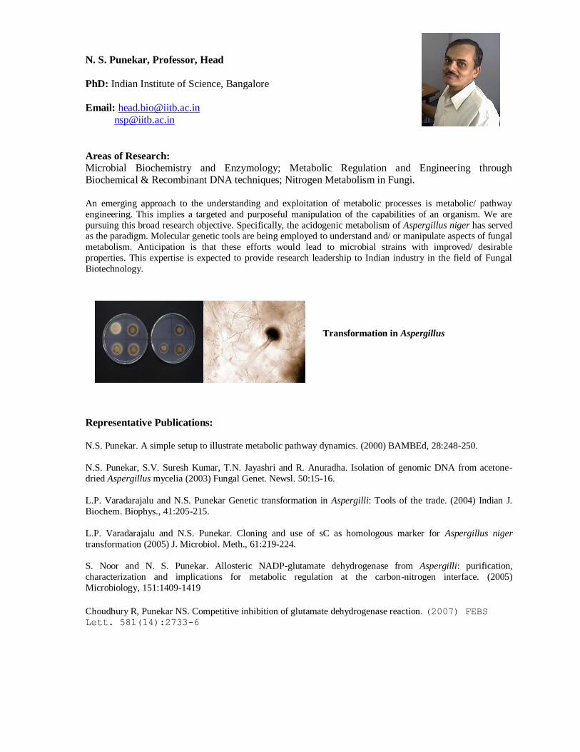
Transformation in Aspergillus
K. Krishnamurthy Rao, Professor
PhD: Tata Institute of Fundamental Research, Bombay, India
Email: [email protected]
Areas of Research:
Gene regulation, Bacillus subtilis, extracellular protease, motility, signal transduction
My laboratory is interested in understanding transcriptional regulation of genes in Bacillus subtilis. One of the
genes that we have been studying is a minor extracellular protease, Epr. We have recently demonstrated that epr
is transcribed by a σD form of RNA polymerase and the protease appears to be involved in providing signals for
the swarming motility of this organism. Currently the focus of our research is to determine how this gene is
regulated and the signaling mechanisms that lead to swarming. My laboratory is also engaged in a collaborative
research with Prof. G.K. Sureshkumar of IIT Madras, towards understanding the role of shear stress in a
bioreactor on the physiology of Bacillus subtilis. We have recently demonstrated that elevated shear stress leads
to suppression of sporulation of B.subtilis through the formation of a reactive oxygen species, leading to an
apoptotic form of cell death. At present we are attempting to dissect the molecular path that culminates in this
phenomenon.
Representative Publications:
Madhulika Dixit, Charuta S.Murudkar and K.Krishnamurthy Rao. (2002) epr is transcribed from a ċD
Promoter and is involved in Swarming of Bacillus subtilis. J. Bacteriol, 184: 596-599
Susmita Sahoo, Rajesh Verma, AK Suresh, Krishnamurthy Rao K, Jayesh Bellare and GK Suraishkumar(2003).
Macro-level and Genetic-level Responses of Bacillus subtilis to Shear Stress. Biotechnology Progress, 19:1689-
1696
Susmita Sahoo, Krishnamurthy Rao K, AK Suresh and GK Suraishkumar (2004). Intracellular Reactive Oxygen
Species Mediate Suppression of Sporulation in Bacillus subtilis Under Shear Stress. Biotechnology and
Bioengineering, 87: 81-89.
Susmita Sahoo, K. Krishnamurthy Rao, and G. K. Suraishkumar (2006). Reactive Oxygen Species Induced by
Shear Stress Mediate Cell Death in Bacillus subtilis. Biotechnology and Bioengineering, , 94 : 118-127
Charuta S. Murudkar, Prashant Kodgire and K. Krishnamurthy Rao (2006). The carboxy terminal domain of
Epr, a minor extracellular serine protease, is essential for the swarming motility of Bacillus subtilis 168. FEMS
Microbiology letters , 257: 24-31
Prashant Kodgire, Madhulika Dixit and K. Krishnamurthy Rao (2006) ScoC and SinR negatively regulate epr
by corepression in Bacillus subtilis. J. Bacteriol, 188: 6425-6428.
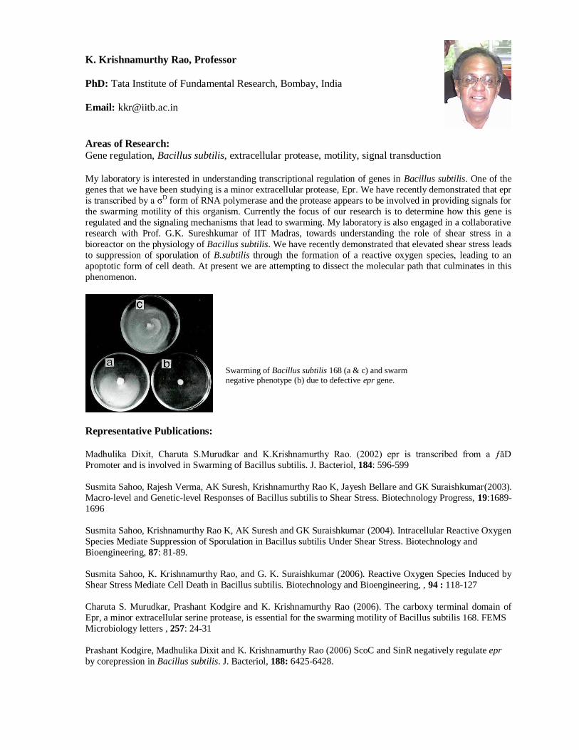
Swarming of Bacillus subtilis 168 (a & c) and swarm negative phenotype (b) due to defective epr gene.

Paike Jayadeva Bhat, Professor
PhD: Indian Institute of Science, Bangalore
Email: [email protected]
Areas of Research: Functional genomics, Differentiation in unicellular eukaryotes, Nucleocytoplasmic signal
transduction, Modeling of biological systems.
Organisms adapt to changing environmental conditions by regulating their physiology, mainly through the
genetic and metabolic circuitry. These circuits constitute a complex network involving a variety of molecular
interactions. Our focus is to understand the network of molecular interactions to unearth the regulatory themes that biological systems have evolved over millions of years. For this purpose, we have chosen to understand the
regulation of galactose metabolism in Yeast Saccharomyces cerevisiae. We have identified a new member of
the genetic component that participates in this regulation. Preliminary analysis has indicated that it is a
component that is not only involved in the regulation of galactose metabolism, but also regulates developmental
process in response to other nutritional cues. In collaboration with Prof. K.V. Venkatesh, we have developed a
model which predicts the behavior of the GAL genetic switch to known experimental perturbation. Using this
model we have hypothesized that the long term adaptation phenotype is due to stochastic variation in the
repressor concentration. Besides, we are also trying to use yeast as a surrogate system to understand human
genetic disorders such as galactosemia.
Representative Publications:
Khandey, F. A., Saha, M. and Bhat, P. J. Molecular characterization of MRG19 of Saccharomyces cerevisiae:
Implication in the regulation of galactose and non-fermnetable carbon source utilization. (2002) Eur.J.
Biochem. 269: 5840-5850
Bhat, P. J. Galactose-1-phosphate is a regulator of inositol monophosphatase: a fact or a fiction. (2003) Medical
Hypothesis 60: (1)123-128
Das, M and Bhat, P. J. Disruption of MRG19 results in altered nitrogen metabolic status and defective
pseudohyphal development in Saccharomyces cerevisiae (2005) Microbiology 151: 91-98
Bhat, P. J. and Venkatesh, K. V. Stochastic variations in the concentration of the repressor activates GAL
genetic switch: Implication in evolution of regulatory network. (2005) FEBS Letters 579: 597-603
Lakshminarasimhan, L and Bhat, P. J. Replacement of a conserved tyrosine by tryptophan in Gal3 of
Saccharomyces cerevisiae reduces constitutive activity: Implications for signal transduction of the GAL regulon
(2005) Mol Gen Genomics. In press.
Genetically regulated morphogenetic transition, from the yeast to the filamentous form, in
response to nutrient signaling in S. cerevisiae.
Rohit Manchanda, Professor
PhD: University of Oxford, U.K
Email: [email protected]
Areas of Research:
Computational neurophysiology, compartmental modeling, synaptic transmission, synaptic plasticity,
dendritic integration
Our lab has been involved in neurophysiological research, focusing particularly on aspects of synaptic
neurotransmission. A major part of the work has been electrophysiological; the approaches, both experimental
and computational. Experimental studies of neurotransmission, conducted until recently, concentrated on the
peripheral sympathetic nerve - smooth muscle junction. Here, we looked at the biophysics of the
neurotransmitter actions of ATP, and at how the syncytial electrical properties of smooth muscle influence the
synaptic potentials and contractions at these junctions. More recently, we have moved towards computational
studies of synaptic integration in detailed compartmental models of CNS (especially hippocampal) neurons,
aided by simulation platforms such as NEURON. We are interested in exploring the effects of factors such as
input synchrony, dendritic active conductances and their distributions on modes of novel electrical signalling by
these neurons, e.g. dendritic spikes and back-propagating action potentials, and their significance for physiological phenomena related to learning and cognition.
Representative Publications:
Manchanda, R. and Venkateswarlu, K. Quantal evoked depolarizations underlying the excitatory junction
potential of the guinea-pig isolated vas deferens. J. Physiol. 520.2: 527-537 (1999).
Godbole P., K. Venkateswarlu, R. Manchanda and U. B. Desai. Analysis of synaptic quantal depolarizations in
smooth muscle using the wavelet transform. IEEE Trans. Biomed. Engg., 47, 701-708 (2000).
Turale, N., A. Devulapalli, R. Manchanda, K. Moudgalya, G. Sivakumar A simulation framework for
electrophysiological networks: application to synaptic potentials in syncytical smooth muscle. Medical &
Biological Engineering & Computing, 41, 589-594 (2003).
P. Ghildyal, R. Manchanda Effects of cooling and ARL 67156 on synaptic ecto-ATPase activity in guinea pig and mouse vas deferens. Autonomic Neuroscience: Basic and Clinical 115, 28-34 (2004).
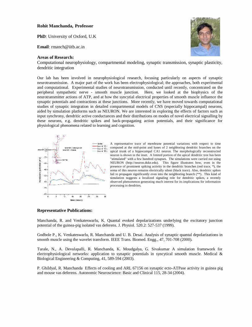
A representative trace of membrane potential variations with respect to time
computed at the mid-point and bases of 2 neighboring dendritic branches on the
apical trunk of a hippocampal CA1 neuron. The morphologically reconstructed
neuron is shown in the inset. A limited portion of the apical dendritic tree has been
"stimulated" with a few hundred synapses. The simulations were carried out using
NEURON (http://neuron.duke.edu). This figure illustrates how, even in the
presence of prominent spiking activity in the dendritic branches (red trace, *), the
soma of this neuron remains electrically silent (black trace). Also, dendritic spikes
fail to propagate significantly even into the neighboring branch (**). This kind of
simulation suggests a localized signaling role for dendritic spikes, a recently
observed phenomenon generating much interest for its implications for information
processing in dendrites.
Gosukonda Subrahmanyam, Professor
PhD: Indian Agricultural Research Institute, New Delhi
Email: [email protected]
Areas of Research:
Molecular Immunology and cell signaling mechanisms, Role of lipid, protein kinases and
phosphatases in T cell and mast cell biology, Signaling mechanisms in micro algae, Biochemistry and genetics of Dunaliella sp.
The complexity and fine tuning of T cell and mast activation signaling cascades are the focus of the lab. Our
studies have shown that a type II PtdIns 4-kinase activity is associated with CD3, CD4, CD7 and CD28
receptors in T cells and with FcƒÕRI in mast cells. These studies suggest that type II PtdIns 4-kinases may generate a threshold of signals at plasma membrane that may regulate immune synapse formation, T cell
adhesion and infiltration to sites of inflammation. Understanding of type II PtdIns 4-kinases function and
regulation in immune cells help us in development of rational drug design for immunomodulators.
The other project that has been initiated recently in our lab is on osmotic / stress signaling mechanisms in a
marine alga Dunaliella salina. This alga responds to different stress conditions by accumulating a large quantity
of carotenoids and the focus of the lab is to dissect out these signaling pathways and generate stable mutants in
these pathways.
Representative Publications:
Srivastava R, Sinha RK, Subrahmanyam G. Type II phosphatidylinositol 4-kinase beta associates with TCR-
CD3 zeta chain in Jurkat cells.(2006) Molecular Immunology 43(5):454-63.
Srivastava R., Ratheesh A., Rajiv K. Gude, R.K., Rao K.V.K ., Panda D and Subrahmanyam G. Resveratrol
inhibits type II phosphatidylinositol 4-kinase: A key component in pathways of phosphoinositide turn over.
(2005) Biochemical Pharmacology, 70: 1048-1055
Fernandis A.Z., Srivastava R., Sinha R.K and Subrahmanyam G. A type II phosphatidylinositol 4-kinase
associates with T cell receptor £a chain in Con A stimulated splenic lymphocytes through tyrosyl
phosphorylation -dependent mechanisms. (2005) Molecular Immunology 42,: 561-568
Naveen B., Shankar B.S and Subrahmanyam G. Fc(epsilon)RI cross-linking activates a type II
phosphatidylinositol 4-kinase in RBL 2H3 cells. (2005) Molecular Immunology, 42: 1541-1549
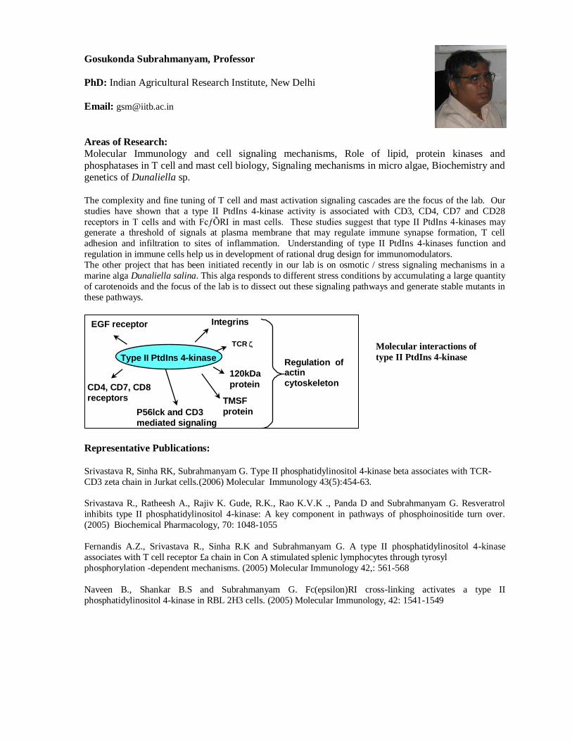
Integrins
TCR
120kDa
protein
TMSF
protein P56lck and CD3
mediated signaling
CD4, CD7, CD8
receptors
EGF receptor
Type II PtdIns 4-kinase Regulation of actin
cytoskeleton
Molecular interactions of
type II PtdIns 4-kinase
Petety V. Balaji, Professor
Ph.D: Indian Institute of Science, Bangalore. Email: [email protected]
Area of Research: Glycolipid dynamics in lipid environment, Structure-function studies of human
ST3 SiaTs.
Our research interest is in studying the recognition and binding of carbohydrates by proteins. MD simulations are being used to probe the conformational dynamics of the glycan headgroups of gangliosides in lipid bilayer
environment. This is related to the recognition and binding of the glycans by receptor proteins and enzymes of
biosynthesis / degradation pathways. The 3-D structures of saccharide-binding proteins are also being analyzed
to delineate and characterize the recognition templates shared by proteins of same saccharide-specificity. Our
laboratory is also interested in the structure-function relationship in human SiaTs; specifically, 3-D structures of
human ST3Sias have been modeled and experimental studies to validate the predicted models and to generate
mutants with novel substrate specificities are underway. Motivated by the problems encountered by the over-
expression of proteins in the insoluble fraction, the relationship between the primary structure of proteins and
their propensity to aggregate was also investigated. This work is being extended to study the sequence-structure
relationship in amyloids.
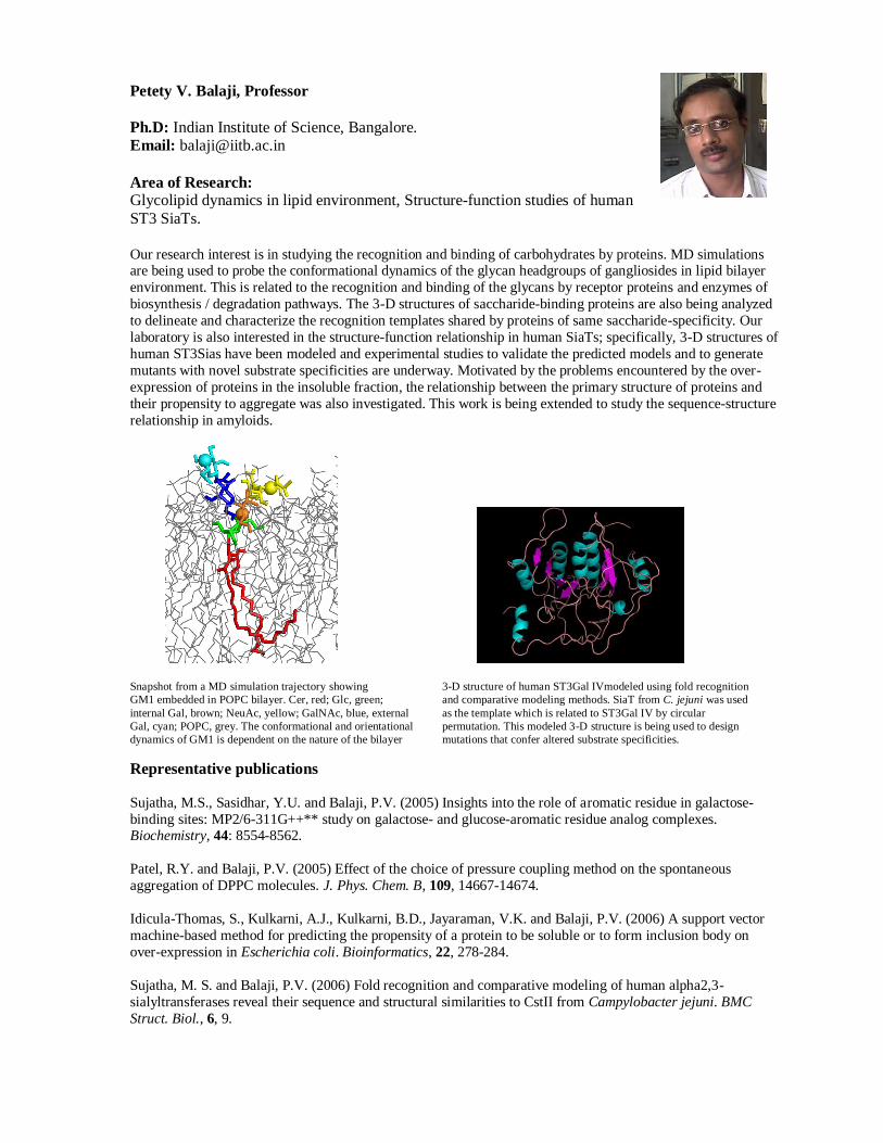
Snapshot from a MD simulation trajectory showing 3-D structure of human ST3Gal IVmodeled using fold recognition
GM1 embedded in POPC bilayer. Cer, red; Glc, green; and comparative modeling methods. SiaT from C. jejuni was used
internal Gal, brown; NeuAc, yellow; GalNAc, blue, external as the template which is related to ST3Gal IV by circular
Gal, cyan; POPC, grey. The conformational and orientational permutation. This modeled 3-D structure is being used to design
dynamics of GM1 is dependent on the nature of the bilayer mutations that confer altered substrate specificities.
Representative publications
Sujatha, M.S., Sasidhar, Y.U. and Balaji, P.V. (2005) Insights into the role of aromatic residue in galactose-
binding sites: MP2/6-311G++** study on galactose- and glucose-aromatic residue analog complexes. Biochemistry, 44: 8554-8562.
Patel, R.Y. and Balaji, P.V. (2005) Effect of the choice of pressure coupling method on the spontaneous
aggregation of DPPC molecules. J. Phys. Chem. B, 109, 14667-14674.
Idicula-Thomas, S., Kulkarni, A.J., Kulkarni, B.D., Jayaraman, V.K. and Balaji, P.V. (2006) A support vector
machine-based method for predicting the propensity of a protein to be soluble or to form inclusion body on
over-expression in Escherichia coli. Bioinformatics, 22, 278-284.
Sujatha, M. S. and Balaji, P.V. (2006) Fold recognition and comparative modeling of human alpha2,3-
sialyltransferases reveal their sequence and structural similarities to CstII from Campylobacter jejuni. BMC
Struct. Biol., 6, 9.
Dulal Panda, Professor
Recipient of the 2004-2005 Swarnajayanti Award, Govt. of India
PhD : Bose Institute Kolkata Email: [email protected]
Area of Research:
Microtubule dynamics, mitosis, anticancer drugs, FtsZ assembly, bacterial cell division Microtubules, the dynamic cytoskeletal polymers of eukaryotic cells, play essential roles in several cellular
tasks including mitosis and neuronal growth and development. My lab is interested to understand the molecular
mechanism of regulation of microtubule assembly dynamics and the mechanism of action of microtubule-
targeted anticancer drugs. We are using cell-permeable small molecules to understand roles of microtubules
during mitosis. Our goal is to understand the linkage between perturbation of microtubule assembly dynamics,
inhibition of cell proliferation, mitotic block and cell death. We are also investigating the mechanism of
regulation of the assembly dynamics of FtsZ, a protein that plays a critical role during bacterial cytokinesis. The
lab has reconstituted FtsZ protofilaments in vitro that can serve as a screen for finding antibacterial drugs with
novel mechanism of action. In addition, we are interested to understand the mechanism of protein folding.
Representative Publications:
Rai D, Singh JK, Roy N, Panda D. (2008), Curcumin inhibits FtsZ assembly: an attractive mechanism for its
antibacterial activity. Biochem J.; 410:147-55
Ray S, Mohan R, Singh JK, Samantaray MK, Shaikh MM, Panda D., Ghosh P (2007), Anticancer and
Antimicrobial Metallopharmaceutical agents based on Palladium, Gold and Silver N-hetrocyclic carbine complexes. J.Am.Chem.Soc; 129:15042-53
Singh JK, Makde RD, Kumar V, Panda D. (2007), A membrane protein, EzrA, regulates assembly dynamics of
FtsZ by interacting with the C-terminal tail of FtsZ. Biochemistry; 46(38):11013-22.
Srivastava P, Panda D. (2007), Rotenone inhibits mammalian cell proliferation by inhibiting microtubule
assembly through tubulin binding. FEBS J.; 274(18):4788-801.
Jaiswal R, Beuria TK, Mohan R, Mahajan SK, Panda D. (2007), Totarol inhibits bacterial cytokinesis by
perturbing the assembly dynamics of FtsZ. Biochemistry; 46(14):4211-20.
Santra MK, Panda D. (2007), Acid-induced loss of functional properties of bacterial cell division protein FtsZ:
evidence for an alternative conformation at acidic pH. Proteins; 67(1):177-88.
Gupta KK, Bharne SS, Rathinasamy K, Naik NR, Panda D., (2006)Dietary antioxidant curcumin inhibits
microtubule assembly through tubulin binding. FEBS J.; 273(23):5320-32.
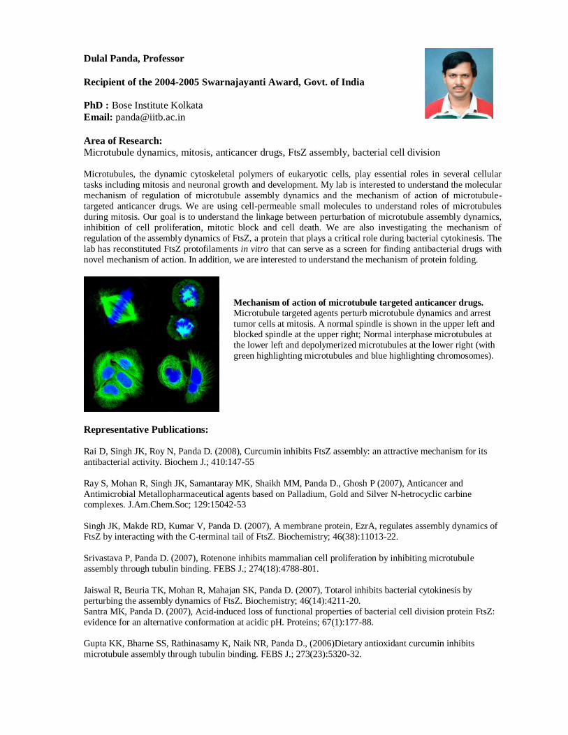
Mechanism of action of microtubule targeted anticancer drugs.
Microtubule targeted agents perturb microtubule dynamics and arrest
tumor cells at mitosis. A normal spindle is shown in the upper left and blocked spindle at the upper right; Normal interphase microtubules at
the lower left and depolymerized microtubules at the lower right (with
green highlighting microtubules and blue highlighting chromosomes).
Soumyo Mukherji, Professor
PhD: University of North Carolina (Chapel Hill, USA)
Email: [email protected]
Areas of Research:
Bioinstrumentation and Biosensors, particularly focussing on low cost diagnostic and monitoring
devices; BioMEMS ; Cardiac Electrophysiology.
Cardiac and allied problems are amongst the major causes of morbidity and mortality in western countries and
the incidences of such diseases are on the rise in urban as well as rural India. To effectively diagnose and treat
cardiac dysfunctions, one not only needs to diagnose diseases early but also understand the pathophysiology of
such diseases. In the Cardiac Electrophysiology and Biosensors group of the of the School of Biosciences and
Bioengineering, in collaboration with the Microelectronics group (Electrical Engineering) and Chemistry
department, inexpensive sensors for early detection of myocardial infarction are being developed. In the course
of such development various techniques to immobilize proteins on micromachinable surfaces (e.g. silicon and
its derivatives, photoresists and epoxies) have been developed in this laboratory. These techniques can be used
for development of other biosensors as well. Generic methods for quantitative assessment of immobilization
density on solid surfaces have also been developed. It is only natural that one extends these techniques towards developing sensors for other biological entities, both in blood and water. Research is also conducted to try to
understand the mechanism of translation of myocardial ischemia into ventricular fibrillation, which can cause
death in a matter of minutes. Other collaborative activities include the development of conducting polymer
based sensing systems for ionic and inorganic contamintants of water (with Chemistry and Electrical
Engineering); and the development of an inexpensive wearable ECG recorder.
Representative Publications: Manoj Joshi , Richard Pinto , V. Ramgopal Rao , Soumyo Mukherji "Silanization and antibody immobilization
on SU-8" , (2007)* Applied Surface Science, 253, Page(s): 3127-3132.
Manoj Joshi , Nitin Kale , Rakesh Lal , V. Ramgopal Rao, Soumyo Mukherji "A Novel Dry Method for Surface
Modification of SU-8 for Immobilization of Biomolecules in Bio-MEMS" ,(2006) Biosensors & Bioelectronics.
V.R. Sai Vemulakonda, S. Mahajan, A.Q. Contractor, S. Mukherji "Immobilization of antibodies on polyaniline
films and its application in a piezoelectric immunosensor", . (2006) Applied Chemistry Journal, 78(24), Page(s):
8368-8373.
Amol V. Patil, Soumyo Mukherji, Uday B. Desai,"Optimal Objective Functional Selection for Image
Reconstruction in Diffuse Optical Tomography", Proceedings of the International Conference on Computing: Theory and Applications (ICCTA'07).
Anupama V. Govindarajan, Manoj Joshi, Ritu Rashmi, Soumyo Mukherji, Ramgopal Rao and Karl F.
Böhringer, "Engineering the Hydrophobicity of Oxide Surfaces by Controlling the
Dehydration Temperature of Silanization", , E-MRS 2007 Spring Meeting, Strausburg, France.
Manoj Joshi, Gajanan Nagre, Seena V., V. Ramgopal Rao, Soumyo Mukherji, "Functionalization of Hydrogen
Silsesquioxane (HSQ) surface for biosensor applications", E-MRS 2007 Spring Meeting, Strausburg, France.
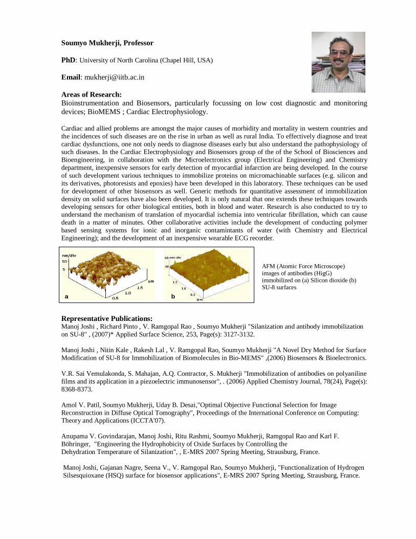
a b
AFM (Atomic Force Microscope) images of antibodies (HigG) immobilized on (a) Silicon dioxide (b) SU-8 surfaces
Prashant S. Phale, Professor
Ph D: Indian Institute of Science, Bangalore
Email: [email protected]
Areas of Research:
Pseudomonas, Biodegradation, Oxygenases, Carbon catabolite repression, Metabolic engineering of
the degradative pathways
Our current research focuses on isolating novel microorganisms and understandng metabolic regulations at the
biochemical and genetic level. Pseudomonas putida CSV86 isolated by us utilizes hydrocarbons preferentially
over glucose. Several biochemical experiments indicate that regulation is at the enzyme and glucose uptake
level. Such a novel strain has tremendous applications in bioremediation. We have isolated two bacterial strains,
which utilizes isophthalate (a competitive inhibitor of glutamate dehydrogenase, GDH) and showed the
presence of two isozymes for GDH. We are characterizing these isozymes in order to understand the
physiological significance in isophthalate degradation. Recently, we have isolated three bacterial species, which
degrades carbaryl via 1,2-dihydroxynaphthalene pathway and found to have new enzyme 1-naphthol
hydroxylase, not reported so far in the literature. Also, we are characterizing the third group of ring-cleaving
enzymes viz. gentisate dioxygenase, 1-hydroxy-2-naphthoate dioxygenase from hydrocarbon degrading strains.
Our long-term objective is to engineer a microbe that can metabolize large number of aromatic compounds.
Time, h
0 5 10 15 20 25
pm
ole
s.m
g-1
0
4
8
50
100
150
200
Gro
wth
(O
. D
. 540 n
m);
Glu
cose (
mg.m
l-1)
0
1
2
3
Representative Publications:
Basu A & Phale PS (2007) Conjugative transfer of preferential utilization of aromatic compounds from
Pseudomonas putida CSV8. Biodegradation (In Press).
Phale PS, Basu A, Majhi PD, Deveryshetty J, Vamsee-Krishna C & Shrivastava R (2007) Metabolic diversity
in bacterial degradation of aromatic compounds. OMICS Jr of Integrative biology (In Press)
Basu A, Das D, Bapat P, Wangikar PP & Phale PS (2007) Sequential utilization of substrates by Pseudomonas putida CSV86: Signatures of intermediate metabolites and on-line measurements. Microbiological Res (In
Press).
Swetha VP, Basu A & Phale PS (2007) Purification and characterization of 1-naphthol-2-hydroxylase from
carbaryl degrading Pseudomonas strain C4. J Bacteriol 189:2660-2666.
Deveryshetty J, Suvekbala V, Varadamshetty G & Phale PS (2007) Metabolism of 2-, 3-, and 4-
hydroxybenzoates by soil isolates Alcaligenes sp. strain PPH and Pseudomonas sp. strain PPD. FEMS
Microbiol Lett 268:59-66.
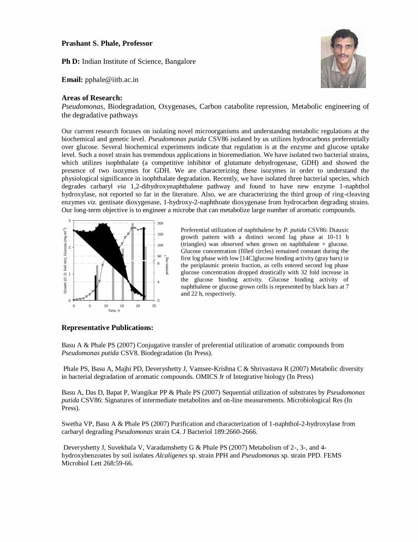
Preferential utilization of naphthalene by P. putida CSV86: Diauxic growth pattern with a distinct second lag phase at 10-11 h (triangles) was observed when grown on naphthalene + glucose. Glucose concentration (filled circles) remained constant during the first log phase with low [14C]glucose binding activity (gray bars) in the periplasmic protein fraction, as cells entered second log phase glucose concentration dropped drastically with 32 fold increase in
the glucose binding activity. Glucose binding activity of naphthalene or glucose grown cells is represented by black bars at 7 and 22 h, respectively.
Rinti Banerjee, Professor
PhD: Indian Institute of Technology Bombay AICTE Career Award for Young Teachers 2005-2006
Email: [email protected]
Areas of Research:
Biological surfactant replacements, biomaterials, bio-interfaces, drug delivery, bionanotechnology
My research interests are development of surfactant replacements, biomaterials, evaluation of interfacial
phenomena in medicine, biorheology, and development of liposomal and nanoparticulate drug delivery systems.
In my group, we have been developing promising inhalable aerosols of lung surfactants and dual functional
drug loaded surfactants for respiratory diseases (NRDS, ARDS, tuberculosis, asthma). In the area of
biomaterials, we have focused on ocular biomaterials like vitreous humor replacements, on in situ gels for
cartilage replacement and bio-fluid replacements having desirable rheological properties. We are also working
on the biological applications of colloids and interfaces to study drug interactions using langmuir monolayers as
biomembrane models and tissue tensiometry as a prognostic tool in cancers. In the area of drug delivery, our
research interests include use of drug encapsulated liposomes and nanoparticles for sustained delivery and use
of the respiratory route for systemic delivery. We are also developing nanoparticles of biological origin to overcome several anatomical barriers for example in non-viral gene delivery and targeted anticancer therapy.
Representative Publications: Banerjee R. Nanotechnology in respiratory medicine. (2004) Editorial Chest (Ind. Ed) 5(1) 5
Chimote G and Banerjee R. Effect of antitubercular drugs on dipalmitoyl phosphatidylcholine monolayers:
implication for drug loaded surfactants. (2005) Respir. Physiol. & Neurobiol., 145: 65-77.
Suri S, Banerjee R. In vitro evaluation of in situ biopolymeric gels as short term vitreous substitutes. (2006) J.
Biomed. Mater. Res A In press
Preetha A, Huilgol N and Banerjee R. Comparison of paclitaxel penetration in normal and cancerous cervical
model monolayer membranes. (2006) Colloids and Surfaces B: Biointerfaces. In press.
Banerjee R. Nanoparticles for respiratory drug delivery. (2006) Invited chapter (Chapter 19) in “Nanoparticles
for Pharmaceutical Applications” edited by A J Domb, Y Tabata and MNV Ravikumar, American Scientific
Publishers. In press.
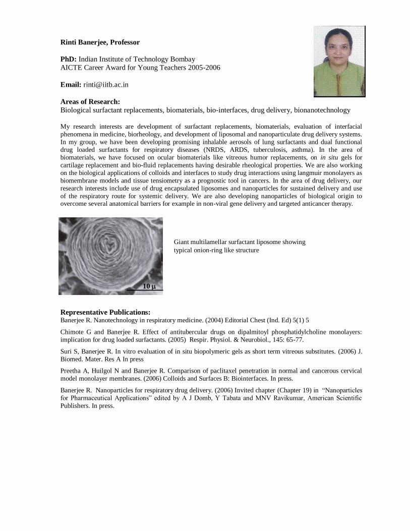
10
Giant multilamellar surfactant liposome showing
typical onion-ring like structure
Swati Patankar, Associate Professor
PhD: Tufts University, Boston
Email: [email protected]
Areas of Research:
Malaria, regulation of gene expression, noncoding RNAs, TATA-binding protein, anti-malarial lead
compound screening
Malaria, caused by the protozoan parasite Plasmodium falciparum, is responsible for disease and death of
millions in developing countries. Our laboratory studies this pathogen with the aim of understanding its basic
biology specifically differences between the parasite and its human host. One such example of a biological
phenomenon where the parasite differs greatly from humans is the process of regulation of gene expression.
Mechanisms of transcriptional and post-transcriptional regulation are textbook knowledge for higher eukaryotes
like humans but poorly understood in the malaria parasite. We study regulation of gene expression in two ways:
first we are trying to understand how the TATA-binding protein of Plasmodium differs from its human
homologue at the level of DNA-binding specificity and second we have uncovered several noncoding RNAs
from the Plasmodium genome and are in the process of understanding whether they may be a novel strategy
used by the parasite to regulate genes at the post-transcriptional level. Additionally, we also collaborate with laboratories that synthesize lead compounds for anti-malarial drug discovery by carrying out in vitro screening
of these compounds against P. falciparum.
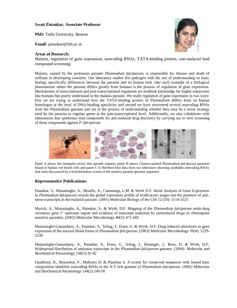
Panel A shows the mosquito vector that spreads malaria; panel B shows Giemsa-stained Plasmodium falciparum parasites found in human red blood cells and panel C is Northern blot data from our laboratory showing candidate noncoding RNAs that were discovered by a bioinformatics screen of the malaria parasite genome sequence.
Representative Publications:
Patankar, S., Munasinghe, A., Shoaibi, A., Cummings, L.M. & Wirth D.F. Serial Analysis of Gene Expression
in Plasmodium falciparum reveals the global expression profile of erythrocytic stages and the presence of anti-sense transcripts in the malarial parasite. (2001) Molecular Biology of the Cell 12 (10): 3114-3125
Myrick, A., Munasinghe, A., Patankar, S., & Wirth, D.F. Mapping of the Plasmodium falciparum multi-drug
resistance gene 5’ upstream region and evidence of transcript induction by antimalarial drugs in chloroquine
sensitive parasites. (2003) Molecular Microbiology 49(3): 671-683
Munasinghe-Gunasekera, A., Patankar, S., Schug, J., Eisen, G. & Wirth, D.F. Drug-induced alterations in gene
expression of the asexual blood forms of Plasmodium falciparum. (2003) Molecular Microbiology 50(4): 1229-
1239
Munasinghe-Gunasekera, A., Patankar, S., Eisen, G., Schug, J., Kissinger, J., Roos, D. & Wirth, D.F. Widespread distribution of antisense transcripts in the Plasmodium falciparum genome. (2004) Molecular and
Biochemical Parasitology 136(1):35-42
Upadhyay, R., Bawankar, P., Malhotra D. & Patankar S. A screen for conserved sequences with biased base
composition identifies noncoding RNAs in the A-T rich genome of Plasmodium falciparum. (2005) Molecular
and Biochemical Parasitology 144(2):149-58
A B C
Rohit Srivastava, Associate Professor
PhD: Louisiana Tech University, May 2005
Email: [email protected]
Areas of Research: Fluorescent Biosensors, Nanoengineered Sensors, Controlled Release, Layer-by-Layer Assembly,
BioMEMS
The concept of tattooing, which were viewed as a form of body art, fashion and rebellion is now being used by
researchers for embedding smart sensors for monitoring vital analytes in the body, and it seems that the
development of micro- and nanoscale sensors and actuators might prove to be instrumental in curing the human
body of its various ills. I have been part of an interdisciplinary team comprising engineers, chemists, biologists,
and physicists conducting research on glucose sensors and novel drug delivery systems. I plan to build upon my
research experience in endeavoring to build a sensing platform for cells to understand the dynamic response of
cells to chemical stimuli. This work will involve integrating the fields of MEMS and Nanotechnology with tissue engineering and biosensing and can be easily extended to monitoring the effects of drugs, especially the
release of immunomodulating agents.
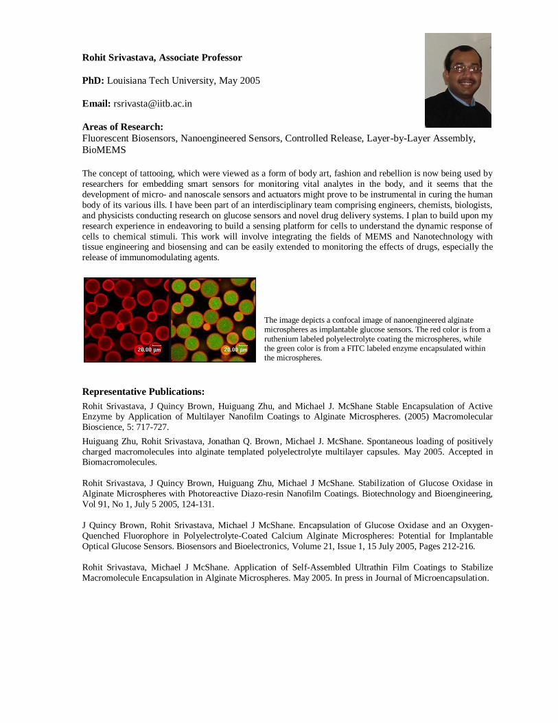
The image depicts a confocal image of nanoengineered alginate microspheres as implantable glucose sensors. The red color is from a ruthenium labeled polyelectrolyte coating the microspheres, while the green color is from a FITC labeled enzyme encapsulated within the microspheres.
Representative Publications:
Rohit Srivastava, J Quincy Brown, Huiguang Zhu, and Michael J. McShane Stable Encapsulation of Active Enzyme by Application of Multilayer Nanofilm Coatings to Alginate Microspheres. (2005) Macromolecular
Bioscience, 5: 717-727.
Huiguang Zhu, Rohit Srivastava, Jonathan Q. Brown, Michael J. McShane. Spontaneous loading of positively
charged macromolecules into alginate templated polyelectrolyte multilayer capsules. May 2005. Accepted in Biomacromolecules.
Rohit Srivastava, J Quincy Brown, Huiguang Zhu, Michael J McShane. Stabilization of Glucose Oxidase in
Alginate Microspheres with Photoreactive Diazo-resin Nanofilm Coatings. Biotechnology and Bioengineering,
Vol 91, No 1, July 5 2005, 124-131.
J Quincy Brown, Rohit Srivastava, Michael J McShane. Encapsulation of Glucose Oxidase and an Oxygen-
Quenched Fluorophore in Polyelectrolyte-Coated Calcium Alginate Microspheres: Potential for Implantable
Optical Glucose Sensors. Biosensors and Bioelectronics, Volume 21, Issue 1, 15 July 2005, Pages 212-216.
Rohit Srivastava, Michael J McShane. Application of Self-Assembled Ultrathin Film Coatings to Stabilize
Macromolecule Encapsulation in Alginate Microspheres. May 2005. In press in Journal of Microencapsulation.
Samir K Maji, Assistant Professor
PhD: Indian Association for the Cultivation of Science,
Jadavpur, Kolkata, India
Email: [email protected]
Areas of Research: protein aggregation and amyloid formation associated with neurodegenerative diseases (Alzheimer's, Parkinson's, Prion diseases) and native biological functions.
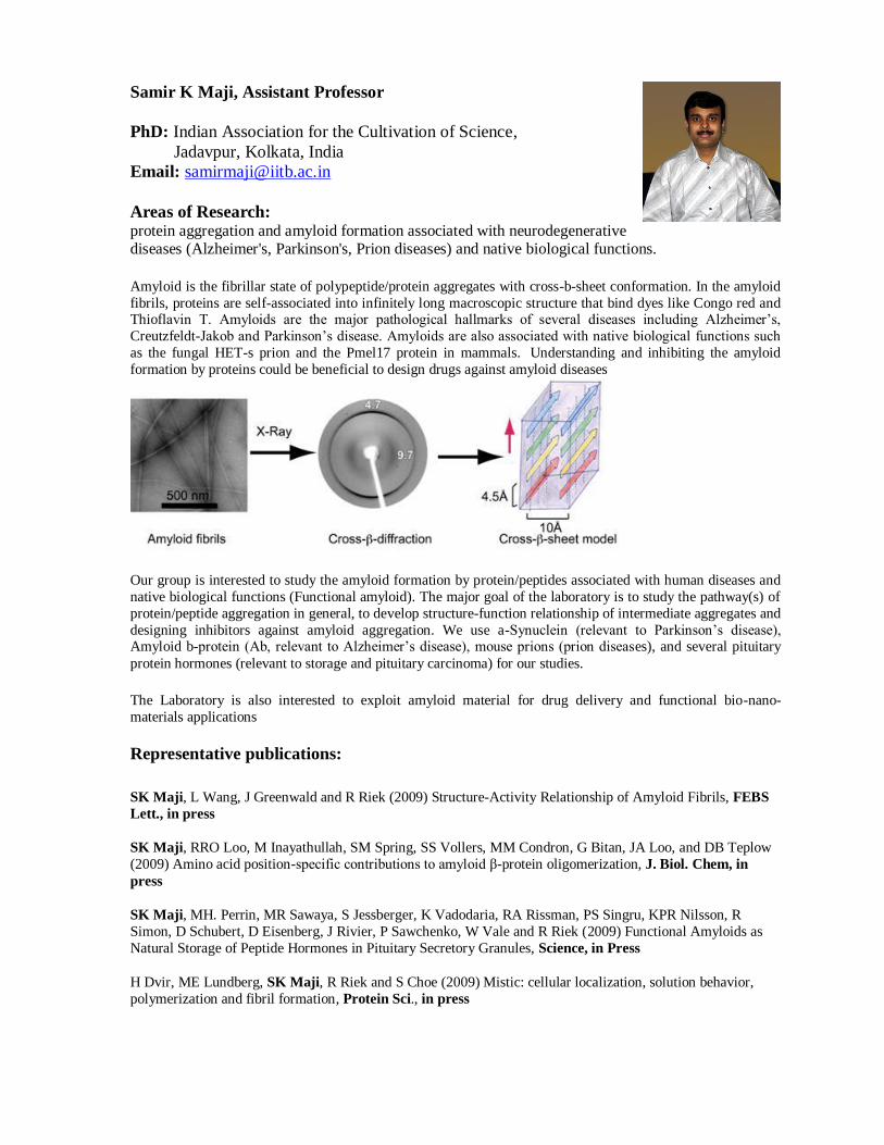
Amyloid is the fibrillar state of polypeptide/protein aggregates with cross-b-sheet conformation. In the amyloid
fibrils, proteins are self-associated into infinitely long macroscopic structure that bind dyes like Congo red and Thioflavin T. Amyloids are the major pathological hallmarks of several diseases including Alzheimer’s,
Creutzfeldt-Jakob and Parkinson’s disease. Amyloids are also associated with native biological functions such
as the fungal HET-s prion and the Pmel17 protein in mammals. Understanding and inhibiting the amyloid
formation by proteins could be beneficial to design drugs against amyloid diseases
Our group is interested to study the amyloid formation by protein/peptides associated with human diseases and
native biological functions (Functional amyloid). The major goal of the laboratory is to study the pathway(s) of
protein/peptide aggregation in general, to develop structure-function relationship of intermediate aggregates and
designing inhibitors against amyloid aggregation. We use a-Synuclein (relevant to Parkinson’s disease), Amyloid b-protein (Ab, relevant to Alzheimer’s disease), mouse prions (prion diseases), and several pituitary
protein hormones (relevant to storage and pituitary carcinoma) for our studies.
The Laboratory is also interested to exploit amyloid material for drug delivery and functional bio-nano-
materials applications
Representative publications:
SK Maji, L Wang, J Greenwald and R Riek (2009) Structure-Activity Relationship of Amyloid Fibrils, FEBS
Lett., in press
SK Maji, RRO Loo, M Inayathullah, SM Spring, SS Vollers, MM Condron, G Bitan, JA Loo, and DB Teplow
(2009) Amino acid position-specific contributions to amyloid β-protein oligomerization, J. Biol. Chem, in
press
SK Maji, MH. Perrin, MR Sawaya, S Jessberger, K Vadodaria, RA Rissman, PS Singru, KPR Nilsson, R
Simon, D Schubert, D Eisenberg, J Rivier, P Sawchenko, W Vale and R Riek (2009) Functional Amyloids as
Natural Storage of Peptide Hormones in Pituitary Secretory Granules, Science, in Press
H Dvir, ME Lundberg, SK Maji, R Riek and S Choe (2009) Mistic: cellular localization, solution behavior,
polymerization and fibril formation, Protein Sci., in press
Sanjeeva Srivastava, Assistant Professor
PhD: University of Alberta, Canada
Email: [email protected]
Award:
Apple Research Technology Support (ARTS) award (2009)
Areas of Research: Proteomics, Biomarker discovery, Protein-protein interactions
One of the most important pursuits in post-genome era is to understand the function of gene-
expressed proteins. Despite immense progress in molecular biology and genetics only a small fraction
of the proteome is understood at the biochemical level. Systems biology and proteomics (study and
characterization of complete set of proteins) strive to create detailed predictive models for molecular pathways based upon the quantitative behavior of proteins. Understanding these dynamic networks
provides clues into the consequence of aberrant interactions and why they lead to diseases like cancer;
however, collecting biochemical data about protein behavior at scale has been daunting. Our laboratory is applying high throughput proteomic techniques such as protein microarrays, surface
plasmon resonance, two-dimensional electrophoresis and mass spectrometry for biomarker discovery,
protein-protein interactions and drug target discovery. Information obtained from research program is also used for in silico studies and computing models to enhance our understanding in systems
approach.
Representative publications:
Srivastava S. and LaBaer J. (2008) Nanotubes light up protein arrays. Nature Biotechnology 26, 1244-1246.
Srivastava S., Ramachandran N. and LaBaer J. (2008) Applications of protein microarrays for biomarker
discovery. Proteomics Clinical Applications 2, 1444-14459.
Spera R., Badino F., Hainsworth, G., Fuentes, M., Srivastava, S., LaBaer, J. and Nicolini, C. (2009) “Label free
detection of NAPPA: III. Mass spectrometry”. In: The Proteome: Technologies and Applications. Pan Stanford
Series on Nanobiotechnology - Volume 3 (Eds: Josh LaBaer and Claudio Nicolini) (in press).
Kav N.N.V., Srivastava S., Yajima W. and Ali S. (2008) “Proteomics in developing countries”. In: Plant
proteomics: Technologies, strategies, and applications (Eds: Ganesh K. Agrawal and Randeep Rakwal), Wiley-
Interscience, USA 570-581.
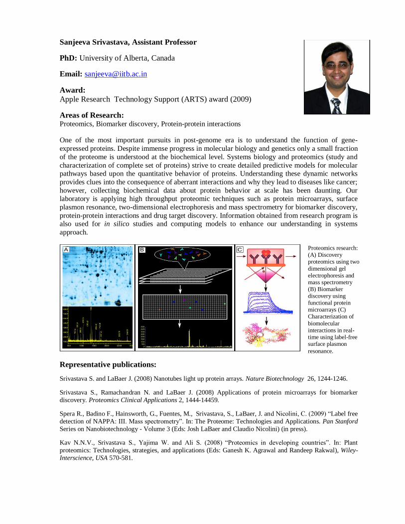
Proteomics research: (A) Discovery proteomics using two
dimensional gel electrophoresis and mass spectrometry (B) Biomarker discovery using functional protein microarrays (C) Characterization of
biomolecular interactions in real-time using label-free surface plasmon
resonance.
Santanu Kumar Ghosh, Assistant Professor
PhD: Bose Institute, Kolkata, India
Email: [email protected]
Areas of Research: Chromosome segregation, Saccharomyces cerevisiae, meiosis and mitosis, plasmid maintenance The molecular mechanisms ensuring accurate chromosome segregation during meiosis and mitosis are critical
to the conservation of euploidy. Errors in these processes lead to dire consequences like cancer, infertility and
congenital disorders. My group is interested in elucidating the factors responsible for successful execution of
these two cell division processes. Kinetochore, a key player in orchestrating the movement of chromosomes
during cell division, will be probed in a cell cycle-dependent manner to analyze its role in specifying meiotic or
mitotic mode of segregation. Cohesin, which holds replicated chromatids together, also specify the mode of
segregation through its degradation in a temporal fashion. We are interested in finding out factors which
regulate cohesin destruction. My group is also interested in studying the mechanism behind the remarkable stability of yeast endogenous plasmid. How the partitioning system communicates with the in-built
amplification system of the plasmid is of great interest of my group.
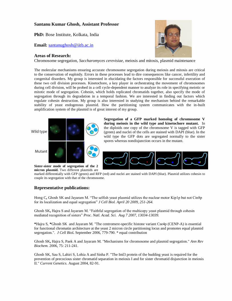
Segregation of a GFP marked homolog of chromosome V
during meiosis in the wild type and kinetochore mutant. In
the diploids one copy of the chromosome V is tagged with GFP (green) and nuclei of the cells are stained with DAPI (blue). In the
wild type the GFP dots are segregated normally to the sister
spores whereas nondisjunction occurs in the mutant.
Sister-sister mode of segregation of the 2 micron plasmid. Two different plasmids are marked differentially with GFP (green) and RFP (red) and nuclei are stained with DAPI (blue). Plasmid utilizes cohesin to
couple its segregation with that of the chromosome.
Representative publications:
Hong C, Ghosh SK and Jayaram M. “The selfish yeast plasmid utilizes the nuclear motor Kip1p but not Cin8p
for its localization and equal segregation” J Cell Biol. April 20 2009, 251-264.
Ghosh SK, Hajra S and Jayaram M. “Faithful segregation of the multicopy yeast plasmid through cohesin
mediated recognition of sisters” Proc. Natl. Acad. Sci. Aug 7 2007, 13034-13039.
*Hajra S, *Ghosh SK and Jayaram M. "The centromere-specific histone variant Cse4p (CENP-A) is essential
for functional chromatin architecture at the yeast 2 micron circle partitioning locus and promotes equal plasmid segregation.". J Cell Biol. September 2006, 779-790. * equal contribution
Ghosh SK, Hajra S, Paek A and Jayaram M. "Mechanisms for chromosome and plasmid segregation." Ann Rev
Biochem. 2006, 75: 211-241.
Ghosh SK, Sau S, Lahiri S, Lohia A and Sinha P. "The Iml3 protein of the budding yeast is required for the
prevention of precocious sister chromatid separation in meiosis I and for sister chromatid disjunction in meiosis
II." Current Genetics. August 2004, 82-91.
Ranjit Padinhateeri, Assistant Professor
PhD: Indian Institute of Technology Madras, Chennai, India
Email: [email protected]
Areas of Research: Nucleosome dynamics & Chromatin assembly, Dynamics of Actin & Microtubules, Mechanics of DNA
My broad areas of interest are biological physics and soft-matter physics. I do theoretical studies to
understand various physical and biological phenomena, using a variety of tools from physics
including equilibrium and non-equilibrium statistical mechanics, polymer physics, and soft-matter
theory. I tackle research problems using a combination of computational (numerical) and analytical
methods. Specific areas of interest include Nucleosome dynamics, Chromatin assembly, Dynamics of
Actin & Microtubules, and DNA mechanics.
Representative publications:
Non-equilibrium self-assembly of a filament coupled to ATP/GTP hydrolysis P. Ranjith, D. Lacoste, K. Mallick
and J.-F. Joanny Biophysical Journal 96 2146-2159, 2009
Equilibrium properties of a grafted polyelectrolyte with explicit counterions K. Jayasree, P. Ranjith, Madan Rao
and P. B. Sunil Kumar J. Chem. Phy. 130, 094901 (2009)
Nucleosome hopping and sliding kinetics determined from dynamics of single chromatin fibers in Xenopus egg
extracts P. Ranjith, J Yan and J. F. Marko PNAS, 104, 13649-13654 (2007)
Filling of a one-dimensional lattice by k-mers proceeds via fast power-law-like kinetics P. Ranjith and John F.
Marko Phys. Rev. E. 74, 041602 (2006)
Distribution Functions, Loop Formation Probabilities and Force-Extension Relations in a Model for Short
Double-Stranded DNA Molecules P. Ranjith , P. B. Sunil Kumar and Gautam I. Menon Phys. Rev. Lett. 94,
138102 (2005)
Dynamics of a Semiflexible filament under external force P. Ranjith and P.B. Sunil Kumar Physica A 318, 220-
229 (2003)
Dynamics of folding in Semiflexible filaments P. Ranjith and P.B. Sunil Kumar Phys. Rev. Lett. 89, 018302
(2002)
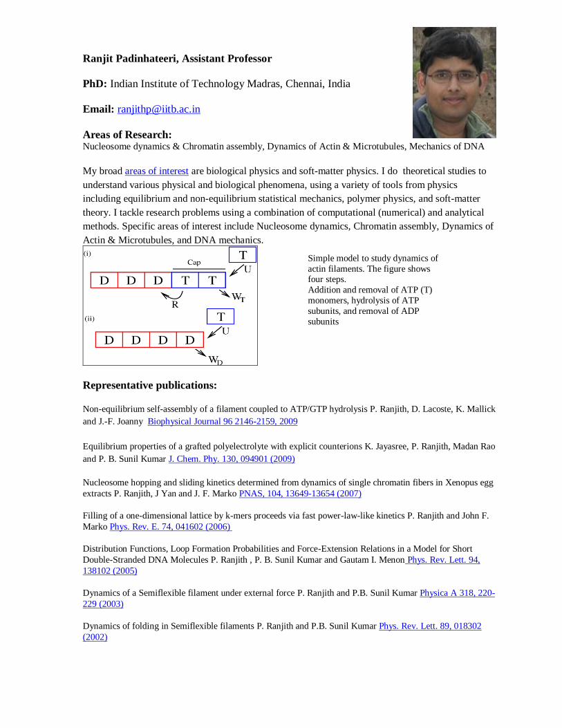
Simple model to study dynamics of
actin filaments. The figure shows four steps.
Addition and removal of ATP (T)
monomers, hydrolysis of ATP
subunits, and removal of ADP
subunits
Shamik Sen, Assistant Professor
PhD: University of Pennsylvania, Philadelphia
Email: [email protected]
Areas of Research: Cellular Biophysics, Bioengineering
It is increasingly understood that physical properties of the extracellular matrix (ECM) have a
profound influence on cell behavior. These include the geometry, topography and stiffness of the
ECM. This has led to the growth of the new and exciting field of mechanobiology, where the goal is
to study the influence of mechanics on various biological processes.
Our aim is to explore how development and cancer progression are regulated by the physical crosstalk
between the cell cytoskeleton and the ECM. To contribute to this understanding we combine cell and
molecular biology techniques with biophysical tools and computational approaches.
Representative publications:
Sen S, Ng WP, Kumar S. “Contractility Dominates Adhesive Ligand Density in Regulating Cellular De-
adhesion and Retraction Kinetics.” Ann Biomed. Engg, 2011. 39(4):1163-73.
Lee J, Chu BH, Sen S, Gupte A, Chancellor TJ, Chang C, Ren F, Kumar S, Lele TP. “Modulating malignant
epithelial tumor cell adhesion, migration and mechanics with nanorod surfaces.” Biomedical Microdevices,
2011. 13(1):89-95.
Sen S*, Tewari M*, Zajac A, Barton E, Sweeney HL, Discher DE. “Upregulation of paxillin and focal adhesion
signaling follows Dystroglycan Complex deletions and promotes a hypertensive state of differentiation,” Eur. J.
Cell Biology, 2011. 90(2-3):249-60.
Sen S, Dong M, Kumar S. “Isoform-specific contributions of -actinin to glioma cell mechanobiology.” PLoS
ONE, 2009. 4(12), e8427.
Sen S, Kumar S. “Cell-matrix de-adhesion dynamics reflect contractile mechanics,” Cellular and Molecular
Bioengineering, 2009. 2(2): 218-230.
Engler AJ, Sen S, Sweeney HL, Discher DE. “Matrix Elasticity Directs Stem Cell Lineage Specification,” Cell,
2006. 126(4): 677-698.
Sen S, Subramanian S, Discher DE. “Indentation and adhesive probing of a cell membrane with AFM:
theoretical model and experiments,” Biophysical J., 2005. 89(5): 3203-3213.
Ashutosh Kumar, Assistant Professor PhD: Tata Institute of Fundamental Research (TIFR), Mumabi, India
Email: [email protected]
Research Interest: NMR based structural biology
We intend to develop and make use of Nuclear Magnetic Resonance techniques to explore the structural and mechanistic aspects of biological macromolecules. We will deduce the atomic-
resolution structural and dynamics information to understand how bio-molecules communicate. Our
specific goal is to understand mechanism of protein aggregation and to elucidate the structural bases
of protein-lipid and protein-protein interactions for the membrane assembly.
Publications:
1. Kumar A, Heise H, Blommers M, Krastel P, Schmitt E, Petersen F, Mandelkow E, Carlomagno T, Griesinger C, Baldus M (2010) Interaction of epothilone B (patupilone) with microtubules as detected
by two-dimensional Solid-state NMR. Angw. Chem. Int. Ed. 49 (41), 7504-7.
2. Kim HY, Cho MK, Kumar A, Maier E, Siebenhaar C, Becker S, Fernandez CO, Lashuel HA, Benz
R, Lange A, Zweckstetter M. (2009) Structural Properties of Pore-Forming Oligomers of alpha-
Synuclein. J. Am. Chem. Soc. 131, 17482-17489.
3. Kumar A, Srivastava S, Hosur RV (2007) NMR Characterisation of the energy landscape of SUMO-1
in the native-state ensemble. J. Mol. Biol. 367, 1480-1493.
4. Kumar A, Srivastava S, Mishra RK, Mittal R, Hosur RV (2006) Residue Level NMR View of the
Urea Driven Equilibrium Folding pathway of SUMO-1 (1-97): Native Preferences do not Increase
Monotonously J. Mol. Biol. 361, 180-194.
5. Chatterjee A., Kumar A., Hosur RV (2006) Alanine check-points in HNN and HN(C)N spectra J.
Mag. Res. 181, 21-28
Kiran Kondabagil, Assistant Professor
PhD: University of Mysore (work done at CFTRI, Mysore)
Email: [email protected]
Areas of Research: Molecular virology, DNA packaging motors of bacteriophages, complex dsDNA viruses; DNA
unwinding proteins involved in replication of large DNA viruses; structural and functional analysis of the
virus assembly; phage DNA packaging machinery manipulation for nanotechnology applications
We are interested in understanding the molecular mechanisms of DNA packaging in large double-stranded DNA viruses and phages. We employ bioinformatics, molecular genetics, recombinant DNA, biochemical and
structural biology approaches to unravel these mechanisms. Genome encapsidation into preformed empty
capsid shell is a critical step in assembly of many bacteriophages (e.g. T4 and lambda), eukaryotic viruses (e.g.
herpes viruses) and nucleo-cytoplasmic large DNA viruses (NCLDVs) such as mimivirus. Two major types of
DNA packaging systems are found in viruses; terminase-portal mediated DNA packaging system that is
operative in many tailed bacteriophages and HerA/FtsK-type ATPases that are predicted to be involved in genome encapsidation in many viruses with inner lipid membranes. While the terminase-portal packaging
systems have been studied in many viruses like T4, lambda and HCMV for over two decades, the FtsK-ATPase
mediated packaging mechanisms have not been addressed in any system so far. We seek to employ complex
DNA viruses such as algae and protist-infecting viruses (e.g. mimivirus and PBCV) and virophages (e.g.
sputnik) as models systems.
Mimivirus and other NCLDVs encode for a number proteins involved in DNA precursor biosynthesis,
replication and repair. Some of these are predicted to be of ancient lineage with probable pre-eukaryotic origins
and are thought to be pivotal in major evolutionary transitions. Understanding molecular mechanisms
underlying the function of some of these important enzymes will help us decipher how large, complex double
stranded DNA viruses are assembled. It will also help in delineating many impending controversies about the
origins and evolution of large DNA viruses and their role, if any, in the emergence of eukaryotes.
Representative Publications
Abdulrahman Al-Zahrani*, Kiran Kondabagil*, Song Gao, Noreen Kelly, Manjira Ghosh-Kumar, and
Venigalla B. Rao1 The Small Terminase, gp16, of Bacteriophage T4 is a Global Regulator of the DNA
Packaging Motor. J. Biol. Chem. 284:24490-500, 2009.
Siyang Sun*, Kiran Kondabagil*, Bonnie Draper, Tanfis I Alam, Valorie D Bowman, Zhihong Zhang Z,
Shylaja Hegde, Andrie Fokine, Michael G Rossmann and Venigalla B Rao. The structure of the phage T4 DNA
packaging motor suggests a mechanism dependent on electrostatic forces. Cell 135:1251-1262, 2008.
Leading Edge Article; previewed by Williams RS, Williams GJ, Tainer JA. A charged performance by gp17 in
viral packaging. Cell 135:1169-1171, 2008.
Tanfis Alam, Bonnie Draper, Kiran Kondabagil, Frank J Rentas, Manjira Ghosh-Kumar, Siyang Sun, Michael
G Rossmann and Venigalla B Rao. The headful packaging nuclease of bacteriophage T4. Mol.
Microbiol. 69:1180-1190, 2008.
Siyang Sun, Kiran Kondabagil, Petra Gentz Michael G Rossmann and Venigalla B Rao. The structure of the ATPase that powers DNA packaging into bacteriophage T4 procapsids. Mol. Cell. 25:943-949, 2007.
Kiran R Kondabagil, Zhang Zhihong, and Venigalla B Rao. The DNA Translocating ATPase of Bacteriophage T4 Packaging Motor.J.Mol.Biol. 363:786-799,2006.
*-Contributed equally and share first authorship.