N-acetylcysteine for the prevention of stricture after
8
RESEARCH Open Access N-acetylcysteine for the prevention of stricture after circumferential endoscopic submucosal dissection of the esophagus: a randomized trial in a porcine model Maximilien Barret 1 , Frédéric Batteux 2 , Frédéric Beuvon 3 , Luigi Mangialavori 1 , Ariane Chryssostalis 1 , Carlos Pratico 1 , Stanislas Chaussade 1 and Frédéric Prat 1* Abstract Background: Circumferential endoscopic submucosal dissection (CESD) of the esophagus would allow for both the eradication of Barrett’s esophagus and its related complications, such as advanced neoplasia. However, such procedures generally induce inflammatory repair resulting in a fibrotic stricture. N-acetylcysteine (NAC) is an antioxidant that has shown some efficacy against pulmonary and hepatic fibrosis. The aim of our study was to evaluate the benefit of NAC in the prevention of esophageal cicatricial stricture after CESD in a swine model. Animals and methods: Two groups of six pigs each were subjected to general anesthesia and CESD: after randomization, a first group received an oral NAC treatment regimen of 100 mg/kg/day, initiated one week before the procedure, whereas a second group was followed without any prophylactic treatment. Follow-up endoscopies took place seven, fourteen, twenty-one, and twenty-eight days after CESD. Necropsy, histological assessment of esophageal inflammation, and fibrosis were performed on day 28. Results: The median esophageal lumen diameter on day 21 (main judgment criterion) was 4 mm (range 2 to 5) in group 1 and 3 mm (range 1 to 7) in group 2 (P = 0.95). No significant difference was observed between the two groups regarding clinical evaluation (time before onset of clinically significant esophageal obstruction), number of dilations, esophageal inflammation and fibrosis, or oxidative stress damage on immunohistochemistry. Conclusions: Despite its antioxidant effect, systemic administration of NAC did not show significant benefit on esophageal fibrosis in our animal model of esophageal wound healing within the experimental conditions of this study. Since the administered doses were relatively high, it seems unlikely that NAC might be a valuable option for the prevention of post-endoscopic esophageal stricture. Keywords: Endoscopic submucosal dissection (ESD), Esophageal stricture, Esophageal fibrosis, Barrett’s esophagus, Early esophageal adenocarcinoma Background Endoscopic resection of Barrett’ s esophagus with a high- grade dysplasia or early esophageal cancer has become an acceptable therapeutic option [1-3], alongside surgical eso- phagectomy. Endoscopic mucosal resection is easily feas- ible but leads to a piecemeal, non-carcinologic resection of neoplastic tissue, whereas endoscopic submucosal dis- section (ESD) is more carcinologically efficient, although more technically challenging. Nonetheless, both techni- ques result in cicatricial fibroinflammatory esophageal strictures when 75% or more of the mucosal circumfer- ence is removed [4,5]. These strictures may require nu- merous endoscopic dilations, jeopardizing the patients’ quality of life and risking esophageal perforation. This is believed to result from local inflammatory reactions, as well as subsequent cicatricial fibrosis [4,6-8]. Furthermore, * Correspondence: [email protected] 1 Department of Gastroenterology, Cochin Hospital, 27, rue du Faubourg St Jacques, Paris 75014, France Full list of author information is available at the end of the article © 2012 Barret et al.; licensee BioMed Central Ltd. This is an Open Access article distributed under the terms of the Creative Commons Attribution License (http://creativecommons.org/licenses/by/2.0), which permits unrestricted use, distribution, and reproduction in any medium, provided the original work is properly cited. Barret et al. Fibrogenesis & Tissue Repair 2012, 5:8 http://www.fibrogenesis.com/content/5/1/8
Transcript of N-acetylcysteine for the prevention of stricture after

Barret et al. Fibrogenesis & Tissue Repair 2012, 5:8http://www.fibrogenesis.com/content/5/1/8
RESEARCH Open Access
N-acetylcysteine for the prevention of strictureafter circumferential endoscopic submucosaldissection of the esophagus: a randomized trialin a porcine modelMaximilien Barret1, Frédéric Batteux2, Frédéric Beuvon3, Luigi Mangialavori1, Ariane Chryssostalis1, Carlos Pratico1,Stanislas Chaussade1 and Frédéric Prat1*
Abstract
Background: Circumferential endoscopic submucosal dissection (CESD) of the esophagus would allow for both theeradication of Barrett’s esophagus and its related complications, such as advanced neoplasia. However, suchprocedures generally induce inflammatory repair resulting in a fibrotic stricture. N-acetylcysteine (NAC) is anantioxidant that has shown some efficacy against pulmonary and hepatic fibrosis. The aim of our study was toevaluate the benefit of NAC in the prevention of esophageal cicatricial stricture after CESD in a swine model.
Animals and methods: Two groups of six pigs each were subjected to general anesthesia and CESD: afterrandomization, a first group received an oral NAC treatment regimen of 100 mg/kg/day, initiated one week beforethe procedure, whereas a second group was followed without any prophylactic treatment. Follow-up endoscopiestook place seven, fourteen, twenty-one, and twenty-eight days after CESD. Necropsy, histological assessment ofesophageal inflammation, and fibrosis were performed on day 28.
Results: The median esophageal lumen diameter on day 21 (main judgment criterion) was 4 mm (range 2 to 5) ingroup 1 and 3 mm (range 1 to 7) in group 2 (P= 0.95). No significant difference was observed between the twogroups regarding clinical evaluation (time before onset of clinically significant esophageal obstruction), number ofdilations, esophageal inflammation and fibrosis, or oxidative stress damage on immunohistochemistry.
Conclusions: Despite its antioxidant effect, systemic administration of NAC did not show significant benefit onesophageal fibrosis in our animal model of esophageal wound healing within the experimental conditions of thisstudy. Since the administered doses were relatively high, it seems unlikely that NAC might be a valuable option forthe prevention of post-endoscopic esophageal stricture.
Keywords: Endoscopic submucosal dissection (ESD), Esophageal stricture, Esophageal fibrosis, Barrett’s esophagus,Early esophageal adenocarcinoma
BackgroundEndoscopic resection of Barrett’s esophagus with a high-grade dysplasia or early esophageal cancer has become anacceptable therapeutic option [1-3], alongside surgical eso-phagectomy. Endoscopic mucosal resection is easily feas-ible but leads to a piecemeal, non-carcinologic resection
* Correspondence: [email protected] of Gastroenterology, Cochin Hospital, 27, rue du Faubourg StJacques, Paris 75014, FranceFull list of author information is available at the end of the article
© 2012 Barret et al.; licensee BioMed Central LCommons Attribution License (http://creativecreproduction in any medium, provided the or
of neoplastic tissue, whereas endoscopic submucosal dis-section (ESD) is more carcinologically efficient, althoughmore technically challenging. Nonetheless, both techni-ques result in cicatricial fibroinflammatory esophagealstrictures when 75% or more of the mucosal circumfer-ence is removed [4,5]. These strictures may require nu-merous endoscopic dilations, jeopardizing the patients’quality of life and risking esophageal perforation. This isbelieved to result from local inflammatory reactions, aswell as subsequent cicatricial fibrosis [4,6-8]. Furthermore,
td. This is an Open Access article distributed under the terms of the Creativeommons.org/licenses/by/2.0), which permits unrestricted use, distribution, andiginal work is properly cited.
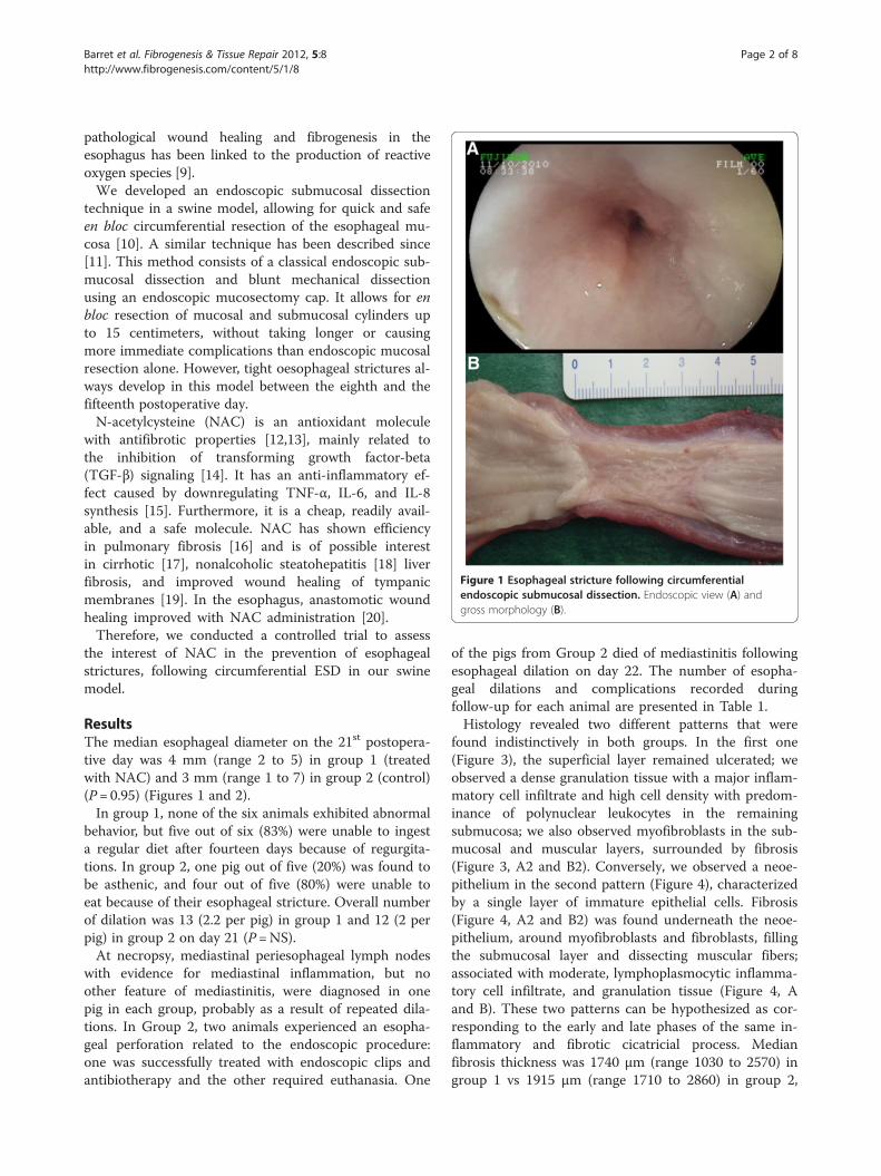
Figure 1 Esophageal stricture following circumferentialendoscopic submucosal dissection. Endoscopic view (A) andgross morphology (B).
Barret et al. Fibrogenesis & Tissue Repair 2012, 5:8 Page 2 of 8http://www.fibrogenesis.com/content/5/1/8
pathological wound healing and fibrogenesis in theesophagus has been linked to the production of reactiveoxygen species [9].We developed an endoscopic submucosal dissection
technique in a swine model, allowing for quick and safeen bloc circumferential resection of the esophageal mu-cosa [10]. A similar technique has been described since[11]. This method consists of a classical endoscopic sub-mucosal dissection and blunt mechanical dissectionusing an endoscopic mucosectomy cap. It allows for enbloc resection of mucosal and submucosal cylinders upto 15 centimeters, without taking longer or causingmore immediate complications than endoscopic mucosalresection alone. However, tight oesophageal strictures al-ways develop in this model between the eighth and thefifteenth postoperative day.N-acetylcysteine (NAC) is an antioxidant molecule
with antifibrotic properties [12,13], mainly related tothe inhibition of transforming growth factor-beta(TGF-β) signaling [14]. It has an anti-inflammatory ef-fect caused by downregulating TNF-α, IL-6, and IL-8synthesis [15]. Furthermore, it is a cheap, readily avail-able, and a safe molecule. NAC has shown efficiencyin pulmonary fibrosis [16] and is of possible interestin cirrhotic [17], nonalcoholic steatohepatitis [18] liverfibrosis, and improved wound healing of tympanicmembranes [19]. In the esophagus, anastomotic woundhealing improved with NAC administration [20].Therefore, we conducted a controlled trial to assess
the interest of NAC in the prevention of esophagealstrictures, following circumferential ESD in our swinemodel.
ResultsThe median esophageal diameter on the 21st postopera-tive day was 4 mm (range 2 to 5) in group 1 (treatedwith NAC) and 3 mm (range 1 to 7) in group 2 (control)(P= 0.95) (Figures 1 and 2).In group 1, none of the six animals exhibited abnormal
behavior, but five out of six (83%) were unable to ingesta regular diet after fourteen days because of regurgita-tions. In group 2, one pig out of five (20%) was found tobe asthenic, and four out of five (80%) were unable toeat because of their esophageal stricture. Overall numberof dilation was 13 (2.2 per pig) in group 1 and 12 (2 perpig) in group 2 on day 21 (P=NS).At necropsy, mediastinal periesophageal lymph nodes
with evidence for mediastinal inflammation, but noother feature of mediastinitis, were diagnosed in onepig in each group, probably as a result of repeated dila-tions. In Group 2, two animals experienced an esopha-geal perforation related to the endoscopic procedure:one was successfully treated with endoscopic clips andantibiotherapy and the other required euthanasia. One
of the pigs from Group 2 died of mediastinitis followingesophageal dilation on day 22. The number of esopha-geal dilations and complications recorded duringfollow-up for each animal are presented in Table 1.Histology revealed two different patterns that were
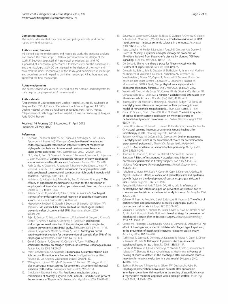
found indistinctively in both groups. In the first one(Figure 3), the superficial layer remained ulcerated; weobserved a dense granulation tissue with a major inflam-matory cell infiltrate and high cell density with predom-inance of polynuclear leukocytes in the remainingsubmucosa; we also observed myofibroblasts in the sub-mucosal and muscular layers, surrounded by fibrosis(Figure 3, A2 and B2). Conversely, we observed a neoe-pithelium in the second pattern (Figure 4), characterizedby a single layer of immature epithelial cells. Fibrosis(Figure 4, A2 and B2) was found underneath the neoe-pithelium, around myofibroblasts and fibroblasts, fillingthe submucosal layer and dissecting muscular fibers;associated with moderate, lymphoplasmocytic inflamma-tory cell infiltrate, and granulation tissue (Figure 4, Aand B). These two patterns can be hypothesized as cor-responding to the early and late phases of the same in-flammatory and fibrotic cicatricial process. Medianfibrosis thickness was 1740 μm (range 1030 to 2570) ingroup 1 vs 1915 μm (range 1710 to 2860) in group 2,

Figure 2 Esophageal stricture following circumferentialendoscopic submucosal dissection, endoscopic view (A) andmicroscopic view (B) (Masson’s trichrome, original magnifiedx12.5) showing the corresponding histological feature.
Table 1 Number of esophageal dilations and occurrenceof complications
Animalnumber
Number ofdilations
Earlycomplication
Latecomplication
Otherfindings
1** 2 - -
2* 3 - -
3** 0 Post-endoscopicesophagealperforation,requiringeuthanasia
4* 3 - -
5** 3 - -
6* 1 - -
7** 3 - Mediastinitison day 22,following thethird dilation
8* 2 - -
9** 2 Post-endoscopicesophagealperforation,successfullytreated
-
10* 2 - -
11** 2 - - Mediastinal inflammation+
at necropsy
12* 2 - - Mediastinalinflammation+
at necropsy
*Group 1, treated by N-acetylcysteine; **group 2, control group;+periesophageal lymphadenopathies without actual mediastinitis.
Barret et al. Fibrogenesis & Tissue Repair 2012, 5:8 Page 3 of 8http://www.fibrogenesis.com/content/5/1/8
P=NS; median thickness of the granulation tissue was1000 μm (range 925 to 1100) in group 1 vs 843 μm(range 868 to 2440) in group 2, P=NS. The granulationtissue presented with features of acute inflammation in30% of cases in group 1 vs 25% in group 2. Immunohis-tochemistry staining with 8-hydroxy-2’ deoxyguanosine(8-OH DG) antibody showed a more intense signal inthe esophageal wall of control animals as compared toNAC-treated animals (Figure 5). This finding was con-sistently observed on all slides. However, signal measure-ments did not show a statistically significant differencebetween the groups: median: 148 (range 122 to 169) ingroup 1 vs 136 (range 90 to 168) in group 2, P= 0.54.
DiscussionThis study is the first one to evaluate, on a swine model,the systemic administration of an antioxidant moleculein the prevention of esophageal strictures.Nonetheless, this work did not demonstrate any clin-
ical, endoscopical, or histological benefit of NAC to pre-vent oesophageal stenosis after circumferential ESD. Afirst hypothesis is an insufficient oral intake of NAC inregurgitating animals, despite repeated esophageal dila-tions. However, if solid food intake is impaired by
esophageal stricture, liquid intake is almost always pos-sible. Moreover, food intake remained normal during thefirst 10 days, the period during which NAC wasexpected to exert its maximal beneficial effect, if any. Anintravenous route for NAC would have required daily in-vasive treatment administration, and sedation of the ani-mals, which did not seem acceptable; furthermore, oraland intravenous administrations have been reported tobe equivalent [21]. Doses of NAC vary from 20 mg/kg to400 mg/kg of body weight per day. Given that the bio-availability of orally administrated NAC is low (<10%),we decided to use high doses, closer to those used inacetaminophen poisoning than in chronic fibrotic dis-eases [22]. The pigs were put on a 60 mg/kg/day dose ofNAC as part of a preliminary experiment and resultsshowed improvement of the esophageal diameter at day15 with no side effects. This propelled us to increase theNAC dose to 100 mg/kg/day. The use of higher doses,up to 400 mg/kg/day, has been reported, but is com-monly associated with frequent side effects [23]. Suchanaphylactoïd reactions, including vomiting, could havebiased the evaluation of food intake, esophageal mucosa,
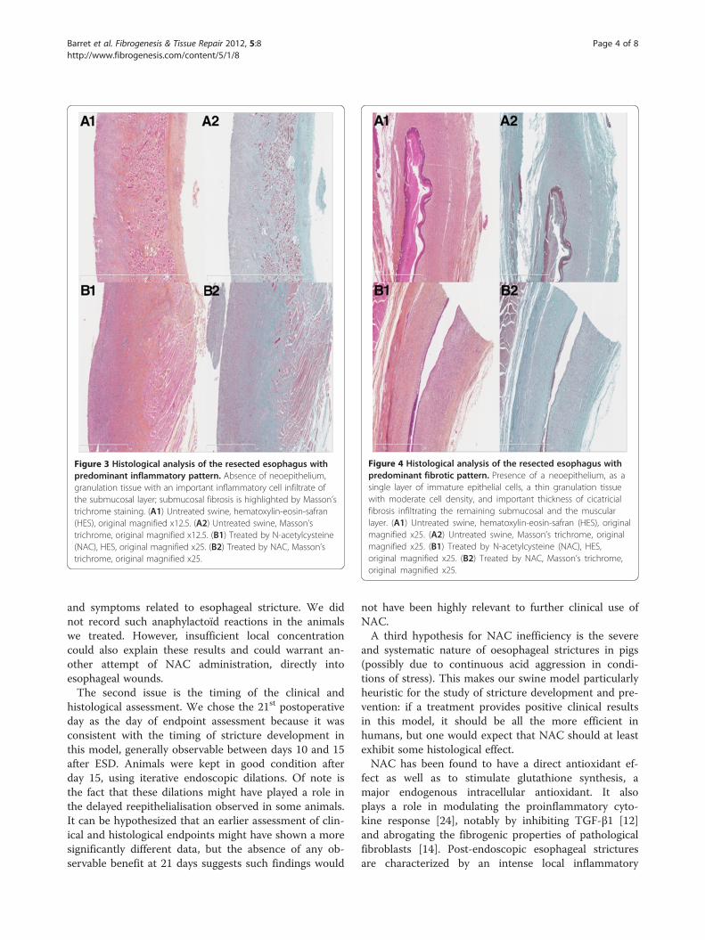
Figure 3 Histological analysis of the resected esophagus withpredominant inflammatory pattern. Absence of neoepithelium,granulation tissue with an important inflammatory cell infiltrate ofthe submucosal layer; submucosal fibrosis is highlighted by Masson’strichrome staining. (A1) Untreated swine, hematoxylin-eosin-safran(HES), original magnified x12.5. (A2) Untreated swine, Masson’strichrome, original magnified x12.5. (B1) Treated by N-acetylcysteine(NAC), HES, original magnified x25. (B2) Treated by NAC, Masson’strichrome, original magnified x25.
Figure 4 Histological analysis of the resected esophagus withpredominant fibrotic pattern. Presence of a neoepithelium, as asingle layer of immature epithelial cells, a thin granulation tissuewith moderate cell density, and important thickness of cicatricialfibrosis infiltrating the remaining submucosal and the muscularlayer. (A1) Untreated swine, hematoxylin-eosin-safran (HES), originalmagnified x25. (A2) Untreated swine, Masson’s trichrome, originalmagnified x25. (B1) Treated by N-acetylcysteine (NAC), HES,original magnified x25. (B2) Treated by NAC, Masson’s trichrome,original magnified x25.
Barret et al. Fibrogenesis & Tissue Repair 2012, 5:8 Page 4 of 8http://www.fibrogenesis.com/content/5/1/8
and symptoms related to esophageal stricture. We didnot record such anaphylactoïd reactions in the animalswe treated. However, insufficient local concentrationcould also explain these results and could warrant an-other attempt of NAC administration, directly intoesophageal wounds.The second issue is the timing of the clinical and
histological assessment. We chose the 21st postoperativeday as the day of endpoint assessment because it wasconsistent with the timing of stricture development inthis model, generally observable between days 10 and 15after ESD. Animals were kept in good condition afterday 15, using iterative endoscopic dilations. Of note isthe fact that these dilations might have played a role inthe delayed reepithelialisation observed in some animals.It can be hypothesized that an earlier assessment of clin-ical and histological endpoints might have shown a moresignificantly different data, but the absence of any ob-servable benefit at 21 days suggests such findings would
not have been highly relevant to further clinical use ofNAC.A third hypothesis for NAC inefficiency is the severe
and systematic nature of oesophageal strictures in pigs(possibly due to continuous acid aggression in condi-tions of stress). This makes our swine model particularlyheuristic for the study of stricture development and pre-vention: if a treatment provides positive clinical resultsin this model, it should be all the more efficient inhumans, but one would expect that NAC should at leastexhibit some histological effect.NAC has been found to have a direct antioxidant ef-
fect as well as to stimulate glutathione synthesis, amajor endogenous intracellular antioxidant. It alsoplays a role in modulating the proinflammatory cyto-kine response [24], notably by inhibiting TGF-β1 [12]and abrogating the fibrogenic properties of pathologicalfibroblasts [14]. Post-endoscopic esophageal stricturesare characterized by an intense local inflammatory

Figure 5 Immunohistochemical staining with 8-hydroxy-2’deoxyguanosine antibody. Strong signal predominant inperivascular region in control (A) and almost absent signal in treatedswine (B).
Barret et al. Fibrogenesis & Tissue Repair 2012, 5:8 Page 5 of 8http://www.fibrogenesis.com/content/5/1/8
reaction, which prevents reepithelialisation taking placeand quickly makes way for cicatricial fibrosis. Anincreased level of reactive oxygen species (ROS) hasbeen reported in esophageal ulcers, with the benefit ofan antioxidant molecule (vitamin E) administration onesophageal wound healing [9]; along with these results,many stricture-preventing drugs, either antioxidant[25], antifibrotic [26-30], or anti-inflammatory [4,7,31],have been tested on esophageal wounds. The onlytreatment reported to be beneficial against esophagealstrictures in human studies is local corticosteroid in-jection [4], although these results are contradictory,with previous animal studies reporting severe compli-cations related to triamcinolone injection [7]. Thesedata have led us to consider NAC for post-endoscopicesophageal stricture prevention. Despite an antioxidanteffect suggested by the results of immunohistochemis-try (although the difference between both signals onquantitative measurements did not reach statisticalsignificance), NAC did not, in our work, prevent esopha-geal stricture.Considering our results, it seems that several mechan-
isms could be at stake in cicatricial esophageal fibrosis.In particular, mucosal defect and resulting acid, and
bacterial aggressions of the submucosa could play a role;therefore, stricture prevention strategies focusing oncovering and protecting the injured esophagus should bedeveloped, even if recent studies, including ones doneon humans, either report insufficient efficacy [32], orshow an improvement of esophageal wound healing onlyin partial mucosal defects [7,8,33-35], where stricturesare uncommon. However, the use of growth factors,extracellular membranes, or bio-scaffolds or cellulartherapy, makes their translation into daily practice un-likely for the present moment. This data confirm thatpreventive treatment should prevail against curative onesregarding post-endoscopic esophageal strictures.
ConclusionsOur study failed to prove that an antioxidant such asNAC can improve the outcome of tissue repair in amodel of severe post-ESD esophageal stricture, whenused as a single agent. Its antioxidant activity did notprevent fibroinflammatory cicatricial esophageal stric-ture formation at the 21st postoperative day. Extensiveesophageal endoscopic submucosal dissection might be-come an alternative choice to esophagectomy forBarrett’s esophagus with high-grade dysplasia or earlyadenocarcinoma; hence, further analysis of esophagealwound healing will be needed, in order to develop newstricture-preventing treatments.
MethodsAnimal experimentsThe experimental protocol received approval from the sci-entific committee of the Surgical School of Paris (Ecole deChirurgie de l’Assistance Publique des Hôpitaux de Paris,7 rue du Fer à Moulin, 75005 Paris, France), and experi-ments were performed according to the standard guide-lines of the French Ministry of Agriculture that regulatesanimal research in France.Twelve 30 to 35 kg pigs, originating from the same
farm, were used in the study. The pigs were accommo-dated at our facility 48 hours before the procedure tookplace. Endoscopies were performed under generalanesthesia. All animals were prepared for anesthesia witha 12-hour diet and were administered an intramuscularinjection of 10 mg/kg ketamine and 2 mg/kg azaperone,30 minutes before induction. Following induction with8 mg/kg intravenous 1% propofol and endotracheal in-tubation, anesthesia was maintained through inhalationof 1% to 2% isoflurane. All animals received an intraven-ous infusion of 10 mg/kg/h crystalloid solution.
Endoscopic submucosal dissection procedureUpper gastrointestinal endoscopies were performed using astandard gastroscope (Fujinon EG450D; Fujinon, Sataima,Japan), through an overtube. A senior endoscopist carried

Barret et al. Fibrogenesis & Tissue Repair 2012, 5:8 Page 6 of 8http://www.fibrogenesis.com/content/5/1/8
out and/or directly supervised the procedure at all times.Circumferential submucosal endoscopic dissection of thedistal third of the esophagus was performed according tothe previously reported technique [10]. Briefly, two circum-ferential incisions reaching the superficial submucosal layerwere made following a submucosal injection of indigocarmine-stained saline; these two incisions defined a muco-sal cylinder of 5 cm in the lower third of the esophagus,5 cm above the gastroesophageal junction. The cylinderwas removed by blunt submucosal dissection, using amucosectomy cap (ST hood DH-19 GR; Fujinon, Sataima,Japan), which allowed for cleaving of the mucosa andsuperficial submucosa from the underlying submucosa bygently scraping the esophageal wall in a back and forth mo-tion, from the proximal to the distal incision. The resectedmucosal cylinder was then retrieved using the endoscope’ssuction channel.
Study designTwelve pigs were randomly allocated to a treatment and acontrol group. The first group of six pigs was given100 mg/kg/day of NAC (Mucomyst; Bristol-Myers Squibb,Princeton, NJ, USA) in drinking water for seven days be-fore the procedure. After circumferential endoscopic sub-mucosal dissection (CESD), the pigs received NACtreatment, at the same dose, until the 21st postoperativeday. A seven-day course of omeprazole 40 mg/day andbroad-spectrum antibiotics was also given: 10 mg/kgextended-release benzathine benzylpenicillin G+penicillinprocaine (Duphapen; Pfizer, NewYork, NY, USA) for thefirst four days; then 1 g/day amoxicillin (ClamoxylW,GlaxoSmithKline, Brentford, UK) for three days. The con-trol group (group 2) received proton-pump inhibitors andbroad-spectrum antibiotics at the same doses.The animals were given a liquid diet following each
procedure, and were gradually introduced to a solid fooddiet. The pigs were kept in the Surgical School of Pariswith daily examination and weekly veterinary follow-upand endoscopic examination. An endoscopic balloondilation was performed if clinical symptoms suggestedthat an esophageal stricture had occurred. Investigatorsprospectively collected clinical data on a weekly basis,such as global clinical status, which included general be-havior, food intake, and occurrence of regurgitations orvomiting. Investigators also recorded any endoscopicfindings, width of esophageal lumen (evaluated with abiopsy forceps), and number of balloon dilations.Twenty-eight days after the procedure, or earlier in
case of major complication (esophageal perforation,death), animals were euthanized with 100 mg/kg intra-venous injection of 7 mg pentobarbital (Dolethal;Vétoquinol, Paris, France), and underwent a necropsywith esophagectomy afterward.
Histological analysisSpecimens were fixed in 10% buffered formalin, embed-ded in paraffin, and processed into 5 μm-thick sections,after gross morphological examination of the size, weight,and thickness of the esophageal wall. The specimens werethen stained with hematoxylin-eosin-safran (HES), andMasson’s trichrome. Histological slides were digitizedwith the NDPI Nanozoomer (Hamamatsu Photonics,Hamamatsu City, Japan), and submitted to quantitativeanalysis of fibrosis, granulation tissue, and inflammatorycell infiltrate by a senior pathologist expert in digestivepathology. The following measurements were made oneach digitized image of trichrome-stained slides: maximalthickness of fibrosis, from the muscularis propria to theepithelium, and maximal thickness of the granulation tis-sue, from the esophageal lumen to the submucosal fibro-sis. Furthermore, the inflammatory cell infiltrate wascharacterized as being acute (predominance of poly-nuclear cells, high cell density) or chronic (predominanceof lymphocytes or plasmocytes, low cell density).Immunohistochemistry on paraffin sections was per-
formed with monoclonal antibodies to 8-hydroxy-2’ deox-yguanosine (8-OH DG) (ab26842; Abcam, Cambridge,UK) and goat polyclonal secondary antibodies to mouseIgG1 (ab 97240; Abcam, Cambridge, UK), in order toassess the intensity of oxidative stress in the esophagealwall. The intensity of the signal was analyzed using theImageJ software, the highest numbers corresponding tothe lowest signal.
Statistical analysisStatistical analysis was performed using GraphPad Prism(GraphPad Software, La Jolla, CA, USA). Continuousdata is expressed as median values and range, and com-pared with a nonparametric Mann–Whitney test. Cat-egorical data is expressed in percentages and comparedwith a Fisher’s exact test.
End pointsThe primary end point was the diameter of the esopha-gus measured via endoscopy on day 21 following theoperation.The secondary end points were: global clinical evalu-
ation of the pigs on day 21, number of endoscopic dila-tions required, thickness of the granulation tissue andthe fibrotic layer in the esophageal wall, and antioxidanteffect of NAC, as measured by 8-OH DG staining.
AbbreviationsCESD: Circumferential endoscopic submucosal dissection; HES: Hematoxylin-eosin-safran; IL: Interleukin; NAC: N-acetylcysteine; NS: Not significant;ROS: Reactive oxygen species; 8-OH DG: 8-hydroxy-2’ deoxyguanosine;TGF: Transforming growth factor; TNF-α: Tumor necrosis factor-alpha.
Barret et al. Fibrogenesis & Tissue Repair 2012, 5:8 Page 7 of 8http://www.fibrogenesis.com/content/5/1/8
Competing interestsThe authors declare that they have no competing interests, and do notreport any funding source.
Authors’ contributionsMB carried out the endoscopies and histologic study, the statistical analysisand drafted the manuscript. F. Batteux participated in the design of thestudy. F. Beuvon supervised all histological evaluations. LM and ACsupervised all endoscopic procedures. CP helped carry out the endoscopiesand the histologic study. SC participated in the design of the study andcorrected the draft. FP conceived of the study, and participated in its designand coordination and helped to draft the manuscript. All authors read andapproved the final manuscript.
AcknowledgementsThe authors thank Mrs Michelle Reichard and Mr Antoine Dechezleprêtre fortheir help in the preparation of the manuscript.
Author details1Department of Gastroenterology, Cochin Hospital, 27, rue du Faubourg StJacques, Paris 75014, France. 2Department of Immunology and EA 1833,Cochin Hospital, 27, rue du Faubourg St Jacques, Paris 75014, France.3Department of Pathology, Cochin Hospital, 27, rue du Faubourg St Jacques,Paris 75014, France.
Received: 14 February 2012 Accepted: 11 April 2012Published: 28 May 2012
References1. Chennat J, Konda VJ, Ross AS, de Tejada AH, Noffsinger A, Hart J, Lin S,
Ferguson MK, Posner MC, Waxman I: Complete Barrett's eradicationendoscopic mucosal resection: an effective treatment modality forhigh-grade dysplasia and intramucosal carcinoma–an Americansingle-center experience. Am J Gastroenterol 2009, 104:2684–2692.
2. Ell C, May A, Pech O, Gossner L, Guenter E, Behrens A, Nachbar L, HuijsmansJ, Vieth M, Stolte M: Curative endoscopic resection of early esophagealadenocarcinomas (Barrett's cancer). Gastrointest Endosc 2007, 65:3–10.
3. Pech O, May A, Gossner L, Rabenstein T, Manner H, Huijsmans J, Vieth M,Stolte M, Berres M, Ell C: Curative endoscopic therapy in patients withearly esophageal squamous-cell carcinoma or high-grade intraepithelialneoplasia. Endoscopy 2007, 39:30–35.
4. Hashimoto S, Kobayashi M, Takeuchi M, Sato Y, Narisawa R, Aoyagi Y: Theefficacy of endoscopic triamcinolone injection for the prevention ofesophageal stricture after endoscopic submucosal dissection. GastrointestEndosc 2011, 74:1389–1393.
5. Katada C, Muto M, Manabe T, Boku N, Ohtsu A, Yoshida S: Esophagealstenosis after endoscopic mucosal resection of superficial esophageallesions. Gastrointest Endosc 2003, 57:165–169.
6. Nieponice A, McGrath K, Qureshi I, Beckman EJ, Luketich JD, Gilbert TW,Badylak SF: An extracellular matrix scaffold for esophageal strictureprevention after circumferential EMR. Gastrointest Endosc 2009,69:289–296.
7. Rajan E, Gostout C, Feitoza A, Herman L, Knipschield M, Burgart L, Chung S,Cotton P, Hawes R, Kalloo A, Kantsevoy S, Pasricha P: Widespreadendoscopic mucosal resection of the esophagus with strategies forstricture prevention: a preclinical study. Endoscopy 2005, 37:1111–1115.
8. Sakurai T, Miyazaki S, Miyata G, Satomi S, Hori Y: Autologous buccalkeratinocyte implantation for the prevention of stenosis after EMR of theesophagus. Gastrointest Endosc 2007, 66:167–173.
9. Gunel E, Caglayan F, Caglayan O, Canbilen A, Tosun M: Effect ofantioxidant therapy on collagen synthesis in corrosive esophageal burns.Pediatr Surg Int 2002, 18:24–27.
10. Prat F, Chryssostalis A, Chaussade S: Esophageal Circumferential En-BlocSubmucosal Dissection in a Porcine Model. In Digestive Disease Week,Volume 63. Los Angeles: Gastrointest Endosc; 2006:AB133.
11. Willingham FF, Gee DW, Sylla P, Lauwers GY, Rattner DW, Brugge WR: Enbloc esophageal mucosectomy for concentric circumferential mucosalresection (with video). Gastrointest Endosc 2009, 69:147–151.
12. Knobloch K, Redeker J, Vogt PM: Antifibrotic medication using acombination of N-acetyl-L-cystein (NAC) and ACE inhibitors can preventthe recurrence of Dupuytren's disease. Med Hypotheses 2009, 73:659–661.
13. Servettaz A, Goulvestre C, Kavian N, Nicco C, Guilpain P, Chereau C, VuibletV, Guillevin L, Mouthon L, Weill B, Batteux F: Selective oxidation of DNAtopoisomerase 1 induces systemic sclerosis in the mouse. J Immunol2009, 182:5855–5864.
14. Kopp J, Seyhan H, Muller B, Lanczak J, Pausch E, Gressner AM, Dooley S,Horch RE: N-acetyl-L-cysteine abrogates fibrogenic properties offibroblasts isolated from Dupuytren's disease by blunting TGF-betasignalling. J Cell Mol Med 2006, 10:157–165.
15. Del Sorbo L, Zhang H: Is there a place for N-acetylcysteine in thetreatment of septic shock? Crit Care 2004, 8:93–95.
16. Demedts M, Behr J, Buhl R, Costabel U, Dekhuijzen R, Jansen HM, MacNeeW, Thomeer M, Wallaert B, Laurent F, Nicholson AG, Verbeken EK,Verschakelen J, Flower CD, Capron F, Petruzzelli S, De Vuyst P, van denBosch JM, Rodriguez-Becerra E, Corvasce G, Lankhorst I, Sardina M,Montanari M, IFIGENIA Study Group: High-dose acetylcysteine inidiopathic pulmonary fibrosis. N Engl J Med 2005, 353:2229–2242.
17. Vercelino R, Crespo I, de Souza GF, Cuevas MJ, de Oliveira MG, Marroni NP,Gonzalez-Gallego J, Tunon MJ: S-nitroso-N-acetylcysteine attenuates liverfibrosis in cirrhotic rats. J Mol Med (Berl) 2010, 88:401–411.
18. Baumgardner JN, Shankar K, Hennings L, Albano E, Badger TM, Ronis MJ:N-acetylcysteine attenuates progression of liver pathology in a ratmodel of nonalcoholic steatohepatitis. J Nutr 2008, 138:1872–1879.
19. Ozcan C, Gorur K, Cinel L, Talas DU, Unal M, Cinel I: The inhibitory effectof topical N-acetylcysteine application on myringosclerosis inperforated rat tympanic membrane. Int J Pediatr Otorhinolaryngol 2002,63:179–184.
20. Demir EO, Cakmak GK, Bakkal H, Turkcu UO, Kandemir N, Demir AS, TascilarO: N-acetyl-cysteine improves anastomotic wound healing afterradiotherapy in rats. J Investig Surg 2011, 24:151–158.
21. Buckley NA, Whyte IM, O'Connell DL, Dawson AH: Oral or intravenousN-acetylcysteine: which is the treatment of choice for acetaminophen(paracetamol) poisoning? J Toxicol Clin Toxicol 1999, 37:759–767.
22. Heard KJ: Acetylcysteine for acetaminophen poisoning. N Engl J Med2008, 359:285–292.
23. Knudsen TT, Thorsen S, Jensen SA, Dalhoff K, Schmidt LE, Becker U,Bendtsen F: Effect of intravenous N-acetylcysteine infusion onhaemostatic parameters in healthy subjects. Gut 2005, 54:515–521.
24. Moldeus P: Cotgreave IA: N-acetylcysteine. Methods Enzymol 1994,234:482–492.
25. Koltuksuz U, Mutus HM, Kutlu R, Ozyurt H, Cetin S, Karaman A, Gurbuz N,Akyol O, Aydin NE: Effects of caffeic acid phenethyl ester and epidermalgrowth factor on the development of caustic esophageal stricture inrats. J Pediatr Surg 2001, 36:1504–1509.
26. Apaydin BB, Paksoy M, Artis T, Sahin DA, Aki H, Uslu E: Influence ofpentoxifylline and interferon-alpha on prevention of stricture due tocorrosive esophagitis. An experimental study in the rat. Eur Surg Res 2001,33:225–231.
27. Cakmak M, Nayci A, Renda N, Erekul S, Gokcora H, Yucesan S: The effect ofcorticosteroids and pentoxifylline in caustic esophageal burns. Aprospective trial in rats. Int Surg 1997, 82:371–375.
28. Mizutani T, Tadauchi A, Arinobe M, Narita Y, Kato R, Niwa Y, Ohmiya N, ItohA, Hirooka Y, Honda H, Ueda M, Goto H: Novel strategy for prevention ofesophageal stricture after endoscopic surgery. Hepatogastroenterology2010, 57:1150–1156.
29. Ozcelik MF, Pekmezci S, Saribeyoglu K, Unal E, Gumustas K, Dogusoy G: Theeffect of halofuginone, a specific inhibitor of collagen type 1 synthesis,in the prevention of esophageal strictures related to caustic injury.Am J Surg 2004, 187:257–260.
30. Turkyilmaz Z, Sonmez K, Demirtola A, Karabulut R, Poyraz A, Gulen S, DincerS, Basaklar AC, Kale N: Mitomycin C prevents strictures in causticesophageal burns in rats. J Surg Res 2005, 123:182–187.
31. Honda M, Nakamura T, Hori Y, Shionoya Y, Nakada A, Sato T, Yamamoto K,Kobayashi T, Shimada H, Kida N, Hashimoto A, Hashimoto Y: Process ofhealing of mucosal defects in the esophagus after endoscopic mucosalresection: histological evaluation in a dog model. Endoscopy 2010,42:1092–1095.
32. Badylak SF, Hoppo T, Nieponice A, Gilbert TW, Davison JM, Jobe BA:Esophageal preservation in five male patients after endoscopicinner-layer circumferential resection in the setting of superficial cancer:a regenerative medicine approach with a biologic scaffold. Tissue EngPart A 2011, 17:1643–1650.

Barret et al. Fibrogenesis & Tissue Repair 2012, 5:8 Page 8 of 8http://www.fibrogenesis.com/content/5/1/8
33. Baatar D, Kawanaka H, Szabo IL, Pai R, Jones MK, Kitano S, Tarnawski AS:Esophageal ulceration activates keratinocyte growth factor and itsreceptor in rats: implications for ulcer healing. Gastroenterology 2002,122:458–468.
34. Nieponice A, Gilbert TW, Badylak SF: Reinforcement of esophagealanastomoses with an extracellular matrix scaffold in a canine model.Ann Thorac Surg 2006, 82:2050–2058.
35. Ohki T, Yamato M, Murakami D, Takagi R, Yang J, Namiki H, Okano T,Takasaki K: Treatment of oesophageal ulcerations using endoscopictransplantation of tissue-engineered autologous oral mucosal epithelialcell sheets in a canine model. Gut 2006, 55:1704–1710.
doi:10.1186/1755-1536-5-8Cite this article as: Barret et al.: N-acetylcysteine for the prevention ofstricture after circumferential endoscopic submucosal dissection of theesophagus: a randomized trial in a porcine model. Fibrogenesis & TissueRepair 2012 5:8.
Submit your next manuscript to BioMed Centraland take full advantage of:
• Convenient online submission
• Thorough peer review
• No space constraints or color figure charges
• Immediate publication on acceptance
• Inclusion in PubMed, CAS, Scopus and Google Scholar
• Research which is freely available for redistribution
Submit your manuscript at www.biomedcentral.com/submit