Myoepithelial tumor of soft tissue and bone: A current ... · Summary. Myoepithelial tumor (MET) of...
17
Summary. Myoepithelial tumor (MET) of soft tissue and bone is an unusual tumor of uncertain differentiation and histogenesis, but lately has been recognized as a distinct tumor entity. This tumor forms a morphologic continuum with a mixed tumor and a parachordoma, but is different from an extra-axial chordoma or chordoma periphericium. METs display a range of histopathologic features, including architectural arrangements/growth patterns, cell types and intervening stroma, leading to their several differential diagnoses. Presently, moderate nuclear atypia is the acceptable criterion to differentiate a myoepithelial carcinoma from a myoepithelioma. Immunohistochemical (IHC) stains, including epithelial antibody markers, along with S100 protein and GFAP are necessary in confirming a diagnosis of a MET. Certain METs are associated with loss of INI1/SMARCB1. Lately, certain specific “molecular signatures” been described underlying METs, identification of which that can further aid in their accurate diagnosis and in differentiating these tumors from their diagnostic mimics. Complete surgical resection forms the treatment mainstay, irrespective of a myoepithelioma or a myoepithelial carcinoma. This review will focus upon clinicopathologic, immunohisto- chemical and molecular features of METs of soft tissue and bone, along with their differential diagnoses and diagnostic implications. Key words: Myoepithelial tumor of soft tissues, Intraosseous myoepithelial tumor, Myoepithelioma, Myoepithelial carcinoma, EWSR1, INI1 Introduction Primary myoepithelial tumor (MET) of soft tissues is rare, but a well-defined tumor, of uncertain histogenesis. One of the first cases of a MET of the soft tissues was published by Burke et al. (1995) in a retroperitoneal location, diagnosed with the help of immunohistochemical (IHC) stains and further confirmed by ultrastructural examination. This was followed by a series of 19 METs of soft tissues, documented by Kilpatrick and Fletcher (Kilpatrick et al., 1997). Subsequently, there have been case reports and studies regarding these tumors, documented by various investigators (Burke et al., 1995; Kilpatrick et al., 1997; Michal and Miettinen, 1999; Hornick and Fletcher, 2003; Gleason and Fletcher, 2007; Antonescu et al., 2010; Rekhi et al., 2012). These tumors occur over a wide age-range, but are mostly seen in middle-aged patients and affect both the sexes equally (Hornick and Fletcher, 2003; Gleason and Fletcher, 2007; Antonescu et al., 2010). Primary MET has also been described in the bones as a distinct tumor entity (de Pinieux et al., 2001; Kurzawa et al., 2013; Rekhi et al., 2014, 2016). METs of soft tissues and bones exhibit a wide histopathologic spectrum and are synonymous with mixed tumors, myoepitheliomas and parachordomas (Kilpatrick et al., 1997; Antonescu et al., 2010; Rekhi et al., 2012; Fletcher et al., 2013). As a result of significant heterogeneity within METs, several soft tissue and bone tumors constitute as their differential diagnoses. Application of IHC markers is necessary for making a correct diagnosis of a MET (Hornick and Fletcher, 2003; Gleason and Fletcher, 2007; Kurzawa et al., 2013; Rekhi et al., 2012, 2016). The diagnosis of a malignant MET or a myoepithelial carcinoma is based upon the presence of at least moderate nuclear atypia (Hornick and Fletcher, Review Myoepithelial tumor of soft tissue and bone: A current perspective Anuj Verma and Bharat Rekhi Department of Surgical Pathology, Tata Memorial Hospital, Parel, Mumbai, India Histol Histopathol (2017) 32: 861-877 http://www.hh.um.es Offprint requests to: Dr Bharat Rekhi, Professor, Pathologist, Room Number AB:818, Department of Pathology, 8th Floor, Annex Building, Tata Memorial Hospital, Dr E.B. Road, Parel, Mumbai, India, 400012. e- mail: [email protected] DOI: 10.14670/HH-11-879 Histology and Histopathology From Cell Biology to Tissue Engineering
Transcript of Myoepithelial tumor of soft tissue and bone: A current ... · Summary. Myoepithelial tumor (MET) of...

Summary. Myoepithelial tumor (MET) of soft tissue andbone is an unusual tumor of uncertain differentiation andhistogenesis, but lately has been recognized as a distincttumor entity. This tumor forms a morphologic continuumwith a mixed tumor and a parachordoma, but is differentfrom an extra-axial chordoma or chordoma periphericium.METs display a range of histopathologic features,including architectural arrangements/growth patterns, celltypes and intervening stroma, leading to their severaldifferential diagnoses. Presently, moderate nuclear atypiais the acceptable criterion to differentiate a myoepithelialcarcinoma from a myoepithelioma. Immunohistochemical(IHC) stains, including epithelial antibody markers, alongwith S100 protein and GFAP are necessary in confirminga diagnosis of a MET. Certain METs are associated withloss of INI1/SMARCB1. Lately, certain specific“molecular signatures” been described underlying METs,identification of which that can further aid in theiraccurate diagnosis and in differentiating these tumorsfrom their diagnostic mimics. Complete surgical resectionforms the treatment mainstay, irrespective of amyoepithelioma or a myoepithelial carcinoma. Thisreview will focus upon clinicopathologic, immunohisto-chemical and molecular features of METs of soft tissueand bone, along with their differential diagnoses anddiagnostic implications.Key words: Myoepithelial tumor of soft tissues,Intraosseous myoepithelial tumor, Myoepithelioma,Myoepithelial carcinoma, EWSR1, INI1
Introduction
Primary myoepithelial tumor (MET) of soft tissuesis rare, but a well-defined tumor, of uncertainhistogenesis. One of the first cases of a MET of the softtissues was published by Burke et al. (1995) in aretroperitoneal location, diagnosed with the help ofimmunohistochemical (IHC) stains and furtherconfirmed by ultrastructural examination. This wasfollowed by a series of 19 METs of soft tissues,documented by Kilpatrick and Fletcher (Kilpatrick et al.,1997). Subsequently, there have been case reports andstudies regarding these tumors, documented by variousinvestigators (Burke et al., 1995; Kilpatrick et al., 1997;Michal and Miettinen, 1999; Hornick and Fletcher,2003; Gleason and Fletcher, 2007; Antonescu et al.,2010; Rekhi et al., 2012). These tumors occur over awide age-range, but are mostly seen in middle-agedpatients and affect both the sexes equally (Hornick andFletcher, 2003; Gleason and Fletcher, 2007; Antonescuet al., 2010). Primary MET has also been described inthe bones as a distinct tumor entity (de Pinieux et al.,2001; Kurzawa et al., 2013; Rekhi et al., 2014, 2016).METs of soft tissues and bones exhibit a wide
histopathologic spectrum and are synonymous withmixed tumors, myoepitheliomas and parachordomas(Kilpatrick et al., 1997; Antonescu et al., 2010; Rekhi etal., 2012; Fletcher et al., 2013). As a result of significantheterogeneity within METs, several soft tissue and bonetumors constitute as their differential diagnoses.Application of IHC markers is necessary for making acorrect diagnosis of a MET (Hornick and Fletcher, 2003;Gleason and Fletcher, 2007; Kurzawa et al., 2013; Rekhiet al., 2012, 2016). The diagnosis of a malignant MET ora myoepithelial carcinoma is based upon the presence ofat least moderate nuclear atypia (Hornick and Fletcher,
Review
Myoepithelial tumor of soft tissue and bone: A current perspectiveAnuj Verma and Bharat RekhiDepartment of Surgical Pathology, Tata Memorial Hospital, Parel, Mumbai, India
Histol Histopathol (2017) 32: 861-877
http://www.hh.um.es
Offprint requests to: Dr Bharat Rekhi, Professor, Pathologist, RoomNumber AB:818, Department of Pathology, 8th Floor, Annex Building,Tata Memorial Hospital, Dr E.B. Road, Parel, Mumbai, India, 400012. e-mail: [email protected]: 10.14670/HH-11-879
Histology andHistopathology
From Cell Biology to Tissue Engineering
2003; Rekhi et al., 2012, 2016 Kurzawa et al., 2013).Lately, various molecular alterations have beenunraveled that constitute as the molecular signatures ofcertain soft tissue METs, including myoepithelialcarcinomas (Antonescu et al., 2010, 2013; Agaram et al.,2015).This review will focus upon clinical, radiologic,
histopathologic, IHC and molecular features of METs ofsoft tissue and bones, along with a mention of diagnosticimplications, including treatment and outcomes in suchcases.Clinical features
METs of soft tissues and bone occur in patients of allage groups. These tumors have been reported in newborns,as well as in patients in the tenth decade of life. However,these tumors are most commonly seen in middle-agedpatients, with a mean age of 38 years, as documented inthe largest series (Michal and Miettinen, 1999; Hornickand Fletcher, 2003; Gleason and Fletcher, 2007).In a series of 8 primary intraosseous METs, the
reported average age was 33.5 years, while in a recentlydocumented series of 5 intraosseous myoepithelialcarcinomas, the reported average age was 26.2 years(Kurzawa et al., 2013; Rekhi et al., 2016). Gleason et al(Gleason and Fletcher, 2007) observed that METs inchildren are significantly more likely to be malignantand aggressive than those occurring in adults. Theyobserved that nearly 62% METs in children and 42% inadults were malignant (Gleason and Fletcher, 2007).Gender-wise males and females have been found to beequally affected in most documented studies (Hornickand Fletcher, 2003; Gleason and Fletcher, 2007;Antonescu et al., 2010).The presenting clinical symptoms in patients
afflicted with these tumors depend upon the sites ofinvolvement. Patients most commonly present with apainless mass, followed by a painful mass (morecommon in superficial METs). Cutaneous METs areknown to present as cutaneous papules. Rarely, patientspresent with only pain or neurological symptoms, suchas paraesthesia. Deep-seated METs are identifiedincidentally. The duration of symptoms ranges from afew weeks to decades, with a mean of 4 years (Hornickand Fletcher, 2003; Gleason and Fletcher, 2007;Kurzawa et al., 2013).Site-wise, METs mostly occur in the acral region
and in the girdles, commonly in the lower limbs. Othersites include trunk and head and neck region. Rarely,METs have been reported in the scrotal skin and in thevisceral organs, such as lungs (Hornick and Fletcher,2003; Flucke et al., 2011). These tumors are morecommonly superficially located. In the deeper tissues,these might present either in the subfascial,intramuscular regions, or rarely, in the intraosseous sites.Approximately 9% METs are known to present asintraosseous masses (Hornick and Fletcher, 2003;Antonescu et al., 2010). Among intraosseous METs, a
myoepithelioma is seen more frequently in theappendicular bones, while a myoepithelial carcinoma ismore commonly documented in the axial bones(Kurzawa et al., 2013; Rekhi et al., 2016). The size ofthe tumor ranges from 0.3 to 21.6 cm. Cutaneous andsuperficial METs are smaller than those occurring indeeper locations (Hornick and Fletcher, 2003; Flucke etal., 2011; Rekhi et al., 2012). In the largest study onMETs of soft tissues, Hornick et al (Hornick andFletcher, 2003) observed an average tumor size of 4.7cm. They also found that the malignant tumors(average=5.9 cm) were significantly larger than theirbenign counterparts (average 3.8 cm) (p=0.01) (Hornickand Fletcher, 2003).Radiologic features
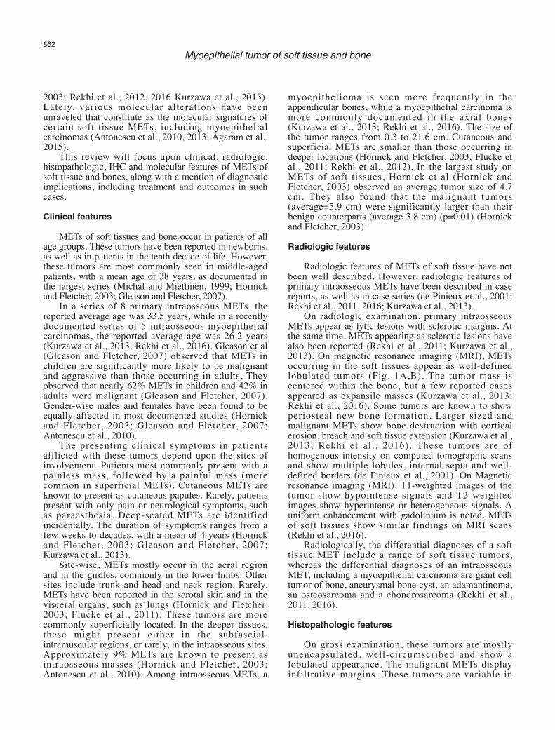
Radiologic features of METs of soft tissue have notbeen well described. However, radiologic features ofprimary intraosseous METs have been described in casereports, as well as in case series (de Pinieux et al., 2001;Rekhi et al., 2011, 2016; Kurzawa et al., 2013).On radiologic examination, primary intraosseous
METs appear as lytic lesions with sclerotic margins. Atthe same time, METs appearing as sclerotic lesions havealso been reported (Rekhi et al., 2011; Kurzawa et al.,2013). On magnetic resonance imaging (MRI), METsoccurring in the soft tissues appear as well-definedlobulated tumors (Fig. 1A,B). The tumor mass iscentered within the bone, but a few reported casesappeared as expansile masses (Kurzawa et al., 2013;Rekhi et al., 2016). Some tumors are known to showperiosteal new bone formation. Larger sized andmalignant METs show bone destruction with corticalerosion, breach and soft tissue extension (Kurzawa et al.,2013; Rekhi et al., 2016). These tumors are ofhomogenous intensity on computed tomographic scansand show multiple lobules, internal septa and well-defined borders (de Pinieux et al., 2001). On Magneticresonance imaging (MRI), T1-weighted images of thetumor show hypointense signals and T2-weightedimages show hyperintense or heterogeneous signals. Auniform enhancement with gadolinium is noted. METsof soft tissues show similar findings on MRI scans(Rekhi et al., 2016).Radiologically, the differential diagnoses of a soft
tissue MET include a range of soft tissue tumors,whereas the differential diagnoses of an intraosseousMET, including a myoepithelial carcinoma are giant celltumor of bone, aneurysmal bone cyst, an adamantinoma,an osteosarcoma and a chondrosarcoma (Rekhi et al.,2011, 2016).Histopathologic features
On gross examination, these tumors are mostlyunencapsulated, well-circumscribed and show alobulated appearance. The malignant METs displayinfiltrative margins. These tumors are variable in
862Myoepithelial tumor of soft tissue and bone

consistency and are soft to firm/ rubbery to hard,depending upon the intervening stroma that could bemyxoid to chondroid to osteoid. The color varies fromwhite to yellow to tan, the latter in case of hemorrhage.The cut surface is glistening, mucoid and myxoid.Trabeculations and microcysts are seen, along withoccasional cases showing foci of calcification, necrosisand hemorrhage (Hornick and Fletcher, 2003; Gleasonand Fletcher, 2007; Rekhi et al., 2012; Kurzawa et al.,2013).Microscopically, the tumors are unencapsulated,
lobulated and circumscribed with focal infiltrativemargins; the latter feature is more commonly identifiedin myoepithelial carcinomas. A range of cellulararrangements, cell types and stroma/ matrix is seenwithin METs. The tumor cells are arranged in nodulesand are arranged in the form of cords, trabeculae, as well
as in a nesting pattern; sheet-like and in a reticularpattern (Hornick and Fletcher, 2003; Gleason andFletcher, 2007). Rare cellular patterns include tubulo-acinar, pseudoacinar, alveolar and ‘rhythmic palisades’(Rekhi et al., 2012). Most common cell-type ispolygonal/epithelioid, containing moderate to abundant,eosinophilic cytoplasm, followed by cells with clearcytoplasm (mostly in parachordomas) and cells withspindle-shaped nuclei. Gradual transition from one celltype to another is often seen. Other cell types seen areplasmacytoid, rhabdoid and small round cell types(Hornick and Fletcher, 2003; Gleason and Fletcher,2007; Rekhi et al., 2012, 2016; Kurzawa et al., 2013).The round cells with scant cytoplasm are morecommonly seen in pediatric cases (approximately 30%of the cases) (Gleason and Fletcher, 2007). Certaintumors show ducts and tubules with sharply defined
863Myoepithelial tumor of soft tissue and bone
Fig. 1. Myoepithelioma of soft tissues. Magnetic resonance imaging (MRI) (post-contrast) showing a well-defined, lobulated soft tissue tumor in thegluteal region. B. T1-axial image showing a well-defined, multilobulated tumor, isointense to the muscles. C. Microscopic examination showingpolygonal cells, including cells with vacuolated cytoplasm (arrows), embedded in a myxochondroid matrix. Hematoxylin-eosin (H and E) x 200.

luminal borders along with myoepithelial cells. Theseare classified as mixed tumors (Hornick and Fletcher,2003). The intervening matrix in most tumors is myxoidor chondromyxoid type. Some tumors display acollagenous stroma or a combination of myxoid andcollagenous stroma. At the same time, a significantproportion of METs might not show any stroma(Hornick and Fletcher, 2003). Metaplastic cartilage,bone formation, calcification and adipocyticdifferentiation are also frequently seen (Hornick andFletcher, 2003; Flucke et al., 2011). Rarely, squamousmetaplasia has been described in certain primaryintraosseous myoepitheliomas, as well as in intraosseousmyoepithelial carcinomas (Rekhi et al., 2011, 2016).Parachordoma and an ectomesenchymal chondro-
myxoid tumor (ECT) of the tongue, initially thought torepresent different tumor entities are now considered as
a part of the common spectrum of METs (Hornick andFletcher, 2003; Argyris et al., 2016). The termparachordoma was first coined by Dabska (Dabska,1977), but this tumor was first described by Laskowski(Dabska, 1977; Fisher and Miettinen, 1997) in 1951, aschordoma periphericum. As a result of its morphologicsimilarity with a classical chordoma, various authorssuggested that this tumor might be representative of anabaxial chordoma (Dabska, 1977; Shin et al., 1994).However, recent studies have shown that a para-chordoma is different from an extra-axial or an abaxialchordoma/ chordoma periphericum (Tirabosco et al.,2008; Rekhi et al., 2012).Microscopically, parachordomas are composed of
polygonal cells with an abundant, pale to eosinophilicand vacuolated cytoplasm, embedded in a hyaline andmyxoid matrix. A transition of these polygonal-shaped
864Myoepithelial tumor of soft tissue and bone
Fig. 2. Immunohistochemical results of the same case. A. Tumor cells displaying pan cytokeratin (AE1/AE3) positivity. B. Focal EMA positivity withintumor cells. C. S100 protein positivity in several tumor cells. D. Low Ki-67/MIB1 highlighting nearly 5% tumor cell nuclei. Diaminobencidine (DAB). x 400.

cells to spindle-shaped cells is noted in parachordomasand is also a rare feature of a chordoma (Fisher andMiettinen, 1997). In a retrospective study, Fisher et al(Fisher and Miettinen, 1997) concluded that a chordomaand a parachordoma are different entities, as chordomashave an aggressive clinical behavior, higher metastaticrate and mortality, in contrast to parachordomas.Kilpatrick et al (Kilpatrick et al., 1997) noted that anMET of soft tissues might focally show vacuolated cells,resembling parachordomas. Furthermore, in a largerstudy, Hornick et al (Hornick and Fletcher, 2003)confirmed that parachordomas are within the spectrumof METs. An ectomesenchymal tumor (ECT) showspolygonal to epithelioid cells in a chondromyxoidstroma (Argyris et al., 2016). This tumor was describedas a pathological entity by Smith et al (Smith et al.,1995), who proposed that these tumors originate fromectomesenchymal cells in the tongue. However, afterfurther IHC and molecular results, ECTs have beenincluded within the morphologic continuum of a MET(Smith et al., 1995; Argyris et al., 2016). As result of their biphenotypic nature, METs express
a range of IHC markers, including epithelial andmyoepithelial markers. In most of the documented series
865Myoepithelial tumor of soft tissue and bone
Table 1. Review of molecular results from various studies on myoepithelial tumors of soft tissues and bone.
Authors, Year Gene Rearrangement Positive Cases Fusion Partner
Brandal et al., 2008 EWSR1 (n=1) 1 ÈWSR1-PBX1Brandal et al., 2009 EWSR1 (n=1) 1 EWSR1-ZNF444
Antonescu et al., 2010 EWSR1(n=66) 30
EWSR1-POU5F1(n=5)EWSR1-PBX1(n=5)EWSR1-ZNF444(n=1)EWSR1-#(n=19)
FUS(n=30§) 1 FUS-#
Flucke et al., 2011 (Cutaneous Mixed tumors/Myoepithelioma) EWSR1(n=16) 7 EWSR1-*FUS(n=9§) 0 -
Rekhi et al., 2012 EWSR1(n=6) 3 EWSR1-*Bahrami et al., 2012 PLAG1(n=11) 8 PLAG1-*Flucke et al., 2012 EWSR1(n=1) 1 EWSR1-ATF1Romeo et al., 2012 EWSR1(n=7) 1 EWSR1-NFATC2
Antonescu et al., 2013 PLAG1(n=35‡) 12PLAG1-LIFR(n=1)PLAG1-CTNNB1(n=0)PLAG1-#(n=11)
Kurzawa et al., 2013 (Intraosseous Myoepithelial tumors) EWSR1(n=7) 5 EWSR1-PBX1(n=1)EWSR1-*(n=4)
Puls et al., 2014 FUS(n=1) 1 FUS-POU5F1Agaram et al., 2015 EWSR1(n=23§§) 3 EWSR1-PBX3
Huang et al., 2015 FUS(n=66‡‡) 6 FUS-KLF17(n=4)FUS-#(n=2)
EWSR1(n=16§§) 1 EWSR1-KLF17Rekhi et al., 2016 EWSR1(n=1) 1 EWSR1-*
Argyris et al., 2016 (Ectomesenchymal chondromyxoid tumor) EWSR1(n=11) 3 EWSR1-*PLAG1(n=7) 0 -
*: Fusion partner not tested, #: Fusion partner not identified, §: Tested in a cohort of cases lacking EWSR1 gene rearrangement, §§: Tested in a cohortof cases with EWSR1 gene rearrangement with an unknown fusion partner, ‡: Tested in a cohort of cases lacking EWSR1 and FUS generearrangement, ‡‡: Tested in a cohort of cases lacking EWSR1 and PLAG1 gene rearrangement.
Fig. 3. Gross appearance of a myoepithelial tumor involving bone andsoft tissues. Cut surface, fresh and fixed state showing a grey-white,lobulated, rubbery tumor with focal glistening areas.

of METs, pan cytokeratin (AE1/AE3), epithelialmembrane antigen (EMA) or cytokeratin (CK) wasfound to be positive in more than 90% cases (Hornickand Fletcher, 2003; Gleason and Fletcher, 2007).Hornick and Fletcher (2003) recommended an optimalIHC panel for diagnosis of a MET, including markers,such as EMA, AE1/AE3, S100 protein and glialfibrillary acidic protein (GFAP) (Hornick and Fletcher,2003). They recommended positive expression of at leasta single epithelial IHC marker (EMA and or AE1/AE3),along with the positive expression of myoepithelialmarkers (S100 protein and/or GFAP), as a minimumcriteria for substantiating a diagnosis of a MET.Subsequently, Rekhi et al. (2012) in a series of 14 METs,including myoepithelial carcinomas of soft tissues,observed EMA positivity in 10/12 tumors (83%), CKpositivity in 3/12 tumors (25%), along with S100 protein
and glial fibrillary acidic protein (GFAP) in (11/13,85%) and, (6/12, 50%) tumors, respectively. Inequivocal cases, they recommended p63, CD10,calponin and SMA as additional, useful, surrogatemarkers (Rekhi et al., 2012).In their study, Kurzawa et al. (2013) reported EMA
positivity in 7/8 primary intraosseous METs andnegativity for keratins (AE1/AE3 or CK or Cam5.2) inall their 8 cases. Recently, Rekhi et al (Rekhi et al.,2016) reported positive expression of various epithelialmakers, such as EMA (5/5), CK (1/1) and CK5/6 (4/4),along with S100 protein (5/5) and GFAP (3/5), in 5 casesof primary intraosseous myoepithelial carcinomas.EMA positivity has been reported within the range
of 20% and 100% and approximately, including in 63%cases, in the largest documented series of METs,including carcinomas (Kilpatrick et al., 1997; Hornick
866Myoepithelial tumor of soft tissue and bone
Fig. 4. Same case. Myoepithelioma. Microscopic findings. A. Cellular tumor composed of benign appearing round to polygonal cells arranged in cordsand nests with intespersed thin walled blood vessels and myxoid stroma. H and E. B. Higher magnification displaying polygonal cells with clearcytoplasm, embedded in a myxoid matrix. H and E. Inset: tumor cells displaying pan cytokeratin (AE1/AE3) positivity. DAB. A, x 200; B, inset, x 400.

and Fletcher, 2003; Gleason and Fletcher, 2007;Kurzawa et al., 2013; Rekhi et al., 2016). S100 proteinimmunoexpression has been reported within the range of72% and 100% within METs of soft tissues and bone(Hornick and Fletcher, 2003; Gleason and Fletcher,2007; Rekhi et al., 2012; Kurzawa et al., 2013). P63positivity is seen between 23% and 70% tumors andGFAP expression within 27% and 60% METs(Kilpatrick et al., 1997; Hornick and Fletcher, 2003;Rekhi et al., 2012, 2016). The most commonly expressedmyogenic marker in these tumors is calponin, which isreported in 86% to 100% of cases, followed by SMAwhich is documented in 36-64% of cases and desmin in0-20% of cases (Kilpatrick et al., 1997; Hornick andFletcher, 2003; Gleason and Fletcher, 2007; Rekhi et al.,2012; Kurzawa et al., 2013). CD10 positivity is seen in
approximately 67% of METs (Rekhi et al., 2012). Focalcytoplasmic and membranous staining for MIC2 (CD99)is reported in approximately 80% of cases, particularlyin the round cell component of METs (Gleason andFletcher, 2007). SOX10 (Schwannian/melanocyticmarker) expression is more commonly seen in benignMETs (80%), as compared to the malignant counterparts(30%) (Miettinen et al., 2015). Nearly 40% METs have been reported to display
loss of integrase interactor 1 (INI1)/SMARCB1/BAF47protein and are therefore included under the expandingspectrum of “INI1 deficient” tumors (Gleason andFletcher, 2007; Rekhi et al., 2012). Nuclear reactivity forPLAG1 antibody, which is seen in pleomorphicadenoma, is only seen in mixed tumors and not inmyoepitheliomas. The staining is more diffuse in the
867Myoepithelial tumor of soft tissue and bone
Fig. 5. A. Tumor composed of polygonal cells arranged in diffuse and tubular manner (mixed epithelial tumor) with interspersed cystic areas andcollagenous stroma. Hematoxylin-eosin (H and E). B. Higher magnification showing rather benign apearing tumor cells with eosinophilic cytoplasm,arranged in cords and trabeculae within abundant myxoid stroma. H and E. C. Tumor cells showing diffuse pan cytokeratin (AE1/AE3) positivity. DAB.D. Tumor cells displaying S100 protein positivity. DAB. A, x 200; B-D, x 400.
myoepithelial component and is focal to absent in theepithelial cells. PLAG1 expression correlates well withPLAG1 gene rearrangement (Jo and Fletcher, 2015).Brachyury is not expressed in a MET/parachordoma, incontrast to an abaxial/extra-axial chordoma or achordoma periphericum, supporting the view that thesetwo categories of tumors are different (Rekhi et al.,2012, 2016).Criteria for malignancy are not well-defined in
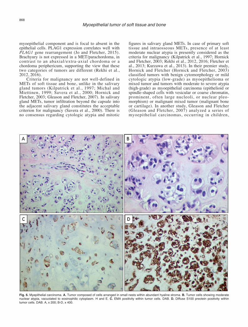
METs of soft tissue and bone, unlike in the salivarygland tumors (Kilpatrick et al., 1997; Michal andMiettinen, 1999; Savera et al., 2000; Hornick andFletcher, 2003; Gleason and Fletcher, 2007). In salivarygland METs, tumor infiltration beyond the capsule intothe adjacent salivary gland constitutes the acceptablecriterion for malignancy (Savera et al., 2000). There isno consensus regarding cytologic atypia and mitotic
figures in salivary gland METs. In case of primary softtissue and intraosseous METs, presence of at leastmoderate nuclear atypia is presently considered as thecriteria for malignancy (Kilpatrick et al., 1997; Hornickand Fletcher, 2003; Rekhi et al., 2012, 2016; Fletcher etal., 2013; Kurzawa et al., 2013). In their premier study,Hornick and Fletcher (Hornick and Fletcher, 2003)classified tumors with benign cytomorphology or mildcytologic atypia (low-grade) as myoepithelioma ormixed tumor and tumors with moderate to severe atypia(high-grade) as myoepithelial carcinoma (epithelioid orspindle-shaped cells with vesicular or coarse chromatin,prominent, often large nucleoli, or nuclear pleo-morphism) or malignant mixed tumor (malignant boneor cartilage). In another study, Gleason and Fletcher(Gleason and Fletcher, 2007) analyzed a series ofmyoepithelial carcinomas, occurring in children,
868Myoepithelial tumor of soft tissue and bone
Fig. 6. Myepithelial carcinoma. A. Tumor composed of cells arranged in small nests within abundant hyaline stroma. B. Tumor cells showing moderatenuclear atypia, vacuolated to eosinophilic cytoplasm. H and E. C. EMA positivity within tumor cells. DAB. D. Diffuse S100 preotein positivity withintumor cells. DAB. A, x 200; B-D, x 400.

considering at least moderate nuclear atypia as thecriterion for malignancy. Lately, in another study, Jo andFletcher (Jo and Fletcher, 2015) reinforced at leastmoderate degree of nuclear atypia as the criterion formalignancy in METs. Mitotic activity in METs has beenfound to be variable, irrespective of benign or malignantsub-types (Hornick and Fletcher, 2003). In their study onMETs, Hornick and Fletcher (Hornick and Fletcher,2003) observed mitotic figures ranging from 0 and 68per 10 high power fields, the average being 4.8. In theirstudy, Rekhi et al. (2012) observed no significant mitoticfigures in 5 myoepitheliomas, whereas mitotic figuresranging from 10-50/10 hpf in 9 cases of myoepithelialcarcinomas of soft tissues, all accompanied with at leastmoderate nuclear atypia. Coagulative tumor necrosis isseen more commonly in malignant METs (Rekhi et al.,2016). Heterologous chondrosarcomatous and osteo-
sarcomatous differentiation have also been described insoft tissue METs (Hornick and Fletcher, 2003). Whilemixed tumors are composed of both epithelial (ductformation/differentiation) and myoepithelial cells invarying proportions, myoepitheliomas and myoepithelialcarcinomas comprise cells lacking ductal differentiation(Kilpatrick et al., 1997; Hornick and Fletcher, 2003).Most cases of malignancy occur de novo. Rarely,malignant METs arise from a pre-existing myo-epithelioma (Hornick and Fletcher, 2003) (Figs. 2-10A).Genetic aberrations
Recent studies have unraveled certain geneticaberrations underlying METs of soft tissue and bone,including EWSR1 gene rearrangement as the commonestgenetic alteration, noted in approximately 45-50% of the
869Myoepithelial tumor of soft tissue and bone
Fig. 7. A. A case of a myoepithelial tumor composed of cells with prominent cytoplasmic vacuolation and focal myxoid stroma, indicative of aparachordoma. H and E. B. Tumor cells displaying EMA positivity. DAB. C. Tumor cells displaying focal S100 protein positivity. D. Tumor cellsdisplaying complete loss of INI1. Interveing lymphocytes and endothelial cells acting as internal positive control. DAB. x 400.

these tumors (Antonescu et al., 2010; Flucke et al., 2011;Rekhi et al., 2012, 2016) (Fig. 10B). Brandal et al.(2008, 2009) reported EWSR1-PBX1 and EWSR1-ZNF444 fusion transcripts in a single case, each of amyoepithelioma and a myoepithelial carcinoma,respectively. Subsequently, Antonescu et al. (2010)demonstrated that EWSR1 rearrangement was consis-tently present in METs albeit with various partner genes.They observed EWSR1 rearrangement with some of thepartner genes associated with specific histopathologicfeatures. In their study, mixed tumors did not showEWSR1 gene rearrangement (Antonescu et al., 2010).Kurzawa et al. (2013) identified EWSR1 gene rearran-gement in 71% cases of intraosseous myoepithelioma.Antonescu et al. (2010) observed EWSR1 rearrangementin 50% malignant METs and in 40% benign METs,mostly in deep-seated tumors. In the same study, among
the cases showing EWSR1 rearrangement, 53% of caseswere malignant and 47% were benign. Rekhi et al.(2012, 2016) identified EWSR1 rearrangement in 3/6 softtissue METs and in a single case of an intraosseousmyoepithelial carcinoma, where this was tested. Fluckeet al. (2011) identified EWSR1 rearran-gement in 44%cases of cutaneous METs. They observed EWSR1rearrangement in myoepitheliomas, as well as in mixedtumors (Flucke et al., 2011). Contrastingly, Bahrami et al(Bahrami et al., 2012) did not identify EWSR1rearrangement in mixed tumors.Fusion with specific partner genes, such as POU5F1
and PBX1 has been reported in 16% METs that showEWSR1 rearrangement (Antonescu et al., 2010). PBX3,ZNF444, ATF1, KLF17, NFATC2 are uncommon genesforming specific transcripts, underlying certain METs(Antonescu et al., 2010; Flucke et al., 2012a,b; Romeo et
870Myoepithelial tumor of soft tissue and bone
Fig. 8. Introassoeus myepithelial carcinoma. Microscopic findings. A. Tumor composed of cells exhibiting moderate nuclear atypia, arranged in sheetsand cords and displaying squmaous differentiation, with areas of prominent chondroid differentiation. H and E. B. Conspicuous areas of chondroiddifferentiation. H and E. x 200.

al., 2012; Agaram et al., 2015; Huang et al., 2015). Incertain cases of METs, the partner genes have not yetbeen identified. Antonescu et al. (2010) observed thattumors showing EWSR1-POU5F1 fusion are seen inyoung patients (mean 21 years; median 26 years). Thesetend to be situated in relatively deeper locations, aremore commonly malignant and have a nestedarrangement of clear cells. They also observed thattumors with EWSR1-PBX1 fusion are seen in middle-aged patients (mean 46 years); in deep soft tissues, bonesand visceral organs and could be either benign ormalignant. These tumors show spindle and epithelioidcells in a sclerotic and fibrotic stroma. In a recent study,Agaram et al. (2015), observed EWSR1-PBX3 mutationmore frequently in the bones. Histopathologically, thesetumors tend to show epithelioid to oval cells, embeddedwithin a sclerotic or myxoid stroma (Agaram et al.,2015). ECT of the tongue is reported to show EWSR1
rearrangement in 25% of cases, further reinforcing itsgenetic link with a MET (Argyris et al., 2016).Nearly 9% of METs that do not show EWSR1 gene
rearrangement are characterized by FUS gene rearran-gement. KLF17 and POU5F1 constitute as the onlyknown fusion partners for EWSR1 gene in these cases. Ina recent study, KLF17 fusion was seen in 66% cases ofFUS rearranged METs. Such tumors tend to occur inyoung patients (mean 28 years; median 32years) andhave a male preponderance. Although soft tissues arepredominantly involved, these tumors are also known tooccur in the bones and visceral organs. Histopatho-logically, these METs are more frequently benign thanmalignant and show the entire morphologic spectrumknown in METs (Huang et al., 2015). FUS-POU5F1fusion transcript has been reported in a single case of anintraosseous myoepithelioma (Puls et al., 2014). Inanother case report of a large intraosseous myo-
871Myoepithelial tumor of soft tissue and bone
Fig. 9. Immunohistochemical findings. A. Tumor cells displaying CK7 positivity. DAB. B. Tumor cells showing distinct, focal GFAP positivity. DAB. C.Tumor cells showing diffuse S100 protein positivity. DAB. x 400.

epithelioma, displaying promineent squamousmetaplasia, EWSR1 rearrangment was absent. The sametumor displayed trisomies of chromosomes 11, 15 and17 and del (16q) and del (22q11) (Rekhi et al., 2011).
PLAG1 gene rearrangement is reported in pleo-morphic adenoma of the salivary gland (Martins et al.,2005). METs lacking EWSR1 and FUS gene re-arrangement have been found to show PLAG1 generearrangement in 37% cases. These METs present assuperficial, as well as deep-seated lesions. Almost allsuch tumors show tubulo-ductular differentiation (mixedtumors). It is noteworthy that a gene rearrangement canbe seen in the form of a balanced translocation, anunbalanced translocation, inversion and interstitialdeletion (Bahrami et al., 2012; Antonescu et al., 2013).LIFR-PLAG1 gene fusion has been reported in a singlecase of a MET (Antonescu et al., 2013). PLAG1 gene
fusions with CTNNB1, FGFR1 and HMGA2 genes,observed in mixed salivary gland tumors, have not beenreported in METs of bone and soft tissues. However, asingle case of a MET showing an increased copy numberof HMGA2 has been reported. An ECT lacks PLAG1mutation (Antonescu et al., 2013).The value of molecular testing lays in differentiating
an MET from its differential diagnoses. EMC, one of theclosest differentials of a MET also shows EWSR1 generearrangement, but the fusion partner in cases of EMC isNR4A3. In a documented study, all cases of EMC showedfusion of NR4A3 gene, either with EWSR1 gene, or withother genes (Flucke et al., 2012a,b). In cases of equivocalfeatures, molecular tests can be used to differentiate aMET from an OFMT. OFMTs show PHF1 generearrangement in 80% cases and lack EWSR1 rearran-gement (Jo, 2015). Adamantinoma-like EFTs also display
872Myoepithelial tumor of soft tissue and bone
Fig. 10. A. Parachordoma. H and E. B. Fluorescent in-situ hybridization technique displaying EWSR1 rearrangement. Double lines show ‘split’orange/green signals, indicative of rearrangement. DAP. Inset displaing ‘split’ orange/green signals (double arrows) along with single fused signal(yellow arrow), indicative of EWSR1 rearrangement. DAPI. A, x 400; B, inset, x 1,000.

EWSR1 rearrangement, but the fusion partner is FLI1 inthose cases (Table 1, Fig. 11) (Bishop et al., 2015).Differential diagnoses
Several differential diagnoses need to be consideredbefore making diagnosis of a MET. Metastatic tumors,especially malignant mixed epithelial tumors andmyoepithelial carcinomas from the salivary gland needto be excluded by clinico-radiologic examination.Metastasis of tumors, such as mucinous adeno-carcinomas can be ruled out with the help of positiveIHC expression of S100 protein and GFAP that is seen inMETs (Hornick and Fletcher, 2003; Gleason andFletcher, 2007).An extraskeletal myxoid chondrosarcoma (EMC)
shows overlapping histopathologic features with a MET.By immunohistochemistry, EMCs are generally negativefor cytokeratins, unlike METs, but do express S100protein and EMA, similar to METs (Hornick andFletcher, 2003; Gleason and Fletcher, 2007; Kurzawa etal., 2013). In such cases, molecular testing is
recommended.An Ossifying fibromyxoid tumor (OFMT) also
shows overlapping histopathologic and IHC featureswith a MET. However, in contrast to a MET, an OFMTinvariably shows monomorphic oval cells with aperipheral rim of ossification. The intervening stroma inan OFMT is invariably myxoid, in contrast to arelatively more variable stroma within cells of a MET,ranging from dense hyaline-like (as noted in a sclerosingepithelioid fibrosarcoma), to chondroid to an osseoustype. There is a considerable overlap of expression ofIHC markers between an OFMT and a MET. In difficultcases, particularly in “non-ossifying” variant of anOFMTs, the molecular tests are useful (Hornick andFletcher, 2003; Antonescu et al., 2010). A sclerosingrhabdomyosarcoma can be differentiated from a MET,based on positive expression of muscle specific markers,such as desmin, along with skeletal muscle specificmarkers, such as MyoD1 and myogenin.Considering 40% cases of MET display loss of
SMARCB1/INI1, various tumor entities showing loss ofINI1 constitute as differential diagnoses of a MET, for
873Myoepithelial tumor of soft tissue and bone
Fig. 11. Schematic representation of various fusiontranscripts underlying myoepithelial tumors of soft tissues.

example, an extra-renal rhabdoid tumor (ERRT) inpediatric patients and an epithelioid sarcoma.Immunoexpression of INI1 is invariably lost, whileexpression of S100 protein is variable in an ERRT(Gleason and Fletcher, 2007; Agaram et al., 2015).Similarly, epithelioid sarcomas (ESs) do not expressS100 protein in a substantial number of tumor cells.Moreover, in nearly 60-70% cases of ES, CD34 ispositive (Hornick and Fletcher, 2003; Agaram et al.,2015). An epithelioid malignant peripheral nerve sheathtumor displays overlapping histopathologic features,IHC profile, including positivity for epithelial markersand S100 protein, as well as loss of INI1 in certain cases.The presence of a variable amount of chondromyxoidstroma is more commonly seen in a MET. Furthermore,molecular testing is recommended, considering METsdisplay specific molecular signatures that are lacking inan epithelioid MPNST.Various differential diagnoses of an intraosseous
MET, include a chondromyxoid fibroma; an adaman-tinoma; a chondrosarcoma, to an osteosarcoma withchondroblastic differetiation (Rekhi et al., 2011, 2016). A chondromyxoid fibroma, an osteosarcoma and a
chondrosarcoma can be differentiated from a MET,based on negative expression of epithelial IHC markersin these tumors, in contrast to a MET (Kurzawa et al.,2013). Squamous differentiation, reported withinintraosseous METs, is characteristically seen in anadamantinoma, including its dedifferentiated subtype(Hazelbag et al., 2003; Kanamori and Hogendoorn,2013; Bishop et al., 2015). In such cases, positiveexpression of S100 protein and GFAP are crucial inconfirmation of a MET, over an adamantinoma (Rekhi etal., 2011, 2016).An abaxial chordoma needs exclusion in cases
where vacuolated cells predominate, for example aparachordoma. S100 protein and epithelial markers aresimilarly expressed by chordomas. However, brachyury,which is a highly specific and a sensitive for chordomas,is consistently negative in METs (Jo, 2015; Rekhi et al.,2016). “Adamantinoma-like” Ewing family tumors(EFT) show uniformly arranged small round cells andmight resemble a myoepithelial carcinoma (Bishop etal., 2015). These tumors show positive expression ofS100 protein and actin as well. Besides overlappinghistopathologic and IHC features, these two tumors
874Myoepithelial tumor of soft tissue and bone
Fig. 12. Flow chart depicting approach to diagnosis of Myoepithelial tumors of Bone and Soft tissue.

show similar molecular features, including EWSR1rearrangement (Fig. 12) (Antonescu et al., 2010; Fluckeet al., 2011; Rekhi et al., 2012; Bishop et al., 2015).However, the fusion partner genes are different.Treatment and outcomes
Diagnosis of a MET has significant treatmentimplications. Surgical resection with clear marginsremains the preferred treatment for choice of METs,irrespective of benign or malignant subtypes. Adjuvantradiation therapy may be offered in cases with marginalor intracapsular resections for a better loco-regionalclearance. The role of chemotherapy is unclear (Rekhi etal., 2012, 2016; Domingo-Musibay et al., 2016). This isin contrast to certain diagnostic mimics of METs, suchas a metastatic adenocarcinoma, where a specificchemotherpay might be offered, as well as a conven-tional high-grade osteosarcoma that is treated with aspecific chemotherapy in neoadjuvant settings.Chances of recurrences in cases of benign METs
range from 18% to 29%, while in malignant METs, theseare between 42% and 64% (Hornick and Fletcher, 2003;Domingo-Musibay et al., 2016). In a recently publishedretrospective clinical analysis, Domingo-Musibay et al(Domingo-Musibay et al., 2016), reported 5-year eventfree survival rates of 88% and 36%, in low-grade andhigh-grade METs, respectively. The low-grade and highgrade METs in their study were equivalent to benign andmalignant METs, respectively (Hornick and Fletcher,2003; Domingo-Musibay et al., 2016). In an earlierstudy, metastasis and death was recorded in 32% and13%, respectively, in cases of malignant METs (Hornickand Fletcher, 2003). Metastasis is more commonly seenin pediatric patients (Gleason and Fletcher, 2007;Domingo-Musibay et al., 2016). Gleason et al (Gleasonand Fletcher, 2007) documented metastasis in 52% casesof pediatric myoepithelial carcinomas, while Domingo-Musibay et al. (Domingo-Musibay et al., 2016) observedmetastasis in 43% cases of adult myoepithelialcarcinomas. Multiple recurrences can be seen in bothbenign and malignant tumors. Recurrence is morecommon in patients undergoing marginal excisions, ascompared to patients undergoing wide-excisions(Gleason and Fletcher, 2007). METs can metastasize tothe regional lymph nodes, or to distant organs, mostcommonly lungs, along with other sites, such as bones,mediastinum, spine, orbit, soft tissues and brain(Hornick and Fletcher, 2003). Tumor size more than,equal to 5 cm and tumor necrosis are importantprognostic markers (Gleason and Fletcher, 2007).Histogenesis of these tumors, especially intraosseous
METs is unclear. Presently METs of soft tissue and boneare included in the category of tumors with uncertainhistogenesis. One of the possible hypotheses isdisplacement of myoepithelial elements duringembryogenesis into deeper soft tissues and bones, wherethese tumors originate (de Pinieux et al., 2001; Rekhi etal., 2011, 2012, 2016). There is an increasing evidence
that epithelial and myoepithelial cell populations sharephenotypic and genotypic characteristics, supporting theconcept of a modified myoepithelial cell model.Probably, a single pluripotent cell is capable ofdifferentiating into a variety of morphologic/phenotypicforms (Antonescu et al., 2013).Conclusions
A MET of soft tissue and bone and is a distincttumor entity, which is diagnosed with the help of certainhistopathologic features, combined with IHC results.METs show a wide range of histopathologic featuresincluding various cell types, architectural patterns andstroma, leading to a range of differential diagnoses. Co-expression of epithelial IHC markers with S100 proteinand or GFAP immunoreactivity along with otheroptional IHC markers is helpful in making an accuratediagnosis of a MET. Certain METs show loss ofSMARCB1/INI1 protein. METs, including a parachor-doma are different from an extra axial chordoma orchordoma periphericum, based on brachyury negativitywithin METs, in contrast to chordomas. Presently,moderate nuclear atypia constitutes as the criterion formalignancy in cases of MET of bone and soft tissues.EWSR1, FUS and PLAG1 gene rearrangements, leadingto formation of specific fusion transcripts, are noted in asubset of METs, constituting as their molecular‘signatures’, associated with certain specific clinico-pathologic features. In certain cases where differentialdiagnoses canot be resolved with IHC, molecular testingis recommended. A correct diagnosis has treatmentimplications. Surgical resection, preferably with clearmargins is a definitive treatment, irrespective of a benignor a malignant MET. Adjuvant radiation therapyfollowing excision may be offered in recurrent andmalignant cases. The role of chemotherapy is unclear. Amalignant or a high-grade MET is associated with asignificantly more aggressive clinical course than abenign or a low-grade MET.References
Agaram N.P., Chen H.W., Zhang L., Sung Y.S., Panicek D., HealeyJ.H., Nielsen G.P., Fletcher C.D. and Antonescu C.R. (2015).EWSR1-PBX3: a novel gene fusion in myoepithelial tumors. GenesChromosomes Cancer 54, 63-71.
Antonescu C.R., Zhang L., Shao S.Y., Mosquera J.M., Weinreb I.,Katabi N. and Fletcher C.D. (2013). Frequent PLAG1 generearrangements in skin and soft tissue myoepithelioma with ductaldifferentiation. Genes Chromosomes Cancer 52, 675-682.
Antonescu C.R., Zhang L., Chang N.E., Pawel B.R., Travis W., KatabiN., Edelman M., Rosenberg A.E., Nielsen G.P., Dal Cin P. andFletcher C.D. (2010). EWSR1-POU5F1 fusion in soft tissuemyoepithelial tumors. A molecular analysis of sixty-six cases,including soft tissue, bone, and visceral lesions, showing commoninvolvement of the EWSR1 gene. Genes Chromosomes Cancer 49,1114-1124.
Argyris P.P., Bilodeau E.A., Yancoskie A.E., Trochesset D.,
875Myoepithelial tumor of soft tissue and bone

Pambuccian S.E., Wetzel S.L., Shah S.S., Edelman M., FreedmanP., Dolan M. and Koutlas I.G. (2016). A subset of ectomesenchymalchondromyxoid tumors of the tongue shows EWSR1 rearran-gements and is genetically linked to soft tissue myoepithelialneoplasms: A study of 11 cases. Histopathology 69, 607-613.
Bahrami A., Dalton J.D., Krane J.F. and Fletcher C.D. (2012). A subsetof cutaneous and soft tissue mixed tumors are genetically linked totheir salivary gland counterpart. Genes Chromosomes Cancer 51,140-148.
Bishop J.A., Alaggio R., Zhang L., Seethala R.R. and Antonescu C.R.(2015). Adamantinoma-like Ewing family tumors of the head andneck: a pitfall in the differential diagnosis of basaloid andmyoepithelial carcinomas. Am. J. Surg. Pathol. 39, 1267-1274.
Brandal P., Panagopoulos I., Bjerkehagen B. and Heim S. (2009).t(19;22)(q13;q12) Translocation leading to the novel fusion geneEWSR1-ZNF444 in soft tissue myoepithelial carcinoma. GenesChromosomes Cancer 48, 1051-1056.
Brandal P., Panagopoulos I., Bjerkehagen B., Gorunova L., Skjeldal S.,Micci F. and Heim S. (2008). Detection of a t(1;22)(q23;q12)translocation leading to an EWSR1-PBX1 fusion gene in amyoepithelioma. Genes Chromosomes Cancer 47, 558-564.
Burke T., Sahin A., Johnson D.E., Ordonez N.G. and Mackay B. (1995).Myoepithelioma of the retroperitoneum. Ultrastruct. Pathol. 19, 269-274.
Dabska M. (1977). Parachordoma: a new clinicopathologic entity.Cancer 40, 1586-1592.
de Pinieux G., Beabout J.W., Unni K.K. and Sim F.H. (2001). Primarymixed tumor of bone. Skeletal Radiol. 30, 534-536.
Domingo-Musibay E., Oliveira A.M., Okuno S.H., Petersen I.A., RoseP.S. and Robinson S.I. (2016). Myoepithelioma of soft tissues: Asingle institution retrospective case series. Am. J. Clin. Oncol. [Epubahead of print].
Fisher C. and Miettinen M. (1997). Parachordoma: a clinicopathologicand immunohistochemical study of four cases of an unusual softtissue neoplasm. Ann. Diagn. Pathol. 1, 3-10.
Fletcher C.D., Antonescu C.R., Heim S. and Hornick J.L. (2013).Myoepithelioma/myoepithelial carcinoma/mixed tumor. In: WorldHealth Organization (WHO) Classification of Tumours of Soft Tissueand Bone, Fletcher C.D., Bridge J.A., Hogendoorn P.C. and MertensF. (eds). IARC Press. Lyon, France. pp 208-209.
Flucke U., Palmedo G., Blankenhorn N., Slootweg P.J., Kutzner H. andMentzel T. (2011). EWSR1 gene rearrangement occurs in a subsetof cutaneous myoepithelial tumors: a study of 18 cases. Mod.Pathol. 24, 1444-1450.
Flucke U., Mentzel T., Verdijk M.A., Slootweg P.J., Creytens D.H.,Suurmeijer A.J. and Tops B.B. (2012a). EWSR1-ATF1 chimerictranscript in a myoepithelial tumor of soft tissue: a case report. Hum.Pathol. 43, 764-768.
Flucke U., Tops B.B., Verdijk M.A., van Cleef P.J., van Zwam P.H.,Slootweg P.J., Bovee J.V., Riedl R.G., Creytens D.H., SuurmeijerA.J. and Mentzel T. (2012b). NR4A3 rearrangement reliablydistinguishes between the clinicopathologically overlapping entitiesmyoepithelial carcinoma of soft tissue and cellular extraskeletalmyxoid chondrosarcoma. Virchows Arch. 460, 621-628.
Gleason B.C. and Fletcher C.D. (2007). Myoepithelial carcinoma of softtissue in children: an aggressive neoplasm analyzed in a series of29 cases. Am. J. Surg. Pathol. 31, 1813-1824.
Hazelbag H.M., Laforga J.B., Roels H.J. and Hogendoorn P.C. (2003).Dedifferentiated adamantinoma with revertant mesenchymalphenotype. Am. J. Surg. Pathol. 27, 1530-1537.
Hornick J.L. and Fletcher C.D. (2003). Myoepithelial tumors of softtissue: a clinicopathologic and immunohistochemical study of 101cases with evaluation of prognostic parameters. Am. J. Surg. Pathol.27, 1183-1196.
Huang S.C., Chen H.W., Zhang L., Sung Y.S., Agaram N.P., Davis M.,Edelman M., Fletcher C.D. and Antonescu C.R. (2015). Novel FUS-KLF17 and EWSR1-KLF17 fusions in myoepithelial tumors. GenesChromosomes Cancer 54, 267-275.
Jo V.Y. (2015). Myoepithelial tumors: An update. Surg Pathol Clin 8,445-466.
Jo V.Y. and Fletcher C.D. (2015). Myoepithelial neoplasms of softtissue: an updated review of the clinicopathologic, immuno-phenotypic, and genetic features. Head Neck Pathol. 9, 32-38.
Kanamori M. and Hogendoorn P.C.W. (2013). Adamantinoma. In: WorldHealth Organization (WHO) Classification of Tumours of Soft Tissueand Bone, Fletcher C.D., Bridge J.A., Hogendoorn P.C. and MertensF. (eds). IARC Press. Lyon, France. pp 343-345.
Kilpatrick S.E., Hitchcock M.G., Kraus M.D., Calonje E. and FletcherC.D. (1997). Mixed tumors and myoepitheliomas of soft tissue: aclinicopathologic study of 19 cases with a unifying concept. Am. J.Surg. Pathol. 21, 13-22.
Kurzawa P., Kattapuram S., Hornicek F.J., Antonescu C.R., RosenbergA.E. and Nielsen G.P. (2013). Primary myoepithelioma of bone: areport of 8 cases. Am. J. Surg. Pathol. 37, 960-968.
Martins C., Fonseca I., Roque L., Pereira T., Ribeiro C., Bullerdiek J.and Soares J. (2005). PLAG1 gene alterations in salivary glandpleomorphic adenoma and carcinoma ex-pleomorphic adenoma: acombined study using chromosome banding, in situ hybridizationand immunocytochemistry. Mod. Pathol. 18, 1048-1055.
Michal M. and Miettinen M. (1999). Myoepitheliomas of the skin and softtissues. Report of 12 cases. Virchows Arch. 434, 393-400.
Miettinen M., McCue P.A., Sarlomo-Rikala M., Biernat W., CzapiewskiP., Kopczynski J., Thompson L.D., Lasota J., Wang Z. and FetschJ.F. (2015). Sox10--a marker for not only schwannian andmelanocytic neoplasms but also myoepithelial cell tumors of softtissue: a systematic analysis of 5134 tumors. Am. J. Surg. Pathol.39, 826-835.
Puls F., Arbajian E., Magnusson L., Douis H., Kindblom L.G. andMertens F. (2014). Myoepithelioma of bone with a novel FUS-POU5F1 fusion gene. Histopathology 65, 917-922.
Rekhi B., Amare P., Gulia A., Baisane C., Patil A., Agarwal S., Puri A.and Jambhekar N.A. (2011). Primary intraosseous myoepitheliomaarising in the iliac bone and displaying trisomies of 11, 15, 17 withdel (16q) and del (22q11)--A rare case report with review ofliterature. Pathol. Res. Pract. 207, 780-785.
Rekhi B., Sable M. and Jambhekar N.A. (2012). Histopathological,immunohistochemical and molecular spectrum of myoepithelialtumours of soft tissues. Virchows Arch. 461, 687-697.
Rekhi B., Desai S.S., Gulia A., Puri A. and Jambhekar N.A. (2014).Intraosseous myoepithelioma: a rare, distinct tumor entity. Indian J.Pathol. Microbiol. 57, 269-271.
Rekhi B., Joshi S., Panchwagh Y., Gulia A., Borges A., Bajpai J.,Jambehekar N.A., Pant V., Mandholkar M., Byregowda S. and PuriA. (2016). Clinicopathological features of five unusual cases ofintraosseous myoepithelial carcinomas, mimicking conventionalprimary bone tumours, including EWSR1 rearrangement in onecase. APMIS 124, 278-290.
Romeo S., Bovee J.V., Kroon H.M., Tirabosco R., Natali C., Zanatta L.,Sciot R., Mertens F., Athanasou N., Alberghini M., Szuhai K.,
876Myoepithelial tumor of soft tissue and bone
Hogendoorn P.C. and Dei Tos A.P. (2012). Malignant fibroushistiocytoma and fibrosarcoma of bone: a re-assessment in the lightof currently employed morphological, immunohistochemical andmolecular approaches. Virchows Arch. 461, 561-570.
Savera A.T., Sloman A., Huvos A.G. and Klimstra D.S. (2000).Myoepithelial carcinoma of the salivary glands: a clinicopathologicstudy of 25 patients. Am. J. Surg. Pathol. 24, 761-774.
Shin H.J., Mackay B., Ichinose H., Ayala A.G. and Romsdahl M.M.(1994). Parachordoma. Ultrastruct. Pathol. 18, 249-256.
Smith B.C., Ellis G.L., Meis-Kindblom J.M. and Williams S.B. (1995).Ectomesenchymal chondromyxoid tumor of the anterior tongue.
Nineteen cases of a new clinicopathologic entity. Am. J. Surg.Pathol. 19, 519-530.
Tirabosco R., Mangham D.C., Rosenberg A.E., Vujovic S., Bousdras K.,Pizzolitto S., De Maglio G., den Bakker M.A., Di Francesco L., KalilR.K., Athanasou N.A., O'Donnell P., McCarthy E.F. and FlanaganA.M. (2008). Brachyury expression in extra-axial skeletal and softtissue chordomas: a marker that distinguishes chordoma from mixedtumor/myoepithelioma/parachordoma in soft tissue. Am. J. Surg.Pathol. 32, 572-580.
Accepted February 1, 2017
877Myoepithelial tumor of soft tissue and bone