Myocardial Pathophysiology - CNIC
21
2 Vascular Pathophysiology 3 Cell and Developmental Biology Myocardial Pathophysiology
Transcript of Myocardial Pathophysiology - CNIC

2 Vascular Pathophysiology
3 Cell and Developmental Biology
Myocardial Pathophysiology

The Myocardial Pathophysiology Area brings together scientists from a broad range of disciplines. Basic scientists, cardiologists and engineers work together to generate invaluable knowledge about the molecular mechanisms regulating the cardiovascular system in homeostasis and disease. This strategy enables the development of noninvasive technologies through the identification of imaging, genetic, molecular and metabolic markers for understanding health and disease, and has the potential to lead to improvements in both diagnosis and treatment. Our experimental strategy comprises in vitro, animal and human studies, and the range of topics includes the oxidative phosphorylation system, the role of nuclear receptors in lipid metabolism and inflammatory responses, metabolic syndrome and stress kinases, immunobiology of inflammation, inherited cardiomyopathies, cardiac arrhythmias, epigenetic regulation, alternative splicing in cardiac development and heart disease, and cardioprotection during myocardial infarction.
Myocardial Pathophysiology
José Antonio Enríquez Enrique Lara
Area coordinators:
Cross-section of a mouse heart 7 days after infarction, stained for nuclei (blue) and CD68 macrophages (red).
Colocalization of endogenous respiratory complex II subunit (anti-Fp70; red) and complex III subunit (anti-Rieske; green) in mouse fibroblasts. The large image shows a single confocal Z-stack, and the gray boxed area shows a single STED microscopy Z-stack. The white-boxed area in the STED image is shown at high magnification in the inset.
Masson’s trichrome staining of an aortic root section from C57BL/6J mouse transduced with an adeno-associated virus (AAV) encoding D374Y-mutated proprotein convertase subtilin/kexin type 9 (AAV-C57-PCSK9DY) and fed a high-fat diet.

S C I E N T I F I C R E P O R T 2 0 1 5 15
1. Myocardial Pathophysiology
Our research into cardiovascular disease is based on a simple principle: create to understand, create to treat.
Animal models are essential investigative tools for expanding our understanding of disease; however, the generation and maintenance of genetically modified mouse colonies for research is costly. We have developed an alternative method that uses adeno-associated virus (AAV) vectors, widely used for gene-therapy approaches, to express disease-causing dominant-negative mutants to generate disease models in wild-type mice. Single systemic injection of AAV virus is more versatile, cost-effective, simpler, and time-efficient than transgenic approaches for generating mutant animals.
Our major area of interest is arrhythmogenic right ventricular cardiomyopathy (ARVC). This heart muscle disease is characterized by right ventricular anatomical abnormalities and ventricular arrhythmias that can lead to sudden cardiac death, especially in young athletes. To be able to study the effect of exercise on hearts of mice carrying the most prevalent ARVC-associated mutation in plakophilin-2 (PKP2), we used AAV to express the R735X mutant in wild-type mice. Our work shows that injected AAV-R735X animals develop an overt ARVC phenotype when subjected to endurance training, supporting the recommendation for exercise cessation in carriers of this mutation.
We have applied the same principle to a subtype of familiar hypercholesterolemia induced by a PCSK9 mutant. We have shown that AAV-PCSK9DY-transformed mice develop the disease and could be used as a platform for testing specific PCSK9-targeted therapies. These findings demonstrate that AAV-transfer methodology has the potential to make valuable contributions to the specific understanding of cardiovascular diseases.
Juan A. Bernal
Inherited cardiomyopathies
Predoctoral Researchers:Francisco M. CruzMarta Roche-MolinaCristina del Carmen Roselló
Masters Student: Silvia Sacristán
Technician:Andrés González Guerra
General working-model used in the laboratory to understand and test compounds in a specific disease. For example, for ARVC pathology we have already developed a cellular model in human induced pluripotent stem cells (iPS) and a mouse model. In the near future we plan to develop a pig model of ARVC, to take advantage of the pigs’s closer similarity to human physiology.

1. Myocardial Pathophysiology
S C I E N T I F I C R E P O R T 2 0 1 516
- Ministerio de Economía y Competitividad (BFU2012-35258)- Ministerio de Economía y Competitividad (RYC-2009-04341)
González-Terán B, López JA, Rodríguez E, Leiva L, Martínez-Martínez S, Bernal JA, Jiménez-Borreguero LJ, Redondo JM, Vázquez J, and Sabio G. p38γ and δ promote heart hypertrophy by targeting the mTOR-inhibitory protein DEPTOR for degradation. Nat Commun (accepted)
Cruz FM, Tomé M, Bernal JA*, Bernad A. miR-300 mediates Bmi1 function and regulates differentiation in primitive cardiac progenitors. Cell Death Dis (2015) 6, e1953* Co-Corresponding Author
Cruz FM, Sanz-Rosa D, Roche-Molina M, García-Prieto J, García-Ruiz JM, Pizarro G, Jiménez-Borreguero LJ, Torres M, Bernad A, Ruíz-Cabello J, Fuster V, Ibáñez B, Bernal JA.Exercise triggers ARVC phenotype in mice expressing a disease-causing mutated version of human plakophilin-2. J Am Coll Cardiol (2015) 65: 1438-50
Nakagawa Y, Sedukhina AS, Okamoto N, Nagasawa S, Suzuki N, Ohta T, Hattori H, Roche-Molina M, Narváez AJ, Jeyasekharan AD, Bernal JA, Sato K. NF-κB signaling mediates acquired resistance after PARP inhibition. Oncotarget (2015) 6: 3825-39
Roche-Molina M, Sanz-Rosa D, Cruz FM, García-Prieto J, López S, Abia R, Muriana FJ, Fuster V, Ibáñez B, Bernal JA. Induction of sustained hypercholesterolemia by single adeno-associated virus-mediated gene transfer of mutant hPCSK9. Arterioscler Thromb Vasc Biol (2015) 35: 50-9
ARVC is considered a desmosomal disease. (A) Desmosomal structure and protein components in which mutation has been linked to ARVC. (B) Representative transmission electron microscopy images showing inter-cardiomyocyte desmosome organization. PM, plasma membrane; DP, dense plaque; ES, extracellular space. Heat-map color code conversions of these images are also shown.
Staining of atheroma plaques in the aortas of mice fed a high-cholesterol diet. The images show en face Oil red O staining in whole aorta (A) and Masson’s trichrome and Oil red O staining in transverse sections (B). We have demonstrated the ability of FDA approved compounds, including NMP (shown in the figure), to reduce inflammation and atherosclerosis development in hypercholesterolemic animals.

S C I E N T I F I C R E P O R T 2 0 1 5 17
1. Myocardial Pathophysiology
Our aim is to better understand the role of mammalian oxidative phosphorylation (OXPHOS) in the homeostasic response in health and disease from a variety of perspectives. We cover the whole spectrum of regulation from the molecular structure of the respiratory complexes and supercomplexes to the adaptation to metabolic and cardiac challenging. We are studying the organization of the respiratory complexes by stimulated emission depletion (STED) superresolution microscopy (Fig.1) to identify interaction partners. This goal is complemented with mitochondrial high-throughput proteomics aimed at defining the protein interactome and also identifying posttranslational modifications in healthy, heart-stressed and metabolically altered animals. High-throughput omics are also implemented through transcriptomic, metabolomic and sh-library approaches in order to identify new targets responsible for mitochondrial function and maintenance.
One of the main objectives of the group is to reveal the role of mitochondria in metabolism, cardiac insult, and drug responses. We work with models in which mitochondrial function is mildly or severely affected, and use mice with the same nuclear background but carrying different non-pathological variants of mitochondrial DNA in the same cell (heretoplasmic) or in the whole organism (conplastic) to study the response to metabolic challenges, aging (Fig.2), angiogenesis, and cardiac performance. We also extend those studies to mice in which mitochondrial function has been genetically modified by altering its respiratory subunits, chaperones, mitochondrial ultrastructure or signaling. A new line of research involves the study of the less known structural genes of the mitochondrial ATPase in embryo development, differentiation and function (Fig. 3).
José Antonio Enríquez
Functional genetics of the oxidative phosphorylation system
Research Scientist:Rebeca Acín Pérez
Support Scientist:María Concepción Jiménez Gómez
Postdoctoral Researchers:Umut CaginSergio Caja GalánSara CogliatiCarmen Colás EstébanTanja Celic
Predoctoral Researchers:Adela María Guarás RubioAna Victoria Lechuga ViecoElena Martín GarcíaRocío Nieto Arellano Carolina García Poyatos
Masters Student:Alvaro Serrano
Technicians:Isabel Martínez CarrascosoMaría del Mar Muñóz HernándezClara López
Visiting Scientists:Mª Eugenia SorianoNerea RamosIrene LópezDiana Moroni (Cicerone 2015)Marina MojenaEligio F. Iannetti
Qualitative colocalization of (a) CIV (Cox5a) and (b) CI (NDUFB8) with FC57, C57, and a22. Columns in the left columns show 3D maximum projections of confocal microscopy images of immunolabeled cultured cell lines. Images in the right columns show high magnification analysis by STED microscopy. c) Overlays of dual-color STED microscopy images.

1. Myocardial Pathophysiology
S C I E N T I F I C R E P O R T 2 0 1 518
- Ministerio de Economía y Competitividad (SAF2015-71521-REDC)- Ministerio de Economía y Competitividad (BFU2013-50448) - Ministerio de Economía y Competitividad (SAF2012-32776) - Marie Curie Initial Training Networks (ITN). Mitochondrial European Educational Training (GA Nº 317433).- Comunidad de Madrid. Programa de Biomedicina (S2011/BMD-2402). - Ministerio de Economía y Competitividad (RyC 2011-07826). PI: Rebeca Acín- European Commission. Marie Curie Career Integration Grant. PI: Rebeca Acín
Enríquez JA. Supramolecular organization of respiratory complexes. Annu Rev Physiol (doi: 10.1146/annurev-physiol-021115-105031. Epub 2015 Dec 21).
Baixauli F, Acín-Pérez R, Villarroya-Beltrí C, Mazzeo C, Nuñez-Andrade N, Gabandé-Rodriguez E, Ledesma MD, Blázquez A, Martin MA, Falcón-Pérez JM, Redondo JM, Enríquez JA, Mittelbrunn M. Mitochondrial respiration controls lysosomal Function during inflammatory T cell responses. Cell Metab (2015) 22: 485-98
Cagin U, Enríquez J.A. The complex crosstalk between mitochondria and the nucleus: What goes in between? Int J Biochem Cell Biol (2015) 63: 10-5
Acín-Pérez R, Carrascoso I, Baixauli F, Roche-Molina M, Latorre-Pellicer A, Fernández-Silva P, Mittelbrunn M, Sanchez-Madrid F, Pérez-Martos A, Lowell CA, Manfredi G, Enríquez JA. ROS-triggered phosphorylation of complex II by Fgr kinase regulates cellular adaptation to fuel use. Cell Metab (2014) 19: 1020-33
Quirós PM, Español Y, Acín-Pérez R, Rodríguez F, Bárcena C, Watanabe K, Calvo E, Loureiro M, Fernández-García MS, Fueyo A, Vázquez J, Enríquez JA, López-Otín C. ATP-dependent Lon protease controls tumor bioenergetics by reprogramming mitochondrial activity. Cell Rep (2014) 8: 542-56
CT analysis of kyphosis in heteroplasmic mice. Damaged skeletal muscle, cardiac muscle and kyphosis are signatures of premature aging in mice with two physiological mtDNA haplotypes. A) Skeleton of a control BL/6C57 mouse. B) Conplastic BL/6NZB mouse. C) Heteroplasmic BL/6C57-NZB mouse.
β-Gal staining of an embryo section. The endogenous expression of the LacZ gene led us to characterize the expression pattern of ATP synthase related gene in the heart (A) and eye ganglion neurons (B).
S C I E N T I F I C R E P O R T 2 0 1 5 19
1. Myocardial Pathophysiology
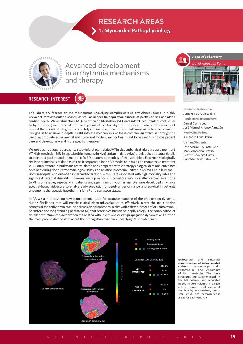
The laboratory focuses on the mechanisms underlying complex cardiac arrhythmias found in highly prevalent cardiovascular diseases, as well as in specific population subsets at particular risk of sudden cardiac death. Atrial fibrillation (AF), ventricular fibrillation (VF) and infarct scar-related ventricular tachycardia (VT) are three of the most prevalent cardiac rhythm disorders, in which the capacity of current therapeutic strategies to accurately eliminate or prevent the arrhythmogenic substrate is limited. Our goal is to achieve in-depth insight into the mechanisms of these complex arrhythmias through the use of appropriate experimental and numerical models, and for this insight to be used to improve patient care and develop new and more specific therapies.
We use a translational approach to study infarct scar-related VT in pigs and clinical infarct-related reentrant VT. High-resolution MRI images, both in humans (in vivo) and animals (ex vivo) provide the structural details to construct patient and animal-specific 3D anatomical models of the ventricles. Electrophysiologically realistic numerical simulations can be incorporated in the 3D model to induce and characterize reentrant VTs. Computational simulations are validated and compared with electropysiological data and outcomes obtained during the electrophysiological study and ablation procedure, either in animals or in humans. Both in-hospital and out-of-hospital cardiac arrest due to VF are associated with high mortality rates and significant cerebral disability. However, early prognosis in comatose survivors after cardiac arrest due to VF is unreliable, especially in patients undergoing mild hypothermia. We have developed a reliable spectral-based risk-score to enable early prediction of cerebral performance and survival in patients undergoing therapeutic hypothermia for VF and comatose status.
In AF, we aim to develop new computational tools for accurate mapping of the propagation dynamics during fibrillation that will enable clinical electrophysiologists to effectively target the main driving sources of the arrhythmia. We use a translational approach in pigs with different stages of AF (paroxysmal, persistent and long-standing persistent AF) that resembles human pathophysiology. The combination of detailed structural characterization of the atria with in vivo and ex vivo propagation dynamics will provide the most precise data to date about the propagation dynamics underlying AF maintenance.
David Filgueiras RamaAdvanced developmentin arrhythmia mechanismsand therapy
Graduate Technician:Jorge García Quintanilla
Predoctoral Researchers:Daniel García LeónJosé Manuel Alfonso Almazán
Res@CNIC Fellow:Alejandro Cruz Utrilla
Visiting Students:José María Lillo CastellanoManuel Marina BreysseBeatriz Domingo GarcíaConrado Javier Calvo Sainz
Endocardial and epicardial reconstruction of infarct-related susbtrate. Voltage maps of the endocardium and epicardium of both ventricles. The three structures are superimposed in the left column, and separated in the middle column. The right column shows quantification of the healthy myocardium, dense scar areas, and heterogeneous areas for each ventricle.

1. Myocardial Pathophysiology
S C I E N T I F I C R E P O R T 2 0 1 520
- Spanish Society of Cardiology (Electrophysiology & Arrhythmia Division).- Salud 2000 Foundation.- Jesús Serra Foundation.- Pro-CNIC Foundation.
Filgueiras-Rama D, Calvo CJ, Salvador-Montañés Ó, Cádenas R, Ruiz-Cantador J, Armada E, Rey JR, Merino JL, Peinado R, Pérez-Castellano N, Pérez-Villacastín J, Quintanilla JG, Jiménez S, Castells F, Chorro FJ, López-Sendón JL, Berenfeld O, Jalife J, López de Sá E, Millet J. Spectral analysis-based risk score enables early prediction of mortality and cerebral performance in patients undergoing therapeutic hypothermia for ventricular fibrillation and comatose status. Int J Cardiol (2015) 186: 250-8
Quintanilla JG, Moreno J, Archondo T, Usandizaga E, Molina-Morúa R, Rodríguez-Bobada C, González P, García-Torrent MJ, Filgueiras-Rama D, Pérez-Castellano N, Macaya C, Pérez-Villacastín J. Increased intraventricular pressures are as harmful as the electrophysiological substrate of heart failure in favoring sustained reentry in the swine heart. Heart Rhythm (2015) 12: 2172-83
Filgueiras-Rama D, de Torres-Alba F, Castrejón-Castrejón S, Estrada A, Figueroa J, Salvador-Montañés O, López T, Moreno-Yanguela M, López Sendón JL, Merino JL. Utility of intracardiac echocardiography for catheter ablation of complex cardiac arrhythmias in a medium-volume training center. Echocardiography (2015) 32: 660-70
Jalife J, Filgueiras Rama D, Berenfeld O. Letter by Jalife et al Regarding Article, “Quantitative Analysis of Localized Sources Identified by Focal Impulse and Rotor Modulation Mapping in Atrial Fibrillation”. Circ Arrhythm Electrophysiol (2015) 8: 1296-8
Toniolo M, Estrada A, Filgueiras-Rama D, Merino JL. Revolving thrombus within the left atrium at atrial fibrillation ablation. Herzschrittmacherther Elektrophysiol (2015) 26: 54-5
VF_Spectral_Based_Score. Risk score based on the predictive performance of the spectral-based model. A. Observed (triangles) and predicted (circles) probability of FNP for the entire population. Blue and red represent FNP (favorable neurological performance) and non-FNP, respectively (dark fill, retrospective; light fill, prospective). We defined four risk groups of non-FNP performance according to their risk scores as follows: expected FNP; very low (VL) and low risk (L) and expected non-FNP; high (H) and very high risk (VH). B. Percentage of patients (predicted, dark gray and observed, light gray) who belong to each of the risk score groups in both the retrospective (B1) and prospective cohorts (B2). (α) and (β) represent false negative and false positive individuals, respectively.

1. Myocardial Pathophysiology
Gonzalez-Valdes I, Bujarrabal A., Hidalgo I., Padron L., Garcia-Pavia P., Lara-Pezzi E., Gomez P., Redondo J.M., Jimenez-Borreguero L.J., Ruiz-Cabello, J.M., de la Pompa, J.L., Enriquez, J.A., Hidalgo A., and Gonzalez S. Bmi1 limits dilated cardiomyopathy and heart failure by inhibiting cardiac senescence. Nat Commun (2015) 6: 6473
Lopez-Arribillaga E, Rodilla V, Pellegrinet L, Guiu J, Iglesias M, Roman AC, Gutarra S, Gonzalez S, Munoz-Canoves P, Fernandez-Salguero P, Radtke F, Bigas A, Espinosa LL. Bmi1 regulates murine intestinal stem cell proliferation and self-renewal downstream of Notch. Development (2015) 142: 41-50
Branco AF, Pereira SP, Gonzalez S, Gusev O, Rizvanov AA, Oliveira PJ. Gene Expression Profiling of H9c2 Myoblast Differentiation towards a Cardiac-Like Phenotype. PLoS One (2015) 10: e0129303
Sousa-Victor P, Gutarra S, García-Prat L, Rodriguez-Ubreva J, Ortet L, Ruiz-Bonilla V, Jardí M, Ballestar E, González S, Serrano AL, Perdiguero E, Muñoz-Cánoves P. Geriatric muscle stem cells switch reversible quiescence into senescence. Nature (2014) 506:316-21
S C I E N T I F I C R E P O R T 2 0 1 5 21
The regeneration of adult tissues after injury involves tight homeostatic control by adult stem cells through their ability to self-renew and differentiate into multiple lineages. These characteristics are strongly affected with aging, leading to a loss in tissue regeneration capacity. Emerging evidence suggests that polycomb-group (PcG)-mediated alteration of the epigenetic status in hematopoietic stem cells (HSCs) is a major driving force behind many age-related HSC changes. Interestingly, PcG is often misregulated in human malignancies. Protection of the transcriptional “stemness” network is thus essential for the maintenance of a healthy HSC compartment throughout life. Whether the functional decline in adult stem cells is related to reversible chromatin modifications remains a key unanswered question in the field. We propose that changes to the chromatin state can restore the regenerative capacity of stem cells. To investigate this hypothesis, we are exploring the role of the epigenetic polycomb-mediated silencing mechanism in stemness maintenance, with particular emphasis on the self-renewal capacity and the microenvironment of HSCs, an important adult stem cell population with diverse regenerative abilities. Unraveling the molecular mechanisms by which polycomb members control stem cell fate will provide new insights into hematopoietic stem cell biology and increase the understanding of neoplastic transformation.
We have a particularly strong interest in the emerging role of different classes of chromatin regulators and how their dysregulation in the adult heart alters specific gene programs, with subsequent development of major cardiomyopathies. While dilated cardiomyopathy (DCM) is as the third most common cause of heart failure, it is still poorly modeled in nonhuman species. We propose that epigenetic remodeling could provide an important means of modulating the transcriptional reprogramming of cardiac gene expression in this condition. Understanding the action of Polycomb factors will allow the development of strategies to control physiological and pathological gene expression.
Susana GonzálezEpigenetic regulation in cardiac aging and disease Postdoctoral Researcher:
Anne Marie Bleau
Predoctoral Researchers:Isabel Hidalgo GavilánItziar Cossío CuarteroVera Lúcia Ferreira OliveiraIleana Beatriz González ValdésEsmeralda Armando LewisMaría Inmaculada Martos FolgadoCarlos José Martos RodríguezEleni Petra
Technician:Rebeca Diges López
Masters StudentAlicia González Martínez
- European Commission. European Research Council Consolidator Grant (ERC-CoG-647670)- Ministerio de Economía y Competitividad (SAF2013-42252-R)
The epigenetic basis of cardiac rejuvenation. Aging is the greatest risk factor for cardiovascular disease. The aging process involves chromatin modifications by polycomb group proteins in HSCs, leading to a reduced stemness phenotype. By modulating PcG epigenetic status, we aim to restore the regeneration capacity of stem cells in adult tissues.
epigenetic behind cardiac rejuvenation

S C I E N T I F I C R E P O R T 2 0 1 522
1. Myocardial Pathophysiology
Our laboratory focuses on the study of myocardial diseases, ranging from ischemia/reperfusion to heart failure. Our studies span the molecular origins of disease and their manifestations at the macro-anatomical and physiological levels, and our group includes experts in molecular biology, clinical cardiology and cardiovascular imaging. Our evaluation of experimental animal models makes use of advanced imaging techniques that can also be applied to humans, strengthening the translational potential of our research. To exploit this potential, we work on multidisciplinary programs in close collaboration with hospitals and clinical researchers.
A major interest of the group is cardioprotection during myocardial infarction (MI). We have established models of MI in rodents and large animals, and use these to study the mechanisms underlying the beneficial effects of various cardioprotective strategies, mainly related to modulation of the adrenergic system. We are pioneering the use state-of-the-art magnetic resonance imaging (MRI) to better characterize post-infarcted myocardial healing by combining studies in large animal models and human study participants. An example of this work is our leadership of the randomized METOCARD-CNIC clinical trial, which used MRI to evaluate the effectiveness of early intravenous metoprolol in patients suffering a myocardial infarction. The primary objective of this trial is already reported and we now are preparing a large multinational clinical trial based on these results (MOVE ON!). MOVE ON! will address the effect of this protective strategy not only on infarct size, but more importantly on long term mortality and morbidity. In parallel with these clinical trials, we study the cellular and molecular mechanisms underlying the observed cardioprotection in in vitro and genetically modified small animal models. We are also opening new fields of research focused on the metabolism of heart failure and the study of nutritional approaches to treat this condition.
We are part of the Spanish network for inherited cardiomyopathies, where our major interest is the development of better imaging-based strategies for improved risk stratification of patients carrying malignant mutations. This clinical work is combined with preclinical and basic studies to better understand the genotype-phenotype correlations of these mutations.
We are also interested in the myocardial response to pulmonary hypertension. We have developed small and large animal models of pulmonary hypertension and use imaging technology to evaluate the response to different therapies. We have identified a novel therapeutic approach for the treatment of pulmonary hypertension in preclinical studies and we have been funded to bring this therapy into a pilot clinical trial that will start during the coming year.
Borja Ibáñez(CNIC, Fund. Jiménez Díaz Hospital) Translational laboratory for
cardiovascular imaging and therapy
Postdoctoral Researchers:Rodrigo Fernández-Jiménez(CNIC, Hospital Clínico San Carlos)Gonzalo Pizarro(CNIC, Hospital Universitario Quirón)José Manuel García Ruíz(CNIC, Hospital Universitario Centralde Asturias)Predoctoral Researchers:Jaime García-Prieto CuestaAndrés Pun GarcíaJaume Agüero Ramón-LlinFederico Sierra Rodríguez de la RubiaCarlos Galán ArriolaPiotr Waldemar WilczynskiRobert Austin Bruce BennResearch Coordinator:Noemí Escalera BiendichoTechnicians:Mónica Gómez ParrizasParvin Rupa KhatonRes@CNIC Fellows:Julio C. García RubioIsabel Valandrón SucasasInvesmir Fellows:Ali Ayaon AlbarránManuel Lobo GonzálezVisiting Students:Rocío Villena GutiérrezEster Jiménez ArroyoSulayman Lazaar SolerBlanca Sanz MagallónAndrés Escudero DíazConstanza Ballesteros MartínezAndrés Escudero DíazVisiting Scientists:Jesús González Mirelis Alonso Mateos Rodríguez Daniel Pereda Arnau Jorge Solís Martín Mauro EchavarríaMontserrat Rigol Muxart Núria Solanes BatllóSantiago Roura FerrerJoaquim Bobi i GibertIker Rodríguez ArabaolazaEvelyn Santiago VacasMónica García BouzaBunty Kishore RamchandaniRamchandani

1. Myocardial Pathophysiology
S C I E N T I F I C R E P O R T 2 0 1 5 23
Mechanisms underlying the Bimodal Edema Phenomenon after myocardial I/R.(from Fernández-Jiménez R et al., J Am Coll Cardiol, 2015 66(7): 816-28).
- Ministerio de Economía y Competitividad - EXPLORA CIENCIA (SAF2013-49663-EXP)- Ministerio de Economía y Competitividad - Acciones de Dinamización Europa investigación (EUIN2013-50881)- Ministerio de Economía y Competitividad. ISCIII-FIS (PI13/01979)- Ministerio de Economía y Competitividad. ISCIII-RETICS (RiC, RD12/0042/0054) - European Commision FP7-ICT-2011-8 (LIPHOS-317916)- Marató, Fundación TV3 (REF: 70/C/2012)- European Commision FP7-PEOPLE-2013-ITN (CARDIONEXT).
Wai T†, García-Prieto J†, Baker MJ, Merkwirth C, Benit P, Rustin P, Rupérez FJ, Barbas C, Ibañez B*, Langer T*. Imbalanced OPA1 processing and mitochondrial fragmentation cause heart failure in mice. Science (2015) 350: aad0116-1-11†Equal contribution; *co-corresponding authors
Fernández-Jiménez R, García-Prieto J, Sánchez-González J, Agüero J, López-Martín GJ, Galán-Arriola C, Molina-Iracheta A, Doohan R, Fuster V, Ibanez B. Pathophysiology underlying the bimodal edema phenomenon after myocardial ischemia/reperfusion. J Am Coll Cardiol (2015) 66:816-28
Ibanez B*, Heusch G, Ovize M, Van de Werf F. Evolving therapies for myocardial ischemia/reperfusion injury. J Am Coll Cardiol (2015) 65:1454-71 *Corresponding author
Fernández-Jiménez R, Silva J, Martínez-Martínez S, López-Maderuelo MD, Nuno-Ayala M, García-Ruiz JM, García-Álvarez A, Fernández-Friera L, Pizarro G, García-Prieto J, Sanz-Rosa D, López-Martin G, Fernández-Ortiz A, Macaya C, Fuster V, Redondo JM, Ibanez B. Impact of left ventricular hypertrophy on troponin release during acute myocardial infarction: new insights from a comprehensive translational study. J Am Heart Assoc (2015) 4: e001218
Fernández-Jiménez R, Sánchez-González J, Aguero J, García-Prieto J, López-Martín GJ, García-Ruiz JM, Molina-Iracheta A, Rosselló X, Fernández-Friera L, Pizarro G, García-Álvarez A, Dall’Armellina E, Macaya C, Choudhury R, V Fuster, B Ibanez. Myocardial edema after ischemia/reperfusion is not stable and follows a bimodal pattern: advanced imaging and histological tissue characterization. J Am Coll Cardiol (2015) 65: 315-23
Players involved in ischemia/reperfusion injury. (from Ibanez B et al., J Am Coll Cardiol, 2015 65(14): 1454-71).
1. Myocardial Pathophysiology
The laboratory centers its research interest on understanding the causes of cardiovascular disease at the molecular, cellular and electrophysiological levels. Our specific research objectives are focused on 1) the mechanisms of atrial and ventricular fibrillation at the structural and functional level; 2) the molecular genetics of cardiac fibrillation; and 3) the cellular basis of cardiac arrhythmia in genetic and rare diseases that can lead to sudden death.
The laboratory has well-established collaborations with expert engineers, biologists and clinicians around the world, as well as collaborations with other CNIC groups. These partnerships provide a unique research environment in which to generate new and clinically relevant breakthroughs on arrhythmia mechanisms that will benefit the medical and basic science communities, and ultimately the patient.
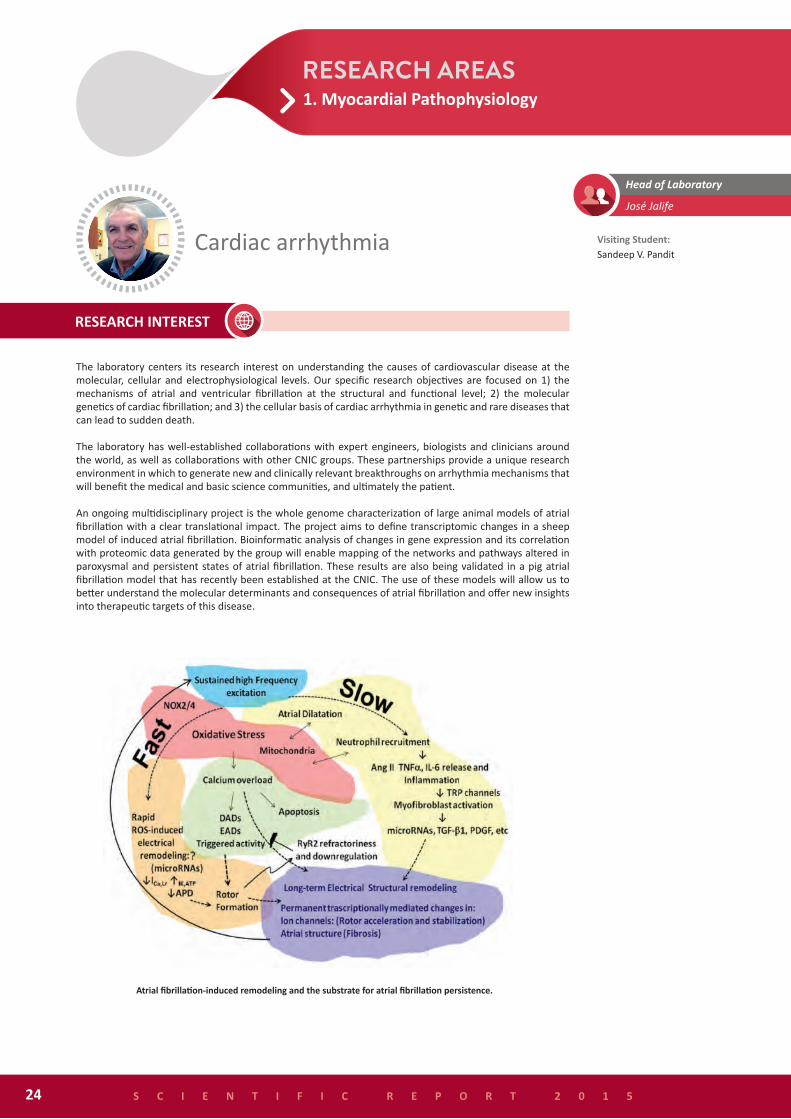
An ongoing multidisciplinary project is the whole genome characterization of large animal models of atrial fibrillation with a clear translational impact. The project aims to define transcriptomic changes in a sheep model of induced atrial fibrillation. Bioinformatic analysis of changes in gene expression and its correlation with proteomic data generated by the group will enable mapping of the networks and pathways altered in paroxysmal and persistent states of atrial fibrillation. These results are also being validated in a pig atrial fibrillation model that has recently been established at the CNIC. The use of these models will allow us to better understand the molecular determinants and consequences of atrial fibrillation and offer new insights into therapeutic targets of this disease.
Cardiac arrhythmia
José Jalife
Visiting Student:Sandeep V. Pandit
S C I E N T I F I C R E P O R T 2 0 1 524
Atrial fibrillation-induced remodeling and the substrate for atrial fibrillation persistence.

1. Myocardial Pathophysiology
- Leducq Foundation Transatlantic Networks of Excellence Program (not CNIC). Co – Investigator- NIH / NHLBI - R01 (HL122352) (not CNIC). Co-Investigator- NIH / NHLBI - T32 (HL125242) (not CNIC). Co-Investigator- The University of Michigan Health Sciences-Peking University Health Science Center Joint Institute. (not CNIC). Co-Investigator.
Quintanilla JG, Pérez-Villacastín J, Pérez-Castellano N, Pandit SV, Berenfeld O, Jalife J, Filgueiras-Rama D. Mechanistic approaches to detect, target, and ablate the drivers of atrial fibrillation. Circ Arrhythm Electrophysiol (doi: 10.1161/CIRCEP.115.002481).
Haemers P, Hamdi H, Guedj K, Suffee N, Farahmand P, Popovic N, Claus P, LePrince P, Nicoletti A, Jalife J, Wolke C, Lendeckel U, Jaïs P, Willems R, Hatem SN. Atrial fibrillation is associated with the fibrotic remodelling of adipose tissue in the subepicardium of human and sheep atria. Eur Heart J (doi: org/10.1093/eurheartj/ehv625. Epub 2015 Nov 26).
Abriel H, Rougier JS, Jalife J. Ion channel macromolecular complexes in cardiomyocytes: roles in sudden cardiac death. Circ Res (2015) 116: 1971-88
Zhao YT, Valdivia CR, Gurrola GB, Powers PP, Willis BC, Moss RL, Jalife J, Valdivia HH. Arrhythmogenesis in a catecholaminergic polymorphic ventricular tachycardia mutation that depresses ryanodine receptor function. Proc Natl Acad Sci U S A. 2015;112:E1669-77.
Filgueiras-Rama D, Calvo CJ, Salvador-Montañés Ó, Cádenas R, Ruiz-Cantador J, Armada E, Rey JR, Merino JL, Peinado R, Pérez-Castellano N, Pérez-Villacastín J, Quintanilla JG, Jiménez S, Castells F, Chorro FJ, López-Sendón JL, Berenfeld O, Jalife J, López de Sá E, Millet J. Spectral analysis-based risk score enables early prediction of mortality and cerebral performance in patients undergoing therapeutic hypothermia for ventricular fibrillation and comatose status. Int J Cardiol (2015) 186: 250-8
S C I E N T I F I C R E P O R T 2 0 1 5 25

Our laboratory investigates the molecular processes underlying heart remodeling, which are still poorly understood. In collaboration with Dr Fernando Rodriguez-Pascual’s group (Centro de Biología Molecular Severo-Ochoa, Madrid) we have unveiled an unexpected beneficial role of lysyl oxidase (Lox) in post-infarction heart remodeling. Lox facilitates the cross-linking of extracellular matrix and thereby contributes to the development of fibrosis. We found that inhibition of Lox activity results in decreased infarct expansion and improved cardiac function one month after myocardial infarction. This effect is similar to that of the calcineurin splicing variant CnAβ1. Expression of this isoform promotes the vascularisation of the infarct region, reinforcing the structure in the infarcted area and preventing infarct expansion and therefore heart remodeling.
The beneficial activity of CnAβ1 in the adult heart stands in stark contrast to the detrimental effects of other calcineurin isoforms, which promote maladaptive cardiac hypertrophy and heart failure by activating the transcription factor NFAT, among other targets. Due to retention of an intron in its mRNA, CnAβ1 has a unique C-terminal domain that has no similarity with any other known protein. This unique domain drives CnAβ1 to the Golgi apparatus and facilitates activation of the Akt signaling pathway. In embryonic stem cells, CnAβ1 is necessary for mesodermal differentiation in a parallel pathway to that activated by the calcineurin isoform CnAβ2 via NFAT for this same purpose. We are now exploring the therapeutic potential of CnAβ1 in a swine model of myocardial infarction using gene therapy based on adeno-associated vectors.
Molecular regulation of heart development and disease
Enrique Lara-Pezzi
Postdoctoral Researcher:Laura Padrón
Predoctoral Researchers:Jesús Gómez SalineroAlberto GattoEnda ClintonGirolamo GiudicePaula Ortiz SánchezJose Javier Larrasa Alonso
Graduate Technician:María Villalba Orero
Technician:Marina López Olañeta
Res@CNIC Fellow:Juan M. Monteagudo Ruiz
Masters Student:Carlos Martí Gómez-Aldaraví
Visiting Scientists:Pablo García PavíaRaquel San José Martín-Albo
S C I E N T I F I C R E P O R T 2 0 1 526
CnAβ1 is localized in the Golgi apparatus. Unlike other calcineurin isoforms, such as CnAβ2, which is freely distributed throughout the cytoplasm, CnAβ1 is compartmentalized to the Golgi apparatus. This localization is necessary for activation of the Akt signaling pathway, a major regulator of cell growth and survival. P19 cells were transfected with a chimeric GFP-CnAβ1 construct and immunostained with antibodies against GFP (green) and the Golgi marker GM130 (red). Nuclei were counterstained with DAPI (blue). Scale bar 25 μm.
1. Myocardial Pathophysiology

27
- European Commission. Marie Curie Action Initial Training Network (ITN) (FP7-PEOPLE-2013-ITN, “CardioNext” 608027)- European Commission. Marie Curie Action Initial Training Network (ITN) (FP7-PEOPLE-2011-ITN, “CardioNeT” 289600)- Comunidad de Madrid (GRUPOSCAM10, “Fibroteam” S2010/BMD-2321)- Ministerio de Economía y Competitividad (SAF2012-31451)- Instituto de Salud Carlos III (MSII14/00027)
González-Santamaría J, Villalba M, Busnadiego O, López-Olañeta MM, Sandoval P, Snabel J, López-Cabrera M, Erler JT, Hanemaaijer R, Lara-Pezzi E*, Rodríguez-Pascual F*. Matrix cross-linking lysyl oxidases are induced in response to myocardial infarction and promote cardiac dysfunction. Cardiovasc Res (doi: 10.1093/cvr/cvv214. Epub 2015 Aug 10)*Co-corresponding authors
Lara-Pezzi E, Menasché P, Trouvin JH, Badimón L, Ioannidis JP, Wu JC, Hill JA, Koch WJ, De Felice AF, de Waele P, Steenwinckel V, Hajjar RJ, Zeiher AM. Guidelines for translational research in heart failure. J Cardiovasc Transl Res (2015) 8: 3-22
López-Olañeta MM, Villalba M, Gómez-Salinero JM, Jiménez-Borreguero LJ, Breckenridge R, Ortiz-Sánchez P, García-Pavía P, Ibáñez B, Lara-Pezzi E. The calcineurin variant CnAβ1 improves post-infarction ventricular remodelling by promoting infarct vascularisation. Cardiovasc Res (2014) 102: 396-406
Gatto A, Torroja-Fungairiño C, Mazzarotto F, Cook SA, Barton PJ, Sánchez-Cabo F, Lara-Pezzi E. FineSplice, enhanced splice junction detection and quantification: a novel pipeline based on the assessment of diverse RNA-Seq alignment solutions. Nucleic Acids Res (2014) 42:e71.
Regulation of mesoderm differentiation in mESCs through CnAβ isoforms. The splicing factor muscle blind like 1 (Mbnl1) promotes CnAβ1 isoform production from the CnAβ locus. CnAβ1 is located at the Golgi apparatus, where it interacts with Cog8. CnAβ1 regulates the phosphorylation levels of AKT from the Golgi. The active form of AKT inhibits GSK3 activation, leading to an increase in the levels of β-catenin that promotes mesoderm specification. CnAβ2 is localized in the cytoplasm and activates NFAT to promote mesoderm specification.
Echocardiographic analysis of an infarcted mouse heart. A, Short axis apical, medium and basal echocardiographic views (a, b and c, respectively) combined with the long axis view (d) for accurate estimation of left ventricle motion and remodeling in a untreated C5/BL6 mice. B, The same analysis as in (A) carried out on a C5/BL6 mouse 28 days after occluding the left anterior descending coronary artery.
S C I E N T I F I C R E P O R T 2 0 1 5
1. Myocardial Pathophysiology

Macrophages are hematopoietic cells of the myeloid lineage with important functions in development, homeostasis, tissue repair and immunity. Macrophages can be found in practically all tissues, making important contributions to homeostasis and protection against injury. Projects in our group focus on elucidating the transcriptional control of macrophages in different tissues, especially in the heart, adipose tissue and bone marrow, with special emphasis on their possible medical utility in the treatment of metabolic and cardiovascular diseases.
A special interest of our group is the transcriptional regulation of macrophage functions by nuclear hormone receptors. Our laboratory has shown that nuclear receptors play a major regulatory role in homeostasis, inflammation and immunity. Our recent studies indicate that retinoid X receptors (RXRs) play a key role in orchestrating macrophage transcriptional programs necessary for debris clearance, proliferation, polarization and lipid metabolism. Moreover, genetic deletion of these receptors in macrophages severely compromises macrophage homeostatic responses, leading to autoimmunity, osteopetrosis and insulin resistance. Based on these findings, we hypothesize that nuclear receptors play important roles in orchestrating hematopoietic stem cell and macrophage transcriptional programs necessary for tissue repair and regeneration. To test this hypothesis we are currently conducting complementary loss-of-function and drug-mediated gain-of-function mouse studies, and also genome-wide studies using transcriptomic (RNA-seq and GRO-seq) and cistromic (ChIP-seq) technologies. We will examine mice lacking RXR in hematopoietic stem cells, macrophages, endothelial, and cardiomyoctes, allowing us to examine the specific role of these receptors in tissue homeostasis and injury.
Nuclear receptor signalingMercedes Ricote
Research Scientist:María Piedad Menéndez Gutiérrez
Postdoctoral Researchers:Lorenzo Veschini
Predoctoral Researchers:Anna KwasniewskaWencke WalterAngel Núñez BuizaLaura Alonso Herranz
Masters Students:Ana ParedesJosé Juan Aparicio
Technician:Vanessa Núñez González
S C I E N T I F I C R E P O R T 2 0 1 528
Defining RXR functions in macrophage biology. Macrophages express RXRα and RXRβ. RXRs play key roles in macrophage homeostasis and disease by controlling transcriptional programs necessary for inflammation, apoptotic cell uptake, proliferation, antiviral response, polarization and lipid metabolism. Macrophage-specific RXRα/β-delition results in sepsis, autoimmunity, osteopetrosis and insulin resistance.
1. Myocardial Pathophysiology

- Ministerio de Economía y Competitividad (SAF2012-31483)- Fundación TV3 Marató- European Commission, 7th Frame Program (FP7-PEOPLE-2013-ITN) (PITN-GA-2013-608027)- Ministerio de Economía y Competitividad (SAF2015-71878-REDT)
Natrajan MS, de la Fuente AG, Crawford AH, Linehan E, Nuñez V, Johnson KR, Wu T, Fitzgerald DC, Ricote M, Bielekova B, Franklin RJ. Retinoid X receptor activation reverses age-related deficiencies in myelin debris phagocytosis and remyelination. Brain (2015) 138: 3581-97
Walter W, Sánchez-Cabo F, Ricote M. GOplot: an R package for visually combining expression data with functional analysis. Bioinformatics (2015) 31: 2912-4
Menéndez-Gutiérrez MP, Rőszer T, Fuentes L, Núñez V, Escolano A, Redondo JM, De Clerck N, Metzger D, Valledor AF, Ricote M. Retinoid X receptors orchestrate osteoclast differentiation and postnatal bone remodeling. J Clin Invest (2015) 125: 809-23
Ballesteros I, Cuartero MI, Pradillo JM, de la Parra J, Perez-Ruiz A, Corbi A, Ricote M, Hamilton JA, Sobrado M, Vivancos J, Nombela F, Lizasoain I and Moro MA. Rosiglitazone-induced CD36 up-regulation resolves inflammation by PPARgamma and 5-LO-dependent pathways. J Leukoc Biol (2014) 95: 587-98
Ma F, Liu SY, Razani B, Arora N, Li B, Kagechika H, Tontonoz P, Nunez V, Ricote M and Cheng G. Retinoid X receptor alpha attenuates host antiviral response by suppressing type I interferon. Nat Commun (2014) 5: 5494
S C I E N T I F I C R E P O R T 2 0 1 5 29
1. Myocardial Pathophysiology
Guadalupe Sabio
Postdoctoral Researchers:Nuria MatesanzAntonia Tomás LobaIvana Nikolic
Predoctoral Researchers:Edgar Bernardo Bárbara GonzálezElisa Manieri María del Valle Montalvo
Technicians:Elena González Luis LeivaAlfonso Mora(since July)Victor Emilio Bondia(since September)
Res@CNIC Fellow:Ana Pardo Sanz
Masters Student: Leticia Herrera
Group photo: Dani Pozo (El Español)
S C I E N T I F I C R E P O R T 2 0 1 530
Metabolic syndrome is a medical disorder defined by the co-occurrence of obesity, impaired glucose tolerance, dyslipidemia and hypertension. The condition is associated with proinflammatory and prothrombotic states, and clinical outcomes include cardiovascular disease and type 2 diabetes. Moreover, metabolic syndrome may be a predisposing factor for the development of some types of cancer, such us hepatocellular carcinoma.
The high cardiovascular risk associated with metabolic syndrome and type 2 diabetes suggests that common mechanisms are involved in the etiology of these conditions, and that agents acting on the same therapeutic targets might improve disease parameters in both. Research suggests that one such target might be the stress activated protein kinases (SAPKs), an important family of kinases implicated in the transduction of stress signals into the cell.
Our group investigates the involvement of SAPKs in the development of cancer, diabetes, cardiac hypertrophy and atherosclerosis induced by obesity. Our research is conducted with a number of disease models in combination with whole-body and tissue-specific knockout mice, and has shown that the p38γ/δ isoforms control IL6 and TNF production in myeloid cells. We are now studying how the regulation of inflammation by these kinases affects the development of metabolic syndrome. We are also studying the function of these kinases in other tissues, such us muscle, heart, the central nervous system and adipose tissue, in order to elucidate the role of these kinases in the development of different diseases associated with obesity (steatosis, diabetes, cardiovascular diseases and some types of cancer).
Stress kinases in diabetes, cancer and cardiovascular disease
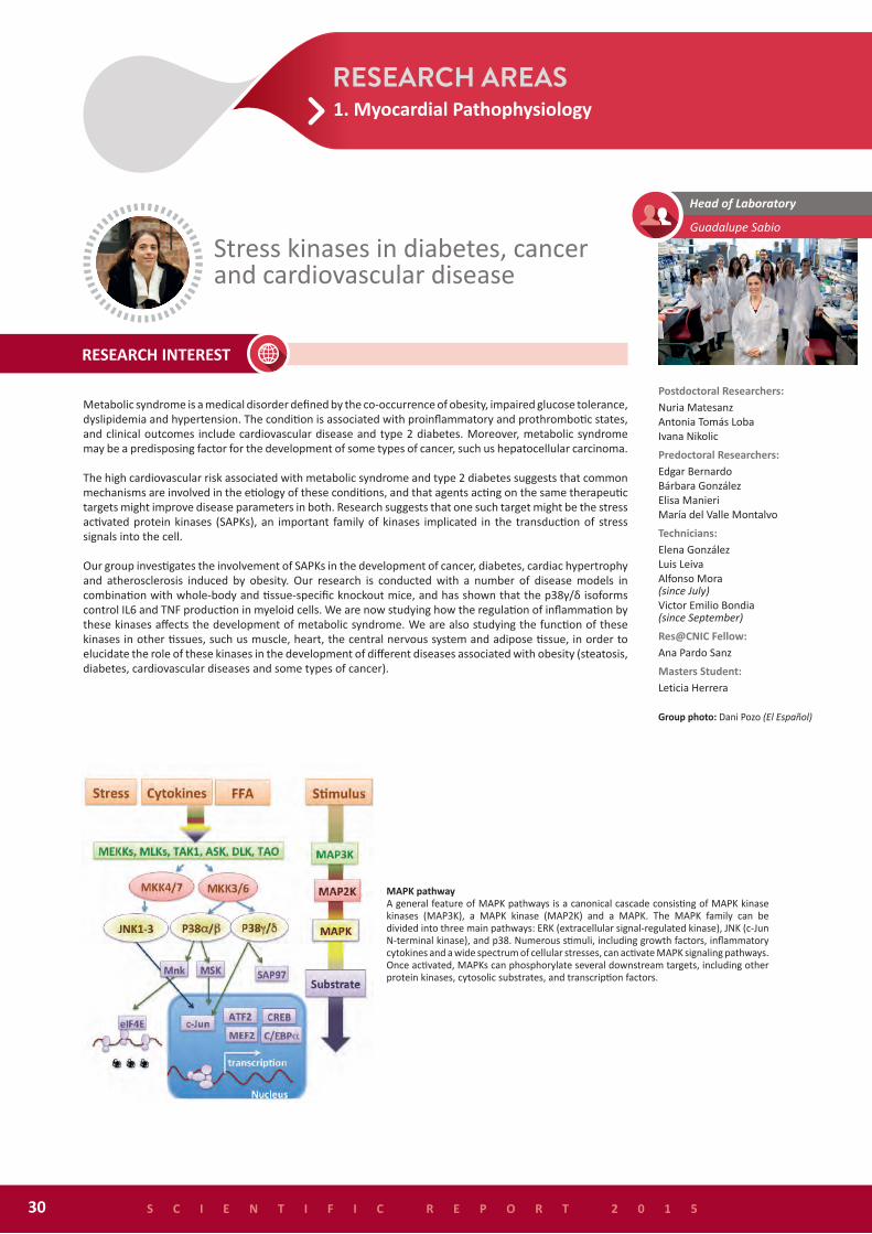
MAPK pathway A general feature of MAPK pathways is a canonical cascade consisting of MAPK kinase kinases (MAP3K), a MAPK kinase (MAP2K) and a MAPK. The MAPK family can be divided into three main pathways: ERK (extracellular signal-regulated kinase), JNK (c-Jun N-terminal kinase), and p38. Numerous stimuli, including growth factors, inflammatory cytokines and a wide spectrum of cellular stresses, can activate MAPK signaling pathways. Once activated, MAPKs can phosphorylate several downstream targets, including other protein kinases, cytosolic substrates, and transcription factors.
1. Myocardial Pathophysiology

- Ministerio de Economía y Competitividad (SAF2013-43506-R)- European Commission. European Research Council Starting Independent Researcher Grant (ERC-StG-260464)- Comunidad de Madrid. INMUNOTHERCAN (S2011/BMD-2326)
González-Terán B, López JA, Rodríguez E, Leiva L, Martínez Martínez S. Jiménez Borreguero LJ, Redondo JM, Vázquez J, Sabio G. p38 and δ promote heart hypertrophy by targeting the mTOR-inhibitory protein DEPTOR for degradation. Nat Commun (accepted)
González-Terán B, Matesanz N, Verdugo MA, Sreeramkumar V, Hernández-Cosido L, Bernardo E, Leiva-Vega L, Rodríguez E, Torres JL, Perez S., Ortega L, Cuenda A, Nogueira R, Hidalgo A, Miguel Marcos M, Sabio G. p38γ and p38δ reprogram liver metabolism by modulating neutrophil infiltration EMBO Journal (accepted)
Manieri E, Sabio G. Stress kinases in the modulation of metabolism and energy balance. J Mol Endocrinol (2015) 55: R11-22
Vernia S, Cavanagh-Kyros J, Garcia-Haro L, Sabio G, Barrett T, Jung DY, Kim JK, Xu J, Shulha HP, Garber M, Gao G, Davis RJ. The PPARα-FGF21 hormone axis contributes to metabolic regulation by the hepatic JNK signaling pathway. Cell Metab (2014) 20: 512-25.
Sabio G, Davis RJ. TNF and MAP kinase signalling pathways. Seminars in Immunology (2014) 26: 237-45
Obesity related dieseasesObesity is one of the leading causes of life-threatening diseases and can compromise health and shorten life expectancy. In our group we study several of themes such as diabetes, cancer, and heart disease.
Obesity-induced iInflamation of white adipose tissue
S C I E N T I F I C R E P O R T 2 0 1 5 31
1. Myocardial Pathophysiology

Our main interest is in how dendritic cells (DCs) and macrophages modulate immunity and inflammation. We are interested in the analysis of DC subsets, their specific functions and plasticity. We have found that Batf3-dependent DCs are crucial for generation of Th1 immunity through the production of IL-12 (Fig. 1 and Martínez-López et al. 2015). Some of our work is conducted with the Th1-immunity model of infection by the eukaryotic parasite Leishmania major; this organism induces tissue damage and mimics many tissue-derived danger signals. We have also found that Batf3-dependent DCs do not play a major role in the development of atherosclerosis. In contrast, cross-presenting DCs are crucial for the generation of a basal immune response that can be rescued by immunostimulatory antibodies for cancer immunotherapy (Sánchez-Paulete et al., in press).
We have also analyzed the modulation of signals through C-type lectin receptors and have found that SHIP-1 associates with the intracellular hemITAM motif in Dectin-1 and selectively modulates its ability to induce reactive oxygen species in DCs (Fig. 2 and Blanco-Menéndez et al. 2015). In addition, we are working on DNGR-1 as a model C-type lectin that detects tissue damage during infection and we have characterized an impact on the CD8+ T cell memory response. We are also analyzing the effects of sensing non-self and damaged-self on the metabolism of myeloid cells. We believe that this research has potential for the development of new vaccines and immunotherapy strategies.
Immunobiology of inflammationDavid Sancho Madrid
Postdoctoral Researchers:Salvador Iborra Martín Johan J.B. GaraudeCarlos del Fresno SánchezLaura Conejero Hall
Predoctoral Researchers:Noelia Blanco Menéndez Helena M. Izquierdo FernándezMaría Martínez LópezNeris M. Enamorado EscalonaPaola Brandi Francisco J. Cueto RodríguezPaula Saz
Masters Student:Sofía Chayeb
Technicians: Ruth Conde GarrosaSarai Martínez CanoJesús Sánchez
S C I E N T I F I C R E P O R T 2 0 1 532
Batf-3 dependent CD103+ DC are major IL-12 producers (A) Production of IL-12p40 in ear dermal CD103+ DCs. Left: representative plots. Right: arithmetic mean + SEM of frequency (upper panel) and absolute numbers (lower panel) from naive mice or mice infected with IL-12p40 producing DCs. (B) Transfer of Batf3-dependent DCs rescues impaired Th1 immunity in Batf3KO mice in a IL12p40-dependent fashion.
1. Myocardial Pathophysiology

- Ministerio de Economía y Competitividad (EUIN2015-62652)- Ministerio de Economía y Competitividad. Programa Redes de Excelencia 2014. (SAF2014-53563- REDT).- EU Framework Programme for Research and Innovation H2020. Call: H2020-PERSONALISING HEALTH AND CARE (GA635122-PROCROP).- Ministerio de Economía y Competitividad (SAF2013-42920-R)- European Commission. European Research Council Starting Independent Researcher Grant (ERC-StG-260414)- Research cooperation agreement with MedImmune (Cambridge, UK)- ERS/EU Marie Curie Post-doctoral Research Fellowships (RESPIRE 2 - 3708-2013).
Sánchez-Paulete AR, Cueto FJ, Martínez-López M, Labiano S, Morales-Kastresana A, Rodríguez-Ruiz ME, Jure-Kunkel M, Azpilikueta A, Aznar MA, Quetglas JI, Sancho D*, Melero I*. Cancer Immunotherapy with immunomodulatory anti-CD137 and anti-PD-1 monoclonal antibodies requires BATF3-dependent dendritic cells. Cancer Discov (doi: 10.1158/2159-8290.CD-15-0510. Epub 2015 Oct 22) *Co-corresponding authors
Blanco-Menéndez N, Del Fresno C, Fernandes S, Calvo E, Conde-Garrosa R, Kerr WG, Sancho D. SHIP-1 couples to the dectin-1 hemITAM and selectively modulates reactive oxygen species production in dendritic cells in response to candida albicans. J Immunol (2015) 195: 4466-78
Hanč P, Fujii T, Iborra S, Yamada Y, Huotari J, Schulz O, Ahrens S, Kjær S, Way M, Sancho D, Namba K, Reis e Sousa C. Structure of the complex of F-actin and DNGR-1, a C-type lectin receptor involved in dendritic cell cross-presentation of dead cell-associated antigens. Immunity (2015) 42: 839-49
Martínez-López M, Iborra S, Conde-Garrosa R, Sancho D. Batf3-dependent CD103+ dendritic cells are major producers of IL-12 that drive local Th1 immunity against Leishmania major infection in mice. Eur J Immunol (2015) 45: 119-29
Iborra S, Sancho D. Signalling versatility following self and non-self sensing by myeloid C-type lectin receptors. Immunobiology (2015) 220: 175-84
SHIP-1 colocalizes with Dectin-1 in the phagosome in a Tyr15-dependent fashion. CHO cells expressing either mCherry-tagged wild type Dectin-1 (WT D-1) or mCherry-tagged Y15F-mutated Dectin-1 (Y15F D-1) were cotransfected with EGFP-SHIP-1 fusion protein. Cells were then exposed to 10 µg/ml zymosan for 20 minutes. Colocalization of WT D-1 and Y15F D-1 with SHIP-1 was examined by confocal microscopy. White lines indicate transverse sections of illustrative phagosomes. Fluorescence intensity profiles for green and red channels are plotted as histograms.
S C I E N T I F I C R E P O R T 2 0 1 5 33
1. Myocardial Pathophysiology