Mutations that alter RNA splicing of the human HPRT gene: a review
36
Ž . Mutation Research 411 1998 179–214 Mutations that alter RNA splicing of the human HPRT gene: a review of the spectrum J. Patrick O’Neill a, ) , Peter K. Rogan b , Neal Cariello c , Janice A. Nicklas a a UniÕersity of Vermont Genetics Laboratory, 32 North Prospect Street, Burlington, VT 05401, USA b Department of Human Genetics, Allegheny UniÕersity of the Health Sciences, 320 E. North AÕenue, Pittsburgh, PA, 15212, USA c Glaxo, 5 Moore DriÕe, Research Triangle Park, NC, 27709, USA Received 25 October 1997; revised 18 June 1998; accepted 19 June 1998 Abstract The human HPRT gene contains spans approximately 42,000 base pairs in genomic DNA, has a mRNA of Ž . approximately 900 bases and a protein coding sequence of 657 bases initiation codon AUG to termination codon UAA . Ž . Ž . This coding sequence is distributed into 9 exons ranging from 18 exon 5 to 184 exon 3 base pairs. Intron sizes range from Ž . Ž . Ž . 170 intron 7 to 13,075 intron 1 base pairs. In a database of human HPRT mutations, 277 of 2224 12.5% mutations result in alterations in splicing of the mRNA as analyzed by both reverse transcriptase mediated production of a cDNA followed by PCR amplification and cDNA sequencing and by genomic DNA PCR amplification and sequencing. Mutations X Ž . X Ž . X have been found in all eight 5 donor and 3 acceptor splice sequences. Mutations in the 5 splice sequences of introns 1 and 5 result in intron inclusion in the cDNA due to the use of cryptic donor splice sequences within the introns; mutations in the other six 5 X sites result in simple exon exclusion. Mutations in the 3 X splice sequences of introns 1, 3, 7 and 8 result in partial exon exclusion due to the use of cryptic acceptor splice sequences within the exons; mutations in the other four 3 X Ž . X Ž . sites result in simple exon exclusion. A base substitution in exon 3 209G ™T creates a new 5 donor splice site which Ž results in the exclusion of 110 bases of exon 3 from the cDNA. Two base substitutions in intron 8 IVS8-16G ™A and . X Ž . IVS8-3T ™G result in the inclusion of intron 8 sequences in the cDNA due to the creation of new 3 acceptor splice sites. Base substitution within exons 1, 3, 4, 6 and 8 also result in splice alterations in cDNA. Those in exons 1 and 6 are at the 3 X end of the exon and may directly affect splicing. Those within exons 3 and 4 may be the result of the creation of nonsense codons, while those in exon 8 cannot be explained by this mechanism. Lastly, many mutations that affect splicing of the HPRT mRNA have pleiotropic effects in that multiple cDNA products are found. q 1998 Elsevier Science B.V. All rights reserved. Keywords: HPRT ; HPRT mutation; Human mutation; Splice site mutation; Exon exlusion or exon skipping ) Corresponding author. Tel.: q1-802-656-8332; Fax: q1-802-656-8333; E-mail: [email protected] 1383-5742r98r$ - see front matter q 1998 Elsevier Science B.V. All rights reserved. Ž . PII: S1383-5742 98 00013-1
Transcript of Mutations that alter RNA splicing of the human HPRT gene: a review
Ž .Mutation Research 411 1998 179–214
Mutations that alter RNA splicing of the human HPRT gene: areview of the spectrum
J. Patrick O’Neill a,), Peter K. Rogan b, Neal Cariello c, Janice A. Nicklas a
a UniÕersity of Vermont Genetics Laboratory, 32 North Prospect Street, Burlington, VT 05401, USAb Department of Human Genetics, Allegheny UniÕersity of the Health Sciences, 320 E. North AÕenue, Pittsburgh, PA, 15212, USA
c Glaxo, 5 Moore DriÕe, Research Triangle Park, NC, 27709, USA
Received 25 October 1997; revised 18 June 1998; accepted 19 June 1998
Abstract
The human HPRT gene contains spans approximately 42,000 base pairs in genomic DNA, has a mRNA ofŽ .approximately 900 bases and a protein coding sequence of 657 bases initiation codon AUG to termination codon UAA .
Ž . Ž .This coding sequence is distributed into 9 exons ranging from 18 exon 5 to 184 exon 3 base pairs. Intron sizes range fromŽ . Ž . Ž .170 intron 7 to 13,075 intron 1 base pairs. In a database of human HPRT mutations, 277 of 2224 12.5% mutations
result in alterations in splicing of the mRNA as analyzed by both reverse transcriptase mediated production of a cDNAfollowed by PCR amplification and cDNA sequencing and by genomic DNA PCR amplification and sequencing. Mutations
X Ž . X Ž . Xhave been found in all eight 5 donor and 3 acceptor splice sequences. Mutations in the 5 splice sequences of introns 1and 5 result in intron inclusion in the cDNA due to the use of cryptic donor splice sequences within the introns; mutations inthe other six 5X sites result in simple exon exclusion. Mutations in the 3X splice sequences of introns 1, 3, 7 and 8 result inpartial exon exclusion due to the use of cryptic acceptor splice sequences within the exons; mutations in the other four 3X
Ž . X Ž .sites result in simple exon exclusion. A base substitution in exon 3 209G™T creates a new 5 donor splice site whichŽresults in the exclusion of 110 bases of exon 3 from the cDNA. Two base substitutions in intron 8 IVS8-16G™A and
. X Ž .IVS8-3T™G result in the inclusion of intron 8 sequences in the cDNA due to the creation of new 3 acceptor splice sites.Base substitution within exons 1, 3, 4, 6 and 8 also result in splice alterations in cDNA. Those in exons 1 and 6 are at the 3X
end of the exon and may directly affect splicing. Those within exons 3 and 4 may be the result of the creation of nonsensecodons, while those in exon 8 cannot be explained by this mechanism. Lastly, many mutations that affect splicing of theHPRT mRNA have pleiotropic effects in that multiple cDNA products are found. q 1998 Elsevier Science B.V. All rightsreserved.
Keywords: HPRT ; HPRT mutation; Human mutation; Splice site mutation; Exon exlusion or exon skipping
) Corresponding author. Tel.: q1-802-656-8332; Fax: q1-802-656-8333; E-mail: [email protected]
1383-5742r98r$ - see front matter q 1998 Elsevier Science B.V. All rights reserved.Ž .PII: S1383-5742 98 00013-1
( )J.P. O’Neill et al.rMutation Research 411 1998 179–214180
1. Introduction
1.1. Splicing in general
ŽMost genes are divided into coding regions ex-. Ž .ons and intervening non-coding regions introns .
Transcription of genomic DNA creates large pre-RNAs from which the intervening non-coding re-
Ž .gions must be precisely removed spliced to createfunctional mRNAs. Splicing is performed by whathas been called the spliceosome of which 5 small
Ž . w xnuclear RNA snRNAs U1, U2, U4, U5 and U6Ž w x.are the major components reviewed in Refs. 1–5 .
A number of proteins are also important in thisŽprocess including the serine-rich proteins SR pro-
. Ž w x.teins reviewed in Ref. 4 and proteins that bindŽ . Žheterogenous nuclear RNA hnRNP proteins re-
w x.viewed in Ref. 6 . SR proteins contain an amino-terminal RNA recognition motif which is widelypresent in RNA binding proteins and an arginine-
Ž .serine dipeptide repeat region RS domain at their Cterminus which is considered diagnostic of splicingproteins. Two examples are SF2rASF and SC35.While these proteins were originally thought to benon-specific as they could be switched between or-ganisms, more recent work has shown specific bind-ing and regulation of different genes by different SR
w xproteins 7 . The SR proteins bind to exon sequences,especially those 25 bases upstream of the 3X end of
w xthe exon 5 . The hnRNP are found only in organ-isms with large introns and contain an RNP domain
w xand protein–protein recognition domains 6 . Theyappear to recognize intronic sequences and could be
w xinvolved in exon juxtaposition 8 .The snRNAs and proteins recognize consensus
sequences on the DNA at or near the splice junc-tions. These consensus sequences will be discussedin detail below but the most important elements are:Ž . Ž X1 a gt as the first 2 bases of the intron the 5 splice
. Ž .or splice donor site , 2 an ag as the last two basesŽ X . Ž .of the intron the 3 splice or splice acceptor site , 3
a pyrimidine-rich tract usually just upstream of theŽ .acceptor site and 4 an ‘a’ upstream of the pyrimi-
Ž .dine-rich tract branch site .Briefly, splicing proceeds by ordered binding of 4
Ž .complexes E, A, B, C at the splice sites. Initial
X Ž .binding of U1 occurs at 5 splice sites E complexŽwhile U2 binds to the branch point region A com-
.plex by complementary base pairing with the helpŽ .of U2 auxiliary factor U2AF bound to the pyrimi-
dine rich sequence. U1 is replaced by U4–U5–U6with complementary base-pairing of U6 with the 5X
splice site. Complementary base-pairing of U2 andŽ .U6 bridges across the intron B complex with re-
w xlease of U4 9 . Splicing commences by U2 facilitat-ing a bulging out of the branch A and the 2X hy-droxyl of A’s ‘attack’ of the phosphodiester bondbetween the last base of the proceeding exon and the
Ž .first base of the intron C complex . This causes alariat to be formed with the branch A attached to thefirst base of the intron. The ‘free’ 3X OH of the 5X
w xexon, held by U5 10 , then ‘attacks’ the last base ofthe intron, attaching the 2 exons together and releas-ing the intron and associated snRNPs and proteins as
Ž .a lariat I complex .At least one way that the SR proteins appear to
facilitate these steps is by strengthening binding ofthe complexes to the RNA, especially by recruitingU1. They are especially crucial when the 5X and 3X
site sequences are less than optimal. In fact, U1 isapparently dispensable if sufficient SR proteins are
w xpresent 11,12 . Heterogenous ribonucleoprotein par-Ž .ticles hnRNPs appear to compete with SR binding
and are important in regulating alternative splicingŽ .see below . For example, adjusting relative levels ofSF2rASF and hnRNP A1 promotes exon inclusion
w xor exclusion, respectively 13,14 . SR proteins can bephosphorylated at the serines in the RS domains andthis may play an important part in their function,
w xregulation or specificity 4 . Polypyrimidine tractŽ .binding protein PTB binds in vitro to sequences
that are very similar to the polypyrimidine tract inthe alternatively spliced exons of rat a and b
tropomyosin and can apparently suppress U2AFw xbinding to pyrimidine sequences 15 .
When introns were first discovered, discussionfocused on the intron as a recognized unit that isspliced out; however, there are a number of difficul-ties with this model. Introns can be hundreds tothousands of basepairs long, and to scan down theDNA from one end to find the next is not a feasibleplan. Also, this model would predict that mutationsof consensus splice sites would cause inclusion ofintrons in the final mutant mRNA because they
( )J.P. O’Neill et al.rMutation Research 411 1998 179–214 181
cannot be spliced out. This is rarely observed. In-stead, the most common result of a splice site muta-tion is exclusion of the adjacent exon from the
Ž XmRNA i.e., the upstream exon for 5 or donor sitemutations and the downstream exon for 3X or accep-
.tor site mutations . In mammalian genes, splice mu-tations occur at these frequencies: exon skipping—51%, splicing at a nearby spot—32%, creation of apseudo exon in an intron—11% and intron retention
w x—only 6% 16 . Also, in in vitro studies, mutationsin the donor site affect splicing of both the previous
w xintron and the intron in which they occur 17 .Lastly, surveys of exons have demonstrated thatexons have a clear maximum size of about 300 bp;
Žvery few exons are larger than this 95% are less. w xthan 300 bp 8,18 . In vitro studies of constructs
with increasing exon lengths show that splicing ei-ther is diminished or cryptic sites within the exons
w xare utilized when the exon reaches 300 bp 8,19 .These studies led investigators to develop the exondefinition model of splicing whereby binding ofsplicing factors at either end of the exon defines theexons; these exons are then spliced together to form
Ž . w xthe mRNA exon juxtaposition 8,17,19 . If eitherone of the splice sites at the ends of the exon do notfunction, then the exon is not recognized—it is justconsidered part of the new large intron made byjoining what were the two smaller introns on eitherside of that exon.
The exon definition model is supported by evi-dence showing that SR proteins bind to exon se-quences, apparently bridging the acceptor and donor
w xbinding snRNPs across the exon 20 . In addition, invitro experiments show that deletion of certain exon
w xsequences abrogates splicing in of the exon 21 .These sites are called exon recognition sequencesŽ .ERS or exonic splicing enhancers or exon splicing
Ž . w xelements ESE 22–24,26 . Splicing enhancers aregenomic sequences in exons or nearby in introns that
Ž .facilitate splicing Table 1 . These sequences aregenerally purine rich.
Ž .Poison sequences the opposite of enhancers alsow x w xexist 28,29 . Del Gatto et al. 28 found that the
exonic sequence TAGG in the alternative K-SAMexon of fibroblast-growth factor receptor-2 inhibitssplicing of that exon; this sequence inhibits splicingin other genes as well, possibly because it is the
X Ž .consensus 3 splice sequence as discussed below . Tab
le1
Splic
ing
enha
ncer
s
Gen
eE
xon
Site
Sequ
ence
Com
men
tsR
efer
ence
Xw
xM
ouse
IgM
M2
5re
gion
ofex
onG
GA
AG
GA
CA
GC
Aw
orks
inot
her
gene
s22
Ž.
wx
Car
diac
trop
onin
exon
5al
texo
nin
exon
AA
AG
AG
GA
exon
5is
only
30bp
,23
wor
ksin
othe
rge
nes
Ž.
wx
Fibr
onec
tinE
D1
alte
xon
cent
er,8
1bp
GA
AG
AA
GA
Cw
orks
inot
her
gene
s24 w
xB
ovin
egr
owth
horm
one
last
exon
inex
onG
GA
AG
GA
bind
sSF
2rA
SF14
,25
Ž.
wx
Rat
fibr
onec
tinE
IIIB
alte
xon
dow
nstr
eam
intr
onT
GC
AT
G—
high
lyre
peat
edw
orks
inot
her
gene
s,26
foun
din
othe
rge
nes
wx
Chi
cken
card
iac
exon
1713
4ba
ses
inpu
rine
rich
,GG
GG
CT
Gex
onis
only
6ba
ses,
27tr
opon
indo
wns
trea
mex
onfi
veof
whi
char
epu
rine
s
( )J.P. O’Neill et al.rMutation Research 411 1998 179–214182
w x XFurdon and Kole 29 found that a 5 region of therabbit globin intron 2 inhibited splicing when down-stream of the 3X splice site.
Ž .Some genes undergo different alternative splic-ing patterns in different tissues or during develop-ment. The classic example is of tra and dsx inDrosophila, which are spliced differently in the two
Žsexes, thereby controlling sexual development re-w x.viewed in Ref. 30 . For tra, choice of an upstream
splice site leads to inclusion of an exon with a stopcodon in the mRNA causing a non-functional protein
Ž .and male development default splice . Use of amore 3X splice site excludes this exon and the result-ing protein controls female development. The gene
Ž .Sex-lethal Sxl , which is active only in females,codes for a snRNP that binds to the U-rich pyrimi-dine sequence at the upstream splice sequence andsuppresses splicing. Sxl is also alternatively splicedin males and females through autoregulation. Tra
Ž .and tra-2 regulate the doublesex gene dsx bybinding to 13 base repeats in the fourth exon andcausing the female alternative splicing pathway. Apurine rich sequence can substitute for this 13 baserepeat. These examples show both positive and nega-tive regulation of splicing.
X X ŽConsensus sequences for the 5 , 3 includes.pyrimidine tract and branch sequences have been
determined and a number of groups have developedmethods to score andror search for sites within
w xgenes or DNA sequences 31–39 . In one model, theX Ž .5 consensus DNA sequence is: AGgt arg agt and
X Ž . Ž .the 3 consensus DNA sequence is: 10 trc n trcagG. Different groups suggest different lengths for
w xthe acceptor associated pyrimidine tract 31,33,35 .The most important feature appears to be the numberof pyrimidines in the stretch; consecutive T’sstrengthen the acceptor while purines weaken itw x w x40,41 . Chu et al. 42 describe a polymorphism inthe pyrimidine tract of exon 9 of the cystic fibrosisgene which affected the level of exon 9-transcript. Inorder to rank potential splice sites and determine
w xpossible cryptic sites, Senapathy et al. 31 usedtables of base frequencies at each position to developad hoc measures of the respective ‘strength’ of 3X
X Ž .and 5 sites a Senapathy score .w xStephens and Schneider 35 have discussed the
features of splice sites in human genes and reviewedthe models offered for this process. Based on the
sequence of 1799 donor sites and 1744 acceptorsites, they have developed an information theory-based model of splicing in which information curvesdefine the relative constraints on the specific base in
w xthat sequence 34,37–39 . This study argues thatprevious models for splice sites do not comprehen-sively define functional splice sites and does notinclude many functional sites with different se-
Ž .quences. The information in bits for a splice site R i
is defined as the dot product of a weight matrixderived from the nucleotide frequencies at each posi-tion of the splice site from the database and thevector of a particular splice junction sequence. Ac-cording to information theory and the second law ofthermodynamics, sites with R -0 cannot be recog-i
nized or bound by the splicing proteins. In practice,sites with R -2.36 bits are not utilized. The se-i
quence logo graphically illustrates the average infor-Ž .mation content in bits flanking each site of the
exon.Using this model to study 1799 donor and 1744
w xacceptor sites, Stephens and Schneider 35 founddonor sites had an average value of 8.4 bits andshowed a significant sequence specificity in the last3 bases in the exon and the first 6 bases of the intronand the acceptor splice sequence had an averagevalue of 9.3 bits and included the last 25 bases of theintron and the first 2 bases of the exon. Since theinformation content of the acceptor excedes that ofthe donor; this supports the exon definition model ofthe spliceosome, in which the acceptor site is recog-nized first prior to finding an appropriate donor sitedownstream. Once 2 exons have been located, the
Žintervening intron is removed. This model does notexplain how the first exon’s donor site is recognized
.or how the last exon’s acceptor site is spliced. Thedonor and acceptor sequence logos contain the com-mon sequence CAGrGT which may have been thesequence of a proto-splice junction that evolved early.The idea of a single splice junction recognitionsequence is attractive because it would maximize theinformation required for recognition on the introniccomponent of both splice sites allowing largely unre-stricted codon choices throughout exons.
Further analysis of 130 mutations in 42 genesŽshowed significant losses of information y4.0 bits
.for acceptors and y6.7 bits for donors with moresevere mutations showing more significant decreases
( )J.P. O’Neill et al.rMutation Research 411 1998 179–214 183
w x38 . By contrast, polymorphic variants do not signif-icantly change the R value of a site. The consensusi
donor and acceptor sequences have the maximumpossible R values of 13.1 and 21.4 bits, respectivelyi
while actual donor and acceptor sequences haveaverage R values of 7.1 and 9.4 bits, respectively.i
Cryptic sites were also detected and generally haveR values comparable to the natural splice sequences.i
Ž X.Whereas a large number of donor 5 and accep-Ž X.tor 3 splice sites have been sequenced and col-
lated, the branch site sequence containing the branchwhose ‘attack’ forms the lariat has only rarely beenexperimentally determined. The branch consensussites given in the literature are: ctray or tnctracŽ . w xwhere a is the branch A described above 32,43 .
w xNelson and Green 43 list 31 branch sites; however,a number of these are from in vitro constructs, lowerorganisms or viruses and 8 of the remaining 14 are
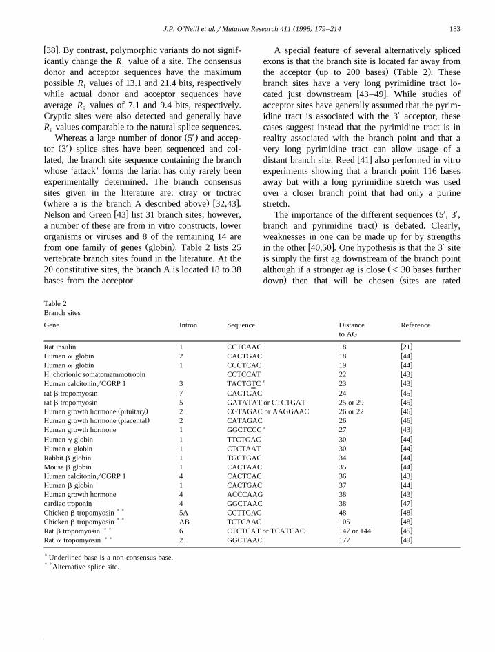
Ž .from one family of genes globin . Table 2 lists 25vertebrate branch sites found in the literature. At the20 constitutive sites, the branch A is located 18 to 38bases from the acceptor.
A special feature of several alternatively splicedexons is that the branch site is located far away from
Ž . Ž .the acceptor up to 200 bases Table 2 . Thesebranch sites have a very long pyrimidine tract lo-
w xcated just downstream 43–49 . While studies ofacceptor sites have generally assumed that the pyrim-idine tract is associated with the 3X acceptor, thesecases suggest instead that the pyrimidine tract is inreality associated with the branch point and that avery long pyrimidine tract can allow usage of a
w xdistant branch site. Reed 41 also performed in vitroexperiments showing that a branch point 116 basesaway but with a long pyrimidine stretch was usedover a closer branch point that had only a purinestretch.
Ž X XThe importance of the different sequences 5 , 3 ,.branch and pyrimidine tract is debated. Clearly,
weaknesses in one can be made up for by strengthsw x Xin the other 40,50 . One hypothesis is that the 3 site
is simply the first ag downstream of the branch pointŽalthough if a stronger ag is close -30 bases further
. Ždown then that will be chosen sites are rated
Table 2Branch sites
Gene Intron Sequence Distance Referenceto AG
w xRat insulin 1 CCTCAAC 18 21w xHuman a globin 2 CACTGAC 18 44w xHuman a globin 1 CCCTCAC 19 44w xH. chorionic somatomammotropin CCTCCAT 22 43
) w xHuman calcitoninrCGRP 1 3 TACTGTC 23 43w xrat b tropomyosin 7 CACTGAC 24 45w xrat b tropomyosin 5 GATATAT or CTCTGAT 25 or 29 45
Ž . w xHuman growth hormone pituitary 2 CGTAGAC or AAGGAAC 26 or 22 46Ž . w xHuman growth hormone placental 2 CATAGAC 26 46
) w xHuman growth hormone 1 GGCTCCC 27 43w xHuman g globin 1 TTCTGAC 30 44w xHuman e globin 1 CTCTAAT 30 44w xRabbit b globin 1 TGCTGAC 34 44w xMouse b globin 1 CACTAAC 35 44w xHuman calcitoninrCGRP 1 4 CACTCAC 36 43w xHuman b globin 1 CACTGAC 37 44w xHuman growth hormone 4 ACCCAAG 38 43w xcardiac troponin 4 GGCTAAC 38 47
) ) w xChicken b tropomyosin 5A CCTTGAC 48 48) ) w xChicken b tropomyosin AB TCTCAAC 105 48
) ) w xRat b tropomyosin 6 CTCTCAT or TCATCAC 147 or 144 45) ) w xRat a tropomyosin 2 GGCTAAC 177 49
) Underlined base is a non-consensus base.) )Alternative splice site.
( )J.P. O’Neill et al.rMutation Research 411 1998 179–214184
. w xcag) tag)aag)gag 51 . This is supported by thefact that ag’s are not commonly found 10–20 bases
w xupstream of acceptor sites 36 and when presentthey are associated with weaker sites. However,other work where the branch points are mutated
Ž .shows that a new branch point always an A ischosen 22 to 37 bases upstream of the acceptorimplying that the 3X site is the more important sitew x w x X Ž .52 . Senapathy et al. 31 state that the 5 donorsite must be recognized first, however, based oninformation content, as stated previously, Stephens
w xand Schneider 35 suggest just the opposite.As discussed above, internal exons have a maxi-
mum size of about 300–400 bp with a mean of 245w xbases and a peak of about 125 bases 8,18 ; however,
they also have a minimum size of approximately 50w xbases 53 . In vitro experiments with constructs have
shown that when exons drop below this size, they arew xskipped 27,29 . Small exons can be spliced how-
w xever, and exons as small as 3 bases are known 8,47 .These small exons apparently all require specificenhancer sequences in order not to be skipped. Forexample in the cardiac troponin gene the 30 base
w xexon 5 has a purine rich enhancer 47 . In vitro,small exons that are skipped can be spliced in if
w xpurines are removed from the pyrimidine tract 53 .The above size constraints concern only internal
X Ž .exons. The average size of the 5 first exon is 200bases with the highest percentage less than 100 basesand ranging up to 1 kb. Deletions in the first exon do
w x Xnot appear to affect splicing of exon 2 29 . The 3Ž .last exon averages 649 bases with a peak at 300
w xbases and a large percentage over 900 bases 18 .These features imply that the splicing signals for thefirst and last exon must be different than for internal
w xexons. For example, Liu and Mertz 54 found thatan acceptor signal that worked well at an internalexon did not work for the last exon unless thepyrimidine tract was strengthened or sequences weredeleted in the last exon. First exons apparently re-quire the presence of the 5X 7-methyl-guanosine capŽ .and its binding proteins which is present on all
w x Ž X.polymerase II transcripts 8,55 . Last 3 exons endat the poly A tail and studies indicate that splicing
Žand polyadenylation factors interact i.e., mutation ofthe last exon inhibits polyadenylation and mutation
.of the adenylation site inhibits last exon splicingw x8,56 .
A minimum intron size has also been determinedw x w x33,49,52,57 . Smith and Nadal-Ginard 49 found aminimum 5X splice site to branch distance of 51–59bases in the a-tropomyosin gene while Wieringa et
w x Žal. 57 found a minimum intron size of 80 bases did.not function at 69 bases or less in the rabbit b-globin
gene and their survey of the literature found nointrons of less than 50 bases. An unusual mutation in
w xthe donor site of exon 8 of the COL1A1 gene 58causes not only skipping of exon 8 but inclusion of96 bases on the 5X end of intron 7 into the mRNA.Looking at the genomic DNA, the new donor siteŽ .Ggtaaga, Senapathy score—86.5 is better than the
Ž .old site Tgtgagt, Senapathy score—75.8 ; the rea-son that the better site must not normally be used isbecause this would reduce the size of intron 7 to
Ž .only 63 bases too small for an intron ; however, inthe mutant where exon 8 is not recognized then
Ž‘intron 7’ extends until the beginning of exon 9 280.bases .
There is also information that suggests that someexons are recognized as a cluster or cooperative unitw x w x59–61 . Sterner and Berget 59 found that a mini-exon requires the preceding exon to be spliced in;mutation of the 5X splice site of the upstream exon
w xleads to intron inclusion. Ge et al. 60 found cooper-ation of two 3X sites in order to splice a 48 bp exon.
Ž .Both exon 8 44 bp and exon 9 are skipped with amutation in the exon 9 donor site in the p67-PJOX
w xgene 61 .Splicing can also be influenced by pre-mRNA
w xsecondary structure 46,48,62 . In the human growthhormone gene, stabilization of a stem loop causes
w xshifting to the alternative splice site 46 while asingle nucleotide polymorphism in exon 2 of the
w xepisialin gene 62 causes use of different spliceacceptor sites; this polymorphism is hypothesized tocause the loss of a hairpin loop. In the tropomyosingene, a large hairpin encompassing the alternativeexon 6B causes it to be spliced out. Mutations thatdestabilize the loop cause the exon to be spliced in
w xRef. 48 .Mutations leading to splicing errors are a signifi-
cant proportion of mutations leading to human dis-Žease i.e., 101 of a total of 659 point mutations
w x.causing human diseases are in splice junctions 63 .w x w xKrawczak et al. 63 and Nakai and Sakamoto 16
review 101 human and 209 mammalian splicing

( )J.P. O’Neill et al.rMutation Research 411 1998 179–214 185
w xmutations, respectively. Krawczak et al. 63 reportŽ X. Ž X.62 donor 5 splice site mutations, 26 acceptor 3
splice site mutations and 13 creation of novel sites.Ž X.For the donor 5 splice site mutations, 60% involve
the invariant gt with increased mutations at IVSq1Ž X .and IVSq2. For the acceptor 3 splice site, 87%
involve the invariant ag with an excess of mutationsat IVS-2. Of interest, 12 of 13 of the new creatednovel sites are upstream of the normal site and only4 of 13 have lower Senapathy scores than the origi-nal site. For the 5X splice site mutations, 16 lead toskipping, 7 to cryptic utilization and 5 to both. Forthe 3X splice mutants, 4 result in exon skipping and 6in cryptic site utilization. For both sites, purines aremost likely to be introduced in the mutation. Nakai
w xand Sakamoto 16 found that 92% of mutationsoccur at consensus sites, 97 at 5X sites and 55 at 3X
sites with 15% causing novel sites. For the 5X sitemutations, 48 lead to skipping and 33 to cryptic andfor the 3X site 29 lead to skipping and 21 to cryptic.They also found that novel sites are all upstream ofthe normal site. As indicated above, intron retentionis rare. Among the intron retention mutants, 4 arevery short introns and 3 are terminal introns; how-ever, 3 are large internal introns.
Mutations at splice sites often lead to the observa-tion of multiple cDNA species after RTrPCR analy-sis of intracellular mRNA. Sometimes utilization of acryptic site is incomplete with some normal messageobserved or both skipped and normal or skipped andcryptic site mRNAs seen. These indicate competitionbetween splice sites. In addition, sometimes mRNAswith multiple skipped exons are observed. Nonsensemutations in exons have also been found to lead to
Žboth low levels of mRNA andror mRNAs some-.times multiple usually excluding the exon contain-
Žw xing the nonsense mutation 64–72 ; reviewed inw x.Ref. 73 . It has been suggested that the premature
termination of translation caused by the nonsensemutation causes mRNA instability and, thus, lowlevels of mRNA. This is supported by the observa-tion that nonsense mutants do not affect mRNAlevels if they occur at the very 5X end of the geneŽ .re-initiation of transcription possible or at the very
X Ž3 end transcription goes long enough that the mRNA.is not unstable . Often, if the exon containing the
nonsense mutation is skipped and an inframe dele-tion results, premature termination is avoided and
that mRNA will be stable. In general, it is notsuggested that the nonsense mutation causes theexon skipping, but merely that it stabilizes what isnormally a low level product that is seen in the
Žmutant only because the major full length nonsense.containing product is degraded. The exon skipped
product seen in nonsense mutants has been demon-strated at a low level in normal cells for some genesw x68,69 . However, some groups suggest that the non-sense mutation does directly affect splicing becausechanges in mRNA are observed in the nucleus and
w xtranslation effects must be cytoplasmic 64,70 . It isdifficult to model how a nonsense mutation could berecognized by the spliceosome as this requires recog-nition of mRNA frame in the nucleus and many‘nonsense codons’ obviously exist out of frame innormal mRNAs. Some of these nonsenserexon skip-ping mutations could occur because the nonsensemutation just happens to be within an enhancer
w xwhich is disrupted 71 . Models have been proposedwhereby a link exists between splicing and transla-tion; the ribosome pulls the hnRNA through the
w xnuclear pore driving splicing 65 . If the ribosomereleases the RNA, splicing ceases. Caenorhabditiselegans with mutations in their smg genes do nothave nonsense mutation reductions in mRNA in
w xother genes 72 implying that specific genes exist todegrade prematurely terminated mRNAs.
1.2. The HPRT gene
The X-linked hypoxanthine-guanine phosphoribo-Ž .syltransferase HPRT gene is widely used in muta-
tion studies because of the ease of selection ofmutant cells. HPRTase phosphoribosylates hypoxan-thine and guanine in the purine salvage pathway.Although constitutively produced, the HPRT enzymeproduct is non-essential as purines can also be cre-ated de novo. HPRTase deficient mutants can beselected in the presence of purine analogues such as
Ž .6-thioguanine TG which kill normal, wild typecells; only cells containing a mutation in their HPRTgene are able to survive and proliferate. AlthoughHPRT is dispensable at the cellular level, germinalHPRT deficiency does lead to human disease, either
Ž .Lesch–Nyhan syndrome complete deficiency orŽ .X-linked gout partial deficiency , apparently caused
by the high levels of purine metabolites such as uric
( )J.P. O’Neill et al.rMutation Research 411 1998 179–214186
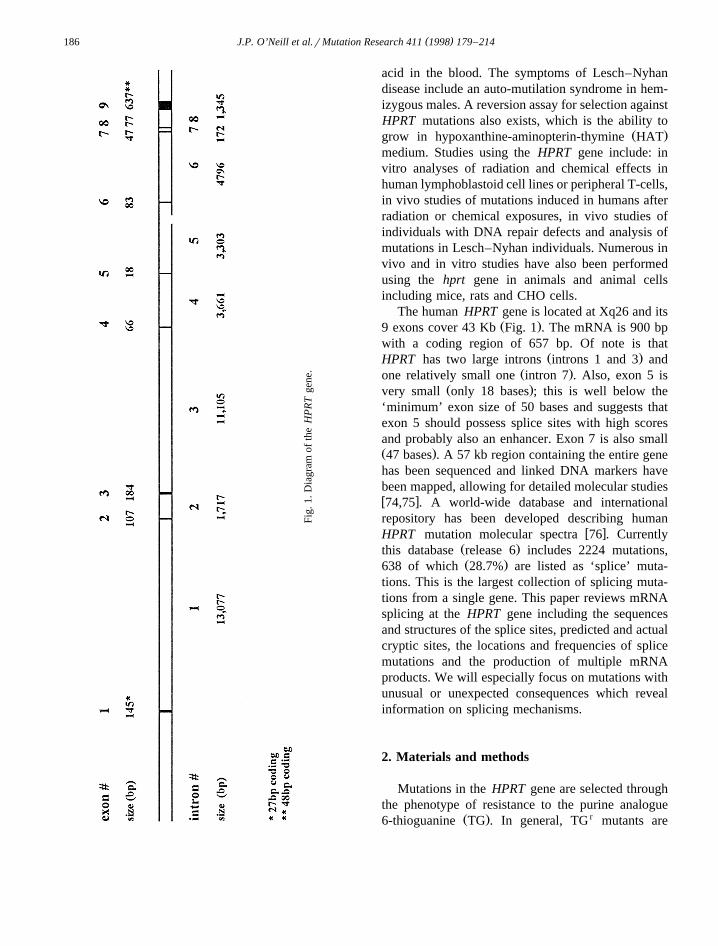
Fig.
1.D
iagr
amof
the
HP
RT
gene
.
acid in the blood. The symptoms of Lesch–Nyhandisease include an auto-mutilation syndrome in hem-izygous males. A reversion assay for selection againstHPRT mutations also exists, which is the ability to
Ž .grow in hypoxanthine-aminopterin-thymine HATmedium. Studies using the HPRT gene include: invitro analyses of radiation and chemical effects inhuman lymphoblastoid cell lines or peripheral T-cells,in vivo studies of mutations induced in humans afterradiation or chemical exposures, in vivo studies ofindividuals with DNA repair defects and analysis ofmutations in Lesch–Nyhan individuals. Numerous invivo and in vitro studies have also been performedusing the hprt gene in animals and animal cellsincluding mice, rats and CHO cells.
The human HPRT gene is located at Xq26 and itsŽ .9 exons cover 43 Kb Fig. 1 . The mRNA is 900 bp
with a coding region of 657 bp. Of note is thatŽ .HPRT has two large introns introns 1 and 3 and
Ž .one relatively small one intron 7 . Also, exon 5 isŽ .very small only 18 bases ; this is well below the
‘minimum’ exon size of 50 bases and suggests thatexon 5 should possess splice sites with high scoresand probably also an enhancer. Exon 7 is also smallŽ .47 bases . A 57 kb region containing the entire genehas been sequenced and linked DNA markers havebeen mapped, allowing for detailed molecular studiesw x74,75 . A world-wide database and internationalrepository has been developed describing human
w xHPRT mutation molecular spectra 76 . CurrentlyŽ .this database release 6 includes 2224 mutations,Ž .638 of which 28.7% are listed as ‘splice’ muta-
tions. This is the largest collection of splicing muta-tions from a single gene. This paper reviews mRNAsplicing at the HPRT gene including the sequencesand structures of the splice sites, predicted and actualcryptic sites, the locations and frequencies of splicemutations and the production of multiple mRNAproducts. We will especially focus on mutations withunusual or unexpected consequences which revealinformation on splicing mechanisms.
2. Materials and methods
Mutations in the HPRT gene are selected throughthe phenotype of resistance to the purine analogue
Ž . r6-thioguanine TG . In general, TG mutants are
( )J.P. O’Neill et al.rMutation Research 411 1998 179–214 187
selected by growth in the presence of 10 mM TG.The mutations described here are from a variety ofhuman sources. Most are in vitro derived from eitherT-lymphocytes or lymphoblastoid cell lines, oftentreated with mutagens. However, many are in vivoderived somatic mutations in T-lymphocytes fromnormal or mutagen-exposed individuals and someare from individuals with Lesch–Nyhan syndrome or
Ž .X-linked gout germinal mutations . The HPRT geneis an ideal source to probe hypotheses of splicingrecognition. Analyses of mutations that are known toaffect splicing provide unequivocable information onthe nature of splice recognition in this human gene.
The splice mutations discussed in this review area compilation of the mutations described in thereferences listed in the Appendix A to this paper.
ŽThey are part of an HPRT mutation database Ref.w x .76 , release 6 containing 2224 mutations, of which
Ž .638 28.7% are classified as splice mutations. Mostof the HPRT splice mutations were discovered be-cause they gave abnormal products after RT-PCRanalysis; however, some were originally discoveredbased on genomic sequencing. The RT products areoften smaller than the expected full length cDNA,with sometimes several products due to multiplesplicing events. DNA sequencing of the RT product
Žreveals the absence of one or more exons exon.exclusion or the presence of inserted intron se-Ž .quences intron inclusion . When coupled with ge-
nomic PCR studies, it is clear that the excludedexons are present in the genomic DNA and thealterations in mRNA splicing must be due to pointmutations in splice sites. Genomic DNA sequencingis necessary to define the exact mutation. This re-view focuses on the 277 of the 638 splice mutations
Žfor which the actual genomic basis i.e., the exactŽ . .basepair s changes for the splicing alteration has
Žbeen determined. For 341 mutants the genomicmutation is not known, only that the mRNA isaltered; for example, the cDNA lacks a single exon.This could be due to a small deletion, a pointmutation in the 5X splice site, a point mutation in the
X .3 splice site, etc.Donor and acceptor splice site Senapathy scores
were calculated at each nucleotide of the entireŽ .HPRT gene bases 1701–41,501 using the equa-
w xtions and tables in 31 and the program MathematicaŽ .Wolfram . The information content in bits for donor
Ž .and acceptor sites R was also calculated at eachi
nucleotide of the HPRT gene using the equations inw x35 . To assess the effects of mutations, R werei
computed for the normal and mutated site using theprogram Scan and displayed with MakeWalker,
w xDNAplot and Lister as in Ref. 33 .
3. Results
This paper attempts to summarize the types ofsplice site mutations observed in the human HPRTgene and to discern any ‘rules’ which apply to thisparticular gene. In this review, we will use thefollowing splice recognition sequence terminology.The splice sequences in the intron are designated 5X
and 3X splice sites. Relative to an exon, the intron 3X
splice sequence is termed ‘donor’ and the intron 5X
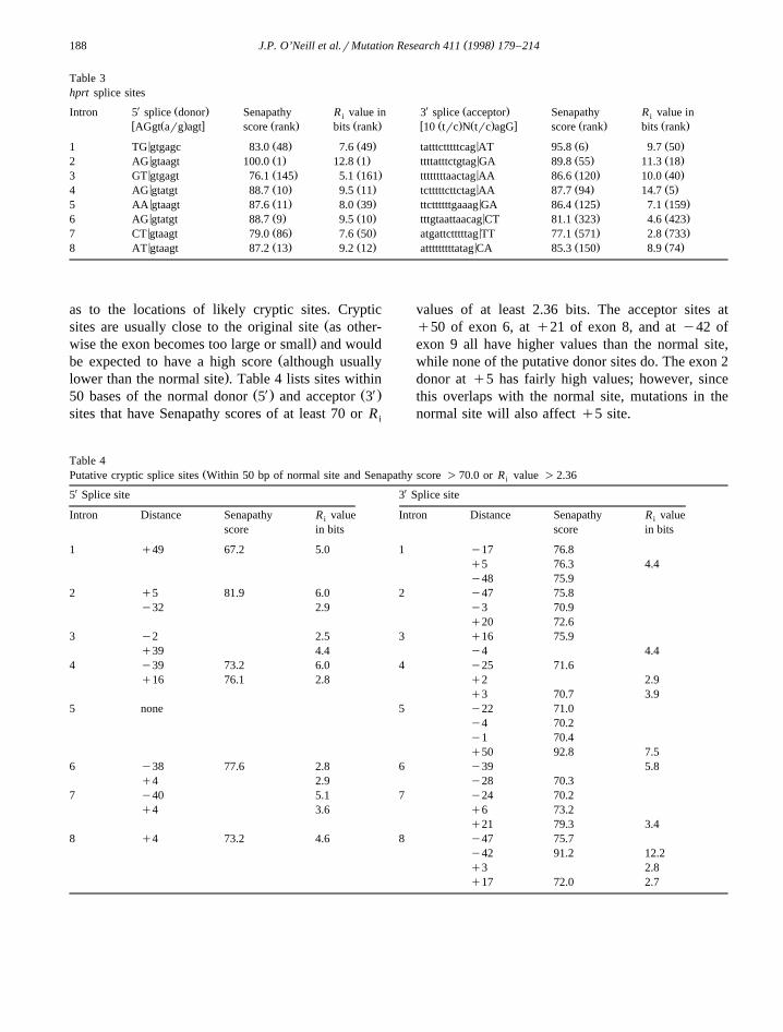
splice sequence is termed ‘acceptor.’ Table 3 givesX Ž . X Ž .the HPRT 3 donor and 5 acceptor site se-
w xquences and their Senapathy scores 31 and R iw xvalues 38 . By the Senapathy method, a perfect
Ž .score requires a donor sequence of AGgt arg agtŽ . Ž .and an acceptor sequence of 10 trc n trc agG. Us-
ing the information theory based model, a minimalR of 0 is required for splicing but in general a cutoffi
of 2.36 bits is utilized; however, values can be ashigh as 21 although most sites have R values in thei
w x7–12 bit range 38 . Also given is the rank of thatsite among all possible sites of the HPRT geneŽdonor and acceptor splice site scores and valueswere calculated for each base from the beginning ofexon 1 to the end of exon 9 with the genomic
.sequence of 43 kb . Although some sites are veryŽgood the exon 2 donor splice site has a Senapathy
score of 100 and an R value of 12.7, both ranking iti. Ža1 , others are quite poor the exon 8 acceptor has a
Senapathy score of only 77.1 and is ranked a571.and an R value of 2.8 and is ranked a733 . Never-i
theless, these rankings indicate that the exon 3 donor,exon 7 donor, and exon 8 acceptors might be particu-larly sensitive to mutation. Exon 2 has a very strongacceptor and donor indicating it might be moreresistant to mutation. As for the small exon 5, bothacceptor and donor are strong.
The determination of Senapathy scores and R i
values for the entire HPRT gene allows speculations
( )J.P. O’Neill et al.rMutation Research 411 1998 179–214188
Table 3hprt splice sites
X XŽ . Ž .Intron 5 splice donor Senapathy R value in 3 splice acceptor Senapathy R value ini iw Ž . x Ž . Ž . w Ž . Ž . x Ž . Ž .AGgt arg agt score rank bits rank 10 trc N trc agG score rank bits rank
< Ž . Ž . < Ž . Ž .1 TG gtgagc 83.0 48 7.6 49 tatttctttttcag AT 95.8 6 9.7 50< Ž . Ž . < Ž . Ž .2 AG gtaagt 100.0 1 12.8 1 ttttatttctgtag GA 89.8 55 11.3 18< Ž . Ž . < Ž . Ž .3 GT gtgagt 76.1 145 5.1 161 ttttttttaactag AA 86.6 120 10.0 40< Ž . Ž . < Ž . Ž .4 AG gtatgt 88.7 10 9.5 11 tctttttcttctag AA 87.7 94 14.7 5< Ž . Ž . < Ž . Ž .5 AA gtaagt 87.6 11 8.0 39 ttcttttttgaaag GA 86.4 125 7.1 159< Ž . Ž . < Ž . Ž .6 AG gtatgt 88.7 9 9.5 10 tttgtaattaacag CT 81.1 323 4.6 423< Ž . Ž . < Ž . Ž .7 CT gtaagt 79.0 86 7.6 50 atgattctttttag TT 77.1 571 2.8 733< Ž . Ž . < Ž . Ž .8 AT gtaagt 87.2 13 9.2 12 atttttttttatag CA 85.3 150 8.9 74
as to the locations of likely cryptic sites. CrypticŽsites are usually close to the original site as other-
.wise the exon becomes too large or small and wouldŽbe expected to have a high score although usually
.lower than the normal site . Table 4 lists sites withinŽ X. Ž X.50 bases of the normal donor 5 and acceptor 3
sites that have Senapathy scores of at least 70 or R i
values of at least 2.36 bits. The acceptor sites atq50 of exon 6, at q21 of exon 8, and at y42 ofexon 9 all have higher values than the normal site,while none of the putative donor sites do. The exon 2donor at q5 has fairly high values; however, sincethis overlaps with the normal site, mutations in thenormal site will also affect q5 site.
Table 4ŽPutative cryptic splice sites Within 50 bp of normal site and Senapathy score )70.0 or R value )2.36i
X X5 Splice site 3 Splice site
Intron Distance Senapathy R value Intron Distance Senapathy R valuei i
score in bits score in bits
1 q49 67.2 5.0 1 y17 76.8q5 76.3 4.4y48 75.9
2 q5 81.9 6.0 2 y47 75.8y32 2.9 y3 70.9
q20 72.63 y2 2.5 3 q16 75.9
q39 4.4 y4 4.44 y39 73.2 6.0 4 y25 71.6
q16 76.1 2.8 q2 2.9q3 70.7 3.9
5 none 5 y22 71.0y4 70.2y1 70.4q50 92.8 7.5
6 y38 77.6 2.8 6 y39 5.8q4 2.9 y28 70.3
7 y40 5.1 7 y24 70.2q4 3.6 q6 73.2
q21 79.3 3.48 q4 73.2 4.6 8 y47 75.7
y42 91.2 12.2q3 2.8q17 72.0 2.7
( )J.P. O’Neill et al.rMutation Research 411 1998 179–214 189
Use of the calculated Senapathy scores across thegene also allows prediction of exons within the
Žregion i.e., would the observed exons be predicted,andror do other ‘exon-like’ sequences exist that
.would be predicted to be exons but are not? . Usingacceptors with Senapathy scores greater than 85.2and donors with Senapathy scores greater than 83.0Ž .7 of 9 HPRT exons have these scores , and thecriterion that an exon must be between 50 and 300
Žbp, leads to the prediction of 26–27 exons there are.two that overlap of which only 3 are actual HPRT
Ž .exons exons 2, 4 and 6 . Two exons would beŽpredicted around the 18 bp exon 5 the overlapping
.set . These new exons are defined by either the exon5 donor or acceptor splice sequence being used withanother ‘splice sequence’ to yield a larger exon of190 or 85 bases, respectively. This reaffirms thatsomething must be enhancing exon 5 splicing sinceother better choices exist. For exons 3 and 8, theacceptor and donor splice sites, respectively, arepresent, however, they have no cognate 5X or 3X
splice site, respectively, within the 50–300 baserange. For exon 7, neither splice site makes the list.
An advantage of HPRT mutations for analyzingsplice recognition site mutations is that polymor-phisms are not a complication since a mutant pheno-type has been selected. In fact, no polymorphisms inthe coding region of the human HPRT gene havebeen described. The wild type sequence can bedirectly compared to that in the mutant cell. Thereare, however, some complications. HPRT splice mu-tations are often defined by RTrPCR methods; de-termining the exact mutation often requires a ge-nomic DNA PCR assay and sequencing. Unfortu-nately, for many of the ‘splice mutations’ in theHPRT database only the RT analysis or RT analysis
Ž . Žand genomic multiplex PCR but not sequencing of.the genomic PCR products has been performed.
Thus, many of these ‘splice’ mutants show the phe-notype of exon exclusion in cDNA with the exon’spresence only confirmed in genomic DNA. There-fore, this database is a work in progress, as moremutations will certainly be determined; at present,the precise mutation has been determined in 277splicing mutants.
As discussed in the introduction, the human HPRTŽ .gene contains 8 introns and 9 exons Fig. 1 . Exclu-
sion of a single exon due to aberrant splicing can
Ž .yield an in-frame reading frame exon 4 or 5 only orŽ .out-of-frame reading frame rest of exons in the
mRNA. Since the usual method of analysis isRTrPCR with primers 5X and 3X to the HPRT cod-ing sequence, a splicing alteration which excludesexon 1 or 9 will not be detected because there willbe no cDNA produced by the RTrPCR procedure.
The bases in the HPRT gene are designated bytwo numbering systems in this review. A nucleotide’sposition in the HPRT gene is indicated by subscriptsafter the base. The base numbers are given bothaccording to the genomic sequence of 56,736 bases
w x Žof Edwards et al. 74 Note: the GenBank fileM26434 has an inserted T between 22,227 and 22,228and thus numbering differs by one between the
.Edwards system and M26434 after base 22,227 andŽaccording to the cDNA sequence base 1 is the A in
.the AUG initiation codon . To easily discriminatebetween the two numbering systems, if the number isless than 700 it is cDNA numbering while thosenumbers greater than 1700 are genomic numbering.Intronic bases may also labeled by an intervening
Ž .sequence nomenclature; the intron IVS bases arenumbered away from the nearest exon, bases 5X to
Ž . Xthe exon given as negative y and bases 3 to theŽ .exon given as positive q . For example, the first g
X Ž .of the intron 1 splice 5 donor site is designatedbase number 1704 in genomic DNA or intron 1,
Ž . Xbaseq1 IVS1q1 . The first base 5 to exon 2Ž .exon 2 acceptor site is designated base number
Ž .14,779 or intron 1, base y1 IVS1-1 . Exonic basesare designated by uppercase letters and intronic basesby lowercase letters.
3.1. Mutations in intron splice recognition sequences
Table 5 lists the number of mutations seen forX Ž . X Ž .each of the 5 donor and 3 acceptor splice sites
and the data on splice mutations are presented indi-vidually below for each of the 9 HPRT exons.
( )3.1.1. Exon 1 Table 6: cDNA bases 1–27 , 9 donormutations
The first exon of the HPRT gene contains theŽ .first 27 coding bases 1–27 plus approximately 60
bases 5X to the AUG initiation triplet. The splice

( )J.P. O’Neill et al.rMutation Research 411 1998 179–214190
Table 5Frequencies of splice site mutations
X XIntron No. of 5 Splice No. of 3 SpliceŽ . Ž .Site donor Site acceptor
Ž . Ž .Mutations % ) Mutations % )
Ž . Ž .1 9 8.0 9 10.1Ž . Ž .2 5 4.5 5 5.6Ž . Ž .3 3 2.7 11 12.4Ž . Ž .4 15 13.4 5 5.6Ž . Ž .5 21 18.8 7 7.9Ž . Ž .6 13 11.6 10 11.2Ž . Ž .7 18 16.1 21 23.6Ž . Ž .8 28 25.0 21 23.6
Total 112 89
X Ž .)An equal distribution among the 9 exons would be 14 5 donorŽ . X Ž .splice site mutations 112r8s14, 12.5% and 11 3 acceptorŽ .splice site mutations 89r8s11, 12.5% .
donor sequence is GTG gtgagc. Mutations at IVS127
q1, q2 or q5 have been observed to result in theinclusion of the first 49 bases of intron 1 in thecDNA. This is the result of the use of a cryptic splice
Ždonor sequence g tggcg Senapathy score 67.2, R1753 i.value 5.0 bits . Although the values for the cryptic
site are low, it is the best in the area and it isŽ .essential that very large intron 1 13,075 bp be
spliced out to make a mRNA. From these results, itappears that an exon 1 splice donor sequence muta-tion always results in the use of this cryptic donorsequence and creates a 49 base intron inclusion inthe mRNA. This inclusion is out of frame and yieldsa chain terminating codon TAG at new codon num-
Žber 27 initiation codon AUG still designated codon.1 .
In summary, exon 1 splice donor site mutationshave been found in base 1, 2 or 5 of the 6 splicesequence bases and in the last base of the exon
Ž .27G and all lead to an intron inclusion. This in-cluded sequence contains the mutated base.
( )3.1.2. Exon 2 Table 7: cDNA bases 28–134 , 9acceptor and 5 donor mutations
The splicing of exon 1 to exon 2 removes theŽ .largest HPRT intron IVS1—13,075 bases . The
X Ž .intron 1 5 splice exon 2 acceptor sequence iscagA T and mutations at IVS1-1 or -2 have been28
reported. Mutations at both sites have pleiotropiceffects, yielding multiple species of mRNA. The
Žlargest cDNA is missing exon 2 bases 1–5 HPRT.bases 28–32:ATTAG due to the use of a cryptic
Ž . Žsplice acceptor sequence TAG rT a potential32
cryptic splice sequence, CAG rG, does not appear46.to be used . In addition, cDNA species missing exon
2 or exon 2 and 3 have been reported.Mutations in the exon 2 acceptor splice site can
cause different mRNA spectra depending on theparticular type of nucleotide replacement. Changing
Ž .of the invariant g at IVS1-1 IVS1-1G leads to usewof a cryptic site 5 bases into exon 2 normal—tatttct-
ttttcagATTAGTG; mutant—tatttctttttcanATTAGTGxwhich is spliced as tatttctttttcanattagTG . The newŽalternate site has Senapathy scores of 82.6 R valuei
. Ž . Žof 7.3 bits , 81.1 R value of 7.1 bits and 75.8 Ri i.value of 4.6 bits for g™ t, g™c, and g™a, re-
w Ž .spectively vs. 95.8 R value of 9.7 bits for theiŽ .usual unmutated site and 75.8 R value of 4.4 bitsi
xfor the unmutated site . The g™ t and g™c muta-tions lead to exclusive use of the cryptic site whilethe g™a mutation leads to mixed exon 2 loss andcryptic site mRNAs. This is understandable based onthe lower values for the g™a mutation site. Chang-
Ž .ing of the invariant a at IVS1-2 to t IVS1-2 A™TŽleads to use of the cryptic site Senapathy score 76.6,
Table 6Ž .aSplice site mutations involving exon 1 27 Bases; 1–27 A TG . . . GTG gtgagc1 27
X Ž .5 Donor Splice Mutation Effect in cDNASequence
GTGg tgagc g ™a or t or t ™a Inclusion of intron 1, b 1–49 due to use of a cryptic splice1704 1704 1705Ž < .or g ™a or t sequence cag gtggcg1708 1752
Ž .D TG gtgagc . . . c Deletion of E1, b 26–27 and insertion of intron 1, b 35–4927 1737Ž < .due to use of a cryptic splice sequence cag gtggcg1752
a Ž . Ž .Nine donor site mutations have been reported. A mutation at 27G G™A has been reported to affect splicing see Table 15 .

( )J.P. O’Neill et al.rMutation Research 411 1998 179–214 191
Table 7Ž .aSplice site mutations involving exon 2 107 bases; 28–134 atatttcttttt cag A TT . . . AG gtaagt28 134
X Ž .3 Acceptor Splice Mutation Effect in cDNASequence
Ž < .cag ATT g ™c or a or t Loss of E2, b 28–32 due to use of a cryptic splice site ATTAG T14779 14779 32
or a ™g or t andror loss of E2 andror loss of E2q314778
X Ž .5 Donor Splice Mutation Effect in cDNASequence
AG g taagt g ™a or t ™g Loss of E214887 14887 14888
a Ž .Nine acceptor and five donor site mutations have been reported. No mutations in 28A have been reported; mutations at 134G G™A or Thave been reported with no effect on splicing.
.R value of 6.5 bits while changing to a g leads toiŽexon 2 andror exon 2–3 loss old mutated site
.Senapathy score 71.1, R value of 4.3 bits . Thei
latter still has an acceptable R value, it is not cleari
why it is not used.The intron 2 5X splice site sequence is
CAG gtaagt and mutations at IVS2q1 and IVS2134
q2 have been reported. All reported mutations re-sult in the exclusion of exon 2 from the cDNA.Mutations at 134G do not affect splicing. Mutationsin the exon 2 splice donor sequence do not lead tothe use of a cryptic splice site in intron 2, althoughthe sequences gtaaga and gtggga exist 4 and 191bases downstream, respectively.
In summary, mutations in either base of the agdinucleotide in the exon 2 splice acceptor sequenceor mutations in base 1 or 2 of the 6 bases in the exon2 donor sequence lead to exclusion of part or all ofexon 2 in the cDNA. The exclusion of exon 2 bases1–5 results in an immediate chain terminating TGAat new codon 10. The exclusion of exon 2 results ina chain terminating codon TGA at new codon 11.The exclusion of exon 2q3 results in an in-framemRNA lacking 291 bases. The observed exclusion ofboth exon 2 and 3 only with acceptor site mutationssuggests that exons 2 and 3 are spliced together prior
Ž .to splicing to exons 1 and 4 this is discussed below .
( )3.1.3. Exon 3 Table 8: cDNA bases 135–318 , 5acceptor and 3 donor mutations
The splicing of exon 2 to exon 3 removes theŽ .relatively small intron 2 of 1715 bases IVS2 . Muta-
tions in the exon 3 tagG A acceptor splice site135
have been found only at IVS2-1G. Mutations in the
exon 3 donor splice TGT gtgagt site have been318
found only at IVS3q1G. Mutations in both splicesequences result in the exclusion of exon 3 androrexon 2 and 3 from the cDNA. Mutations of g™a atIVS3q1 cause exon 2–3 loss while g™ t mutationscause only exon 3 loss. There appears to be noexplanation for this. The differences could be purelycoincidence related to variations in reporting of mul-tiple cDNAs by different researchers. The use of apotential cryptic donor site, gtaggt, 39 bases down-stream has not been reported. The exclusion of exon3 results in a chain terminating T AA at new341
codon 53. As pointed out above, the exclusion ofexon 2 and 3 results in the in-frame loss of 291 basesfrom the mRNA.
The exclusion of exon 2q3 is quite commonwith mutations in the 3X splice sequences of both
Table 8Ž .aSplice site mutations involving exon 3 184 bases; 135–318
attttatttctg tagG ACT. . . TGT gtgagt135 318X Ž .3 Acceptor Splice Mutation Effect in cDNA
Sequence
tag GAC g ™c or a or t Loss of E3 andr16602 16602
or E2q3
X Ž .5 Donor Splice Mutation Effect in cDNASequence
TGTg tgagt g ™a or t Loss of E3 andr16787 16787
or E2q3
a Five acceptor and three donor site mutations have been reported.Ž . Ž .Mutations at 135G G™T or C and at 318T T™A have been
reported with no effect on splicing.
( )J.P. O’Neill et al.rMutation Research 411 1998 179–214192
intron 1 and 2 and the 5X splice sequence in intron 3.This exclusion is not reported with intron 2 5X splicesequence mutations, which suggests that the resultantsplicing of exon 1 to exon 4 is the frequent result ofinterference with the splicing between exon 2 and 3.Alternatively, the mRNA missing both exon 2 and 3is probably more stable than the mRNA missingexon 2 or 3 alone because of the chain terminatingcodons created by the loss of either single exon.
In summary, mutations affecting the splicing ofexon 3 are relatively infrequent and all reported sofar cause the exclusion of exon 3 andror exons 2and 3. It should be noted, however, that the exclu-sion of exons 2 and 3 in cDNA has been reportedoften in the database and many of these could alsobe splice sequence mutations. However, since thegenomic alteration has not been defined, these could
Ž .be V D J recombinase mediated deletions—whichw xare common in T-lymphocytes 77,78 —or other
genomic deletions of exons 2 and 3. In conclusion,exon 3 splice mutations make up only 4% of thetotal-well below the expected 12.5%.
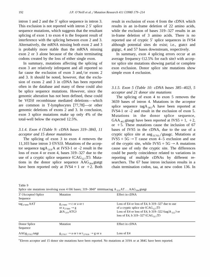
( )3.1.4. Exon 4 Table 9: cDNA bases 319–384 , 11acceptor and 15 donor mutations
The splicing of exon 3 to exon 4 removes theŽ .11,103 base intron 3 IVS3 . Mutations of the accep-
tor sequence tagA A at IVS3-1 or -2 result in the319
loss of exon 4 or exon 4, bases 319–327 due to theŽ < .use of a cryptic splice sequence CAG T . Muta-327
tions in the donor splice sequence AAG gtatgt384
have been reported only at IVS4q1 or q2. Both
result in exclusion of exon 4 from the cDNA whichresults in an in-frame deletion of 22 amino acids,while the exclusion of bases 319–327 results in anin-frame deletion of 3 amino acids. There is noreported use of cryptic 5X splice sequences in IVS4although potential sites do exist; i.e., gtatct andgtgtgc, 4 and 57 bases downstream, respectively.
In summary, exon 4 splicing errors occur at anŽ .average frequency 12.5% for each site with accep-
tor splice site mutations showing partial or completeexon exclusion. Donor splice site mutations showsimple exon 4 exclusion.
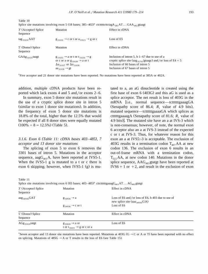
( )3.1.5. Exon 5 Table 10: cDNA bases 385–402 , 5acceptor and 21 donor site mutations
The splicing of exon 4 to exon 5 removes the3659 bases of intron 4. Mutations in the acceptorsplice sequence tagA A have been reported at385
IVS4-1 or -2 and result in the exclusion of exon 5.Mutations in the donor splice sequence,GAA gtaagt have been reported at IVS5q1, q2,402
or q5. These mutations cause the inclusion of 67bases of IVS5 in the cDNA, due to the use of acryptic splice site at aag rgtaagc. Mutations at31701
IVS5q5G™T cause exon 4–5 exclusion and useof the cryptic site, while IVS5q5G™A mutationscause use of only the cryptic site. The differencescould be purely coincidence related to variations inreporting of multiple cDNAs by different re-searchers. The 67 base intron inclusion results in achain termination codon, taa, at new codon 136. In
Table 9Ž .aSplice site mutations involving exon 4 66 bases; 319–384 tttttttttaactag A AT . . . AAG gtatgt319 384
X Ž .3 Acceptor Splice Mutation Effect in cDNASequence
tag AAT g ™c or a or t Loss of E4 or loss of E4, b 319–327 due to use27890 27890Ž < .or a ™g of a cryptic splice site CAG T27889 327
Ž . Ž < .D A ATG Loss of E4 or loss of E4, b 319–322 tag A or319 322Ž < .loss of E4, b 319–327 CAG T327
Donor Splice Mutation Effect in cDNASequence
AAGg tatgt g ™a or t or t ™g or a Loss of E427957 27957 27958
a Eleven acceptor and 15 donor site mutations have been reported. No mutations at 319A or at 384G have been reported.
( )J.P. O’Neill et al.rMutation Research 411 1998 179–214 193
Table 10Ž .aSplice site mutations involving exon 5 18 bases; 385–402 ctctttttcttctagA AT . . . GAA gtaagt385 402
X Ž .3 Acceptor Splice Mutation Effect in cDNASequence
tag AAT g ™c or t or a ™g or c Loss of E531616 31616 31615
X Ž .5 Donor Splice Mutation Effect in cDNASequence
GAAg taagt g ™a or t or t ™g Inclusion of intron 5, b 1–67 due to use of a31635 31635 31636Ž < .or c or a or g ™a or t cryptic splice site aag gtaagc andror loss of E4q531639 31701
Da or Da Inclusion of 66 bases of intron 531637 31638
aa ™gt Inclusion of 67 bases of intron 531638
a Five acceptor and 21 donor site mutations have been reported. No mutations have been reported at 385A or 402A.
addition, multiple cDNA products have been re-ported which lack exons 4 and 5 andror exons 2–6.
In summary, exon 5 donor site mutations result inthe use of a cryptic splice donor site in intron 5Ž .similar to exon 1 donor site mutations . In addition,the frequency of exon 5 donor site mutations is18.8% of the total, higher than the 12.5% that wouldbe expected if all 8 donor sites were equally mutatedŽ . Ž .100%%8s12.5% Table 5 .
( )3.1.6. Exon 6 Table 11: cDNA bases 403–485 , 7acceptor and 13 donor site mutations
The splicing of exon 5 to exon 6 removes the3301 bases of intron 5. Mutations in the acceptorsequence, aagG A, have been reported at IVS5-1.403
When the IVS5-1 g is mutated to a t or c there isŽ .exon 6 skipping; however, when IVS5-1 g is mu-
tated to a, an aG dinucleotide is created using theŽ .first base of exon 6 403G and this aG is used as a
splice acceptor. The net result is loss of 403G in thewmRNA i.e., normal sequence—tcttttttgaaagGA
Ž .Senapathy score of 86.4; R value of 4.9 bits ,i
mutated sequence—tcttttttgaaaaGA which splices asŽcttttttgaaaagA Senapathy score of 81.6; R value ofi
.x4.9 bits . The mutated site have an a at IVS-3 whichis non-consensus; however, of note, the normal exon6 acceptor also an a at IVS-3 instead of the expectedc or t at IVS-3. Thus, for whatever reason for thisexon an a at IVS5:-3 is acceptable. The exclusion of403G results in a termination codon T AA at new407
codon 136. The exclusion of exon 6 results in anout-of-frame mRNA with a termination codon,T AA, at new codon 140. Mutations in the donor522
splice sequence, AAG gtatgt have been reported at485
IVS6q1 or q2, and result in the exclusion of exon
Table 11Ž .aSplice site mutations involving exon 6 83 bases; 403–485 cttcttttttgaaagG AT . . . AG gtatgt403 485
X Ž .3 Acceptor Splice Mutation Effect in cDNASequence
aag GAT g ™a Loss of E6 andror loss of E6, b 403 due to use of34936 34936Ž < .new splice site aaa G A34936
g ™c or t Loss of E634936
X Ž .5 Donor Splice Mutation Effect in cDNASequence
AGg tatgt g ™a or Loss of E635020 35020
t or t ™g or c or a35021
a Ž .Seven acceptor and 13 donor site mutations have been reported. Mutations at 403G G™C or A or T have been reported with no effectŽ .on splicing. Mutations of 485G™A or T results in the loss of E6 see Table 15 .
( )J.P. O’Neill et al.rMutation Research 411 1998 179–214194
6. There is no reported use of a cryptic donor site,Žsuch as g tatga the next closest donor sequence is5
.g tgagg .310
In summary, mutations affecting exon 6 splicingŽmake up 10.0% of the total splicing mutations 12.5%
.expected by equal distribution and demonstrate nouse of cryptic sites.
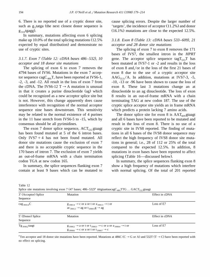
( )3.1.7. Exon 7 Table 12: cDNA bases 486–532 , 10acceptor and 18 donor site mutations
The splicing of exon 6 to exon 7 removes the4794 bases of IVS6. Mutations in the exon 7 accep-tor sequence cagC T, have been reported at IVS6-1,486
-2, -3, and -12. All result in the loss of exon 7 fromthe cDNA. The IVS6-12 T™A mutation is unusual
Ž .in that it creates a purine dinucleotide ag whichcould be recognized as a new acceptor splice site butis not. However, this change apparently does causeinterference with recognition of the normal acceptorsequence nine bases downstream. This sensitivitymay be related to the normal existence of 4 purinesin the 11 base stretch from IVS6-5 to -15, which byconsensus should be all pyrimidines.
The exon 7 donor splice sequence, ACT gtaagt532
has been found mutated at 5 of the 6 intron bases.Only IVS7q4 has not been found mutated. Alldonor site mutations cause the exclusion of exon 7and there is no acceptable cryptic sequence in the170 bases of intron 7. The exclusion of exon 7 yieldsan out-of-frame mRNA with a chain terminationcodon TGA at new codon 165.
In summary, the splice sequences flanking exon 7contain at least 9 bases which can be mutated to
cause splicing errors. Despite the larger number ofŽ .‘targets’, the incidence of acceptor 11.2% and donor
Ž .16.1% mutations are close to the expected 12.5%.
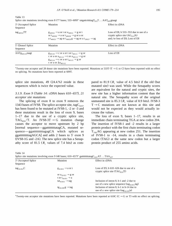
( )3.1.8. Exon 8 Table 13: cDNA bases 533–609 , 21acceptor and 28 donor site mutations
The splicing of exon 7 to exon 8 removes the 171bases of IVS7, the smallest intron in the HPRTgene. The acceptor splice sequence tagT T has533
been mutated at IVS7-1 or -2 and results in the lossof exon 8 andror in the loss of the first 21 bases ofexon 8 due to the use of a cryptic acceptor siteAAG rA. In addition, mutations at IVS7-3, -5,553
-10, -13 or -96 have been shown to cause the loss ofexon 8. These last 3 mutations change an atdinucleotide to an ag dinucleotide. The loss of exon8 results in an out-of-frame mRNA with a chainterminating TAG at new codon 187. The use of thecryptic splice acceptor site yields an in frame mRNAwhich predicts a protein lacking 7 amino acids.
The donor splice site for exon 8 is AAT gtaagt609
and all 6 bases have been reported to be mutated andresult in the loss of exon 8. There is no use of acryptic site in IVS8 reported. The finding of muta-tions in all 6 bases of the IVS8 donor sequence mayreflect the high frequency of IVS8 donor site muta-tions in general; i.e., 28 of 112 or 25% of the totalcompared to the expected 12.5%. In addition, 8mutations in exon bases have been reported to affect
Ž .splicing Table 16—discussed below .In summary, the splice sequences flanking exon 8
show a high frequency of mutations which interferewith normal splicing. Of the total of 201 reported
Table 12Ž .aSplice site mutations involving exon 7 47 bases; 486–532 ttttgtaattaacagC TTG . . . GACT gtaagt486 532
X Ž .3 Acceptor Splice Mutation Effect in cDNASequence
cag C g ™c or a or t or a ™ t or Loss of E739814 39814 39813
ac ™ag or t g™ag39812 39803
X Ž .5 Donor Splice Mutation Effect in cDNASequence
Tg taagt g ™a or t or t ™c or a or a ™ t or Loss of E739862 39862 39863 39864
g ™c or a or t or t ™c39866 39867
a Ž . Ž .Ten acceptor and 18 donor site mutations have been reported. Mutations at 486C C™G or A and 532T T™C have been reported withno effect on splicing.
( )J.P. O’Neill et al.rMutation Research 411 1998 179–214 195
Table 13Ž .aSplice site mutations involving exon 8 77 bases; 533–609 tatgattctttttagT T . . . AAT gtaagt533 609
X Ž .3 Acceptor Splice Mutation Effect in cDNASequence
tag TT g ™a or t or a ™g or t Loss of E8, b 533–553 due to use of a40032 40032 40031Ž < .or t ™a or t ™g or a or cryptic splice site AG A40030 40028 553
a t ™ag or t g™ag or a t ™ag andror loss of E8; Loss of E840023 40020 39937
X Ž .5 Donor Splice Mutation Effect in cDNASequence
AATg taagt g ™c or a or t or t ™g or Loss of E840110 40110 40111
c or a or a ™ t or a ™g or40112 40113
g ™c or a or t ™g or40114 40115
c or a or Da40112
a Ž .Twenty-one acceptor and 28 donor site mutations have been reported. Mutations at 533T T™G or C have been reported with no effecton splicing. No mutations have been reported at 609T.
Ž .splice site mutations, 49 24.4.% reside in thesesequences which is twice the expected value.
( )3.1.9. Exon 9 Table 14: cDNA bases 610–657 , 21acceptor site mutations
The splicing of exon 8 to exon 9 removes the1343 bases of IVS8. The splice acceptor site, tagC ,610
has been found to be mutated at IVS8-1, -2 or -3 andthese mutations result in the loss of exon 9, bases1–17 due to the use of a cryptic splice site,TAG rT. An IVS8-3T ™ G mutation change626
causes the acceptor to move upstream by 2 bpwnormal sequence—ggatttttttttatagCA, mutated se-quence— ggattttttttttagagCA which splices as
x XggattttttttttagAGCA and adds 2 bases to 5 exon 9Ž .IVS8-1G and -2A . The new splice site has a Senap-
Ž .athy score of 81.5 R values of 7.4 bits as com-i
Ž . Žpared to 81.9 R value of 4.5 bits if the old buti.mutated site was used. While the Senapathy scores
are equivalent for the natural and cryptic sites, thenew site has a higher information content than thenatural site. The Senapathy score of the original
Ž .unmutated site is 85.3 R value of 8.9 bits . IVS8-3i
T™C mutations are not known at this site andwould not be expected as they would actually in-crease the values.
The loss of exon 9, bases 1–17, results in animmediate chain terminating TGA at new codon 204.The insertion of IVS8-1 and -2 results in a largerprotein product with the first chain terminating codonT AG appearing at new codon 251. The insertion749
of IVS8-1 to -14, results in a chain terminatingŽ .codon TAG at the same new codon but a larger
protein product of 255 amino acids.
Table 14Ž .aSplice site mutations involving exon 9 48 bases; 610–657 gatttttttttatagC AT . . . TAA610 657
X Ž .3 Acceptor Splice Mutation Effect in cDNASequence
tag C g ™a or t Loss of E9, b 610–626 due to use of a41453 41453Ž < .cryptic splice site TAG T626
or a ™g or41452
t or t ™a41451
tat ™ tag Inclusion of intron 8, b-1 and -2 due to41451Ž < .use of a new splice sequence tag ag41451
tg g™ tag Inclusion of intron 8, b-1 to b-14 due to41438Ž < .use of a new splice site tag a41439
a Ž .Twenty-one acceptor site mutations have been reported. Mutations have been reported at 610C C™G or T with no effect on splicing.
( )J.P. O’Neill et al.rMutation Research 411 1998 179–214196
In summary, the exon 9 acceptor site shows anŽ .elevated frequency of mutations 21 of 89s23.6%
in comparison with the 3X sites in IVS1 to IVS6.Only the IVS7 3X site shows a similar elevatedfrequency and 47.2% of the total acceptor site muta-tions reside in these 2 introns. Mutations in the IVS83X sequence which block splicing of exon 8 to exon 9cannot be determined because such a mRNA will notbe detected by the conventional RTrPCR assay.
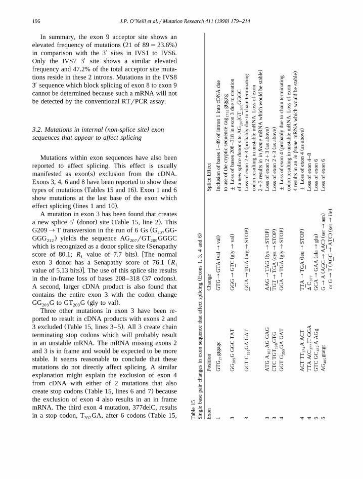
( )3.2. Mutations in internal non-splice site exonsequences that appear to affect splicing
Mutations within exon sequences have also beenreported to affect splicing. This effect is usually
Ž .manifested as exon s exclusion from the cDNA.Exons 3, 4, 6 and 8 have been reported to show these
Ž .types of mutations Tables 15 and 16 . Exon 1 and 6show mutations at the last base of the exon which
Ž .effect splicing lines 1 and 10 .A mutation in exon 3 has been found that creates
X Ž . Ž .a new splice 5 donor site Table 15, line 2 . ThisŽG209™T transversion in the run of 6 Gs G GG-207
.GGG yields the sequence AG rGT GGGC212 207 209Žwhich is recognized as a donor splice site Senapathy
. wscore of 80.1; R value of 7.7 bits . The normaliŽexon 3 donor has a Senapathy score of 76.1 R i
.xvalue of 5.13 bits . The use of this splice site resultsŽ .in the in-frame loss of bases 208–318 37 codons .
A second, larger cDNA product is also found thatcontains the entire exon 3 with the codon change
Ž .GG G to GT G gly to val .209 209
Three other mutations in exon 3 have been re-ported to result in cDNA products with exons 2 and
Ž .3 excluded Table 15, lines 3–5 . All 3 create chainterminating stop codons which will probably resultin an unstable mRNA. The mRNA missing exons 2and 3 is in frame and would be expected to be morestable. It seems reasonable to conclude that thesemutations do not directly affect splicing. A similarexplanation might explain the exclusion of exon 4from cDNA with either of 2 mutations that also
Ž .create stop codons Table 15, lines 6 and 7 becausethe exclusion of exon 4 also results in an in framemRNA. The third exon 4 mutation, 377delC, results
Žin a stop codon, T GA, after 6 codons Table 15,392 Tab
le15
Ž.
Sing
leba
sepa
irch
ange
sin
exon
sequ
ence
that
affe
ctsp
licin
gE
xons
1,3,
4an
d6
Exo
nPo
sitio
nC
hang
eSp
lice
Eff
ect
Ž.
1G
TG
gtga
gcG
TG
™G
TA
val™
val
Incl
usio
nof
base
s1
–49
ofin
tron
1in
tocD
NA
due
27
tous
eof
the
cryp
ticse
quen
ceca
ggt
ggcg
1752
Ž.
3G
GG
GG
CT
AT
GG
G™
GT
Cgl
y™
val
"L
oss
ofba
ses
208–
318
inex
on3
due
tocr
eatio
n20
9
<of
ane
wsp
lice
dono
rsi
teA
GG
TG
GG
C20
720
9Ž
.Ž
3G
CT
CG
AG
AT
GG
A™
TG
Aar
g™
STO
PL
oss
ofex
on2q
3pr
obab
lydu
eto
chai
nte
rmin
atin
g15
1
codo
nre
sulti
ngin
unst
able
mR
NA
.Los
sof
exon
.2q
3re
sults
inin
fram
em
RN
Aw
hich
wou
ldbe
stab
leŽ
.Ž
.3
AT
GA
AG
GA
GA
AG
™T
AG
lys™
STO
PL
oss
ofex
on2q
3as
abov
e16
3
Ž.
Ž.
3C
TC
TG
TG
TG
TG
T™
TG
Acy
s™ST
OP
Los
sof
exon
2q3
asab
ove
198
Ž.
Ž4
GG
TG
GA
GA
TG
GA
™T
GA
gly
™ST
OP
"L
oss
ofex
on4
prob
ably
due
toch
ain
term
inat
ing
355
codo
nre
sulti
ngin
unst
able
mR
NA
.Los
sof
exon
.4
resu
ltsin
anin
fram
em
RN
Aw
hich
wou
ldbe
stab
leŽ
.Ž
.4
AC
TT
TA
AC
TT
TA
™T
GA
leu
™ST
OP
"L
oss
ofex
on4
asab
ove
374
Ž.
4T
TA
AC
TG
GA
DC
Los
sof
exon
4–
837
737
7Ž
.6
GT
CG
CA
AG
gG
CA
™G
AA
ala™
glu
Los
sof
exon
648
2
Ž.Ž
.6
AG
gtat
gtG
™A
AG
C™
AA
Cse
r™as
nL
oss
ofex
on6
485
Ž.Ž
.or
G™
TA
GC
™A
TC
ser™
ile
( )J.P. O’Neill et al.rMutation Research 411 1998 179–214 197
Table 16Ž .Single base pair changes in exon sequence that affect splicing Exon 8
Exon Position Change Splice Effect
Ž .8 GTT G GA TTT GGA™TGA gly™STOP " Loss of exon 8538
Ž .orGGA™AGA gly™arg " Loss of exon 8Ž .8 GTT GG A TTT GGA™GTA gly™val Loss of exon 8539
Ž .8 GTT G AA ATT GAA™AAA glu™ lys Loss of exon 8544
Ž .orGAA™TAA glu™STOP " Loss of exon 8Ž .8 ATT CC A GAC CCA™CTA pro™ leu " Loss of exon 8551
Ž .8 CTT G AC TAT GAC™TAC asp™ tyr Loss of exon 8580
Ž .8 AAT G AA TAC GAA™AAA glu™ lys " Loss of exon 8589
Ž .orGAA™TAA glu™STOP " Loss of exon 8Ž .8 AAT GA A TAC GAA™GTA glu™val " Loss of exon 8590
Ž .8 TAC TTC AGG TTC™TTT asp™val " Loss of exon 8597Ž .8 AGG GA T TTG GAT™GTT asp™val " Loss of exon 8602
.line 8 . However, the reported cDNA is missingŽ .exons 4–8 also in frame and not the simple exon 4
exclusion.Exon 6 contains 2 bases which, when mutated,
result in exon 6 exclusion. Both bases are near theŽ .exon 6 splice donor site Table 15, lines 9 and 10 .
The mutation at 482C does not reside within theconventional splice site, but the effect on splicingimplies that this ‘weak’ 5X site relies on this base aswell. Changing 482C to A does not appear to createa new donor or a hairpin which might be hypothe-sized to effect splicing. The splice donor sequence is
not very close to the consensus sequence with apyrimidine t at IVS6q4 in place of the more com-
Ž .mon purine a or g . G is the consensus base for thelast exonic base in a donor site and 485G is the lastbase of exon 6. G ™A or T splice mutations have485
been reported 3 times each and so this is not a rareevent. Both mutations change the R value from 9.5i
to 6.4 bits which is a significant decrease and theSenapathy score drops from 88.7 to 75.4.
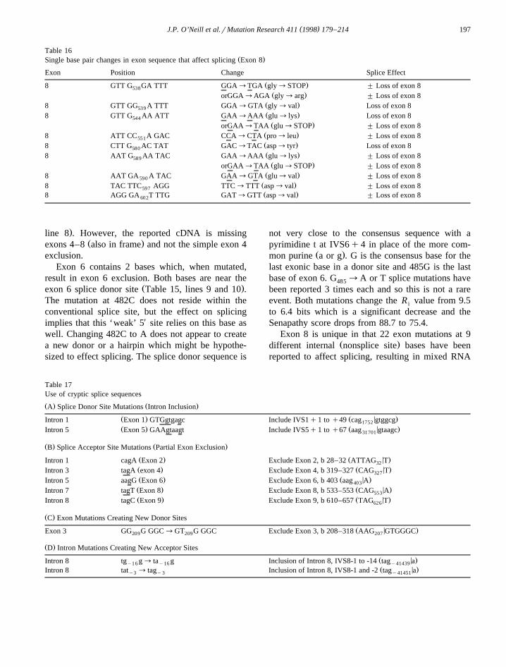
Exon 8 is unique in that 22 exon mutations at 9Ž .different internal nonsplice site bases have been
reported to affect splicing, resulting in mixed RNA
Table 17Use of cryptic splice sequences
Ž . Ž .A Splice Donor Site Mutations Intron Inclusion
Ž . Ž < .Intron 1 Exon 1 GTGgtgagc Include IVS1q1 to q49 cag gtggcg1752
Ž . Ž < .Intron 5 Exon 5 GAAgtaagt Include IVS5q1 to q67 aag gtaagc31701
Ž . Ž .B Splice Acceptor Site Mutations Partial Exon Exclusion
Ž . Ž < .Intron 1 cagA Exon 2 Exclude Exon 2, b 28–32 ATTAG T32
Ž . Ž < .Intron 3 tagA exon 4 Exclude Exon 4, b 319–327 CAG T327
Ž . Ž < .Intron 5 aagG Exon 6 Exclude Exon 6, b 403 aag A403
Ž . Ž < .Intron 7 tagT Exon 8 Exclude Exon 8, b 533–553 CAG A553
Ž . Ž < .Intron 8 tagC Exon 9 Exclude Exon 9, b 610–657 TAG T626
Ž .C Exon Mutations Creating New Donor Sites
Ž < .Exon 3 GG G GGC™GT G GGC Exclude Exon 3, b 208–318 AAG GTGGGC209 209 207
Ž .D Intron Mutations Creating New Acceptor Sites
Ž < .Intron 8 tg g™ ta g Inclusion of Intron 8, IVS8-1 to -14 tag ay1 6 y16 y41439Ž < .Intron 8 tat ™ tag Inclusion of Intron 8, IVS8-1 and -2 tag ay3 y3 y41451
( )J.P. O’Neill et al.rMutation Research 411 1998 179–214198
Ž .species " exon 8 of variable percentages of fullŽ .length and exon 8 exclusion Table 16 . Five muta-
tions each at 538G, 544G and 551C have beenw xreported. Steingrimsdottir et al. 79 have presented a
model of exon 8 secondary structure to explain thisunusual splicing sensitivity. It is possible that otherexon 8 mutations also affect splicing but have notbeen reported because a full length cDNA productwas also obtained and only it was noted.
In summary, mutations in exon sequences havebeen reported to affect splicing in exons 1, 3, 4, 6and 8. In exon 3, a new splice donor site can becreated by a single base change and, in exons 1 and6, mutation of the exon base G adjacent to the splicedonor sequence blocks splicing.
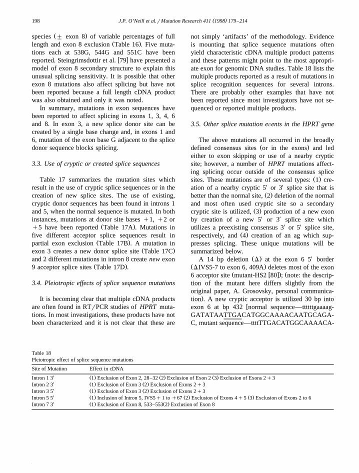
3.3. Use of cryptic or created splice sequences
Table 17 summarizes the mutation sites whichresult in the use of cryptic splice sequences or in thecreation of new splice sites. The use of existing,cryptic donor sequences has been found in introns 1and 5, when the normal sequence is mutated. In bothinstances, mutations at donor site bases q1, q2 or
Ž .q5 have been reported Table 17A . Mutations infive different acceptor splice sequences result in
Ž .partial exon exclusion Table 17B . A mutation inŽ .exon 3 creates a new donor splice site Table 17C
and 2 different mutations in intron 8 create new exonŽ .9 acceptor splice sites Table 17D .
3.4. Pleiotropic effects of splice sequence mutations
It is becoming clear that multiple cDNA productsare often found in RTrPCR studies of HPRT muta-tions. In most investigations, these products have notbeen characterized and it is not clear that these are
not simply ‘artifacts’ of the methodology. Evidenceis mounting that splice sequence mutations oftenyield characteristic cDNA multiple product patternsand these patterns might point to the most appropri-ate exon for genomic DNA studies. Table 18 lists themultiple products reported as a result of mutations insplice recognition sequences for several introns.There are probably other examples that have notbeen reported since most investigators have not se-quenced or reported multiple products.
3.5. Other splice mutation eÕents in the HPRT gene
The above mutations all occurred in the broadlyŽ .defined consensus sites or in the exons and led
either to exon skipping or use of a nearby crypticsite; however, a number of HPRT mutations affect-ing splicing occur outside of the consensus splice
Ž .sites. These mutations are of several types: 1 cre-ation of a nearby cryptic 5X or 3X splice site that is
Ž .better than the normal site, 2 deletion of the normaland most often used cryptic site so a secondary
Ž .cryptic site is utilized, 3 production of a new exonby creation of a new 5X or 3X splice site whichutilizes a preexisting consensus 3X or 5X splice site,
Ž .respectively, and 4 creation of an ag which sup-presses splicing. These unique mutations will besummarized below.
Ž . XA 14 bp deletion D at the exon 6 5 borderŽ .DIVS5-7 to exon 6, 409A deletes most of the exon
Ž w x. Ž6 acceptor site mutant-HS2 80 ; note: the descrip-tion of the mutant here differs slightly from theoriginal paper, A. Grosovsky, personal communica-
.tion . A new cryptic acceptor is utilized 30 bp intowexon 6 at bp 432 normal sequence—ttttttgaaaag-
GATATAATTGACATGGCAAAACAATGCAGA-C, mutant sequence—ttttTTGACATGGCAAAACA-
Table 18Pleiotropic effect of splice sequence mutations
Site of Mutation Effect in cDNAX Ž . Ž . Ž .Intron 1 3 1 Exclusion of Exon 2, 28–32 2 Exclusion of Exon 2 3 Exclusion of Exons 2q3X Ž . Ž .Intron 2 3 1 Exclusion of Exon 3 2 Exclusion of Exons 2q3X Ž . Ž .Intron 3 5 1 Exclusion of Exon 3 2 Exclusion of Exons 2q3X Ž . Ž . Ž .Intron 5 5 1 Inclusion of Intron 5, IVS5q1 to q67 2 Exclusion of Exons 4q5 3 Exclusion of Exons 2 to 6X Ž . Ž .Intron 7 3 1 Exclusion of Exon 8, 533–553 2 Exclusion of Exon 8
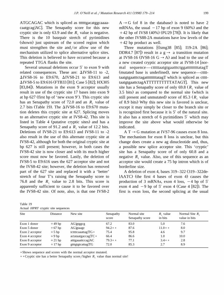
( )J.P. O’Neill et al.rMutation Research 411 1998 179–214 199
ATGCAGAC which is spliced as ttttttgacatggcaaaa-< xcaatgcag AC . The Senapathy score for this new
cryptic site is only 63.9 and the R value is negative.i
There is the 10 basepair stretch of pyrimidinesŽ .shown just upstream of the scored region whichmust strengthen the site andror allow use of themechanism utilized to splice alternative splice sites.This deletion is believed to have occurred because arepeated TTGA flanks the site.
Several unusual deletions occur 5X to exon 9 withrelated consequences. These are: DIVS8-11 to -2,DIVS8-16 to E9:670, DIVS8-21 to E9:613 and
Ž w x w xDIVS8-5 to E9:616 FTB33 81 ; Case 5 82 ; HX305w x.83,84 . Mutations in the exon 9 acceptor usuallyresult in use of the cryptic site 17 bases into exon 9
Ž .at bp 627 first bp of ‘new exon 9’ . This cryptic sitehas an Senapathy score of 72.0 and an R value ofi
Ž .2.7 bits Table 19 . The DIVS8-16 to E9:670 muta-tion deletes this cryptic site at 627. Splicing movesto an alternative cryptic site at IVS8-42. This site is
Ž .listed in Table 4 putative cryptic sites and has aSenapathy score of 91.2 and a R value of 12.2 bits.i
Deletions of IVS8-21 to E9:613 and IVS8-11 to -2also result in the use of this alternate cryptic site atIVS8-42, although for both the original cryptic site atbp 627 is still present; however, in both cases theIVS8-42 site is now closer and with its much higherscore must now be favored. Lastly, the deletion ofIVS8-5 to E9:616 uses the 627 acceptor site and notthe IVS8-42 site; however, the deletion has removedpart of the 627 site and replaced it with a ‘better’stretch of four T’s raising the Senapathy score to76.8 and the R value to 2.8 bits. This score isi
apparently sufficient to cause it to be favored overthe IVS8-42 site. Of note, also, is that one IVS8-2
Ž .A™G of 8 in the database is noted to have 2Ž .mRNAs, the usual y17 bp of exon 9 60% and the
Ž . Ž w x.q42 bp of IVS8 40% PU29 78 . It is likely thatthe other IVS88-2A mutations have low levels of theq42 bp product as well.
wŽ w x w xThree mutations lsmg38 85 ; I19-2A 86 ;w x.DDR4.7 87 result in a g™ a transition mutation
Ž .at IVS8-16 IVS8-16 G™A and lead to the use ofwa new created cryptic acceptor site at IVS8-14 nor-
mal sequence— ctttttaatgtgaatttctggatttttttttatagTŽ .mutated base is underlined , new sequence—ctttt-taatgtgaatttctagatttttttttatagT which is spliced as ctttt-
xtaatgtgaattctagATTTTTTTTTTATAGT . This newŽsite has a Senapathy score of only 69.8 R value ofi
. Ž3.5 bits as compared to the normal site which is. Žstill present and unmutated score of 85.3 R valuei
.of 8.9 bits Why this new site is favored is unclear,except it may simply be closer to the branch site oris recognized first because it is 5X of the natural site.It also has a stretch of 6 pyrimidines 5X which mayimprove the site above what would otherwise beindicated.
A T™G mutation at IVS7-96 causes exon 8 loss.The mechanism for exon 8 loss is unclear, but thischange does create a new ag dinucleotide and, thus,a possible new splice acceptor site. This ‘cryptic’site has a Senapathy score of of only 60.8 and anegative R value. Also, use of this sequence as ani
acceptor site would create a 75 bp intron which is ofborderline size.
ŽA deletion of exon 4, bases 319–322 319–322de-. Ž .lAATC the first 4 bases of exon 4 causes the
production of 3 mRNAs, exon 4 loss, y4 bp of 5X
X Ž w x.exon 4 and y9 bp of 5 exon 4 Case 4 82 . Thefirst is exon loss, the second splicing at the usual
Table 19Actual HPRT cryptic site sequences
Site Distance New site Senapathy Normal site R value Normal Site Ri i
score Senapathy score in bits value in bits
<Exon 1 donor q49 bp AG gtggcg 67.2 83.0 5.0 7.6<Exon 5 donor q67 bp AG gtaagc 94.2)) 87.6 11.0)) 8.0
<Exon 2 acceptor q5 bp tcttttcaaattag TG) 75.4 95.8 4.6 9.7<Exon 4 acceptor q9 bp actataatgaccag TC) 66.4 86.6 1.0 10.0
<Exon 8 acceptor q21 bp atttgaaattccag AC 79.3)) 77.1 3.4)) 2.8<Exon 9 acceptor q17 bp gtttgtgtcattag TG 72.0 85.3 2.7 8.9
)Shows sequence and scores with the normal acceptor mutated.))Cryptic site has a better Senapathy scorerhigher R value than normal site!i
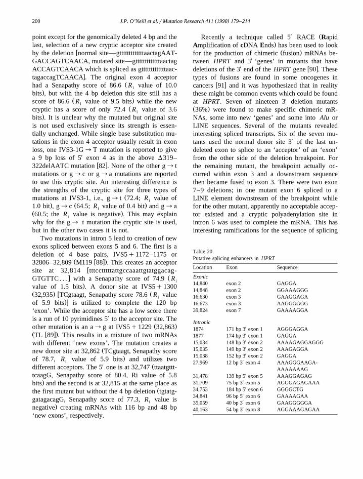
( )J.P. O’Neill et al.rMutation Research 411 1998 179–214200
point except for the genomically deleted 4 bp and thelast, selection of a new cryptic acceptor site created
wby the deletion normal site—gtttttttttttttaactagAAT-GACCAGTCAACA, mutated site—gtttttttttttttaactagACCAGTCAACA which is spliced as gtttttttttttttaac-
xtagaccagTCAACA . The original exon 4 acceptorŽhad a Senapathy score of 86.6 R value of 10.0i
.bits , but with the 4 bp deletion this site still has aŽ .score of 86.6 R value of 9.5 bits while the newi
Žcryptic has a score of only 72.4 R value of 3.6i.bits . It is unclear why the mutated but original site
is not used exclusively since its strength is essen-tially unchanged. While single base substitution mu-tations in the exon 4 acceptor usually result in exonloss, one IVS3-1G™T mutation is reported to givea 9 bp loss of 5X exon 4 as in the above D319–
w x322delAATC mutation 82 . None of the other g™ tmutations or g™c or g™a mutations are reportedto use this cryptic site. An interesting difference isthe strengths of the cryptic site for three types of
Žmutations at IVS3-1, i.e., g™ t 72.4; R value ofi. Ž .1.0 bit , g™c 64.5; R value of 0.4 bit and g™ai
Ž .60.5; the R value is negative . This may explaini
why for the g™ t mutation the cryptic site is used,but in the other two cases it is not.
Two mutations in intron 5 lead to creation of newexons spliced between exons 5 and 6. The first is adeletion of 4 base pairs, IVS5q1172–1175 or
Ž w x.32806–32,809 M119 88 . This creates an acceptorwsite at 32,814 tttcctttttattgccaaattgtatggacag-
x ŽGTGTTC . . . with a Senapathy score of 74.9 R i.value of 1.5 bits . A donor site at IVS5q1300
Ž . w Ž32,935 TCgtaagt, Senapathy score 78.6 R valuei.xof 5.9 bits is utilized to complete the 120 bp
‘exon’. While the acceptor site has a low score thereis a run of 10 pyrimidines 5X to the acceptor site. The
Ž .other mutation is an a™g at IVS5q1229 32,863Ž w x.TL 89 . This results in a mixture of two mRNAswith different ‘new exons’. The mutation creates a
Žnew donor site at 32,862 TCgtaagt, Senapathy score.of 78.7, R value of 5.9 bits and utilizes twoi
X Ždifferent acceptors. The 5 one is at 32,747 ttaatgttt-tcaagG, Senapathy score of 80.4, Ri value of 5.8
.bits and the second is at 32,815 at the same place asŽthe first mutant but without the 4 bp deletion tgtatg-
gatagacagG, Senapathy score of 77.3, R value isi.negative creating mRNAs with 116 bp and 48 bp
‘new exons’, respectively.
X ŽRecently a technique called 5 RACE Rapid.Amplification of cDNA Ends has been used to look
Ž .for the production of chimeric fusion mRNAs be-tween HPRT and 3X ‘genes’ in mutants that have
X w xdeletions of the 3 end of the HPRT gene 90 . Thesetypes of fusions are found in some oncogenes in
w xcancers 91 and it was hypothesized that in realitythese might be common events which could be foundat HPRT. Seven of nineteen 3X deletion mutantsŽ .36% were found to make specific chimeric mR-NAs, some into new ‘genes’ and some into Alu orLINE sequences. Several of the mutants revealedinteresting spliced transcripts. Six of the seven mu-tants used the normal donor site 3X of the last un-deleted exon to splice to an ‘acceptor’ of an ‘exon’from the other side of the deletion breakpoint. Forthe remaining mutant, the breakpoint actually oc-curred within exon 3 and a downstream sequencethen became fused to exon 3. There were two exon7–9 deletions; in one mutant exon 6 spliced to aLINE element downstream of the breakpoint whilefor the other mutant, apparently no acceptable accep-tor existed and a cryptic polyadenylation site inintron 6 was used to complete the mRNA. This hasinteresting ramifications for the sequence of splicing
Table 20Putative splicing enhancers in HPRT
Location Exon Sequence
Exonic14,840 exon 2 GAGGA14,848 exon 2 GGAAAGGG16,630 exon 3 GAAGGAGA16,673 exon 3 AAGGGGGG39,824 exon 7 GAAAAGGA
IntronicX1874 171 bp 3 exon 1 AGGGAGGAX1877 174 bp 3 exon 1 GAGGAX15,034 148 bp 3 exon 2 AAAAGAGGAGGGX15,035 149 bp 3 exon 2 AAAGAGGAX15,038 152 bp 3 exon 2 GAGGA
X27,969 12 bp 3 exon 4 AAAGGGAAGA-AAAAAAAG
X31,478 139 bp 5 exon 5 AAAGGAGAGX31,709 75 bp 3 exon 5 AGGGAGAGAAA
X34,753 184 bp 5 exon 6 GGGGCTGX34,841 96 bp 5 exon 6 GAAAAGAAX35,059 40 bp 3 exon 6 GAAGGGGGAX40,163 54 bp 3 exon 8 AGGAAAGAGAA
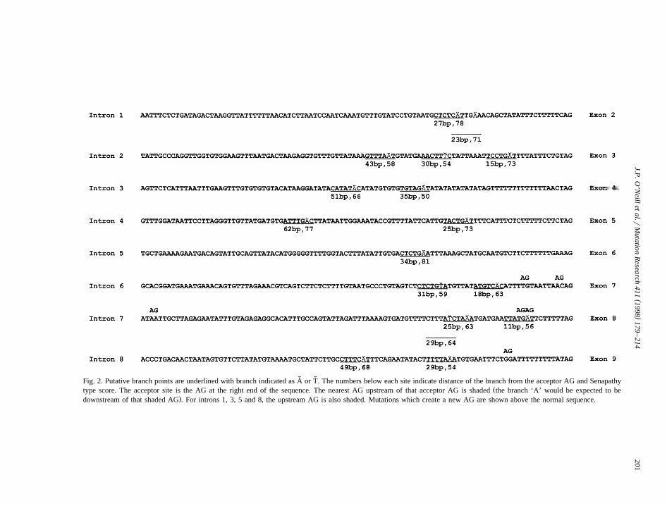
()
J.P.O
’Neillet
al.rM
utationR
esearch411
1998179
–214
201
˘ ˇFig. 2. Putative branch points are underlined with branch indicated as A or T. The numbers below each site indicate distance of the branch from the acceptor AG and SenapathyŽtype score. The acceptor site is the AG at the right end of the sequence. The nearest AG upstream of that acceptor AG is shaded the branch ‘A’ would be expected to be
.downstream of that shaded AG . For introns 1, 3, 5 and 8, the upstream AG is also shaded. Mutations which create a new AG are shown above the normal sequence.
( )J.P. O’Neill et al.rMutation Research 411 1998 179–214202
and polyadenylation and recognition of the last exon.Splicing must occur first or otherwise HPRTmRNAs would always end at exon 6. However, ifthat intron 6 polyadenylation site is quickly splicedout, polyadenylation cannot occur. Only when adownstream acceptor is not found and no splicingoccurs, does polyadenylation occur in intron 6. Forone mutant, it could be shown that exon 8 wasspliced to a 117 bp sequence located 10kb 3X to exon9 in the wildtype HPRT gene. There is an excellent
X Žacceptor on the 5 end of the sequence ttttttttttttttt-.tgtagA, Senapathy score 88.2, R value of 16.4 bitsi
X Žand a good donor on the 3 end TGgtaact, Senapa-.thy score 77.9, R value of 6.0 bits which spliced ini
turn to a downstream microsatellite repeat.
3.6. Other aspects of HPRT splice recognition
The HPRT gene was scanned for the enhancersshown in Table 1 as well as a generic eight purine
Ž .stretch most enhancers are purine-rich . Table 20shows a partial listing of those results. Since mostenhancers are exonic, all the exonic enhancer-likesequences are listed first. Sequences were found inexon 2, exon 3 and exon 7. In the bottom half of thetable are shown enhancers-like sequences within 200bp of the exons. In their original descriptions, the
w x w xTGAATG 26 and GGGGCTG 27 enhancers wererepeated; although these sequences were found inHPRT there were only single copies near any exon.There are no potential enhancer elements near exon8 which appears to be the exon most susceptible tomutation resulting in splicing errors. Enhancerswould also be expected near the ‘too small’, 18basepair exon 5 and there are several nearby, al-though none is in exon 5 itself. Exon 5 itself is in
Ž .fact fairly T rich AATGTCTTGATTGTGGAA andhas no run of purines greater than 4 bases.
The branch sites for HPRT have not been experi-mentally determined; however, based on the consen-
Ž .sus sequence tnctray or ctray , educated conjecturesas to the positions of branch points can be made. Fig.
Ž2 gives the 100 bases upstream of each exon theconsensus acceptor ag dinucleotide is the last 2 bases
.shown . Putative branch points are underlined withtheir distance from the exon shown as well as a
w xSenapathy type score 31 based on the frequenciesof nucleotides at the different positions for the con-
stitutive branch sites in Table 2. Good putative branchsites are found for exon 2, exon 5, exon 6 and
Žpossibly exon 3 this site is only 15 bases away,.however . Only poor sites are found for exons 7 and
Ž .9 scores-70 . The exon 4 branch site is especiallyunusual. There is a pyrimidine stretch of 13 t’s
Žimmediately preceded by an ag dinucleotide ags arethought to be rare within 10–20 bases of an accep-
.tor . Upstream of that are two stretches of 8–9 atdinucleotides with 4 gt dinucleotides in the middle;this could form a hairpin. The presence of the agdinucleotide suggests that the branch should be veryclose upstream but this would place it in the hairpinin a low score region of ats. For introns 1, 3, 5 and 8,the ag upstream of the putative branch points is thesecond ag upstream of the acceptor ag. In intron 6, 7and 8, mutations that create new ag dinucleotides
Ž .either cause exon exclusion intron 6 and 7 or createa new splice acceptor site resulting in intron inclu-
Ž .sion intron 8 .
4. Discussion
Several conclusions can be drawn from study ofHPRT splicing mutations. The first is that the HPRTsplice sites often show large deviations from theconsensus sequences especially in the first and lastbases of the exons. This probably reflects the lowerinformation content contributed by these bases. Ac-cording to the consensus sequence, the first and lastbases of the exon should be G. However, only exons
Ž X3 and 6 have a G as the first exonic base 3 splice.site and for both of these, mutations at the G are
missense mutations, with no reported effect on splic-ing. In the donor splice sites, only 4 of the 8 sitesŽ .exons 1, 2, 4, 6 have a consensus G as the last baseof the exon. Exon 1 has one reported mutation at thelast base G which because it is 3rd position wobble
Ž .causes no amino acid change Val™Val but doesaffect splicing. Exon 2 has missense mutations atthat site that do not affect splicing; this is notsurprising as the Senapathy score remains a high
Ž .86.7 R value of 9.7 or 9.4 even with a mutation ati
the G. No mutations, either splice mutations or mis-Ž . Žsense, have been reported at 384G exon 4 . The
exon 4 G is a partial wobble, a 384G™C or Twould change lysine to asparagine but 384G™A

( )J.P. O’Neill et al.rMutation Research 411 1998 179–214 203
.remains lysine . Mutation at the last base of exon 6cause splicing changes; of note, there is also a reportthat mutation three bases into the exon also causesexon 6 loss. Thus, exon 6 is unique in that severalbases at the 3X end of the exon appears critical forsplicing. Table 21 summarizes the Senapathy scores
Žand R values for observed mutations splice andi.non-splice at the exonic consensus G’s. While all
mutations reduce scores, there are some ‘interesting’observations. The exon 1 donor G™A mutationreduces the Senapathy score to 70.6 and a R valuei
of 4.6 bits, while the cryptic has a Senapathy scoreof 67.2 and a R value of 5.0 bits. The Senapathyi
score would predict that splicing would not be af-fected while information theory would predict thatthe cryptic site would be utilized which is what isobserved. Thus, information theory more accuratelypredicts the result here. Of special note, the scoresand mutations are identical for the exon 4 and exon 6donors, but only for the exon 6 donor has a splicemutation been observed at the exonic G. The reasonfor this is unclear but may be due to exon 4’sslightly better acceptor or to enhancer sequences orsimply to chance.
Fig. 3 summarizes all the simple base changes inthe consensus donor and acceptor sites. The relativefrequencies at the various bases should be takenlightly as these mutations include both spontaneous
and mutagen induced; i.e., some mutagens may have‘hotspots’ that will skew the frequencies. However,from this figure, it can be seen at which basesmutation affects splicing and which probably do not.Obviously, saturation has not been reached as one
Ž .invariant acceptor a exon 3; IVS2-2A , one invari-Ž .ant acceptor a exon 6; IVS5-2A and one invariant
Ž .donor t exon 3; IVS3q2G have not been foundmutated. Considering only the ‘spontaneous’ muta-tions, the frequencies of base changes at the four
Žinvariant sites 2 G:C basepairs and 2 A:T basepairs,.45 mutations are: 7% G:C™C:G, 44% G:C™A:T,
11% G:C™T:A, 15% A:T™T:A, 4% A:T™C:G,and 18% A:T™G:C.
We found that in our 201 simple splice sitemutations that 56% were in the donor sites and 44%in the acceptor sites. Other compilations of splicesites have found much higher frequencies of donor
w x w x w xmutations, e.g., 79% 39 , 68% 16 , and 70% 63 .The high frequency of acceptor site mutations atHPRT is predominantly due to the high number of
Žexon 8 and exon 9 acceptor mutations 22 and 21.mutations, respectively .
The information theory model predicts that accep-tors are recognized first and that cryptic acceptors
Žeither preexisting which are only used if the normal.site is mutated or those created by mutation can be
either upstream or downstream of the the normal
Table 21Ž .Mutations in exonic consensus G’s last and first base of the exon
Site Normal Senapathy R value Mutation) Senapathy R value Resultsi i
in bits in bits
Ex 1 donor TG gtgagc 83.0 7.6 TA gtgagc 70.6 4.6 no effect on splicingw xno use of cryptic score 67.2, 5.0 bits
TT gtgagc 70.6 4.3 no effect on splicingw xno use of cryptic score 67.2, 5.0 bits
Ex 2 donor AG gtaagt 100.0 12.8 AA gtaagt 86.7 9.7 no effect on splicingAT gtaagt 86.7 9.4 no effect on splicing
Ex 4 donor AG gtatgt 88.7 9.5 AA gtatgt 75.4 6.4 no effect on splicingAT gtatgt 75.4 6.4 no effect on splicing
Ex 6 donor AG gtatgt 88.7 9.5 AT gtatgt)) 75.4 6.4 ex 6 skipAA gtatgt)) 75.4 6.4 ex 6 skip
Ex 3 acceptor ttttatttctgtag G 89.8 11.3 ttttatttctgtag A 86.4 10.3 no effect on splicingEx 6 acceptor ttcttttttgaaag G 86.4 7.1 ttcttttttgaaag A 83.0 6.2 no effect on splicing
ttcttttttgaaag T 80.1 4.9 no effect on splicingttcttttttgaaag C 80.6 5.2 no effect on splicing
)Mutated base is underlined and change is shown.))The only mutations which do affect splicing.
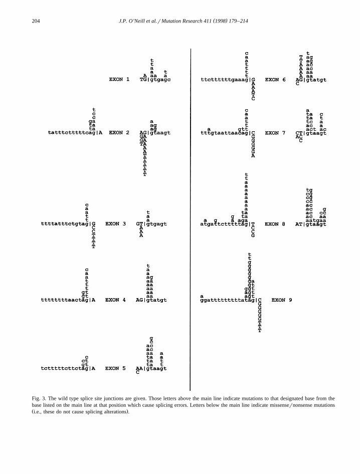
( )J.P. O’Neill et al.rMutation Research 411 1998 179–214204
Fig. 3. The wild type splice site junctions are given. Those letters above the main line indicate mutations to that designated base from thebase listed on the main line at that position which cause splicing errors. Letters below the main line indicate missensernonsense mutationsŽ .i.e., these do not cause splicing alterations .
( )J.P. O’Neill et al.rMutation Research 411 1998 179–214 205
site. Scanning then proceeds downstream through theexon to find a donor site. Preexisting cryptic donorsused after mutation of the normal site must thus bedownstream of the normal site while newly createddonors must be upstream of the normal donor site.Human HPRT mutations support this model. The
Ž .preexisting cryptic donor sites exons 1 and 5 areboth downstream of the normal donor while the
Ž .created donor site exon 3 is upstream. HPRT cryp-tic acceptors are both upstream and downstreamalthough all of the preexisting cryptics are down-stream and the newly created cryptics are upstream.This may be coincidence as both preexisting andcreated cryptic acceptors occur both upstream and
w xdownstream in other genes 39 .All 8 intron 3X splice sequences have shown muta-
Žtions in the invariant base IVS-1G and 6 of 8 excep-.tions are introns 2 and 5 in the invariant base
Ž .IVS-2A. Three introns 6, 7 and 8 show mutationsin base IVS-3 also. Two introns have the more
Ž .prevalent c at IVS-3 introns 1 and 6 . Mutation hasbeen observed only at the intron 6 site. The intron 7and 8 IVS-3 mutations change the lower consensus tto an a or g. No mutations were found in base IVS-4as expected from the consensus sequence hypothesisof no specific base preference at this site. In exons 3and 6, which have a G as the first base, g to a
Žmutations have been reported in IVS-1 exon 3,.tagG™ taaG and exon 6, aagG™aaaG . However,
this potential new splice acceptor site is used only inexon 6 and not in exon 3.
The polypyrimidine tract residing in bases y5 toŽ .y23 shown in Fig. 2 is fairly variable among the 8
introns. Introns range from 94.7% polypyrimidineŽ . Ž .intron 4 to 57.9% polypyrimidine introns 6 and 7in this 19 base stretch. As discussed in the introduc-tion, ag’s are rare in this region as presumably theybecome putative acceptors. Only introns 1 and 3
Žnormally contain an ag dinucleotide in this stretch at.IVS2-18 and IVS3-20, respectively ; possibly these
are too close to the branch ‘a’ to be utilized. Thecreation of an ag dinucleotide within the normalsplice site causes splicing errors in introns 6, 7 and8. In introns 6 and 7, the exon is excluded, whileonly in intron 8 is this new ag sequence used as a
Žsplice acceptor site. See Table 22 for a summary of.Senapathy splice site scores and R values. Bothi
IVS8-3T™G and IVS8-16G™A cause utilization
of the new ag as a new cryptic acceptor site. TheIVS8-3 mutation yields reasonable new R valuesi
although the original site is still better. Use of theIVS-14 site is surprising based on its poor values;however, if exon 9 were skipped, no mRNA wouldhave been detected; we may be detecting only aminor product which utilizes the cryptic site. Thatthe other mutant ag’s in introns 6 and 7 are not usedas acceptors is not surprising based on their values.Somehow these new ag dinucleotides in the otherintrons must interfere with splicing so the wholeexon is skipped. The other 5 introns contain at least16 possible sites to create a new ag dinucleotide,while introns 6, 7 and 8 contain at least 11 othersites. The sensitivity of these sequences to mutationmay be determined as additional splice mutations aredefined.
For exon 8, mutations also occur at IVS7-5T™GŽ .or A and IVS7-3T™A Table 13 . The scores for
the exon 8 acceptor site are by far the lowest of allthe HPRT acceptor sites. Loss of either of these t’slowers the values considerably which appears to besufficient to cause bypass of the exon. The two othermutations mentioned above at IVS7-10 and IVS7-13also cause loss of t’s and would reduce values
Ž .similarly see Table 22 , thus, they both create agdinucleotides and reduce values.
The intron 5X splice site sequences all fit aŽ .gt arg agt consensus sequence, except for intron 1
Ž . Ž . Ž .gtgagc , intron 4 gtatgt and intron 6 gtatgt . Muta-tions in all 8 intron sequences have been found in the
Ž .invariant base q1 g; IVSq1G and in 7 of the 8Ž .introns in the invariant base q2 t; IVSq2T .
ŽMutations in intron 1, 5, 7 and 8 at base q5 g;.IVSq5G have also been found. Three splice sites
have mutations of their a at IVSq3 and 2 havemutations of their t at IVSq6. Only exon 8 has amutation at IVSq4 at its a although 5 other exonshave a at this position. Tables 23 and 24 show themutations that have and that have not been observed,respectively, in the IVSq3 to IVSq6 positions. Ingeneral, the IVS bases where mutation has beenobserved to occur create sites with lower values thanwhat would occur at the bases where no mutationhas been seen. For example, changes in IVS1 wouldstill have very good scores and are not observed asmight be expected. However, IVS2 changes wouldlower values significantly but have not been ob-
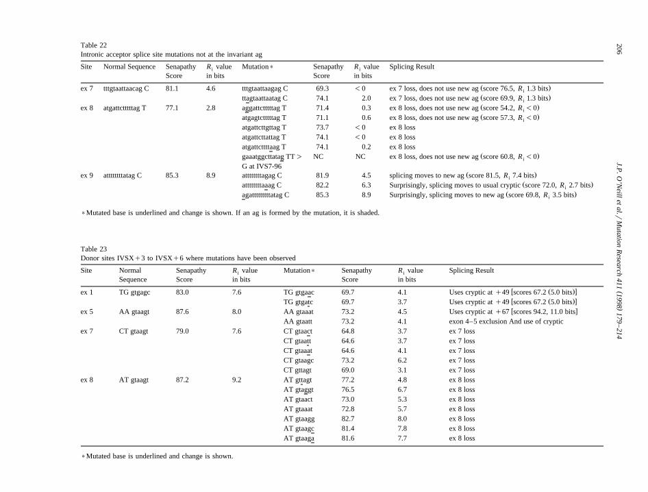
()
J.P.O
’Neillet
al.rM
utationR
esearch411
1998179
–214
206Table 22Intronic acceptor splice site mutations not at the invariant ag
Site Normal Sequence Senapathy R value Mutation) Senapathy R value Splicing Resulti i
Score in bits Score in bits
Ž .ex 7 tttgtaattaacag C 81.1 4.6 tttgtaattaagag C 69.3 -0 ex 7 loss, does not use new ag score 76.5, R 1.3 bitsi
Ž .ttagtaattaatag C 74.1 2.0 ex 7 loss, does not use new ag score 69.9, R 1.3 bitsi
Ž .ex 8 atgattctttttag T 77.1 2.8 aggattctttttag T 71.4 0.3 ex 8 loss, does not use new ag score 54.2, R -0i
Ž .atgagtctttttag T 71.1 0.6 ex 8 loss, does not use new ag score 57.3, R -0i
atgattcttgttag T 73.7 -0 ex 8 lossatgattcttattag T 74.1 -0 ex 8 lossatgattcttttaag T 74.1 0.2 ex 8 loss
Ž .gaaatggcttatag TT) NC NC ex 8 loss, does not use new ag score 60.8, R -0i
G at IVS7-96Ž .ex 9 attttttttatag C 85.3 8.9 attttttttagag C 81.9 4.5 splicing moves to new ag score 81.5, R 7.4 bitsi
Ž .attttttttaaag C 82.2 6.3 Surprisingly, splicing moves to usual cryptic score 72.0, R 2.7 bitsi
Ž .agatttttttttatag C 85.3 8.9 Surprisingly, splicing moves to new ag score 69.8, R 3.5 bitsi
)Mutated base is underlined and change is shown. If an ag is formed by the mutation, it is shaded.
Table 23Donor sites IVSXq3 to IVSXq6 where mutations have been observed
Site Normal Senapathy R value Mutation) Senapathy R value Splicing Resulti i
Sequence Score in bits Score in bits
w Ž .xex 1 TG gtgagc 83.0 7.6 TG gtgaac 69.7 4.1 Uses cryptic at q49 scores 67.2 5.0 bitsw Ž .xTG gtgatc 69.7 3.7 Uses cryptic at q49 scores 67.2 5.0 bitsw xex 5 AA gtaagt 87.6 8.0 AA gtaaat 73.2 4.5 Uses cryptic at q67 scores 94.2, 11.0 bits
AA gtaatt 73.2 4.1 exon 4–5 exclusion And use of crypticex 7 CT gtaagt 79.0 7.6 CT gtaact 64.8 3.7 ex 7 loss
CT gtaatt 64.6 3.7 ex 7 lossCT gtaaat 64.6 4.1 ex 7 lossCT gtaagc 73.2 6.2 ex 7 lossCT gttagt 69.0 3.1 ex 7 loss
ex 8 AT gtaagt 87.2 9.2 AT gttagt 77.2 4.8 ex 8 lossAT gtaggt 76.5 6.7 ex 8 lossAT gtaact 73.0 5.3 ex 8 lossAT gtaaat 72.8 5.7 ex 8 lossAT gtaagg 82.7 8.0 ex 8 lossAT gtaagc 81.4 7.8 ex 8 lossAT gtaaga 81.6 7.7 ex 8 loss
)Mutated base is underlined and change is shown.

( )J.P. O’Neill et al.rMutation Research 411 1998 179–214 207
Table 24Donor sites IVSXq3 to IVSXq6 where mutations have not been observed to date
Site Normal Senapathy Mutation site Senapathy Score withŽ .Score underlined worst base at site
ex 1 TG gtgagc 83.0 TG gtgagc 76.3TG gtgagc 71.5TG gtgagc ))
ex 2 AG gtaagt 100.0 AG gtaagt 90.0AG gtaagt 88.5AG gtaagt 85.6AG gtaagt 94.2
ex 3 GT gtgagt 76.1 GT gtgagt 69.3GT gtgagt 64.6GT gtgagt 61.7GT gtgagt 70.3
ex 4 AG gtatgt 88.7 AG gtatgt 78.7AG gtatgt ))
AG gtatgt 74.3AG gtatgt 82.9
ex 5 AA gtaagt 87.6 AA gtaagt 77.6AA gtaagt 76.1AA gtaagt 81.8
ex 6 AG gtatgt 88.7 AG gtatgt 78.7AG gtatgt ))
AG gtatgt 74.3AG gtatgt 82.9
ex 7 CT gtaagt 79.0 CT gtaagt 67.5
))All mutations at that site improve or do not change the score—therefore, it would not be expected to find mutations.
Žserved see further discussion on exon 3 splice site.mutations below . Of note also is that for mutations
at IV1:q5, the values are above that of the crypticbut the cryptic is utilized. Two observations shouldbe noted: first, that mutation at some bases baseswhere a non-consensus nucleotide is present canactually improve values and second, that just becausea mutation has not yet been observed at a positiondoes not mean that one will not be seen in the future.
Exon 3 is especially intriguing in that only muta-tions at the invariant q1g are known; this is surpris-ing in light of the fact that its Senapathy score andR value is so low. While this paper summarizes 277i
splicing mutants; there are an additional 341 exonexclusion mutations in the database where the ge-nomic mutation has not been determined. These
Ž .include 26 7.6% exon 3 exclusion mutations whichŽcould contain missing IVS3q2–5 mutations some.IVS3q1 mutations cause exon 2–3 exclusion , this
percentage is still lower than expected. For example,
there are 27 exon 2 exclusion and 58 exon 4 exclu-sion mutants in the uncharacterized data set.
Mutations have been observed at all 6 intronicdonor positions for exon 8. This is interesting in that,unlike its acceptor site, this donor site is quite strongŽ .Senapathy score 87.2, R value of 14.7 bits . Exoni
8 is also unique in that there are a number ofmissense mutations within the exon that cause eitherexon 8 loss or mixed cDNAs of exon 8 loss and full
w xlength. Steingrimsdottir et al. 79 hypothesized ahairpin structure for exon 8 and that these exonicmissense mutations disrupted this hairpin. However,the placement of the mutated bases from all thedatabase mutations does not clearly support thatmodel, although the unusual mutations observed inand around exon 8 do suggest that DNA secondarystructure may be important for splicing. Some addi-tional unusual mutations have also been found in thisexon. A deletion of IVS7-45 to -22 causes loss ofexon 8 from the mRNA. This is outside of the
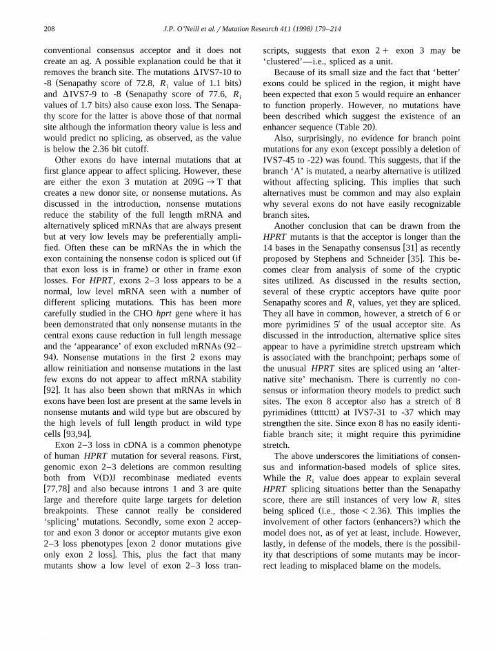
( )J.P. O’Neill et al.rMutation Research 411 1998 179–214208
conventional consensus acceptor and it does notcreate an ag. A possible explanation could be that itremoves the branch site. The mutations DIVS7-10 to
Ž .-8 Senapathy score of 72.8, R value of 1.1 bitsiŽand DIVS7-9 to -8 Senapathy score of 77.6, R i
.values of 1.7 bits also cause exon loss. The Senapa-thy score for the latter is above those of that normalsite although the information theory value is less andwould predict no splicing, as observed, as the valueis below the 2.36 bit cutoff.
Other exons do have internal mutations that atfirst glance appear to affect splicing. However, theseare either the exon 3 mutation at 209G™T thatcreates a new donor site, or nonsense mutations. Asdiscussed in the introduction, nonsense mutationsreduce the stability of the full length mRNA andalternatively spliced mRNAs that are always presentbut at very low levels may be preferentially ampli-fied. Often these can be mRNAs the in which the
Žexon containing the nonsense codon is spliced out if.that exon loss is in frame or other in frame exon
losses. For HPRT , exons 2–3 loss appears to be anormal, low level mRNA seen with a number ofdifferent splicing mutations. This has been morecarefully studied in the CHO hprt gene where it hasbeen demonstrated that only nonsense mutants in thecentral exons cause reduction in full length message
Žand the ‘appearance’ of exon excluded mRNAs 92–.94 . Nonsense mutations in the first 2 exons may
allow reinitiation and nonsense mutations in the lastfew exons do not appear to affect mRNA stabilityw x92 . It has also been shown that mRNAs in whichexons have been lost are present at the same levels innonsense mutants and wild type but are obscured bythe high levels of full length product in wild type
w xcells 93,94 .Exon 2–3 loss in cDNA is a common phenotype
of human HPRT mutation for several reasons. First,genomic exon 2–3 deletions are common resulting
Ž .both from V D J recombinase mediated eventsw x77,78 and also because introns 1 and 3 are quitelarge and therefore quite large targets for deletionbreakpoints. These cannot really be considered‘splicing’ mutations. Secondly, some exon 2 accep-tor and exon 3 donor or acceptor mutants give exon
w2–3 loss phenotypes exon 2 donor mutations givexonly exon 2 loss . This, plus the fact that many
mutants show a low level of exon 2–3 loss tran-
scripts, suggests that exon 2q exon 3 may be‘clustered’—i.e., spliced as a unit.
Because of its small size and the fact that ‘better’exons could be spliced in the region, it might havebeen expected that exon 5 would require an enhancerto function properly. However, no mutations havebeen described which suggest the existence of an
Ž .enhancer sequence Table 20 .Also, surprisingly, no evidence for branch point
Žmutations for any exon except possibly a deletion of.IVS7-45 to -22 was found. This suggests, that if the
branch ‘A’ is mutated, a nearby alternative is utilizedwithout affecting splicing. This implies that suchalternatives must be common and may also explainwhy several exons do not have easily recognizablebranch sites.
Another conclusion that can be drawn from theHPRT mutants is that the acceptor is longer than the
w x14 bases in the Senapathy consensus 31 as recentlyw xproposed by Stephens and Schneider 35 . This be-
comes clear from analysis of some of the crypticsites utilized. As discussed in the results section,several of these cryptic acceptors have quite poorSenapathy scores and R values, yet they are spliced.i
They all have in common, however, a stretch of 6 ormore pyrimidines 5X of the usual acceptor site. Asdiscussed in the introduction, alternative splice sitesappear to have a pyrimidine stretch upstream whichis associated with the branchpoint; perhaps some ofthe unusual HPRT sites are spliced using an ‘alter-native site’ mechanism. There is currently no con-sensus or information theory models to predict suchsites. The exon 8 acceptor also has a stretch of 8
Ž .pyrimidines ttttcttt at IVS7-31 to -37 which maystrengthen the site. Since exon 8 has no easily identi-fiable branch site; it might require this pyrimidinestretch.
The above underscores the limitiations of consen-sus and information-based models of splice sites.While the R value does appear to explain severali
HPRT splicing situations better than the Senapathyscore, there are still instances of very low R sitesi
Ž .being spliced i.e., those-2.36 . This implies theŽ .involvement of other factors enhancers? which the
model does not, as of yet at least, include. However,lastly, in defense of the models, there is the possibil-ity that descriptions of some mutants may be incor-rect leading to misplaced blame on the models.
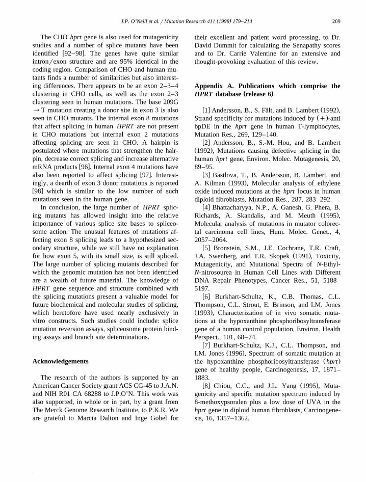
( )J.P. O’Neill et al.rMutation Research 411 1998 179–214 209
The CHO hprt gene is also used for mutagenicitystudies and a number of splice mutants have been
w xidentified 92–98 . The genes have quite similarintronrexon structure and are 95% identical in thecoding region. Comparison of CHO and human mu-tants finds a number of similarities but also interest-ing differences. There appears to be an exon 2–3–4clustering in CHO cells, as well as the exon 2–3clustering seen in human mutations. The base 209G™T mutation creating a donor site in exon 3 is alsoseen in CHO mutants. The internal exon 8 mutationsthat affect splicing in human HPRT are not presentin CHO mutations but internal exon 2 mutationsaffecting splicing are seen in CHO. A hairpin ispostulated where mutations that strengthen the hair-pin, decrease correct splicing and increase alternative
w xmRNA products 96 . Internal exon 4 mutations havew xalso been reported to affect splicing 97 . Interest-
ingly, a dearth of exon 3 donor mutations is reportedw x98 which is similar to the low number of suchmutations seen in the human gene.
In conclusion, the large number of HPRT splic-ing mutants has allowed insight into the relativeimportance of various splice site bases to spliceo-some action. The unusual features of mutations af-fecting exon 8 splicing leads to a hypothesized sec-ondary structure, while we still have no explanationfor how exon 5, with its small size, is still spliced.The large number of splicing mutants described forwhich the genomic mutation has not been identifiedare a wealth of future material. The knowledge ofHPRT gene sequence and structure combined withthe splicing mutations present a valuable model forfuture biochemical and molecular studies of splicing,which heretofore have used nearly exclusively invitro constructs. Such studies could include: splicemutation reversion assays, spliceosome protein bind-ing assays and branch site determinations.
Acknowledgements
The research of the authors is supported by anAmerican Cancer Society grant ACS CG-45 to J.A.N.and NIH R01 CA 68288 to J.P.O’N. This work wasalso supported, in whole or in part, by a grant fromThe Merck Genome Research Institute, to P.K.R. Weare grateful to Marcia Dalton and Inge Gobel for
their excellent and patient word processing, to Dr.David Dummit for calculating the Senapathy scoresand to Dr. Carrie Valentine for an extensive andthought-provoking evaluation of this review.
Appendix A. Publications which comprise the( )HPRT database release 6
w x Ž .1 Andersson, B., S. Falt, and B. Lambert 1992 ,¨Ž .Strand specificity for mutations induced by q -anti
bpDE in the hprt gene in human T-lymphocytes,Mutation Res., 269, 129–140.
w x2 Andersson, B., S.-M. Hou, and B. LambertŽ .1992 , Mutations causing defective splicing in thehuman hprt gene, Environ. Molec. Mutagenesis, 20,89–95.
w x3 Bastlova, T., B. Andersson, B. Lambert, andŽ .A. Kilman 1993 , Molecular analysis of ethylene
oxide induced mutations at the hprt locus in humandiploid fibroblasts, Mutation Res., 287, 283–292.
w x4 Bhattacharyya, N.P., A. Ganesh, G. Phera, B.Ž .Richards, A. Skandalis, and M. Meuth 1995 ,
Molecular analysis of mutations in mutator colorec-tal carcinoma cell lines, Hum. Molec. Genet., 4,2057–2064.
w x5 Bronstein, S.M., J.E. Cochrane, T.R. Craft,Ž .J.A. Swenberg, and T.R. Skopek 1991 , Toxicity,
Mutagenicity, and Mutational Spectra of N-Ethyl-N-nitrosourea in Human Cell Lines with DifferentDNA Repair Phenotypes, Cancer Res., 51, 5188–5197.
w x6 Burkhart-Schultz, K., C.B. Thomas, C.L.Thompson, C.L. Strout, E. Brinson, and I.M. JonesŽ .1993 , Characterization of in vivo somatic muta-tions at the hypoxanthine phosphoribosyltransferasegene of a human control population, Environ. HealthPerspect., 101, 68–74.
w x7 Burkhart-Schultz, K.J., C.L. Thompson, andŽ .I.M. Jones 1996 , Spectrum of somatic mutation at
Ž .the hypoxanthine phosphoribosyltransferase hprtgene of healthy people, Carcinogenesis, 17, 1871–1883.
w x Ž .8 Chiou, C.C., and J.L. Yang 1995 , Muta-genicity and specific mutation spectrum induced by8-methoxypsoralen plus a low dose of UVA in thehprt gene in diploid human fibroblasts, Carcinogene-sis, 16, 1357–1362.
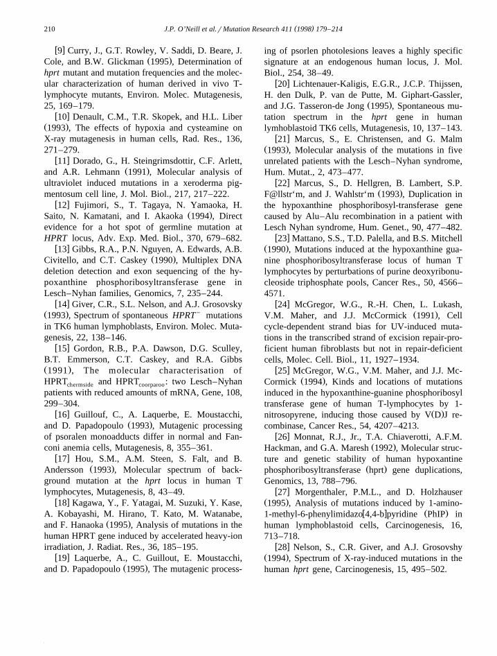
( )J.P. O’Neill et al.rMutation Research 411 1998 179–214210
w x9 Curry, J., G.T. Rowley, V. Saddi, D. Beare, J.Ž .Cole, and B.W. Glickman 1995 , Determination of
hprt mutant and mutation frequencies and the molec-ular characterization of human derived in vivo T-lymphocyte mutants, Environ. Molec. Mutagenesis,25, 169–179.
w x10 Denault, C.M., T.R. Skopek, and H.L. LiberŽ .1993 , The effects of hypoxia and cysteamine onX-ray mutagenesis in human cells, Rad. Res., 136,271–279.
w x11 Dorado, G., H. Steingrimsdottir, C.F. Arlett,Ž .and A.R. Lehmann 1991 , Molecular analysis of
ultraviolet induced mutations in a xeroderma pig-mentosum cell line, J. Mol. Biol., 217, 217–222.
w x12 Fujimori, S., T. Tagaya, N. Yamaoka, H.Ž .Saito, N. Kamatani, and I. Akaoka 1994 , Direct
evidence for a hot spot of germline mutation atHPRT locus, Adv. Exp. Med. Biol., 370, 679–682.
w x13 Gibbs, R.A., P.N. Nguyen, A. Edwards, A.B.Ž .Civitello, and C.T. Caskey 1990 , Multiplex DNA
deletion detection and exon sequencing of the hy-poxanthine phosphoribosyltransferase gene inLesch–Nyhan families, Genomics, 7, 235–244.
w x14 Giver, C.R., S.L. Nelson, and A.J. GrosovskyŽ . y1993 , Spectrum of spontaneous HPRT mutationsin TK6 human lymphoblasts, Environ. Molec. Muta-genesis, 22, 138–146.
w x15 Gordon, R.B., P.A. Dawson, D.G. Sculley,B.T. Emmerson, C.T. Caskey, and R.A. GibbsŽ .1991 , The molecular characterisation ofHPRT and HPRT : two Lesch–Nyhanchermside coorparoo
patients with reduced amounts of mRNA, Gene, 108,299–304.
w x16 Guillouf, C., A. Laquerbe, E. Moustacchi,Ž .and D. Papadopoulo 1993 , Mutagenic processing
of psoralen monoadducts differ in normal and Fan-coni anemia cells, Mutagenesis, 8, 355–361.
w x17 Hou, S.M., A.M. Steen, S. Falt, and B.Ž .Andersson 1993 , Molecular spectrum of back-
ground mutation at the hprt locus in human Tlymphocytes, Mutagenesis, 8, 43–49.
w x18 Kagawa, Y., F. Yatagai, M. Suzuki, Y. Kase,A. Kobayashi, M. Hirano, T. Kato, M. Watanabe,
Ž .and F. Hanaoka 1995 , Analysis of mutations in thehuman HPRT gene induced by accelerated heavy-ionirradiation, J. Radiat. Res., 36, 185–195.
w x19 Laquerbe, A., C. Guillout, E. Moustacchi,Ž .and D. Papadopoulo 1995 , The mutagenic process-
ing of psorlen photolesions leaves a highly specificsignature at an endogenous human locus, J. Mol.Biol., 254, 38–49.
w x20 Lichtenauer-Kaligis, E.G.R., J.C.P. Thijssen,H. den Dulk, P. van de Putte, M. Giphart-Gassler,
Ž .and J.G. Tasseron-de Jong 1995 , Spontaneous mu-tation spectrum in the hprt gene in humanlymhoblastoid TK6 cells, Mutagenesis, 10, 137–143.
w x21 Marcus, S., E. Christensen, and G. MalmŽ .1993 , Molecular analysis of the mutations in fiveunrelated patients with the Lesch–Nyhan syndrome,Hum. Mutat., 2, 473–477.
w x22 Marcus, S., D. Hellgren, B. Lambert, S.P.Ž .F@llstr‘m, and J. Wahlstr‘m 1993 , Duplication in
the hypoxanthine phosphoribosyl-transferase genecaused by Alu–Alu recombination in a patient withLesch Nyhan syndrome, Hum. Genet., 90, 477–482.
w x23 Mattano, S.S., T.D. Palella, and B.S. MitchellŽ .1990 , Mutations induced at the hypoxanthine gua-nine phosphoribosyltransferase locus of human Tlymphocytes by perturbations of purine deoxyribonu-cleoside triphosphate pools, Cancer Res., 50, 4566–4571.
w x24 McGregor, W.G., R.-H. Chen, L. Lukash,Ž .V.M. Maher, and J.J. McCormick 1991 , Cell
cycle-dependent strand bias for UV-induced muta-tions in the transcribed strand of excision repair-pro-ficient human fibroblasts but not in repair-deficientcells, Molec. Cell. Biol., 11, 1927–1934.
w x25 McGregor, W.G., V.M. Maher, and J.J. Mc-Ž .Cormick 1994 , Kinds and locations of mutations
induced in the hypoxanthine-guanine phosphoribosyltransferase gene of human T-lymphocytes by 1-
Ž .nitrosopyrene, inducing those caused by V D J re-combinase, Cancer Res., 54, 4207–4213.
w x26 Monnat, R.J., Jr., T.A. Chiaverotti, A.F.M.Ž .Hackman, and G.A. Maresh 1992 , Molecular struc-
ture and genetic stability of human hypoxantineŽ .phosphoribosyltransferase hprt gene duplications,
Genomics, 13, 788–796.w x27 Morgenthaler, P.M.L., and D. Holzhauser
Ž .1995 , Analysis of mutations induced by 1-amino-w x Ž .1-methyl-6-phenylimidazo 4,4-b pyridine PhIP in
human lymphoblastoid cells, Carcinogenesis, 16,713–718.
w x28 Nelson, S., C.R. Giver, and A.J. GrosovshyŽ .1994 , Spectrum of X-ray-induced mutations in thehuman hprt gene, Carcinogenesis, 15, 495–502.
( )J.P. O’Neill et al.rMutation Research 411 1998 179–214 211
w x29 Papadopoulo, D., A. Laquerbe, C. Guillouf,Ž .and E. Moustacchi 1993 , Molecular spectrum of
mutations induced at the hprt locus by a crosslinking agent in human cell lines with differentrepair capacities, Mutation Res., 294, 167–177.
w x30 Recio, L., J. Cochrane, D. Simpson, T.R.Skopek, J.P. O’Neill, J.A. Nicklas, and R.J. AlbertiniŽ .1990 , DNA sequence analysis of in vivo hprtmutation in human T-lymphocytes, Mutagenesis, 5,505–510.
w x31 Recio, L., D. Simpson, J. Cochrane, H. Liber,Ž .and T.R. Skopek 1990 , Molecular analysis of hprt
mutants induced by 2-cyanoethylene oxide in humanlymphoblastoid cells, Mutation Res., 242, 195–208.
w x32 Rigaud, O., A. Laquerbe, and E. MoustacchiŽ .1995 , DNA sequence analysis of HPRT mutantsinduced in human lymphoblastoid cells adapted toionizing radiation, Rad. Res., 144, 181–189.
w x33 Rossi, A.M., A.D. Tates, A.A. van Zeeland,Ž .and H. Vrieling 1992 , Molecular Analysis of Muta-
tions Affecting hprt mRNA Splicing in Human T-lymphocytes in vivo, Environ. Molec. Mutagenesis,19, 7–13.
w x34 Sege-Peterson, L., J. Chambers, T. Page,Ž .O.W. Jones, and W.L. Nyhan 1992 , Characteriza-
tion of mutations in phenotypic variants of hypoxan-thine phosphoribosyltransferase deficiency, Hum.Molec. Genet., 1, 427–442.
w x35 Steingrimsdottir, H., G. Rowley, A. Waugh,D. Beare, I. Ceccherine, J. Cole, and A.R. LehmannŽ .1993 , Molecular analysis of mutations in the hprtgene in circulating lymphocytes from normal andDNA repair deficient donors, Mutation Res., 294,29–41.
w x36 Vrieling, H., J.C.P. Thijssen, A.M. Rossi,F.J. van Dam, A.T. Natarajan, A.D. Tates, and A.A.
Ž .van Zeeland 1992 , Enhanced hprt mutant fre-quency but no significant difference in mutationspectrum between a smoking and a non-smokinghuman population, Carcinogenesis, 13, 1625–1631.
w x37 Yamada, Y., H. Goto, K. Suzumori, A. Rit-Ž .suko, and N. Ogasawara 1992 , Molecular analysis
of five independent Japanese mutant genes responsi-ble for hypoxanthine guanine phosphoribosyltrans-
Ž .ferase HPRT deficiency, Hum. Genet., 90, 379–384.
w x38 Yamada, Y., H. Goto, S. Tamura, and N.Ž .Osasawara 1993 , Molecular genetic study of a
Japanese family with Lesch–Nyhan syndrome: apoint mutation at the consensus region of RNA
Ž .splicing hprt Keio , Jpn. J. Human. Genet., 38,413–419.
w x Ž .39 Yang, J.L., M.C. Hu, and C.W. Wu 1991 ,Novel mutational spectrum induced by N-methyl-N X-nitro-N-nitrosoguanindine in the coding region of
Ž .the hypoxanthine guanine phosphoribosyltrans-ferase gene in diploid human fibroblasts, J. Mol.Biol., 221, 421–430.
w x40 Yang, J.L., P.C. Lee, S.R. Lin, and J.G. LinŽ .1994 , Comparison of mutation spectra induced byN-ethyl-N-nitrosourea in the hprt gene of Merq andMery diploid fibroblasts, Carcinogenesis, 15, 939–945.
w x41 Yang, J.L., J.G. Lin, M.C. Hu, and C.W. WuŽ .1993 , Mutagenicity and mutational specificity ofN-methyl-N X-nitro-N-nitrosguanidine in the hprt genein G1-S and late S phase of diploid human fibrob-lasts, Cancer Res., 53, 2865–2873.
w x42 Yang, S., J.G. Lin, C.C. Chiou, L.Y. Chen,Ž .and J.L. Yang 1994 , Mutation specificity of 8-
methoxypsoralen plus two doses of UVA irradiationin the hprt gene in diploid human fibroblasts, Car-cinogenesis, 15, 201–207.
References
w x1 A. Kramer, The structure and function of proteins involvedin mammalian pre-mRNA splicing, Annu. Rev. Biochem. 65Ž .1996 367–409.
w x2 C. Will, R. Luhrmann, Protein functions in pre-mRNA splic-Ž .ing, Curr. Opin. Cell Biol. 9 1997 320–328.
w x3 R. Reed, Initial splice-site recognition and pairing duringŽ .pre-mRNA splicing, Curr. Opin. Genet. Dev. 6 1996 215–
220.w x4 X.D. Fu, The superfamily of argininerserine rich splicing
Ž .factors, RNA 1 1995 663–680.w x5 M.D. Chiara, O. Gozani, M. Bennett, P. Champion-Arnaud,
L. Palanfjian, R. Reed, Identification of proteins that interactwith exon sequences, splice sites, and the branchpoint se-quence during each stage of spliceosome assembly, Mol.
Ž .Cell. Biol. 16 1996 3317–3326.w x6 G. Dreyfuss, M.J. Matunis, S. PiZol-Roma, C.G. Burd, hn-
RNP proteins and the biogenesis of mRNA, Annu. Rev.Ž .Biochem. 62 1993 289–321.
w x7 X.-D. Fu, Specific commitment of different pre-mRNAs toŽ .splicing by single SR proteins, Nature 365 1993 82–85.
w x8 S.M. Berget, Exon recognition in vertebrate splicing, J. Biol.Ž .Chem. 270 1995 2411–2414.
( )J.P. O’Neill et al.rMutation Research 411 1998 179–214212
w x9 J.-S. Sun, J.L. Manley, A novel U2–U6 snRNA structure isnecessary for mammalian mRNA splicing, Genes Dev. 9Ž .1995 843–854.
w x10 E.J. Sontheimer, J.A. Steitz, The U5 and U6 small nuclearRNAs as active site components of the spliceosome, Science
Ž .262 1993 1989–1991.w x11 J.D. Crispino, B.J. Blencowe, P.A. Sharp, Complementation
by SR proteins of pre-mRNA splicing reactions depleted ofŽ .U1 snRNP, Science 265 1994 1866–1869.
w x X12 W.Y. Tarn, J.A. Steitz, Modulation of 5 splice site choice inpre-messenger RNA by two distinct steps, Proc. Natl. Acad.
Ž .Sci. U.S.A. 92 1995 2504–2508.w x13 J.F. Ca-ceres, S. Stamm, D.M. Helfman, A.R. Krainer, Regu-
lation of alternative splicing in vivo by overexpression ofŽ .antagonistic splicing factors, Science 265 1994 1706–1708.
w x14 Q. Sun, A. Mayeda, R.K. Hampson, A.R. Krainer, F.M.Rottman, General splicing factor SF2rASF promotes alterna-tive splicing by binding to an exonic splicing enhancer,
Ž .Genes Dev. 7 1993 2598–2608.w x15 R. Singh, J. Valc- rcel, M.R. Green, Distinct binding speci-
ficities and functions of higher eukaryotic polypyrimidineŽ .tract-binding proteins, Science 268 1995 1173–1176.
w x16 K. Nakai, H. Sakamoto, Construction of a novel databasecontaining aberrant mutations of mammalian genes, Gene
Ž .141 1994 171–177.w x X17 M. Talerico, S.M. Berget, Effect of 5 splice site mutations
on splicing of the preceding intron, Mol. Cell. Biol. 10Ž .1990 6299–6305.
w x18 S. Brunak, J. Engelbrecht, S. Knudsen, Prediction of humanmRNA donor and acceptor sites from the DNA sequence, J.
Ž .Mol. Biol. 220 1991 49–65.w x19 B.L. Robberson, G.J. Cote, S.M. Berget, Exon definition
may facilitate splice site selection in RNAs with multipleŽ .exons, Mol. Cell. Biol. 10 1990 84–94.
w x20 J.Y. Wu, T. Maniatis, Specific interactions between proteinsimplicated in splice site selection and regulated alternative
Ž .splicing, Cell 75 1993 1061–1070.w x21 R. Reed, T. Maniatis, A role for exon sequences and splice-
Ž .site proximity in splice-site selection, Cell 46 1986 681–690.
w x22 A. Watakabe, K. Tanaka, Y. Shimura, The role of exonŽ .sequences in splice site selection, Genes Dev. 7 1993
407–418.w x23 R. Xu, J. Teng, T.A. Cooper, The cardiac troponin T alterna-
tive exon contains a novel purine-rich positive splicing ele-Ž .ment, Mol. Cell. Biol. 13 1993 3660–3674.
w x24 A. Laviguer, H. La Branche, A.R. Kornblihtt, B. Chabot, Asplicing enhancer in the human fibronectin alternate exonED1 exon interacts with SR proteins and stimulates U2
Ž .snRNP binding, Genes Dev. 7 1993 2405–2417.w x25 R.K. Hampson, L. La Follette, F.M. Rottman, Alternative
processing of bovine growth hormone mRNA is influencedŽ .by downstream exon sequences, Mol. Cell. Biol. 9 1989
1604–1610.w x26 G.S. Huh, R.O. Hynes, Regulation of alternative pre-mRNA
splicing by a novel repeated hexanucleotide element, GenesŽ .Dev. 8 1994 1561–1574.
w x27 T. Carlo, D.A. Sterner, S.M. Berget, An intron splicingenhancer containing a G-rich repeat facilitates inclusion of a
Ž .vertebrate micro-exon, RNA 2 1996 342–353.w x28 F. Del Gatto, M.-C. Gesnel, R. Breathnach, The exon se-
quence TAGG can inhibit splicing, Nucl. Acids Res. 24Ž .1996 2017–2021.
w x29 P.J. Furdon, R. Kole, The length of the downstream exon andthe substitution of specific sequences affect pre-mRNA splic-
Ž .ing in vitro, Mol. Cell. Biol. 8 1988 860–866.w x30 K. Inoue, M. Ohno, Y. Shimura, Aspects of splice site
selection in constitutive and alternative pre-mRNA splicing,Ž .Gene Express. 4 1995 177–182.
w x31 P. Senapathy, M.B. Shapiro, N.L. Harris, Splice junctions,branch point sites, and exons: sequence statistics, identifica-tion, and applications to genome project, Meth. Enzymol.
Ž .183 1990 252–278.w x32 N.L. Harris, P. Senapathy, Distribution and consensus of
branch point signals in eukaryotic genes: a computerizedŽ .statistical analysis, Nucl. Acids Res. 18 1990 3015–3019.
w x33 F.E. Penotti, Human pre-mRNA splicing signals, J. Theor.Ž .Biol. 150 1991 385–420.
w x34 T.D. Schneider, Sequence walkers: a graphical method todisplay how binding proteins interact with DNA or RNA
Ž .sequences, Nucl. Acids Res. 25 1997 4408–4415.w x35 R.M. Stephens, T.D. Schneider, Features of spliceosome
evolution and function inferred from an analysis of theŽ .information at human splice sites, J. Mol. Biol. 24 1992
1124–1136.w x36 M.B. Shapiro, P. Senapathy, RNA splice junctions of differ-
ent classes of eukaryotes: sequence statistics and functionalŽ .implications in gene expression, Nucl. Acids Res. 15 1987
7155–7174.w x37 B.M. Faux, T.D. Schneider, P.K. Rogan, Information analy-
sis of human splice site mutations, Am. J. Hum. Genet. 59Ž .1996 A257.
w x38 P.K. Rogan, T.D. Schneider, Using information content andbase frequencies to distinguish mutations from genetic poly-morphisms in splice junction recognition sites, Hum. Mutat.Ž .6 1995 74–76.
w x39 P.K. Rogan, B.M. Faux, T.D. Schneider, Information analy-Ž .sis of human splice site mutations, Hum. Mut. 12 1998
153–171.w x40 R.F. Roscigno, M. Weiner, M.A. Garcia-Blanco, A muta-
tional analysis of the polypyrimidine tract of introns, J. Biol.Ž .Chem. 268 1993 11222–11229.
w x X41 R. Reed, The organization of 3 splice-site sequences inŽ .mammalian introns, Genes Dev. 3 1989 2113–2123.
w x42 C.-S. Chu, B.C. Trapnell, S. Curristin, G.R. Cutting, R.G.Crystal, Genetic basis of variable exon 9 skipping in cysticfibrosis transmembrane conductance regulator mRNA, Na-
Ž .ture Genet. 3 1993 151–156.w x43 K.K. Nelson, M.R. Green, Mammalian U2 snRNP has a
Ž .sequence-specific RNA-binding activity, Genes Dev. 3 19891562–1571.
w x44 R. Reed, T. Maniatis, Intron sequences involved in lariatŽ .formation during pre-mRNA splicing, Cell 41 1985 95–105.
w x45 D.M. Helfman, W.M. Ricci, Branch point selection in alter-
( )J.P. O’Neill et al.rMutation Research 411 1998 179–214 213
native splicing of tropomyosin pre-mRNAs, Nucl. Acids Res.Ž .17 1989 5633–5650.
w x46 P.A. Estes, N.E. Cooke, S.A. Liebhaber, A native RNAsecondary structure controls alternative splice-site selectionand generates two human growth hormone isoforms, J. Biol.
Ž .Chem. 267 1992 14902–14908.w x47 T.A. Cooper, In vitro splicing of cardiac troponin precursors,
Ž .J. Biol. Chem. 267 1992 5330–5338.w x48 D. Libri, M. Goux-Pelletan, E. Brody, M.Y. Fiszman, Exon
as well as intron sequences are cis-regulating elements forthe mutually exclusive alternative splicing of the a
Ž .tropomyosin gene, Mol. Cell. Biol. 10 1990 5036–5046.w x49 C.W.J. Smith, B. Nadal-Ginard, Mutually exclusive splicing
of a-tropomyosin exons enforced by an unusual lariat branchpoint location: implications for constitutive splicing, Cell 56Ž .1989 749–758.
w x50 A.M. Carothers, G. Urlaub, D. Grunberger, L.A. Chasin,Splicing mutants and their second-site suppressors at thedihydrofolate reductase locus in Chinese hamster ovary cells,
Ž .Mol. Cell. Biol. 13 1993 5085–5098.w x51 W.J.T. Smith, C.T. Chu, B. Nadal-Ginard, Scanning and
competition between AGs are involved in 3X splice siteŽ .selection in mammalian introns, Mol. Cell. Biol. 13 1993
4939–4952.w x52 B. Ruskin, J.M. Greene, M.R. Green, Cryptic branch point
activation allows accurate in vitro splicing of human $-globinŽ .intron mutants, Cell 41 1985 833–844.
w x53 Z. Dominiski, R. Kole, Selection of splice sites in pre-mRNAsŽ .with short internal exons, Mol. Cell. Biol. 11 1991 6075–
6083.w x54 X. Liu, J.E. Mertz, Sequence of the polypyrimidine tract of
the 3X-terminal 3X splicing signal can affect intron-dependentŽ .pre-mRNA processing in vivo, Nucl. Acids Res. 24 1996
1765–1773.w x55 E. Izaurraide, J. Lewis, C. McGuigan, M. Jankowska, E.
Darzynkiewicz, I.W. Mattaj, A nuclear cap binding proteinŽ .complex involved in pre-mRNA splicing, Cell 78 1994
657–668.w x56 M. Niwa, S.D. Rose, S.M. Berget, In vitro polyadenylation is
stimulated by the presence of an upstream intron, Genes Dev.Ž .4 1990 1552–1559.
w x57 B. Wieringa, E. Hofer, C. Weissman, A minimal intronlength but no specific internal sequence is required for
Ž .splicing the large rabbit $-globin intron, Cell 37 1984915–925.
w x58 J.F. Bateman, D. Chan, I. Moeller, M. Hannagan, W.G. Cole,A 5X splice site mutation affecting the pre-mRNA splicing oftwo upstream exons in the collagen COL 1A1 gene, Biochem.
Ž .J. 302 1994 729–735.w x59 D.A. Sterner, S.M. Berget, In vivo recognition of a vertebrate
mini-exon as an exon–intron–exon unit, Mol. Cell. Biol. 13Ž .1993 2677–2687.
w x60 H. Ge, J. Noble, J. Colgan, J.L. Manley, Polyoma virus smalltumor antigen pre-mRNA splicing requires cooperation be-tween two 3X splice sites, Proc. Natl. Acad. Sci. U.S.A. 87Ž .1990 3338–3342.
w x61 M. Aoshima, H. Nunoi, M. Shimazu, S. Shimizu, O. Tat-
suzawa, R.T. Kenney, S. Kanegasaki, Two-exon skippingdue to a point mutation in p67-phox-deficient chronic granu-
Ž .lomatous disease, Blood 88 1996 1841–1845.w x62 M.J.L. Ligtenberg, A.M.C. Gennissen, H.L. Vos, J. Hilkens,
A single nucleotide polymorphism in an exon dictates alleledependent differential splicing of episialin mRNA, Nucl.
Ž .Acids Res. 19 1991 297–304.w x63 M. Krawczak, J. Reiss, D.N. Cooper, The mutational spec-
trum of single base-pair substitutions in the mRNA splicejunctions of human genes: causes and consequences, Hum.
Ž .Genet. 90 1992 41–54.w x64 H.C. Dietz, D. Valle, C.A. Francomano, R.J. Kendzior Jr.,
R.E. Pyeritz, G.R. Cutting, The skipping of constitutiveexons in vivo induced by nonsense mutations, Science 259Ž .1993 680–683.
w x65 G. Urlaub, P.J. Mitchell, C.J. Ciudad, L.A. Chasin, Nonsensemutations in the dihydrofolate reductase gene affect RNA
Ž .processing, Mol. Cell. Biol. 9 1989 2868–2880.w x66 D. Weil, M. D’Alessio, F. Ramirez, W. de Wet, W.G. Cole,
D. Chan, J.F. Bateman, A base substitution in the exon of acollagen gene causes alternative splicing and generates astructurally abnormal polypeptide in a patient with Ehlers–
Ž .Danlos syndrome Type VII, EMBO J. 8 1989 1705–1710.w x67 C.W. Fisher, C.R. Fisher, J.L. Chuang, K.S. Lau, D.T.
Ž .Chuang, R.P. Cox, Occurrence of a 2-bp AT deletion alleleŽ . Ž .and a nonsense G-to-T mutant allele at the E2 DBT locus
of six patients with maple syrup urine disease: multiple-exonskipping as a secondary effect of the mutations, Am. J. Hum.
Ž .Genet. 52 1993 414–424.w x68 M.H. Ricketts, M.J. Simons, J. Parma, L. Mercken, Q. Dong,
G. Vassart, A nonsense mutation causes hereditary goitre inthe Afrikaner cattle and unmasks alternative splicing ofthyroglobulin transcripts, Proc. Natl. Acad. Sci. U.S.A. 84Ž .1987 3181–3184.
w x69 P.M. Smooker, J. Christodoulou, R.R. McInnes, R.G.H. Cot-ton, A mutation causing DHPR deficiency results in aframeshift and a secondary splicing defect, J. Med. Genet. 32Ž .1995 220–223.
w x70 S. Aoufouchi, J. Yjlamos, C. Milstein, Nonsense mutationsinhibit RNA splicing in a cell-free system: recognition ofmutant codon is independent of protein synthesis, Cell 85Ž .1996 415–422.
w x71 I. Santisteban, F.X. Arredondo-Vega, S. Kelly, M. Loubser,N. Meydan, C. Roifman, P.L. Howell, T. Bowen, K.I. Wein-berg, M.L. Schroeder, M.S. Hershfield, Three new adenosinedeaminase mutations that define a splicing enhancer andcause severe and partial phenotypes: implications for evolu-tion of a CpG hotspot and expression of a transduced ADA
Ž .cDNA, Hum. Mol. Genet. 4 1995 2081–2087.w x72 R. Pulak, P. Anderson, mRNA surveillance by the
Ž .Caenorhabditis elegans smg genes, Genes Dev. 7 19931885–1897.
w x73 L.E. Maquat, Defects in RNA splicing and the consequenceof shortened translational reading frames, Am. J. Hum. Genet.
Ž .59 1996 279–286.w x74 A. Edwards, H. Voss, P. Rice, A. Civitello, J. Stegemann, C.
Schwager, J. Zimmerman, H. Erfle, C.T. Caskey, W. An-
( )J.P. O’Neill et al.rMutation Research 411 1998 179–214214
sorge, Automated DNA sequencing of the human HPRTŽ .locus, Genomics 6 1990 593–608.
w x75 J.A. Nicklas, T.C. Hunter, J.P. O’Neill, R.J. Albertini, Finestructure mapping of the hprt gene region of the human X
Ž . Ž .chromosome Xq26 , Am. J. Hum. Genet. 49 1990 267–278.
w x76 N.F. Cariello, T.R. Craft, H. Vrieling, A.A. van Zeeland, T.Adams, T.R. Skopek, Human HPRT mutant database: soft-ware for data entry and retrieval, Environ. Mol. Mutagenesis
Ž .20 1992 81–83.w x77 J.C. Fuscoe, L.J. Zimmerman, M.L. Lippert, J.A. Nicklas,
Ž .J.P. O’Neill, R.J. Albertini, V D J recombinase-like activitymediates hprt gene deletion in human fetal T-lymphocytes,
Ž .Cancer Res. 51 1991 6001–6005.w x78 J.C. Fuscoe, L.J. Zimmerman, K. Harrington-Brock, L. Bur-
nette, M.M. Moore, J.A. Nicklas, J.P. O’Neill, R.J. Albertini,Ž .V D J recombinase-mediated deletion of the hprt gene in
Ž .T-lymphocytes from adult humans, Mutation Res. 283 199213–20.
w x79 H. Steingrimsdottir, G. Rowley, G. Dorado, J. Cole, A.R.Lehmann, Mutations which alter splicing in the human hy-poxanthine-guanine phosphoribosyltransferase gene, Nucl.
Ž .Acids Res. 20 1992 1201–1208.w x80 C.R. Giver, S.L. Nelson, A.J. Grosovsky, Spectrum of spon-
taneous HPRTy mutations in TK6 human lymphoblasts,Ž .Environ. Mol. Mutagenesis 22 1993 138–146.
w x81 C. Guillouf, A. Laquerbe, E. Moustacchi, D. Papadopoulo,Mutagenic processing of psoralen monoadducts differ in
Ž .normal and Fanconi anemia cells, Mutagenesis 8 1993355–361.
w x82 Y. Yamada, H. Goto, K. Suzumori, A. Ritsuko, N. Oga-sawara, Molecular analysis of five independent Japanesemutant genes responsible for hypoxanthine guanine phospho-
Ž . Ž .ribosyltransferase HPRT deficiency, Hum. Genet. 90 1992379–384.
w x83 S. Nelson, C.R. Giver, A.J. Grosovshy, Spectrum of X-ray-induced mutations in the human hprt gene, Carcinogenesis
Ž .15 1994 495–502.w x84 S. Marcus, D. Hellgren, B. Lambert, S.P. Fallstrom, J.¨ ¨
W ahlstrom , D uplication in the hypoxanthine¨phosphoribosyl-transferase gene caused by Alu–Alu recom-bination in a patient with Lesch–Nyhan syndrome, Hum.
Ž .Genet. 90 1993 477–482.w x85 S. Yang, J.G. Lin, C.C. Chiou, L.Y. Chen, J.L. Yang,
Mutation specificity of 8-methoxypsoralen plus two doses ofUVA irradiation in the hprt gene in diploid human fibrob-
Ž .lasts, Carcinogenesis 15 1994 201–207.w x86 T. Bastlova, B. Andersson, B. Lambert, A. Kilman, Molecu-
lar analysis of ethylene oxide induced mutations at the hprtŽ .locus in human diploid fibroblasts, Mutation Res. 287 1993
283–292.w x87 A.M. Rossi, A.D. Tates, A.A. van Zeeland, H. Vrieling,
Molecular Analysis of Mutations Affecting hprt mRNASplicing in Human T-lymphocytes in vivo, Environ. Mol.
Ž .Mutagenesis 19 1992 7–13.w x88 E.G.R. Lichtenauer-Kaligis, J.C.P. Thijssen, H. den Dulk, P.
van de Putte, M. Giphart-Gassler, J.G. Tasseron-de Jong,Spontaneous mutation spectrum in the hprt gene in human
Ž .lyphoblastoid TK6 cells, Mutagenesis 10 1995 137–143.w x89 L. Sege-Peterson, J. Chambers, T. Page, O.W. Jones, W.L.
Nyhan, Characterization of mutations in phenotypic variantsof hypoxanthine phosphoribosyltransferase deficiency, Hum.
Ž .Mol. Genet. 1 1992 427–442.w x90 M.J. Lippert, I.R. Rainville, J.A. Nicklas, R.J. Albertini,
Large deletions partially external to the human hprt geneŽ .result in chimeric transcripts, Mutagenesis 12 1997 185–
190.w x91 M.L. Cleary, Oncogenic conversion of transcription factors
Ž .by chromosomal translocations, Cell 66 1991 619–622.w x92 M.G. Manjanatha, L.A. Lindsey, R.A. Mittelstaedt, R.H.
Heflich, Low hprt mRNA levels and multiple hprt mRNAspecies in 6-thioguanine-resistant Chinese hamster cell mu-tants possessing nonsense mutations, Mutation Res. 308Ž .1994 65–75.
w x93 C.R. Valentine, R.H. Heflich, The association of nonsensemutation with exon-skipping in hprt mRNA of Chinesehamster ovary cells results from an artifact of RT-PCR, RNAŽ .3 1997 660–676.
w x Ž .94 Valentine, C.R. in press , Origin of exon-deleted mRNAsassociated with nonsense mutations, Mutation Research.
w x95 P. Menichini, A. Inga, G. Fronza, R. Iannone, P. Degan, P.Campomenosi, A. Abbondandolo, Defective splicing inducedby 4NQO in the hamster hprt gene, Mutation Res. 323Ž .1994 159–165.
w x96 E.E. Hennig, A.H. Conney, S.-J.C. Wei, Characterization ofhprt splicing mutations induced by the ultimate carcinogenic
w xmetabolite of benzo a pyrene in Chinese Hamster V-79 cells,Ž .Cancer Res. 55 1995 1550–1558.
w x97 L.-H. Zhang, H. Vrieling, A.A. van Zeeland, D. Jenssen,Spectrum of spontaneously occurring mutations in the hprt
Ž .gene of V79 Chinese hamster cells, J. Mol. Biol. 223 1992627–635.
w x98 C.R. Valentine, R.H. Heflich, Genomic DNA sequencing ofmRNA splicing mutants in the hprt gene of Chinese Hamster
Ž .ovary cells, Environ. Mol. Mutagenesis 25 1995 85–96.