Mutational analysis of the Aspergillus ambient pH receptor PalH ... · Since the pioneering studies...
45
Mutational analysis of the Aspergillus ambient pH receptor PalH underscores its potential as a target for antifungal compounds Daniel Lucena-Agell 1 , América Hervás-Aguilar 1 , Tatiana Munera-Huertas 2 , Olga Pougovkina 1 , Joanna Rudnicka 2 , Antonio Galindo 1 , Joan Tilburn 2 , Herbert N. Arst, Jr. 1,2 , and Miguel A. Peñalva 1 1 Department of Cellular and Molecular Biology, Centro de Investigaciones Biológicas CSIC, Ramiro de Maeztu 9, Madrid 28040, Spain. 2 Section of Microbiology, Imperial College London, Flowers Building, Armstrong Road, London SW7 2AZ, UK. Short title: PalH as a druggable antifungal target This article has been accepted for publication and undergone full peer review but has not been through the copyediting, typesetting, pagination and proofreading process which may lead to differences between this version and the Version of Record. Please cite this article as an ‘Accepted Article’, doi: 10.1111/mmi.13438 This article is protected by copyright. All rights reserved.
Transcript of Mutational analysis of the Aspergillus ambient pH receptor PalH ... · Since the pioneering studies...
Mutational analysis of the Aspergillus ambient pH receptor
PalH underscores its potential as a target for antifungal compounds
Daniel Lucena-Agell1, América Hervás-Aguilar1, Tatiana Munera-Huertas2, Olga
Pougovkina1, Joanna Rudnicka2, Antonio Galindo1, Joan Tilburn2, Herbert N. Arst,
Jr.1,2, and Miguel A. Peñalva1
1 Department of Cellular and Molecular Biology, Centro de Investigaciones Biológicas
CSIC, Ramiro de Maeztu 9, Madrid 28040, Spain.
2 Section of Microbiology, Imperial College London, Flowers Building, Armstrong Road,
London SW7 2AZ, UK.
Short title: PalH as a druggable antifungal target
This article has been accepted for publication and undergone full peer review but has not beenthrough the copyediting, typesetting, pagination and proofreading process which may lead todifferences between this version and the Version of Record. Please cite this article as an‘Accepted Article’, doi: 10.1111/mmi.13438
This article is protected by copyright. All rights reserved.
2
Abstract The pal/RIM ambient pH signaling pathway is crucial for the ability of pathogenic fungi
to infect hosts. The Aspergillus nidulans 7-TMD receptor PalH senses alkaline pH,
subsequently facilitating ubiquitination of the arrestin PalF. Ubiquitinated PalF triggers
downstream signaling events. The mechanism(s) by which PalH transduces the
alkaline pH signal to PalF is poorly understood.
We show that PalH is phosphorylated in a signal dependent manner,
resembling mammalian GPCRs, although PalH phosphorylation, in contrast to
mammalian GPCRs, is arrestin dependent. A genetic screen revealed that an ambient-
exposed region comprising the extracellular loop connecting TM4-TM5 and ambient-
proximal residues within TM5 is required for signaling. In contrast, substitution by
alanines of four aromatic residues within TM6 and TM7 results in a weak ‘constitutive’
activation of the pathway. Our data support the hypothesis that PalH mechanistically
resembles mammalian GPCRs that signal via arrestins, such that the relative positions
of individual helices within the heptahelical bundle determines the Pro316-dependent
transition between inactive and active PalH conformations, governed by an ambient-
exposed region including critical Tyr259 that potentially represents an agonist binding
site. These findings open the possibility of screening for agonist compounds stabilizing
the inactive conformation of PalH, which might act as antifungal drugs against
ascomycetes.
Abbreviated abstract
Fungi colonize environments with different values of pH by adapting their patterns of
gene expression to the needs imposed by ambient pH. pH regulation is important for
fungal pathogenicity, but therapeutically useful drugs targeting the pH signaling
pathway are yet to be developed. We show that the ascomycete ambient pH receptor
PalH resembles a major class of mammalian receptors, denoted GPCRs. Thus our
data pave the way for the identification of specific inhibitors of the ambient pH receptor
for antifungal intervention.
Page 2 of 45Molecular Microbiology
This article is protected by copyright. All rights reserved.

3
Introduction Regulation of gene expression by ambient pH, a transcriptional response that tailors
the biosynthesis of extracellular enzymes, permeases and exported metabolites to the
needs imposed by the pH of the growth medium, crucially contributes to the ability of
many fungi to thrive in disparate pH environments. The molecular wiring of the pH
regulatory circuit has been extensively studied in the filamentous fungus Aspergillus
nidulans and in yeasts, particularly in Saccharomyces cerevisiae. It includes a
dedicated signal transduction pathway, denoted the pal/RIM pathway, that triggers,
with assistance of the endosomal sorting complex required for transport (ESCRT)
machinery, the proteolytic activation of the executive transcription factor
PacC/Rim101p in response to alkaline pH (Peñalva & Arst, 2002, Peñalva & Arst,
2004, Peñalva et al., 2014, Peñalva et al., 2008).
As fungal pathogens invading mammalian hosts are exposed to systemic pH values
above neutrality, it is unsurprising that the pal/RIM pathway has been identified as an
important factor determining pathogenicity (Davis, 2009, Cornet & Gaillardin, 2014).
Since the pioneering studies revealing the involvement of this pathway in systemic
candidiasis (Davis et al., 2000), studies with ascomycetes as diverse as the
Saccharomycotina Candida albicans (Nobile et al., 2008) and the Pezizomycotina A.
nidulans (Bignell et al., 2005) and A. fumigatus (Bertuzzi et al., 2014) have uncovered
its key involvement in the ability of these fungi to infect mammalian hosts.
Overwhelming evidence indicates that, in ascomycetes, the pal/RIM pathway ambient
pH sensor is a transmembrane protein denoted PalH in A. nidulans and Rim21p in S.
cerevisiae (Herranz et al., 2005, Hervás-Aguilar et al., 2007, Galindo et al., 2012,
Obara et al., 2012, Herrador et al., 2015). This pH sensor is the most appealing
component of the pathway to serve as druggable target, although it would be
unsuitable for infections caused by the basidiomycetes, for example Cryptococcus
neoformans, given that although the RIM pathway is involved in its pathogenicity
(O'Meara et al., 2013, O'Meara et al., 2010, O'Meara et al., 2014), PalH/Rim21p (and
their associated arrestin, see below) appear to be missing in basidiomycete lineage
(Ost et al., 2015, Cervantes-Chávez et al., 2010). However, the fact that ascomycetes
including pathogenic species such as A. fumigatus, A. niger, C. albicans, C. glabrata,
Penicillum marneffei and Histoplasma capsulatum account for a large share of fungal
human infections makes the possibility of targeting PalH potentially very useful.
The direct or indirect activation of this PalH sensor by ambient pH is transmitted to the
internal leaflet of the plasma membrane by a transducer module involving an arrestin-
Page 3 of 45 Molecular Microbiology
This article is protected by copyright. All rights reserved.
4
like protein, denoted PalF in A. nidulans and Rim8p in S. cerevisiae, an ubiquitin ligase
(Rsp5p in S. cerevisiae) and the ESCRT-I protein Vps23. PalF becomes ubiquitinated
in a PalH-dependent manner following exposure of cells to alkaline pH (Herranz et al.,
2005), and this PalF ubiquitination triggers downstream signaling events (Hervás-
Aguilar et al., 2010). This occurs because ubiquitinated PalF/Rim8 in turn recruits
Vps23 to plasma membrane-associated signaling foci, sparking ESCRT-III
polymerization in these locales, which most likely serves as an amplification step
(Herrador et al., 2009, Galindo et al., 2012, Obara & Kihara, 2014) (see (Peñalva et al.,
2014) for a review). ESCRT-III recruits to these foci the downstream pal/RIM pathway
components PalC/Ygr122w (Rim23) (Galindo et al., 2007, Rothfels et al., 2005),
PalA/Rim20p (which serves as adaptor for PacC/Rim101p (Xu & Mitchell, 2001,
Vincent et al., 2003)) and the calpain-like protease PalB/Rim13p (Lucena-Agell et al.,
2015, Obara & Kihara, 2014, Rodríguez-Galán et al., 2009), which results in the
proteolytic processing activation of the transcription factor (Peñalva et al., 2014). It is
now agreed that despite the link of pH signaling to endosomal protein complexes, the
pathway functions at the plasma membrane and does not require endocytosis (Lucena-
Agell et al., 2015, Galindo et al., 2012, Obara & Kihara, 2014, Calcagno-Pizarelli et al.,
2011).
Suggestively, PalH/Rim21p shares features with members of the seven
transmembrane protein family of receptors (7-TMRs). 7-TMRs, usually denoted G
protein-coupled receptors (GPCRs), are the ‘most druggable’ family of proteins for
pharmacological intervention (30% of the top-selling prescription drugs are directed
against GPCRs (Chalmers & Behan, 2002)), in part due to the fact that small
molecules are able to target them with high specificity and affinity. Therefore, should
the mechanism of PalH activation resemble that of its mammalian counterparts, the
possibility of targeting the fungal ambient pH receptor with molecules interfering with its
normal activity appears feasible, beside being theoretically attractive.
GPCRs do not solely transduce signals by way of heterotrimeric G proteins. Arrestins
are a class of proteins previously thought to act exclusively to desensitize stimulated
GPCRs. However, when engaged to the cytosolic tails of stimulated receptors,
‘classical’ β-arrestins can recruit, activate and scaffold downstream signaling
complexes without involvement of heterotrimeric G-proteins, thus playing positive roles
in signaling (Lefkowitz & Shenoy, 2005, Violin & Lefkowitz, 2007). Unlike β-arrestins, α-
arrestins, a subfamily including the fungal clade, characteristically contain docking sites
for the Rsp5 ubiquitin ligase (Alvarez, 2008) and indeed many of them act as adaptors
Page 4 of 45Molecular Microbiology
This article is protected by copyright. All rights reserved.
5
targeting integral membrane protein partners for ubiquitin-mediated endocytic down-
regulation (Nikko & Pelham, 2009, Lin et al., 2008, Karachaliou et al., 2013). In marked
contrast, the α-arrestin PalF/Rim8p engaging the PalH/Rim21p receptor acts positively,
apparently by mediating its own requisite ubiquitination (Herrador et al., 2015, Herrador
et al., 2009). PalF is necessary and, if artificially ubiquitinated, sufficient for the alkaline
pH-dependent activation of the pal pathway (Hervás-Aguilar et al., 2010, Galindo et al.,
2012). However, how under normal circumstances PalH/Rim21 stimulation results in
PalF/Rim8p ubiquitination is currently unknown.
GPCR receptors consist of a bundle of helices embedded in the membrane, connected
by cytosolic and external loops. Agonist binding to an inactive receptor R promotes a
conformational transition R > R* resulting in subtle movements of the TMs that expose
the otherwise inaccessible G protein-binding sites (Sprang, 2011, Katritch et al., 2013).
In contrast, the so denoted inverse agonists displace the equilibrium towards the
inactive R state, thereby suppressing basal (i.e. in the absence of agonist) activity.
However this model is an oversimplification as receptors can adopt more than two
conformations in response to different ligands, a fact that gave rise to the concept of
‘biased ligands’, i.e. ligands that specifically stabilize one particular receptor
conformation. This concept is ultimately translated in that, for example, a ‘biased
agonist’ elicits only a subset of the full repertoire of activities of any given receptor
(Violin & Lefkowitz, 2007). Thus for receptors that can signal by either G proteins or
arrestins, certain agonists are biased towards arrestin signaling, strongly supporting
the view that ‘arrestin-specific conformations’ do exist (Wisler et al., 2007).
A. nidulans PalH consisting of an N-terminal integral membrane moiety (residues 1-
352) and a long C-terminal tail (residues 353-760) (Figure 1) not only engages an
arrestin, but it appears to conform to the topology of 7-TMRs. According to TMpred
(http://www.ch.embnet.org) the membrane moiety consists of seven TMs, with TM-1
crossing the membrane in the outside-in direction, such that its long C-terminal tail is
exposed to the cytosol. This tail contains two PalF binding regions (Figure 1, pink
residues) one adjacent to TM-7 (residues 349-386) and the second bound by C-
terminal residues 657-760 (Herranz et al., 2005).
Here we exploit the amenability of A. nidulans to classical and reverse genetic analysis
to obtain evidence that PalH is indeed a pal pathway receptor that upon exposure to
alkaline pH undergoes a conformational change similar to those experienced by
mammalian GPCRs, and that this change is transmitted to the cytosolic tail to
Page 5 of 45 Molecular Microbiology
This article is protected by copyright. All rights reserved.

6
transduce the alkaline pH signal by way of arrestin activation. Given that pH signaling
does not involve G-proteins, PalH/Rim21p would be a prototypical example of a class
of receptors naturally biased towards arrestin signaling.
Results
Gene replacement procedure for PalH analysis A. nidulans is haploid, facilitating the analysis of PalH function genetically, using a
gene replacement procedure. We first constructed a pyrG89 palH∆ strain in which the
palH coding region had been substituted by A. fumigatus pyroA (pyroAAf). This
substitution/recombination event was confirmed by Southern blotting and phenotype
testing. This palH∆::pyroAAf strain was used as recipient of gene-replacement linear
DNA fragments consisting of wild-type or mutant HA3-tagged palH alleles and A.
fumigatus pyrG as selective marker (Supplemental Figure 1). Transformants in which
palH had been reconstituted with mutant versions were identified by their pyridoxine
auxotrophy resulting from the exchange of pyroAAf by palH-HA3::pyrGAf cassettes.
Diagnostic plate tests for pH regulation demonstrated that strains carrying a wild-type
palH-HA3 gene-replaced allele were indistinguishable from the authentic wild-type, and
western blot (WB) analyses following a shift from acidic to alkaline conditions revealed
that the proteolytic activation processing patterns of PacC in palH-HA3 and palH+
strains were indistinguishable (Supplemental Figure 1), thereby showing that the
genetic manipulations do not by themselves impair PalH function.
PalH is phosphorylated in an ambient pH- and PalF-dependent manner The pH signaling pathway responds to alkaline pH. Under acidic conditions the
transcription factor PacC is not proteolytically activated, being present as the primary
translation product, PacC72. Alkalinization of the medium activates the pal pathway,
triggering the proteolytic activation of PacC72 to PacC27 via the committed
intermediate PacC53 (Díez et al., 2002) (anti-PacC western blots, Figure 2A). In anti-
HA3 WBs of these cells cultured under acidic conditions PalH appears as a single
band. However, PalH is modified upon exposing cells to alkalinity, such that an
additional band(s) of reduced mobility become(s) the predominating species (Figure
2A). This reduction in the mobility of PalH was reversed by treatment with lambda
phosphatase (Figure 2B), indicating that the mobility shift(s) result(s) from
phosphorylation.
Page 6 of 45Molecular Microbiology
This article is protected by copyright. All rights reserved.

7
PalH remained phosphorylated in cells kept at alkaline pH for as long as 4 h (Figure
2A). When alkaline-exposed cells were shifted back to acidic pH, the relative level of
phosphorylated species declined slowly, correlating with the ‘recovery’ of PacC72
caused by the absence of signaling (i.e. the lack of proteolytic processing of freshly
synthesized PacC)(Figure 2C). Complete dephosphorylation took as long as 45-90 min
after returning cells to acidic pH, a time at which the pool of PacC27, which turns over
also slowly (Mingot et al., 1999), becomes barely detectable (note that PacC72 does
not increase further because transcriptional autorepression counteracts the absence of
its proteolytic processing (Bussink et al., 2015)) (Figure 2D). All the above data are
consistent with PalH becoming accessible to phosphorylation as a consequence of
alkaline pH sensing. The correlation between pathway activation and PalH
phosphorylation was suggestive as upon their agonist-mediated activation mammalian
GPCRs are rapidly phosphorylated by GRKs (G protein receptor kinases), hampering
signaling (Shukla et al., 2011, Lefkowitz, 2013). Notably, although PalH
phosphorylation is independent of PalI (a PalH helper) and of PalA (acting downstream
of ESCRT recruitment), it is strictly dependent on PalF (Figure 2E), indicating that PalH
phosphorylation requires the functional engagement of the arrestin to the receptor.
Mutant PalH with a cytosolic tail restricted to the two PalF arrestin binding sites retains some function Equipped with the gene replacement procedure, we first constructed an allele encoding
a PalH protein in which cytosolic residues 385 through 653 separating the two PalF
binding sites had been substituted by a synthetic linker consisting of a Gly-Ala
pentamer (palH654, Figure 3A). WB showed that the protein is stably expressed
(Figure 3B). Null palH alleles such as palH72 truncating PalH after residue 12, or
alleles truncating PalH within the TM domain do not grow at alkaline pH, display
exacerbated sensitivity to Li+ and MoO42- ion toxicity and enhanced resistance to
neomycin (Negrete-Urtasun et al., 1999, Herranz et al., 2005) (Figure 3C).
Remarkably, palH654 strains are, like palH72 strains, highly sensitive to lithium. Yet,
unlike palH72, palH654 strains grow at alkaline pH and are more resistant than palH72
strains to molybdate, showing that the mutant palH654 product is, to a significant
extent, functional. WB analysis of PacC processing buttressed this conclusion. In pH
shift experiments the null palH72 mutation blocked the proteolytic processing activation
of PacC (Hervás-Aguilar et al., 2010). In contrast, palH654 impaired it relatively weakly
(Figure 3B, bottom), although this allele led to a reduction in the overall PacC levels.
We conclude that although the 269 cytosolic residues of PalH located between the
Page 7 of 45 Molecular Microbiology
This article is protected by copyright. All rights reserved.

8
previously delimited PalF binding regions play some role, this region is not critical for
pH signaling.
Phosphorylation is largely dispensable for PalH function Anti-HA3 WBs of palH654 cells strongly suggested that the phosphorylation-mediated
shift in PalH mobility requires amino acids located within the large 269 residue region
deleted in the mutant (Figure 3B). This large segment of the protein might contain
phosphoacceptor residues or it might be required indirectly, for example if it contributes
a docking site for the phosphorylating kinase(s). To distinguish between these
possibilities we set out to identify phosphorylatable residues throughout the protein. We
first ruled out that a region containing five Ser residues between positions 130 and 138,
within the first intracellular loop (IL1) connecting TM1 and TM2 (Figure 1) serves as
phosphoacceptor. We generated palH138 encoding a mutant PalH with Ala
substitutions for all five of these serines. WBs revealed normal PalH phosphorylation,
eliminating these serine residues as phosphoacceptors (data not shown). In passing,
palH138 behaved as wt in diagnostic tests of pH regulation and showed a normal
pattern of PacC72 processing in response to alkaline pH, indicating that these serines
are not functionally/structurally important (data not shown).
In view of this we focused on a highly conserved region located between Pro450 and
Pro486, rich in Pro, Thr and Ser residues (Figure 4A). We generated palH486
encoding a mutant PalH deleted for this 37-residue motif. palH486 had four phenotypic
effects. Firstly, it resulted in an increase, albeit very modest, in the steady state levels
of PalH (Figure 4B). Secondly, the mutant receptor did not undergo any changes in
mobility upon shifting cells to alkaline pH, showing that the deleted region is required
for phosphorylation (Figure 4C). Thirdly, although palH486 strains behave essentially
as the wild-type in alkaline pH, molybdate and neomycin sensitivity growth tests
(Figure 4D), they display weak hypersensitivity to lithium chloride (Figure 4E). Amongst
48 progeny obtained from a backcross between a palH486 strain and the wild-type 20
were phenotypically wild-type and 28 were lithium hypersensitive. According to a psi-
square test these figures are not significantly different from those expected for a single
Mendelian character distribution. Six wild-type and six lithium hypersensitive clones
showed the expected PCR amplification bands for wild-type palH and palH486,
respectively, indicating that lithium hypersensitivity co-segregates with the palH486
deletion, and thus that the phenotype is unlikely to be caused by a secondary mutation.
Lastly, analysis of PacC processing showed that palH486 causes a slight delay in the
Page 8 of 45Molecular Microbiology
This article is protected by copyright. All rights reserved.

9
activation of the pathway within 2 h after shifting cells to alkaline conditions (Figure 4F),
a defect which appears insufficient to cause detectable effects in diagnostic tests of pH
regulation (with the exception of the highly sensitive Li+ toxicity test), perhaps because
these growth tests are scored after a 48 h incubation. Thus phosphorylation (i.e.: those
phosphorylation events detectable by mobility shift in SDS-PAGE) is largely
dispensable for PalH function.
We thus considered the possibility that PalH phosphorylation is required for the
turnover of the receptor by endocytosis. To test this, we used fimA∆ ablating the
endocytic patch component fimbrin, which results in a major defect in endocytosis
(Upadhyay & Shaw, 2008). Figure 5 shows that in sharp contrast with the wild-type, a
proportion of phosphorylated PalH is detectable in fimA∆ cells under acidic conditions,
strongly indicating that fimA∆ stabilizes this form. As fimA∆ prevents the endocytic
internalization of PalH-GFP (Lucena-Agell et al., 2015), this build-up of the
phosphorylated form suggests that phosphorylation tags the protein for endocytic
turnover, a possibility that is consistent with the increase in the steady state levels of
PalH detected in fimA∆ (and in palH654) cells.
Investigating PalH function by site directed mutagenesis To gain further insight into the mechanisms of alkaline ambient pH sensing, we
performed a genetic screen using the GABA (gamma amino-butyrate) technique.
Briefly, areAr mutants that cannot utilize nitrogen sources other than ammonium are
unable to grow on GABA as nitrogen source unless the pH signaling pathway is
switched off, because the GABA permease gene is repressible by PacC27, the end
product of the PacC proteolytic activation cascade (Caddick et al., 1986, Hutchings et
al., 1999, Espeso & Arst, 2000). To select pH signal-impairing mutations specifically
mapping to palH we exploited the procedure of Tilburn et al. (2005) (Tilburn et al.,
2005), screening for GABA utilizers among mutagenized spores of a diploid strain
homozygous for areAr and heterozygous for a null mutation in palH. Besides less
informative early truncating mutations, which are not discussed for simplicity, eleven
novel palH missense mutations resulting in partial loss-of-function were identified by
DNA sequencing (Figures 1 and 6). A twelfth mutation, palH402, is a two-codon
deletion.
palH402 removing E350 and W351 results in complete loss-of-function. These
residues belong to Trp349-Glu350-Trp351 motif located in the interface between the
Page 9 of 45 Molecular Microbiology
This article is protected by copyright. All rights reserved.
10
cytosol-proximal C-terminus of TM7 and the cytosolic tail (Figure 1). The functional
importance of this motif is supported by its strong conservation in ascomycetes,
including PalH/Rim21 receptors from organisms as distant from filamentous
ascomycetes as S. cerevisiae and Yarrowia lipolytica (Tréton et al., 2000). The Trp-
Glu-Trp motif is contained within a 36 residue sequence (residues 349-385 of the PalH
cytosolic tail) which is sufficient to interact with PalF in two-hybrid assays (Herranz et
al., 2005), strongly indicating that this motif plays a key role in receptor-arrestin
binding/coupling. Another mutation almost certainly impairing PalF binding to PalH is
palH422, which affects the conserved Leu368 also located within the 36-residue N-
terminal (i.e. membrane-proximal) PalF binding site in the cytosolic tail. This position
cannot tolerate Phe. Substitution by Ala of adjacent Gly369 debilitates the interaction
with PalF (Herranz et al., 2005). The isolation of these mutations validated the efficacy
of the genetic screen.
Among the remaining ten missense mutations, two, palH13 and palH1415 affect
residues located in the N-terminal extracellular tail, indicating that this region of the
protein has a functional role. Remarkably eight of these mapped to a region
encompassing the 23 residue-long extracellular loop connecting TM4 with TM5 and the
N-terminal half (the ‘outside’ half) of TM5. Notable among these are those affecting
Leu258 and Tyr259, located within a conserved motif of this loop. Tyr259 (Phe
tolerated in other fungi) is highly conserved (Figure 6B). Four different substitutions (to
Asn, His, Ser, Asp) involving this residue resulted in partial loss-of-function, indicating
that Tyr259 plays an important physiological role. Pro264Leu also affects this TM4-
TM5 loop, whereas Tyr268Cys affects the loop-TM5 interface. Both Pro264 and Tyr268
are conserved. Lastly Asn275Ile affects a residue located in the extracellular space-
proximal half of TM5. In other PalH orthologues, a polar residue generally occupies the
position of PalH Asn275. These polar residues must be protected from the lipid
environment within the 3D structure, arguably by a compensating charge that would
become ‘unpaired’ if position 275 were occupied by a purely aliphatic side chain
residue. In any case this genetic analysis strongly indicates that residues within both
TM5 and the loop connecting it with TM4 are important for PalH activity.
PalH phosphorylation can be uncoupled from PalH activation We next examined the effects that hypomorphic palH alleles isolated in the GABA
screen have on the proteolytic activation of PacC. We generated palH13 (W13L),
palH258 (L258P), palH2593 (Y259D), palH264 (P264L), palH268 (Y268C) and
palH275 (N275I) HA3-tagged alleles by gene replacement. All six prevented growth on
Page 10 of 45Molecular Microbiology
This article is protected by copyright. All rights reserved.
11
0.3 M Li+ plates and resulted in hypersensitivity to molybdate albeit to a lesser extent
than a palH72 null (Supplemental Figure 2), consistent with their causing partial loss-
of-function.
Y259D, P264L, Y268C and, more conspicuously, N275I affected the persistence of
signaling, as noted by the abnormal accumulation of unprocessed PacC72 at the 120
min time points (and at the 90 min time point in the case of N275I) (Figure 7). All of
these led to a reduction in PacC levels, in some cases very noticeably, possibly
reflecting that the steady state levels of PacC are reduced when PacC proteolytic
activation is impaired, as a consequence of the negative auto-regulatory loop that
determines the steady-state situation (Bussink et al., 2015). Notably, none of these
mutations decreased PalH levels (Figure 7, anti-HA blots), and thus their effects can
only be attributable to PalH dysfunction. Thus, these data confirm the inferences
derived from growth tests, establishing that the above missense mutations impair pH
signaling.
We next considered the possibility that, as in mammalian GPCRs, PalH
phosphorylation occurred upon receptor stimulation. However, all of the mutant PalH
proteins showed normal in vivo phosphorylation upon a shift to alkaline pH (Figure 7),
indicating that PalH phosphorylation does not correlate with receptor function. However
virtually all the above mutations led to lower levels of the PalH phosphorylated form at
the 120 min time point, hinting at some degree of connection between reduced PalH
function and the inability of these mutant PalH proteins to maintain the phosphorylated
state.
In view of the above, we considered the possibility that the extent to which the above
missense mutations impair signaling is insufficient to be reflected in the steady-state
levels of phosphorylation. Thus we constructed a palH2264 allele encoding a double
mutant protein combining Y259D and P264L. Unlike either parental, the double mutant
was as sensitive to molybdate as the null and showed markedly decreased growth at
alkaline pH, indicating additivity (Figure 8A). The double substitution did not affect the
steady state PalH levels nor the alkaline pH-induced phosphorylation of PalH beyond
the reduction in relative levels of phosphorylated form observed in the respective single
mutants at the 120 min time point (Figure 8B; for comparison see Figure 7). By anti-
PacC WB, palH2264 led to an abnormal accumulation of PacC72 already at 60 min
after an alkaline pH shift, which was even more striking at the 120 min time point
(Figure 8C). Thus the synthetic negative effect in pH signaling displayed by these two
Page 11 of 45 Molecular Microbiology
This article is protected by copyright. All rights reserved.

12
mutations does not correlate with decreased PalH phosphorylation, indicating that PalH
phosphorylation cannot solely reflect the activation of the pathway.
Gain-of-function mutations in PalH obtained by site-directed mutagenesis Studies with GPCRs have uncovered the ‘molecular switches’, which are interactions
between residues located within the heptahelical bundle that stabilize the inactive
conformation of the receptor and that are disrupted after ligand binding (Xie &
Chowdhury, 2013) (Schwartz et al., 2006). At the cytoplasmic side of GPCRs,
activation results in conformational changes of the helical bundle generally involving
shifts in the positions of helices V, VI, and VII (Katritch et al., 2013). In the β2-
adrenergic receptor (β2-AR) both TM5 and TM6 contain one Pro residue each that
introduce kinks in the helices. Agonist binding modifies these kinks’ angles, causing a
swing in the cytosolic part of TM6 that exposes receptor epitopes to downstream
transducers (Rasmussen et al., 2011, Rosenbaum et al., 2011, Warne et al., 2011). In
certain types of GPCRs aromatic residues located within TM6 and TM7 modulate the
basal activity of the receptor through the bend angle kink (Shi et al., 2002, Xie &
Chowdhury, 2013).
Inspired by these studies we addressed, by site directed mutagenesis of TM6 and TM7
residues, the possibility that PalH undergoes conformational changes resulting in shifts
in the positions of the TMs decodable by the arrestin module. TM6 contains a proline
residue, Pro316, located within a conserved 316-PVVFF-320 motif (Figure 9A),
suggesting that, by analogy to GPCRs, this Pro may kink the helix, and that the nearby
Phe residues might regulate the bending angle. Thus in a first approach we substituted
Pro316 by Ala. This allele (palH316, Figure 6) resulted in weak hypersensitivity to Li+
and MoO42- ions and increased tolerance to neomycin, and impaired PacC processing
markedly, indicating loss-of-function (Supplemental Figure 3). However, these
phenotypic characteristics are attributable to the rapid and marked decrease in this
mutant PalH levels at alkaline pH (Supplemental Figure 3). Encouraged by such pH-
induced instability, arguably consistent with a requirement for this Pro in pH-mediated
conformational rearrangements involving TM6, we undertook further mutagenesis
studies aimed at ‘releasing’ a hypothetical molecular switch maintaining PalH in an
inactive conformation. We focused on a potential aromatic toggle involving TM6 and
TM7 residues, substituting by Ala four conserved aromatic amino acids including
Phe319 and Phe320 in the proximity of Pro316 in TM6 and Trp332 and Trp338 in TM7
(Figure 9B). The quadruple substitution allele was denoted palH4338. Remarkably
palH4338 strains showed hyper-resistance to both Li+ and MoO42- and hypersensitivity
Page 12 of 45Molecular Microbiology
This article is protected by copyright. All rights reserved.

13
to neomycin (Figure 9B), strongly indicating that the quadruple substitution activates
the pathway. We next established rigorously that these gain-of-function phenotypes
result from palH4338 after outcrossing the mutant and confirming that they segregated
like a single Mendelian character linked to the mutant allele, genotyped by PCR (data
not shown). Furthermore, as palH is tightly linked to the cloramphenicol resistance
gene camC, we demonstrated that the molybdate resistance phenotype associated
with palH4338 is closely linked to camC (no recombinants obtained in n=49 progeny of
a camC108 x palH4338 heterozygous cross), largely ruling out the possibility that it is
caused by an unrelated mutation.
Thus we studied the effect of palH4338 in PacC processing. Instead of monitoring 2 h
time courses after shifting cells to alkaline pH we used overnight cultures adjusted to
acidic or neutral pH to approximate the steady-state conditions of (neutral pH) plate
tests. In the wild-type cultured under neutral conditions PacC appeared as a mixture of
PacC72, PacC53 and PacC27 (Figure 9C). In contrast, under acidic conditions wild
type cells contained only PacC72, in sheer contrast with palH4338 cells, which
contained detectable levels of PacC27, indicative of pathway activation (Figure 9C).
Thus PacC processing data agree with the palH4338 gain-of-function phenotype.
During the course of these experiments we also noticed that at pH 5.5 palH4338 cells
but not palH+ cells display a faint band of phosphorylated PalH (Figure 9C). This
observation indicates that the quadruple substitution indeed affects the basal
conformation of the receptor, and might suggest that such conformational change
facilitates, under acidic pH conditions, the engagement of the PalF arrestin in a manner
conductive to PalH phosphorylation [which is alkaline pH- and PalF-dependent (Figure
2E)]
Next we performed epistasis analysis. palF15 and palI32 are null mutations in the
genes encoding the PalF arrestin-like and the PalI ‘helper’, respectively (PalI is a
protein acting upstream of PalH assisting its plasma membrane localization (Calcagno-
Pizarelli et al., 2007); the localization of PalI to the plasma membrane is PalH-
independent, see Supplemental Figure 4). palF15 prevents growth at pH 8.3, whereas
palI32 allows some growth at this pH because the absence of PalI does not impede pal
signaling completely. We crossed palH4338 into null palI32 and palF15 backgrounds.
Notably a double palH4338 palI32 mutant grew like the wild-type, i.e. much better than
the single palI32 parental mutant, at alkaline pH (Figure 9D), confirming that palH4338
has a signal-promoting effect even without PalI. In contrast palH4338 did not rescue to
any extent the inability of palF15 strains to grow at alkaline pH, demonstrating that the
Page 13 of 45 Molecular Microbiology
This article is protected by copyright. All rights reserved.
14
pH signaling activity resulting from palH4338 requires PalF. Similar results were
obtained with molybdate or lithium toxicity tests, where palH4338 suppressed the
hypersensitivity phenotype resulting from palI32 but not that resulting from palF15
(Figure 9D). Thus, all the above data strongly support the contention that palH4338
abnormally stimulates pH signaling under acidic conditions by promoting an arrestin-
activating conformation of PalH.
Contribution of individual substitutions in palH4338 to gain-of-function In an attempt to identify the individual contributions of the residues mediating the
effects of palH4338 we constructed four alleles each containing one of its four
missense mutations. In plate tests Phe319Ala and Trp332Ala led to weak gain of
function (Figure 10A), whereas Phe320Ala led to weak loss-of-function and Trp338Ala
behaved as wild-type (data not shown). Thus we combined Phe319Ala with Trp332Ala
in palH2332 (Figure 10A and B). PacC processing assays showed that palH2332
extracts displayed detectable bands of PacC27 in acidic conditions that were not seen
in wild-type controls (Figure 10B). The double substitution resulted in slightly higher
neomycin sensitivity than its constituents, suggesting gain of function, but did not
increase MoO42- tolerance any further, showing that its activating effect is weak (Figure
10A). Thus, Phe319 and Trp332 in TM6 and TM7, respectively, may contribute to
maintain PalH in the basal conformation, and these combined with the above data
suggest that the relative position of the helices within the TM bundle participates in pH
signal reception, with possible involvement of not only Phe319 in TM6 but also of
Trp332 in TM7. This is very suggestive given that in the β2-AR arrestin-biased ligands
preferentially affect the conformational states of TM7 (Liu et al., 2012).
Discussion PalH is the ascomycete alkaline pH signal sensor and PalF is the key component of a
signal-transducing module also involving ESCRT-I Vps23, which is recruited to pH
signaling complexes by ubiquitinated PalF/Rim8 (Herrador et al., 2009, Hervás-Aguilar
et al., 2010, Galindo et al., 2012, Obara & Kihara, 2014). In all likelihood Vps23 triggers
an amplification step that involves ESCRT-III polymerization, thus creating multiple
docking sites for the downstream’ pal/RIM signaling components
PalC/Ygr122w/Rim23, PalA/Rim20 and PalB/Rim13 and, by way of its interaction with
PalA/Rim20, for PacC72/Rim101 (Figure 11) (Vincent et al., 2003, Xu et al., 2004,
Galindo et al., 2007, Rodríguez-Galán et al., 2009, Herrador et al., 2009, Calcagno-
Page 14 of 45Molecular Microbiology
This article is protected by copyright. All rights reserved.

15
Pizarelli et al., 2011, Galindo et al., 2012, Lucena-Agell et al., 2015, Obara & Kihara,
2014, Herrador et al., 2015, Peñalva et al., 2014). Vps4-mediated ESCRT-III de-
polymerization terminates signaling (Galindo et al., 2012). However, despite this
detailed understanding of the pathway, the mechanistic bases of pH signal reception
(i.e. the modification(s) that PalH undergoes in response to alkaline pH) and
transduction (i.e., how are these conformational changes in the PalH TM receptor
result in PalF ubiquitination) are insufficiently understood.
This work provides two important novel pieces of information when considered in the
light of extensive knowledge available on the mechanisms of 7-TMR activation. One is
that an extracellular region comprising the TM4-TM5 extracellular loop and residues in
the ambient-proximal half of TM5 are required for signaling. By analogy to mammalian
GPCRs (Katritch et al., 2013), this region might contribute to an agonist binding site.
The second is that aromatic residues embedded within TM6 and TM7 are necessary to
maintain PalH in an inactive state, as their substitution by alanines weakly activates the
pathway. An attractive interpretation of this observation is that TM6/TM7 substitutions
result in rearrangements of the heptahelical bundle that are transmitted to PalF,
promoting its transition from the basal to the active conformation. PalF activation
results in ubiquitination of the arrestin (Herranz et al., 2005). Upon binding to cognate
active receptors mammalian arrestins undergo conformational changes (Hirsch et al.,
1999, Shukla et al., 2013), which expose otherwise masked epitopes to effectors. One
such PalF effector must be the NEDD4 ubiquitin ligase Rsp5 (in S. cerevisiae
(Herrador et al., 2009), denoted HulA in A. nidulans (Karachaliou et al., 2013)). As
overwhelming evidence indicates that Vps23 binds only to ubiquitinated PalF (Galindo
et al., 2012), the hypothetical conformational change that PalF undergoes would be the
actual transducer of the alkaline pH signal.
In both two-hybrid assays and pull-downs using a bacterially expressed GST-PalH tail
bait, PalF and the cytosolic tail of PalH are strong interactors (Herranz et al., 2005,
Galindo et al., 2012), indicating that binding of the arrestin to the receptor does not
require phosphorylation of the latter, contrary to the situation reported for mammalian
GPCR/arrestin complexes. In agreement, a PalH mutant deficient in phosphorylation is
largely functional (Figure 4). Thus, even though our results cannot rule out a
contribution of PalH phosphorylation to arrestin binding, this contribution would be
largely inconsequential. Co-expression experiments in vivo indicated that PalF binds to
PalH even under acidic conditions (Hervás-Aguilar et al., 2010), and Rim21p has been
shown to interact with Rim8p in a ubiquitin split assay on standard yeast media
Page 15 of 45 Molecular Microbiology
This article is protected by copyright. All rights reserved.
16
(Herrador et al., 2015), supporting a scenario in which the PalH/PalF complex is pre-
formed in vivo, irrespective of the pH conditions. Strikingly, the cytosolic tail (without
the TMs) of yeast Rim21p (Rim21CT) localizes in part to the PM (Herrador et al.,
2015), which might help to position Rim8 (yeast PalF) in this locale. The intrinsically
unstable active conformation of GPCRs is stabilized by G protein binding at the
cytosolic side of the membrane (Sprang, 2011). PalF/Rim8 binding to PalH/Rim21
might reduce the energy required by the receptor to acquire the signaling conformation,
thereby increasing its basal activity (i.e. the probability that any one PalH receptor at
any one moment is signaling under acidic conditions). This would ensure that there is
some ‘housekeeping’ activity of the pathway under acidic circumstances. Such
interpretation is consistent with the observation that PalF overexpression results in
weak activation of the pathway (Hervás-Aguilar et al., 2010).
Transduction of the alkaline pH signal essentially requires, besides the TM region of
PalH, the two PalF binding sites present in its cytosolic tail (Figure 1). The one
adjacent to TM7 contains 12 acidic (Asp/Glu) residues out of a total 36. Thus a highly
speculative possibility is that this region acts as a phosphomimetic epitope, playing the
role of the GRK-phosphorylated residues in mammalian GPCRs. Direct signal-
independent engagement of PalF to this region might facilitate the transmission of
structural rearrangements in PalH to the PalF fold. According to our mutagenesis
analysis aromatic residues in the highly conserved PalH TM7 might be implicated in
structural rearrangements, which is a very suggestive finding in view of the fact that
conformational changes in TM7 have been implicated in arrestin-biased signaling by
GPCRs (Wacker et al., 2013)
A major unresolved question is how is alkaline pH sensed by PalH/Rim21, including
whether it is directly activated or not by alkaline pH. A potentially important observation
was that the RIM pathway is activated by altering lipid asymmetry (Ikeda et al., 2008),
raising the intriguing possibility that the receptor actually senses a lipid whose
ionization status or whose localization changes rapidly in response to alkalinity. It has
been proposed that the ability of the C-terminal cytosolic moiety of Rim21p (Rim21CT)
to bind the PM is itself modulated by pH-dependent changes in lipid asymmetry,
implying that the cytosolic region may contribute to signal reception (Nishino et al.,
2015).
A GPCR receptor responding to the lipid sphingosine-1-phosphate has been
characterized structurally (Hanson et al., 2012). Here the lipid ligand gains access to its
Page 16 of 45Molecular Microbiology
This article is protected by copyright. All rights reserved.

17
pocket laterally, within the transmembrane moiety of the receptor, because access to
the extracellular milieu from the top is blocked by the amino terminus and extracellular
loops. This situation would be consistent with our genetic data, such that Trp13 and
Ala14/Arg15 in the amino terminal region together with Leu258, Tyr259 and Pro264 in
EL2 could form the ‘roof’ of a lipid pocket embedded in the outer leaflet of the plasma
membrane and contribute to stabilizing agonist binding, thereby explaining why these
residues are required for signaling. A model involving cooperation between the N-
terminal external domain and a ligand binding site contributed by the transmembrane
moiety has been proposed to explain the regulation of the 7-TM receptor Smoothened,
the key transducer of the metazoan Hedgehog pathway (Rana et al., 2013). We have
previously noted the similarities between the pal/RIM and Hedgehog pathways (Arst &
Peñalva, 2003), similarities further highlighted by the subsequent seminal finding that
β-arrestin 2 mediates Smoothened signaling (Chen et al., 2004). It is worth
emphasizing that Tyr259 within the EL2 in PalH appears to be critical. Theoretically
there are six possible missense mutations resulting from single nucleotide substitution
within the TAT Tyr259 codon. Four, involving substitutions of Tyr259 by Asn, His, Asp
and Ser were isolated as partial loss-of-function mutations, suggesting that rather than
any ionization status of Tyr259 it is the aromatic moiety which is physiologically
important for receptor activation.
Herrador et al. have recently reported that the activity of yeast Rim8 (PalF) is
antagonized by CK1-mediated, Rim21-dependent phosphorylation (Herrador et al.,
2015). We have shown that PalH is phosphorylated in alkaline pH- and PalF-
dependent manner, raising the possibility that the same kinase that is recruited by the
activated arrestin phosphorylates the receptor. However, PalH phosphorylation does
not seem to have any important role in promoting signaling. An instructive finding was
that despite the fact that phosphorylation is normally seen only when the pathway is
activated, phosphorylated PalH is detectable at acidic pH in a mutant deficient in
endocytosis, indicating that the cells preferentially turnover phosphorylated PalH as
compared to the non-phosphorylated pool. Thus we speculate that phosphorylation
contributes to maintaining the steady-state levels of PalH at the plasma membrane.
In summary our results are consistent with a mechanism of PalH activation bearing
resemblance to that of mammalian 7-TM receptors: a ‘pocket’ accommodates an
unidentified ‘ligand’ that triggers a shift in the relative positions of individual TM helices
within the helical bundle. Therefore our data strongly support the possibility that PalH is
Page 17 of 45 Molecular Microbiology
This article is protected by copyright. All rights reserved.

18
a druggable target, potentially alleviating the devastating impacts of ascomycete fungi
not only in animal and plant pathogenesis, but also in toxin production, food spoilage
and building material damage, all of which are subordinated to the jurisdiction of the pH
signaling pathway (Peñalva & Arst, 2002, Peñalva & Arst, 2004, Peñalva et al., 2008).
Page 18 of 45Molecular Microbiology
This article is protected by copyright. All rights reserved.

19
Experimental Procedures
Media and growth tests
Aspergillus complete (MCA) and synthetic complete medium (SC) (Cove, 1966),
containing 1% glucose and, unless otherwise indicated, 5 mM ammonium tartrate as
carbon and nitrogen sources, respectively, were used for growth tests and strain
maintenance. Strains, which carried markers in standard use, are listed in Table 1. pH
shift experiments involving transfer of cells from acidic (pH 4) to alkaline conditions
(pH ∼ 8) have been described (Hervás-Aguilar et al., 2010, Hervás-Aguilar et al., 2007,
Lucena-Agell et al., 2015). Overnight cultures under acidic (final pH ∼ 5) or neutral
(final pH ∼ 6.5) pH conditions (Hervás-Aguilar et al., 2010) were used for some
experiments involving palH4338 (see text for details).
Selection and characterization of previously undescribed palH mutations by
random mutagenesis
Essentially the diploid γ-aminobutyrate (GABA) selection method of Tilburn et al.
(Tilburn et al., 2005) was used to select new palH mutations. A diploid of genotype
pabaA1 yA2 aroC660 palH72 camC108; areAr-5; pantoB100/areAr-5; inoB2; glrA1;
fwA1 was constructed by several meiotic crosses. palH (AN6886) is closely linked and
centromere-distal to aroC (AN6866) (http://www.aspgd.org/) and camC is even more
tightly linked to palH (Arst et al., 1994). Inclusion of aroC660 and camC108 in coupling
to palH72 minimizes the selection of mitotic recombinant palH72 homozygous diploids:
The absence of aromatic amino acids in the selection medium virtually rules out
obtaining single crossover events centromere-proximal to aroC660 and screening for
reduction of chloramphenicol resistance in diploids eliminates most single crossover
events centromere-distal to aroC660. (camC108 is partially dominant (Gunatilleke et
al., 1975)). After UV mutagenesis, conidiospores of the above diploid were top-layered
into glucose-minimal medium containing 5 mM GABA as sole nitrogen source and
0.054% (w/v) sodium deoxycholate and incubated for 4-5 days at 37ºC. Diploids able to
use GABA as nitrogen source were picked off and tested phenotypically at 37ºC for
sensitivity to 3.5 mg/ml chloramphenicol and GABA utilization, and at both 37ºC and
20ºC for growth at pH 8 and for sensitivity to 20 mM MoO42- on glucose minimal
medium with 5 mM ammonium tartrate as nitrogen source. Resulting diploids impaired
for growth at pH 8 and hyper-sensitive to MoO42- toxicity were haploidized on complete
medium containing benlate (Hastie, 1970) but without additional supplementation of
pabaA1 or aroC660 and yA+ colonies were purified on otherwise supplemented glucose
Page 19 of 45 Molecular Microbiology
This article is protected by copyright. All rights reserved.
20
minimal medium lacking p-aminobenzoate and aromatic amino acids. This ensured
that virtually all haploids recovered carried the palH allele in repulsion to palH72. The
haploids were tested phenotypically for other markers present in the diploids along with
GABA utilization, chloramphenicol and MoO42- sensitivity and growth at pH 8. As
experiments to obtain palH mutations having a null phenotype yielded mainly frame-
shift and chain termination alleles lacking major regions of the protein (with the
exception of palH402), we mostly focused on obtaining partial loss-of-function
mutations in order to maximize chances of getting single residue substitutions and
other phenotypically less drastic sequence changes. Phenotypically promising palH
alleles were then sequenced.
Site-directed mutagenesis and construction of palH alleles by gene replacement
This procedure is graphically summarized in Supplemental Fig. 1. A strain (MAD1737
in our collection, MAD indicating Madrid) with genotype pyrG89; pyroA4 nkuA::bar;
pacC900 (the latter encoding wild-type Myc-tagged PacC (Hervás-Aguilar et al., 2007))
(Table I), requiring both pyrimidines and pyridoxine for growth, was used to substitute
the complete coding region of palH by A. fumigatus pyroA. The resulting strain,
denoted MAD2668 (pyrG89 palH∆::pyroAAf; pyroA4 nkuA∆::bar; pacC900) still requires
pyrimidines but it is prototrophic for pyridoxine. This strain was used as recipient for
transformation with mutated palH alleles, all constructed after mutagenesis of plasmid
p1949. This plasmid contains a DNA fragment including ∼ 1,500 bps of palH 5’-flanking
region followed by the complete palH coding region, tagged with HA3 in the C-
terminus, followed by A. fumigatus pyrG (pyrGAf) as selective marker and then by ∼
1,500 bps of 3’-flanking region facilitating recombination. The wild-type and mutated
versions of this fragment can be excised as a linear DNA molecules by appropriate
restriction enzyme digestion and used to replace palH∆::pyroAAf in MAD2668 by
engineered palH alleles. As control we reconstructed a wild-type palH::HA3 allele
denoted palH806. Strains containing this allele were indistinguishable from a ‘true’ wild-
type in diagnostic tests of pH regulation (Supplemental Fig. 1). Mutagenesis was
carried out using the Stratagene QuickChange site-directed mutagenesis kit and
mutagenic primers. All mutant plasmids were confirmed by DNA sequencing and, when
appropriate (internal palH deletions), by restriction enzyme digestion.
Gene replaced transformants selected by their pyrimidine-independent growth were
tested on plates lacking pyridoxine, as the gene replacement event made these
transformants auxotrophic for pyridoxine. Clones thus selected were confirmed to carry
Page 20 of 45Molecular Microbiology
This article is protected by copyright. All rights reserved.
21
gene-replaced palH alleles by Southern blotting. A preliminary assessment of their
phenotypes was carried out and, if necessary, a selected primary transformant for any
given mutation was outcrossed to confirm that the phenotype segregated as a single
Mendelian character, linked to the palH::HA3 allele, whose presence was diagnosed by
PCR.
Protein extraction and western blots
With the single exception described below, Western blot analyses were made using
‘total protein samples’ obtained by a direct alkaline solubilization of lyophilized
mycelium (Hervás-Aguilar & Peñalva, 2010), which minimizes protein degradation and
dephosphorylation. The sources of antibodies and the dilutions used for detection of
Myc-PacC, PalF-HA3 (or PalH-HA3), hexokinase and actin have been detailed
previously (Lucena-Agell et al., 2015). Diagnostic tests of pH regulation based on
alkaline pH (pH 8.3), lithium (0.3 or 0.5 M), sodium molybdate (10, 20 or 30 mM, as
required) or neomycin sulfate (1 mg/ml) sensitivity have all been described (Tilburn et
al., 1995, Peñas et al., 2007, Lucena-Agell et al., 2015).
To isolate membrane fractions used for lambda phosphatase treatment, lyophilized
mycelia (collected from cultures shifted for 15 min at pH 8.3 to trigger phosphorylation
of PalH) were ground to a fine powder with a FP120 FastPrep and a ceramic sphere.
80 mg of powdered mycelia were hydrated with 1 ml of buffer MPE (100 mM Tris HCl,
150 mM NaCl, 5 mM EDTA and 25 mM N-ethylmaleimide, pH 7.5, containing Roche’s
EDTA-free protease inhibitor cocktail) and mixed with 0.45 µm glass beads. The
resulting slurry was FastPrep-homogenized again. Large debris were pelleted by
centrifugation at 3000 rpm for 3 min at 4ºC in a microcentrifuge. The supernatant was
spun for 45 min at 14,000 g and the resulting pellet was resuspended in 1 mL MPE,
washed once in the same buffer and finally resuspended in 180 µl. Aliquots were mixed
with lambda phosphatase buffer (New England Biolabs) and, where indicated, with
lambda phosphatase. 10 mM sodium orthovanadate was used for the ‘plus inhibitor’
control reaction. Reactions were incubated for 20 min at 30ºC before precipitating
proteins with 10% TCA and proceeding with anti-HA western blotting of the
immunoprecipitates.
Acknowledgements
We thank Elena Reoyo and Lily Rudman for technical assistance and the Wellcome
Trust (grants 067878/Z/02/Z), the Spanish Ministerio de Economía y Competitividad
(MINECO; grants BIO2009-7281 and BIO2012-30965) and the Comunidad de Madrid
Page 21 of 45 Molecular Microbiology
This article is protected by copyright. All rights reserved.

22
(grant S2010/BMD-2414) for support. DL-A was a predoctoral fellow of the MINECO
‘Formación de Personal Investigador’ Program. The authors declare that they do not
have any conflict of interest.
Authors’ contributions DL-A, AH-A, TM-H, OP, JR, and HNA carried out the experimental work. DL-A, AH-A,
AG, JT, HNA and MAP designed and interpreted experiments. MAP and HNA wrote
the paper.
Page 22 of 45Molecular Microbiology
This article is protected by copyright. All rights reserved.
23
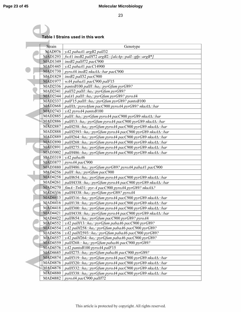
Table I Strains used in this work Strain Genotype
MAD976 yA2 pabaA1 argB2 palI32
MAD1293 fwA1 inoB2 palH72 argB2::[alcAp::palI::gfp::argB*]
MAD1349 inoB2 palH72 pacC900
MAD1445 yA2 pabaA1 pacC14900
MAD1730 pyroA4 inoB2 nkuA∆::bar pacC900
MAD1829 inoB2 palI32 pacC900
MAD1977 wA4 pabaA1 pacC900 palF15
MAD2336 pantoB100 palH::ha3::pyrGfum pyrG89?
MAD2341 palI32 palH::ha3::pyrGfum pyrG89?
MAD2344 palA1 palH::ha3::pyrGfum pyrG89? pyroA4
MAD2337 palF15 palH::ha3::pyrGfum pyrG89? pantoB100
MAD2668 palH∆::pyroAfum pacC900 pyroA4 pyrG89? nkuA∆::bar
MAD2743 yA2 pyroA4 pantoB100
MAD2885 palH::ha3::pyrGfum pyroA4 pacC900 pyrG89 nkuA∆::bar
MAD2886 palH13::ha3::pyrGfum pyroA4 pacC900 pyrG89 nkuA∆::bar
MAD2887 palH258::ha3::pyrGfum pyroA4 pacC900 pyrG89 nkuA∆::bar
MAD2888 palH2593::ha3::pyrGfum pyroA4 pacC900 pyrG89 nkuA∆::bar
MAD2889 palH264::ha3::pyrGfum pyroA4 pacC900 pyrG89 nkuA∆::bar
MAD2890 palH268::ha3::pyrGfum pyroA4 pacC900 pyrG89 nkuA∆::bar
MAD2891 palH275::ha3::pyrGfum pyroA4 pacC900 pyrG89 nkuA∆::bar
MAD3002 palH486::ha3::pyrGfum pyroA4 pacC900 pyrG89 nkuA∆::bar
MAD3319 yA2 pabaA6
MAD3877 pyroA4 pacC900
MAD3880 palH486::ha3::pyrGfum pyrG89? pyroA4 pabaA1 pacC900
MAD4256 palH::ha3::pyrGfum pacC900
MAD4258 palH654::ha3::pyrGfum pyroA4 pacC900 pyrG89 nkuA∆::bar
MAD4261 palH4338::ha3::pyrGfum pyroA4 pacC900 pyrG89 nkuA∆::bar
MAD4270 fimA::Tn431::pyr-4 pacC900 pyroA4 pyrG89? nkuA∆?
MAD4336 palH4338::ha3::pyrGfum pyrG89? pyroA4
MAD4413 palH316::ha3::pyrGfum pyroA4 pacC900 pyrG89 nkuA∆::bar
MAD4416 palH138::ha3::pyrGfum pyroA4 pacC900 pyrG89 nkuA∆::bar
MAD4418 palH300::ha3::pyrGfum pyroA4 pacC900 pyrG89 nkuA∆::bar
MAD4421 palH4338::ha3::pyrGfum pyroA4 pacC900 pyrG89 nkuA∆::bar
MAD4422 palH654::ha3::pyrGfum pacC900 pyrG89? pyroA4
MAD4552 yA2 palH13::ha3::pyrGfum pabaA6 pacC900 pyrG89?
MAD4554 yA2 palH258::ha3::pyrGfum pabaA6 pacC900 pyrG89?
MAD4556 yA2 palH2593::ha3::pyrGfum pabaA6 pacC900 pyrG89?
MAD4557 yA2 palH264::ha3::pyrGfum pabaA6 pacC900 pyrG89?
MAD4559 palH268:: ha3::pyrGfum pabaA6 pacC900 pyrG89?
MAD4576 yA2 pantoB100 pyroA4 palF15
MAD4685 palH275::ha3::pyrGfum pabaA6 pacC900 pyrG89?
MAD4874 palH319::ha3::pyrGfum pyroA4 pacC900 pyrG89 nkuA∆::bar
MAD4876 palH320::ha3::pyrGfum pyroA4 pacC900 pyrG89 nkuA∆::bar
MAD4878 palH332::ha3::pyrGfum pyroA4 pacC900 pyrG89 nkuA∆::bar
MAD4880 palH338::ha3::pyrGfum pyroA4 pacC900 pyrG89 nkuA∆::bar
MAD4882 pyroA4 pacC900 palH72
Page 23 of 45 Molecular Microbiology
This article is protected by copyright. All rights reserved.

24
MAD5140 palH2332::ha3::pyrGfum pyroA4 pacC900 pyrG89 nkuA∆::bar
MAD5142 palH25964::ha3::pyrGfum pyroA4 pacC900 pyrG89 nkuA∆::bar
MAD5205 palI32 palH4338::ha3::pyrGfum pyroA4 pacC900 pyrG89? nkuA∆?
MAD5207 wA2 palH::ha3::pyrGfum fimA::Tn431::pyr-4 pacC900 pyroA4 pyrG89? nkuA∆?
MAD5216 yA2 pabaA6 palH316::ha3::pyrGfum pyrG89? pacC900
MAD5218 pabaA6 palH138::ha3::pyrGfum pyrG89? pacC900
MAD5220 yA2 palH300::ha3::pyrGfum pyroA4 pabaA6 pyrG89? pacC900
MAD5224 palH654::ha3::pyrGfum pyroA4 pacC900 pyrG89? nkuA∆::bar
MAD5225 palF15 palH4338::ha3::pyrGfum pyroA4 pacC900 nkuA∆?
Page 24 of 45Molecular Microbiology
This article is protected by copyright. All rights reserved.
25
Figure legends
Figure 1: Scheme of PalH. The positions of the predicted TM domains and connecting loops relative to the lipid bilayer (olive green) are depicted. Amino acid substitutions corresponding to ‘classical’ and engineered missense mutations, the positions of truncations by ‘classical’ nonsense or frameshift alleles, the PalF arrestin binding regions (FABRs) in the cytosolic tail and the residues and borders of regions removed by deletions designed for this work are indicated.
Figure 2: PalH phosphorylation in alkaline pH conditions (A) Top, time course of PalH phosphorylation upon shifting cells from acidic to alkaline pH conditions for the indicated times. PalH-P is phosphorylated PalH. Bottom, time-course of PacC processing in the same cells. In all PacC western blots the ‘empty’ gel regions located between PacC72 and PacC53 and between the latter and PacC27 are not shown. Actin serves as loading control. (B) The two bands of PalH with different mobility coalesce into a single faster moving band after lambda bacteriophage phosphatase treatment (samples from cells shifted to alkaline pH for 15 min). (C) Time course of PalH dephosphorylation in cells that had been shifted from acidic to alkaline pH for 150 min and then back to acidic conditions for the indicated times. The bottom panel shows the corresponding anti-PacC western blot. The relative levels of PacC53 and PacC27 change little over the time scale of this experiment, whereas PacC72 accumulates gradually, reflecting the fact that the pathway is inactive. (D) A similar time course experiment in which cells were sampled between 45 min and 4.5 h after shifting them down to acidic pH. Note how PacC27 gradually disappears due to degradation and the absence of signaling proteolysis, combined with transcriptional repression of the pacC promoter mediated by PacC72 (Bussink et al., 2015). (E) PalH phosphorylation is PalI- and PalA-dependent and PalF-independent. palF15, palI32 and palA1 are phenotypically null mutations in the corresponding genes.
Figure 3: mutant PalH with a cytosolic tail containing just the arrestin binding regions (FABRs) retains substantial function. (A) Scheme of the mutant palH654 product compared to the wild-type. (B) Time course of PalH phosphorylation and PacC processing in pH shift experiments with palH654 and wild-type cells. (C) Diagnostic growth tests of pH signaling in the indicated strains. Note that although almost unable to grow on plates containing Li+, palH654 grows like the wild-type at pH 8.3, a pH at which the null palH72 strain is unable to grow. palH654 also displays a noticeably stronger resistance to molybdate than palH72, although lesser than the wild-type.
Figure 4: A conserved region rich in Pro, Ser and Thr residues is required for PalH phosphorylation (A) Multiple sequence alignment showing conservation of the PalH cytosolic tail region located between resides 450 and 486. Blue and red dots indicate Pro and Ser/Thr residues, respectively, in A. nidulans PalH. Amino acid highlighting in yellow and blue indicates strong and weak conservation, respectively. ANID, A. nidulans; ANIG, A. niger; SSCL, Sclerotinia sclerotium; PMAR, Penicillium marneffei; PTRI, Pyrenophora tritici-repentis; BCYN, Botrytis cinerea; CIMM, Coccidioides immitis; MGRI, Magnaporthe grisea; NCRA, Neurospora crassa. (B) Western blot analysis of equally loaded samples showing slightly elevated levels of the palH486 mutant product. (C) pH shift experiment showing the apparent absence of alkaline pH-induced phosphorylation
Page 25 of 45 Molecular Microbiology
This article is protected by copyright. All rights reserved.

26
(as determined by changes in SDS-PAGE mobility) of the palH486 mutant product. (D). palH486 is indistinguishable from the wild-type on pH 8.3, molybdate and neomycin media. (E) palH486 is hypersensitive to Li+. Different clones of a cross between palH486 and palH+ parental strains were point inoculated onto medium containing 0.3 M Li+. Yellow and green dots indicate strains that had been genotyped as palH486 and palH+, respectively. Red dots indicate a palH72 strain included as control. Yellow and green surface palH+ colonies reflect the fact that the yellow spore marker yA2 is segregating in the cross (wild-type spores are green; spores are not visible in palH486 clones due their impaired growth on lithium medium). (F) Comparison of the PacC proteolytic processing activation pattern of palH486 with the wild-type during a pH shift experiment. Note the slightly delayed processing and the slower increase in PacC72 in the mutant after long times at alkaline pH.
Figure 5: iImpairment of endocytosis results in phosphorylated PalH under acidic conditions and elevates the levels of the receptor Western blot analysis of PalH in a null fimbrin (fimA∆) mutant compared to the wild-type. Two different exposures of same the blot are shown. Hexokinase was used as loading control.
Figure 6: Mutational analysis of PalH: a potential agonist binding site (A) Complete catalogue of mutations reported here (with the exception of internal deletion alleles described in the text and Figure 1). For missense mutations (top), ‘classic’ and HA3 indicate whether the allele was obtained by UV or site-directed mutagenesis, respectively (the latter were all HA3-tagged). N.D. is ‘not done’ whereas N.S. is ‘not selected’. The red line boxes those missense mutations that we hypothesize comprise an agonist binding site. For nonsense/frameshifting mutations (bottom) their phenotypes are indicated. Note that mutant PalH proteins encoded by leaky truncating alleles contain the TM domain-proximal PalF binding region (Herranz et al., 2005), which explains their partial loss-of-function phenotype. palH45 and palH47, described by Negrete-Urtasun et al. (Negrete-Urtasun et al., 1999) and Herranz et al. (Herranz et al., 2005) are also in this category. (B) Multiple sequence alignment in the PalH region containing missense mutations hypothetically representing an agonist binding site. Species are as in Figure 4 with the exception of S. cerevisiae, whose Rim21p is included to underline the remarkable sequence conservation existing between filamentous fungi and budding yeast homologues in TM5.
Figure 7: Molecular characterization of single missense mutants affecting extracellular residues Time courses of PalH phosphorylation and PacC processing in pH shift experiments with cells carrying single residue substitutions in residues exposed to the ambient. The colors of the dots indicating mutated residues in the PalH scheme correspond with those of the different panel labels. Mutations are denoted by the resulting substitutions, in single letter amino acid code (see Fig. 6 for standard allele nomenclature and corresponding DNA changes).
Figure 8. Phenotypic additivity of Y259D and P264L: molecular characterization (A) Diagnostic growth tests showing phenotypic additivity between palH259 (Y259D) and palH264 (P264L). palH2264 encodes a doubly-substituted PalH. palH72 is a null palH mutation. (B) The time courses of alkaline pH-induced wild-type and Y259D, P264L double mutant PalH phosphorylation are very similar. (C) Effects of the double
Page 26 of 45Molecular Microbiology
This article is protected by copyright. All rights reserved.

27
mutant substitution on the signaling proteolysis of PacC72; (Note that for emphasis only the region of the gel containing PacC72 and PacC53 is included).
Figure 9. Characterization of palH4338 resulting in pH signaling pathway activation at acidic pH (A) Multiple sequence alignment showing the region corresponding to TM6 and TM7, as in Fig. 6 (Rim21 is from S. cerevisiae). The positions of residues that were mutated to investigate the hypothetical involvement in alkaline pH signaling of conformational rearrangements of the helical bundle of transmembrane helices are indicated. (B) Diagnostic growth tests of pH signaling in which palH4338 is compared to the wild-type and to a strong alkalinity-mimicking pacCc14 allele. Note the intermediate response of palH4338 on molybdate and neomycin plates (boxed in red). (C) palH4338 results in weak processing of PacC72 at acidic pH (pH 5.5) (the arrow indicates PacC27, the end product of the proteolytic cascade). Note the faint degree of PalH phosphorylation at pH 5.5 seen with the mutant but not with the wild-type. Cells were cultured overnight at ‘constant’ pH before proceeding to total protein extraction followed by anti-PacC (Myc3) and anti-PalH (HA3) western blotting. Actin was used as loading control (D) Diagnostic tests of pH regulation showing that palF15 is epistatic to palH4338 whereas palI32 is hypostatic. Figure 10. Phe319 and Trp332 seem important to maintain the basal activity of PalH (A) Diagnostic growth tests of pH regulation in the indicated strains. (B) Scheme showing the positions of Phe319 and Trp332 in PalH. (C) Effects of the double mutant substitution on the proteolytic processing of PacC72 in the steady state situation at the indicated pH; Note the weak activation of the pathway as indicated by detection of a faint band of PacC27.
Figure 11: Hypothetical model for the transduction of the alkaline ambient pH signal in A. nidulans
PalH alternates between basal and active conformations. Unlike β−arrestins PalF is bound to the receptor even under acidic (i.e. non-signaling) conditions, which drive the receptor towards the basal conformation (A). (B) Alkalization of the medium generates an ‘agonist’, perhaps a lipid of the external leaflet of the plasma membrane, that upon change of its ionization status binds a region located at the ambient-exposed interface between TM4 and TM5. Agonist binding drives the equilibrium towards the active conformation. We speculate that the transition from the basal to the active conformation results from an alkaline pH- and TM6 Pro316-dependent shift in the orientation of TM6 and TM7, a shift that is also promoted by palHc4338 independently of ambient pH (C). This shift would be then transmitted to arrestin-binding regions in the cytosolic PalH tail (D), leading to a conformational change that facilitates the recruitment of the HulA/Rsp5 ubiquitin ligase (E) and the subsequent ubiquitination of PalF (F). Next ubiquitinated PalF recruits ESCRT-I Vps23 (G), which primes ESCRT-III polymerization that, we hypothesize, serves as a signal amplification step because the remaining Pal proteins acting downstream of PalF (PalA, PalC and PalB) are all ESCRT-III interactors that are transiently localized to punctate structures at the plasma membrane when ambient pH is alkalinized.
Page 27 of 45 Molecular Microbiology
This article is protected by copyright. All rights reserved.

28
References Alvarez, C. E. (2008) On the origins of arrestin and rhodopsin. BMC Evol Biol 8: 222. Arst, H. N., Jr., Bignell, E., and Tilburn, J. (1994) Two new genes involved in signalling
ambient pH in Aspergillus nidulans Mol Gen Genet 245: 787-790. Arst, H. N., Jr., and Peñalva, M. A. (2003) pH regulation in Aspergillus and parallels
with higher eukaryotic regulatory systems. Trends Genet. 19: 224-231. Bertuzzi, M., Schrettl, M., Alcazar-Fuoli, L., Cairns, T. C., Munoz, A., Walker, L. A.,
Herbst, S., Safari, M., Cheverton, A. M., Chen, D., Liu, H., Saijo, S., Fedorova, N. D., Armstrong-James, D., Munro, C. A., Read, N. D., Filler, S. G., Espeso, E. A., Nierman, W. C., Haas, H., and Bignell, E. M. (2014) The pH-responsive PacC transcription factor of Aspergillus fumigatus governs epithelial entry and tissue invasion during pulmonary aspergillosis. PLoS Pathog 10: e1004413.
Bignell, E., Negrete-Urtasun, S., Calcagno, A. M., Haynes, K., Arst, H. N., Jr., and Rogers, T. (2005) The Aspergillus pH-responsive transcription factor PacC regulates virulence. Mol Microbiol 55 1072-1084.
Bussink, H. J., Bignell, E. M., Munera-Huertas, T., Lucena-Agell, D., Scazzocchio, C., Espeso, E. A., Bertuzzi, M., Rudnicka, J., Negrete-Urtasun, S., Peñas-Parilla, M. M., Rainbow, L., Peñalva, M. A., Arst, H. N., Jr., and Tilburn, J. (2015) Refining the pH response in Aspergillus nidulans: a modulatory triad involving PacX, a novel zinc binuclear cluster protein. Mol Microbiol.
Caddick, M. X., Brownlee, A. G., and Arst, H. N., Jr. (1986) Regulation of gene expression by pH of the growth medium in Aspergillus nidulans Mol Gen Genet 203: 346-353.
Calcagno-Pizarelli, A. M., Hervás-Aguilar, A., Galindo, A., Abenza, J. F., Peñalva, M. A., and Arst, H. N., Jr. (2011) Rescue of Aspergillus nidulans severely debilitating null mutations in ESCRT-0, I, II and III genes by inactivation of a salt-tolerance pathway allows examination of ESCRT gene roles in pH signalling. J Cell Sci 124: 1-13.
Calcagno-Pizarelli, A. M., Negrete-Urtasun, S., Denison, S. H., Rudnicka, J. D., Bussink, H. J., Munera-Huertas, T., Stanton, L., Hervás-Aguilar, A., Espeso, E. A., Tilburn, J., Arst, H. N., Jr., and Peñalva, M. A. (2007) Establishment of the ambient pH signaling complex in Aspergillus nidulans : PalI assists plasma membrane localization of PalH. Eukaryot Cell 6: 2365-2375.
Cervantes-Chávez, J. A., Ortiz-Castellanos, L., Tejeda-Sartorius, M., Gold, S., and Ruiz-Herrera, J. (2010) Functional analysis of the pH responsive pathway Pal/Rim in the phytopathogenic basidiomycete Ustilago maydis. Fungal Genet Biol 47: 446-457.
Chalmers, D. T., and Behan, D. P. (2002) The use of constitutively active GPCRs in drug discovery and functional genomics. Nat Rev Drug Discov 1: 599-608.
Chen, W., Ren, X. R., Nelson, C. D., Barak, L. S., Chen, J. K., Beachy, P. A., de Sauvage, F., and Lefkowitz, R. J. (2004) Activity-dependent internalization of Smoothened mediated by ß-arrestin 2 and GRK2. Science 306: 2257-2260.
Cornet, M., and Gaillardin, C. (2014) pH Signaling in Human Fungal Pathogens: a New Target for Antifungal Strategies. Eukaryot Cell 13: 342-352.
Cove, D. J. (1966) The induction and repression of nitrate reductase in the fungus Aspergillus nidulans. Biochim Biophys Acta 113: 51-56.
Davis, D., Edwards, J. E., Jr., Mitchell, A. P., and Ibrahim, A. S. (2000) Candida albicans RIM101 pH response pathway is required for host-pathogen interactions. Infect Immun 68: 5953-5959.
Davis, D. A. (2009) How human pathogenic fungi sense and adapt to pH: the link to virulence. Curr Opin Microbiol 12: 365-370.
Díez, E., Álvaro, J., Espeso, E. A., Rainbow, L., Suárez, T., Tilburn, J., Arst, H. N., Jr., and Peñalva, M. A. (2002) Activation of the Aspergillus PacC zinc-finger transcription factor requires two proteolytic steps EMBO J 21 1350-1359.
Page 28 of 45Molecular Microbiology
This article is protected by copyright. All rights reserved.

29
Espeso, E. A., and Arst, H. N., Jr. (2000) On the mechanism by which alkaline pH prevents expression of an acid-expressed gene. Mol Cell Biol 20: 3355-3363.
Galindo, A., Calcagno-Pizarelli, A. M., Arst, H. N., Jr., and Peñalva, M. A. (2012) An ordered pathway for the assembly of ESCRT-containing fungal ambient pH signalling complexes at the plasma membrane. J Cell Sci 125: 1784-1795.
Galindo, A., Hervás-Aguilar, A., Rodríguez-Galán, O., Vincent, O., Arst, H. N., Jr., Tilburn, J., and Peñalva, M. A. (2007) PalC, one of two Bro1 domain proteins in the fungal pH signaling pathway, localizes to cortical structures and binds Vps32. Traffic 8: 1346-1364.
Gunatilleke, I. A. U. N., Scazzocchio, C., and Arst, H. N., Jr. (1975) Cytoplasmic and nuclear mutations to chloramphenicol resistance in Aspergillus nidulans. Mol Gen Genet 137: 269-276.
Hanson, M. A., Roth, C. B., Jo, E., Griffith, M. T., Scott, F. L., Reinhart, G., Desale, H., Clemons, B., Cahalan, S. M., Schuerer, S. C., Sanna, M. G., Han, G. W., Kuhn, P., Rosen, H., and Stevens, R. C. (2012) Crystal structure of a lipid G Protein–coupled receptor. Science 335: 851-855.
Hastie, A. C. (1970) Benlate-induced Instability of Aspergillus diploids. Nature 226: 771-771.
Herrador, A., Herranz, S., Lara, D., and Vincent, O. (2009) Recruitment of the ESCRT machinery to a putative seven-transmembrane-domain receptor is mediated by an arrestin-related protein. Mol Cell Biol 30: 897-907.
Herrador, A., Livas, D., Soletto, L., Becuwe, M., Leon, S., and Vincent, O. (2015) Casein kinase 1 controls the activation threshold of an alpha-arrestin by multisite phosphorylation of the interdomain hinge. Mol Biol Cell 26: 2128-2138.
Herranz, S., Rodríguez, J. M., Bussink, H. J., Sánchez-Ferrero, J. C., Arst, H. N., Jr., Peñalva, M. A., and Vincent, O. (2005) Arrestin-related proteins mediate pH signaling in fungi. Proc Natl Acad Sci USA 102: 12141-12146.
Hervás-Aguilar, A., Galindo, A., and Peñalva, M. A. (2010) Receptor-independent ambient pH signaling by ubiquitin attachment to fungal arrestin-like PalF. J Biol Chem 285: 18095-18102.
Hervás-Aguilar, A., and Peñalva, M. A. (2010) Endocytic machinery protein SlaB is dispensable for polarity establishment but necessary for polarity maintenance in hyphal tip cells of Aspergillus nidulans. Eukaryot Cell 9: 1504-1518.
Hervás-Aguilar, A., Rodríguez, J. M., Tilburn, J., Arst, H. N., Jr., and Peñalva, M. A. (2007) Evidence for the direct involvement of the proteasome in the proteolytic processing of the Aspergillus nidulans zinc finger transcription factor PacC. J Biol Chem 282: 34735-34747.
Hirsch, J. A., Schubert, C., Gurevich, V. V., and Sigler, P. B. (1999) The 2.8 A crystal structure of visual arrestin: a model for arrestin's regulation. Cell 97: 257-269.
Hutchings, H., Stahmann, K., Roels, S., Espeso, E. A., Timberlake, W. E., Arst, H. N., Jr., and Tilburn, J. (1999) The multiply-regulated gabA gene encoding the GABA permease of Aspergillus nidulans : a score of exons. Mol Microbiol 32: 557-568.
Ikeda, M., Kihara, A., Denpoh, A., and Igarashi, Y. (2008) The Rim101 pathway is involved in Rsb1 expression induced by altered lipid asymmetry. Mol Biol Cell 19: 1922-1931.
Karachaliou, M., Amillis, S., Evangelinos, M., Kokotos, A. C., Yalelis, V., and Diallinas, G. (2013) The arrestin-like protein ArtA is essential for ubiquitination and endocytosis of the UapA transporter in response to both broad-range and specific signals. Mol Microbiol 88: 301-317.
Katritch, V., Cherezov, V., and Stevens, R. C. (2013) Structure-function of the G protein-coupled receptor superfamily. Annu Rev Pharmacol Toxicol 53: 531-556.
Page 29 of 45 Molecular Microbiology
This article is protected by copyright. All rights reserved.

30
Lefkowitz, R. J., (2013) Chapter One - Arrestins Come of Age: A Personal Historical Perspective. In: Progress in Molecular Biology and Translational Science. M. L. Louis (ed). Academic Press, pp. 3-18.
Lefkowitz, R. J., and Shenoy, S. K. (2005) Transduction of receptor signals by beta-arrestins. Science 308: 512-517.
Lin, C. H., MacGurn, J. A., Chu, T., Stefan, C. J., and Emr, S. D. (2008) Arrestin-related ubiquitin-ligase adaptors regulate endocytosis and protein turnover at the cell surface. Cell 135: 714-725.
Liu, J. J., Horst, R., Katritch, V., Stevens, R. C., and Wüthrich, K. (2012) Biased signaling pathways in β2-ddrenergic receptor characterized by 19F-NMR. Science 335: 1106-1110.
Lucena-Agell, D., Galindo, A., Arst, H. N., Jr., and Peñalva, M. A. (2015) Aspergillus nidulans ambient pH signaling does not require endocytosis. Eukaryot Cell 14: 545-553.
Mingot, J. M., Tilburn, J., Díez, E., Bignell, E., Orejas, M., Widdick, D. A., Sarkar, S., Brown, C. V., Caddick, M. X., Espeso, E. A., Arst, H. N., Jr., and Peñalva, M. A. (1999) Specificity determinants of proteolytic processing of Aspergillus PacC transcription factor are remote from the processing site, and processing occurs in yeast if pH signalling is bypassed. Mol Cell Biol 19: 1390-1400.
Negrete-Urtasun, S., Reiter, W., Díez, E., Denison, S. H., Tilburn, J., Espeso, E. A., Peñalva, M. A., and Arst, H. N., Jr. (1999) Ambient pH signal transduction in Aspergillus : completion of gene characterization. Mol Microbiol 33: 994-1003.
Nikko, E., and Pelham, H. R. (2009) Arrestin-mediated endocytosis of yeast plasma membrane transporters. Traffic 10: 1856-1867.
Nishino, K., Obara, K., and Kihara, A. (2015) The C-terminal cytosolic region of Rim21 senses alterations in plasma membrane lipid composition: Insights into sensing mechanisms for plasma membrane lipid asymmetry. J Biol Chem 290: 30797-30805.
Nobile, C. J., Solis, N., Myers, C. L., Fay, A. J., Deneault, J.-S., Nantel, A., Mitchell, A. P., and Filler, S. G. (2008) Candida albicans transcription factor Rim101 mediates pathogenic interactions through cell wall functions. Cellular Microbiology 10: 2180-2196.
O'Meara, T. R., Holmer, S. M., Selvig, K., Dietrich, F., and Alspaugh, J. A. (2013) Cryptococcus neoformans Rim101 Is associated with cell wall remodeling and evasion of the host immune responses. mBio 4: e00522-00512.
O'Meara, T. R., Norton, D., Price, M. S., Hay, C., Clements, M. F., Nichols, C. B., and Alspaugh, J. A. (2010) Interaction of Cryptococcus neoformans Rim101 and protein kinase A regulates capsule. PLoS Pathog 6: e1000776.
O'Meara, T. R., Xu, W., Selvig, K. M., O'Meara, M. J., Mitchell, A. P., and Alspaugh, J. A. (2014) The Cryptococcus neoformans Rim101 transcription factor directly regulates genes required for adaptation to the host. Mol Cell Biol 34: 673-684.
Obara, K., and Kihara, A. (2014) Signaling events of the Rim101 pathway occur at the plasma membrane in a ubiquitination-dependent manner. Mol Cell Biol 34: 3525–3534.
Obara, K., Yamamoto, H., and Kihara, A. (2012) Membrane protein Rim21 plays a central role in sensing ambient pH in Saccharomyces cerevisiae. J Biol Chem 287: 38473-38481.
Ost, K. S., O’Meara, T. R., Huda, N., Esher, S. K., and Alspaugh, J. A. (2015) The Cryptococcus neoformans alkaline response pathway: identification of a novel Rim pathway activator. PLoS Genet 11: e1005159.
Peñalva, M. A., and Arst, H. N., Jr. (2002) Regulation of gene expression by ambient pH in filamentous fungi and yeasts. Microbiol Mol Biol Rev 66: 426-446.
Peñalva, M. A., and Arst, H. N., Jr. (2004) Recent advances in the characterization of ambient pH regulation of gene expression in filamentous fungi and yeasts. Annu Rev Microbiol. 58 425-451.
Page 30 of 45Molecular Microbiology
This article is protected by copyright. All rights reserved.

31
Peñalva, M. A., Lucena-Agell, D., and Arst, H. N., Jr. (2014) Liaison alcaline: Pals entice non-endosomal ESCRTs to the plasma membrane for pH signaling. Curr Opin Microbiol 22: 49-59.
Peñalva, M. A., Tilburn, J., Bignell, E., and Arst, H. N., Jr. (2008) Ambient pH gene regulation in fungi: making connections. Trends Microbiol. 16: 291-300.
Peñas, M. M., Hervás-Aguilar, A., Múnera-Huertas, T., Reoyo, E., Peñalva, M. A., Arst, H. N., Jr., and Tilburn, J. (2007) Further characterization of the signaling proteolysis step in the Aspergillus nidulans pH signal transduction pathway. Eukaryot Cell 6 960-970.
Rana, R., Carroll, C. E., Lee, H.-J., Bao, J., Marada, S., Grace, C. R. R., Guibao, C. D., Ogden, S. K., and Zheng, J. J. (2013) Structural insights into the role of the Smoothened cysteine-rich domain in Hedgehog signalling. Nat Commun 4: 2965.
Rasmussen, S. G., Choi, H. J., Fung, J. J., Pardon, E., Casarosa, P., Chae, P. S., Devree, B. T., Rosenbaum, D. M., Thian, F. S., Kobilka, T. S., Schnapp, A., Konetzki, I., Sunahara, R. K., Gellman, S. H., Pautsch, A., Steyaert, J., Weis, W. I., and Kobilka, B. K. (2011) Structure of a nanobody-stabilized active state of the beta(2) adrenoceptor. Nature 469: 175-180.
Rodríguez-Galán, O., Galindo, A., Hervás-Aguilar, A., Arst, H. N., Jr., and Peñalva, M. A. (2009) Physiological involvement in pH signalling of Vps24-mediated recruitment of Aspergillus PalB cysteine protease to ESCRT-III. J Biol Chem 284: 4404-4412.
Rosenbaum, D. M., Zhang, C., Lyons, J. A., Holl, R., Aragao, D., Arlow, D. H., Rasmussen, S. G., Choi, H. J., Devree, B. T., Sunahara, R. K., Chae, P. S., Gellman, S. H., Dror, R. O., Shaw, D. E., Weis, W. I., Caffrey, M., Gmeiner, P., and Kobilka, B. K. (2011) Structure and function of an irreversible agonist-ß2 adrenoceptor complex. Nature 469: 236-240.
Rothfels, K., Tanny, J. C., Molnar, E., Friesen, H., Commisso, C., and Segall, J. (2005) Components of the ESCRT pathway, DFG16, and YGR122w are required for Rim101 to act as a corepressor with Nrg1 at the negative regulatory element of the DIT1 gene of Saccharomyces cerevisiae Mol Cell Biol 25: 6772-6788.
Schwartz, T. W., Frimurer, T. M., Holst, B., Rosenkilde, M. M., and Elling, C. E. (2006) Molecular mechanism of 7TM receptor activation - a global toggle switch model. Annu Rev Pharmacol Toxicol 46: 481-519.
Shi, L., Liapakis, G., Xu, R., Guarnieri, F., Ballesteros, J. A., and Javitch, J. A. (2002) ß2 adrenergic receptor activation. Modulation of the proline kink in transmembrane 6 by a rotamer toggle switch. J Biol Chem 277: 40989-40996.
Shukla, A. K., Manglik, A., Kruse, A. C., Xiao, K., Reis, R. I., Tseng, W.-C., Staus, D. P., Hilger, D., Uysal, S., Huang, L.-Y., Paduch, M., Tripathi-Shukla, P., Koide, A., Koide, S., Weis, W. I., Kossiakoff, A. A., Kobilka, B. K., and Lefkowitz, R. J. (2013) Structure of active ß-arrestin-1 bound to a G-protein-coupled receptor phosphopeptide. Nature 497: 137-141.
Shukla, A. K., Xiao, K., and Lefkowitz, R. J. (2011) Emerging paradigms of β-arrestin-dependent seven transmembrane receptor signaling. Trends Biochem Sci 36: 457-469.
Sprang, S. R. (2011) Cell signalling: Binding the receptor at both ends. Nature 469: 172-173.
Tilburn, J., Sánchez-Ferrero, J. C., Reoyo, E., Arst, H. N., Jr., and Peñalva, M. A. (2005) Mutational analysis of the pH signal transduction component PalC of Aspergillus nidulans supports distant similarity to BRO1 domain family members. Genetics 171: 393-401.
Tilburn, J., Sarkar, S., Widdick, D. A., Espeso, E. A., Orejas, M., Mungroo, J., Peñalva, M. A., and Arst, H. N., Jr. (1995) The Aspergillus PacC zinc finger transcription factor mediates regulation of both acid- and alkaline-expressed genes by ambient pH. EMBO J 14: 779-790.
Page 31 of 45 Molecular Microbiology
This article is protected by copyright. All rights reserved.

32
Tréton, B., Blanchin-Roland, S., Lambert, M., Lepingle, A., and Gaillardin, C. (2000) Ambient pH signalling in ascomycetous yeasts involves homologues of the Aspergillus nidulans genes palF and paIH Mol Gen Genet 263: 505-513.
Upadhyay, S., and Shaw, B. D. (2008) The role of actin, fimbrin and endocytosis in growth of hyphae in Aspergillus nidulans. Mol Microbiol 68: 690-705.
Vincent, O., Rainbow, L., Tilburn, J., Arst, H. N., Jr., and Peñalva, M. A. (2003) YPXL/I is a protein interaction motif recognised by Aspergillus PalA and its human homologue AIP1/Alix. Mol Cell Biol 23: 1647-1655.
Violin, J. D., and Lefkowitz, R. J. (2007) ß-arrestin-biased ligands at seven-transmembrane receptors. Trends Pharmacol Sci 28: 416-422.
Wacker, D., Wang, C., Katritch, V., Han, G. W., Huang, X.-P., Vardy, E., McCorvy, J. D., Jiang, Y., Chu, M., Siu, F. Y., Liu, W., Xu, H. E., Cherezov, V., Roth, B. L., and Stevens, R. C. (2013) Structural features for functional selectivity at serotonin receptors. Science 340: 615-619.
Warne, T., Moukhametzianov, R., Baker, J. G., Nehme, R., Edwards, P. C., Leslie, A. G., Schertler, G. F., and Tate, C. G. (2011) The structural basis for agonist and partial agonist action on a ß1-adrenergic receptor. Nature 469: 241-244.
Wisler, J. W., DeWire, S. M., Whalen, E. J., Violin, J. D., Drake, M. T., Ahn, S., Shenoy, S. K., and Lefkowitz, R. J. (2007) A unique mechanism of ß-blocker action: carvedilol stimulates beta-arrestin signaling. Proc Natl Acad Sci USA 104: 16657-16662.
Xie, X.-Q., and Chowdhury, A., (2013) Chapter Seven - Advances in Methods to Characterize Ligand-Induced Ionic Lock and Rotamer Toggle Molecular Switch in G Protein-Coupled Receptors. In: Methods in Enzymology. P. M. Conn (ed). Academic Press, pp. 153-174.
Xu, W., and Mitchell, A. P. (2001) Yeast PalA/AIP1/Alix homolog Rim20p associates with a PEST-like region and is required for its proteolytic cleavage. J Bacteriol 183: 6917-6923.
Xu, W., Smith, F. J., Jr., Subaran, R., and Mitchell, A. P. (2004) Multivesicular body-ESCRT components function in pH response regulation in Saccharomyces cerevisiae and Candida albicans Mol Biol Cell 15: 5528-5537.
Page 32 of 45Molecular Microbiology
This article is protected by copyright. All rights reserved.

203x240mm (300 x 300 DPI)
Page 33 of 45 Molecular Microbiology
This article is protected by copyright. All rights reserved.
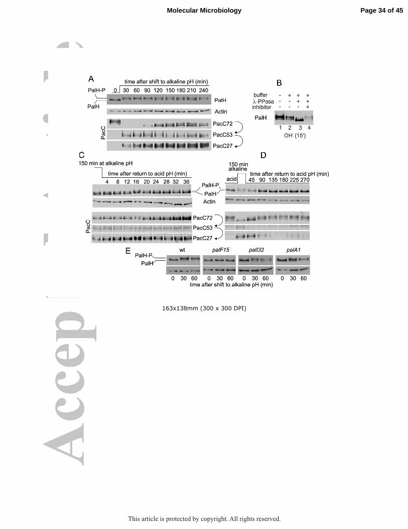
163x138mm (300 x 300 DPI)
Page 34 of 45Molecular Microbiology
This article is protected by copyright. All rights reserved.
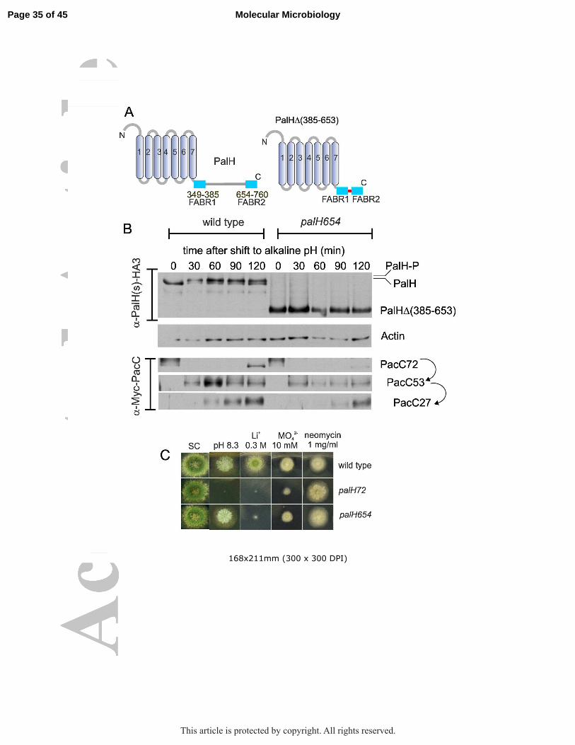
168x211mm (300 x 300 DPI)
Page 35 of 45 Molecular Microbiology
This article is protected by copyright. All rights reserved.
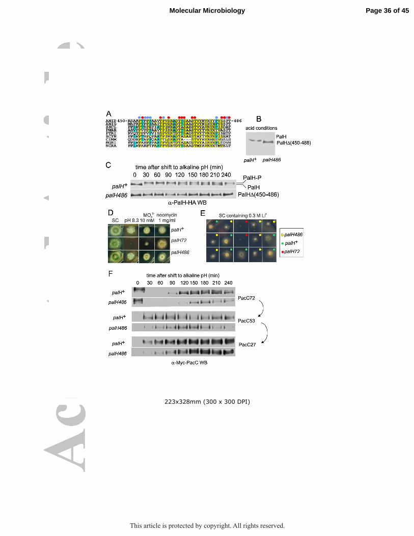
223x328mm (300 x 300 DPI)
Page 36 of 45Molecular Microbiology
This article is protected by copyright. All rights reserved.

31x10mm (300 x 300 DPI)
Page 37 of 45 Molecular Microbiology
This article is protected by copyright. All rights reserved.

173x251mm (300 x 300 DPI)
Page 38 of 45Molecular Microbiology
This article is protected by copyright. All rights reserved.

217x303mm (300 x 300 DPI)
Page 39 of 45 Molecular Microbiology
This article is protected by copyright. All rights reserved.

143x148mm (300 x 300 DPI)
Page 40 of 45Molecular Microbiology
This article is protected by copyright. All rights reserved.
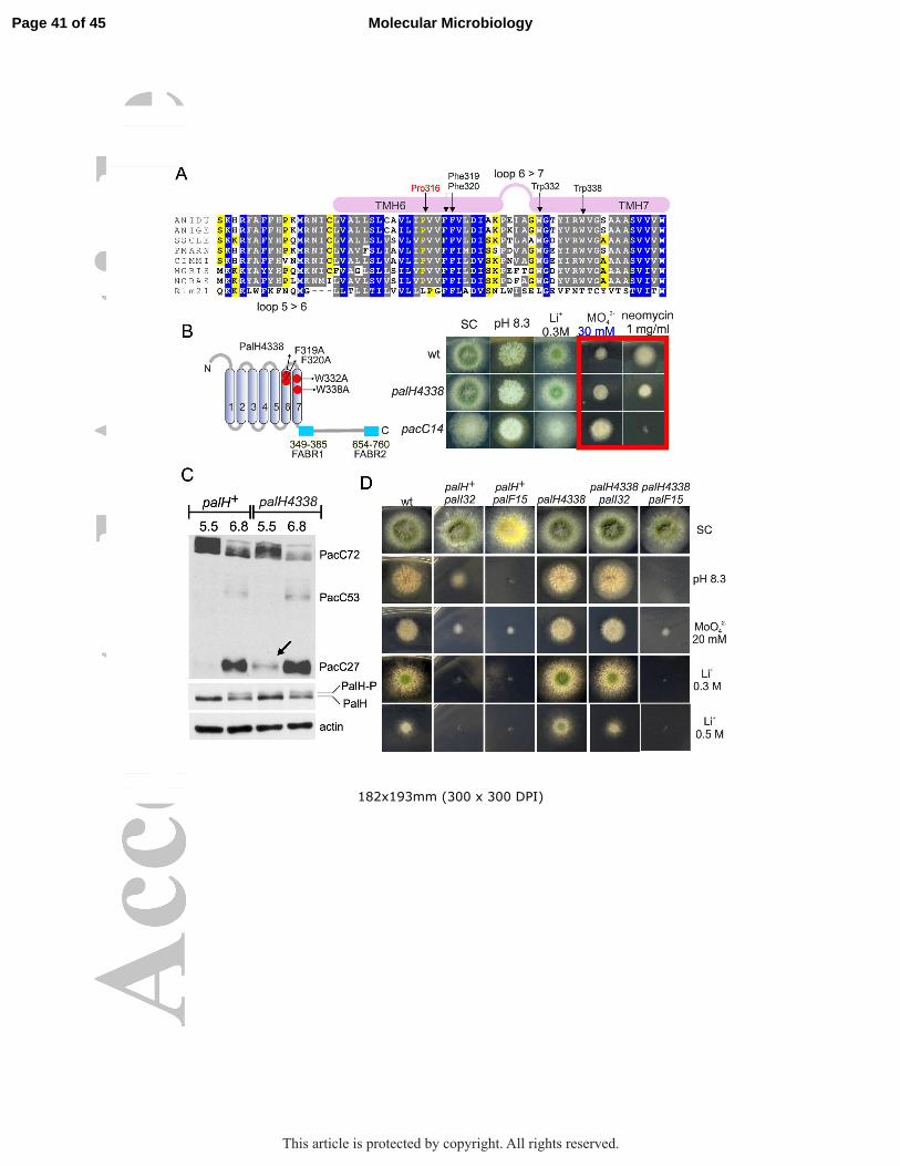
182x193mm (300 x 300 DPI)
Page 41 of 45 Molecular Microbiology
This article is protected by copyright. All rights reserved.

189x351mm (300 x 300 DPI)
Page 42 of 45Molecular Microbiology
This article is protected by copyright. All rights reserved.

259x168mm (300 x 300 DPI)
Page 43 of 45 Molecular Microbiology
This article is protected by copyright. All rights reserved.

103x65mm (300 x 300 DPI)
Page 44 of 45Molecular Microbiology
This article is protected by copyright. All rights reserved.

Abbreviated abstract
Fungi colonize environments with different values of pH by adapting their patterns of
gene expression to the needs imposed by ambient pH. pH regulation is important for
fungal pathogenicity, but therapeutically useful drugs targeting the pH signaling
pathway are yet to be developed. We show that the ascomycete ambient pH receptor
PalH resembles a major class of mammalian receptors, denoted GPCRs. Thus our
data pave the way for the identification of specific inhibitors of the ambient pH receptor
for antifungal intervention.
Page 45 of 45 Molecular Microbiology
This article is protected by copyright. All rights reserved.