Muscular and Nervous Tissue Chapter 4.3 Human Anatomy & Physiology.
24
Muscular and Nervous Tissue Chapter 4.3 Human Anatomy & Physiology
-
Upload
vivien-heath -
Category
Documents
-
view
231 -
download
1
Transcript of Muscular and Nervous Tissue Chapter 4.3 Human Anatomy & Physiology.

Muscular and Nervous Tissue
Chapter 4.3
Human Anatomy & Physiology

Muscular Tissue
• Function• Contraction• Attachment by
tendons to bones for movement
• Movement: Voluntary and involuntary


Muscular Tissue
• Appearance – striated (striped) – Alternating light and
dark bands
• Location – Usually attached to skeleton
• Each cell has a nucleus that is centrally located

Types of Muscular Tissue
• Types
A. Skeletal
B. Smooth
C. Cardiac

Characteristics of Skeletal Muscle
• Appearance: striated
• Location: Attached primarily to bones
• Control: Voluntary (conscious)
• Contracts quickly, tires easily (fatigable)
• Allows for wide range of forces to be generated

Skeletal Muscle Tissue - 400X

Smooth Muscle• Appearance: spindle-shaped• Location: wall of hollow
organs– example: Intestines, urinary
bladder, ureters, urinary bladder, blood vessels
• Control: Involuntary • Contracts rhythmically and
quickly, tires easily (fatigable)• Allows for wide range of
forces to be generated

Smooth Muscle

Smooth Muscle Tissue - 400X

Cardiac Muscle
• Has features of both skeletal and smooth muscle- Strong contractions and striated appearance is similar to skeletal muscle- Involuntary control and rhythmic contraction is similar to smooth muscle

• Appearance: striated and branched (like skeletal muscle)
• Location: heart• Function: contraction
of heart pumps blood and causes the heartbeat
• Control: Involuntary (like smooth muscle)

Cardiac Muscle Tissue - 400X

Nervous Tissue
• The ultimate control of all the organ systems is done by the nervous system.– The nervous system controls and coordinates
functions throughout the body and responds to internal and external stimuli.

Nervous Tissue
• Found: brain, spinal cord, has specialized cells
• The cells that transmit these impulses are called neurons.


Structure of a Neuron
Axon terminals
Myelin sheath
Cell body
Nodes Axon
Dendrites
Nucleus

Neuron Structure
• The largest part of a typical neuron is the cell body.
• It contains the nucleus and much of the cytoplasm.
Neuron Structure
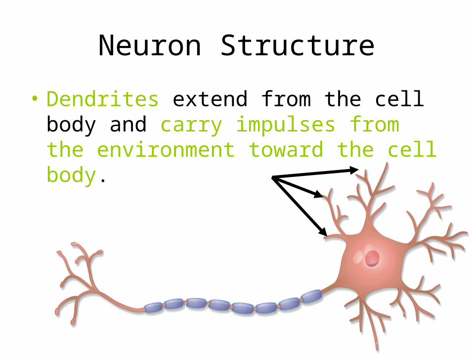
• Dendrites extend from the cell body and carry impulses from the environment toward the cell body.

Neuron Structure
• The axon is the long fiber that carries impulses away from the cell body.

Neuron Structure
• The axon is sometimes surrounded by an insulating membrane called the myelin sheath.


Neuron Structure
There are gaps in the myelin sheath, called nodes, where the membrane is exposed.
• Impulses jump from one node to the next.