Multigate Transcranial Doppler Ultrasound System … transactions on ultrasonics, ferroelectrics,...
9
ieee transactions on ultrasonics, ferroelectrics, and frequency control, vol. 53, no. 10, october 2006 1853 Multigate Transcranial Doppler Ultrasound System with Real-Time Embolic Signal Identification and Archival Lingke Fan, Member, IEEE, Enrico Boni, Piero Tortoli, Senior Member, IEEE, and David H. Evans, Senior Member, IEEE Abstract—An integrated system for acquisition and pro- cessing of intracranial and extracranial Doppler signals and automatic embolic signal detection has been developed. The hardware basis of the system is a purpose-built acquisi- tion/processing board that includes a multigate Doppler unit controlled through a computer. The signal-processing engine of the system contains a fast Fourier transform (FFT)-based, spectral-analysis unit and an embolic signal- detection unit using expert system reasoning theory. The system is designed so that up to four receive gates from a single transducer can be used to provide useful reasoning information to the embolic signal-detection unit. Alterna- tively, two transducers can be used simultaneously, either for bilateral transcranial Doppler (TCD) investigations or for simultaneous intra- and extracranial investigation of dif- ferent arteries. The structure of the software will allow the future implementation of embolus detection algorithms that use the information from all four channels when a single transducer is used, or of independent embolus detection in two sets of two channels when two transducers are used. The user-friendly system has been tested in-vitro, and it has demonstrated a 93.6% sensitivity for micro-embolic signal (MES) identification. Preliminary in-vivo results also are encouraging. I. Introduction D oppler ultrasound is a valuable technique for the de- tection of circulating emboli, whether they are solid or gaseous in nature [1]–[4]. The basis of the detection tech- nique is that, if the acoustic properties of the embolus are sufficiently different from those of the surrounding blood, then when it enters the Doppler sample volume there will be a transient increase in the power of the Doppler signal at a frequency corresponding to the velocity of the embolus through the sample volume [5]. This is not, of course, the only mechanism that can cause a transient rise in Doppler signal power; therefore, careful analysis of the signal is nec- essary to distinguish between signals arising from embolic events and those arising from other mechanisms such as probe movement or electrical interference. Traditionally, trained human observers have carried out this task, but Manuscript received July 7, 2005; accepted November 26, 2005. The authors gratefully acknowledge the financial support of the Eu- ropean Union (UMEDS-QLG1-CT-2002-01518). L. Fan and D. H. Evans are with the Medical Physics Group, De- partment of Cardiovascular Sciences, University of Leicester, Leices- ter, UK (e-mail: [email protected]). E. Boni and P. Tortoli are with the Department of Electronics and Telecommunications, University of Florence, Florence, Italy. Digital Object Identifier 10.1109/TUFFC.2006.117 there are many difficulties with this approach. Periods of monitoring may extend for several hours, and not only does this put great demands on the concentration of the observer, but it is costly. The environment in which the monitoring must take place is often both busy and noisy, which diminishes the ability of the observer to detect what may be subtle increases in power. And there is consid- erable evidence that, for many embolic signals, observer reproducibility is poor. Because of these difficulties, vari- ous systems have been developed in order to automate the detection task [6]–[18]. But to date no system has been acknowledged as being sufficiently reliable to replace the human observer in the clinical environment. This paper de- scribes the design and implementation of a new automated embolic signal identification and archival system (ESIAS) with flexible features that is intended to overcome some of the drawbacks of currently available systems. The new system consists of a custom-built board containing both a programmable dual-channel/multigate transcranial Doppler (TCD) unit and a digital signal pro- cessor (DSP), performing embolus detection. The embolus detection function was realized using expert system rea- soning theory to process information extracted from the time, frequency, energy, and neighborhood domains. The expert system structure was described previously in detail for an off-line implementation [12] and for a unidirectional, real-time implementation using an external TCD machine with a single gate [17]. The latest implementation, with a programmable dual-channel TCD unit, allows the de- velopment of algorithms incorporating multigate informa- tion from bi-directional Doppler signals. The expectation is that this will further improve the sensitivity and speci- ficity of the system. A user-friendly, Windows-based system control enables the operator to view, in real time, four spectrograms re- lated to echo-signals from either one or two probes con- nected to the unit. When an embolic event is detected, the data archival structure of the system automatically stores a pair of one-second long raw audio Doppler signals (for- ward and reverse flow directions) for each gate. The same structure also allows the manually triggered storage of the raw signals corresponding to the currently displayed sono- grams (a pair of 3.6-second segments of raw signal com- ponents for each gate), if the operator decides to further investigate what has been displayed on the screen. The system specification is summarized in Table I. 0885–3010/$20.00 c 2006 IEEE
Transcript of Multigate Transcranial Doppler Ultrasound System … transactions on ultrasonics, ferroelectrics,...

ieee transactions on ultrasonics, ferroelectrics, and frequency control, vol. 53, no. 10, october 2006 1853
Multigate Transcranial Doppler UltrasoundSystem with Real-Time Embolic Signal
Identification and ArchivalLingke Fan, Member, IEEE, Enrico Boni, Piero Tortoli, Senior Member, IEEE,
and David H. Evans, Senior Member, IEEE
Abstract—An integrated system for acquisition and pro-cessing of intracranial and extracranial Doppler signals andautomatic embolic signal detection has been developed. Thehardware basis of the system is a purpose-built acquisi-tion/processing board that includes a multigate Dopplerunit controlled through a computer. The signal-processingengine of the system contains a fast Fourier transform(FFT)-based, spectral-analysis unit and an embolic signal-detection unit using expert system reasoning theory. Thesystem is designed so that up to four receive gates from asingle transducer can be used to provide useful reasoninginformation to the embolic signal-detection unit. Alterna-tively, two transducers can be used simultaneously, eitherfor bilateral transcranial Doppler (TCD) investigations orfor simultaneous intra- and extracranial investigation of dif-ferent arteries. The structure of the software will allow thefuture implementation of embolus detection algorithms thatuse the information from all four channels when a singletransducer is used, or of independent embolus detectionin two sets of two channels when two transducers are used.The user-friendly system has been tested in-vitro, and it hasdemonstrated a 93.6% sensitivity for micro-embolic signal(MES) identification. Preliminary in-vivo results also areencouraging.
I. Introduction
Doppler ultrasound is a valuable technique for the de-tection of circulating emboli, whether they are solid or
gaseous in nature [1]–[4]. The basis of the detection tech-nique is that, if the acoustic properties of the embolus aresufficiently different from those of the surrounding blood,then when it enters the Doppler sample volume there willbe a transient increase in the power of the Doppler signalat a frequency corresponding to the velocity of the embolusthrough the sample volume [5]. This is not, of course, theonly mechanism that can cause a transient rise in Dopplersignal power; therefore, careful analysis of the signal is nec-essary to distinguish between signals arising from embolicevents and those arising from other mechanisms such asprobe movement or electrical interference. Traditionally,trained human observers have carried out this task, but
Manuscript received July 7, 2005; accepted November 26, 2005.The authors gratefully acknowledge the financial support of the Eu-ropean Union (UMEDS-QLG1-CT-2002-01518).
L. Fan and D. H. Evans are with the Medical Physics Group, De-partment of Cardiovascular Sciences, University of Leicester, Leices-ter, UK (e-mail: [email protected]).
E. Boni and P. Tortoli are with the Department of Electronics andTelecommunications, University of Florence, Florence, Italy.
Digital Object Identifier 10.1109/TUFFC.2006.117
there are many difficulties with this approach. Periods ofmonitoring may extend for several hours, and not onlydoes this put great demands on the concentration of theobserver, but it is costly. The environment in which themonitoring must take place is often both busy and noisy,which diminishes the ability of the observer to detect whatmay be subtle increases in power. And there is consid-erable evidence that, for many embolic signals, observerreproducibility is poor. Because of these difficulties, vari-ous systems have been developed in order to automate thedetection task [6]–[18]. But to date no system has beenacknowledged as being sufficiently reliable to replace thehuman observer in the clinical environment. This paper de-scribes the design and implementation of a new automatedembolic signal identification and archival system (ESIAS)with flexible features that is intended to overcome some ofthe drawbacks of currently available systems.
The new system consists of a custom-built boardcontaining both a programmable dual-channel/multigatetranscranial Doppler (TCD) unit and a digital signal pro-cessor (DSP), performing embolus detection. The embolusdetection function was realized using expert system rea-soning theory to process information extracted from thetime, frequency, energy, and neighborhood domains. Theexpert system structure was described previously in detailfor an off-line implementation [12] and for a unidirectional,real-time implementation using an external TCD machinewith a single gate [17]. The latest implementation, witha programmable dual-channel TCD unit, allows the de-velopment of algorithms incorporating multigate informa-tion from bi-directional Doppler signals. The expectationis that this will further improve the sensitivity and speci-ficity of the system.
A user-friendly, Windows-based system control enablesthe operator to view, in real time, four spectrograms re-lated to echo-signals from either one or two probes con-nected to the unit. When an embolic event is detected, thedata archival structure of the system automatically storesa pair of one-second long raw audio Doppler signals (for-ward and reverse flow directions) for each gate. The samestructure also allows the manually triggered storage of theraw signals corresponding to the currently displayed sono-grams (a pair of 3.6-second segments of raw signal com-ponents for each gate), if the operator decides to furtherinvestigate what has been displayed on the screen. Thesystem specification is summarized in Table I.
0885–3010/$20.00 c© 2006 IEEE

1854 ieee transactions on ultrasonics, ferroelectrics, and frequency control, vol. 53, no. 10, october 2006
TABLE IThe Specifications of the ESIAS.
System Specification
Ultrasound transmission and reception(TX-RX) functions
Built-in programmable TCD unit supporting two transmitchannels and four gates
Microembolic signal identification Automated embolic event detection with a detection reso-lution of 2.56 ms
Signal source for embolic event identification The demodulated digital signals from the on-board TCDunit or the analogue directional signals from an externalDoppler source
Frequency resolution Better than 100 Hz
Sonogram displayReal-time sonograms updated every 5.12 ms for each offour gates, showing Doppler frequencies for forward andreverse flow components
Microembolic event data file Eight raw data segments (each of one second duration)saved for each detected event
Identification histogram display Most recent 4-hour histogram of detected results displayed,updated every 10 minutes
Identification histogram data file The number of detected events in every 10-minute detec-tion period for the whole monitoring history saved in a datafile
System control User-friendly WindowsTM based system control
Details of the system design and implementation aregiven in the next section, which also describes the crite-ria used for micro-embolic signal (MES) identification andarchival. Section III reports on in-vitro tests based on aflow phantom for which both solid and gaseous emboli canbe introduced. Some preliminary in-vivo tests on healthyvolunteers also are described. Current and potential futurebenefits of the system are discussed in Section IV.
II. System Design and Implementation
A. Hardware and Software Description
The system hardware consists of a computer, a DSPboard based on a TMS320C6713 DSP processor (TexasInstruments Inc., Dallas, TX) and an I/O daughter board.
In addition to general-purpose DSP hardware support,all the necessary electronics for transmission (TX), recep-tion (RX), and processing of ultrasound (US) signals aredesigned and implemented on the DSP board as partsof a built-in TCD unit. A field programmable gate ar-ray (FPGA) EP1S10 (Altera Inc., San Jose, CA) from theStratix family is used to carry out hardware control andsome of the digital computation tasks. A block-diagram ofthe data processing carried out by the TX-RX hardware,the FPGA, and the DSP processor is given in Fig. 1.
Two US probes can be connected to the two fully inde-pendent TX-RX channels of the TCD unit. For each probe,an arbitrary TX excitation burst is produced using an 8-bitdigital-to-analog (D/A) converter that reads digital codesat 64 mega samples per second (MSPS) from a look-up ta-ble updated by DSP software. Currently, the transmittedtime averaged acoustic power is limited at 56 mW whenusing a 2 MHz transducer with a diameter of 14 mm. Inthe receiver section, the US echoes are amplified with a
Fig. 1. Block-diagram of the ESIAS.
low noise (30 dB) amplifier followed by a programmablegain (−10 to 30 dB) amplifier and analog-to-digital (A/D)converted with 14-bit resolution at 64 MSPS. This highresolution is needed to preserve the high dynamic rangenecessary in MES detection. The digital samples are sentto the FPGA where the digital quadrature demodulationfor both channels is performed. Demodulation frequencyand low-pass filter bandwidth are fully programmable.
The signals demodulated by the FPGA are convertedinto 32-bit floating-point format and transferred to a DSPbuffer as the inputs to an automated embolus detection en-gine run by the TMS320C6713 processor. The DSP boardis equipped with a 64 MB SDRAM memory block in whichraw data can be stored then transferred to the host com-puter through a USB 2.0 connection for further signal pro-cessing, storage or display purposes. This USB communi-cation channel makes the system portable and easily in-
fan et al.: automated embolic signal identification/archival system 1855
Fig. 2. Block-diagram of the system software.
stallable, and provides a peak data transfer speed higherthan 50 MB/s.
The audio codec PCM3003 (Texas Instruments Inc.,Dallas, TX), containing two channels with 16-bit A/D andD/A converters, is mounted onboard to meet basic au-dio requirements. The dual-channel output of the codeccan be used to send directional Doppler signals to speak-ers and/or a tape recorder, and its dual-channel inputscan be used to digitize directional signals from an externalDoppler signal source such as a commercial TCD machineor tape recorder. The audio facility of the system can befurther exploited by attaching the I/O daughter board tothe main DSP board through an onboard digital expan-sion port. This board is equipped with six channels of 16-bit A/D and D/A converters, which can be used to pro-duce analogue outputs containing multigate information(for recording purposes) or digital inputs from multipleanalogue sources (for data processing purposes).
The system software is divided into two parts: theTMS320C6713 program running on the DSP board, andthe MicroSoft VISUAL C++ (MicroSoft Corp., Redmond,WA) Windows programs running on the computer. TheTCD unit provides demodulated digital signals at a sam-pling frequency of 12.5 kHz to the other digital process-ing blocks shown in Fig. 2. The tasks of the DSP pro-grams include the control of the TCD unit, preprocess-ing of Doppler signals from the TCD unit, identificationof embolic signals, data storage of identified events, dis-play data preparation, and real-time data processing tim-ing. The computer programs carry out the tasks of datatransfer, real-time display, operator control interface, sav-ing data files, and display/control timing.
B. System Controls
1. The System Input Control: A system menu allowsthe system input to be chosen from external sources orfrom the onboard TCD unit. In the former case analogueDoppler signals from external TCD machines or from tapeplayers are captured by the on-board A/D converters, andtheir digital counterparts are analyzed to obtain micro-embolic event detection results.
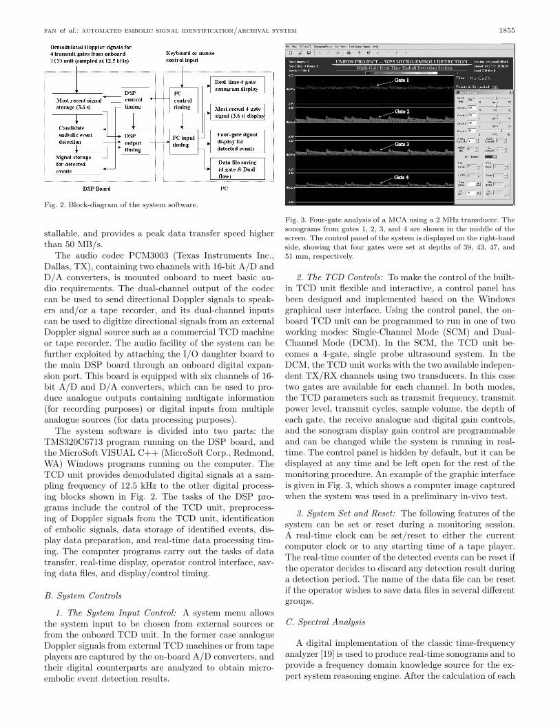
Fig. 3. Four-gate analysis of a MCA using a 2 MHz transducer. Thesonograms from gates 1, 2, 3, and 4 are shown in the middle of thescreen. The control panel of the system is displayed on the right-handside, showing that four gates were set at depths of 39, 43, 47, and51 mm, respectively.
2. The TCD Controls: To make the control of the built-in TCD unit flexible and interactive, a control panel hasbeen designed and implemented based on the Windowsgraphical user interface. Using the control panel, the on-board TCD unit can be programmed to run in one of twoworking modes: Single-Channel Mode (SCM) and Dual-Channel Mode (DCM). In the SCM, the TCD unit be-comes a 4-gate, single probe ultrasound system. In theDCM, the TCD unit works with the two available indepen-dent TX/RX channels using two transducers. In this casetwo gates are available for each channel. In both modes,the TCD parameters such as transmit frequency, transmitpower level, transmit cycles, sample volume, the depth ofeach gate, the receive analogue and digital gain controls,and the sonogram display gain control are programmableand can be changed while the system is running in real-time. The control panel is hidden by default, but it can bedisplayed at any time and be left open for the rest of themonitoring procedure. An example of the graphic interfaceis given in Fig. 3, which shows a computer image capturedwhen the system was used in a preliminary in-vivo test.
3. System Set and Reset: The following features of thesystem can be set or reset during a monitoring session.A real-time clock can be set/reset to either the currentcomputer clock or to any starting time of a tape player.The real-time counter of the detected events can be reset ifthe operator decides to discard any detection result duringa detection period. The name of the data file can be resetif the operator wishes to save data files in several differentgroups.
C. Spectral Analysis
A digital implementation of the classic time-frequencyanalyzer [19] is used to produce real-time sonograms and toprovide a frequency domain knowledge source for the ex-pert system reasoning engine. After the calculation of each

1856 ieee transactions on ultrasonics, ferroelectrics, and frequency control, vol. 53, no. 10, october 2006
frame, the 128-sample data window (data length 10.24 ms)is moved forward by 32 samples (2.56 ms). Thus the over-lap between adjacent frames is 75%, which helps to ensureno very short events are missed.
D. Identification of Micro-Embolic Events amongArtifacts and Background Signals
The task of identification of micro-embolic events is car-ried out in the unit labeled ‘Candidate Embolic EventDetection’ (CEED) in Fig. 2, which is the key computa-tional unit of the system. To cope with the highly com-plicated nature of embolic signals, the different types ofartifacts, and the background Doppler signal (clutter), ex-pert system technology was used to emulate human cog-nitive skills during the decision-making procedure whendetecting emboli and rejecting artifacts. The unit carriesout intensive signal analyses and evaluations in the time,frequency, energy, and neighborhood domains to build itsknowledge source data base, which then is processed usinga blackboard-type intermediate hypotheses and decisionsystem [20]. The detailed reasoning procedure of the expertsystem structure is reported in [12]. The expert system wasoriginally designed to have 14 knowledge source groups,and 76 knowledge sources derived from these groups. Theblackboard evaluation used 164 rules and 45 blackboardlevels in the reasoning and decision-making process, whichdifferentiates possible embolic events from background sig-nals and artifacts. As shown in Table II, four extra ba-sic knowledge sources, unavailable in the previous system,have been introduced into the expert system to improvethe detection performance of the system, and an additional11 rules have been derived from these knowledge sourcesand added to the blackboard-type CEED unit.
E. System Display
The sonograms from four gates are displayed in real-time; each contains forward and reverse flow components.The sonogram display resolution is 5.12 ms, and the mostrecent 3.58 s sonograms are always shown on the screen.
A real-time, updated histogram is displayed, showingdetection results during each 10-minute detection periodfor the most recent 4 hours. During each such period, thehistogram shows the total number of detected events aswell as the number of those with a measured embolus-to-blood-ratio (MEBR) higher than 7 dB.
Optionally, the outputs of the onboard A/D convertersor the inputs of the D/A converters can be displayed onthe screen as a digital oscilloscope. This can be useful whencontrolling the gain of external analogue signal sources orthe magnitudes of output analogue signals.
F. Signal Archival Facilities of the System
1. Archiving Signals Corresponding to Detected Events:Each micro-embolus monitoring session is divided into
multiple, 10-minute detection periods. If any event is de-tected during such a period, a 1 second segment of direc-tional Doppler signal for each gate is automatically storedin the DSP data buffer. These Doppler signals correspondto the blood velocities of the forward and reverse bloodflow calculated from the four programmable gates. Signalstorage starts at half a second before and ends at half asecond after the detection triggering point. Normally, thesesignals are automatically up-loaded to the computer andsaved into a data file at the end of each 10-minute detec-tion period. But, because of DSP memory limitations, if50 events are detected before the end of the period, thecomputer immediately will read the raw data correspond-ing to these events from the DSP data buffer before savingthem into a binary data file. After a data file is saved, theindex part contained in the file name is updated incremen-tally, ready to be used for the next file. The DSP carrieson the real-time task of embolic signal identification every2.56 ms, even when the computer is busy with saving thedata from previously detected events. Up to 10,000 datafiles can be saved for a given file name. Dedicated softwarehas been written to read these data files and check theircontents at a later time.
The stored signal data corresponding to any event de-tected during a 10-minute detection period can be chosenby the operator and displayed on the computer screen astime-domain signals, which enables the operator to checkthe events that already have been detected.
2. Archiving the Signals Corresponding to UnknownEvents Displayed on the Screen: Unknown events seenon the display screen during real-time monitoring can befurther investigated and the corresponding raw data canbe saved, regardless of whether these events are detectedas micro-embolic candidates or not. When operators no-tice such an event, they can “freeze” the running real-time displays and choose any part of the displayed sono-grams before displaying the corresponding eight segments(from four gates, each with forward and reverse flow com-ponents) of the time-domain signals on the screen. Thesetime-domain signals also can be saved to a data file forfurther analysis at a later time. The maximum length ofthe signals that can be saved is 3.58 seconds (×8), whichcorresponds to a full-screen display of sonograms.
III. System Test and Preliminary Results
A. Preliminary In-Vivo Tests
A number of preliminary in-vivo tests have been per-formed on healthy volunteers. These tests have been car-ried out to evaluate the ability of the system to work cor-rectly in a clinical environment. The results shown in Fig. 3were obtained from the middle cerebral artery (MCA) ofa healthy volunteer using a transmit frequency of 2 MHz.Four gates, at depths of 39, 43, 47, and 51 mm, were used.Fig. 4 shows the results of monitoring both the left and

fan et al.: automated embolic signal identification/archival system 1857
TABLE IIThe Recently Introduced Basic Knowledge Sources for the Expert System.
Basic knowledge sourcesKnowledge origin introduced Purpose of introduction
The directional characteristics ofsignals from emboli and artifacts
The correlation coefficients cal-culated using signals correspond-ing to the forward and reverseflows from the same gate
To provide useful decision-making information based onthe unidirectional property ofembolic signals
The directional characteristics ofsignals from emboli and artifacts
The correlation coefficients cal-culated using the MEBRs corre-sponding to the forward and re-verse flows from the same gate
To provide useful decision-making information based onthe unidirectional property ofembolic signals
The spatial distribution of thereceive gates
The correlation coefficients cal-culated using signals correspond-ing to forward or reverse flowpairs from different gates
To provide useful decision-making information based onthe non-traveling property ofartifact signals
The spatial distribution of thereceive gates
The correlation coefficients cal-culated using the MEBRs corre-sponding to the forward or re-verse flow pairs from differentgates
To provide useful decision-making information based onthe nontraveling property ofartifact signals
Fig. 4. Bilateral monitoring of MCAs. Two 2-MHz transducers wereused to obtain sonograms from a left MCA [Gate 1 (L) and Gate 2(L)] and a right MCA [Gate 1 (R) and Gate 2 (R)]. Gate depths forthe left MCA were 43 and 47 mm, and for the right MCA, 41 and45 mm.
right MCAs simultaneously. Signals were obtained fromthe left and right temporal windows of a 28-year old vol-unteer using two 2 MHz probes, with two gates for eachprobe.
Fig. 5 shows a typical result obtained during simulta-neous monitoring of MCA and common carotid arteries(CCA). Two probes, one of 2 MHz and another of 8 MHz,were used here for simultaneous intracranial and extracra-nial investigation.
B. Experimental In-Vitro Test
Preliminary in-vitro experiments have been carried outto test the sensitivity performance of the embolus detec-
Fig. 5. Simultaneous monitoring of a MCA and a CCA. A 2 MHztransducer was used to obtain the two-gate sonograms from theMCA, and an 8 MHz transducer was used for the two-gate sono-grams from the CCA. The gate depths are 43 and 47 mm for theMCA, 9 and 11 mm for the CCA.
tion system (single gate only) using an embolus flow phan-tom. The phantom consisted of a gear pump and a tank(L×H×W = 25×19.5×10 cm3) filled with tissue mimick-ing material (TMM). The pump generated a steady flow ofa blood mimicking fluid (BMF) through a 3-mm wall-lesschannel within the TMM, at a velocity of approximately35 cm s−1. Details of the TMM, BMF, and constructionof the wall-less TMM channel can be found in [21]. Ar-tificial emboli consisting of small particles of TMM wereinjected into the flow to produce embolus-like Doppler sig-nals. In addition to these particles, air bubbles also wereintroduced to simulate gaseous emboli. As the main pur-pose was to test sensitivity at this stage, no artifacts werecreated during the experiment. A 2 MHz transducer was

1858 ieee transactions on ultrasonics, ferroelectrics, and frequency control, vol. 53, no. 10, october 2006
Fig. 6. Example of a detected event when the system was testedusing a flow phantom. The top most sonogram corresponds to gate1, which shows that a signal from an embolus-like particle (arrow1) was detected by the system, and an indicator line automaticallydisplayed (arrow 2). The embolic signal can be seen on the sonogramscorresponding to gates 2 and 3 (arrows 3 and 4), but not on thesonogram from gate 4, which was deliberately set outside the vessel ofthe flow phantom. The histogram of detected events (arrow 5) showsthat the system detected a total of 160 candidate embolic events inthe first 50 minutes. The two columns of figures to the left of thehistogram bars detail the total number of embolic events detectedin any one 10-minute period (left), and the number of events withan MEBR greater than 7 dB (right). The depths of the four receivegates were 46, 49, 52, and 15 mm, respectively.
connected to the on-board TCD unit; it was fixed on topof the tank and angled nearly parallel to the flow direction.A 10-mm sample volume was used, and the depth was setat 45 mm. During the automated detection, the demod-ulated directional analogue Doppler signals produced bythe on-board TCD unit were recorded in real time usinga tape recorder that had its clock synchronized to that ofthe monitoring system. The recorded 420 s signal was latermanually examined by an experienced human expert, andall events, irrespective of their MEBR, used in a compari-son with those detected by the automated system.
The results of the laboratory tests indicate that, with-out using an MEBR threshold, the system reached a sen-sitivity of 93.60% and a specificity of 99.99% (recordedsignal duration, 420 s; all candidate events, 10500; true em-bolic events, 203; mean MEBR of the true embolic events,14.9 dB) when compared to a human observer. An ex-ample of the detection results saved as a screen image isgiven in Fig. 6, showing signals from an embolus-like par-ticle detected by the system (this image was saved duringa separate laboratory test).
Fig. 7 shows an example of an embolus travelingthrough gates 1–3. The grey rectangle defines the time pe-riod for which the time-domain signals from the four gateswere displayed. Signals from an embolus-like particle canbe seen in the first three gates, with evident time delaysamong them. No signal appears on gate 4, which was delib-erately set outside the vessel in the phantom. Figs. 8 and 9were obtained after an artifact was created by touching the
Fig. 7. Example of embolic event traveling among three gates. Thiscaptured computer image is the same as the one shown in Fig. 6,except that time signals were displayed to show their mutual delay.A time interval was selected from the sonogram display as indicatedby a rectangle (below arrow 1) to display time-domain signals fromthis period. The time-domain signals from the detected embolic eventcan be seen at gates 1–3 (arrows 2–4).
probe. The artifact appears in the sonograms for all gates,including gate 4 that was, also in this case, set outside thevessel.
C. Initial Tests on the Improvement of Embolus Detection
In order to explore the potential for the improvementin performance using the new knowledge sources, prelimi-nary tests were carried out to analyze data files containingmultigate and bidirectional Doppler signals. These datafiles were recorded digitally from a volunteer and from apatient during cardiac surgery, using the new system (butwith a version of software written primarily for recordingpurposes).
The data from the healthy volunteer were embolus-free, but they contain a number of deliberately introducedartifacts such as talking, probe taping/moving, etc. Thedepths of gates 1 and 2 were set at 44 and 25 mm dur-ing the 162 s recording. The data from the patient wererecorded during surgery to replace an aortic valve. Therecorded signal duration was 406 s, with the depths ofgates 1 and 2 being set at 45 and 30 mm, respectively. A2 MHz transducer and a sampling frequency of 8.06 kHzwere used during both recordings.
The Doppler signals generated from the data files wereanalyzed in real time using both the original expert system(i.e., the one reported in [12]) and an improved version us-ing the basic knowledge sources shown in Table II (this iscurrently in the form of separate stand-alone embolus de-tection software, to be finalized and incorporated into theTCD integrated system). The results from the original andnovel systems were carefully examined by an experiencedhuman expert, and the sensitivity/specificity performanceswere compared.

fan et al.: automated embolic signal identification/archival system 1859
Fig. 8. An example of signals due to an artifact. The four gates werearranged in the same configuration as in Fig. 6. An artifact wascreated by touching the probe that was fixed at the top of the flowphantom. It can be seen that the artifact event appeared in the sono-grams for all gates (arrows 1–4), including gate 4 (arrow 4), whichwas deliberately set outside the vessel of the flow phantom. The fig-ure also shows that the artifact produced frequency components thatwere symmetric about the base line.
The results showed that, using a 7 dB MEBR thresh-old, the new system reached a sensitivity of 98.68% and aspecificity of 100.00% (all candidate events, 7877; true em-bolic events, 302; mean MEBR of the true embolic events,28.2 dB) compared with the corresponding performance ofthe original system of 89.07% and 99.99%, respectively.
An example of the detection results from the novel sys-tem is given as a saved screen image in Fig. 10, showing agroup of embolic signals detected by the new expert sys-tem.
IV. Discussion
The TCD integrated ESIAS was designed and imple-mented with state-of-art DSP hardware, into which spec-tral analysis and expert system reasoning algorithms havebeen integrated. The system has been adapted from apreviously described, single-gate embolic event detectionsystem to provide further improvement in system perfor-mance by gathering bidirectional information from multi-ple gates.
The TCD integrated ESIAS has the following advan-tages over the system reported in [17]:
• Flexibility. The CEED of the system can be configuredto directly process digital data from the on-board TCDunit. It also can be configured to process data from anexternal TCD unit, using the on-board A/D convert-ers. The on-board FPGA can be programmed to takeover some of the workload from the DSP processor ifnecessary.
• User-friendly system control. The on-board TCD andthe embolus detection and archiving functions can be
Fig. 9. An example of signals from an artifact. This captured com-puter image is the same as the one shown in Fig. 8, except thattime-domain signals are displayed. The image shows that a part ofthe sonograms was selected (indicated by the grey rectangle—arrow1), and the corresponding time-domain signals for the four gates weredisplayed. Signals from an artifact can be seen in both flow direc-tions in the first gate (arrows 2 and 3). Similarly, signals from theartifact appeared in all other gates with no time delay among them(displayed below those of the first gate), including gate 4, which wasdeliberately set outside the vessel of the flow phantom.
Fig. 10. Example of a group of events detected using multigate anddual-directional information. The top sonogram corresponds to gate1 (arrow 1), which shows that a group of embolic signals were de-tected by the system, and the corresponding indicator lines auto-matically displayed (arrow 2). The embolic signals do not appear onthe bottom sonogram corresponding to gate 2 (arrow 3), which wasdeliberately set outside the vessel of the patient. These results wereobtained by playing back and processing data files, which were dig-itally recorded during surgery for an aortic valve substitution. Thedepths of gates 1 and 2 were set at 45 and 30 mm, respectively.
controlled more easily and intuitively, using a singledisplay-and-control center.
• Achievement of floating-point dynamic range integrityof the entire system. The fixed-point data I/O inter-face (usually a D/A followed by an A/D structure)between an external TCD unit and the embolus detec-tion unit is eliminated when using the on-board TCD.
• Small dimensions. A programmable TCD unit anda real-time MES identification and archiving sys-tem have been integrated into one state-of-art device(23 × 21 × 6.2 cm3).
1860 ieee transactions on ultrasonics, ferroelectrics, and frequency control, vol. 53, no. 10, october 2006
Although our previously described, single-gate systemreached very high levels of specificity in in-vivo tests(99.2%), during long monitoring sessions there may bevery large numbers of significant increases in Doppler sig-nal power caused by mechanisms unrelated to emboli, andtherefore, even higher levels of specificity are desirable.The current system allows us to develop additional strate-gies for rejecting artifacts based both on the time rela-tionship between the signals in separate gates and theirdirectional properties. Comparison of Figs. 7 and 9 showshow, in the case of simple events, there are clear differ-ences between the characteristics of embolic signals andartifacts. Embolic signals show a progressive time delaybetween their arrivals in adjacent gates and are unidirec-tional, and artifacts appear simultaneously in all gates (in-cluding the gate placed outside the vessel) and are bidi-rectional [8], [11], [14], [22]–[27]. The inclusion of theseproperties as inputs into our expert reasoning system willfurther enhance its high performance levels.
V. Conclusions
A real-time TCD integrated embolic signal identifica-tion/archival system has been designed and implemented.The system is based on state-of-art hardware componentsincluding a TMS320C6713 DSP processor, and a novelexpert system software structure. The integrated, pro-grammable TCD unit can be configured either as a single-channel, four-gate ultrasound system or as a dual-channel,dual-gate system using two ultrasound transmit frequen-cies. The system has provided real-time sonogram dis-play and real-time automated embolic signal acquisitionfor bidirectional signal pairs from four gates.
In-vitro experiments have been carried out to test thesensitivity performance of the system (single gate only)using an embolus flow phantom. The preliminary exper-imental results show a sensitivity of 93.6% and a speci-ficity of 99.99% when using the system to identify the solidand gaseous emboli introduced into the flow system of thephantom.
Initial in-vivo experiments indicate that, by analyzingmultigate and bidirectional signals, embolus detection per-formance can be further improved. For the short-signalsample tested, sensitivity and specificity improved from89.07% and 99.99%, to 98.68% and 100.00%, respectively.The new embolus detection program will require some fur-ther improvement before being evaluated using a muchlarger data set.
Acknowledgments
The authors thank Dr. K. Ramnarine and Dr. J.Gittings for their help with the in-vitro experiments andP. Fidanzati for assistance in software development.
References
[1] M. P. Spencer, “Detection of cerebral arterial emboli,” in Tran-scranial Doppler Ultrasonography. D. W. Newell and R. Aaslid,Eds. New York: Raven, 1992, pp. 215–230.
[2] D. H. Evans, “Detection of microemboli,” in TranscranialDoppler Ultrasonography. 2nd ed. V. L. Babikian and L. R.Wechsler, Eds. Boston: Butterworth Heinemann, 1999, pp. 141–155.
[3] H. Markus, “Microembolic signal detection in cerebrovasculardisease,” in Transcranial Doppler Ultrasonography. 2nd ed. V. L.Babikian and L. R. Wechsler, Eds. Boston: Butterworth Heine-mann, 1999, pp. 167–174.
[4] R. Dittrich, M. A. Ritter, and D. W. Droste, “Microembolusdetection by transcranial Doppler ultrasonography,” Eur. J. Ul-trasound, vol. 16, pp. 21–30, Nov. 2002.
[5] D. H. Evans, “Ultrasonic detection of cerebral emboli,” in Proc.IEEE Ultrason. Symp., 2003, pp. 316–326.
[6] H. Markus, A. Loh, and M. M. Brown, “Computerized detec-tion of cerebral emboli and discrimination from artifact usingDoppler ultrasound,” Stroke, vol. 24, pp. 1667–1672, Nov. 1993.
[7] M. Siebler, G. Rose, M. Sitzer, A. Bender, and H. Steinmetz,“Real-time identification of cerebral microemboli with US fea-ture detection by a neural network,” Radiology, vol. 192, pp.739–742, Sep. 1994.
[8] D. Georgiadis, A. Wenzel, H. R. Zerkowski, S. Zierz, and A.Lindner, “Automated intraoperative detection of Doppler mi-croembolic signals using the bigate approach,” Stroke, vol. 29,pp. 137–139, Jan. 1998.
[9] E. Roy, S. Montresor, P. Abraham, and J. L. Saumet, “Spectro-gram analysis of arterial Doppler signals for off-line automatedhits detection,” Ultrasound Med. Biol., vol. 25, pp. 349–359,Mar. 1999.
[10] V. Kemeny, D. W. Droste, S. Hermes, D. G. Nabavi, G. Schulte-Altedorneburg, M. Siebler, and E. B. Ringelstein, “Automaticembolus detection by a neural network,” Stroke, vol. 30, pp.807–810, Apr. 1999.
[11] W. H. Mess, B. M. Titulaer, and R. G. Ackerstaff, “A new al-gorithm for off-line automated emboli detection based on thepseudo-wigner power distribution and the dual gate TCD tech-nique,” Ultrasound Med. Biol., vol. 26, pp. 413–418, Mar. 2000.
[12] L. Fan, D. H. Evans, and A. R. Naylor, “Automated embo-lus identification using a rule-based expert system,” UltrasoundMed. Biol., vol. 27, pp. 1065–1077, Aug. 2001.
[13] D. Georgiadis, F. Uhlmann, M. Astler, S. Cencetti, and S.Zierz, “Automated identification of Doppler microembolic sig-nals: Comparison of two techniques,” Neurol. Res., vol. 22, pp.738–740, Oct. 2000.
[14] G. Devuyst, G. A. Darbellay, J. M. Vesin, V. Kemeny, M. Ritter,D. W. Droste, C. Molina, J. Serena, R. Sztajzel, P. Ruchat, C.Lucchesi, G. Dietler, E. B. Ringelstein, P. A. Despland, and J.Bogousslavsky, “Automatic classification of HITS into artifactsor solid or gaseous emboli by a wavelet representation combinedwith dual-gate TCD,” Stroke, vol. 32, pp. 2803–2809, Dec. 2001.
[15] R. Brucher and D. Russell, “Automatic online embolus detectionand artifact rejection with the first multifrequency transcranialDoppler,” Stroke, vol. 33, pp. 1969–1974, Aug. 2002.
[16] M. Cullinane, Z. Kaposzta, S. Reihill, and H. S. Markus, “Onlineautomated detection of cerebral embolic signals from a variety ofembolic sources,” Ultrasound Med. Biol., vol. 28, pp. 1271–1277,Oct. 2002.
[17] L. Fan, D. H. Evans, A. R. Naylor, and P. Tortoli, “Real-timeidentification and archival of microembolic Doppler signals usinga knowledge-based system,” Med. Biol. Eng. Comput., vol. 42,pp. 193–200, Mar. 2004.
[18] S. Marvasti, D. Gillies, F. Marvasti, and H. S. Markus, “Onlineautomated detection of cerebral embolic signals using a wavelet-based system,” Ultrasound Med. Biol., vol. 30, pp. 647–653,May 2004.
[19] L. Cohen, “Time-frequency distributions—A review,” Proc.IEEE, vol. 77, pp. 941–981, July 1989.
[20] P. Jackson, Introduction to Expert Systems. 2nd ed. Harlow, UK:Addison-Wesley, 1990, pp. 368–383.
[21] K. V. Ramnarine, T. Anderson, and P. R. Hoskins, “Construc-tion and geometric stability of physiological flow rate wall-lessstenosis phantoms,” Ultrasound Med. Biol., vol. 27, no. 2, pp.245–250, 2001.
fan et al.: automated embolic signal identification/archival system 1861
[22] J. L. Smith, D. H. Evans, L. Fan, P. R. F. Bell, and A. R. Naylor,“Differentiation between emboli and artefacts using dual-gatedtranscranial Doppler ultrasound,” Ultrasound Med. Biol., vol.22, pp. 1031–1036, 1996.
[23] D. Georgiadis, J. Goeke, M. Hill, M. Konig, D. G. Nabavi, F.Stogbauer, P. Zunker, and E. B. Ringelstein, “A novel techniquefor identification of Doppler microembolic signals based on thecoincidence method: In vitro and in vivo evaluation,” Stroke,vol. 27, pp. 683–686, Apr. 1996.
[24] G. D. Nabavi, D. Georgiadis, T. Mumme, P. Zunker, and E. B.Ringelstein, “Detection of microembolic signals in patients withmiddle cerebral artery stenosis by means of a bigate probe. Apilot study,” Stroke, vol. 27, pp. 1347–1349, Aug. 1996.
[25] J. Molloy and H. S. Markus, “Multigated Doppler ultrasound inthe detection of emboli in a flow model and embolic signals inpatients,” Stroke, vol. 27, pp. 1548–1552, Sep. 1996.
[26] R. Brucher, D. Russell, and H. Frenkenberger, “Automatic cere-bral embolus detection using dual-gate Doppler,” in New Trendsin Cerebral Hemodynamics and Neurosonology. J. Klingelhofer,E. Bartels, and E. B. Ringelstein, Eds. Amsterdam: Elsevier Sci-ence, 1997, pp. 215–230.
[27] D. Georgiadis, F. Uhlmann, A. Lindner, and S. Zierz, “Differ-entiation between true microembolic signals and artefacts usingan arbitrary sample volume,” Ultrasound Med. Biol., vol. 26, pp.493–496, Mar. 2000.
Lingke Fan (M’99) received his B.Eng. andM.Eng. degrees in acoustic engineering fromHarbin Engineering University (HEU), Peo-ples Republic of China, in 1982 and 1985, re-spectively. He received his Ph.D. degree in ul-trasound signal processing in 1996 from theUniversity of Leicester, Leicester, England.
He worked as a teaching assistant then alecturer in the HEU from 1985 to 1989. Hewas a visiting researcher in University of Tech-nology, Loughborough, England, from 1989 to1991. He worked in University Hospitals of
Leicester, National Health Service Trust (UHL NHS Trust), Leices-ter, England, from 1991 to 1996, as a research scientist. He was alecturer in Singapore Polytechnic, Singapore, from 1996 to 1998. Herejoined the Department of Medical Physics of the UHL NHS Trustin 1998, where he currently works as a senior physicist. Dr. Fan is achartered engineer registered with the Engineering Council (UK).
Enrico Boni graduated in electronic engi-neering in 2001 at the University of Florence,Italy. He received his Ph.D. degree from thesame University in 2005. He currently holds apost-Doctoral position at the MicroelectronicSystems Design Laboratory of the Universityof Florence.
His interests include analog/digital sys-tems design and the development of digitalsignal processing algorithms for applicationto Doppler ultrasound with emphasis on mi-croemboli detection.
Piero Tortoli (M’91–SM’96) received theLaurea degree in Electronic Engineering fromthe University of Florence in 1978. Since then,he has been with the Electronics and Telecom-munications Department of the University ofFlorence, where he is currently a full profes-sor of Electronics. His main interests are inthe area of signal processing systems with ap-plication to biomedical instrumentation. Hehas published over 120 papers related to hisresearch activity on ultrasonic imaging andDoppler techniques.
Piero Tortoli is on the Editorial Board of Ultrasound in Medicineand Biology. He has been a member of the Technical Program Com-mittee of international conferences, including the IEEE InternationalUltrasonics Symposium. He was also chairman of the 22nd Interna-tional Symposium on Acoustical Imaging (1995), co-Chairman of theICB Seminar on Ultrasound in Biomeasurements, Diagnostics andTherapy (1998), and Chairman of the 12th New England DopplerConference (2003).
David H. Evans (SM’92) was born inCheshire, England, in 1950. He received aB.Sc. degree in physics from the University ofSurrey, Guildford, England, in 1972, a Ph.D.degree in medical physics from the Universityof Leicester, Leicester, England, in 1979, anda D.Sc. degree from the University of Surreyin 2001 for his contribution to Doppler Ultra-sound in Medicine.
He has worked in Leicester, England, since1974, except for a 2-year period between 1980and 1982 when he was a visiting professor of
biomedical engineering at the Federal University of Rio de Janeiro,Brazil. He was appointed to the Chair of Medical Physics at theUniversity of Leicester in 1990, and he is an Honorary Consultant inthe University Hospitals of Leicester, National Health Service Trust,where he heads the Department of Medical Physics.
Dr. Evans is a Chartered Physicist, a Chartered Scientist, a Fellowof the Institute of Physics, a Fellow of the Institute of Physics andEngineering in Medicine, a Senior Member of IEEE, and an Honorarymember of the British Medical Ultrasound Society, having served asHonorary Secretary, as Honorary Treasurer, and as President from1996–1998. He is currently President of the European Federation ofSocieties for Ultrasound in Medicine and Biology, previously havingserved as its Honorary Secretary. He was Editor of the journal Phys-iological Measurement from 1993 to 1997, and Instrumentation andTechniques Editor of the Journal of Clinical Ultrasound from 1993to 1996.