Applications of transcranial Doppler in the ICU: a revie of transcranial doppler in the... ·...
14
Intensive Care Med (2006) 32:981–994 DOI 10.1007/s00134-006-0173-y REVIEW Hayden White Balasubramanian Venkatesh Applications of transcranial Doppler in the ICU: a review Received: 17 June 2005 Accepted: 16 March 2006 Published online: 10 May 2006 © Springer-Verlag 2006 H. White Queen Elizabeth II Hospital, Department of Anaesthesia, Coopers Plains, 4108, QLD, Australia B. Venkatesh (✉) University of Queensland, Princess Alexandra and Wesley Hospitals, Department of Critical Care Medicine, Woollongabba, 4102, QLD, Australia e-mail: [email protected] Tel.: +61-7-32402111 Fax: +61-7-32402155 Abstract Objective: Transcranial Doppler (TCD) ultrasonography is a technique that uses a hand-held Doppler transducer (placed on the surface of the cranial skin) to measure the velocity and pulsatility of blood flow within the intracranial and the extracranial arteries. This review critically evaluates the evidence for the use of TCD in the critical care population. Discussion: TCD has been frequently employed for the clinical evaluation of cerebral vasospasm following subarachnoid haemorrhage (SAH). To a lesser degree, TCD has also been used to evaluate cerebral autoregulatory capacity, monitor cerebral circula- tion during cardiopulmonary bypass and carotid endarterectomies and to diagnose brain death. Techno- logical advances such as M mode, colour Doppler and three-dimensional power Doppler ultrasonography have extended the scope of TCD to include other non-critical care applications including assessment of cerebral emboli, functional TCD and the management of sickle cell disease. Conclusions: Despite pub- lications suggesting concordance between TCD velocity measurements and cerebral blood flow there are few randomized controlled studies demonstrating an improved outcome with the use of TCD monitoring in neurocritical care. Newer develop- ments in this technology include venous Doppler, functional Doppler and use of ultrasound contrast agents. Keywords Transcranial Doppler · Subarachnoid haemorrhage · Intracranial pressure · Brain death · Neurocritical care History In the middle 1800s Christian Andreas Doppler observed that when a sound wave with a certain frequency strikes a moving object, it is reflected with a different frequency. This phenomenon came to be known as the Doppler effect. The principle was utilized by Spencer and Reid [1] who popularized the concept of imaging blood vessels using ultrasound. In the late twentieth century Aaslid and col- leagues (1982) introduced transcranial Doppler (TCD) ul- trasonography into clinical practice for the evaluation of cerebral haemodynamics, ushering in a new era of cere- bral circulation monitoring [2]. Their successful adapta- tion of ultrasound technology was due to the following factors: (a) the use of a relatively low ultrasonic frequency (2 MHz), with better bone penetration, and (b) insonation of the intracranial vessels via the temporal area, which is the thinnest part of the skull. Basic concepts Ultrasound technology is based on the Doppler effect [3]. The TCD probe emits a wave with known frequency f O and propagating speed c towards a moving target and receives the echo. The echo is the reflected wave with an altered frequency fe. The difference in frequency between the incident and the reflected waves is known as the
Transcript of Applications of transcranial Doppler in the ICU: a revie of transcranial doppler in the... ·...

Intensive Care Med (2006) 32:981–994DOI 10.1007/s00134-006-0173-y R E V I E W
Hayden WhiteBalasubramanian Venkatesh Applications of transcranial Doppler
in the ICU: a review
Received: 17 June 2005Accepted: 16 March 2006Published online: 10 May 2006© Springer-Verlag 2006
H. WhiteQueen Elizabeth II Hospital,Department of Anaesthesia,Coopers Plains, 4108, QLD, Australia
B. Venkatesh (�)University of Queensland, PrincessAlexandra and Wesley Hospitals,Department of Critical Care Medicine,Woollongabba, 4102, QLD, Australiae-mail: [email protected].: +61-7-32402111Fax: +61-7-32402155
Abstract Objective: TranscranialDoppler (TCD) ultrasonography isa technique that uses a hand-heldDoppler transducer (placed on thesurface of the cranial skin) to measurethe velocity and pulsatility of bloodflow within the intracranial and theextracranial arteries. This reviewcritically evaluates the evidencefor the use of TCD in the criticalcare population. Discussion: TCDhas been frequently employed forthe clinical evaluation of cerebralvasospasm following subarachnoidhaemorrhage (SAH). To a lesserdegree, TCD has also been usedto evaluate cerebral autoregulatorycapacity, monitor cerebral circula-tion during cardiopulmonary bypassand carotid endarterectomies andto diagnose brain death. Techno-logical advances such as M mode,colour Doppler and three-dimensional
power Doppler ultrasonographyhave extended the scope of TCDto include other non-critical careapplications including assessmentof cerebral emboli, functional TCDand the management of sickle celldisease. Conclusions: Despite pub-lications suggesting concordancebetween TCD velocity measurementsand cerebral blood flow there arefew randomized controlled studiesdemonstrating an improved outcomewith the use of TCD monitoring inneurocritical care. Newer develop-ments in this technology includevenous Doppler, functional Dopplerand use of ultrasound contrast agents.
Keywords TranscranialDoppler · Subarachnoidhaemorrhage · Intracranial pressure ·Brain death · Neurocritical care
History
In the middle 1800s Christian Andreas Doppler observedthat when a sound wave with a certain frequency strikesa moving object, it is reflected with a different frequency.This phenomenon came to be known as the Doppler effect.The principle was utilized by Spencer and Reid [1] whopopularized the concept of imaging blood vessels usingultrasound. In the late twentieth century Aaslid and col-leagues (1982) introduced transcranial Doppler (TCD) ul-trasonography into clinical practice for the evaluation ofcerebral haemodynamics, ushering in a new era of cere-bral circulation monitoring [2]. Their successful adapta-tion of ultrasound technology was due to the following
factors: (a) the use of a relatively low ultrasonic frequency(2 MHz), with better bone penetration, and (b) insonationof the intracranial vessels via the temporal area, which isthe thinnest part of the skull.
Basic concepts
Ultrasound technology is based on the Doppler effect [3].The TCD probe emits a wave with known frequency f Oand propagating speed c towards a moving target andreceives the echo. The echo is the reflected wave with analtered frequency fe. The difference in frequency betweenthe incident and the reflected waves is known as the

982
Doppler shift, fd, and can be determined as: fd = fe – fo.A shift to a higher frequency is termed positive Dopplershift, and to a lower frequency is a negative shift. Accord-ing to the Doppler equation for a reflector, the velocity vof the moving target can be calculated as:
v = c × fd
2 × fo(1)
where c is the speed of the incident wave. This assumesthat the target moves directly towards the device. If the tar-get moves in a direction that has an angle θ (Doppler angle)towards the device, the equation becomes:
v = c × fd
2 × fo × cos θ(2)
If the angle ranges from 0° to 30°, its cosine varies between1 and 0.86. Thus the maximum error due to θ will be lessthan 15%.
The TCD system uses this principle to measure cere-bral blood flow (CBF) velocity and displays the informa-tion as a velocity-time waveform. The peak systolic (PSV)and end-diastolic (EDV) blood flow velocities (FV) aremeasured directly from the waveform display. The meanvelocity (MV), resistance index (RI) and pulsatility index(PI) [4, 5, 6], may be calculated as follows:
MV = PSV + (EDV × 2)
3(3)
PI =(
PSV − EDV
MV
)(4)
RI =(
PSV − EDV
PV
)(5)
TCD systems use pulsed-wave Doppler which employsa single transducer for emitting and receiving pressurewaves [3]. In contrast to continuous-wave Doppler, pulsesare emitted in discrete packages. The time interval duringwhich the transducer accepts returning signals is mani-pulated to allow sampling from a specific range of depths.The pulse repetition frequency (PRF) describes how manypulses per second are generated [7]. The pulsed-wavebeam has its energy concentrated in the pulse-durationand no energy distributed in pulse interval. Thereforethe pulsed-wave beam has much greater strength perwave-unit, resulting in better penetration.
The Nyquist-Shannon sampling theorem which gov-erns the frequency of sampling a signal also applies to theDoppler system [8]. According to this theorem, in order toperfectly reconstruct the original signal from the sampledversion the sampling frequency must be at least twice thefrequency of the signal itself. Furthermore, accurate depic-tion of a Doppler shift requires at least two sampling pulsesper cycle of the shifted waveform. There is therefore anupper limit to the Doppler shifts that can be detected by
pulsed Doppler instruments, the Nyquist limit. As pulse-wave equipment collects samples of waves at the rate ofPRF, a pulsed Doppler device cannot accurately determineDoppler shifts exceeding PRF/2 [9].
Technique
TCD uses a hand-held microprocessor-controlled trans-ducer. It transmits a low-frequency (2 MHz) pulse-wavedultrasonic signal from skin surface across the cranial vaultto the intracerebral arteries and receives the echoes alongthe same path. In an adult human, cranial bones fullyencase the cranial cavity, leaving only small foraminafor vessels and nerves. Bone tissue heavily attenuatesultrasound with at best, only 6% of the intensity of theultrasound reaching the brain substance.
Acoustic windows are thin areas of bone or naturallyoccurring foramina or fissures that allow the best trans-mission of TCD signal [9]. There are three naturally oc-curring acoustic windows: transtemporal, transorbital andtransforaminal windows (Fig. 1). The transtemporal win-dow is used to study the middle, anterior and posteriorcerebral arteries. The transorbital window is used to ex-amine the ophthalmic artery and the three segments of thecavernous portion (siphon) of the internal carotid artery.
Fig. 1 The temporal window allows insonation of the anterior,middle and posterior cerebral arteries. (Reproduced with permissionfrom [9])
983
The transforaminal window allows assessment of the in-tracranial portions of the basilar and vertebral arteries. Itshould be noted that up to 8% of persons do not have anadequate acoustic window [10].
In general, six factors are used to identify the insonatedartery (Table 1): (a) the window through which the vesselis insonated, (b) the depth of sample volume, (c) the orien-tation of the transducer during insonation, (d) the directionof blood flow with respect to the transducer, (a) the rela-tionship of the vessel to the junction of middle (MCA) andanterior cerebral arteries (ACA) and the terminal portionof the internal carotid artery, and (f) the response to dy-namic maneuvers (for example, compression of the com-mon carotid results in a temporary decrease in MCA ve-locity on the ipsilateral side) [9].
Systolic, diastolic and time-averaged mean values canbe estimated from waveform analysis of reflected Dopplershift echoes from red blood cells. These echoes are re-ceived and converted into electric voltages that pass intothe electronic component of the instrument. The signal isprocessed by demodulation and fast Fourier transformationand Doppler shifts are obtained and displayed in spectralform. The frequency shift is on the vertical axis and timeon the horizontal. FV varies with CBF, angle of insonation,vessel diameter and collateral flow.
Normal values are influenced by a number of physio-logical and demographic factors. Physiological determi-nants of velocity are similar to those influencing CBFand include mean arterial blood pressure (MAP), partialpressure of arterial carbon dioxide (PaCO2) and haema-tocrit. Demographic factors include age, sex, pregnancyand arousal state. At birth MCA FV is approx. 24 cm/s,increasing to 100 cm/s at the age of 4–6 years. Thereafterit decreases steadily to about 40 cm/s in the seventhdecade of life. Women generally have a higher FV thanmen, although the difference is usually small (10–15%).During normal pregnancy FV is maintained for the firsttwo trimesters but decreases during the third. FV drops byapproximately 15% during sleep [11].
The range of normal values for adults was determinedby Aaslid et al. [2]. The mean velocities obtained in themiddle cerebral artery, anterior cerebral artery, and poste-rior cerebral circulation (PCA) were: 62 ± 12, 51 ± 12 and44 ± 11 cm/c, respectively (Table 1). These values were inthe same ranges as those found by direct Doppler tech-niques during surgery [12, 13]. The procedure introducedby Aaslid et al. has now been adopted by other researchers
Artery Window Depth (mm) Transducer orientation Flow direction Velocity (cm/s)
OA Orbital 40–50 Slightly medial Toward 16–26MCA Temporal 35–60 En face Toward 46–86ACA Temporal 60–75 Anteriorly Away 41–76PCA Temporal 60–75 Posteriorly Toward 33–64VA Foraminal 45–75 Superiorly and obliquely Away 27–55BA Foraminal 70–120 Superiorly Away 30–57
Table 1 Criteria for arteryidentification (OA ophthalmicartery, MCA middle cerebralartery, ACA anterior cerebralartery, PCA posterior cerebralartery, VA vertebral artery, BAbasilar artery)
as a standard method to detect the FV of basal cerebralarteries [2, 14].
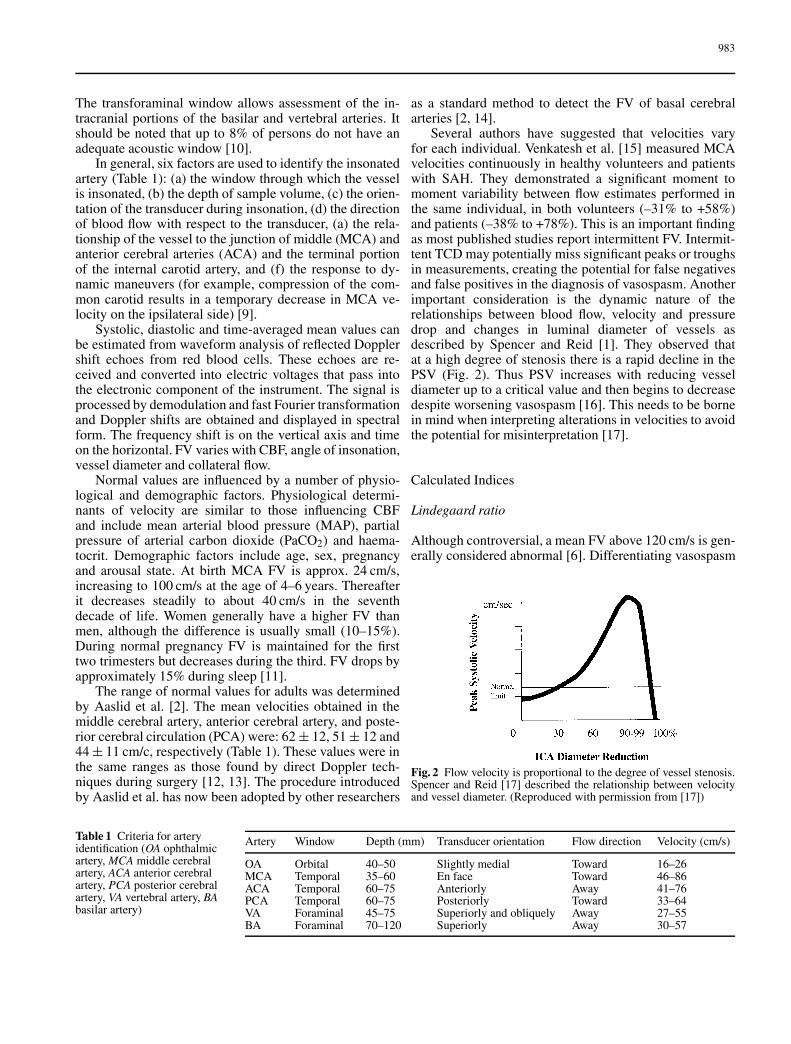
Several authors have suggested that velocities varyfor each individual. Venkatesh et al. [15] measured MCAvelocities continuously in healthy volunteers and patientswith SAH. They demonstrated a significant moment tomoment variability between flow estimates performed inthe same individual, in both volunteers (–31% to +58%)and patients (–38% to +78%). This is an important findingas most published studies report intermittent FV. Intermit-tent TCD may potentially miss significant peaks or troughsin measurements, creating the potential for false negativesand false positives in the diagnosis of vasospasm. Anotherimportant consideration is the dynamic nature of therelationships between blood flow, velocity and pressuredrop and changes in luminal diameter of vessels asdescribed by Spencer and Reid [1]. They observed thatat a high degree of stenosis there is a rapid decline in thePSV (Fig. 2). Thus PSV increases with reducing vesseldiameter up to a critical value and then begins to decreasedespite worsening vasospasm [16]. This needs to be bornein mind when interpreting alterations in velocities to avoidthe potential for misinterpretation [17].
Calculated Indices
Lindegaard ratio
Although controversial, a mean FV above 120 cm/s is gen-erally considered abnormal [6]. Differentiating vasospasm
Fig. 2 Flow velocity is proportional to the degree of vessel stenosis.Spencer and Reid [17] described the relationship between velocityand vessel diameter. (Reproduced with permission from [17])

984
from hyperaemia using TCD can be problematic as bothconditions may result in elevated FV. In 1988 Lindegaardet al. [18] put forward an index to distinguish between thetwo by comparing the FV of the MCA with that of the ex-tracranial portion of the ipsilateral internal carotid artery.They compared the estimates to cerebral angiographic as-sessments. The ratio of FV in the MCA to that in the exter-nal carotid artery increased with the severity of vasospasm.This ratio (Lindegaard ratio, LR) ranged from 1.1 to 2.3,median 1.7 at days 1–2, but rose to over 10 in patients withthe most severe MCA lumen narrowing. In the setting ofelevated FV a ratio lower than 3 is considered hyperaemia,a ratio of 3–6 mild vasospasm and a ratio higher than 6consistent with severe vasospasm.
Pulsatility index
The most widely used measures of intracranial resistanceare Pourcelot’s resistance index (RI) and the pulsatility in-dex (PI) developed by Gosling and King [4] (see also [19]).The “pulsatility” of the FV waveform reflects the resis-tance of the more distal cerebral vasculature. Both RI andPI are influenced by physiological factors including arte-rial pressure, vascular resistance and changes in PaCO2.The PI has been evaluated as an alternative to direct in-tracranial pressure (ICP) measurement. Bellner et al. [20]compared the PI index with ICP measurements from intra-ventricular monitors in 81 patients with various intracra-nial disorders. A strong correlation was found between theICP and the PI (r = 0.938, p < 0.001) within the ICP rangesof 5–40 mmHg. Omitting repeated measurements and con-sidering only the initial TCD ICP recording in each sub-ject (n = 81) for the interval 5–40 mmHg, the R2 value was0.733 (p < 0.0001) with an SD of 2.5. The regression lineis: ICP (PI) = 11.1 × PI – 1.43. The correlation betweenthe cerebral perfusion pressure (CPP) and PI was also sig-nificant but less strong (r = –0.493, p < 0.001). Similarly,the RI has been used to demonstrate the presence ofraised ICP. Several authors have reported good correlationbetween RI and raised ICP in cases involving various intra-cranial pathological processes including hydrocephalus [21,22]. Table 2 summarizes the differential diagnoses ofa perturbation in the measured and calculated indices.
Assessment of cerebral perfusion pressure andcerebral blood flow using TCD
Relationship between TCD measurements and CPP
CPP is the difference between arterial pressure (AP) andthe effective downstream pressure (EDP) of the cerebralcirculation. Because of a Starling resistor phenomenon lo-cated at the level of cerebral veins ICP is thought to rep-resent the EDP of the cerebral circulation [23]. The Brain
Table 2 Differential diagnosis of changes in flow velocity and resis-tivity indices during transcranial Doppler examination (from [5, 6,18, 20, 115, 116])
Transcranial Doppler flow velocityIncrease
VasospasmHyperaemiaLoss of autoregulation↑ PaCO2Intracranial arterial stenosisIncreasing ageHyperdynamic circulationVolatile anaesthetic agentsSickle cell anaemiaArteriovenous malformationBacterial meningitisPre-eclampsia
DecreaseHypotension↓ CBFBrain deathRaised ICP↓ PaCO2↑ angle of insonationPregnancyAnaesthetic Induction agents (except ketamine)HypothermiaFulminant hepatic failure
Pulsatility index, resistance indexIncrease
Raised ICPHydrocephalusTraumatic brain injuryIntracerebral haemorrhageFulminant hepatic failureStrokeBrain deathIntracranial artery occlusionBacterial meningitis
DecreaseVasospasmArteriovenous malformationRewarming following hypothermiaHyperaemia
Lindegaard ratioIncrease
VasospasmDecrease
Hyperaemia
Trauma Foundation’s guidelines recommend a CPP of atleast 60 mmHg [24]. Direct measurement of ICP is not al-ways possible when the risk of a reduction in CPP is max-imal, for example, during the early management of brain-injured patients. In this instance TCD evaluation of MCAvelocity has been proposed as an alternative for neurologi-cal monitoring.
Several methods of estimating CPP using measuredTCD velocities have been described: Aaslid et al. [25]determined CPP using the following TCD parameters:

985
where F1 and A1 represent amplitude of the fundamentalfrequency components of FV and arterial pressure, respec-tively. The fundamental frequency is determined by fastFourier analysis of the waveform, and is equivalent to theheart rate.
Czosnyka et al. [26] used measured and calculatedTCD variables from 96 patients with head injury to derivea different formula to estimate CPP:using time averaged mean, systolic and diastolic valuesof FV from the MCA. A subsequent validation study ofthe above method reported the absolute difference betweenmeasured CPP and calculated CPP (daily averages) to beless than 10 mmHg in 89% of measurements and less than13 mmHg in 92% of measurements [27].
Edouard et al. [28] investigated a simplified model ofCPP originally described in pregnant patients. They com-pared conventional measurements of CPP (difference be-tween MAP and ICP) with an estimated CPP (CPPe). CPPewas derived using a formula combining the phasic valuesof FV and arterial pressure:
CPPe =(
vmean
vmean − vdiast
)× (APmean − APdiast) (6)
where vmean and vdiast are the mean and diastolic MCAvelocities, and APmean and APdiast are the mean and di-astolic arterial pressures, respectively. Twenty adults withbilateral and diffuse brain injuries were included in thestudy. CPPe and CPP were correlated (slope 0.76. intercept+10.9, 95% CI 3.5 to +25.4). The intercept of the regres-sion line equaled the zero flow pressure (ZFP), in the MCAand represents the EDP of the cerebral circulation. Nonin-vasive measurement of CPP is a promising technique. Cur-rent evidence suggests that EDP as predicted by the criticalclosing pressure is as useful as ICP in describing cerebralhaemodynamics, and in some instances better. Weyland etal. [29] observed that during hypocapnia cerebrovasculartone rather than ICP determines the EDP and thereforeCPP. Thus MAP EDP would give a better indication ofCPP in this situation. More work is needed as none ofthe recently described methods has been fully validated; inparticular, it is not clear from the existing literature whichformula is most appropriate under given pathophysiologi-cal conditions.
Relationship between TCD and cerebral blood flow
TCD velocities have been used to provide informationabout CBF. A number of studies have assessed the validityof comparing FV with techniques for measuring CBF,including IV xenon, the Kety-Schmidt method, magneticresonance imaging, single photon emission computedtomography and laser Doppler flowmetry [30, 31]. Resultshave been inconsistent. Brauer et al. [32] compared TCDwith xenon-enhanced computed tomography in 32 patientswith varying intracerebral pathologies. Acetozolamide
or CO2 was used to induce a change in CBF. Theyobserved that changes in FV were correlated well withCBF measures for the group as a whole (r = 0.82) butnot subgroups classified according to diagnosis. Not allauthors agree. Minhas et al. [33] studied cerebral perfusionpatterns (positron emission tomography) and associatedTCD indices in 25 patients who developed clinical signsof delayed ischaemic deficits (DID) following SAH.They identified a markedly heterogeneous pattern of CBFdistribution, with hyperaemia, normal CBF values andreduced flow among patients with DID. TCD indiceswere not indicative of the cerebral perfusion findings.In summary, whilst the TCD represents a non-invasivemethod of obtaining information about CBF, the linearrelationship between CBF and FV is only present ifneither the diameter of the insonated vessel nor the angleof insonation change during the examination.
Clinical applications of TCD in the ICU
Subarachnoid haemorrhage
Vasospasm is a major cause of morbidity and mortality fol-lowing aneurysmal SAH (Fig. 3). Clinically important va-sospasm (or delayed ischaemic deficit) occurs in 20–30%of patients within 3–14 days after bleed. Angiographic va-sospasm however, can be demonstrated in up to 60% of pa-tients. Given that vasospasm is associated with significantmorbidity and mortality, it is imperative not only to diag-nose but also predict those patients who are likely to be-come symptomatic. Aaslid et al. [34] investigated the useof TCD technology for the diagnosis and management ofvasospasm. An inverse relationship between vessel diam-eter and TCD velocities was observed. Their group wenton to define mean TCD velocities higher than 120 cm/sas mild and those higher than 200 cm/s as indicative ofsevere vasospasm [2, 34, 35]. Several authors have con-firmed the Aaslid et al. findings, but it was not until 1987that a controlled trial was performed by Compton et al. [36,37]. They compared the Doppler FV in three groups of pa-tients, a SAH group, a control group and a group of pa-tients with other cerebral pathologies. They observed thatin the SAH group FV tended to be higher. In 80% of pa-tients with MCA FV higher than 100 cm/s they demon-strated vasospasm on angiography. However, a strong cor-relation with the clinical grade of SAH or onset of neuro-logical deficit was not demonstrable [37].
Comparison of TCD with angiography for detection ofvasospasm
Attempts to establish a correlation between TCD velocityand angiographic vasospasm have met with mixed results.A variety of end-points have been investigated to improvesensitivity and specificity. Lindegaard et al. [18] measured

986
Fig. 3 M mode (above) andpulsed-wave Doppler (below)illustrating increased meanvelocity in MCA consistent withmild to moderate vasospasm
the diameter of the proximal segment of the MCA, ACAand PCA from angiograms. There was an inverse relation-ship between MCA diameter and MCA FV. Eleven of the13 MCAs with diameter of 1.5 mm or less exhibited FVgreater than 140 cm/s. This was adopted as a useful limit todiagnose marked MCA spasm (50% diameter reduction).
The sensitivity (MCA) ranges from 59% for velocitieshigher than 100 cm/s to 94% for those above 140 cm/s [38,39, 40, 41, 42, 43, 44, 45, 46, 47, 48, 49]. Vora et al. [50]compared angiographic vasospasm, independently gradedby observers, with TCD measurements. Inter-observeragreement regarding angiographic vasospasm was good(κ = 0.86). Despite significant correlation between meanvelocities and the degree of vasospasm the TCD velocityestimates were not dependable. The positive predictivevalue of an FV of 200 cm/s or greater for moderate/severeangiographic vasospasm was 87% but that of a lowerFV was only 50%. The negative predictive value of FVless than 120 cm/s was 94% but that of higher velocitiesapprox. 75%. LR values did not improve the predictivevalue of TCD monitoring.
In 2001 Lysakowski et al. [51] performed a systematicreview comparing TCD with cerebral angiography todiagnose vasospasm following SAH. Meta-analyses asperformed with data from 7 of 26 trials. They found thatfor the MCA sensitivity was 67% (95% CI 48–87%)and specificity 99% (CI 98–100%). For the anteriorcerebral artery (3 trials, 171 tests), sensitivity was 42%
(CI 11–72%) and specificity 76% (CI 53–100%). Otherarteries were tested in only one trial each. The authorsconcluded that for the MCA TCD is unlikely to indicatespasm when angiography is negative (high specificity) andmay be used to identify patients with spasm (high positivepredictive value) [51].
Ability of TCD to predict onset of delayed ischaemicdeficit
Many of the published reports have attempted to find a cor-relation between TCD velocity and angiographic evidenceof vasospasm. It could be argued that the presence of clini-cal deficits is more important then radiological vasospasmas only 50% of patients develop DID. Several trials haveattempted to compare TCD velocities with clinical evi-dence of cerebral hypoperfusion [52]. Again, the lack ofwell-designed trials hampers interpretation of results.
A correlation between TCD velocity and DID has beenconfirmed by several authors. Sekhar et al. [53] followed21 patients after SAH, 8 of whom developed neuro-logical deficits. They found a good correlation betweenTCD velocity in the MCA and ACA and neurologicaldeficits. Similarly, Vora et al. [50] found the specificityof a TCD for diagnosing DID approached 95% (velocity> 200 cm/s). Lam et al. [54] demonstrated that patientswith an abnormal result on the transient hyperaemic

987
response test (see below) after SAH were likely to developincreased FV and DID (Fisher’s exact test, p = 0.0004).
Other investigators have reported conflicting results.Grosset et al. [55, 56] found that although the high-est recorded velocity was greater in the patients whodeveloped DID (186 ± 6 vs. 149 ± 5 cm/s, p < 0.05),peak velocity was often recorded only after the onset ofneurological deficit. When only the readings made beforethe onset of DID were considered, there was no significantdifference in peak velocity between the groups (157 ± 8vs. 149 ± 5 cm/s, respectively). The rate of increase inTCD velocity recorded during the first few days afterSAH was significantly higher in the patients who laterdeveloped DID. They concluded that a rise of more than50 cm/s in 24 h predicts the onset of DID following SAH.
Klingelhofer et al. [57] failed to identify a consistentrelationship between the occurrence and extent of DID andthe relative change in mean FV during the clinical course.In contrast, there was a significant correlation between therelative change in the cerebral circulatory resistance in-dex and the occurrence of DID. Mizuno found that only61% of patients with DID demonstrated increased FV onTCD. Interestingly, subsequent angiograms of the other39% showed distal vasospasm [58].
Role of TCD for the detection of posterior circulationvasospasm
Only few studies have investigated the role of TCDfor evaluation of the posterior circulation. One of thefirst compared cerebral angiography and conventionalhand-held TCD to determine sensitivity and specificity indetection of vertebral and basilar artery vasospasm. TCDwas highly specific (100%) for vertebral and basilar arteryvasospasm when FV was at least 80 and at least 95 cm/s,respectively [14]. The ability to predict DID, however, isless clear. Clinical studies and those using single photonemission computed tomography confirm that an FVgreater than 115 cm/s is required before ischaemia canreliably be predicted [59, 60]. In an attempt to differentiatebetween hyperaemia and vasospasm Soustiel et al. [61]developed a ratio of intracranial basilar artery (BA) toextracranial vertebral artery velocities, similar to the LR.Comparative analysis between computed-tomographyangiography and TCD findings showed a ratio greater than2 in all patients with BA vasospasm (100% sensitivity)and one less than 2 in patients without BA vasospasm(95% specificity).
Summary
The inconsistency between studies is not surprising andmay be attributable to the lack of correlation betweenangiographic vasospasm and DID, moment to mo-
ment variation in velocities [15], potential for probedisplacement, spasm distal to the major intracerebralvessels [62], the occurrence of hyperaemia in a subgroupof patients [63] and technical factors including operatorvariability [64] and difficulty insonating non-MCA ves-sels. Furthermore, CBF and TCD readings are not alwayscorrelated [65, 66, 67]. Spasm may be an episodic phe-nomenon. This, combined with the fact that intermittentTCD may potentially miss significant peaks or troughsin measurements, creates the potential for false negativesand false positives in the diagnosis of vasospasm. Finally,the theoretical relationship between blood flow, velocityand pressure drop and changes in vessel diameter needto be borne in mind when interpreting alterations in FVto avoid the potential for misinterpretation. With thesecaveats, TCD measurements can be used to assess the siteand severity of vasospasm. Improvements in probe designand fixation together with refinements in signal qualityanalysis have the potential to lead to greater measurementaccuracy.
Traumatic brain injury
A major cause of morbidity and mortality in the West-ern world is traumatic brain injury (TBI). The pathophysi-ology and management is complex and poorly understood.The fact that TCD is non-invasive and can be applied atthe bedside makes it an ideal tool for investigation of thechanging cerebral haemodynamics of the head-injured pa-tient. The technique has been used for the non-invasive as-sessment of ICP, cerebral autoregulation and vasospasm.Each of these is described in detail below.
Alternations in TCD velocities following TBI
TCD has been used to provide a noninvasive assessmentof CBF in TBI (Fig. 4). Published data indicate that FVdecreases during the first 48 h after head trauma andsubsequently increases between 48–120 h. The delayedincrease in velocity may indicate either vasospasm orcerebral hyperaemia [68, 69]. Zurynski et al. [70] mea-sured CBF velocity in 50 severely head-injured patients(Glasgow Coma Scale 8 or less). LR was used to distin-guish between hyperaemia and vasospasm. Although theproportion of patients with poor neurological outcomewas comparable in the two groups (47% vs. 40%), it wasconcluded that the ability of TCD to distinguish betweenthe two processes facilitated clinical management. Finally,TCD measurements have demonstrated that low CBF(as indicated by FV < 35 cm/s) is associated with poorneurological outcome [71, 72].

988
Fig. 4 Spike-wave pattern withraised PI, typical of MCA flowvelocity found in patients withintracranial hypertension
Correlation with ICP
The use of TCD as a marker of ICP was initially describedby Klingerhofer et al. [73]. In a pilot study changes in ICPwere compared with the TCD findings of the MCA. Datafrom five patients with brain death showed that changes inthe ICP influenced the flow patterns considerably. Thesechanges could be recorded quantitatively by means of thePI and the mean FV. Increases in ICP were accompaniedby an increase in PI (owing to a decrease in diastolic andmean velocities). The authors subsequently demonstratedan association between ICP and flow patterns in a subgroupof neurosurgical patients with cerebral hypertension [74].Whilst it is unlikely that TCD will replace invasive ICPmonitoring in the near future, it may have a role in situa-tions where the role of invasive monitoring is not clearlyestablished: stroke, paediatric cases, liver failure, minorhead injury assessed on accident and emergency wards andpreeclampsia [20, 75].
Vasospasm following TBI
Compton and Teddy [76] highlighted the importance of va-sospasm as a contributor to poor neurological outcome fol-lowing head trauma and emphasized the potential role ofTCD in the detection of vascular spasm. Using LR valueshigher than 3 as indicative of spasm, Weber et al. [77] and
Martin et al. [78] confirmed the presence of spasm follow-ing head injury in 40% of patients. Spasm was not noted inTBI patients without SAH.
Although difficult to diagnose, vasospasm may beidentified in the posterior circulation. Soustiel et al. [59,79] examined the FV of the basilar artery post SAH (bothspontaneous and traumatic). BA vasospasm was definedas moderate whenever FV was higher than 60 cm/s andsevere when higher than 85 cm/s. BA vasospasm wassignificantly more common following traumatic SAH(59.7%) than spontaneous SAH (40.3%, p = 0.041). Per-manent neurological deficit was associated with moderateBA vasospasm while severe vasospasm led to a persistentvegetative state. These findings have been confirmed inseveral other studies [80, 81, 82].
Assessment of cerebrovascular autoregulation
TCD has been used extensively in the study of autoregula-tion in the critical care setting. The ability of the cerebralvascular system to constrict and dilate in response tochanges in perfusion pressure is termed autoregulation.Cerebral autoregulation can be explained at least partiallyby a tight coupling between O2 supply and demand ofthe brain. Under normal conditions CBF is maintained ata constant flow rate of 50–60 ml per 100 g/min, with 50 mloxygen being extracted every minute from 700–800 ml

989
blood [83]. This occurs despite changes in MAP withinthe range of 60–160 mmHg. Outside this range flowbecomes proportional to pressure, potentially leading toepisodes of hypo- and hyper-perfusion [84]. Changesin FV in response to fluctuations in blood pressure andarterial carbon dioxide tension have been used as markersof cerebrovascular regulation. Thus autoregulation of thecerebral circulation can be assessed by examining thechanges in FV in response to changes in PaCO2 and MAP.
Assessment of autoregulation in response to changesin PaCO2 is termed CO2 reactivity. In the range of PaCO2between 20 and 60 mmHg, CBF changes by approx.3%/mmHg change in PaCO2 [85, 86]. It is important toensure that hypotension is corrected before testing forCO2 reactivity. Hypoventilation causes vasodilatation andincreased CBF and velocity, while hyperventilation resultsin vasoconstriction and decreased velocity [87].
Autoregulation in response to changes in MAP canbe assessed using a static or a dynamic manoeuvre. Inthe static technique MCA FV is measured under restingconditions and again following a 20- to 30-mmHg increasein MAP [88]. An index of autoregulation can be calculatedas a percentage change in cerebrovascular resistance(MAP/FV) per 1% change in MAP. A value lower than0.4 suggests impaired autoregulation. The main disad-vantage of this technique is the need for pharmacologicalintervention, which in itself may alter vascular reactivityand has the potential for serious adverse effects. Conse-quently, less invasive, non-pharmacological, “dynamic”methods have been developed. (a) The classical thighcuff technique of Aaslid et al. [89] elicits a stepwise dropin AP by applying a thigh pressure cuff, which is thenrapidly deflated. Normally, both FV and MAP decreaseinitially, but FV recovers more quickly. If autoregulation isimpaired, FV recovery passively follows the improvementin MAP. (b) Tiecks et al. [90] developed a simpler, lesstime consuming method using a valsalva maneuver, whichevokes a transient decrease in AP and FV. If autoregulationis intact, there is a stronger restoration of FV than of APduring phase II. (c) Giller et al. [91] described the transienthyperaemic response test which evaluates the changes inMCA FV following brief compression of the ipsilateralcommon carotid artery. The sudden decrease in CPPprovokes vasodilatation in the vascular bed distal to theMCA, leading to a transient increase in FV followingrelease of the compression. If autoregulation is lost, FVreturns to pretest values without a transient increase.
Loss of autoregulation is common following head in-jury and is associated with severity of injury and increasedmortality [92, 93]. Knowledge of the state of autoregula-tion may permit individualization of therapy [94, 95, 96,97]. The process is rapid, but not instantaneous, allowingfor assessment using both static and dynamic tests. Al-though CBF is not directly measured by TCD, changesin velocity are correlated with flow if the vessels cross-sectional area remains constant.
Summary
Despite initial enthusiasm for the technique, to date therehave been no well-designed studies demonstrating a pos-itive outcome effect from the use of TCD in head injury.The main role of TCD in neurotrauma appears to be inits ability to provide information on cerebrovascular reac-tivity and CBF. Whilst the PI may be used as a surrogatefor ICP, it is unlikely to replace invasive pressure monitor-ing. Its role in the identification of vasospasm in the settingof traumatic SAH needs further evaluation. TCD may alsohave a role as an adjunct in the diagnosis of brain death.
Brain death
The law in many countries requires the demonstrationof loss of cerebral function (via electroencephalography)and/or CBF before brain death can be confirmed [98]. An-giography is considered the gold standard but is laboriousand logistically complicated, as the patient must be movedfrom the ICU environment for the study to be performed.
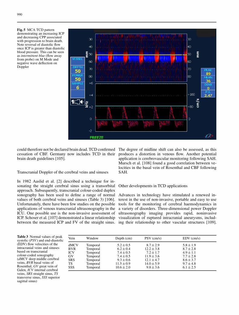
Over the past two decades case reports detailing the useof TCD to confirm cerebral circulatory arrest and by infer-ence brain death have emerged [99, 100]. As an acousticwindow is absent in 8% of patients, it is necessary to havedemonstrated cerebral perfusion on TCD prior to the onsetof brain death. With increasing ICP, TCD waveforms ex-hibit characteristic high-resistance profiles, first with low,then zero, and then reversed diastolic FV [101] (Fig. 5).The flow patterns depend on the relationship between ICPand CPP and are correlated with the level of cerebral cir-culatory arrest as demonstrated by angiography. As CPPapproaches zero, three patterns of TCD waveforms are ob-served: oscillating flow, small systolic spikes and no sig-nal [101, 102]. It is not clear which pattern is the most sen-sitive or specific for diagnosing cerebral circulatory arrest.Furthermore, studies differ in their description of “TCDbrain death” making comparisons difficult. In the ICU set-ting the most commonly insonated artery is the MCA. Theposterior circulation should also be insonated, especiallyin patients with cerebellar pathology. There are addition-ally reports of Doppler studies of the ophthalmic arteriescorrelating with signs of brain death [103].
Although the sensitivity of TCD for the diagnosis ofbrain death is 91.2–100%, the specificity is reported to bevirtually 100% [99]. There have been several case reportsof patients with demonstrable CBF on TCD who were clin-ically brain dead. In these cases the pathology was limitedto the cerebellum or brainstem leaving blood flow in theanterior cerebral circulation relatively intact. For this rea-son it is important that patients show clinical signs of braindeath before performing TCD.
TCD is especially useful in patients who are known tohave received sedative drugs. Hadani et al. [104] reportedthat 31.4% of comatose patients had received sedation and
990
Fig. 5 MCA TCD patterndemonstrating an increasing ICPand decreasing CPP associatedwith progression to brain death.Note reversal of diastolic flowonce ICP is greater than diastolicblood pressure. This can be seenas intermittent blue (flow awayfrom probe) on M Mode andnegative wave deflection onDoppler
could therefore not be declared brain dead. TCD confirmedcessation of CBF. Germany now includes TCD in theirbrain death guidelines [105].
Transcranial Doppler of the cerebral veins and sinuses
In 1982 Aaslid et al. [2] described a technique for in-sonating the straight cerebral sinus using a transorbitalapproach. Subsequently, transcranial colour-coded duplexsonography has been used to define a range of normalvalues of both cerebral veins and sinuses (Table 3) [106].Unfortunately, there have been few studies on the possibleapplications of venous transcranial ultrasonography in theICU. One possible use is the non-invasive assessment ofICP. Schoser et al. [107] demonstrated a linear relationshipbetween the measured ICP and FV of the straight sinus.
Vein Window Depth (cm) PSV (cm/s) EDV (cm/s)
dMCV Temporal 5.2 ± 0.5 8.7 ± 2.9 5.8 ± 1.9BVR Temporal 6.2 ± 0.4 12.2 ± 3.8 8.7 ± 2.8ICV Temporal 7.4 ± 0.5 7.2 ± 1.7 4.9 ± 1.1GV Temporal 7.4 ± 0.5 11.9 ± 3.6 7.7 ± 2.8SRS Temporal 9.3 ± 0.6 12.1 ± 4.7 8.6 ± 3.7TS Temporal 11.5 ± 0.9 14.0 ± 5.9 9.7 ± 4.8SSS Temporal 10.6 ± 2.0 9.8 ± 3.6 6.1 ± 2.5
Table 3 Normal values of peaksystolic (PSV) and end-diastolic(EDV) flow velocities of theintracranial veins and sinusesbased on transcranialcolour-coded sonography(dMCV deep middle cerebralveins, BVR basal veins ofRosenthal, GV great vein ofGalen, ICV internal cerebralveins, SRS straight sinus, TStransverse sinus, SSS superiorsagittal sinus)
The degree of midline shift can also be assessed, as thisproduces a distortion in venous flow. Another potentialapplication is cerebrovascular monitoring following SAH.Mursch et al. [108] found a good correlation between ve-locities in the basal vein of Rosenthal and CBF followingSAH.
Other developments in TCD applications
Advances in technology have stimulated a renewed in-terest in the use of non-invasive, portable and easy to usetools for the monitoring of cerebral haemodynamics ina variety of disorders. Three-dimensional power Dopplerultrasonography imaging provides rapid, noninvasivevisualization of ruptured intracranial aneurysms, includ-ing their relationship to other vascular structures [109].
991
Contrast agents such as Levovist, Sonovue and Echovistare being employed to provide better visualization of themicrocirculation. These are largely gaseous microbubblesstabilized in a surrounding shell. Insonation of these con-trast agents at a fundamental frequency leads to minimaltissue resonance, facilitating improved visualization of themicrocirculation. They appear to have a high sensitivityand specificity in the detection of intracranial stenosiswhen compared to magnetic resonance angiography [110].Ultrasound probes attached to stethoscopes (Neurodop)allow the rapid assessment of MCA velocities in theED and may prove useful in the management of strokepatients [111]. Larsen et al. [112] have investigated CBF,FV and autoregulation following fulminant hepatic failureand during liver transplantation. They suggested that theCBF is not autoregulated in patients with hepatic failureand therefore CBF should be “clamped” within the normalphysiological range by manipulation of arterial blood pres-sure in order to avoid cerebral hypoxia and/or hypertensiveinduced cerebral oedema. Functional transcranial Doppler
is being developed as a tool in psycho-physiological re-search [113], although immediate application in intensivecare is not available. Ultrasound has also been reportedto have therapeutic benefits through enhancement ofliposome-mediated gene transfer to eucaryotic cells in cul-ture [114]. These effects are thought to be mediated by cav-itation. As in the case of functional Doppler, this modalityhas no routine use in clinical intensive care practice.
The future of TCD in intensive care
Whilst class I data exist for the role of TCD in the man-agement of patients with sickle cell disease [115], robustevidence for its usefulness in the critically ill is lacking.Although evidence of its diagnostic capabilities in a varietyof conditions is emerging, its current application in inten-sive care is confined largely to the management of patientswith SAH, assessment of brain death and monitoring ofcerebral haemodynamics in neurotrauma.
References
1. Reid J, Spencer MP (1972) UltrasonicDoppler technique for imaging bloodvessels. Science 176:1235–1236
2. Aaslid R, Markwalder TM, Nornes H(1982) Noninvasive transcranialDoppler ultrasound recording of flowvelocity in basal cerebral arteries.J Neurosurg 57:769–774
3. Aaslid R (1986) The Doppler principleapplied to measurement of bloodflow velocities in cerebral arteries. In:Aaslid R (ed) The Doppler principleapplied to measurement of blood flowvelocities in cerebral arteries. Springer,Vienna, pp 22–38
4. Gosling RG, King DH (1974) Arterialassessment by Doppler-shift ultra-sound. Proc R Soc Med 67:447–449
5. Summors AC, Gupta AK, Matta BF(1999) Dynamic cerebral autoregu-lation during sevoflurane anesthesia:a comparison with isoflurane. AnesthAnalg 88:341–345
6. Moppett IK, Mahajan RP (2004)Transcranial Doppler ultrasonographyin anaesthesia and intensive care. Br JAnaesth 93:710–724
7. Tegeler C, Ratanakorn D (1999)Physics and principles. In: Babikian V,Wechsler L (eds) Physics and prin-ciples. Butterworth-Heinemann,Woburn, pp 3–13
8. Tortoli P (1989) A tracking FFTprocessor for pulsed Doppler analysisbeyond the Nyquist limit. IEEE TransBiomed Eng 36:232–237
9. Santalucia P, Feldmann E (1999)The basic transcranial Doppler ex-amination, technique and anatomy.In: Babikian V, Wechsler L (eds)The basic transcranial Doppler ex-amination, technique and anatomy.Butterworth-Heinemann, Woburn, pp13–33
10. Maeda H, Matsumoto M, Handa N,Hougaku H, Ogawa S, Itoh T,Tsukamoto Y, Kamada T (1993) Reac-tivity of cerebral blood flow to carbondioxide in various types of ischemiccerebrovascular disease: evaluationby the transcranial Doppler method.Stroke 24:670–675
11. Tong D, Albers G (1999) NormalValues. In: Babikian V, Wechsler L(eds) Normal values. Butterworth-Heinemann, Woburn, pp 33–49
12. Nornes H, Grip A, Wikeby P (1979)Intraoperative evaluation of cerebralhemodynamics using directionalDoppler technique. II. Saccularaneurysms. J Neurosurg 50:570–577
13. Nornes H, Grip A, Wikeby P (1979)Intraoperative evaluation of cerebralhemodynamics using directionalDoppler technique. I. Arteriove-nous malformations. J Neurosurg50:145–151
14. Sloan MA, Burch CM, Wozniak MA,Rothman MI, Rigamonti D, Permutt T,Numaguchi Y (1994) TranscranialDoppler detection of vertebrobasilarvasospasm following subarachnoidhemorrhage. Stroke 25:2187–2197
15. Venkatesh B, Shen Q, Lipman J(2002) Continuous measurement ofcerebral blood flow velocity usingtranscranial Doppler reveals significantmoment-to-moment variability of datain healthy volunteers and in patientswith subarachnoid hemorrhage. CritCare Med 30:563–569
16. Alexandrov AV, Brodie DS, McLean A,Hamilton P, Murphy J, Burns PN(1997) Correlation of peak systolic ve-locity and angiographic measurementof carotid stenosis revisited. Stroke28:339–342
17. Spencer MP, Reid J (1979) Quan-titation of carotid stenosis withcontinuous-wave (C-W) Dopplerultrasound. Stroke 10:326–330
18. Lindegaard KF, Nornes H, Bakke SJ,Sorteberg W, Nakstad P (1988) Cere-bral vasospasm after subarachnoidhaemorrhage investigated by means oftranscranial Doppler ultrasound. ActaNeurochir Suppl (Wien) 42:81–84
19. Ursino M, Giulioni M, Lodi CA(1998) Relationships among cerebralperfusion pressure, autoregulation, andtranscranial Doppler waveform: a mod-eling study. J Neurosurg 89:255–266
20. Bellner J, Romner B, Reinstrup P,Kristiansson KA, Ryding E, Brandt L(2004) Transcranial Doppler sonog-raphy pulsatility index (PI) reflectsintracranial pressure (ICP). SurgNeurol 62:45–51

992
21. Westra SJ, Lazareff J, Curran JG,Sayre JW, Kawamoto H Jr (1998)Transcranial Doppler ultrasonographyto evaluate need for cerebrospinal fluiddrainage in hydrocephalic children.J Ultrasound Med 17:561–569
22. De Oliviera R, Machado H (2003)Transcranial color-coded Dopplerultrasonography for evaluation of chil-dren with hydrocephalus. NeurosurgFocus 15:1–7
23. Panerai RB (2003) The critical closingpressure of the cerebral circulation.Med Eng Phys 25:621–632
24. Brain Trauma Foundation (2000) TheAmerican Association of Neurolog-ical Surgeons. The Joint Section onNeurotrauma and Critical Care. Guide-lines for cerebral perfusion pressure.J Neurotrauma 17:507–511
25. Aaslid R, Lundar T, Linde-gaard KF, HN (1986) Estimation ofcerebral perfusion pressure fromarterial blood pressure and transcranialDoppler recordings. In: Miller J,Teasdale G, Rowan J, Galbraith SL,Mendelow A (eds) Estimation ofcerebral perfusion pressure fromarterial blood pressure and transcranialDoppler recordings. Springer, BerlinHeidelberg New York, pp 226–229
26. Czosnyka M, Matta BF, Smielewski P,Kirkpatrick PJ, Pickard JD (1998)Cerebral perfusion pressure inhead-injured patients: a noninva-sive assessment using transcranialDoppler ultrasonography. J Neurosurg88:802–808
27. Schmidt EA, Czosnyka M, Gooskens I,Piechnik SK, Matta BF, Whitfield PC,Pickard JD (2001) Preliminary expe-rience of the estimation of cerebralperfusion pressure using transcranialDoppler ultrasonography. J NeurolNeurosurg Psychiatry 70:198–204
28. Edouard AR, Vanhille E, Le Moigno S,Benhamou D, Mazoit JX (2005)Non-invasive assessment of cerebralperfusion pressure in brain injuredpatients with moderate intracranialhypertension. Br J Anaesth 94:216–221
29. Weyland A, Buhre W, Grund S,Ludwig H, Kazmaier S, Weyland W,Sonntag H (2000) Cerebrovasculartone rather than intracranial pressuredetermines the effective downstreampressure of the cerebral circulationin the absence of intracranial hy-pertension. J Neurosurg Anesthesiol12:210–216
30. Weyland A, Stephan H, Kazmaier S,Weyland W, Schorn B, Grune F,Sonntag H (1994) Flow velocity mea-surements as an index of cerebral bloodflow. Validity of transcranial Dopplersonographic monitoring during cardiacsurgery. Anesthesiology 81:1401–1410
31. De Georgia MA, Deogaonkar A (2005)Multimodal monitoring in the neuro-logical intensive care unit. Neurologist11:45–54
32. Brauer P, Kochs E, Werner C,Bloom M, Policare R, Pentheny S,Yonas H, Kofke WA, Schulte amEsch J (1998) Correlation of transcra-nial Doppler sonography mean flowvelocity with cerebral blood flow inpatients with intracranial pathology.J Neurosurg Anesthesiol 10:80–85
33. Minhas PS, Menon DK, Smielewski P,Czosnyka M, Kirkpatrick PJ, Clark JC,Pickard JD (2003) Positron emissiontomographic cerebral perfusion dis-turbances and transcranial Dopplerfindings among patients with neu-rological deterioration after sub-arachnoid hemorrhage. Neurosurgery52:1017–1022
34. Aaslid R, Huber P, Nornes H (1984)Evaluation of cerebrovascular spasmwith transcranial Doppler ultrasound.J Neurosurg 60:37–41
35. Aaslid R, Huber P, Nornes H (1986)A transcranial Doppler method in theevaluation of cerebrovascular spasm.Neuroradiology 28:11–16
36. Seiler R, Grolimund P, Huber P (1986)Transcranial Doppler sonography.An alternative to angiography inthe evaluation of vasospasm aftersubarachnoid hemorrhage. Acta RadiolSuppl 369:99–102
37. Compton JS, Redmond S, Symon L(1987) Cerebral blood velocity in sub-arachnoid haemorrhage: a transcranialDoppler study. J Neurol NeurosurgPsychiatry 50:1499–1503
38. Mascia L, Fedorko L, terBrugge K,Filippini C, Pizzio M, Ranieri VM,Wallace MC (2003) The accuracyof transcranial Doppler to detectvasospasm in patients with aneurysmalsubarachnoid hemorrhage. IntensiveCare Med 29:1088–1094
39. Suarez JI, Qureshi AI, Yahia AB,Parekh PD, Tamargo RJ, Williams MA,Ulatowski JA, Hanley DF, Razu-movsky AY (2002) Symptomaticvasospasm diagnosis after subarach-noid hemorrhage: evaluation oftranscranial Doppler ultrasound andcerebral angiography as related tocompromised vascular distribution.Crit Care Med 30:1348–1355
40. Proust F, Debono B, Gerardin E, Han-nequin D, Derrey S, Langlois O, WeberJ, Freger P (2002) Angiographic cere-bral vasospasm and delayed ischemicdeficit on anterior part of the circleof Willis. Usefulness of transcranialDoppler. Neurochirurgie 48:489–499
41. Jarus-Dziedzic K, Juniewicz H, Wron-ski J, Zub WL, Kasper E, Gowacki M,Mierzwa J (2002) The relation be-tween cerebral blood flow velocitiesas measured by TCD and the inci-dence of delayed ischemic deficits.A prospective study after subarachnoidhemorrhage. Neurol Res 24:582–592
42. Proust F, Callonec F, Clavier E,Lestrat JP, Hannequin D, Thiebot J,Freger P (1999) Usefulness of tran-scranial color-coded sonography inthe diagnosis of cerebral vasospasm.Stroke 30:1091–1098
43. Wozniak MA, Sloan MA, Roth-man MI, Burch CM, Rigamonti D,Permutt T, Numaguchi Y (1996)Detection of vasospasm by transcranialDoppler sonography. The challengesof the anterior and posterior cerebralarteries. J Neuroimaging 6:87–93
44. Burch CM, Wozniak MA, Sloan MA,Rothman MI, Rigamonti D, Permutt T,Numaguchi Y (1996) Detection ofintracranial internal carotid arteryand middle cerebral artery vasospasmfollowing subarachnoid hemorrhage.J Neuroimaging 6:8–15
45. Giller CA, Purdy P, Giller A, Bat-jer HH, Kopitnik T (1995) Elevatedtranscranial Doppler ultrasound ve-locities following therapeutic arterialdilation. Stroke 26:123–127
46. Creissard P, Proust F, Langlois O(1995) Vasospasm diagnosis: theo-retical and real transcranial Dopplersensitivity. Acta Neurochir (Wien)136:181–185
47. Sander D, Klingelhofer J (1993)Cerebral vasospasm following post-traumatic subarachnoid hemorrhageevaluated by transcranial Dopplerultrasonography. J Neurol Sci 119:1–7
48. Lennihan L, Petty GW, Fink ME,Solomon RA, Mohr JP (1993) Tran-scranial Doppler detection of anteriorcerebral artery vasospasm. J NeurolNeurosurg Psychiatry 56:906–909
49. Sloan MA, Haley EC Jr, Kassell NF,Henry ML, Stewart SR, Beskin RR,Sevilla EA, Torner JC (1989) Sensi-tivity and specificity of transcranialDoppler ultrasonography in thediagnosis of vasospasm followingsubarachnoid hemorrhage. Neurology39:1514–1518
50. Vora YY, Suarez-Almazor M,Steinke DE, Martin ML, Find-lay JM (1999) Role of transcranialDoppler monitoring in the diagnosisof cerebral vasospasm after sub-arachnoid hemorrhage. Neurosurgery44:1237–1247

993
51. Lysakowski C, Walder B,Costanza MC, Tramer MR (2001)Transcranial Doppler versusangiography in patients withvasospasm due to a ruptured cerebralaneurysm: A systematic review. Stroke32:2292–2298
52. Laumer R, Steinmeier R, Gonner F,Vogtmann T, Priem R, Fahlbusch R(1993) Cerebral hemodynamics insubarachnoid hemorrhage evaluatedby transcranial Doppler sonography.Part 1. Reliability of flow velocitiesin clinical management. Neurosurgery33:1–8
53. Sekhar LN, Wechsler LR, Yonas H,Luyckx K, Obrist W (1988) Value oftranscranial Doppler examination inthe diagnosis of cerebral vasospasmafter subarachnoid hemorrhage.Neurosurgery 22:813–821
54. Lam JM, Smielewski P, Czosnyka M,Pickard JD, Kirkpatrick PJ (2000) Pre-dicting delayed ischemic deficits afteraneurysmal subarachnoid hemorrhageusing a transient hyperemic responsetest of cerebral autoregulation. Neuro-surgery 47:819–825
55. Grosset DG, Straiton J, McDonald I,Cockburn M, Bullock R (1993) Useof transcranial Doppler sonographyto predict development of a delayedischemic deficit after subarachnoidhemorrhage. J Neurosurg 78:183–187
56. Grosset DG, McDonald I, Cock-burn M, Straiton J, Bullock RR (1994)Prediction of delayed neurologicaldeficit after subarachnoid haemor-rhage: a CT blood load and Dopplervelocity approach. Neuroradiology36:418–421
57. Klingelhofer J, Sander D, Hakk K,Schwarze J, Dressnandt J, Bischoff C(1996) Relationships between delayedischemic dysfunctions and intracranialhemodynamics following subarachnoidhemorrhage. J Neurol Sci 143:72–78
58. Mizuno M, Nakajima S, Sampei T,Nishimura H, Hadeishi H, Suzuki A,Yasui N, Nathal-Vera E (1994) Serialtranscranial Doppler flow velocity andcerebral blood flow measurements forevaluation of cerebral vasospasm aftersubarachnoid hemorrhage. Neurol MedChir (Tokyo) 34:164–171
59. Soustiel JF, Bruk B, Shik B, Hadani M,Feinsod M (1998) TranscranialDoppler in vertebrobasilar vasospasmafter subarachnoid hemorrhage.Neurosurgery 43:282–291
60. Sviri GE, Lewis DH, Correa R,Britz GW, Douville CM, Newell DW(2004) Basilar artery vasospasmand delayed posterior circulation is-chemia after aneurysmal subarachnoidhemorrhage. Stroke 35:1867–1872
61. Soustiel JF, Shik V, Shreiber R,Tavor Y, Goldsher D (2002) Basilarvasospasm diagnosis: investigation ofa modified “Lindegaard Index” basedon imaging studies and blood velocitymeasurements of the basilar artery.Stroke 33:72–77
62. Newell DW, Grady MS, Eskridge JM,Winn HR (1990) Distribution of angio-graphic vasospasm after subarachnoidhemorrhage: implications for diagnosisby transcranial Doppler ultrasonogra-phy. Neurosurgery 27:574–577
63. Meixensberger J, Hamelbeck B,Dings J, Ernemann U, Roosen K(1996) Critical increase of blood flowvelocities after subarachnoid haemor-rhage: vasospasm versus hyperaemia.Zentralbl Neurochir 57:70–75
64. Shen Q, Stuart J, Venkatesh B,Wallace J, Lipman J (1999) Interobserver variability of the transcranialDoppler ultrasound technique: impactof lack of practice on the accuracy ofmeasurement. J Clin Monit Comput15:179–184
65. Rothoerl RD, Woertgen C, Brawan-ski A (2004) Hyperemia followinganeurysmal subarachnoid hemorrhage:incidence, diagnosis, clinical features,and outcome. Intensive Care Med30:1298–1302
66. Clyde BL, Resnick DK, Yonas H,Smith HA, Kaufmann AM (1996)The relationship of blood velocityas measured by transcranial dopplerultrasonography to cerebral bloodflow as determined by stable xenoncomputed tomographic studies afteraneurysmal subarachnoid hemorrhage.Neurosurgery 38:896–904
67. Rajendran JG, Lewis DH, Newell DW,Winn HR (2001) Brain SPECT used toevaluate vasospasm after subarachnoidhemorrhage: correlation with angio-graphy and transcranial Doppler. ClinNucl Med 26:125–130
68. Steiger HJ, Aaslid R, Stooss R,Seiler RW (1994) TranscranialDoppler monitoring in head injury:relations between type of injury, flowvelocities, vasoreactivity, and outcome.Neurosurgery 34:79–85
69. Muttaqin Z, Uozumi T, Kuwabara S,Arita K, Kurisu K, Ohba S, Kohno H,Ogasawara H, Ohtani M, Mikami T(1993) Hyperaemia prior to acutecerebral swelling in severe head in-juries: the role of transcranial Dopplermonitoring. Acta Neurochir (Wien)123:76–81
70. Zurynski YA, Dorsch NW, Fearn-side MR (1995) Incidence and effectsof increased cerebral blood flowvelocity after severe head injury:a transcranial Doppler ultrasoundstudy II. Effect of vasospasm andhyperemia on outcome. J Neurol Sci134:41–46
71. Santbrink H van, Schouten JW,Steyerberg EW, Avezaat CJ, Maas AI(2002) Serial transcranial Dopplermeasurements in traumatic braininjury with special focus on the earlyposttraumatic period. Acta Neurochir(Wien) 144:1141–1149
72. Chan KH, Miller JD, Dearden NM(1992) Intracranial blood flow velocityafter head injury: relationship to sever-ity of injury, time, neurological statusand outcome. J Neurol NeurosurgPsychiatry 55:787–791
73. Klingelhofer J, Conrad B, Benecke R,Sander D (1987) Intracranial flowpatterns at increasing intracranial pres-sure. Klin Wochenschr 65:542–545
74. Klingelhofer J, Conrad B, Benecke R,Sander D, Markakis E (1988) Eval-uation of intracranial pressure fromtranscranial Doppler studies in cerebraldisease. J Neurol 235:159–162
75. Goraj B, Rifkinson-Mann S, Leslie DR,Lansen TA, Kasoff SS, Tenner MS(1994) Correlation of intracranial pres-sure and transcranial Doppler resistiveindex after head trauma. AJNR Am JNeuroradiol 15:1333–1339
76. Compton JS, Teddy PJ (1987) Cerebralarterial vasospasm following severehead injury: a transcranial Dopplerstudy. Br J Neurosurg 1:435–439
77. Weber M, Grolimund P, Seiler RW(1990) Evaluation of posttraumaticcerebral blood flow velocities bytranscranial Doppler ultrasonography.Neurosurgery 27:106–112
78. Martin NA, Doberstein C, Zane C,Caron MJ, Thomas K, Becker DP(1992) Posttraumatic cerebral ar-terial spasm: transcranial Dopplerultrasound, cerebral blood flow, andangiographic findings. J Neurosurg77:575–583
79. Soustiel JF, Shik V, Feinsod M (2002)Basilar vasospasm following spon-taneous and traumatic subarachnoidhaemorrhage: clinical implications.Acta Neurochir (Wien) 144:137–144
80. Hadani M, Bruk B, Ram Z, Knoller N,Bass A (1997) Transiently increasedbasilar artery flow velocity followingsevere head injury: a time course tran-scranial Doppler study. J Neurotrauma14:629–636
81. Bazzocchi M, Quaia E, Zuiani C, Mo-roldo M (1998) Transcranial Doppler:state of the art. Eur J Radiol 27 [Suppl2]:S141–S148

994
82. Bakshi A, Mahapatra AK (1998)Basilar artery vasospasm after severehead injury: a preliminary transcranialDoppler ultrasound study. Natl Med JIndia 11:220–221
83. Panerai RB (1998) Assessment ofcerebral pressure autoregulation inhumans—a review of measurementmethods. Physiol Meas 19:305–338
84. Paulson OB, Strandgaard S, Edvins-son L (1990) Cerebral autoregulation.Cerebrovasc Brain Metab Rev2:161–192
85. Stocchetti N, Maas AI, Chieregato A,van der Plas AA (2005) Hyperventi-lation in head injury: a review. Chest127:1812–1827
86. Maeda H, Matsumoto M, Handa N,Hougaku H, Ogawa S, Itoh T,Tsukamoto Y, Kamada T (1994) Reac-tivity of cerebral blood flow to carbondioxide in hypertensive patients:evaluation by the transcranial Dopplermethod. J Hypertens 12:191–197
87. Clivati A, Ciofetti M, Cavestri R,Longhini E (1992) Cerebral vascularresponsiveness in chronic hypercapnia.Chest 102:135–138
88. Diehl RR (2002) Cerebral autoregula-tion studies in clinical practice. Eur JUltrasound 16:31–36
89. Aaslid R, Lindegaard KF, Sorteberg W,Nornes H (1989) Cerebral autoreg-ulation dynamics in humans. Stroke20:45–52
90. Tiecks FP, Douville C, Byrd S,Lam AM, Newell DW (1996) Evalu-ation of impaired cerebral autoregula-tion by the Valsalva maneuver. Stroke27:1177–1182
91. Giller CA (1991) A bedside test forcerebral autoregulation using tran-scranial Doppler ultrasound. ActaNeurochir (Wien) 108:7–14
92. Vavilala MS, Lee LA, Boddu K,Visco E, Newell DW, Zimmerman JJ,Lam AM (2004) Cerebral autoregula-tion in pediatric traumatic brain injury.Pediatr Crit Care Med 5:257–263
93. Soehle M, Czosnyka M, Pickard JD,Kirkpatrick PJ (2004) Continuousassessment of cerebral autoregulationin subarachnoid hemorrhage. AnesthAnalg 98:1133–1139
94. Schmidt EA, Czosnyka M,Smielewski P, Piechnik SK, Pickard JD(2002) Asymmetry of cerebralautoregulation following head injury.Acta Neurochir Suppl 81:133–134
95. Schmidt EA, Czosnyka M, Steiner LA,Balestreri M, Smielewski P, PiechnikSK, Matta BF, Pickard JD (2003)Asymmetry of pressure autoregu-lation after traumatic brain injury.J Neurosurg 99:991–998
96. Waran V, Menon DK (2000) Multi-modality monitoring and the diagnosisof traumatic caroticocavernous fistulafollowing head injury. Br J Neurosurg14:469–471
97. Nortje J, Menon DK (2004) Traumaticbrain injury: physiology, mechanisms,and outcome. Curr Opin Neurol17:711–718
98. Ducrocq X, Hassler W, Moritake K,Newell DW, von Reutern GM, Shio-gai T, Smith RR (1998) Consensusopinion on diagnosis of cerebralcirculatory arrest using Doppler-sonography: Task Force Group oncerebral death of the NeurosonologyResearch Group of the World Fed-eration of Neurology. J Neurol Sci159:145–150
99. Ducrocq X, Braun M, Debouverie M,Junges C, Hummer M, Vespignani H(1998) Brain death and transcranialDoppler: experience in 130 casesof brain dead patients. J Neurol Sci160:41–46
100. Feri M, Ralli L, Felici M, Vanni D,Capria V (1994) Transcranial Dopplerand brain death diagnosis. Crit CareMed 22:1120–1126
101. Azevedo E, Teixeira J, Neves JC, Vaz R(2000) Transcranial Doppler and braindeath. Transplant Proc 32:2579–2581
102. Hassler W, Steinmetz H, Gawlowski J(1988) Transcranial Doppler ultra-sonography in raised intracranialpressure and in intracranial circulatoryarrest. J Neurosurg 68:745–751
103. Karaali K, Cevikol C, Senol U, Arici G,Kabaalioglu A, Ramazanoglu A, Bir-can O (2000) Orbital Doppler sonog-raphy findings in cases of brain death.AJNR Am J Neuroradiol 21:945–947
104. Hadani M, Bruk B, Ram Z, Knoller N,Spiegelmann R, Segal E (1999)Application of transcranial dopplerultrasonography for the diagnosisof brain death. Intensive Care Med25:822–828
105. Haupt WF, Rudolf J (1999) Europeanbrain death codes: a comparisonof national guidelines. J Neurol246:432–437
106. Stolz E, Kaps M, Kern A, Babacan SS,Dorndorf W (1999) Transcranialcolor-coded duplex sonography ofintracranial veins and sinuses in adults.Reference data from 130 volunteers.Stroke 30:1070–1075
107. Schoser BG, Riemenschneider N,Hansen HC (1999) The impact ofraised intracranial pressure on cerebralvenous hemodynamics: a prospec-tive venous transcranial Dopplerultrasonography study. J Neurosurg91:744–749
108. Mursch K, Wachter A, Radke K,Buhre W, Al-Sufi S, Munzel U,Behnke-Mursch J, Kolenda H (2001)Blood flow velocities in the basalvein after subarachnoid haemorrhage.A prospective study using transcranialduplex sonography. Acta Neurochir(Wien) 143:793–799
109. Perren F, Horn P, Kern R, Buelt-mann E, Hennerici M, Meairs S(2004) A rapid noninvasive method tovisualize ruptured aneurysms in theemergency room: three-dimensionalpower Doppler imaging. J Neurosurg100:619–622
110. Zunker P, Wilms H, Brossmann J,Georgiadis D, Weber S, Deuschl G(2002) Echo contrast-enhanced tran-scranial ultrasound: frequency of use,diagnostic benefit, and validity ofresults compared with MRA. Stroke33:2600–2603
111. Kidwell CS, Martin NA, Saver JL(2000) A new pocket-sized transcra-nial ultrasound device (NeuroDop):comparison with standard TCD.J Neuroimaging 10:91–95
112. Larsen FS, Ejlersen E, Strauss G, Ras-mussen A, Kirkegaard P, Hansen BA,Secher N (1999) Cerebrovascularmetabolic autoregulation is impairedduring liver transplantation. Transplan-tation 68:1472–1476
113. Deppe M, Ringelstein EB, Knecht S(2004) The investigation of functionalbrain lateralization by transcranialDoppler sonography. Neuroimage21:1124–1146
114. Koch S, Pohl P, Cobet U, Rainov NG(2000) Ultrasound enhancement ofliposome-mediated cell transfec-tion is caused by cavitation effects.Ultrasound Med Biol 26:897–903
115. Sloan MA, Alexandrov AV,Tegeler CH, Spencer MP, Caplan LR,Feldmann E, Wechsler LR,Newell DW, Gomez CR, Babikian VL,Lefkowitz D, Goldman RS,Armon C, Hsu CY, Goodin DS(2004) Assessment: transcranialDoppler ultrasonography: report ofthe Therapeutics and TechnologyAssessment Subcommittee of theAmerican Academy of Neurology.Neurology 62:1468–1481
116. Moppett IK, Mahajan RP (2004)Transcranial Doppler ultrasonographyin anaesthesia and intensive care. Br JAnaesth 93:710–724


![Review Article Transcranial Doppler Ultrasound: A Review ...downloads.hindawi.com/journals/ijvm/2013/629378.pdf · Transcranial Doppler (TCD), rst described in [ ], is a noninvasive](https://static.fdocuments.net/doc/165x107/5f56cc40d1215262b86320d4/review-article-transcranial-doppler-ultrasound-a-review-transcranial-doppler.jpg)