Multi-Electrode Microfluidic Platform for Protein...
5
POSTER 2015, PRAGUE MAY 14 1 Multi-Electrode Microfluidic Platform for Protein Detection Using Electrochemical Impedance Spectroscopy Jaroslav LAZAR 1 , Magdalena BIALON 2 1 Institute of Materials in Electrical Engineering 1, RWTH Aachen University, Sommerfeldstr. 24, Aachen, Germany 2 Helmholtz Institute for Biomedical Engineering, Institute of Applied Medical Engineering, Dept. of Experimental Medicine and Immunotherapy, RWTH Aachen University, Pauwelsstr. 20, 52074 Aachen, Germany [email protected], [email protected] Abstract. Impedimetric sensor was developed based on parallel detection at two identical microfluidic channels with 3 pairs of interdigitated electrodes in each. Preparation and cleaning of the gold electrodes were examined and optimized. A self assembling monolayer of 11-mercaptopropionic acid was established to form a base for later covalent binding of antibody proteins via EDC/NHS chemistry. Two immunoassays containing immune reaction of a 214D4 antibody and a corresponding MUC1ex antigen and a prostate antigen antibody immunoassay with positive and negative controls were performed in two parallel microfluidic channels. Chip regeneration with glycin at pH 2.3 is shown. Presented platform enabled a reliable and simultaneous analysis of the protein reactions on multiple electrodes leading to an improvement in on-time diagnostics and point-of-care. Keywords Impedance sensor, protein adhesion, antigen – antibody interaction. 1. Introduction Electrochemical impedance spectroscopy (EIS) measures the electrical current response to an AC potential applied between electrodes, being extremely sensitive to adsorptions on the electrode surface in the frequency region below 1 kHz in the presence of an oxidation reduction system [1]. Among SPR [2], surface acoustic waves (SAW) and quartz crystal microbalance (QCM) [3, 4] EIS was widely used as a detection method for label-free molecular binding processes (e.g. antigen-antibody, protein-DNA, DNA hybridization, cellular processes) [5, 6, 12]. EIS utilizing interdigital microelectrodes is sensitive to surface modifications in the vicinity of the electrode surface, as well as on the space between the electrodes [7]. A simple electrical equivalent circuit model consisting of double layer capacity in series with solution resistance can be used to describe the non-faradaic system [8]. A presence of a reduction–oxidation couple implying a faradaic system is needed to overcome the polarization effect on the electrode surface. The surface impedance can be calculated at the applied frequencies [12]. Antigen – antibody reactions are used in medical diagnostics and commonly examined by Enzyme Linked Immunosorbent Assay (ELISA) tests which are costly and time inefficient. The combination of the microfluidic and the biochemical technology allows the development of a microfluidic biosensors for such medical applications. A typical microfluidic biosensor consists of a transducer with an immobilized biochemical active compound on the surface and a microfluidic device allowing usage of a minimal volume of an analyte of interest [1]. The immobilization of the biochemical active compound plays one of the crucial roles in the stability and reproducibility of the sensing mechanism. Furthermore, covalent coupling enables a possibility of a chip regeneration since the biochemical compound is fixed on the surface and the protein – protein coupling can be dissociated by a change of pH [13]. In this study we demonstrate a biosensing platform based on the electrochemical impedance spectroscopy on multiple interdigitated electrodes incorporated in a microfluidic channel as an approach to detect antibody – antigen interaction. 2. Materials and Methods 2.1 Reagents and Stock Solutions All reagents are analytical grade unless stated otherwise. Nanopure water was used for preparation of all aqueous solutions described in this paper. Phosphate Buffered Saline (PBS) solution, pH 7.4 with 5mM potassium ferri/ferrocyanide (HCF) was prepared from stocks purchased from Carl Roth GmbH + Co. KG, Germany. Bovine Serum Albumin (BSA) powder bought from Carl Roth was solved to 0.1 % and 1 % in PBS. 11- mercaptoundecanoic acid (MPA) was purchased from Sigma-Aldrich, Germany. N-hydroxysuccinimide (NHS) and 1-ethyl-3- (dimethyxlaminopropyl) carboiimide was sold by Sigma-Aldrich, Germany.
Transcript of Multi-Electrode Microfluidic Platform for Protein...
POSTER 2015, PRAGUE MAY 14 1
Multi-Electrode Microfluidic Platform for Protein Detection Using Electrochemical Impedance Spectroscopy
Jaroslav LAZAR1, Magdalena BIALON2
1 Institute of Materials in Electrical Engineering 1, RWTH Aachen University, Sommerfeldstr. 24, Aachen, Germany 2 Helmholtz Institute for Biomedical Engineering, Institute of Applied Medical Engineering, Dept. of Experimental
Medicine and Immunotherapy, RWTH Aachen University, Pauwelsstr. 20, 52074 Aachen, Germany
[email protected], [email protected]
Abstract. Impedimetric sensor was developed based on parallel detection at two identical microfluidic channels with 3 pairs of interdigitated electrodes in each. Preparation and cleaning of the gold electrodes were examined and optimized. A self assembling monolayer of 11-mercaptopropionic acid was established to form a base for later covalent binding of antibody proteins via EDC/NHS chemistry. Two immunoassays containing immune reaction of a 214D4 antibody and a corresponding MUC1ex antigen and a prostate antigen antibody immunoassay with positive and negative controls were performed in two parallel microfluidic channels. Chip regeneration with glycin at pH 2.3 is shown. Presented platform enabled a reliable and simultaneous analysis of the protein reactions on multiple electrodes leading to an improvement in on-time diagnostics and point-of-care.
Keywords Impedance sensor, protein adhesion, antigen – antibody interaction.
1. Introduction Electrochemical impedance spectroscopy (EIS)
measures the electrical current response to an AC potential applied between electrodes, being extremely sensitive to adsorptions on the electrode surface in the frequency region below 1 kHz in the presence of an oxidation reduction system [1]. Among SPR [2], surface acoustic waves (SAW) and quartz crystal microbalance (QCM) [3, 4] EIS was widely used as a detection method for label-free molecular binding processes (e.g. antigen-antibody, protein-DNA, DNA hybridization, cellular processes) [5, 6, 12]. EIS utilizing interdigital microelectrodes is sensitive to surface modifications in the vicinity of the electrode surface, as well as on the space between the electrodes [7]. A simple electrical equivalent circuit model consisting of double layer capacity in series with solution resistance can be used to describe the non-faradaic system [8]. A presence of a reduction–oxidation couple implying a faradaic system is needed to overcome the polarization effect on the electrode
surface. The surface impedance can be calculated at the applied frequencies [12].
Antigen – antibody reactions are used in medical diagnostics and commonly examined by Enzyme Linked Immunosorbent Assay (ELISA) tests which are costly and time inefficient. The combination of the microfluidic and the biochemical technology allows the development of a microfluidic biosensors for such medical applications. A typical microfluidic biosensor consists of a transducer with an immobilized biochemical active compound on the surface and a microfluidic device allowing usage of a minimal volume of an analyte of interest [1]. The immobilization of the biochemical active compound plays one of the crucial roles in the stability and reproducibility of the sensing mechanism. Furthermore, covalent coupling enables a possibility of a chip regeneration since the biochemical compound is fixed on the surface and the protein – protein coupling can be dissociated by a change of pH [13].
In this study we demonstrate a biosensing platform based on the electrochemical impedance spectroscopy on multiple interdigitated electrodes incorporated in a microfluidic channel as an approach to detect antibody – antigen interaction.
2. Materials and Methods
2.1 Reagents and Stock Solutions
All reagents are analytical grade unless stated otherwise. Nanopure water was used for preparation of all aqueous solutions described in this paper. Phosphate Buffered Saline (PBS) solution, pH 7.4 with 5mM potassium ferri/ferrocyanide (HCF) was prepared from stocks purchased from Carl Roth GmbH + Co. KG, Germany. Bovine Serum Albumin (BSA) powder bought from Carl Roth was solved to 0.1 % and 1 % in PBS. 11-mercaptoundecanoic acid (MPA) was purchased from Sigma-Aldrich, Germany. N-hydroxysuccinimide (NHS) and 1-ethyl-3- (dimethyxlaminopropyl) carboiimide was sold by Sigma-Aldrich, Germany.
2 J. LAZAR, M. BIALON, MULTI-ELECTRODE MICROFLUIDIC PLATFORM FOR PROTEIN DETECTION USING EIS
The monoclonal antibody (mAb) M2D11 [10] (conc. 45 µg/ml, murine IgG) recognizing a non-related antigen was used as a negative control and the breast-cancer specific mAb 214D4 [11] (conc. 45 µg/ml, murine IgG) as a positive probe. Both antibodies were produced in house.
The recombinant protein MUC1ex (extracellular domain of Mucin 1, conc. 10 µg/ml, mw = 65 kDa), which is recognized by mAb 214D4 was also produced in-house. Prostate specific antigen-antibody assay kit was purchased at Sigma-Aldrich, Germany.
2.2 Chip Fabrication
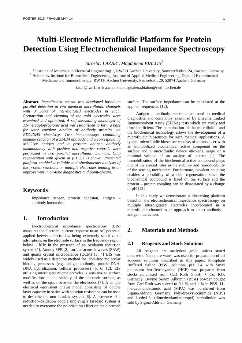
Gold interdigital electrodes were fabricated by standard thin-film technology with a finger width of 4 µm, a gap between the fingers of 3 µm and the length of fingers of 3885 µm, as shown in figure 1 A.
Fig. 1. A Gold interdigital electrodes for electrical impedance spectroscopy. B Microfluidic chip with six sensor areas in two microfluidic channels made of SU-8 photoresist with polydimethylsiloxane covers.
First a nLOF 2035 negative photoresist was spin-coated on thermally oxidized silicon wafers RC 8 spin coater (SÜSS MicroTec AG, Germany) and photolithographically structured in MA 6 mask aligner (SÜSS MicroTec AG, Germany), followed by sputtering of 200 nm gold layer in a Nordiko NS 2550 magnetron sputtering tool. Lift-off acetone process and ultrasound bath was utilized to form the wanted structures. The chips were diced out finally.
2.3 Microfluidic Fabrication and Instrumentation Setup
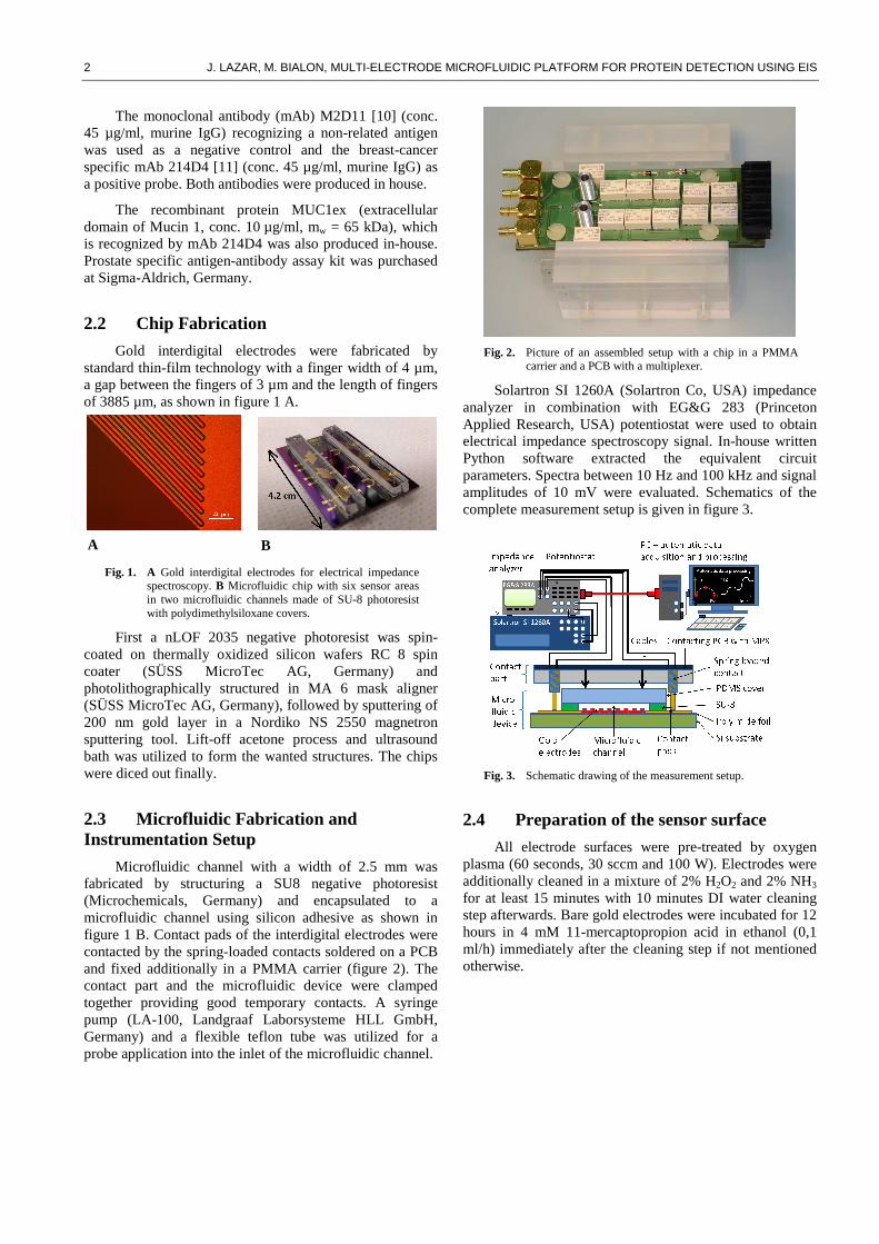
Microfluidic channel with a width of 2.5 mm was fabricated by structuring a SU8 negative photoresist (Microchemicals, Germany) and encapsulated to a microfluidic channel using silicon adhesive as shown in figure 1 B. Contact pads of the interdigital electrodes were contacted by the spring-loaded contacts soldered on a PCB and fixed additionally in a PMMA carrier (figure 2). The contact part and the microfluidic device were clamped together providing good temporary contacts. A syringe pump (LA-100, Landgraaf Laborsysteme HLL GmbH, Germany) and a flexible teflon tube was utilized for a probe application into the inlet of the microfluidic channel.
Fig. 2. Picture of an assembled setup with a chip in a PMMA
carrier and a PCB with a multiplexer.
Solartron SI 1260A (Solartron Co, USA) impedance analyzer in combination with EG&G 283 (Princeton Applied Research, USA) potentiostat were used to obtain electrical impedance spectroscopy signal. In-house written Python software extracted the equivalent circuit parameters. Spectra between 10 Hz and 100 kHz and signal amplitudes of 10 mV were evaluated. Schematics of the complete measurement setup is given in figure 3.
Fig. 3. Schematic drawing of the measurement setup.
2.4 Preparation of the sensor surface
All electrode surfaces were pre-treated by oxygen plasma (60 seconds, 30 sccm and 100 W). Electrodes were additionally cleaned in a mixture of 2% H2O2 and 2% NH3 for at least 15 minutes with 10 minutes DI water cleaning step afterwards. Bare gold electrodes were incubated for 12 hours in 4 mM 11-mercaptopropion acid in ethanol (0,1 ml/h) immediately after the cleaning step if not mentioned otherwise.
A B
POSTER 2015, PRAGUE MAY 14 3
3. Measurement and Results
3.1 Covalent Coupling Test on BSA
All electrode surfaces were cleaned as described above. The function of SAM based on covalent bond via S-H thiol coupling was further tested.
Two groups of electrodes were tested. Electrodes in the first group (‘+’ group) were activated by a 1:1 fresh mixture of 400 mM EDC and 100 mM NHS coupling chemistry for 10 minutes. Subsequently 0.1 % BSA solution in 5 mM HCF in PBS, pH 7.4 was applied on the electrodes for 30 minutes. Glycin of pH 2.3 (HCl titrated) was used to clean the electrodes of remaining BSA. EIS measurement in HCF solution was performed before and after BSA incubation and after cleaning process with the glycin solution. The result was compared with a second electrode group (‘-‘ group), in which the EDC/NHS coupling was not performed and therefore no covalent binding could exist. Curves after formation of SAM (after MPS), after incubation in BSA (after BSA) and after wash out step (after glycin) in figure 4 clearly show the successful function of activated SAM. The BSA protein was fully washed out of the surface from the electrode without any activated surface (hollow points in figure 4), while BSA on activated electrode stayed covalently bonded and only the BSA from medium was washed out (filled points in figure 4). This knowledge has further improved the stability of the immunoassay approach.
Fig. 4. Cole-Cole plots (100 kHz and 1 Hz) of electrodes with
EDC/NHS covalent coupling (+, filled points) and without EDC/NHS covalent coupling of BSA protein (-, hollow points).
3.2 Simplified Immunoassays
A simplified immunoassay was performed. Therefore, the following compounds were successively added: mAb 214D4 (conc. 45 µg/ml) in 0.1 % BSA in HCF buffer and the corresponding protein MUC1ex (conc. 10 µg/ml, mw = 65 kDa) in 0.1 % BSA in HCF buffer. The flow rate in all presented measurements was adjusted to 0.5 ml/hour.
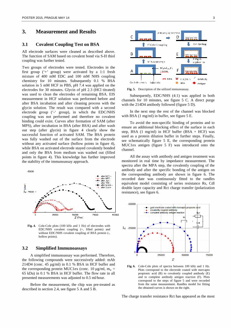
Before the measurement, the chip was pre-treated as described in section 2.4, see figure 5 A and 5 B.
Fig. 5. Description of the utilized immunoassay.
Subsequently, EDC/NHS (4:1) was applied in both channels for 10 minutes, see figure 5 C. A direct purge with the 214D4 antibody followed (figure 5 D).
In the next step the rest of the channel was blocked with BSA (1 mg/ml) in buffer, see figure 5 E.
To avoid the non-specific binding of proteins and to ensure an additional blocking effect of the surface in each step, BSA (1 mg/ml) in HCF buffer (BSA + HCF) was used as a protein dilution buffer in further steps. Finally, see schematically figure 5 E, the corresponding protein MUC1ex antigen (figure 5 F) was introduced onto the channel.
All the assay with antibody and antigen treatment was monitored in real time by impedance measurement. The spectra after the MPA step, the covalently coupling of the antibody and after the specific bonding of the antigen on the corresponding antibody are shown in figure 6. The recorded date was continuously fitted to the randles equivalent model consisting of series resistance Rs, Cdl double layer capacity and Rct charge transfer (polarization resistance), see figure 6.
Fig. 6. Cole-Cole plots of spectra between 100 kHz and 1 Hz.
Plots correspond to the electrode coated with mercapto propionic acid (B) to covalently coupled antibody (E) and to complete antibody antigen reaction (F). Plots correspond to the steps of figure 5 and were recorded from the same measurement. Randles model for fitting the obtained curves is shown on the right.
The charge transfer resistance Rct has appeared as the most
4 J. LAZAR, M. BIALON, MULTI-ELECTRODE MICROFLUIDIC PLATFORM FOR PROTEIN DETECTION USING EIS
sensitive to the protein adhesion. The continuous curve of the normalized Rct parameter for all the applied immunoassay is presented in figure 7. Data was normalized in the buffer level before antigen application. The increase in amplitude after an application of the corresponding antigen and the level of the amplitude after the last buffer purge clearly show the functionality of the assay.
Fig. 7. Normalized values of charge transfer resistance Rct from
the randles equivalent circuit is shown to present an interaction of covalently coupled 214D4 antibody and MUC1ex protein on EDC/NHS pre-treated electrode surface. Sections B to F correspond to the steps in figure 5. Dots correspond to spectra in figure 6.
Glycin titrated to pH 2.3 has shown an excellent possibility of the sensor cleaning in the paragraph 3.1. This property can be also used for sensor regeneration, see figure 8. Three electrodes in the channel were regenerated two times which gives 6 measurements in total. 10 µg/ml MUC1ex protein was injected after each cleaning. After the initial BSA+HCF purge the following measurement was normalized to this impedance level. The impedance values increased in the three devices due to the deposition of the antigen MUC1ex protein. During a final BSA+PBS purge, neither a decrease nor a protein desorption were observed, which differs from the first measurement at the sensor. This can be explained by a saturation of the unspecific regions on the sensor surface.
Fig. 8. Normalized values of Rct showing a binding of MUC1ex (10 µg/ml) antigen on 6 by glycin regenerated electrodes. Antibody protein stays on the electrode surface after glycin washing step.
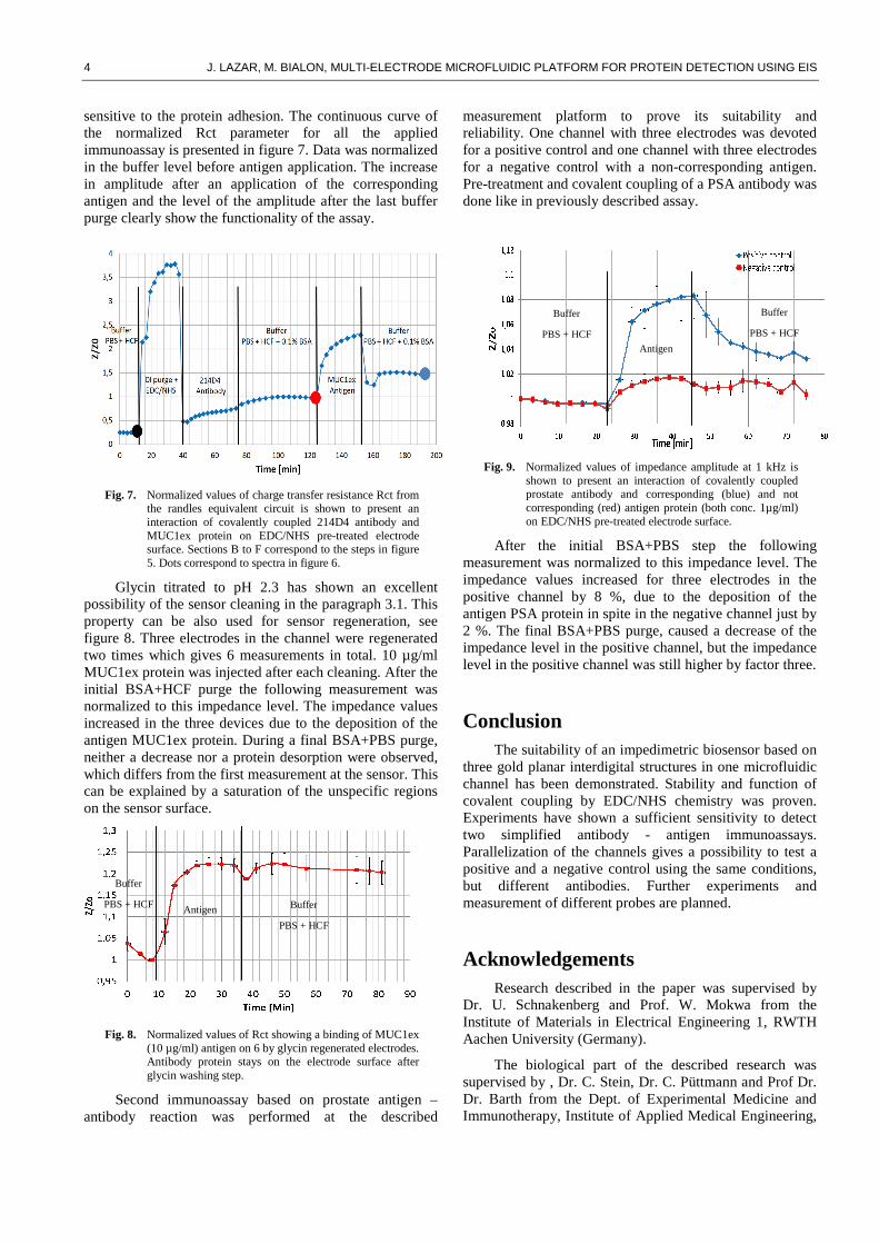
Second immunoassay based on prostate antigen – antibody reaction was performed at the described
measurement platform to prove its suitability and reliability. One channel with three electrodes was devoted for a positive control and one channel with three electrodes for a negative control with a non-corresponding antigen. Pre-treatment and covalent coupling of a PSA antibody was done like in previously described assay.
Fig. 9. Normalized values of impedance amplitude at 1 kHz is shown to present an interaction of covalently coupled prostate antibody and corresponding (blue) and not corresponding (red) antigen protein (both conc. 1µg/ml) on EDC/NHS pre-treated electrode surface.
After the initial BSA+PBS step the following measurement was normalized to this impedance level. The impedance values increased for three electrodes in the positive channel by 8 %, due to the deposition of the antigen PSA protein in spite in the negative channel just by 2 %. The final BSA+PBS purge, caused a decrease of the impedance level in the positive channel, but the impedance level in the positive channel was still higher by factor three.
Conclusion The suitability of an impedimetric biosensor based on
three gold planar interdigital structures in one microfluidic channel has been demonstrated. Stability and function of covalent coupling by EDC/NHS chemistry was proven. Experiments have shown a sufficient sensitivity to detect two simplified antibody - antigen immunoassays. Parallelization of the channels gives a possibility to test a positive and a negative control using the same conditions, but different antibodies. Further experiments and measurement of different probes are planned.
Acknowledgements Research described in the paper was supervised by
Dr. U. Schnakenberg and Prof. W. Mokwa from the Institute of Materials in Electrical Engineering 1, RWTH Aachen University (Germany).
The biological part of the described research was supervised by , Dr. C. Stein, Dr. C. Püttmann and Prof Dr. Dr. Barth from the Dept. of Experimental Medicine and Immunotherapy, Institute of Applied Medical Engineering,
Buffer
PBS + HCF
Buffer
PBS + HCF
Antigen
Buffer
PBS + HCF Buffer
PBS + HCF
Antigen
POSTER 2015, PRAGUE MAY 14 5
Helmholtz Institute for Biomedical Engineering, RWTH Aachen University (Germany). We would like to thank Dr. W. T. V. Germeraad from the Dept. of Internal Medicine, Division of Haematology, Maastricht University Medical Center (Netherlands), for his cooperation and for providing us with the 214D4 hybridoma clone.
This work was gratefully supported by INTERREG IV-A 2007-2013 EUREGIO MAAS-RIJN under grant INTERREG.EMR.INT4-1.2.-2009-11/058-MICROBIOMED.
References [1] DANIELS, J. S., POURMAND, N. Labell-Free Impedance
Biosensors: Opportunities and Challenges. Electroanalysis, 2007, vol. 19, p. 1239 – 1257.
[2] SCARANO, S., MASCINI, M., TURNER, A. P. F., MINUNNI, M., Surface plasmon resonance imaging for affinity-based biosensors, Biosensors and Bioelectronics, 2010, vol. 25/5, p. 957 – 966.
[3] VIDAL, J. C., DUATO, P., BONEL, L., CASTILLO, J. R., Use of polyclonal antibodies to ochratoxin A with a quartz–crystal microbalance for developing real-time mycotoxin piezoelectric immunosensors, Analytical and Bioanalytical Chemistry, 2009, vol. 394/2, p. 572 – 582
[4] SANGDAE, L., KI-BOK, K., YONG-IL, K., Love wave SAW biosensors for detection of antigen-antibody binding and comparison with SPR biosensor, Food Science and Biotechnolog, 2011, vol. 20/5, p. 1413-1418.
[5] TANG, X. H., et al. Direct Protein Detection with a Nano-interdigitated Array Gate MOSFET. Biosens.Bioelectronics, 2009 24.12, p. 3531-37.
[6] BERDAT, D., RODRIGUEZ, A. C. M., HERRERA, F., GIJS, M. A. M., Label-free detection of DNA with interdigitated micro-electrodes in a fluidic cell. Lab Chip, 2008, vol. 8, p. 302–308.
[7] WANG, R., et al., Interdigitated array microelectrode based impedance immunosensor for detection of avian irus H5N1. Talanta, 2009, vol. 79, p 159 – 164.
[8] VAN GERWEN, P., et al. Nanoscaled interdigitated electrode arrays for biochemical sensors, Sens. Actuat. B, 1998, vol. 49, p.73-80.
[9] NARAKATHU, B. B., ATASHBAR, M. Z., BEJCEK B. E., Improved detection limits of toxic biochemical species based on impedance measurements in electrochemical biosensors. Biosens.Bioelectron., 2010, 26.2, p.923-928.
[10] PUETTMANN, C., KOLBERG, K., HAGEN, S., SCHMIES, S., FISCHER, R., NAEHRING, J., BARTH, S., A monoclonal antibody for the detection of SNAP/CLIP-tagged proteins. Immunology letters, 2012, PMID 23085606.
[11] VAN ELSSEN, C. H., FRINGS, P. W., BOT, F. J., VAN DE VIJVER, K. K., HULS, M. B., MEEK, B., HUPPERETS, P., GERMERAAD, W. T., BOS, G.M. Expression of aberrantly glycosylated Mucin-1 in ovarian cancer. Histopathology, 2010, 57/4, p. 597-606.
[12] ZOU, Z., et al. Functionalized nano interdigitated electrodes arrays on polymer with integrated microfluidics for direct bio-affinity sensing using impedimetric measurement. Sensors and Actuators A: Physical, 2007, Vol. 136, Nr. 2, p. 518-526.
[13] ZHANG, X., et al. A reusable electrochemical immunosensor for carcinoembryonic antigen via molecular recognition of glycoprotein antibody by phenylboronic acid self-assembly layer on gold. Analyst, 2008, Vol 133, Nr. 4, p. 485-492.
About Authors... Jaroslav Lazar was born in Kutná Hora in Czech Republic in 1982. He received the title Dipl.-Ing. from RWTH Aachen in 2008 with final thesis on optical coherence tomography for ophthalmological application. After receiving his diploma he continued to work on various optical systems at the Institute of Semiconductor Electronics of RWTH Aachen. During his studies he worked for Philips Research Aachen on development of a magnetic induction tomography device for two years on part time basis. Since 2010 he has been working on his PhD at the Institute of Materials in Electrical Engineering 1 at RWTH Aachen. To his interests belong impedimetric sensing of protein reactions, reactions on glycopolymers and SPR sensing in microfluidic chips or magnetic tweezers for a cell biology applications.
Magdalena Bialon was born in Tychy in Upper Silesia (Poland) in 1985. She received the Abitur in June 2005 at the Hildegardis school in Hagen, NRW, Germany. After that she studied Chemical Biology at the Technical University of Dortmund, NRW, Germany. The Bachelor thesis was written in 2008 at the Max-Planck-Institute of Molecular Physiology (Dortmund) with the title "Optimization of the protein identification and quantification using the SILAC method in HeLa cells". After the Bachelor she started the Master program also in Chemical Biology in Dortmund. In 2010 she wrote the thesis with the title "Expression of phosphorylated ErbB3 in primary ovarian carcinoma in comparison to ovarian carcinoma recurrence". The thesis was prepared at the Leibniz Research Centre for Working Environment and Human Factors in Dortmund. Since May 2011 she is working on her PhD at the Helmholtz-Institute for Biomedical Engineering in the group Experimental Medicine and Immunotherapy (Aachen). The goal of the project is to develop, in cooperation with different engineers, a biochip system for the early detection of breast cancer in women by using cancer specific markers.