MR imaging of the cavernous sinus: Value of spin echo and gradient recalled echo images
1
88 ABSTRACTS CLINICAL IMAGING VOL. 13, NO. 1 rasellar mass, with supplementation when necessary by CT and angiography. Author’s summary MR IMAGING OF THE CAVERNOUS SINUS: VALUE OF SPIN ECHO AND GRADIENT RE- CALLED ECHO IMAGES Daniels DL, Czerviionke LF, Bonneville JF, Cattin F, Mark LP, Pech P, Hendrix LE, Smith DF, Haugh- ton VM, Williams AL (Department of Radiology, Medical College of Wisconsin, Froedtert Memorial Lutheran Hospital, 9200 W. Wisconsin Ave., Milwaukee, WI 53226, U.S.A) AJR 1988;151:1009- 10014. spinal cord, observed on the phase images were most marked at the cervical-upper thoracic level. Cord motion was found to be significantly decreased in cases in which the cord was either tethered or compressed. Cord enlargement due to an intrame- dullarv lesion generally did not lead to decreased cord motion. Imaging of pulsatile cord motion may be clinically useful in evaluating diseases restricting cord motion or changing the status of parenchymal compliance. A detailed evaluation of the MR appearance of the pituitary gland-cavernous sinus junction has not been described. In a series of coronal Tl-weighted spin-echo images without and with IV Gadolinium, we noted the variable size and signal intensity of cavernous venous spaces adjacent to the pituitary gland and the inconsistent visualization of the dural membrane just lateral to the gland. Correlation of coronal Tl-weighted spin-echo and gradient recalled echo images (the latter with high- signal-intensity vascular structures) proved to be an effective means of identifying cavernous venous spaces, connective tissue and cranial nerves, and the lateral margins of the pituary gland, and of differen- tiating tumor tissue from cavernous venous spaces. Further work is needed to develop criteria to distin- guish cavernous sinus compression from compres- sion from actual tumor invasion. Author’s summary Author’s summary CT-GUIDED CUTTING-NEEDLE BIOPSIES OF SELECTED CHEST LESIONS Goralnik CH, O’Connell DM, El Yousef S, Haaga JR [Radiology Department, San Antonio Community Hospital, 999 San Bernadino Rd, Upland CA 91786, USA) AJR 1988;151:903-907. In an effort to improve on our diagnostic yield transthoracic biopsy, we used 14-gauge cutting nee- dles in 56 selected patients. These biopsies were preceded by 18-,20-, or 22-gauge aspirations in 42 patients, allowing a direct comparison of the efficacy of the needle types. Specific diagnoses were made by cutting-needle biopsy in 78% (25132) of patients with nonlymphoproliferative malignancies, in 73% (8/11) with lymphoma or thymoma, and 54% (7113) of patients with benign diseases. In those in whom both aspiration and cutting needles were employed, a higher percentage of specific diagnoses was achieved by cutting-needle biopsy than by aspiration biopsy: 72% vs 64% in nonlymphoproliferative ma- lignancies, 62% vs 12% in the lymphoproliferative group, and 55% vs 22% in benign disorders. Compli- cations were encountered in 20% of all patients studied. FIXED SPINAL CORD: DIAGNOSIS WITH MR IMAGING Levy LM, Di Chiro G, McCullough DC, Dwyer AJ, Johnsson DL, Yang SSL. (G.D.C.: Neuroimaging This study shows that, for all selected chest le- sions, CT-guided cutting-needle biopsies can be per- formed safely and are useful, especially in the diag- nosis of lymphoproliferative or benign disease. Section, Bldg. 10, RM lC451, National Institutes of Health, Bethesda, M.D. 20892, U.S.A) Radiology 1988;169:773-778. Author’s summary Pulsatile motion of the spinal cord was examined PRIMARY LIVER TUMORS IN CHILDREN: with phase imaging techniques. Sagittal images of COMPARISON OF CT AND MR IMAGING the spinal cord were obtained at different times of Boechat MI, Kangarloo H, Ortega J, Hall T, Feig S, the cardiac cycle in healthy volunteers, as well as in Stanley PS, Gilanz V (Department of Radiological patients in whom the spinal cord either was teth- Sciences, UCLA Medical Center, CHS B2-252, Los ered, was compressed, or contained an intrame- Angeles, CA 90024, U.S.A.) Radiology dullary lesion. Pulsatile velocity changes of the 1988;169:727-732.
-
Upload
trannguyet -
Category
Documents
-
view
214 -
download
2
Transcript of MR imaging of the cavernous sinus: Value of spin echo and gradient recalled echo images
88 ABSTRACTS CLINICAL IMAGING VOL. 13, NO. 1
rasellar mass, with supplementation when necessary by CT and angiography.
Author’s summary
MR IMAGING OF THE CAVERNOUS SINUS: VALUE OF SPIN ECHO AND GRADIENT RE- CALLED ECHO IMAGES Daniels DL, Czerviionke LF, Bonneville JF, Cattin F, Mark LP, Pech P, Hendrix LE, Smith DF, Haugh- ton VM, Williams AL (Department of Radiology, Medical College of Wisconsin, Froedtert Memorial Lutheran Hospital, 9200 W. Wisconsin Ave., Milwaukee, WI 53226, U.S.A) AJR 1988;151:1009- 10014.
spinal cord, observed on the phase images were most marked at the cervical-upper thoracic level. Cord motion was found to be significantly decreased in cases in which the cord was either tethered or compressed. Cord enlargement due to an intrame- dullarv lesion generally did not lead to decreased cord motion. Imaging of pulsatile cord motion may be clinically useful in evaluating diseases restricting cord motion or changing the status of parenchymal compliance.
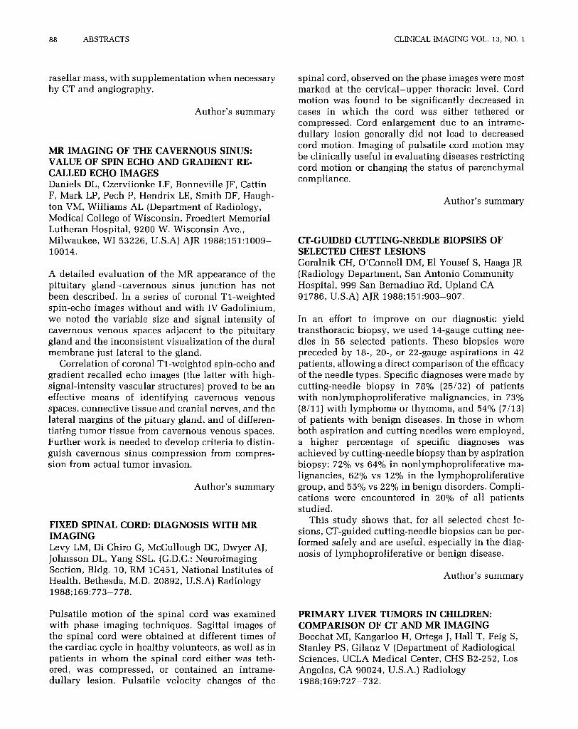
A detailed evaluation of the MR appearance of the pituitary gland-cavernous sinus junction has not been described. In a series of coronal Tl-weighted spin-echo images without and with IV Gadolinium, we noted the variable size and signal intensity of cavernous venous spaces adjacent to the pituitary gland and the inconsistent visualization of the dural membrane just lateral to the gland.
Correlation of coronal Tl-weighted spin-echo and gradient recalled echo images (the latter with high- signal-intensity vascular structures) proved to be an effective means of identifying cavernous venous spaces, connective tissue and cranial nerves, and the lateral margins of the pituary gland, and of differen- tiating tumor tissue from cavernous venous spaces. Further work is needed to develop criteria to distin- guish cavernous sinus compression from compres- sion from actual tumor invasion.
Author’s summary
Author’s summary
CT-GUIDED CUTTING-NEEDLE BIOPSIES OF SELECTED CHEST LESIONS Goralnik CH, O’Connell DM, El Yousef S, Haaga JR [Radiology Department, San Antonio Community Hospital, 999 San Bernadino Rd, Upland CA 91786, USA) AJR 1988;151:903-907.
In an effort to improve on our diagnostic yield transthoracic biopsy, we used 14-gauge cutting nee- dles in 56 selected patients. These biopsies were preceded by 18-, 20-, or 22-gauge aspirations in 42 patients, allowing a direct comparison of the efficacy of the needle types. Specific diagnoses were made by cutting-needle biopsy in 78% (25132) of patients with nonlymphoproliferative malignancies, in 73% (8/11) with lymphoma or thymoma, and 54% (7113) of patients with benign diseases. In those in whom both aspiration and cutting needles were employed, a higher percentage of specific diagnoses was achieved by cutting-needle biopsy than by aspiration biopsy: 72% vs 64% in nonlymphoproliferative ma- lignancies, 62% vs 12% in the lymphoproliferative group, and 55% vs 22% in benign disorders. Compli- cations were encountered in 20% of all patients studied.
FIXED SPINAL CORD: DIAGNOSIS WITH MR IMAGING Levy LM, Di Chiro G, McCullough DC, Dwyer AJ, Johnsson DL, Yang SSL. (G.D.C.: Neuroimaging
This study shows that, for all selected chest le- sions, CT-guided cutting-needle biopsies can be per- formed safely and are useful, especially in the diag- nosis of lymphoproliferative or benign disease.
Section, Bldg. 10, RM lC451, National Institutes of Health, Bethesda, M.D. 20892, U.S.A) Radiology 1988;169:773-778.
Author’s summary
Pulsatile motion of the spinal cord was examined PRIMARY LIVER TUMORS IN CHILDREN: with phase imaging techniques. Sagittal images of COMPARISON OF CT AND MR IMAGING the spinal cord were obtained at different times of Boechat MI, Kangarloo H, Ortega J, Hall T, Feig S, the cardiac cycle in healthy volunteers, as well as in Stanley PS, Gilanz V (Department of Radiological patients in whom the spinal cord either was teth- Sciences, UCLA Medical Center, CHS B2-252, Los ered, was compressed, or contained an intrame- Angeles, CA 90024, U.S.A.) Radiology dullary lesion. Pulsatile velocity changes of the 1988;169:727-732.