Motor tracts
39
MOTOR TRACTS Naresh Mullaguri MD Resident Physician PGY3 Department of Neurology University of Missouri
-
Upload
university-of-missouri -
Category
Health & Medicine
-
view
65 -
download
1
Transcript of Motor tracts

MOTOR TRACTS
Naresh Mullaguri MD
Resident Physician PGY3Department of Neurology
University of Missouri

LEARNING PLAN Cerebral areas of Motor control Corticospinal tracts Corticobulbar tracts (largely covered under
Brainstem nuclei) Rubrospinal tracts Reticulospinal tracts Vestibulospinal tracts Tectospinal tracts
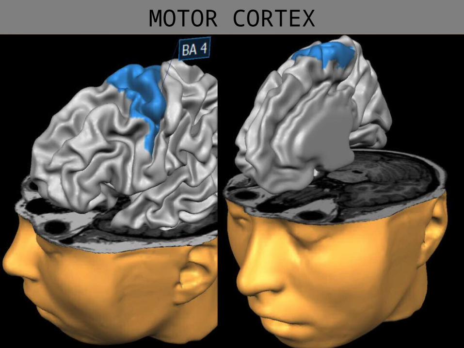
MOTOR CORTEX


PREMOTOR AREAS

https://youtu.be/9BaWBGRVxp8

CORTICOSPINAL TRACTS Aka Pyramidal tract Contains Axons and cell bodies present in the
motor cortex of the Brain Originates from the Pyramidal cells of Betz –
Layer V of Primary Motor cortex (30%), Premotor area and supplementary Motor area(30%) and Somatic sensory area (40%)


MOTOR HOMUNCULUS


FACTS ABOUT CORTICOSPINAL TRACTS
Lateral CST: Crossed, Lateral funicuus, terminate in the Internuncial Neurons of the Spinal grey matter, some fibers end directly on the AH cells, Throughout the length of the cord
Uncrossed Pyramidal fibers, about 10%, anterior funiculus and run until mid thoracic level, Cross the other side at the appropriate spinal segment and terminate in the same way as the lateral CST
Unmyeinated at birth, Myelinates from 2nd week after birth to 2 years of age, small and large fibers. Senile degeneration of the large fibers leading to shaky movements.


Extrapyramidal tracts Motor pathways act as an alternative route for
volitional impulses and which form the platform on which the pyramidal system works skillfully.
Integrated at various levels from cerebral cortex to spinal cord
Cortical areas controlling these tracts are areas 6 and 8

Types Rubrospinal tract Reticulospinal tract Olivospinal tract Vestibulospinal tract Tectospinal tract Medial Longitudinal Fasciculus
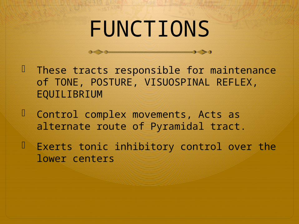
FUNCTIONS These tracts responsible for maintenance of
TONE, POSTURE, VISUOSPINAL REFLEX, EQUILIBRIUM
Control complex movements, Acts as alternate route of Pyramidal tract.
Exerts tonic inhibitory control over the lower centers


Rubrospinal tract


RED NUCLEUS Prominent structure in the rostral midbrain just dorsal to substantia nigra.
High in Iron content and is more vascular than the surrounding tissue Most important efferent projection is to the contralateral spinal cord i.e.,
Rubrospinal tract Course: Dorsally to pons and ventral to the lateral reticular nuclei of the
Medulla. Then enters the lateral white column of the spinal cord and terminate in the internuncial neurons at the base of the anterior horn cells
Extension: Cervical cord receives max. number of fibers. In Humans this tract do not
extend beyond thoracic segments Facilitatory influence over the flexor muscle tone, Acts as alternate route to
the Pyramidal tract.


RETICULOSPINAL TRACT Divided into Medial and Lateral tracts MEDIAL RETICULOSPINAL TRACT(Pontine Reticular Tract)
Origin from Pontine reticular formation from Reticularis pontis oralis and Reticularis Pontis Caudalis
Runs ipsilateral through Longitudinal Fasciculus
Medial part of the anterior funiculus in the Spinal cord
Terminates ipsilaterally at all spinal levels

Lateral Reticulospinal fibers(Medullary Reticulospinal tract)
Origin from Medullary reticular formation – Nucleus reticularis gigantocellularisSome fibers cross and rest of them remain ipsilaterallyDescend in the anterior part of the lateral funiculusTerminate in the internuncial neurons of the anterior horn of the Spinal cord


FunctionsCORTICO_RETICULOSPINAL PATHWAY Integrates information from the motor systems to coordinate
automatic movements of locomotion and posture (antigravity muscles)
Facilitates and inhibits voluntary movement, influences MUSCLE TONE
Conveys autonomic fibers from higher centers Modulates pain impulses MRST: Facilitate voluntary control and reflex movements,
control of Muscle tone through Gamma Neurons, Favors expiration and facilitates Vasoconstriction
LRST: Inhibits voluntary control and reflex movements, Decrease muscle tone, favors inspiration and responsible for vasodilatation.

OLIVOSPINAL TRACT Also called Bulbospinal tract or tract of Helweg Course: Arise in the inferior Olivary nucleus Enter
anterior part of the Lateral funiculus Terminate in the anterior horn cells of Spinal cord
Found in Cervical region only Function: Reflex movements arising from proprioception,
existence is doubtful.

TECTOSPINAL TRACT Origin: Superior colliculus of the midbrain Fibers cross in the Dorsal tegmental decussation (Decussation of
Meynert) which is ventral to the Cerebral aqueduct Descend to the anterior white funiculus and terminate in the
anterior horn of the spinal cord
Function: Control head and arm movements (posture control) in response to visual and auditory stimuli


Vestibulospinal Tracts Two typesMedial Vestibulospinal Tract : Medial Vestibular nucleus in Medulla Descend upto upper thoracic spinal segment Enter uncrossed into Anterior white funiculus and terminate in the anterior horn cellFunction: Concerned with adjustment of head and body during angular and linear acceleration and conjugate horizontal eye movements, integration of eye and neck movements.Lateral Vestibulospinal tract: Lateral Vestibular nucleus (Dieter’s Nucleus) in medulla on both sides Tract descend throughout the spinal cord length Run uncrossed in the anterior white funiculus and terminate in the anterior horn cell of the spinal cord.Function: Essentially the same as the Anterior Reticulospinal tract with facilitatory influence on reflex spinal activities and Spinal mechanisms underlying Muscle tone. Facilitates extensor muscles and inhibit flexor muscles. Maintenance of balance.

Medial Longitudinal Fasciculus Extends from midbrain downwards. Fibers take origin from Vestibular nuclei,
Reticular formation, superior colliculus, Interstitial nucleus of cajal, Posterior commissure and has connections to CN III, IV, VI, VII, VIII, XII
Anterior horn cells of the Neck Function: Hormonious movements of the eyes
and Neck

VESTIBULOSPINAL SYSTEM


LATERAL VESTIBULOSPINAL SYSTEM


THANK YOU