Morgellons Disease - KoreaMed · 2017-03-22 · Morgellons Disease Vol. 29, No. 2, 2017225 Many...
3
Morgellons Disease Vol. 29, No. 2, 2017 223 Received May 30, 2016, Revised July 26, 2016, Accepted for publication August 4, 2016 Corresponding author: Kyu Han Kim, Department of Dermatology, Seoul National University College of Medicine, 101 Daehak-ro, Jongno-gu, Seoul 03080, Korea. Tel: 82-2-2072-2410, Fax: 82-2-742-7344, E-mail: kyuhkim@ snu.ac.kr This is an Open Access article distributed under the terms of the Creative Commons Attribution Non-Commercial License (http://creativecommons. org/licenses/by-nc/4.0) which permits unrestricted non-commercial use, distribution, and reproduction in any medium, provided the original work is properly cited. Copyright © The Korean Dermatological Association and The Korean Society for Investigative Dermatology pISSN 1013-9087ㆍeISSN 2005-3894 Ann Dermatol Vol. 29, No. 2, 2017 https://doi.org/10.5021/ad.2017.29.2.223 CASE REPORT Fig. 1. Photographs taken by the patient with a magnifying device (×200): black, brown, and red fibers were tangled on the erosive skin. Morgellons Disease Jungyoon Ohn, Seon Yong Park, Jungyoon Moon, Yun Seon Choe, Kyu Han Kim Department of Dermatology, Seoul National University College of Medicine, Seoul, Korea Morgellons disease is a rare disease with unknown etiology. Herein, we report the first case of Morgellons disease in Korea. A 30-year-old woman presented with a 2-month his- tory of pruritic erythematous patches and erosions on the arms, hands, and chin. She insisted that she had fiber-like ma- terials under her skin, which she had observed through a magnifying device. We performed skin biopsy, and observed a fiber extruding from the dermal side of the specimen. Histopathological examination showed only mild lympho- cytic infiltration, and failed to reveal evidence of any microorganism. The polymerase chain reaction for Borrelia burgdorferi was negative in her serum. (Ann Dermatol 29(2) 223∼225, 2017) -Keywords- Asian continental ancestry group, Borrelia burgdorferi, Morgellons disease INTRODUCTION Morgellons disease is a mysterious disease with unknown etiology. It is characterized by fibers appearing in slow- or non-healing skin lesions and even beneath unbroken skin, along with abnormal (crawling, stinging, or biting) sensa- tions of skin. Extracutaneous manifestations (fatigue, joint pain, fibromyalgia, or sleep disorders) have been reported to co-exist 1 . It has been recognized as a delusional in- festation 2 . However, recent studies are suggesting that it could be associated with an infectious organism: Borrelia burgdorferi 3,4 . There have been a number of reports and investigations about Morgellons disease in the US or Europe, but there are none so far in Asia. CASE REPORT A 30-year-old woman presented with a 2-month history of pruritic cutaneous lesions on the hands and arms. She in- sisted that she had fiber-like materials under the skin, and could find fiber-like materials when she ripped the skin off. She also complained of a stinging sensation like hav- ing a splinter in the fingers. She brought some pictures of the fiber-like materials ‘in situ,’ taken by her using a mag- nifying device. Twisted black, brown, and red fibers were
Transcript of Morgellons Disease - KoreaMed · 2017-03-22 · Morgellons Disease Vol. 29, No. 2, 2017225 Many...
Morgellons Disease
Vol. 29, No. 2, 2017 223
Received May 30, 2016, Revised July 26, 2016, Accepted for publication August 4, 2016
Corresponding author: Kyu Han Kim, Department of Dermatology, Seoul National University College of Medicine, 101 Daehak-ro, Jongno-gu, Seoul 03080, Korea. Tel: 82-2-2072-2410, Fax: 82-2-742-7344, E-mail: kyuhkim@ snu.ac.kr
This is an Open Access article distributed under the terms of the Creative Commons Attribution Non-Commercial License (http://creativecommons.org/licenses/by-nc/4.0) which permits unrestricted non-commercial use, distribution, and reproduction in any medium, provided the original work is properly cited.
Copyright © The Korean Dermatological Association and The Korean Society for Investigative Dermatology
pISSN 1013-9087ㆍeISSN 2005-3894Ann Dermatol Vol. 29, No. 2, 2017 https://doi.org/10.5021/ad.2017.29.2.223
CASE REPORT
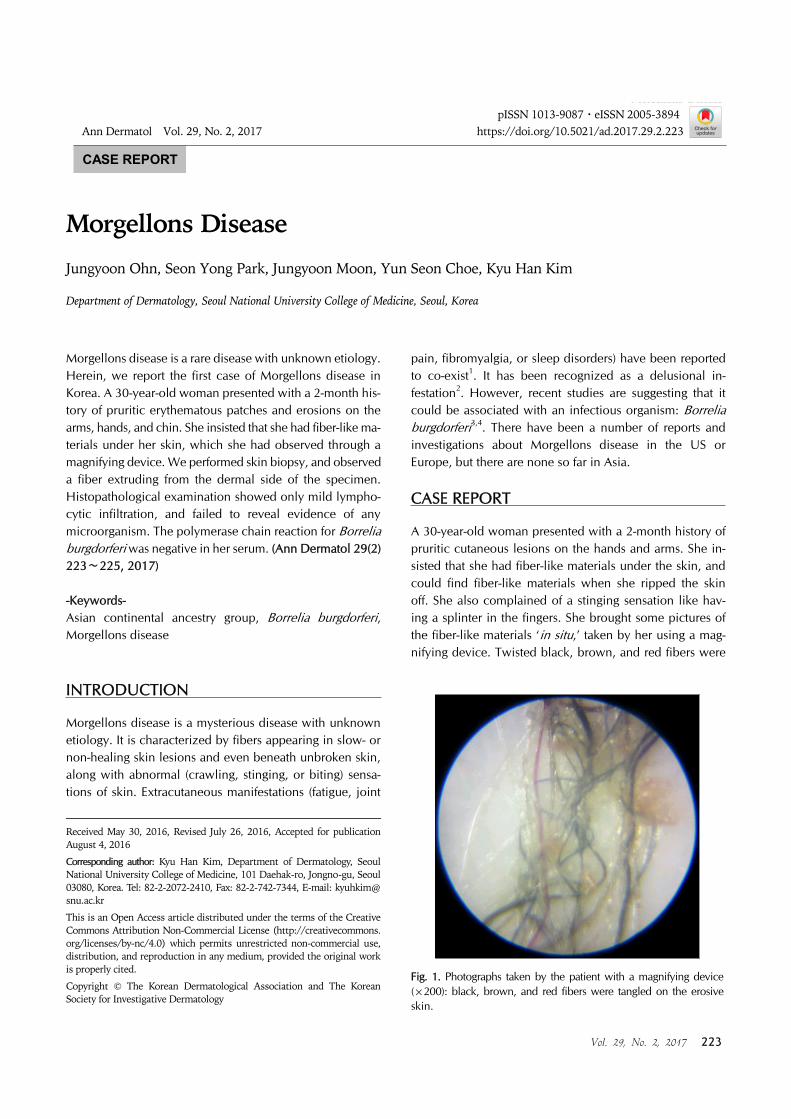
Fig. 1. Photographs taken by the patient with a magnifying device (×200): black, brown, and red fibers were tangled on the erosive skin.
Morgellons Disease
Jungyoon Ohn, Seon Yong Park, Jungyoon Moon, Yun Seon Choe, Kyu Han Kim
Department of Dermatology, Seoul National University College of Medicine, Seoul, Korea
Morgellons disease is a rare disease with unknown etiology. Herein, we report the first case of Morgellons disease in Korea. A 30-year-old woman presented with a 2-month his-tory of pruritic erythematous patches and erosions on the arms, hands, and chin. She insisted that she had fiber-like ma-terials under her skin, which she had observed through a magnifying device. We performed skin biopsy, and observed a fiber extruding from the dermal side of the specimen. Histopathological examination showed only mild lympho-cytic infiltration, and failed to reveal evidence of any microorganism. The polymerase chain reaction for Borrelia burgdorferi was negative in her serum. (Ann Dermatol 29(2) 223∼225, 2017)
-Keywords-Asian continental ancestry group, Borrelia burgdorferi, Morgellons disease
INTRODUCTION
Morgellons disease is a mysterious disease with unknown etiology. It is characterized by fibers appearing in slow- or non-healing skin lesions and even beneath unbroken skin, along with abnormal (crawling, stinging, or biting) sensa-tions of skin. Extracutaneous manifestations (fatigue, joint
pain, fibromyalgia, or sleep disorders) have been reported to co-exist1. It has been recognized as a delusional in-festation2. However, recent studies are suggesting that it could be associated with an infectious organism: Borrelia burgdorferi3,4. There have been a number of reports and investigations about Morgellons disease in the US or Europe, but there are none so far in Asia.
CASE REPORT
A 30-year-old woman presented with a 2-month history of pruritic cutaneous lesions on the hands and arms. She in-sisted that she had fiber-like materials under the skin, and could find fiber-like materials when she ripped the skin off. She also complained of a stinging sensation like hav-ing a splinter in the fingers. She brought some pictures of the fiber-like materials ‘in situ,’ taken by her using a mag-nifying device. Twisted black, brown, and red fibers were
J Ohn, et al
224 Ann Dermatol
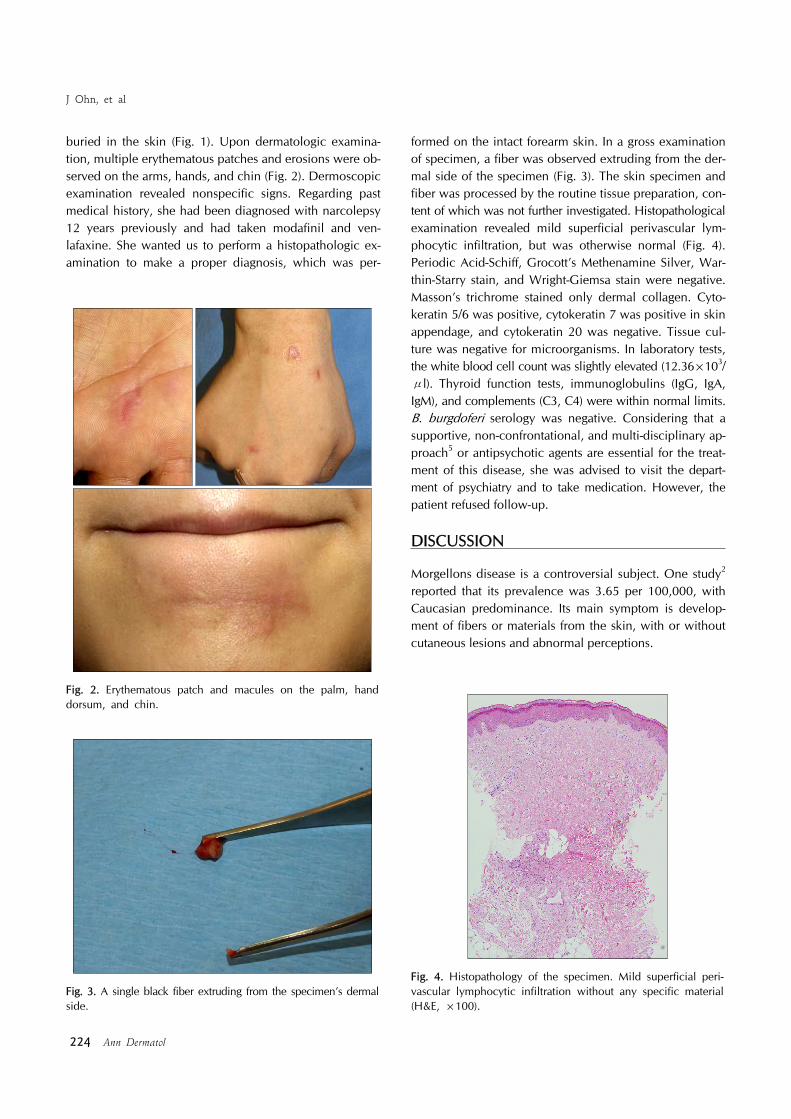
Fig. 2. Erythematous patch and macules on the palm, hand dorsum, and chin.
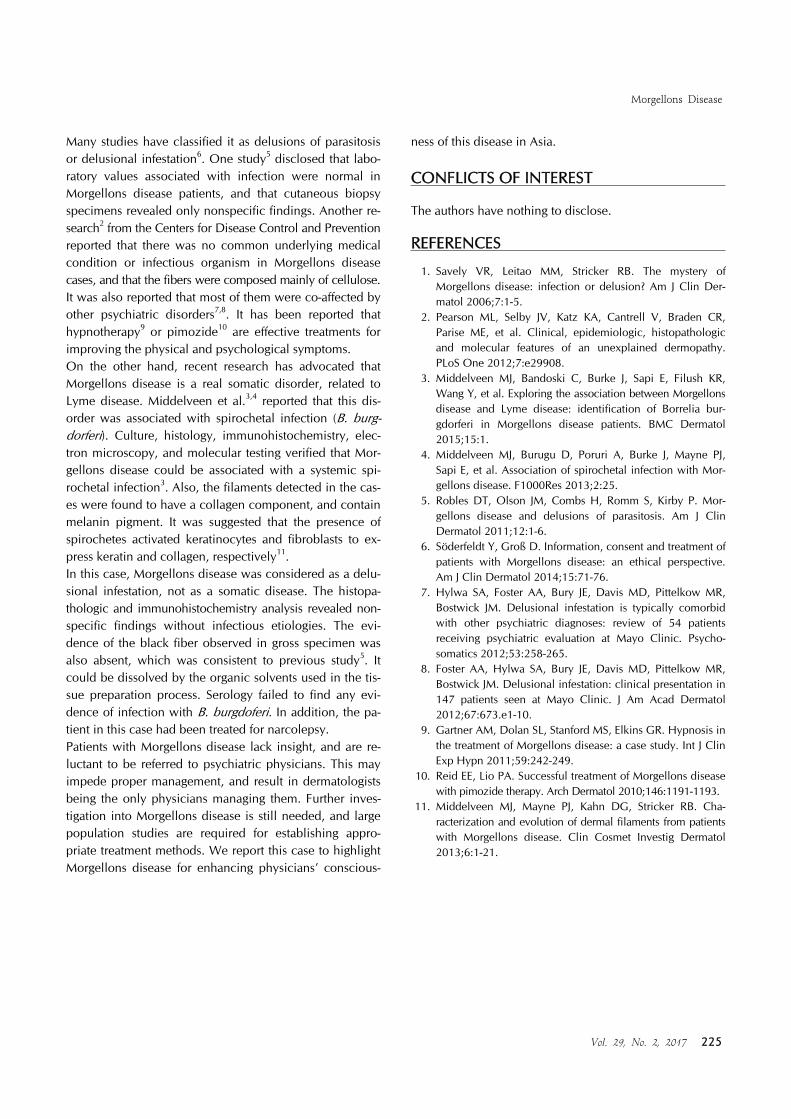
Fig. 3. A single black fiber extruding from the specimen’s dermal side.
Fig. 4. Histopathology of the specimen. Mild superficial peri-vascular lymphocytic infiltration without any specific material (H&E, ×100).
buried in the skin (Fig. 1). Upon dermatologic examina-tion, multiple erythematous patches and erosions were ob-served on the arms, hands, and chin (Fig. 2). Dermoscopic examination revealed nonspecific signs. Regarding past medical history, she had been diagnosed with narcolepsy 12 years previously and had taken modafinil and ven-lafaxine. She wanted us to perform a histopathologic ex-amination to make a proper diagnosis, which was per-
formed on the intact forearm skin. In a gross examination of specimen, a fiber was observed extruding from the der-mal side of the specimen (Fig. 3). The skin specimen and fiber was processed by the routine tissue preparation, con-tent of which was not further investigated. Histopathological examination revealed mild superficial perivascular lym-phocytic infiltration, but was otherwise normal (Fig. 4). Periodic Acid-Schiff, Grocott’s Methenamine Silver, War-thin-Starry stain, and Wright-Giemsa stain were negative. Masson’s trichrome stained only dermal collagen. Cyto-keratin 5/6 was positive, cytokeratin 7 was positive in skin appendage, and cytokeratin 20 was negative. Tissue cul-ture was negative for microorganisms. In laboratory tests, the white blood cell count was slightly elevated (12.36×10³/μl). Thyroid function tests, immunoglobulins (IgG, IgA, IgM), and complements (C3, C4) were within normal limits. B. burgdoferi serology was negative. Considering that a supportive, non-confrontational, and multi-disciplinary ap-proach5 or antipsychotic agents are essential for the treat-ment of this disease, she was advised to visit the depart-ment of psychiatry and to take medication. However, the patient refused follow-up.
DISCUSSION
Morgellons disease is a controversial subject. One study2 reported that its prevalence was 3.65 per 100,000, with Caucasian predominance. Its main symptom is develop-ment of fibers or materials from the skin, with or without cutaneous lesions and abnormal perceptions.
Morgellons Disease
Vol. 29, No. 2, 2017 225
Many studies have classified it as delusions of parasitosis or delusional infestation6. One study5 disclosed that labo-ratory values associated with infection were normal in Morgellons disease patients, and that cutaneous biopsy specimens revealed only nonspecific findings. Another re-search2 from the Centers for Disease Control and Prevention reported that there was no common underlying medical condition or infectious organism in Morgellons disease cases, and that the fibers were composed mainly of cellulose. It was also reported that most of them were co-affected by other psychiatric disorders7,8. It has been reported that hypnotherapy9 or pimozide10 are effective treatments for improving the physical and psychological symptoms.On the other hand, recent research has advocated that Morgellons disease is a real somatic disorder, related to Lyme disease. Middelveen et al.3,4 reported that this dis-order was associated with spirochetal infection (B. burg-dorferi). Culture, histology, immunohistochemistry, elec-tron microscopy, and molecular testing verified that Mor-gellons disease could be associated with a systemic spi-rochetal infection3. Also, the filaments detected in the cas-es were found to have a collagen component, and contain melanin pigment. It was suggested that the presence of spirochetes activated keratinocytes and fibroblasts to ex-press keratin and collagen, respectively11.In this case, Morgellons disease was considered as a delu-sional infestation, not as a somatic disease. The histopa-thologic and immunohistochemistry analysis revealed non-specific findings without infectious etiologies. The evi-dence of the black fiber observed in gross specimen was also absent, which was consistent to previous study5. It could be dissolved by the organic solvents used in the tis-sue preparation process. Serology failed to find any evi-dence of infection with B. burgdoferi. In addition, the pa-tient in this case had been treated for narcolepsy.Patients with Morgellons disease lack insight, and are re-luctant to be referred to psychiatric physicians. This may impede proper management, and result in dermatologists being the only physicians managing them. Further inves-tigation into Morgellons disease is still needed, and large population studies are required for establishing appro-priate treatment methods. We report this case to highlight Morgellons disease for enhancing physicians’ conscious-
ness of this disease in Asia.
CONFLICTS OF INTEREST
The authors have nothing to disclose.
REFERENCES
1. Savely VR, Leitao MM, Stricker RB. The mystery of Morgellons disease: infection or delusion? Am J Clin Der-matol 2006;7:1-5.
2. Pearson ML, Selby JV, Katz KA, Cantrell V, Braden CR, Parise ME, et al. Clinical, epidemiologic, histopathologic and molecular features of an unexplained dermopathy. PLoS One 2012;7:e29908.
3. Middelveen MJ, Bandoski C, Burke J, Sapi E, Filush KR, Wang Y, et al. Exploring the association between Morgellons disease and Lyme disease: identification of Borrelia bur-gdorferi in Morgellons disease patients. BMC Dermatol 2015;15:1.
4. Middelveen MJ, Burugu D, Poruri A, Burke J, Mayne PJ, Sapi E, et al. Association of spirochetal infection with Mor-gellons disease. F1000Res 2013;2:25.
5. Robles DT, Olson JM, Combs H, Romm S, Kirby P. Mor-gellons disease and delusions of parasitosis. Am J Clin Dermatol 2011;12:1-6.
6. Söderfeldt Y, Groß D. Information, consent and treatment of patients with Morgellons disease: an ethical perspective. Am J Clin Dermatol 2014;15:71-76.
7. Hylwa SA, Foster AA, Bury JE, Davis MD, Pittelkow MR, Bostwick JM. Delusional infestation is typically comorbid with other psychiatric diagnoses: review of 54 patients receiving psychiatric evaluation at Mayo Clinic. Psycho-somatics 2012;53:258-265.
8. Foster AA, Hylwa SA, Bury JE, Davis MD, Pittelkow MR, Bostwick JM. Delusional infestation: clinical presentation in 147 patients seen at Mayo Clinic. J Am Acad Dermatol 2012;67:673.e1-10.
9. Gartner AM, Dolan SL, Stanford MS, Elkins GR. Hypnosis in the treatment of Morgellons disease: a case study. Int J Clin Exp Hypn 2011;59:242-249.
10. Reid EE, Lio PA. Successful treatment of Morgellons disease with pimozide therapy. Arch Dermatol 2010;146:1191-1193.
11. Middelveen MJ, Mayne PJ, Kahn DG, Stricker RB. Cha-racterization and evolution of dermal filaments from patients with Morgellons disease. Clin Cosmet Investig Dermatol 2013;6:1-21.