Monocytes and macrophages in pregnancy and pre-eclampsia
11
REVIEW ARTICLE published: 30 June 2014 doi: 10.3389/fimmu.2014.00298 Monocytes and macrophages in pregnancy and pre-eclampsia Marijke M. Faas*, Floor Spaans and Paul DeVos Immunoendocrinology, Department of Pathology and Medical Biology, Division of Medical Biology, University Medical Center Groningen, University of Groningen, Groningen, Netherlands Edited by: Sinuhe Hahn, University Hospital Basel, Switzerland Reviewed by: Juan Carlos Salazar, Connecticut Children’s Medical Center, USA Fulvio D’Acquisto, Queen Mary University of London, UK *Correspondence: Marijke M. Faas, Immunoendocrinology, Department of Pathology and Medical Biology, Division of Medical Biology, University Medical Center Groningen, University of Groningen, Hanzeplein 1, Groningen 7913 GZ, Netherlands e-mail: [email protected] Preeclampsia is an important complication in pregnancy, characterized by hypertension and proteinuria in the second half of pregnancy. Generalized activation of the inflamma- tory response is thought to play a role in the pathogenesis of pre-eclampsia. Monocytes may play a central role in this inflammatory response. Monocytes are short lived cells that mature in the circulation and invade into tissues upon an inflammatory stimulus and develop into macrophages. Macrophages are abundantly present in the endometrium and play a role in implantation and placentation in normal pregnancy. In pre-eclampsia, these macrophages appear to be present in larger numbers and are also activated. In the present review, we focused on the role of monocytes and macrophages in the pathophysiology of pre-eclampsia. Keywords: pregnancy, pre-eclampsia, monocytes, macrophages, decidua, placenta INTRODUCTION Preeclampsia is one of the leading complications of pregnancy, characterized by hypertension and proteinuria and developing in the second half of pregnancy (1, 2). Preeclampsia is suggested to be a two stage disease: the first stage being poor placentation (3). The second stage is the production of pro-inflammatory factors by the diseased placenta, which activates the systemic inflammatory response, leading to the signs of pre-eclampsia (3). During normal pregnancy, the circulation of peripheral blood through the placenta results in direct or indirect contact of mater- nal immune cells with the placenta. This may activate circulating immune cells, especially monocytes (4, 5). In pre-eclampsia, due to production of pro-inflammatory factors from the placenta (6–9), monocytes are even further activated and together with activation of other inflammatory cells, such as granulocytes and endothelial cells, finally induce the full blown syndrome of pre-eclampsia (3). At the maternal–fetal interface, from the beginning of a healthy pregnancy, there is an increase of innate immune cells, such as macrophages and NK cells (10). These macrophages and NK cells may have a local immune function, however, they also appear to be important for placental development by promoting trophoblast recruitment, spiral artery remodeling, and angiogenesis (11). The present review will focus on macrophages at the maternal–fetal interface. In normal pregnancy, most of the macrophages at the maternal–fetal interface are M2 macrophages, i.e., immunomod- ulatory macrophages (11). In pre-eclampsia, there appear to be increased numbers of M1 macrophages, suggesting a role for these macrophages in the poor placental development in pre-eclampsia. Monocytes and macrophages may thus play an important role in healthy pregnancy as well in the pathophysiology of pre- eclampsia. Further insight into the role of these cells in these conditions, may lead to a better understating of the inflammatory response in normal pregnancy and in pre-eclampsia. Therefore, the present paper will review the systemic and local changes in the decidua in monocytes and macrophages and their subsets dur- ing healthy human pregnancy and pre-eclampsia. Examples from animal models will also be included. MONOCYTES AND MACROPHAGES AND THEIR SUBSETS MONOCYTES Monocytes arise from precursors in the bone marrow and com- prise about 5–10% of the circulating blood leukocytes. They cir- culate in the blood for a few days before migrating into tissues to become macrophages or dendritic cells (12). They have important functions in homeostasis, tissue repair, and inflammation (12). It has recently become clear that circulating monocytes are a hetero- geneous population (12). In humans, the monocyte subsets can be distinguished based on the expression of CD14, the lipopolysac- charide (LPS) receptor. The main subset (comprising about 90– 95% of the monocytes) is a subset expressing high levels of CD14, but lacking CD16 (FcγR-III) expression. Since this is the main sub- sets and until recently thought to be the only subset, this subset is usually called “classical subset”. The second subset of monocytes is characterized by low expression of CD14 together with CD16. This subset is often called the non-classical subset. More recently, a third, intermediate subset of monocytes has been defined, called the intermediate subset (13). This subset is characterized by high expression of CD14 in combination with expression of CD16 and is a separate subset of monocytes. It has been suggested that classi- cal monocytes arise from the bone marrow and mature into non- classical monocytes via intermediate monocytes (13, 14). These subsets differ in many respects, including expression of adhesion www.frontiersin.org June 2014 |Volume 5 | Article 298 | 1
Transcript of Monocytes and macrophages in pregnancy and pre-eclampsia
REVIEW ARTICLEpublished: 30 June 2014
doi: 10.3389/fimmu.2014.00298
Monocytes and macrophages in pregnancy andpre-eclampsiaMarijke M. Faas*, Floor Spaans and Paul De Vos
Immunoendocrinology, Department of Pathology and Medical Biology, Division of Medical Biology, University Medical Center Groningen, University of Groningen,Groningen, Netherlands
Edited by:Sinuhe Hahn, University HospitalBasel, Switzerland
Reviewed by:Juan Carlos Salazar, ConnecticutChildren’s Medical Center, USAFulvio D’Acquisto, Queen MaryUniversity of London, UK
*Correspondence:Marijke M. Faas,Immunoendocrinology, Departmentof Pathology and Medical Biology,Division of Medical Biology,University Medical Center Groningen,University of Groningen, Hanzeplein1, Groningen 7913 GZ, Netherlandse-mail: [email protected]
Preeclampsia is an important complication in pregnancy, characterized by hypertensionand proteinuria in the second half of pregnancy. Generalized activation of the inflamma-tory response is thought to play a role in the pathogenesis of pre-eclampsia. Monocytesmay play a central role in this inflammatory response. Monocytes are short lived cellsthat mature in the circulation and invade into tissues upon an inflammatory stimulus anddevelop into macrophages. Macrophages are abundantly present in the endometrium andplay a role in implantation and placentation in normal pregnancy. In pre-eclampsia, thesemacrophages appear to be present in larger numbers and are also activated. In the presentreview, we focused on the role of monocytes and macrophages in the pathophysiology ofpre-eclampsia.
Keywords: pregnancy, pre-eclampsia, monocytes, macrophages, decidua, placenta
INTRODUCTIONPreeclampsia is one of the leading complications of pregnancy,characterized by hypertension and proteinuria and developing inthe second half of pregnancy (1, 2). Preeclampsia is suggested tobe a two stage disease: the first stage being poor placentation (3).The second stage is the production of pro-inflammatory factors bythe diseased placenta, which activates the systemic inflammatoryresponse, leading to the signs of pre-eclampsia (3).
During normal pregnancy, the circulation of peripheral bloodthrough the placenta results in direct or indirect contact of mater-nal immune cells with the placenta. This may activate circulatingimmune cells, especially monocytes (4, 5). In pre-eclampsia, due toproduction of pro-inflammatory factors from the placenta (6–9),monocytes are even further activated and together with activationof other inflammatory cells, such as granulocytes and endothelialcells, finally induce the full blown syndrome of pre-eclampsia (3).
At the maternal–fetal interface, from the beginning of a healthypregnancy, there is an increase of innate immune cells, such asmacrophages and NK cells (10). These macrophages and NK cellsmay have a local immune function, however, they also appear tobe important for placental development by promoting trophoblastrecruitment, spiral artery remodeling, and angiogenesis (11). Thepresent review will focus on macrophages at the maternal–fetalinterface. In normal pregnancy, most of the macrophages at thematernal–fetal interface are M2 macrophages, i.e., immunomod-ulatory macrophages (11). In pre-eclampsia, there appear to beincreased numbers of M1 macrophages, suggesting a role for thesemacrophages in the poor placental development in pre-eclampsia.
Monocytes and macrophages may thus play an importantrole in healthy pregnancy as well in the pathophysiology of pre-eclampsia. Further insight into the role of these cells in these
conditions, may lead to a better understating of the inflammatoryresponse in normal pregnancy and in pre-eclampsia. Therefore,the present paper will review the systemic and local changes inthe decidua in monocytes and macrophages and their subsets dur-ing healthy human pregnancy and pre-eclampsia. Examples fromanimal models will also be included.
MONOCYTES AND MACROPHAGES AND THEIR SUBSETSMONOCYTESMonocytes arise from precursors in the bone marrow and com-prise about 5–10% of the circulating blood leukocytes. They cir-culate in the blood for a few days before migrating into tissues tobecome macrophages or dendritic cells (12). They have importantfunctions in homeostasis, tissue repair, and inflammation (12). Ithas recently become clear that circulating monocytes are a hetero-geneous population (12). In humans, the monocyte subsets can bedistinguished based on the expression of CD14, the lipopolysac-charide (LPS) receptor. The main subset (comprising about 90–95% of the monocytes) is a subset expressing high levels of CD14,but lacking CD16 (FcγR-III) expression. Since this is the main sub-sets and until recently thought to be the only subset, this subset isusually called “classical subset”. The second subset of monocytesis characterized by low expression of CD14 together with CD16.This subset is often called the non-classical subset. More recently,a third, intermediate subset of monocytes has been defined, calledthe intermediate subset (13). This subset is characterized by highexpression of CD14 in combination with expression of CD16 andis a separate subset of monocytes. It has been suggested that classi-cal monocytes arise from the bone marrow and mature into non-classical monocytes via intermediate monocytes (13, 14). Thesesubsets differ in many respects, including expression of adhesion
www.frontiersin.org June 2014 | Volume 5 | Article 298 | 1

Faas et al. Monocytes and macrophages in pregnancy and pre-eclampsia
molecules and chemokine receptors and function [reviewed in Ref.(12, 13)]. Classical monocytes are professional phagocytes thatcan generate reactive oxygen species (ROS) and produce cytokinesin response to toll-like receptor dependent activation by f.i. LPS.Non-classical monocytes are weak phagocytes and do not gener-ate ROS, but are more efficient producers of pro-inflammatorycytokines after TLR dependent activation (12). This subset hasbeen shown to have a longer half-life and localize to both rest-ing and inflamed tissue (12). They crawl on the luminal sideof the endothelium and survey endothelial cells and tissues fordamage and infection (13). Upon damage or infection, they mayrapidly invade the tissue and initiate the inflammatory response(15). Non-classical monocytes have been shown to be increased invarious inflammatory diseases (13, 16, 17).
MACROPHAGESMacrophages are located in all body tissues, where they are impor-tant in detecting, ingesting, and processing foreign material, deadcells, and other debris (12). Monocytes are macrophage precursors(12); monocytes can be recruited into tissues, to replenish steadystate macrophages or can be recruited in inflammatory conditions(12), where they mature into macrophages (or dendritic cells)(12). Macrophages play an important role in the innate and adap-tive immune responses to pathogens and are important mediatorsof inflammatory processes (12). However, they also have anti-inflammatory properties, as they are also involved in the resolutionof the inflammation (12). Indeed, several macrophage subsets withdistinct functions have been described. Broadly, they can be clas-sified into two groups: M1 or classically activated macrophages,and M2 or alternatively activated macrophages (18). These sub-sets differ in receptor, cytokine, and chemokine expression andin effector function (18). M1 macrophages are microbicidal andinflammatory, M2 macrophages are immunomodulatory, whichcan induce tolerance and the resolution of inflammation, andare only weak microbicidal (18). It has been suggested that thesetwo populations may be extreme ends of polarization and thatmacrophages may actively switch their phenotype, depending onthe environment (19).
There is debate on the fate of the different monocyte subsets; itis unclear whether tissue macrophages are derived from a specificmonocyte subset or from either subset randomly (12). It has beensuggested that classical monocytes preferentially differentiate intoM1 macrophages, while the non-classical monocytes preferentiallydifferentiate into M2 macrophages during inflammation (20).However, various studies have shown that such a strict distinctionbetween differentiation of classical and non-classical monocytesmay not be very likely and that it may depend on the model andthe inflammatory stimulus whether a monocyte differentiates intoan M1 or M2 macrophage (20, 21).
MONOCYTES IN PREGNANCYDuring normal pregnancy, the female immune system has to adaptto the presence of the semi-allogeneic fetus. Many changes in theperipheral circulation have been observed, both in the specificand innate immune response. In the specific immune response, adecreased Th1/Th2 ratio has been observed in both T cells (22–24)as well as in NK cells (23, 25). These changes may be associated with
changes in regulatory T cells (26, 27) and Th17 cells (27). It hasbeen suggested, that to compensate for such changes in the specificimmune response, also the innate immune response has to adapt topregnancy. This has most often been shown by increased numbersof circulating monocytes and granulocytes, resulting in increasednumber of total leukocytes during pregnancy (28–30). Here, wewill discuss changes in monocytes during healthy pregnancy andin pre-eclampsia.
Although it has been known for a long time that leukocytenumbers increase during pregnancy, at that time this was not rec-ognized as a sign of generalized inflammation in pregnant women.With the introduction of new techniques, most importantly, flowcytometry, function and activation status of leukocytes monocytecould be examined by measuring expression of markers of acti-vation and production of intracellular cytokines. Moreover, theflow cytometric analysis did not require isolation of cells fromwhole blood, as measurements could be done in whole blood.This represents the in vivo situation much better, since isolationof leukocytes from blood may activate these cells (31). Usingthe whole blood method, Sacks et al. (32) showed phenotypicalactivation of monocytes during pregnancy, by showing increasedexpression of the activation markers CD11b, CD14, and CD64 onmonocytes from pregnant women as compared with monocytesfrom non-pregnant women. Afterward, these results have beenconfirmed by others (33–35).
The monocytes are also functionally changed in pregnantwomen. This has, for instance, been demonstrated by measuringthe production of oxygen free radicals (32), which is increased inpregnant women. Although some authors have shown increasedcytokine production by non-stimulated monocytes from pregnantwomen vs. non-pregnant women (34), others could not confirmthis finding and only observed cytokine production by stimulatedmonocytes (8, 30). Whether stimulated cytokine production ofpregnant monocytes is increased or decreased as compared tonon-pregnant women seems to depend on the stimulus. Afterstimulation with only LPS cytokine production by monocytesfrom pregnant women was decreased as compared with cytokineproduction by monocytes from non-pregnant women (30, 36, 37).However, after stimulation of monocytes with both LPS and IFNγ,monocytes of pregnant women showed increased cytokine pro-duction as compared with monocytes from non-pregnant women(38). Although these findings seem contradictory, they can beexplained as follows: decreased cytokine production of mono-cytes from pregnant women following LPS stimulation is a signof activation of monocytes, since activated monocytes becometolerant to LPS (39). IFNγ, however, abrogates LPS tolerance(40). Therefore, if LPS tolerance is abrogated by IFNγ duringpregnancy, monocytes produce increased amounts of cytokinesduring pregnancy. The above mentioned studies have been per-formed in the third trimester of pregnancy and based on allabove mentioned data, it is now generally accepted that mono-cytes are activated during pregnancy. However, little is knownabout monocyte activation during the course of pregnancy. How-ever, gradually developing monocyte activation may occur duringthe course of pregnancy, since one paper showed progressive phe-notypical activation of monocytes from the first trimester to thethird trimester (34).
Frontiers in Immunology | Inflammation June 2014 | Volume 5 | Article 298 | 2
Faas et al. Monocytes and macrophages in pregnancy and pre-eclampsia
MONOCYTE SUBSETS IN PREGNANCYIn the studies presented above, monocytes have been characterizedby CD14 expression, indicating that mainly classical monocyteshave been studied in pregnancy. Recently, we conducted a studyin which we identified the three subtypes of monocytes in preg-nant women (41). We showed a decreased number of classicalmonocytes and an increased number of intermediate monocytes inhealthy pregnancy. These results are in line with the suggestion thatpregnancy is an inflammatory condition, since in other inflamma-tory diseases, this intermediate subset has also been shown to beincreased (42, 43). Our data, however, were not in line with dataof Al-ofi et al. (44), who showed increased numbers of classicalmonocytes and decreased numbers of non-classical monocytes inpregnant vs. non-pregnant women. The reason for this differenceis unclear, but may be due to differences in experimental meth-ods. Further studies are warranted to evaluate whether the subsetsrespond differently to stimulation in pregnant and non-pregnantwomen.
MONOCYTES AND PARTURITIONParturition is associated with an inflammatory response (45). Atthe end of gestation, the number of leukocytes in the uterine tissueare increased (46). Also in the peripheral circulation just beforedelivery, further phenotypical activation of monocytes in com-parison with earlier in pregnancy has been shown (47), indicatingfurther activation of these cells just before delivery. In line withthis suggestion, more recently, Vega-Sanchez et al. (48) showeddifferences in cytokine production of monocytes between preg-nant women in labor and pregnant women not in labor. A rolefor activated monocytes in parturition can also be deduced fromdata from pre-term labor, where increased expression of activationmarkers by monocytes has been observed compared with healthypregnancy (49).
MONOCYTES IN PRE-ECLAMPSIAIt has now been well-established that during pre-eclampsia, theinnate immune system is even further activated as comparedwith normal pregnancy (50). Activation of monocytes has beendemonstrated by increased expression of inflammation associatedadhesion molecules such as CD11b, ICAM-1, and CD14 (5, 32, 51,52). However, monocytes are not only phenotypically activated,they also produced increased amounts of oxygen free radicalsas compared to normal pregnancy (32) and their cytokine pro-duction also differed as compared to monocytes from normalpregnant women (38, 53–56). As for normal pregnancy, the abovementioned studies did not take into account the presence of mono-cyte subsets and monocytes are generally defined as CD14 positive.In our recent study, we observed decreased numbers of classicalmonocytes and an increased numbers of intermediate monocytesin women with pre-eclampsia as compared with normal preg-nant women (41). Although Al-ofi et al. also showed decreasednumbers of classical monocytes, in contrast to our study, theyshowed increased numbers of non-classical monocytes in pre-eclamptic women compared with healthy pregnant women (44).As explained above, this may be due to different techniques used,but may also be due to a different selection of patient groups(we exclusively included early onset pre-eclamptic women, while
Al-ofi et al. included a more heterogeneous group of pre-eclampticwomen).
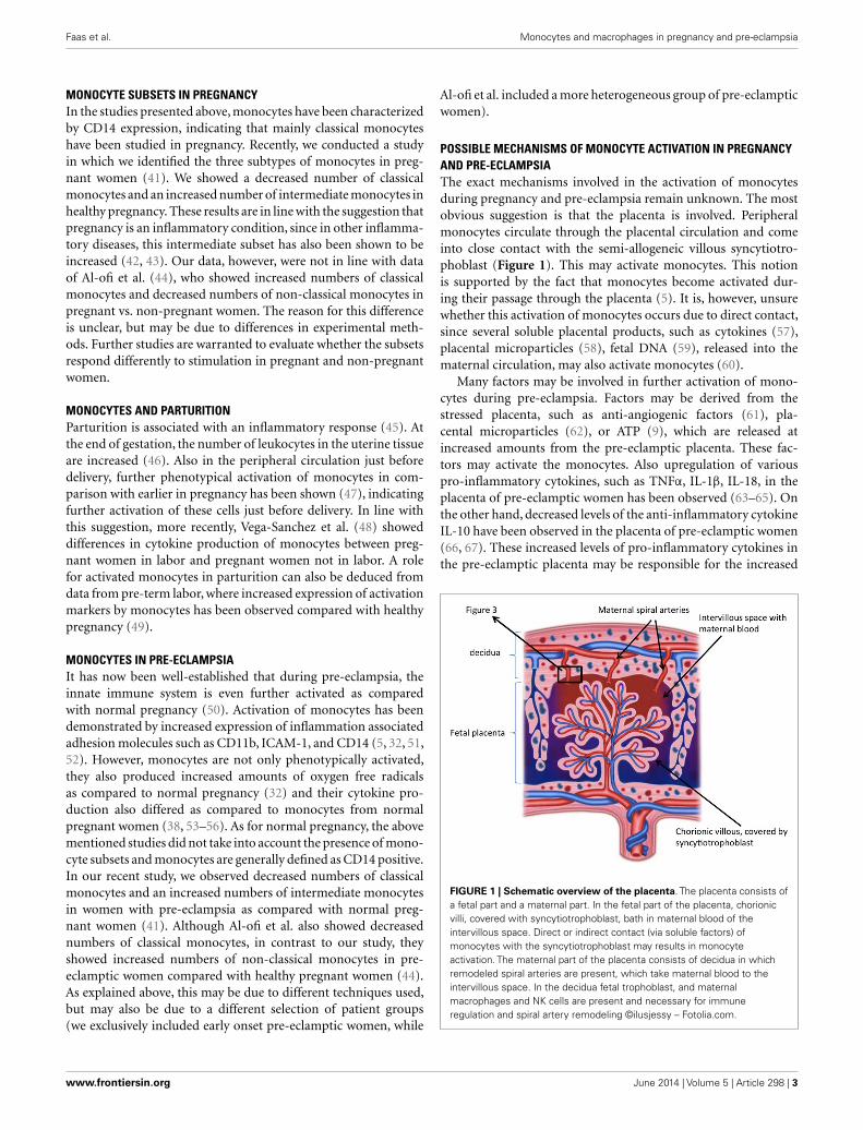
POSSIBLE MECHANISMS OF MONOCYTE ACTIVATION IN PREGNANCYAND PRE-ECLAMPSIAThe exact mechanisms involved in the activation of monocytesduring pregnancy and pre-eclampsia remain unknown. The mostobvious suggestion is that the placenta is involved. Peripheralmonocytes circulate through the placental circulation and comeinto close contact with the semi-allogeneic villous syncytiotro-phoblast (Figure 1). This may activate monocytes. This notionis supported by the fact that monocytes become activated dur-ing their passage through the placenta (5). It is, however, unsurewhether this activation of monocytes occurs due to direct contact,since several soluble placental products, such as cytokines (57),placental microparticles (58), fetal DNA (59), released into thematernal circulation, may also activate monocytes (60).
Many factors may be involved in further activation of mono-cytes during pre-eclampsia. Factors may be derived from thestressed placenta, such as anti-angiogenic factors (61), pla-cental microparticles (62), or ATP (9), which are released atincreased amounts from the pre-eclamptic placenta. These fac-tors may activate the monocytes. Also upregulation of variouspro-inflammatory cytokines, such as TNFα, IL-1β, IL-18, in theplacenta of pre-eclamptic women has been observed (63–65). Onthe other hand, decreased levels of the anti-inflammatory cytokineIL-10 have been observed in the placenta of pre-eclamptic women(66, 67). These increased levels of pro-inflammatory cytokines inthe pre-eclamptic placenta may be responsible for the increased
FIGURE 1 | Schematic overview of the placenta. The placenta consists ofa fetal part and a maternal part. In the fetal part of the placenta, chorionicvilli, covered with syncytiotrophoblast, bath in maternal blood of theintervillous space. Direct or indirect contact (via soluble factors) ofmonocytes with the syncytiotrophoblast may results in monocyteactivation. The maternal part of the placenta consists of decidua in whichremodeled spiral arteries are present, which take maternal blood to theintervillous space. In the decidua fetal trophoblast, and maternalmacrophages and NK cells are present and necessary for immuneregulation and spiral artery remodeling ©ilusjessy – Fotolia.com.
www.frontiersin.org June 2014 | Volume 5 | Article 298 | 3
Faas et al. Monocytes and macrophages in pregnancy and pre-eclampsia
circulating levels of these cytokines in pre-eclamptic patients (68,69). These cytokines may also activate the monocytes. Since mono-cytes themselves are potent producers of cytokines, the activationof monocyte by placental factors and cytokines may in turn resultin a vicious circle of monocytes activation and cytokine pro-duction leading to persistent increased monocyte activation inpre-eclampsia.
It appears to be important for induction of pre-eclamptic signshow monocytes are activated. In pregnant rats, hypertension andproteinuria can only be induced after infusion with E coli LPS(70), not after infusion of LPS from Porphyromonas gingivalis (71),despite the fact that monocytes are activated by this LPS (72).This may explain why certain infections, such as urinary tractinfections or periodontitis, may increase the risk of pre-eclampsia,while other infections, such as CMV or malaria do not increase therisk for pre-eclampsia (73). Apparently, the immune response, andspecifically monocyte activation is different in different infections.Differences may amongst others relate to differences in cytokineproduction between states of monocytes activation, since we havepreviously shown that activation of monocytes with E coli LPS orP. gingivalis LPS resulted in different cytokine production (36).
MONOCYTES DURING PREGNANCY AND EXPERIMENTALPRE-ECLAMPSIA IN ANIMALSAlthough it is now generally accepted that during pregnancymonocytes are activated and that they are even further activatedduring pre-eclampsia, whether this is the cause or consequenceof pre-eclampsia still remains to be shown. It is difficult to studythe role of monocytes in pregnancy and pre-eclampsia in humansubjects. Therefore, animal models are needed. A good animalmodel to study innate immune responses in pregnancy is therat. Although not completely similar, like humans, rats have ahemochorial placenta, showing deep trophoblast invasion into theuterine wall (74) indicating that fetal–maternal interactions may
be similar in rat and human pregnancy. Therefore pregnancy-induced changes in the immune response may also be similar tohuman pregnancy. Indeed, similar phenotypical and functionalactivation of monocytes during the course of pregnancy have beenobserved in rats as compared with humans (75, 76). Moreover, inaccordance with human pregnancy, we found decreased numbersof classical monocytes and increased numbers of non-classicalmonocytes during pregnancy in this species (41).
Various rat models have suggested that activation of mono-cytes, by LPS, ATP, or TNFα during pregnancy, induced pre-eclampsia-like signs (70, 77, 78). Interestingly, such pre-eclampsia-like syndromes were only induced in pregnant rats, not in non-pregnant rats (70, 77). The pathophysiology of the LPS and ATPinduced pre-eclampsia was characterized by a pregnancy-specificinflammatory response, characterized by persistent general (75,76, 79) and glomerular (79, 80) inflammation, in which mono-cytes play a major role. In the ATP model, we have shown that,similar to human pre-eclampsia, non-classical monocytes areincreased and activated by ATP, suggesting an important rolefor this subset in pre-eclampsia. Together, these animal studiessupport the hypothesis that activation of monocytes in preg-nancy may result in pre-eclampsia-like signs, such as hypertensionand proteinuria.
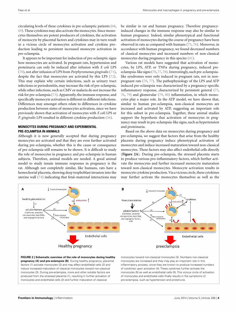
Based on the above data on monocytes during pregnancy andpre-eclampsia, we suggest that factors that arise from the healthyplacenta during pregnancy induce phenotypical activation ofmonocytes and induce increased maturation toward non-classicalmonocytes. These factors may also affect endothelial cells directly(Figure 2A). During pre-eclampsia, the stressed placenta startsto produce various pro-inflammatory factors, which further acti-vate the monocytes and further increased monocyte maturationtoward non-classical monocytes. Monocyte activation results inmonocyte cytokine production.Via a vicious circle, these cytokinesmay further activate the monocytes themselves as well as the
FIGURE 2 | Schematic overview of the role of monocytes during healthypregnancy (A) and pre-eclampsia (B). During healthy pregnancy, placentalfactors (1) activate monocytes (2) and may affect endothelial cells (2) andinduce increased maturation of classical monocytes toward non-classicalmonocytes (3). During pre-eclampsia, more and other soluble factors areproduced from the stressed placenta (1), resulting in further activation ofmonocytes and endothelial cells (2) and further maturation of classical
monocytes toward non-classical monocytes (3). Numbers non-classicalmonocytes are increased and they may play an important role in thisinflammatory process, since they are known to produce increased numbersof cytokines upon activation (4). These cytokines further activate themonocytes (5) as well as endothelial cells (5). This vicious circle of activationof monocytes and endothelial cells finally results in the symptoms ofpre-eclampsia, such as hypertension and proteinuria.
Frontiers in Immunology | Inflammation June 2014 | Volume 5 | Article 298 | 4
Faas et al. Monocytes and macrophages in pregnancy and pre-eclampsia
endothelial cells, finally resulting in the signs of pre-eclampsia,such as proteinuria and hypertension (Figure 2B).
DECIDUAL MACROPHAGESMacrophages are already present in the non-pregnantendometrium, although in low numbers (81). Since their num-bers fluctuate during the menstrual cycle (81, 82), it seems likelythat these are under hormonal control (83). After fertilization, thenumber of uterine macrophages increase, due to expression ofvarious chemokines (84) and during pregnancy macrophages areabundantly present in the decidua at all times of pregnancy (85).During pregnancy, they comprise about 20–30% of all decidualleukocytes (86, 87). The number of decidual macrophages mayvary with gestational age being highest in the first and secondtrimester (88). Macrophages in the decidua are usually associ-ated with spiral arteries and glands as well as with extravilloustrophoblast (86, 89), but are also found in the myometrium(85). When the presence of macrophages in the decidua wasfirst discovered, it was suggested that these cells were recruitedas the result of an immune response to the semi-allogeneic fetus(90). However, it is now generally accepted that macrophages,and other immune cells present in the decidua, are necessaryfor successful implantation (85). Various studies have focusedon specific functions of macrophages in the decidua and ithas been suggested that the decidual macrophages have variousroles during pregnancy, mainly in placentation (91), but theymay also play a role in protecting the fetus against intrauterineinfection (92).
DECIDUAL MACROPHAGES IN EARLY PREGNANCYMost of the studies on macrophages in the decidua have been per-formed in early pregnancy. At this time of pregnancy, macrophagesare located near the spiral arteries during trophoblast invasion andspiral artery remodeling (86, 89). The role of macrophages in spiralartery remodeling was further emphasized by the fact that they arepresent even before the presence of extravillous trophoblast (93).At that time, disruption and disorganization of vascular smoothmuscle cells and endothelial cells was also observed (93). Thissuggests that macrophages may be important in the very earlyphases of spiral artery remodeling, preparing the spiral arteriesfor further remodeling by trophoblast cells (93). Their suggestedrole in vascular remodeling is in accordance with the findingsof production of factors associated with angiogenesis and tissueremodeling by these cells (94, 95). Indeed macrophages, whichwere MMP 9 positive, and which were shown to have phago-cytotic capacities were found to infiltrate spiral arteries duringremodeling (96). Macrophages have also been shown to be impor-tant for clearance of apoptotic cells in the decidua (97). Apoptosisis an important process during spiral artery remodeling and tro-phoblast invasion. During these processes, apoptotic trophoblastcells (98) as well apoptotic cells in the vascular wall that is beingremodeled have been observed (93). By engulfment of the apop-totic cells, macrophages prevent the release of pro-inflammatorysubstances from the apoptotic cells into the decidua [reviewed inRef. (97)].
Decidual macrophages have mainly been classified as M2-like macrophages, i.e., immunomodulatory macrophages (99).
Although they express many markers of M2 macrophages, suchas CD206, CD163, and DC-sign (100–102), they appeared not tobe typical M2 macrophages, since they are not induced by Th2cytokines, such as IL4, but by M-CSF and IL-10 (102). Thesedata are in line with the abundant presence of M-CSF and IL-10 in the decidua (103–105). The M2 phenotype is most likelydue to hypermethylation of genes encoding markers of classicalactivation and hypomethylation of genes encoding markers fornon-classical activation (106). Next to the typical M2 cytokinegene expression, these decidual macrophages also showed geneexpression for inflammatory cytokines such as IL-6 and TNFα
(102, 107). The production of pro-inflammatory cytokines bydecidual macrophages may also be explained by the presence oftwo macrophage subpopulations in the early decidua (107). Oneof these subsets may be a more pro-inflammatory subset, since thissubset expressed genes associated with inflammation. The othersubset, which was higher in number, expressed genes associatedwith extracellular matrix formation, networking, communication,and growth (107).
Apart from the putative role of M-CSF and IL-10 in induc-tion of M2 macrophages in the decidua, other factors have alsobeen suggested to be involved in inducing/maintaining the M2phenotype in decidual macrophages. Decidual macrophages havebeen shown to express inhibitory receptors immunoglobulin liketranscript (ILT)2 and ILT4 (108). These receptors can bind toHLA-G expressed on invading extravillous trophoblast (108),after which a negative signal is delivered to the macrophages,resulting in tolerance to the trophoblast and the induction ofanti-inflammatory cytokines. It has also been suggested that theengulfment of the apoptotic cells induced an immunosuppressiveand anti-inflammatory phenotype of the macrophages (97). Notonly the phagocytosis of apoptotic cells, but also the phagocyto-sis of trophoblast cell debris at the maternal–fetal interface maybe associated with the M2 phenotype of macrophages (109–111).In addition, as it has been suggested that non-classical mono-cytes preferentially differentiate into M2 macrophages (20), itmay be speculated that the increased numbers of non-classicalmonocytes in the circulation during pregnancy (41), results inincreased invasion of these cells into the decidua to become M2macrophages.
DECIDUAL MACROPHAGES IN LATE PREGNANCYMacrophages are present in the decidua throughout pregnancyuntil the end of pregnancy,although their numbers may decrease atthe end of pregnancy (88). The exact role of decidual macrophagesat the end of pregnancy remains to be established, it seems,however, likely that they are still involved in immunoregu-lation and clearance of apoptotic cells. Indeed, many of themacrophages present in the decidua at the end of pregnancy,appeared to be M2 macrophages (112). The potential protectiveeffect of M2 macrophages for the fetus was recently shown byvan Schonkeren et al., who showed the presence of an inflam-matory lesion in placentae from women who underwent eggdonation (113). This lesion consisted of maternal cells, express-ing high levels of CD14 and CD163, suggesting the presenceof M2 macrophages. These lesions appeared to protect againstpre-eclampsia (113).
www.frontiersin.org June 2014 | Volume 5 | Article 298 | 5
Faas et al. Monocytes and macrophages in pregnancy and pre-eclampsia
DECIDUAL MACROPHAGES IN PRE-ECLAMPSIAPreeclampsia is associated with defective trophoblast invasion andspiral artery remodeling: while in healthy pregnancy, spiral arteryremodeling extends into the myometrium, in pre-eclampsia, spiralartery remodeling can only be found in the decidua (3). Unfor-tunately, not very many studies focused on macrophages in thedecidua in pre-eclampsia. Most of the studies in pre-eclampsiawere obviously performed after delivery of the placenta. Someof the studies reported decreased numbers of macrophages inthe decidua of pre-eclamptic patients (114, 115). Most of thestudies, however, found increased numbers of macrophages inpre-eclamptic patients (112, 116–118). These data may not neces-sarily be conflicting, since not only different methods were used(Williams and Burk performed a flow cytometric study, whilethe other studies were immunohistochemical studies), also dif-ferent antibodies were used. Increased numbers of macrophagesin the decidua of pre-eclamptic patients appears to be in line withincreased presence of macrophage chemotactic factors, such asM-CSF, IL-8, and MCP-1 (119–121) in pre-eclamptic patients.Not only numbers of macrophages were found to be different inpre-eclamptic patients, macrophages may also be differently acti-vated in pre-eclampsia (120–122). This may be in line with thepresence of increased pro-inflammatory cytokines (63–65) anddecreased anti-inflammatory cytokines in the placenta of pre-eclamptic women (66, 67). More recently, it has been shown thatin the decidua of pre-eclamptic women decreased numbers of M2macrophages are present (112). Differences in macrophage num-bers may be regional, since increased numbers of macrophageswere found around the spiral arteries of pre-eclamptic patients
(120, 121). The presence of macrophages in the spiral arteriesmay be associated with the development of acute artherosis (120).Acute artherosis is a process mainly seen in poorly remodeled spi-ral arteries at the end of pregnancy, characterized by the presenceof subendothelial CD68 positive foam cells (123). Its presence isassociated with adverse fetal and maternal outcome (124).
The question remains whether the increased presence ofmacrophages in the decidua of pre-eclamptic women is thecause or the result of pre-eclampsia. This question is difficultto answer, due to the difficulties of obtaining material fromearly decidua of women who later in pregnancy developed pre-eclampsia. However, recently we have shown that in early deciduafrom women who later developed pregnancy-induced hyperten-sion (PIH) (including pre-eclampsia) CD68 mRNA expression wasincreased (125), suggesting increased numbers of macrophagesin the early decidua of women who later develop hyperten-sion in pregnancy. Moreover, the CD206/CD68 mRNA ratiowas decreased in PIH women, suggesting that decreased num-bers of M2 macrophages are present in the decidua of womenwho later develop pregnancy-induced hypertension (125). Theincreased numbers of macrophages, with decreased numbers ofM2 macrophages may thus already be present before the onset pre-eclampsia and therefore suggest a role for macrophages in defec-tive trophoblast invasion and spiral artery remodeling. Recentin vitro data showed that macrophages migrate toward invadingtrophoblast (126), while other groups have shown that activatedmacrophages in vitro are able to inhibit trophoblast invasion andspiral artery remodeling (127, 128). In vivo, data have shown thatthere is a reciprocal presence of trophoblast cells and macrophages
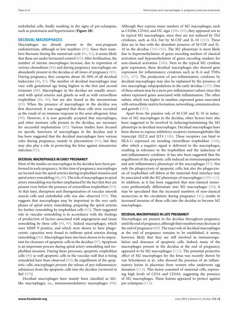
FIGURE 3 | Schematic overview of the role of decidual macrophages inpregnancy (A) and pre-eclampsia (B). During normal pregnancy, M2-likemacrophages are present around spiral arteries and play a role in remodelingof these arteries by producing various factors associated with angiogenesis
and tissue remodeling (such as MMP and VEGF). They also play a role inimmunomodulation, for instance by producing IL-10. During pre-eclampsia,increased numbers of M1-like macrophages are found. They may producepro-inflammatory cytokines, such as TNFα, IL-1β, or IL-18.
Frontiers in Immunology | Inflammation June 2014 | Volume 5 | Article 298 | 6
Faas et al. Monocytes and macrophages in pregnancy and pre-eclampsia
in the spiral arteries of both healthy and pre-eclamptic women(121). Therefore the increased numbers of macrophages in andaround spiral arteries of pre-eclamptic women (121) may inhibitspiral artery remodeling.
Since it is difficult to study the role of macrophages in pre-eclampsia in humans, animal models may help in understandingcritical questions. Studying whether trophoblast invasion andspiral artery remodeling is associated with macrophages in ani-mal models for pre-eclampsia may shed light on the questionwhether increased numbers of macrophages in the decidua arethe cause or the result of pre-eclampsia. In an animal modelfor pre-eclampsia induced by multiple doses of LPS in pregnantrats, decreased trophoblast invasion and spiral artery remodelingafter LPS was associated with increased numbers of macrophages.We studied this subject and showed increased invasion of acti-vated macrophages in the mesometrial triangle (the equivalent ofthe placental bed in humans) before defective trophoblast inva-sion and spiral artery remodeling (129). This appears to be inline with the sparse human data and suggests a role for activatedmacrophages in the pathophysiology of pre-eclampsia.
M2-like macrophages are thus abundantly present in thedecidua of healthy pregnant women. They are observed in thepresence of spiral arteries and extravillous trophoblast cells andmay play a role in spiral artery remodeling by producing factorsassociated with angiogenesis and tissue remodeling, such as MMPsand VEGF (Figure 3A). During pre-eclampsia, increased num-bers of decidual macrophages are observed, which may be of theM1 phenotype and therefore produce pro-inflammatory cytokines(Figure 3B). These activated macrophages may affect spiral arter-ies and may induce acute artherosis, affecting the placental bloodcirculation.
SUMMARYMonocytes and macrophages play important roles in pregnancyand pre-eclampsia. Monocyte activation and increased numbersof non-classical monocytes, is important for normal pregnancy.Monocyte derived macrophages, especially M2-like macrophages(which may be derived from non-classical monocytes) in thedecidua in healthy pregnancy play an important role in blastocystimplantation, trophoblast invasion, and spiral artery remodelingas well as in defense against infection and in immunomodula-tion (Figure 4). During pre-eclampsia, decreased spiral arteryremodeling results in increased production of soluble factors(or different factors), inducing further activation of both classi-cal and non-classical monocytes and further maturation towardnon-classical monocytes. These placental factors as well as theactivated monocytes also induce activation of endothelial cells.Activated monocytes (both classical and non-classical monocytes)may invade into the decidua, resulting in increased numbers ofM1-like macrophages in the decidua of pre-eclamptic women(Figure 4). The M1-like macrophages may affect the spiral arteries,by for instance inducing acute artherosis. This may further affectthe placental blood circulation and stress the placenta.
Unfortunately, most studies on monocytes and macrophagesin pre-eclampsia have been performed during pre-eclampsia.Although we do believe that monocytes and decidual macrophagesdo play a role in inducing the maternal symptoms of pre-eclampsia, it is relatively unknown whether monocytes and
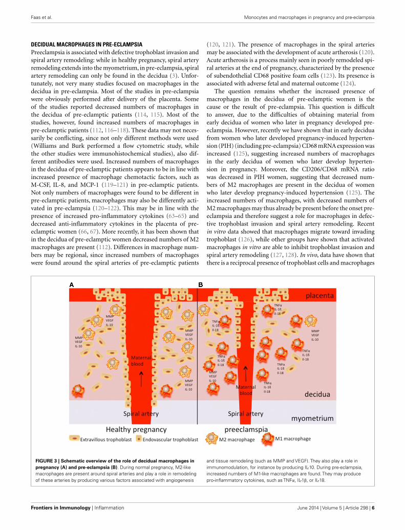
FIGURE 4 | Summary of monocytes and macrophages inpregnancy and pre-eclampsia. In healthy pregnancy, soluble factorsfrom the villous trophoblast activate circulating monocytes, inducematuration of classical monocytes toward non-classical monocytes andaffect endothelial cells. Non-classical monocytes will invade into thedecidua to become M2-like macrophages to support healthyplacentation and immunomodulation. During pre-eclampsia, decreasedremodeling of the spiral arteries will results in a stressed placenta,which produces increased amounts or different soluble factors ascompared with healthy pregnancy. The soluble factors will furtheractivate the monocytes, induce further maturation of classicalmonocytes toward non-classical monocytes and activate endothelialcells. Activated monocytes, by f.i. producing cytokines, further affectmonocytes and endothelial cells. This vicious circle of monocyte andendothelial cell activation results in the maternal symptoms ofpre-eclampsia, i.e., hypertension and proteinuria. Moreover, activatedclassical and non-classical monocytes may invade into the decidua todevelop into M1-like and M2-like macrophages, resulting in increasednumbers of M1-like macrophages in the pre-eclamptic decidua. TheM1-like macrophages may affect the spiral arteries resulting in f.i. acuteatherosis, thereby further affecting the placental blood circulation.
decidual macrophages do also play a role in the aberrant spiralartery remodeling early in pregnancy. The question thus remainsas to what induces the aberrant spiral artery remodeling? Futurestudies should therefore not only focus on the three monocyte sub-sets in pregnancy and pre-eclampsia, but also on the relationshipbetween the circulating monocyte subsets and macrophages in thedecidua. Moreover, since data on macrophages in the decidua inand before pre-eclampsia are relatively scarce future studies shouldtherefore also focus on macrophage function and phenotype inand before pre-eclampsia.
www.frontiersin.org June 2014 | Volume 5 | Article 298 | 7
Faas et al. Monocytes and macrophages in pregnancy and pre-eclampsia
REFERENCES1. Steegers EA, von Dadelszen P, Duvekot JJ, Pijnenborg R. Pre-eclampsia. Lancet
(2010) 376(9741):631–44. doi:10.1016/S0140-6736(10)60279-62. Duley L. The global impact of pre-eclampsia and eclampsia. Semin Perinatol
(2009) 33(3):130–7. doi:10.1053/j.semperi.2009.02.0103. Redman CW, Sargent IL. Placental stress and pre-eclampsia: a revised view.
Placenta (2009) 30(Suppl A):S38–42. doi:10.1016/j.placenta.2008.11.0214. Sacks GP, Sargent IL, Redman CWG. An innate view of human pregnancy.
Immunol Today (1999) 20(3):114–8. doi:10.1016/S0167-5699(98)01393-05. Mellembakken JR, Aukrust P, Olafsen MK, Ueland T, Hestdal K, Videm V.
Activation of leukocytes during the uteroplacental passage in preeclampsia.Hypertension (2002) 39(1):155–60. doi:10.1161/hy0102.100778
6. Hung TH, Charnock-Jones DS, Skepper JN, Burton GJ. Secretion of tumornecrosis factor-alpha from human placental tissues induced by hypoxia-reoxygenation causes endothelial cell activation in vitro: a potential medi-ator of the inflammatory response in preeclampsia. Am J Pathol (2004)164(3):1049–61. doi:10.1016/S0002-9440(10)63192-6
7. Levine RJ, Maynard SE, Qian C, Lim KH, England LJ, Yu KF, et al. Circu-lating angiogenic factors and the risk of preeclampsia. N Engl J Med (2004)350(7):672–83. doi:10.1056/NEJMoa031884
8. Germain SJ, Sacks GP, Soorana SR, Sargent IL, Redman CW. Systemic inflam-matory priming in normal pregnancy and preeclampsia: the role of circu-lating syncytiotrophoblast microparticles. J Immunol (2007) 178(9):5949–56.doi:10.4049/jimmunol.178.9.5949
9. Spaans F, Vos PD, Bakker WW, van Goor H, Faas MM. Danger signals fromATP and adenosine in pregnancy and preeclampsia. Hypertension (2014)63(6):1154–60. doi:10.1161/HYPERTENSIONAHA.114.03240
10. Wallace AE, Fraser R, Cartwright JE. Extravillous trophoblast and decidualnatural killer cells: a remodelling partnership. Hum Reprod Update (2012)18(4):458–71. doi:10.1093/humupd/dms015
11. Svensson-Arvelund J, Ernerudh J, Buse E, Cline JM, Haeger JD, DixonD, et al. The placenta in toxicology. Part II: systemic and local immuneadaptations in pregnancy. Toxicol Pathol (2014) 42(2):327–38. doi:10.1177/0192623313482205
12. Gordon S, Taylor PR. Monocyte and macrophage heterogeneity. Nat RevImmunol (2005) 5(12):953–64. doi:10.1038/nri1733
13. Ziegler-Heitbrock L, Ancuta P, Crowe S, Dalod M, Grau V, Hart DN, et al.Nomenclature of monocytes and dendritic cells in blood. Blood (2010)116(16):e74–80. doi:10.1182/blood-2010-02-258558
14. Sunderkötter C, Nikolic T, Dillon MJ, Van Rooijen N, Stehling M, DrevetsDA, et al. Subpopulations of mouse blood monocytes differ in matura-tion stage and inflammatory response. J Immunol (2004) 172(7):4410–7.doi:10.4049/jimmunol.172.7.4410
15. Auffray C, Fogg D, Garfa M, Elain G, Join-Lambert O, Kayal S, et al. Monitor-ing of blood vessels and tissues by a population of monocytes with patrollingbehavior. Science (2007) 317(5838):666–70. doi:10.1126/science.1142883
16. Fingerle G, Pforte A, Passlick B, Blumenstein M, Strobel M, Ziegler-HeitbrockHW. The novel subset of CD14+/CD16+ blood monocytes is expanded insepsis patients. Blood (1993) 82(10):3170–6.
17. Zimmermann HW, Seidler S, Nattermann J, Gassler N, Hellerbrand C, Zer-necke A, et al. Functional contribution of elevated circulating and hepatic non-classical CD14CD16 monocytes to inflammation and human liver fibrosis.PLoS One (2010) 5(6):e11049. doi:10.1371/journal.pone.0011049
18. Mantovani A, Biswas SK, Galdiero MR, Sica A, Locati M. Macrophage plas-ticity and polarization in tissue repair and remodelling. J Pathol (2013)229(2):176–85. doi:10.1002/path.4133
19. Porcheray F, Viaud S, Rimaniol AC, Léone C, Samah B, Dereuddre-Bosquet N,et al. Macrophage activation switching: an asset for the resolution of inflam-mation. Clin Exp Immunol (2005) 142(3):481–9. doi:10.1111/j.365-2249.2005.02934.x
20. Yang J, Zhang L, Yu C, Yang XF, Wang H. Monocyte and macrophage differ-entiation: circulation inflammatory monocyte as biomarker for inflammatorydiseases. Biomark Res (2014) 2(1):1. doi:10.1186/2050-7771-2-1
21. Spahn JH, Kreisel D. Monocytes in sterile inflammation: recruitment and func-tional consequences. Arch Immunol Ther Exp (Warsz) (2013) 62(3):187–94.doi:10.1007/s00005-013-0267-5
22. Wegmann TG, Lin H, Guilbert L, Mosmann TR. Bidirectional cytokineinteractions in the maternal-fetal relationship: is successful pregnancy a Th2phenomenon? Immunol Today (1993) 14:353–6. doi:10.1016/0167-5699(93)90235-D
23. Veenstra van Nieuwenhoven AL, Bouman A, Moes H, Heineman MJ, de Leij LF,Santema J, et al. Cytokine production in natural killer cells and lymphocytes inpregnant women compared with women in the follicular phase of the ovariancycle. Fertil Steril (2002) 77(5):1032–7. doi:10.1016/S0015-0282(02)02976-X
24. Saito S, Sakai M, Sasaki Y, Tanebe K, Tsuda H, Michimata T. Quantitativeanalysis of peripheral blood Th0, Th1, Th2 and the Th1:Th2 cell ratio dur-ing normal pregnancy and preeclampsia. Clin Exp Immunol (1999) 117:550–5.doi:10.1046/j.1365-2249.1999.00997.x
25. Borzychowski AM, Croy BA, Chan WL, Redman CW, Sargent IL. Changes insystemic type 1 and type 2 immunity in normal pregnancy and pre-eclampsiamay be mediated by natural killer cells. Eur J Immunol (2005) 35(10):3054–63.doi:10.1002/eji.200425929
26. Ernerudh J, Berg G, Mjösberg J. Regulatory T helper cells in pregnancy andtheir roles in systemic versus local immune tolerance. Am J Reprod Immunol(2011) 66(Suppl 1):31–43. doi:10.1111/j.1600-0897.2011.01049.x
27. Saito S, Nakashima A, Shima T, Ito M. Th1/Th2/Th17 and regulatory T-cell paradigm in pregnancy. Am J Reprod Immunol (2010) 63(6):601–10.doi:10.1111/j.1600-0897.2010.00852.x
28. Siegel I, Gleicher N. Changes in peripheral mononuclear cells in pregnancy.Am J Reprod Immunol (1981) 1(3):154–5.
29. Kuhnert M, Strohmeier R, Stegmuller M, Halberstadt E. Changes in lympho-cyte subsets during normal pregnancy. Obstet Gynecol (1998) 76:147–51.
30. Veenstra van Nieuwenhoven AL, Bouman A, Moes H, Heineman MJ, de LeijFMLH, Santema J, et al. Endotoxin-induced cytokine production of mono-cytes of third trimester pregnant women compared to women in the follic-ular phase of the menstrual cycle. Am J Obstet Gynecol (2003) 188:1073–7.doi:10.1067/mob.2003.263
31. Macey MG, McCarthy DA, Vordermeier S, Newland AC, Brown KA. Effects ofcell purification methods on CD11b and L-selectin expression as well as adher-ence and activation of leukocytes. J Immunol Methods (1995) 181(2):211–9.doi:10.1016/0022-1759(95)00003-S
32. Sacks GP, Studena K, Sargent IL, Redman CWG. Normal pregnancy andpreeclampsia both produce inflammatory changes in peripheral blood leuko-cytes akin to those of sepsis. Am J Obstet Gynecol (1998) 179:80–6. doi:10.1016/S0002-9378(98)70254-6
33. Naccasha N, Gervasi MT, Chaiworapongsa T, Berman S, Yoon BH, MaymonE, et al. Phenotypic and metabolic characteristics of monocytes and granulo-cytes in normal pregnancy and maternal infection. Am J Obstet Gynecol (2001)185(5):1118–23. doi:10.1067/mob.2001.117682
34. Luppi P, Haluszczak C, Betters D, Richard CAH, Trucco M, DeLoia JA. Mono-cytes are progressively activated in the circulation of pregnant women. J LeukocBiol (2002) 72(5):874–84.
35. Luppi P, Haluszczak C, Trucco M, DeLoia JA. Normal pregnancy is asso-ciated with peripheral leukocyte activation. Am J Reprod Immunol (2002)47(2):72–81. doi:10.1034/j.1600-0897.2002.1o041.x
36. Faas MM, Kunnen A, Dekker DC, Harmsen HJ, Aarnoudse JG, Abbas F, et al.Porphyromonas gingivalis and E-coli induce different cytokine production pat-terns in pregnant women. PLoS One (2014) 9(1):e86355. doi:10.1371/journal.pone.0086355
37. Beckmann I, Efraim SB, Vervoort M, Visser W, Wallenburg HC. Tumor necro-sis factor-alpha in whole blood cultures of preeclamptic patients and healthypregnant and nonpregnant women. Hypertens Pregnancy (2004) 23(3):319–29.doi:10.1081/PRG-200030334
38. Sacks GP, Redman CWG, Sargent IL. Monocytes are primed to produce theTh1 type cytokine IL-12 in normal human pregnancy: an intracellular flowcytometric analysis of peripheral blood mononuclear cells. Clin Exp Immunol(2003) 131(3):490–7. doi:10.1046/j.1365-2249.2003.02082.x
39. Faas MM, Moes H, Fijen JW, Muller Kobold AC, Tulleken JE, Zijlstra JG.Monocyte intracellular cytokine production during human endotoxemia withor without a second in vitro LPS challenge: effect of RWJ067657, a p36MAP-kinase inhibitor, on LPS-hyporesponsiveness. Clin Exp Immunol (2002)127:337–43. doi:10.1046/j.1365-2249.2002.01765.x
40. Chen J, Ivashkiv LB. IFN-γ abrogates endotoxin tolerance by facilitating toll-like receptor-induced chromatin remodeling. Proc Natl Acad Sci U S A (2010)107(45):19438–43. doi:10.1073/pnas.1007816107
41. Melgert BN, Spaans F, Borghuis T, Klok PA, Groen B, Bolt A, et al. Pregnancyand preeclampsia affect monocyte subsets in humans and rats. PLoS One (2012)7(9):e45229. doi:10.1371/journal.pone.0045229
42. Rossol M, Kraus S, Pierer M, Baerwald C, Wagner U. The CD14(bright)CD16+ monocyte subset is expanded in rheumatoid arthritis and promotes
Frontiers in Immunology | Inflammation June 2014 | Volume 5 | Article 298 | 8
Faas et al. Monocytes and macrophages in pregnancy and pre-eclampsia
expansion of the Th17 cell population. Arthritis Rheum (2012) 64(3):671–7.doi:10.1002/art.33418
43. Moniuszko M, Bodzenta-Lukaszyk A, Kowal K, Lenczewska D, Dabrowska M.Enhanced frequencies of CD14++CD16+, but not CD14+CD16+, periph-eral blood monocytes in severe asthmatic patients. Clin Immunol (2009)130(3):338–46. doi:10.1016/j.clim.2008.09.011
44. Al-ofi E, Coffelt SB, Anumba DO. Monocyte subpopulations from pre-eclamptic patients are abnormally skewed and exhibit exaggerated responses totoll-like receptor ligands. PLoS One (2012) 7(7):e42217. doi:10.1371/journal.pone.0042217
45. Kelly RW. Inflammatory mediators and parturition. Rev Reprod (1996)1(2):89–96.
46. Bokström H, Brännström M, Alexandersson M, Norström A. Leukocyte sub-populations in the human uterine cervical stroma at early and term pregnancy.Hum Reprod (1997) 12(3):586–90. doi:10.1093/humrep/12.3.586
47. Luppi P, Irwin TE, Simhan H, Deloia JA. CD11b Expression on circulatingleukocytes increases in preparation for parturition. Am J Reprod Immunol(2004) 52(5):323–9. doi:10.1111/j.1600-0897.2004.00229.x
48. Vega-Sanchez R, Gomez-Lopez N, Flores-Pliego A, Clemente-Galvan S,Estrada-Gutierrez G, Zentella-Dehesa A, et al. Placental blood leukocytes arefunctional and phenotypically different than peripheral leukocytes duringhuman labor. J Reprod Immunol (2010) 84(1):100–10. doi:10.1016/j.jri.2009.08.002
49. Gervasi MT, Chaiworapongsa T, Naccasha N, Blackwell S, Yoon BH, MaymonE, et al. Phenotypic and metabolic characteristics of maternal monocytes andgranulocytes in preterm labor with intact membranes. Am J Obstet Gynecol(2001) 185(5):1124–9. doi:10.1067/mob.2001.117311
50. Borzychowski AM, Sargent IL, Redman CW. Inflammation and pre-eclampsia.Semin Fetal Neonatal Med (2006) 11(5):309–16. doi:10.1016/j.siny.2006.04.001
51. Gervasi MT, Chaiworapongsa T, Pacora P, Naccasha N, Yoon BH, May-mon E, et al. Phenotypic and metabolic characteristics of monocytes andgranulocytes in preeclampsia. Am J Obstet Gynecol (2001) 185(4):792–7.doi:10.1067/mob.2001.117311
52. Luppi P, Tse H, Lain KY, Markovic N, Piganelli JD, DeLoia JA. Preeclampsia acti-vates circulating immune cells with engagement of the NF-kappaB pathway. AmJ Reprod Immunol (2006) 56(2):135–44. doi:10.1111/j.1600-0897.2006.00386.x
53. Sakai M, Tsuda H, Tanebe K, Sasaki Y, Saito S. Interleukin-12 secretion byperipheral blood mononuclear cells is decreased in normal pregnant sub-jects and increased in preeclamptic patients. Am J Reprod Immunol (2002)47(2):91–7. doi:10.1034/j.1600-0897.2002.1o020.x
54. Peraçoli JC, Rudge MV, Peraçoli MT. Tumor necrosis factor-alpha in gestationand puerperium of women with gestational hypertension and pre-eclampsia.Am J Reprod Immunol (2007) 57(3):177–85. doi:10.1111/j.1600-0897.2006.00455.x
55. Veenstra van Nieuwenhoven AL, Moes H, Heineman MJ, Santema J, Faas MM.Cytokine production by monocytes, NK cells and lymphocytes is different inpreeclamptic patients as compared with normal pregnant women. HypertensPregnancy (2008) 27(3):207–24. doi:10.1080/10641950701885006
56. Brewster JA, Orsi NM, Gopichandran N, Ekbote UV, Cadogan E, WalkerJJ. Host inflammatory response profiling in preeclampsia using an in vitrowhole blood stimulation model. Hypertens Pregnancy (2008) 27(1):1–16.doi:10.1080/10641950701826067
57. Sacks GP, Clover LM, Bainbridge DR, Redman CW, Sargent IL. Flow cyto-metric measurement of intracellular Th1 and Th2 cytokine production byhuman villous and extravillous cytotrophoblast. Placenta (2001) 22(6):550–9.doi:10.1053/plac.2001.0686
58. Redman CW, Tannetta DS, Dragovic RA, Gardiner C, Southcombe JH, CollettGP, et al. Review: does size matter? Placental debris and the pathophysiologyof pre-eclampsia. Placenta (2012) 33(Suppl):S48–54. doi:10.1016/j.placenta.2011.12.006
59. Bianchi DW, Zickwolf GK, Weil GJ, Sylvester S, DeMaria MA. Male fetal prog-enitor cells persist in maternal blood for as long as 27 years postpartum. ProcNatl Acad Sci U S A (1996) 93(2):705–8. doi:10.1073/pnas.93.2.705
60. Faas MM, van Pampus MG, Anninga ZA, Salomons J, Westra IM, Donker RB,et al. Plasma from preeclamptic women activates endothelial cells via mono-cyte activation in vitro. J Reprod Immunol (2010) 87(1–2):28–38. doi:10.1016/j.jri.2010.07.005
61. Steinberg G, Khankin EV, Karumanchi SA. Angiogenic factors and preeclamp-sia. Thromb Res (2009) 123(Suppl 2):S93–9. doi:10.1016/S0049-3848(09)70020-9
62. Redman CW, Sargent IL. Placental debris, oxidative stress and pre-eclampsia.Placenta (2000) 21(7):597–602. doi:10.1053/plac.2000.0560
63. Pang ZJ, Xing FQ. Comparative study on the expression of cytokine – receptorgenes in normal and preeclamptic human placentas using DNA microarrays.J Perinat Med (2003) 31(2):153–62. doi:10.1515/JPM.2003.021
64. Benyo DF, Smarason A, Redman CW, Sims C, Conrad KP. Expression ofinflammatory cytokines in placentas from women with preeclampsia. J ClinEndocrinol Metab (2001) 86(6):2505–12. doi:10.1210/jc.86.6.2505
65. Wang Y, Walsh SW. TNF alpha concentrations and mRNA expression areincreased in preeclamptic placentas. J Reprod Immunol (1996) 32(2):157–69.doi:10.1016/S0165-0378(96)00998-9
66. Hennessy A, Pilmore HL, Simmons LA, Painter DM. A deficiency of placentalIL-10 in preeclampsia. J Immunol (1999) 163(6):3491–5.
67. Rein DT, Breidenbach M, Hönscheid B, Friebe-Hoffmann U, Engel H, GöhringUJ, et al. Preeclamptic women are deficient of interleukin-10 as assessed bycytokine release of trophoblast cells in vitro. Cytokine (2003) 23(4–5):119–25.doi:10.1016/S1043-4666(03)00220-5
68. Vince GS, Starkey PM, Austgulen R, Kwaitkowski D, Redman CWG.Interleukin-6, tumour necrosis factor and soluble tumour necrosis factor recep-tors in women with pre-eclampsia. Br J Obstet Gynaecol (1995) 102:20–5.doi:10.1111/j.1471-0528.1995.tb09020.x
69. Conrad KP, Miles TM, Benyo DF. Circulating levels of immunoreactivecytokines in women with preeclampsia. Am J Reprod Immunol (1998)40(2):102–11. doi:10.1111/j.1600-0897.1998.tb00398.x
70. Faas MM, Schuiling GA, Baller JFW, Visscher CA, Bakker WW. A new animalmodel for human pre-eclampsia: ultralow dose endotoxin infusion in preg-nant rats. Am J Obstet Gynecol (1994) 171:158–64. doi:10.1016/0002-9378(94)90463-4
71. Kunnen A, Van Pampus MG, Aarnoudse JG, van der Schans CP, Abbas F, FaasMM. The effect of Porphyromonas gingivalis lipopolysaccharide on pregnancyin the rat. Oral Dis (2013). doi:10.1111/odi.12177
72. Kunnen A, Dekker DC, van Pampus MG, Harmsen HJ, Aarnoudse JG, AbbasF, et al. Cytokine production induced by non-encapsulated and encapsu-lated Porphyromonas gingivalis strains. Arch Oral Biol (2012) 57(11):1558–66.doi:10.1016/j.archoralbio.2012.07.013
73. Conde-Agudelo A, Villar J, Lindheimer M. Maternal infection and risk ofpreeclampsia: systematic review and metaanalysis. Am J Obstet Gynecol (2008)198(1):7–22. doi:10.1016/j.ajog.2007.07.040
74. Soares MJ, Chakraborty D, Karim Rumi MA, Konno T, Renaud SJ. Rat placen-tation: an experimental model for investigating the hemochorial maternal-fetalinterface. Placenta (2012) 33(4):233–43. doi:10.1016/j.placenta.2011.11.026
75. Faas MM, Schuiling GA, Linton EA, Sargent IL, Redman CW. Activation ofperipheral leukocytes in rat pregnancy and experimental preeclampsia. AmJ Obstet Gynecol (2000) 182(2):351–7. doi:10.1016/S0002-9378(00)70223-7
76. Faas MM, Broekema M, Moes H, van der Schaaf G, Heineman MJ, de Vos P.Altered monocyte function in experimental preeclampsia in the rat. Am J ObstetGynecol (2004) 191(4):1192–8. doi:10.1016/j.ajog.2004.03.041
77. Faas MM, van der Schaaf G, Borghuis T, Jongman RM, van Pampus MG, deVos P, et al. Extracellular ATP induces albuminuria in pregnant rats. NephrolDial Transplant (2010) 25(8):2468–78. doi:10.1093/ndt/gfq095
78. LaMarca B, Speed J, Fournier L, Babcock SA, Berry H, Cockrell K, et al.Hypertension in response to chronic reductions in uterine perfusion in preg-nant rats: effect of tumor necrosis factor-alpha blockade. Hypertension (2008)52(6):1161–7. doi:10.1161/HYPERTENSIONAHA.108.120881
79. Spaans F, Melgert BN, Borghuis T, Klok PA, de Vos P, Bakker WW, et al. Extra-cellular adenosine triphosphate affects systemic and kidney immune cell pop-ulations in pregnant rats. Am J Reprod Immunol (2014). doi:10.1111/aji.12267
80. Faas MM, Schuiling GA, Baller JFW, Bakker WW. Glomerular inflammationin pregnant rats after infusion of low dose endotoxin: an immunohistologicalstudy in experimental pre-eclampsia. Am J Pathol (1995) 147:1510–8.
81. Bulmer JN, Morrison L, Longfellow M, Ritson A, Pace D. Granulated lym-phocytes in human endometrium: histochemical and immunohistochemicalstudies. Hum Reprod (1991) 6(6):791–8.
82. Klentzeris LD, Bulmer JN, Warren A, Morrison L, Li TC, Cooke ID. Endome-trial lymphoid tissue in the timed endometrial biopsy: morphometric and
www.frontiersin.org June 2014 | Volume 5 | Article 298 | 9
Faas et al. Monocytes and macrophages in pregnancy and pre-eclampsia
immunohistochemical aspects. Am J Obstet Gynecol (1992) 167(3):667–74.doi:10.1016/S0002-9378(11)91568-3
83. Hunt JS, Miller L, Platt JS. Hormonal regulation of uterine macrophages. DevImmunol (1998) 6(1–2):105–10.
84. Jones RL, Hannan NJ, Kaitu’u TJ, Zhang J, Salamonsen LA. Identification ofchemokines important for leukocyte recruitment to the human endometriumat the times of embryo implantation and menstruation. J Clin Endocrinol Metab(2004) 89(12):6155–67. doi:10.1210/jc.2004-0507
85. Bulmer JN, Williams PJ, Lash GE. Immune cells in the placental bed. Int J DevBiol (2010) 54(2–3):281–94. doi:10.1387/ijdb.082763jb
86. Bulmer JN, Morrison L, Smith JC. Expression of class II MHC gene prod-ucts by macrophages in human uteroplacental tissue. Immunology (1988)63(4):707–14.
87. Lessin DL, Hunt JS, King CR, Wood GW. Antigen expression by cells near thematernal-fetal interface. Am J Reprod Immunol Microbiol (1988) 16(1):1–7.
88. Williams PJ, Searle RF, Robson SC, Innes BA, Bulmer JN. Decidual leuko-cyte populations in early to late gestation normal human pregnancy. J ReprodImmunol (2009) 82(1):24–31. doi:10.1016/j.jri.2009.08.001
89. Bulmer JN, Johnson PM. Macrophage populations in the human placenta andamniochorion. Clin Exp Immunol (1984) 57(2):393–403.
90. Tafuri A, Alferink J, Moller P, Hammerling GJ, Arnold B. T cell awarenessof paternal alloantigens during pregnancy. Science (1995) 270(5236):630–3.doi:10.1126/science.270.5236.630
91. Renaud SJ, Graham CH. The role of macrophages in utero-placental inter-actions during normal and pathological pregnancy. Immunol Invest (2008)37(5):535–64. doi:10.1080/08820130802191375
92. Singh U, Nicholson G, Urban BC, Sargent IL, Kishore U, Bernal AL. Immuno-logical properties of human decidual macrophages – a possible role inintrauterine immunity. Reproduction (2005) 129(5):631–7. doi:10.1530/rep.1.00331
93. Smith SD, Dunk CE, Aplin JD, Harris LK, Jones RL. Evidence for immune cellinvolvement in decidual spiral arteriole remodeling in early human pregnancy.Am J Pathol (2009) 174(5):1959–71. doi:10.2353/ajpath.2009.080995
94. Engert S, Rieger L, Kapp M, Becker JC, Dietl J, Kämmerer U. Profilingchemokines, cytokines and growth factors in human early pregnancy deciduaby protein array. Am J Reprod Immunol (2007) 58(2):129–37. doi:10.1111/j.1600-0897.2007.00498.x
95. Gustafsson C, Mjösberg J, Matussek A, Geffers R, Matthiesen L, Berg G,et al. Gene expression profiling of human decidual macrophages: evidencefor immunosuppressive phenotype. PLoS One (2008) 3(4):e2078. doi:10.1371/journal.pone.0002078
96. Hazan AD, Smith SD, Jones RL, Whittle W, Lye SJ, Dunk CE. Vascular-leukocyteinteractions: mechanisms of human decidual spiral artery remodeling in vitro.Am J Pathol (2010) 177(2):1017–30. doi:10.2353/ajpath.2010.091105
97. Abrahams VM, Kim YM, Straszewski SL, Romero R, Mor G. Macrophagesand apoptotic cell clearance during pregnancy. Am J Reprod Immunol (2004)51(4):275–82. doi:10.1111/j.1600-0897.2004.00156.x
98. Piacentini M,Autuori F. Immunohistochemical localization of tissue transglut-aminase and Bcl-2 in rat uterine tissues during embryo implantation and post-partum involution. Differentiation (1994) 57(1):51–61. doi:10.1046/j.1432-0436.1994.5710051.x
99. Cupurdija K, Azzola D, Hainz U, Gratchev A, Heitger A, Takikawa O,et al. Macrophages of human first trimester decidua express markers asso-ciated to alternative activation. Am J Reprod Immunol (2004) 51(2):117–22.doi:10.1046/j.8755-8920.2003.00128.x
100. Kämmerer U, Eggert AO, Kapp M, McLellan AD, Geijtenbeek TB, Dietl J, et al.Unique appearance of proliferating antigen-presenting cells expressing DC-SIGN (CD209) in the decidua of early human pregnancy. Am J Pathol (2003)162(3):887–96. doi:10.1016/S00002-9440(10)63884-9
101. Laskarin G, Cupurdija K, Tokmadzic VS, Dorcic D, Dupor J, Juretic K, et al. Thepresence of functional mannose receptor on macrophages at the maternal-fetalinterface. Hum Reprod (2005) 20(4):1057–66. doi:10.1093/humrep/deh740
102. Svensson J, Jenmalm MC, Matussek A, Geffers R, Berg G, Ernerudh J.Macrophages at the fetal-maternal interface express markers of alterna-tive activation and are induced by M-CSF and IL-10. J Immunol (2011)187(7):3671–82. doi:10.4049/jimmunol.1100130
103. Daiter E, Pampfer S, Yeung YG, Barad D, Stanley ER, Pollard JW. Expression ofcolony-stimulating factor-1 in the human uterus and placenta. J Clin EndocrinolMetab (1992) 74(4):850–8. doi:10.1210/jc.74.4.850
104. Thaxton JE, Sharma S. Interleukin-10: a multi-faceted agent of pregnancy.Am J Reprod Immunol (2010) 63(6):482–91. doi:10.1111/j.1600-0897.2010.00810.x
105. Roth I, Corry DB, Locksley RM, Abrams JS, Litton MJ, Fisher SJ. Human pla-cental cytotrophoblasts produce the immunosuppressive cytokine interleukin10. J Exp Med (1996) 184(2):539–48. doi:10.1084/jem.184.2.539
106. Kim SY, Romero R, Tarca AL, Bhatti G, Kim CJ, Lee J, et al. Methylomeof fetal and maternal monocytes and macrophages at the feto-maternal inter-face. Am J Reprod Immunol (2012) 68(1):8–27. doi:10.1111/j.1600-0897.2012.01108.x
107. Houser BL, Tilburgs T, Hill J, Nicotra ML, Strominger JL. Two uniquehuman decidual macrophage populations. J Immunol (2011) 186(4):2633–42.doi:10.4049/jimmunol.1003153
108. Petroff MG, Sedlmayr P, Azzola D, Hunt JS. Decidual macrophages are poten-tially susceptible to inhibition by class Ia and class Ib HLA molecules. J ReprodImmunol (2002) 56(1–2):3–17. doi:10.1016/S0165-0378(02)00024-4
109. Abumaree MH, Chamley LW, Badri M, El-Muzaini MF. Trophoblast debrismodulates the expression of immune proteins in macrophages: a key to mater-nal tolerance of the fetal allograft? J Reprod Immunol (2012) 94(2):131–41.doi:10.1016/j.jri.2012.03.488
110. Fadok VA, Chimini G. The phagocytosis of apoptotic cells. Semin Immunol(2001) 13(6):365–72. doi:10.1006/smim.2001.0333
111. Van Ginderachter JA, Movahedi K, Hassanzadeh Ghassabeh G, Meerschaut S,Beschin A, Raes G, et al. Classical and alternative activation of mononuclearphagocytes: picking the best of both worlds for tumor promotion. Immunobi-ology (2006) 211(6–8):487–501. doi:10.1016/j.imbio.2006.06.002
112. Schonkeren D, van der Hoorn ML, Khedoe P, Swings G, van Beelen E, ClaasF, et al. Differential distribution and phenotype of decidual macrophages inpreeclamptic versus control pregnancies. Am J Pathol (2011) 178(2):709–17.doi:10.1016/j.ajpath.2010.10.011
113. Schonkeren D, Swings G, Roberts D, Claas F, de Heer E, Scherjon S. Pregnancyclose to the edge: an immunosuppressive infiltrate in the chorionic plate of pla-centas from uncomplicated egg cell donation. PLoS One (2012) 7(3):e32347.doi:10.1371/journal.pone.0032347
114. Williams PJ, Bulmer JN, Searle RF, Innes BA, Robson SC. Altered decidualleukocyte populations in the placental bed in pre-eclampsia and foetal growthrestriction: a comparison with late normal pregnancy. Reproduction (2009)138(1):177–84. doi:10.1530/REP-09-0007
115. Bürk MR, Troeger C, Brinkhaus R, Holzgreve W, Hahn S. Severely reducedpresence of tissue macrophages in the basal plate of pre-eclamptic placentae.Placenta (2001) 22(4):309–16. doi:10.1053/plac.2001.0624
116. Reister F, Frank HG, Kingdom JC, Heyl W, Kaufmann P, Rath W, et al.Macrophage-induced apoptosis limits endovascular trophoblast invasion inthe uterine wall of preeclamptic women. Lab Invest (2001) 81(8):1143–52.doi:10.1038/labinvest.3780326
117. Wilczynski JR, Tchórzewski H, Banasik M, Głowacka E, Wieczorek A, Lewkow-icz P, et al. Lymphocyte subset distribution and cytokine secretion in thirdtrimester decidua in normal pregnancy and preeclampsia. Eur J Obstet GynecolReprod Biol (2003) 109(1):8–15. doi:10.1016/S0301-2115(02)00350-0
118. Kim JS, Romero R, Cushenberry E, Kim YM, Erez O, Nien JK, et al. Distribu-tion of CD14+ and CD68+ macrophages in the placental bed and basal plate ofwomen with preeclampsia and preterm labor. Placenta (2007) 28(5–6):571–6.doi:10.1016/j.placenta.2006.07.007
119. Hayashi M, Hoshimoto K, Ohkura T, Inaba N. Increased levels ofmacrophage colony-stimulating factor in the placenta and blood in preeclamp-sia. Am J Reprod Immunol (2002) 47(1):19–24. doi:10.1034/j.1600-0897.2002.1o035.x
120. Katabuchi H, Yih S, Ohba T, Matsui K, Takahashi K, Takeya M, et al. Character-ization of macrophages in the decidual atherotic spiral artery with special ref-erence to the cytology of foam cells. Med Electron Microsc (2003) 36(4):253–62.doi:10.1007/s00795-003-0223-2
121. Reister F, Frank HG, Heyl W, Kosanke G, Huppertz B, Schröder W, et al.The distribution of macrophages in spiral arteries of the placental bed inpre-eclampsia differs from that in healthy patients. Placenta (1999) 20(2–3):229–33. doi:10.1053/plac.1998.0373
122. Haeger M, Unander M, Norder-Hansson B, Tylman M, Bengtsson A. Comple-ment,neutrophil, and macrophage activation in women with severe preeclamp-sia and the syndrome of hemolysis, elevated liver enzymes, and low plateletcount. Obstet Gynecol (1992) 79(1):19–26.
Frontiers in Immunology | Inflammation June 2014 | Volume 5 | Article 298 | 10

Faas et al. Monocytes and macrophages in pregnancy and pre-eclampsia
123. Staff AC, Johnsen GM, Dechend R, Redman CW. Preeclampsia and uteropla-cental acute atherosis: immune and inflammatory factors. J Reprod Immunol(2014) 101-102:120–6. doi:10.1016/j.jri.2013.09.001
124. Staff AC, Dechend R, Redman CW. Review: preeclampsia, acute atherosis of thespiral arteries and future cardiovascular disease: two new hypotheses. Placenta(2013) 34(Suppl):S73–8. doi:10.1016/j.placenta.2012.11.022
125. Prins JR, Faas MM, Melgert BN, Huitema S, Timmer A, Hylkema MN,et al. Altered expression of immune-associated genes in first-trimesterhuman decidua of pregnancies later complicated with hypertension or foetalgrowth restriction. Placenta (2012) 33(5):453–5. doi:10.1016/j.placenta.2012.02.010
126. Helige C, Ahammer H, Hammer A, Huppertz B, Frank HG, Dohr G. Tro-phoblastic invasion in vitro and in vivo: similarities and differences. HumReprod (2008) 23(10):2282–91. doi:10.1093/humrep/den198
127. Renaud SJ, Postovit LM, Macdonald-Goodfellow SK, McDonald GT, CaldwellJD, Graham CH. Activated macrophages inhibit human cytotrophoblast inva-siveness in vitro. Biol Reprod (2005) 73(2):237–43. doi:10.1095/biolreprod.104.038000
128. Renaud SJ, Macdonald-Goodfellow SK, Graham CH. Coordinated regulationof human trophoblast invasiveness by macrophages and interleukin 10. BiolReprod (2007) 76(3):448–54. doi:10.1095/biolreprod.106.055376
129. Spaans F, Melgert BN, Chiang C, Borghuis T, Klok PA, De Vos P, et al. Extra-cellular ATP decreases trophoblast invasion, spiral artery remodeling andimmune cells in the mesometrial triangle in pregnant rats. Placenta (2014).doi:10.1016/j.placenta.2014.05.013
Conflict of Interest Statement: The authors declare that the research was conductedin the absence of any commercial or financial relationships that could be construedas a potential conflict of interest.
Received: 01 May 2014; paper pending published: 26 May 2014; accepted: 12 June 2014;published online: 30 June 2014.Citation: Faas MM, Spaans F and De Vos P (2014) Monocytes and macrophages inpregnancy and pre-eclampsia. Front. Immunol. 5:298. doi: 10.3389/fimmu.2014.00298This article was submitted to Inflammation, a section of the journal Frontiers inImmunology.Copyright © 2014 Faas, Spaans and De Vos. This is an open-access article distributedunder the terms of the Creative Commons Attribution License (CC BY). The use, dis-tribution or reproduction in other forums is permitted, provided the original author(s)or licensor are credited and that the original publication in this journal is cited, inaccordance with accepted academic practice. No use, distribution or reproduction ispermitted which does not comply with these terms.
www.frontiersin.org June 2014 | Volume 5 | Article 298 | 11