
L6- ch inf.ppt - gmch.gov.in lectures/Pathology/L6 ch inf.pdf · Acute • Pathogens, injured...
21
Chronic inflammation • Inflammation of prolonged duration in which active inflammation, tissue destruction and repair proceed simultaneously • May follow ac infl or begins insidiously • Causes: - persistent infections, organisms of low toxicity & evoke delayed HS - prolonged exposure to toxic agents –exogenous (silica) or endogenous (lipid)- Atherosclerosis - autoimmunity- autoAg evoke immune reaction to cause tissue damage eg RA, SLE
Transcript of L6- ch inf.ppt - gmch.gov.in lectures/Pathology/L6 ch inf.pdf · Acute • Pathogens, injured...

Chronic inflammation• Inflammation of prolonged duration in which active
inflammation, tissue destruction and repair proceed simultaneously
• May follow ac infl or begins insidiously• Causes:
- persistent infections, organisms of low toxicity & evoke delayed HS- prolonged exposure to toxic agents –exogenous (silica) or endogenous (lipid)- Atherosclerosis- autoimmunity- autoAg evoke immune reaction to cause tissue damage eg RA, SLE

Acute
• Pathogens, injured tissues
• Neutrophils, monocytes, macrophages
• Vasoactive amines, eicosanoids
• Immediate (Few days)• Resolution, abscess formation,
chronic inflammation
Chronic
• Persistent acute inflammation -non-degradable pathogens, foreign bodies, or autoimmune
• Monocytes, macrophages, lymphocytes, plasma cells,
• IFN-γ and other cytokines, GFs, hydrolytic enzymes
• Delayed ( months or years)• Tissue destruction, fibrosis

Systemic effects of inflammation
• Fever• Anemia• Leucocytosis• ESR• Amyloidosis

Morphologic features of chronic inflammation
• Infiltration with mononuclear cells- by chemotactic factors & adhesion molecules, continuous infiltration- local proliferation- longer survival
• Tissue destruction macrophages release proteases, elastase, collagenase, NO, reactive oxygen radicals, cytokines (IL1,8, TNF)
• Healing by fibrosis & angiogenesis

Types of chronic inflammation
• Chronic non-specificch. osteomyelitis, lung abscess
• Chronic granulomatous inflammation tuberculosis, syphilis, actinomycosis

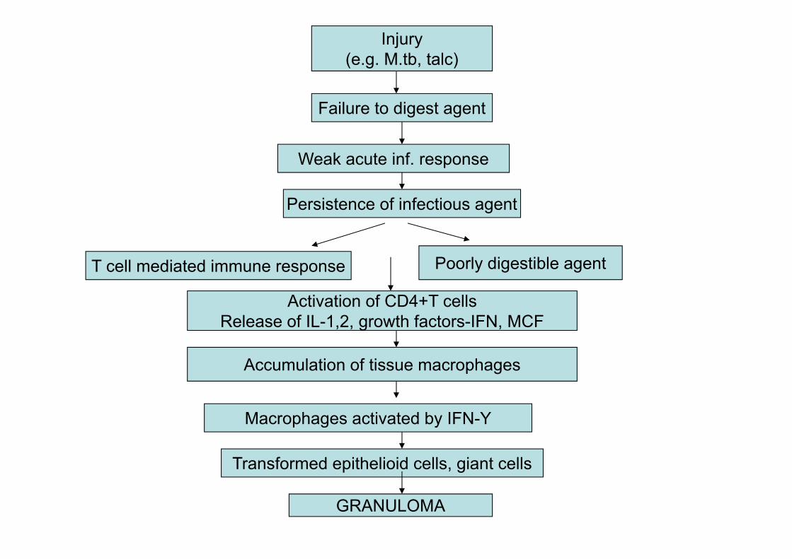
Granulomatous Inflammation
• Distinctive pattern of chronic inflammation, in which the predominant cells are activated macrophages, which are enlarged, oval or elongated with indistinct cell boundary and called epithelioid cells.
• Granuloma – (granule + oma)- circumscribed, tiny lesion (1mm) composed predominantly of collection of epithelioid cells & rimmed at the periphery by lymphoid cells
• Diagnosis of granuloma rests on the identification of epithelioid cells.
• Epithelioid cells may coalesce to form multinucleated giant cells
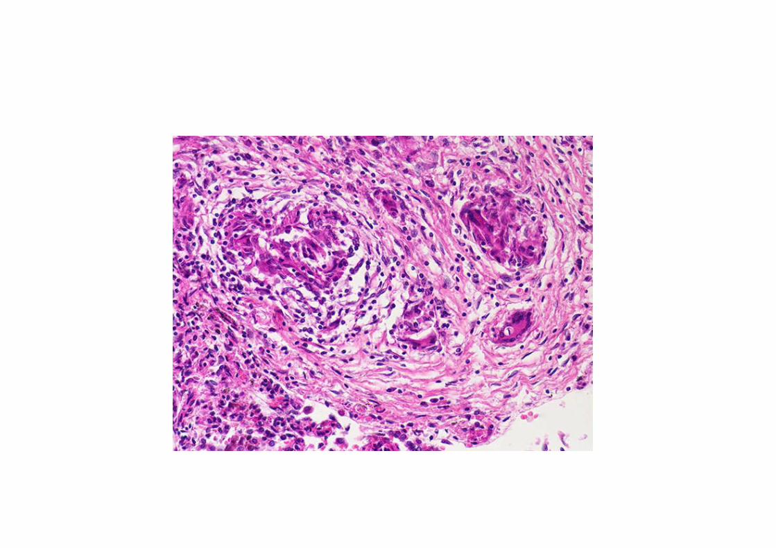
Injury(e.g. M.tb, talc)
Failure to digest agent
Weak acute inf. response
Persistence of infectious agent
T cell mediated immune response Poorly digestible agent
Activation of CD4+T cellsRelease of IL-1,2, growth factors-IFN, MCF
Accumulation of tissue macrophages
Macrophages activated by IFN-Y
Transformed epithelioid cells, giant cells
GRANULOMA


Granulomatous conditions• Bacterial
TuberculosisLeprosySyphilisGranuloma inguinaleCat scratch disease
• FungalActinomycosisBlastomycosisCryptococcosisHistoplasmaCoccidoides immitis
• ParasiticSchistosomiasis

Granulomatous conditions
• Inorganic metals and dusts - Silicosis- Berylliosis- Pneumoconiosis - Asbestosis
• Misc- Sarcoidosis- Crohns disease- Foreign body granuloma

Symptoms
TB can be related to cough, fever, and weight loss.
If untreated, fatal in over 50% of cases.

Giant cells
• Foreign body giant cells• Langhans’ giant cells• Touton giant cells• Aschoff giant cells• Tumor giant cells• Reed-Sternberg cells

Foreign body giant cell

Langhans giant cell

Tuoton giant cell
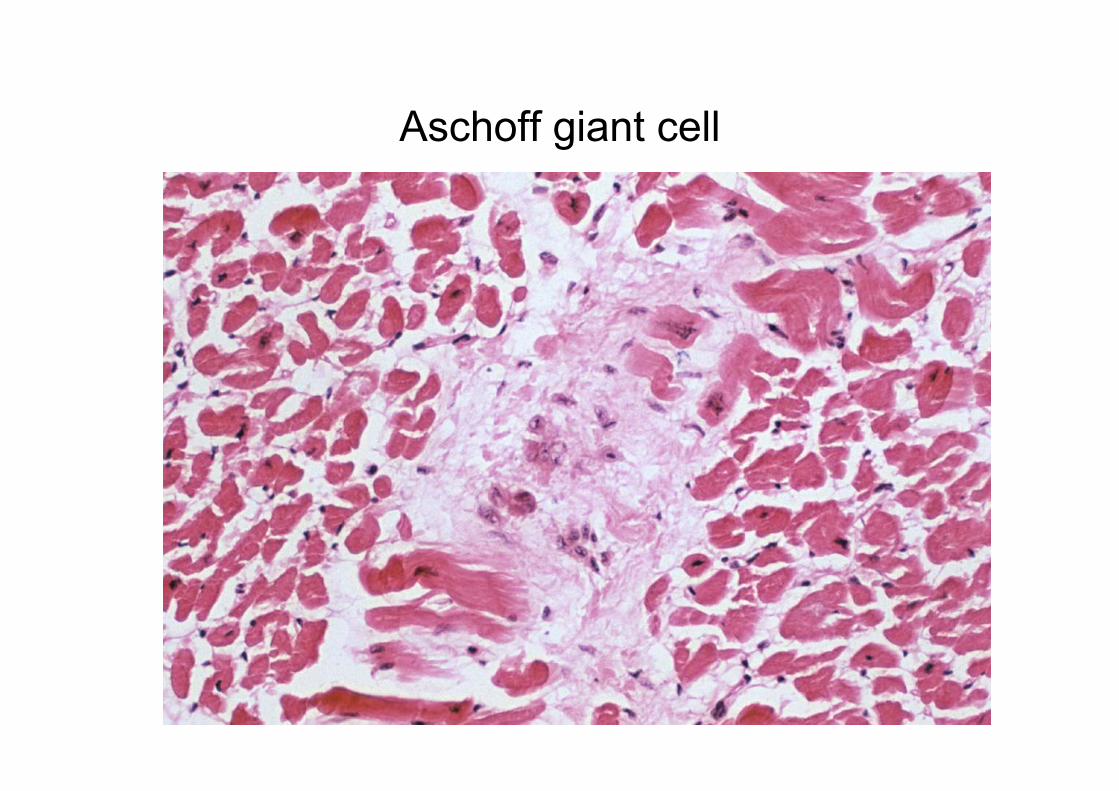
Aschoff giant cell

Tumor giant cell

RS cell Osteoclastic giant cell

Two types of granuloma
(i) Foreign body granulomas: Incited by inert foreign bodies. Example: suture materials, splinter, breast prosthesis, silica, asbestos etc.
(ii) Immune granulomas: It is Type IV hypersensitivity and mediated by T-cells, typically seen in tuberculosis.

Mononuclear phagocyte system
• Blood monocytes• Tissue macrophages
- macrophages in inflammation- kupffer cells- alveolar macrophages- sinus histiocytes- osteoclasts- microglial cells- langerhans’ cells- Hoffbauer cells- mesangial cells


