
Mengangkat potensi pare (Momordica charantia) menjadi produk ...

Asian Pacific Journal of Cancer Prevention, Vol 16, 2015 5507
DOI:http://dx.doi.org/10.7314/APJCP.2015.16.13.5507Momordica cochinchinensis Aril Extract Induces Apoptosis in Human MCF-7 Breast Cancer Cells
Asian Pac J Cancer Prev, 16 (13), 5507-5513
Introduction
Breast cancer is the most frequently diagnosed cancer in women (Bray et al., 2013; Siegel et al., 2013). Chemotherapy is the main method of breast cancer treatment. Due to their ability to arrest cell growth and induce apoptosis, some selective estrogen receptor modulators (SERMs) such as tamoxifen have been considered as drugs for estrogen-dependent cases (Powles, 2008). However, since there are side effects of tamoxifen and other anticancer drugs (Abbasalipourkabir et al., 2010), natural products such as herbs have been used as an alternative therapy (Shukla and George, 2011). Moreover, consumption of phytoestrogens found in foods such as soy beans is associated with a lower risk of breast cancer (Wietrzyk et al., 2005).
Although apoptosis, or type 1 programmed cell death, is a normal physiological process, defects in apoptosis is a major cause of cancer (Elmore, 2007). The apoptotic mechanism is often used as a criterion for discovering new anticancer agents. Several natural compounds such as quercetin and curcumin have shown apoptotic-inducing properties (Kuno et al., 2012). Apoptosis is characterized by specific morphological and biochemical changes of cells, including cell shrinkage, nuclear condensation and DNA fragmentation, dynamic membrane blebbing and loss of cell adhesion, phosphatidylserine externalization and intracellular specific proteolysis (Martin and Green, 1995; Nishida et al., 2008; Ouyang et al., 2012; Mariño et al., 2014).
Several techniques are available to examine cell
Center for Research and Development of Herbal Health Products, Faculty of Pharmaceutical Sciences, Khon Kaen University, Khon Kaen, Thailand *For correspondence: [email protected]
Abstract
Momordica cochinchinensis Spreng (MC) has been used in traditional medicine due to its high carotenoid content. The objective of this study was to investigate mechanisms underlying apoptotic effects of MC on human MCF-7 breast cancer cells. A lycopene-enriched aril extract of MC (AE) showed cytotoxicity and antiestrogenicity to MCF-7 cells. On DAPI staining, AE induced cell shrinkage and chromatin condensation were evident. With flow cytometric analysis, AE increased the percentage of cells in an early apoptosis stage when compared with the control group. RT-PCR analysis showed AE to significantly increase the expression of the proapoptotic bax gene without effect on expression of the anti-apoptotic bcl-2 gene. Moreover, AE enhanced caspase 6, 8 and 9 activity. Taken together, we conclude that AE of MC fruit has anticancer effects on human MCF-7 breast cancer cells by induction of cell apoptosis via both intrinsic and extrinsic pathways of signaling Keywords: Momordica cochinchinensis - breast cancer - apoptosis - bcl-2 and bax gene - caspases activity - MCF-7 cells
RESEARCH ARTICLE
Momordica cochinchinensis Aril Extract Induced Apoptosis in Human MCF-7 Breast Cancer CellsPhuchong Petchsak, Bungorn Sripanidkulchai*
apoptosis. DAPI staining is a simplest technique that based on the complexation of DAPI dye and A-T rich sequences in DNA; the stained cells are visualized under a fluorescence microscope to detect chromatin condensation involved in the apoptotic process (Kapuscinski, 1995). Flow cytometric analysis is commonly used to define the stage of cells. In the early stages of apoptosis, the translocation of membrane phosphatidyl serine to the outer membrane can be detected by high affinity binding fluorescent conjugated annexin V. Further double staining with propidium iodide is used to separate necrotic cells (Vermes et al., 1995). Among several factors regulating the biochemical pathways of signal transduction to trigger apoptosis, Bax and Bcl-2, members of the Bcl-2 protein family, have important regulating roles in the intrinsic pathway. The extrinsic pathway, was regulated through the activation of death receptor. The signal of these two pathways converge to initiator caspases, which are caspase-9 and caspase-8 in the intrinsic and extrinsic pathways respectively, and finally activate effector caspase (caspase-3,6,7) in execution pathway (Krysko et al., 2008).
Momordica cochinchinensis Spreng (MC) is a tropical plant found in Southeast Asia belonging to the Cucurbitaceae family. It is used both as a food and a traditional medicine. In Vietnam, oil of MC fruit is used to treat dry eyes and night blindness due to its high carotenoid content, and the seeds are also used in traditional Chinese medicine (Vuong, 2000; Ishida et al., 2004; Vuong et al., 2006). In terms of anticancer effects, the water extract of MC fruit has been reported to inhibit growth and angiogenesis of the colon 26-20 adenocarcinoma cell
Phuchong Petchsak and Bungorn Sripanidkulchai
Asian Pacific Journal of Cancer Prevention, Vol 16, 20155508
line transplanted in Balb/c mice (Tien et al., 2005), and its ethanolic extract reduced DNA damage in TK6 human lymphocytes (Klungsupya et al., 2012). Due to the high carotenoid content of MC aril and the reported inverse epidemiological relationship of plasma carotenoids and the risk of breast cancer (Huang et al., 2007; Eliassen et al., 2014; Pouchieu et al., 2014), the objective of this study was to investigate the antiestrogenic effect and underlying mechanism of apoptosis-inducing effects of MC aril extract on estrogen receptor positive MCF-7 human breast cancer cells.
Materials and Methods
Chemicals The chemicals in this study included β-carotene,
lycopene, tamoxifen, 17β-estradiol (Sigma, USA); methanol (MeOH) HPLC grade (Fisher, UK); tetrahydrofuran (THF) HPLC grade (Qrec, New Zealand); acetonitrile (ACN) HPLC grade (Labscan , Thailand); agarose gel (Bio-rad, spain); primer bax, bcl-2 and β-actin (Eurofins MWG Operon, Germany); Novel Juice (GeneDirex); charcoal stripped fetal bovine serum (CD-FBS), fetal bovine serum (FBS), sodium pyruvate, annexin-alexa flour 488, annexin binding buffer, 4’,6-diamidino-2-phenylindole (DAPI), Dulbecco’s Modified Eagle’s Medium (DMEM), 3-[4, 5-dimethylthiazol-2-yl]-2,5-dyphenyl tetrazolium bromide (MTT), propidium iodide (Invitrogen, USA) ; caspases colorimetric sampler kit (Novex, UK); RNA extraction kit (GE Healthcare, UK); Omiscript RT Kit, TopTaq MasterMix kit (QIAGEN, Germany) ;1kb DNA ladder, Blue/Orange 6X loading dye (Promega, U.S.A.). All other chemicals were analytical grade.
Plant materials and extractionRipe Momordica cochinchinensis (MC) fruits were
collected from the Faculty of Agriculture, Khon Kaen University. The aril part of fruit was separated and extracted based on the protocol described in our petty patent filing (application no.1403001018). Briefly, the aril was ground and extracted with a mixture of hexane, acetone and ethanol (2:1:1) containing 0.1% butylated hydroxytoluene until the orange color of the aril was bleached. After filtering through Whatman No.1 filter paper, saturated sodium chloride solution was added to the filtrate and gently mixed. The upper phase was collected, and anhydrous sodium sulfate was added. After filtering, the filtrate was dried with a rotary evaporator at a temperature less than 40ºC. The obtained MC aril extract (AE) was kept at -20ºC until use.
HPLC analysis of lycopene and β-carotene contentsUnder light protection, the sample and standard
lycopene and β-carotene were dissolved in a mixture of THF: ACN: MeOH (3:4:3). The system consisted of a C-18 Hypersil® ODS (5 µM, 4.65250 mm) column with isocratic mobile phase ACN: MeOH (70:30). The chromatographic method was run at a flow rate of 1 ml/min, 28ºC column temperature, 20 µl of injection volume and UV detection at 475 nm.
Cell CulturesMCF-7 cells were purchased from American Type
Culture Collection (ATCC) and cultured in Dulbecco’s modified eagle medium (DMEM) supplemented with 10% FBS, 2 mM L-glutamine, 1 mM sodium pyruvate and 1% penicillin-streptomycin solution. The cells were incubated at 37ºC and 5% CO2, and subcultured every 3-4 days or at 80-90% confluence.
Sample preparationMC aril extract and pure lycopene were predissolved
in THF, whereas tamoxifen was predissolved in dimethylsulfoxide (DMSO), after which they were diluted with culture media to the desired concentration. The final concentrations of THF and DMSO were less than 0.5%, which did not affect the test. These were also used as controls.
Cytotoxicity testMCF-7 cells (104 cells/well) were seeded in 96 well
plates and allowed to attach for 24h. The media was replaced with the test sample and incubated for 48h. A MTT assay was used to determine cell viability (Mosman, 1983). Briefly, 50 µl of MTT solution (5mg/ml) were added and incubated at 37ºC for 2-4h until formazan formation was detected by inverted microscopy. The media was discarded and DMSO was added to each well. The plate was shaken until the purple formazan crystals were completely dissolved. The absorbance was determined at 570 nm by a microplate reader (Anthos 2010). The experiment was performed in tripicate and % cell viability was calculated [ Atest
AControl5100].
Antiestrogenic test MCF-7 cells were cultured in DMEM without phenol
red supplemented with 5% CD-FBS, 2 mM L-glutamine, 1 mM sodium pyruvate and 1% penicillin-streptomycin for 3 days before starting the experiments. Then the cells (55103 cells/well) were seeded into 96 well plates and allowed to attach for 24 h. The cells were co-treated with both 10 nM 17β-estradiol and test sample and further incubated for 4 days. A MTT assay was used to determine the cell viability as described above.
DAPI stainingMCF-7 cells (105 cells/well) were seeded into 12
well plates and allowed to attach for 24h. The media was replaced with test sample and further incubated for 48h. After discarding the media, the cells were washed with 10 mM phosphate buffer saline, pH 7.4 (PBS) and incubated with 5% formalin and methanol for 10 min each. DAPI solution (250µl of 1µg/ml concentration) was added and incubated at room temperature for 15 min under light protection. Chromatin condensation of apoptotic cells was observed under a fluorescence inverted microscope. Percentage of apoptotic cells was obtained by counting the apoptotic cells in five fields that contained at least 100 cells/field.
Flow cytometric analysisMCF-7 cells (55105 cells/well) were seeded in 6
Asian Pacific Journal of Cancer Prevention, Vol 16, 2015 5509
DOI:http://dx.doi.org/10.7314/APJCP.2015.16.13.5507Momordica cochinchinensis Aril Extract Induces Apoptosis in Human MCF-7 Breast Cancer Cells
well plates and allowed to attach for 24h. The media was replaced with the test sample and further incubated for 12 h. The cells were trypsinized and washed with ice-cold PBS. After centrifugation at 2000 rpm for 3 min, the cell pellet was collected and 100µl of annexin binding buffer was added and gently mixed. Five µl of Alexa flour 488 conjugated annexin V and 1µl of propidium iodide (PI) were added and incubated at room temperature for 15 min. Finally, 400µl of annexin binding buffer were added and the cell samples were kept on ice, then analyzed using a flow cytometer (Becton Dickinson /FACSCanto II, USA) with the emission wavelengths at 530 and 575 nm. The data were analyzed by BD FACSDiva software. Dot plot analysis was used to separate the cells into four groups in each quadrant, in which quadrant 1 (Q1) referred to necrotic cells that were stained with only PI, quadrant 2 (Q2) referred to late apoptosis/necrosis cells that were stained with both PI and alexa flour 488, quadrant 3 (Q3)
referred to live cells that were unstained, and quadrant 4 (Q4) referred to early apoptosis cells that were stained only with alexa flour 488.
Expression of bax and bcl-2 genes by RT-PCRMCF-7 cells (15106 cells/well) were seeded in 6
well plates. The cells were allowed to attach for 48 h. The media was replaced with the test sample and further incubated for 48 h. After trypsinization, RNA was isolated from the cells using an RNA extraction kit according to the manufacturer’s instructions. The quantity of RNA was measured at an absorbance of 260 nm. cDNA was synthesized from 0.5 µg of RNA with the kit using a thermocycler. Gene amplification was performed using specific nucleotides as previously described (Table 1) (Zhang, 1999; Lovkvist et al., 2008). The PCR profiles were 28 cycles for bax and β-actin, and 30 cycles for bcl-2, denaturation at 95°C for 2 min, annealing for 1 min at 65°C and primer extention at 72°C for 2 min. PCR method was performed and optimized by using RT-PCR kit. The PCR product was analyzed on 1.5% agarose gel with novel juice staining. The qualitative analysis of RT-PCR products density were performed by Gel Documentation and system analysis machine (Gel Documentation InGenius L, Bio-Rad Laboratories, Hercules, CA, USA).The relative expressions of bax and bcl-2 to β-actin from the same treatment were determined.
Determination of caspases activitiesMCF-7 cells (55106 cells/well) were seeded in 100
mm-dishes. The cells were allowed to attach for 48h. The media was replaced with test sample and incubated at 37°C for 48 h. The treated MCF-7 cells were lysed and cell lysis was measured for caspase activity using a test kit (Novex®) according to manufacturer’s protocol. Valine-glutamine-isoleucine-aspartic-acid-para-nitroaniline, (VEID-pNA), isoleucine-glutamine-threonine-aspatic acid-para-nitroaniline, (IETD-pNA) and leucine-glutamine-histidine-aspartic acid-para-nitroaniline, (LEHD-pNA) were used as the specific substrates for caspase 6, 8 and 9, respectively. The incubation was at 27°C for 2h in a light protected container and the absorbance was measured at 405 nm using a microplate reader (DTX 880 Backman Coulter®).
Statistical analysisAll data were expressed as mean±standard error of
mean (SEM) of triplicate experiments. The statistical significance of differences between treated groups was evaluated by One-Way ANOVA and multiple comparisons. A significant difference was defined as P<0.05. Analyses were done using SPSS version 19.0 software.
Table 1. Sequences of Nucleotide Primers for RT-PCRGene Primer Product sizes References
bcl-2 Forward: 5’-GGTGCCACCTGTGGTCCACCTG-3’ 489 Zhang (1999) Reverse: 5’-CTTCACTTGTGGCCCAGATAGG-3’bax Forward: 5’-CAGCTCTGAGCAGATCATGAAGACA-3’ 538 Zhang (1999) Reverse: 5’-GCCCATCTTCTTCCAGATGGTGAGC-3’β-actin Forward: 5’-CGTCATACTCCTGCTTGCTGATC-3’) 661 Lovkvist et al. (2008) Reverse: 5’-AGCACTGTGTTGGCGTACAG-3’
Table 2. IC50 Values of Cytotoxicity and Antiestrogenicity of aril extract to MCF-7 Cellssample IC50 (µg/ml) cytotoxicity antiestrogenicity)
aril extract 117.0±16.7 152.8±8.1lycopene 10.9±0.6 15.3±0.6tamoxifen 4.5±0.1 2.3±0.5
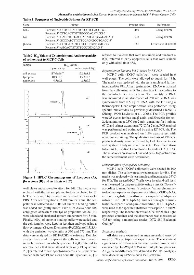
Figure 1. HPLC Chromatograms of Lycopene (A), β-carotene (B) and Aril Extract (C)

Phuchong Petchsak and Bungorn Sripanidkulchai
Asian Pacific Journal of Cancer Prevention, Vol 16, 20155510
Results
Lycopene and β-carotene contents of aril extractAril extract (AE) was obtained at 12% yield. From
HPLC analysis (Figure 1), AE contained both high amounts of lycopene (6.8±0.3mg/g of extract) and β-carotene (2.9±0.1 mg/g of extract).
Cytotoxicity testing of aril extractAE was highly cytotoxic to MCF-7 cells with IC50 at
117.04±16.7 µg/ml. Tamoxifen and standard lycopene showed cytotoxic effects on the cells with IC50 values at 4.5 ± 0.1 and 10.86 ± 0.6 µg/ml, respectively (Table 2).
Antiestrogenic effect of aril extract. As they are well known to be estrogen receptor
positive, MCF-7 cells were used to test for antiestrogenic effects of AE in the presence of 17β-estradiol. When compared to the control, co-treatment of AE with 17β-estradiol significantly decreased cell viability with IC50 at 152.83±8.1 µg/ml. Standard lycopene also showed antiestrogenic effects with IC50 at 15.31±0.6 µg/ml. Tamoxifen, a standard antiestrogenic compound, had an IC50 of 2.25±0.5 µg/ml (Figure 2).
Detection of apoptotic cells by DAPI stainingDAPI was used to stain the chromatin condensation
in the nucleus of apoptotic cells. Apoptotic cells showed smaller and brighter nuclei than normal cells. AE induced cell death by increasing the percentage of apoptotic cells in a similar fashion to tamoxifen (Figure 3 and Figure 4).
Flow cytometric analysis of apoptotic cellsAE significantly increased the number of early
apoptotic cells in Q4, while neither increasing the number of necrotic cells in Q1 nor late apoptotic/necrotic cells in Q2, suggesting that AE specifically induced cell death through the apoptotic process. Tamoxifen similarly induced early apoptosis cells in a dose-dependent fashion. Lycopene did not increase early apoptotic cells in Q4 but significantly increased late apoptotic/necrotic cells in Q2 (Figure 5 and Figure 6).
Effect of AE on the expression of bax and bcl-2 genesBoth AE and lycopene significantly increased
proapoptotic bax gene expression without affecting
Figure 2. Antiestrogenic Effect of AE on Proliferation of MCF-7 Cells when Co-treatment with 17β- Estradiol. *Significant differences from control group at P<0.05
Figure 3. Representative DAPI Staining of MCF-7 cells. The arrows indicated the apoptotic cells with chromatic condensation characteristics. At high magnification, A=control (THF) and B=treated with AE at 800 µg/ml. At lower magnification, C=control (DMSO), D, E, F=treated with AE at 200, 400, 800 µg/ml; G, H, I=treated with lycopene at 2, 4, 8, µg/ml; J=control (DMSO) and K, L, M=treated with tamoxifen at 4, 8, 15 µg/ml
Figure 4. Percentage of Apoptotic Cells of MCF-7 Cells Staining with DAPI after Treatment with AE (A200, A400, A800), Tamoxifen (T4, T8, T15) and Lycopene (L2, L4, L8). *Significant differences from control group at P<0.05.
Figure 5. Dot Plot Analysis from Flow Cytometric Method of MCF-7 Cells Treated with AE, Lycopene and Tamoxifen

Asian Pacific Journal of Cancer Prevention, Vol 16, 2015 5511
DOI:http://dx.doi.org/10.7314/APJCP.2015.16.13.5507Momordica cochinchinensis Aril Extract Induces Apoptosis in Human MCF-7 Breast Cancer Cells
antiapoptosis bcl-2 gene expression in MCF-7 cells. Tamoxifen significantly decreased bcl-2 gene expression without affecting bax gene expression in MCF-7 cells (Figure 7).Effect of AE on caspase activities
AE and tamoxifen significantly increased the activities of caspase-6, 8 and 9, which are involved in both intrinsic and extrinsic pathways in MCF-7 cells. In contrast, lycopene significantly increased only caspase-9 activity (Figure 8).
Discussion
Considerable amounts of research have been performed to discover the potential impact of carotenoids in the prevention and treatment of cancer. The present study provides data on the cytotoxicity of MC aril extract (AE) on estrogen receptor positive human breast cancer MCF-7 cells. The extract induced cell apoptosis via upregulation of proapoptotic bax gene expression and activation of caspase 6, 8 and 9 activities. Besides its two major chemical constitutes, lycopene and β-carotene, other compounds may contribute to the anticancer effect of the aril extract of M. cochinchinensis.
We have optimized the method for extraction of MC aril to obtain a high carotenoid content. In general, the extraction of plant tissue containing water, such as the aril of MC, with a single non-polar solvent such as acetone, hexane or ethyl acetate requires a high volume of solvent for bleaching, due to the immiscibility of water and non-polar solvents. Therefore, in this study a mixture of solvents composed of hexane: acetone: ethanol (2:1:1) was used (Barba et al., 2006) with the addition of an antioxidant
Figure 8. Effect of AE Lycopene and Tamoxifen on the Activities of Caspase-6 (A), Caspase-8 (B) and Caspase-9 (C) in MCF-7 Cells. *Significant differences from control group at P<0.05
Figure 6. Percentage of Early Stage Apoptotic MCF-7 Cell Detected by flow Cytometric Method. *significance differences from control group at p<0.05
Figure 7. Effect of AE, Lycopene and Tamoxifen on the Expression of Proapoptotic Bax Gene (A) and Antiapoptotic bcl-2 (B) in MCF-7 cells (A=aril extract, T=tamoxiffen and L=lycopene. *Significant differences from control group at P < 0.05

Phuchong Petchsak and Bungorn Sripanidkulchai
Asian Pacific Journal of Cancer Prevention, Vol 16, 20155512
0
25.0
50.0
75.0
100.0
New
ly d
iagn
osed
with
out
trea
tmen
t
New
ly d
iagn
osed
with
tre
atm
ent
Pers
iste
nce
or r
ecur
renc
e
Rem
issi
on
Non
e
Chem
othe
rapy
Radi
othe
rapy
Conc
urre
nt c
hem
orad
iatio
n
10.3
0
12.8
30.025.0
20.310.16.3
51.7
75.051.1
30.031.354.2
46.856.3
27.625.033.130.031.3
23.738.0
31.3
0
25.0
50.0
75.0
100.0
New
ly d
iagn
osed
with
out
trea
tmen
t
New
ly d
iagn
osed
with
tre
atm
ent
Pers
iste
nce
or r
ecur
renc
e
Rem
issi
on
Non
e
Chem
othe
rapy
Radi
othe
rapy
Conc
urre
nt c
hem
orad
iatio
n
10.3
0
12.8
30.025.0
20.310.16.3
51.7
75.051.1
30.031.354.2
46.856.3
27.625.033.130.031.3
23.738.0
31.3
(BHT) to decrease the degradation of carotenoids during extraction (Henry et al., 1998). As a result, our AE contained relatively high amounts of carotenoids, of which lycopene and β-carotene were present in the ratio of 2.3 (6.8 versus 2.9 mg/g of extract). These values are in agreement with previous findings that the seed membrane contained high amounts of lycopene, exceeding β-carotene concentrations and considerable amounts of α-tocopherol (Vuong et al., 2006). Moreover, the ratio of lycopene to β-carotene of our AE is lower, which is similar to the finding of Aoki et al. (2002). These results reflect the different carotenoid content of plants grown in different areas. The results of cytotoxicity testing demonstrated that the IC50 value of AE was at 117.04±16.7µg/ml (equivalent to lycopene at 0.8 µg/ml), which is much lower than the value of pure lycopene (10.86±1.1 µg/ml), suggesting that other synergistic ingredients such as β-carotene, alpha-tocopherol and perhaps fatty acids (Ishida et al., 2004) and other unidentified ingredients may confer the overall cytotoxic effect of AE. However, the significant anticancer roles of these compounds needs further investigation.
Estrogen exposure is the main factor in increased risk of breast cancer, and is mediated by stimulation of tissue proliferation (Yager and Davison, 2006; Clarke et al., 2013; Farhat et al., 2013). Several epidemiological studies have demonstrated that lycopene and other carotenoids are inversely associated with breast cancer (Ito et al., 1999; Tamimi et al., 2005). In this study, when co-treated with 17β-estradiol, the lycopene-enriched AE showed antiestrogenic effects on estrogen receptor positive MCF-7 cells, similar to previous reports on lycopene (Hirsch et al., 2007).
To elucidate the mechanism by which AE inhibited the cell growth, DAPI staining was firstly used to demonstrate the typical characteristics of chromatin condensation of apoptotic cells. When compared to the negative control (THF), AE significantly induced apoptosis of MCF-7 cells as observed for the treatments with lycopene and tamoxifen. Further analysis by flow cytometry, which is a more sensitive and accurate method (Hotz et al., 1994), showed that AE increased the number of early stage apoptotic MCF-7 cells in similar fashion as tamoxifen. In contrast, lycopene increased the number of late apoptotic / necrotic cells at concentrations ranging from 2-4 µg/ml. Since the tested concentrations of AE (200-400 µg/ml) were equivalent to lycopene contents at 1.4-2.7 µg/ml, this suggests that AE may not induce apoptosis in exactly the same way as lycopene. Lycopene has been reported to have preventive and anticancer activities as mediated by reducing DNA damage from reactive oxygen species (Kelkel et al., 2011), potential inhibition of proliferation, arresting the cell cycle and increasing apoptosis of MCF-7 cells (Teodoro et al., 2012).
The mechanism of apoptosis occurs due to several factors involving both intrinsic and extrinsic pathways. These pathways converge to the execution pathway that involves the cascade of effector caspases, resulting in DNA fragmentation, degradation of cytoskeletal and nuclear proteins, and cross-linking of proteins to form apoptotic bodies (Elmore, 2007; Mukhopadhyay et al., 2014). Although there are reports concerning apoptotic
induction by MC seed extract in MDA-MB-231 breast cancer and gastric cancer cells lines (Meng et al., 2012; Pratheeshkumar et al., 2012), there are no data on the aril extract. This study of the mechanism of the apoptosis inducing effect of AE in MCF-7 cells focused on the genetic expression of the prominent regulators in the Bcl-2 family, the proapoptotic bax and antiapoptotic bcl-2 genes, and caspase-6, 8, 9 activities. The balance of Bax and Bcl-2 proteins plays a role in controlling the release of cytochrome c by regulating outer mitochondrial membrane permeability in the intrinsic pathway. Our results revealed that AE induced cell apoptosis by increasing proapoptotic bax gene expression and enhancing the activities of caspase 6, 8 and 9. Upregulation of the Bax protein may increase the release of cytochrome c, which can form complexes with procaspase-9 and Apaf-1 in apoptosomes and lead to activation of caspase-9 in the intrinsic pathway, and finally activate other caspases including caspase-6 in the execution pathway. The finding that AE also activates caspase-8 activity suggests additional effects of AE on some parts of the extrinsic pathway as well.
Previous studies of lycopene have reported its apoptosis inducing effect in various breast cancer cell lines such as MCF-7, MDA-MB-235 and MDA-MB-231 (Salman et al., 2007; Teodoro et al., 2012; Gloria et al., 2014). Takeshima et al. (2014) reported that apoptosis induction of lycopene via increasing the expression of the bax gene in MDA- MB-468. However, to the best of our knowledge there are no data on the effect of lycopene on caspase activities in breast cancer cell lines. In this study, lycopene showed similar effects as did AE by upregulating the expression of the bax gene. It is surprising to observe that lycopene increased caspase-9 activity but did not change caspase-6 and 8 activities. Tamoxifen, a selective estrogen receptor modulator, induced apoptosis by decreasing bcl-2 gene expression as previously reported (Zhang et al., 1999) and increased the activities of these three caspases as observed with AE. Since AE increased caspase-6, 8 and 9, whereas lycopene increased only caspase-9, this indicates that some other components may synergize with lycopene to induce apoptosis in MCF-7 cells.
Acknowledgements
The research was funded by the Higher Education Promotion and National Research University Project of Thailand, Office of the Higher Education Commission, through the Food and Functional Food Research Cluster of Khon Kaen University. The authors would like to thank the Center for Research and Development of Herbal Health Products, Faculty of Pharmaceutical Science, Khon Kaen University for providing the facilities in this study.
References
Abbasalipourkabir R, Salehzadeh A, Abdullah R (2010). Antitumor activity of tamoxifen loaded solid lipid nanoparticles on induced mammary tumor gland in Sprague-Dawley rats. Afr J Biotechnol, 9, 7337-45.
Aoki H, Kieu NT, Kuze N, Tomisaka K, Chuyen VN (2002). Carotenoid pigments in gac fruit (Momordica cochinchinensis Spreng). Biosci Biotechnol Biochem, 66,

Asian Pacific Journal of Cancer Prevention, Vol 16, 2015 5513
DOI:http://dx.doi.org/10.7314/APJCP.2015.16.13.5507Momordica cochinchinensis Aril Extract Induces Apoptosis in Human MCF-7 Breast Cancer Cells
2479-82.Barba AIO, Hurtado MC, Mata MCS, Ruiz VF, Tejada MLS
(2006). Application of a UV-vis detection-HPLC method for a rapid determination of lycopene and b-carotene in vegetables. Food Chem, 95, 328-36.
Bray F, Ren JS, Masuyer E, Ferlay J (2013). Global estimates of cancer prevalence for 27 sites in the adult population in 2008. Inter J Cancer, 132, 1133-45.
Clarke LH, Assis SD, Warri A (2013). Exposures to synthetic estrogens at different times during the life, and their effect on breast cancer risk. J Mammary Gland Biol Neoplasia, 18, 25-42.
Eliassen AH, Liao X, Tworoger SS, Hankinson SE (2014). Plasma carotenoids and risk of breast cancer over 20 years of follow-up in the Nurses’ health study. Cancer Res, 74, 2919.
Elmore S (2007). Apoptosis: a review of programmed cell death. Toxicol Pathol, 35, 495-516.
Farhat GN, Parimi N, Chlebowski RT, et al (2013). Sex hormone levels and risk of breast cancer with estrogen plus progestin. J Natl Cancer Inst, 105, 1496-503.
Gloria NF, Soares N, Brand C, et al (2014). Lycopene and beta-carotene induce cell-cycle arrest and apoptosis in human breast cancer cell lines. Anticancer Res, 34, 1377-86.
Henry LK, Catignani GL, Schwartz SJ (1998). Oxidative degradation kinetics of lycopene, lutein, and 9-cis and all-trans β-Carotene. J Am Oil Chem Soc, 75, 823-9.
Hirsch K, Atzmon A, Danilenko M, Levy J, Sharoni Y (2007). Lycopene and other carotenoids inhibit estrogenic activity of 17 β-estradiol and genistein in cancer cells. Breast Cancer Res Treat, 104, 221-30.
Hotz MA, Gong J, Traganos F, Darzynkiewicz Z (1994). Flow cytometric detection of apoptosis: comparison of the assays of in situ DNA degradation and chromatin changes. Cytometry, 15, 237-44.
Huang JP, Zhang M, Holman DC, Xie X (2007). Dietary carotenoids and risk of breast cancer in Chinese women. Asia Pac J Clin Nutr, 16, 437-42.
Ishida BK, Tunner C, Chapman MH, McKeon TA (2004). Fatty acid and carotenoid composition of gac (Momordica cochinchinensis Spreng) fruit. J Agric Food Chem, 52, 274-9.
Ito Y, Gajalakshmi KC, Sasaki R, Suzuki K, Shanta V (1999). A study on serum carotenoid levels in breast cancer patients of Indian women in Chennai (Madras), India. J Epidemiol, 9, 306-14.
Kapuscinski J (1995). DAPI: a DNA specific fluorescent probe. Biotech Histochem, 70, 220-33.
Kelkel M, Schumacher M, Dicato M, Diederich M (2011). Antioxidant and anti-proliferative properties of lycopene. Free Radical Res, 45, 925-40.
Klungsupya P, Saenkhum J, Muangman T, et al (2012). Non-cytotoxic property and DNA protective activity against H2O2 and UVC of Thai GAC fruit extracts in human TK6 cells. J Appl Pharm Sci, 2, 4-8.
Krysko DV, Berghe TV, D’Herde K, Vandenabeele P (2008). Apoptosis and necrosis: Detection, discrimination and phagocytosis. Methods, 44, 205-21.
Kuno T, Tsukamoto T, Hara A, Tanaka T (2012). Cancer chemoprevention through the induction of apoptosis by natural compounds. J Biophys Chem, 3, 156-73.
Lövkvist H, Smith JG, Luthman H, et al (2008). Ischaemic stroke in hypertensive patients is associated with variations in the PDE4D genomeregion. Eur J Human Genet, 16, 1117-25.
Mariño G, Niso-Santano, Baehrecke EH, Kroemer G (2014). Self-consumption: the interplay of autophagy and apoptosis. Nat Rev Mol Cell Biol, 15, 81-94.
Martin SJ, Green DR (1995). Protease activation during
apoptosis: death by a thousand cuts. Cell, 82, 349-52.Meng LY, Liu HR, Shen Y, Yu YQ, Tao X (2012). Cochinchina
momordica seed extract induces G2/M arrest and apoptosis in human breast cancer MDA-MB-231 cells by modulating the PI3K/Akt pathway. Asian Pac J Cancer Prev, 13, 3483-8.
Mosman T (1983). Rapid colorimetric assay for cellular growth and survival: Application to proliferation and cytotoxicity assays. J Immunol Methods, 65, 55-63.
Mukhopadhyay S, Panda PK, Sinha N , Das DN,Bhu SK (2014). Autophagy and apoptosis: where do they meet? Apoptosis, 19, 555-66.
Nishida K, Yamaguchi O, Otsu K (2008). Crosstalk between autophagy and apoptosis in heart disease. Circ Res, 103, 343-51.
Ouyang L, Shi Z, Zhao S, et al (2012). Programmed cell death pathways in cancer: a review of apoptosis, autophagy and programmed necrosis. Cell Prolif, 45, 487-98.
Pouchieu C, Galan P, Ducros V, et al (2014). Plasma carotenoids and retinol and overall and breast cancer risk: a nested case-control study. Nutr Cancer, 66, 980-8.
Powles TJ (2008). Prevention of breast cancer using SERMs. Adv Expl Med Biol, 630, 232-36.
Pratheeshkumar P, Sreekala C, Zhang Z, et al (2012). Cancer prevention with promising natural products: mechanisms of action and molecular targets. Anti-Cancer Agents Med Chem, 12, 1159-84.
Salman H, Bergman M, Djaldetti M, Bessler H (2007). Lycopene affects proliferation and apoptosis of four malignant cell lines. Biomed Pharmacother, 61, 366-69.
Shukla Y, George J (2011). Combinatorial strategies employing nutraceuticals for cancer development. Ann NY Acad Sci, 1229, 162-75.
Siegel R, Naishadham D, Jemal A (2013). Cancer statistics, 2013. CA Cancer J Clin, 63, 11-30.
Takeshima M, Ono M, Higuchi T, et al (2014). Anti-proliferative and apoptosis-inducing activity of lycopene against three subtypes of human breast cancer cell lines. Cancer Sci, 105, 252-7.
Tamimi RM, Hankinson SE, Campos H, et al (2005). Plasma carotenoids, retinol, and tocopherols and risk of breast cancer. Am J Epidemiol, 161, 153-60.
Teodoro A, Oliveira FL, Martins NB, et al (2012). Effect of lycopene on cell viability and cell cycle progression in human cancer cell lines. Cancer Cell Int, 12, 36.
Tien, PG, Kayama F, Konishi F, et al (2005). Inhibition of tumor growth and angiogenesis by water extractof Gac fruit (Momordica cochinchinensis Spreng). Int J Oncol, 26, 882-9.
Vermes I, Haanen C, Steffens-Nakken H, Reutelingsperger C (1995). A novel assay for apoptosis flow cytometric detection of phosphatidylserine early apoptotic cells using fluorescein labeled expression on Annexin V. J Immunol Methods, 184, 39-51.
Vuong LT (2000). Under-utilized beta-carotene-rich crops of Viet Nam. Food Nutr Bull, 2, 173-81.
Vuong LT, Franke AA, Custer LJ, Murphy SP (2006). Momordica cochinchinensis Spreng. (gac) fruit carotenoids reevaluated. J Food Compost Anal, 19, 664-8.
Wietrzyk J, Grynkiewicz G, Opolski A (2005). Phytoestrogens in cancer prevention and therapy-mechanisms of their biological activity. Anticancer Res, 25, 2357-66.
Yager JD, Davidson NE (2006). Estrogen carcinogenesis in breast cancer. New Engl J Med, 354, 270-82.
Zhang GJ, Kimijima I, Onda M (1999). Tamoxifen-induced apoptosis in breast cancer cells relates to down-regulation of bcl-2, but not bax and bcl-X L, without alteration of p53 protein levels. Clin Cancer Res, 5, 2971-7.


![[Aril Daine] Life After Marriage](https://static.fdocuments.net/doc/165x107/55cf9686550346d0338c0e0b/aril-daine-life-after-marriage.jpg)


