Molecular and Functional Characterization of...
24
The Journal of Experimental Biology – ACCEPTED AUTHOR MANUSCRIPT Molecular and Functional Characterization of Hemocyanin of the Giant African Millipede, Archispirostreptus gigas Christian Damsgaard 1 , Angela Fago 1 , Silke Hagner-Holler 2 , Hans Malte 1 , Thorsten Burmester 3 and Roy E. Weber 1 * 1 Zoophysiology, Deparment for Bioscience, Aarhus University, DK 8000 Aarhus, Denmark, 2 Institute of Zoology, Molecular Animal Physiology, University of Mainz, Germany, 3 Institute of Zoology and Zoological Museum, University of Hamburg, D-20146 Hamburg, Germany *Author for correspondence ([email protected] ) Short title: Giant African millipede hemocyanin SUMMARY In contrast to other terrestrial arthropods where gaseous O 2 that fuels aerobic metabolism diffuses to the tissues in tracheal tubes, and most other metazoans where O 2 is transported to tissues by circulating respiratory proteins, the myriapods (millipedes and centipedes) strikingly have tracheal systems as well as circulating hemocyanin (Hc). In order to elucidate the evolutionary origin and biological significance of millipede Hc we report the molecular structure (subunit composition and amino acid sequence) of multimeric (36-mer) Hc from the forest-floor dwelling giant African millipede Archispirostreptus gigas and its allosteric oxygen binding properties under various physico-chemical conditions. A. gigas Hc consists of only a single subunit type with differential glycosylation. Phylogenic analysis reveals that millipede Hc is a sister group to centipede HcA, which supports an early divergence of distinct Hc subunits in myriapods and an ancient origin of multimeric Hcs. A. gigas Hc binds O 2 with a high affinity and shows a strong normal Bohr effect. O 2 binding is moreover modulated by Ca 2+ ions, which increase the O 2 affinity of the Hc in the T (tense; deoxygenated) as well as the R (relaxed; oxygenated) states, and by (L)-lactate, which modulates Hc-O 2 affinity by changing the allosteric equilibrium constant, L. Cooperativity in O 2 -binding at half O 2 -saturation (n 50 ) is pH-dependent and maximal at pH ~7.4 and the number of interacting O 2 binding sites (q) is markedly increased by binding Ca 2+ . The data is discussed in the light of role of mutually supplementary roles of Hc and the tracheal system for tissue O 2 supply. Supplementary material available online at: Keywords: Hemocyanin, oxygen binding, millipede, allosteric regulation, calcium, lactate http://jeb.biologists.org/lookup/doi/10.1242/jeb.080861 Access the most recent version at J Exp Biol Advance Online Articles. First posted online on 24 January 2013 as doi:10.1242/jeb.080861 Copyright (C) 2013. Published by The Company of Biologists Ltd http://jeb.biologists.org/lookup/doi/10.1242/jeb.080861 Access the most recent version at First posted online on 24 January 2013 as 10.1242/jeb.080861
Transcript of Molecular and Functional Characterization of...
The
Jou
rnal
of
Exp
erim
enta
l Bio
logy
– A
CC
EPT
ED
AU
TH
OR
MA
NU
SCR
IPT
Molecular and Functional Characterization of Hemocyanin of the Giant African
Millipede, Archispirostreptus gigas
Christian Damsgaard1, Angela Fago1, Silke Hagner-Holler2, Hans Malte1, Thorsten Burmester3 and
Roy E. Weber1*
1Zoophysiology, Deparment for Bioscience, Aarhus University, DK 8000 Aarhus, Denmark, 2Institute of Zoology, Molecular Animal Physiology, University of Mainz, Germany, 3Institute of Zoology and
Zoological Museum, University of Hamburg, D-20146 Hamburg, Germany
*Author for correspondence ([email protected])
Short title: Giant African millipede hemocyanin
SUMMARY
In contrast to other terrestrial arthropods where gaseous O2 that fuels aerobic metabolism diffuses to the
tissues in tracheal tubes, and most other metazoans where O2 is transported to tissues by circulating
respiratory proteins, the myriapods (millipedes and centipedes) strikingly have tracheal systems as well as
circulating hemocyanin (Hc). In order to elucidate the evolutionary origin and biological significance of
millipede Hc we report the molecular structure (subunit composition and amino acid sequence) of multimeric
(36-mer) Hc from the forest-floor dwelling giant African millipede Archispirostreptus gigas and its allosteric
oxygen binding properties under various physico-chemical conditions. A. gigas Hc consists of only a single
subunit type with differential glycosylation. Phylogenic analysis reveals that millipede Hc is a sister group to
centipede HcA, which supports an early divergence of distinct Hc subunits in myriapods and an ancient
origin of multimeric Hcs. A. gigas Hc binds O2 with a high affinity and shows a strong normal Bohr effect.
O2 binding is moreover modulated by Ca2+ ions, which increase the O2 affinity of the Hc in the T (tense;
deoxygenated) as well as the R (relaxed; oxygenated) states, and by (L)-lactate, which modulates Hc-O2
affinity by changing the allosteric equilibrium constant, L. Cooperativity in O2-binding at half O2-saturation
(n50) is pH-dependent and maximal at pH ~7.4 and the number of interacting O2 binding sites (q) is markedly
increased by binding Ca2+. The data is discussed in the light of role of mutually supplementary roles of Hc
and the tracheal system for tissue O2 supply.
Supplementary material available online at:
Keywords: Hemocyanin, oxygen binding, millipede, allosteric regulation, calcium, lactate
http://jeb.biologists.org/lookup/doi/10.1242/jeb.080861Access the most recent version at J Exp Biol Advance Online Articles. First posted online on 24 January 2013 as doi:10.1242/jeb.080861
Copyright (C) 2013. Published by The Company of Biologists Ltd
http://jeb.biologists.org/lookup/doi/10.1242/jeb.080861Access the most recent version at First posted online on 24 January 2013 as 10.1242/jeb.080861
The
Jou
rnal
of
Exp
erim
enta
l Bio
logy
– A
CC
EPT
ED
AU
TH
OR
MA
NU
SCR
IPT
INTRODUCTION
The size and complexity of metazoan animals compelled the evolution of specific anatomical and molecular
traits (notably circulatory systems and O2-transporting proteins) to secure the transfer of O2 from the
respiratory surfaces to the respiring tissues in support of aerobic metabolism. In contrast to the vast majority
of animals where O2 is transported by respiratory proteins such as hemoglobin (Hb), hemocyanin (Hc) and
hemerythrin circulating in body fluids, terrestrial arthropods possess a tracheal systems that permit gaseous
diffusion of O2 to individual tissue cells (Keilin and Wang, 1946). In this regard, the Myriapoda – that
includes centipedes (class Chilopoda) and millipedes (class Diplopoda) - are of particular interest in having
trachea as well as Hc (Rajulu, 1969; Mangum et.al., 1985; Kusche and Burmester, 2001; Kusche et.al.,
2002). In contrast to Hbs that are widely distributed in the animal kingdom, the copper-containing Hcs are
extracellular proteins found only in arthropods - where Hcs have been recorded in all subphyla (Burmester,
2001) - and molluscs (Mangum et.al., 1985). However, the Hcs of these two phyla are not related but
emerged independently (Burmester, 2001). Arthropod Hcs are highly multimeric. Myriapod Hcs are 6x6, 36-
mer structures (Jaenicke et.al., 1999) composed of up to four different subunits types (Markl et.al., 2009).
Each subunit consists of ~650 amino acid residues that are distributed among three structural domains and
include six highly conserved histidines, which coordinate two copper (I) ions that reversibly bind one O2
molecule (Terwilliger, 1998; Markl and Decker, 1992). The multimerization increases the O2 transport
capacity of the hemolymph without markedly raising its osmotic pressure.
As with Hbs, Hcs are allosteric proteins that are in equilibrium between two conformational states
(Wyman, 1969): a tense (T) state with a low O2 equilibrium association constant (KT), and a relaxed (R) state
with a high O2 equilibrium association constant (KR) (Loewe, 1978). When PO2, and thus O2-saturation, is
high (at the respiratory surfaces) most Hc molecules are in the R state, which favors O2 loading to the
hemolymph. At low PO2 (as in the working muscle) the molecules are predominantly in the T state, which
enhances unloading. Hc-O2 binding is modulated by homotropic interactions (between O2 binding sites) and
heterotropic interactions (between the sites for binding O2 and effectors) (Bonaventura and Bonaventura,
1980; Brouwer and Serigstad, 1989; Van Holde et.al., 2001). Heterotropic effectors modulate O2-affinity by
stabilizing or destabilizing the T and/or the R state or by altering the T-R allosteric equilibrium constant
between the two protein conformations, L.
In contrast to the intensively-studied erythrocytic vertebrate Hbs, where protons, organic phosphates
and chloride ions decrease Hb-O2 affinity by lowering KT, striking variation is encountered amongst the
multimeric invertebrate O2-binding proteins, where effectors, commonly divalent cations may increase or
decrease KT and/or KR (that can graphically be represented by the intercepts of the lower and upper
asymptotes, respectively, of the extended Hill plot with the Y-axis at logPO2 = 0). The diversity in regulatory
mechanisms encountered in extracellular O2-binding proteins calls for parallel analysis of myriapod Hcs.

The
Jou
rnal
of
Exp
erim
enta
l Bio
logy
– A
CC
EPT
ED
AU
TH
OR
MA
NU
SCR
IPT
The diplopod Archispirostreptus gigas is a large, slow-moving, forest-floor dwelling deposit feeder
from East Africa (Pechenik, 2005) predicting markedly different metabolic requirements compared to
chilopods that are fast-moving carnivores. As with insects, myriapods may have occludable spiracles and
exhibit discontinuous gas exchange whereby their tissues may intermittently become hypoxic (Schmitz and
Harrison, 2004). As soil dwellers they moreover are subjected to temporary flooding that decrease O2
availability. Aiming to gain insight into the evolutionary origin of millipede Hc, its functional adaptations
and allosteric regulatory mechanisms, we investigated the primary structure of A.gigas Hc and its O2 binding
properties in the native hemolymph and Hc solutions under differing physico-chemical conditions as regards
pH, (L)-lactate and divalent cation concentrations.
MATERIALS AND METHODS
Animals and hemolymph
Specimens of the giant millipede Archispirostreptus gigas weighing 25-82 g obtained from a dealer (Exotera
GmbH, Holzheim, Germany) were kept in a terrarium at room temperature and fed regularly.
Hemolymph for O2 binding studies were drawn from 12 animals using a hypodermic syringe fitted
with 23Gx1 ¼” gauge needles to pierce the dorsal intersegmental membranes. All subsequent preparative
steps were carried out at 0 – 4 °C. The samples were pooled, and centrifuged for 10 minutes at 10,000g to
remove cellular debris. Samples used to record extended Hill plots (see below) were concentrated by
ultrafiltration in Amicon Ultra Centrifugal filters (cut-off: 100.000 kDa) (Millipore, Billeria, MA, USA). To
study the effect of cations (Na+, Ca2+, Mg2+), the hemolymph was dialyzed against 3 changes of 10 mM Tris-
HCl buffer, pH 7.5 at a 1:300 sample:buffer ratio and concentrated. The dialyzed hemolymph is denoted as
stripped Hc. The hemolymph and Hc preparations were divided into 100 µL aliquots that were stored at -
80oC and thawed individually for measurements of O2 equilibria.
Hc concentration was derived from the absorbance at 335 nm using the ε= 0.0175 M-1·cm-1 reported
for crab Carcinus manaes Hc (Nickerson and Van Holde, 1971; Weber et.al., 2008) and a subunit mass of
73.5 kDa as deduced from cDNA sequence analysis (described below).
Protein biochemistry
A polyclonal antibody was generated against purified A. gigas hemocyanin in guinea pigs. SDS-PAGE was
performed on a 7.5% gel using standard methods as previously described (Jaenicke et.al., 1999). Two-
dimensional gel electrophoresis using pH 3.5-10 ampholines was performed according to O'Farrell
(O'Farrell, 1975). For Western blotting, the proteins were transferred to nitrocellulose at 0.8 mA/cm2. Non-
specific binding sites were blocked by 5% non-fat dry milk in TBST (10 mM Tris-HCl, pH 7.4, 140 mM
NaCl, 0.25% Tween-20). Incubation with the anti-Hc antibody in 5% non-fat dry milk/TBST, was carried
out for 2 h at room temperature. The filters were washed 3 x 10 min in TBST and subsequently incubated for

The
Jou
rnal
of
Exp
erim
enta
l Bio
logy
– A
CC
EPT
ED
AU
TH
OR
MA
NU
SCR
IPT
1 h with a goat-anti-rabbit secondary antibody conjugated with alkaline phosphatase, diluted in 5% non-fat
dry milk/TBST. The membranes were washed as above and the detection was carried out using nitro-blue-
tetrazolium and bromo-chloro-indolyl-phosphate.
Mass spectrometry was carried out by nanoLC-ESI-ion trap analysis after tryptic digest of the
hemocyanin bands from SDS-PAGE. Protein identification was performed with the Mascot software
(Perkins et.al., 1999) using the NCBI nr database.
Cloning of A. gigas hemocyanin cDNA
Total RNA was extracted from the whole animal after removal of the cuticle. Poly(A)+RNA was purified
from total RNA using the PolyATractTM kit (PROMEGA). 5 µg poly(A)+RNA were applied for the
construction of a directionally-cloned cDNA expression library applying the Lambda ZAP-cDNA synthesis
kit (STRATAGENE). The library was screened with the anti-A. gigas hemocyanin antibodies. Positive phage
clones were converted to plasmid vectors using the material provided in the kit. The cDNAs inserted in the
pBK-CMV vector were sequenced on both strands by a commercial sequencing service (GENTERPRISE,
Mainz). A full length hemocyanin cDNA sequence was obtained and is available at the EMBL/GeneBankTM
databases under the accession number HE574799.
Sequence and phylogenetic analyses
The web-based tools provided by the ExPASy Molecular Biology Server of the Swiss Institute of
Bioinformatics (http://www.expasy.org) were used for sequence analyses. A multiple sequence alignment of
the amino acid sequences of selected arthropod hemocyanins and phenoloxidases (Supplemental Table S1)
was constructed with MAFFT 6 (Katoh et.al., 2005) at http://mafft.cbrc.jp/alignment/server/. The L-INS-i
routine and the BLOSUM 45 matrix were selected. The final alignment covered 93 sequences and 906
positions (Supplemental Fig. S1).
Bayesian phylogenetic analysis was performed using MrBayes 3.1.2 (Huelsenbeck and Ronquist,
2001). We assumed the WAG model with a gamma distribution of substitution rates. Metropolis-coupled
Markov chain Monte Carlo (MCMCMC) sampling was performed with one cold and three heated chains.
Two independent runs were performed in parallel for 5 million generations. The average standard deviation
of split frequencies was < 0.005. Starting trees were random and the trees were sampled every 1000th
generation. Posterior probabilities of the nodes were estimated on the final 4,000 trees (burnin = 1,000).
Oxygen equilibrium measurements
O2 equilibria were measured at 25o C using a modified gas-diffusion chamber (Weber, 1981) coupled to two
serially-linked precision Wösthoff gas mixing pumps (Bochum, Germany) for mixing pure (>99.998%) N2
and atmospheric air. In the procedure, absorbance of 3 μL hemolymph samples was recorded at 365 nm

The
Jou
rnal
of
Exp
erim
enta
l Bio
logy
– A
CC
EPT
ED
AU
TH
OR
MA
NU
SCR
IPT
following equilibration with pure O2 and N2, respectively, to obtain the full saturation range and thereafter
monitored during stepwise increases in the PO2 of the equilibrating gas mixture. O2 equilibrium curves were
derived as the relationship between absorbance levels (corresponding to O2 saturation) and calculated O2
tensions at the individual steps. In order to assess the Bohr effect, pH values in the measured samples were
varied by adding 1 M Tris-HCl (for measurements at pH >7) or bisTris buffers (pH <7) to obtain a final
buffer concentration of 0.1 M (unless otherwise indicated). The pH-values were measured at 25oC using a
BMS 2 MK 2 microelectrode coupled to a PHM 64 Research pH meter (Radiometer, Copenhagen,
Denmark).
For each O2 equilibrium curve, P50 and n50 values (O2 tensions and Hill’s cooperativity coefficients,
respectively, at 50% oxygenation) were derived from the zero intercepts and slopes, respectively of Hill plots
(logS/(1-S) vs. logPO2, where S is the fractional saturation) based on at least 4 equilibrium steps in the 25-
75% saturation range. Extended Hill plots for assessment of KT, KR and other allosteric parameters were
obtained by recording at least 10 additional steps at low (<10%) and high (>90%) O2 saturations.
The oxygen fractional saturation, S, at each step was derived as:
� �
����
�������, (1)
where A is the absorbance at each step, and A100 and A0 are the absorbance recorded with pure O2 and pure
N2, respectively.
The allosteric parameters were obtained by fitting the MWC ‘two-state, concerted’ model (Monod
et.al., 1965) to the data by non-linear least squares curve fitting according to the equation:
� �
�·��·�������·��������·�������·���
���
�����·��������·���
�, (2)
where S denotes the fractional saturation, L is the allosteric constant = [T]/[R] in the absence of ligand, PO2
is the partial pressure of O2, and q denotes the number of interacting binding sites. To minimize errors
introduced by incomplete saturation or desaturation when equilibrating with pure oxygen or pure nitrogen,
respectively, the true absorbance at zero and full saturation was found in a single fitting procedure, along
with the allosteric parameters including Pm and nmax (see Table 1), as described by (Fago et.al., 1997). The
Bohr factor (φ, the number of protons released per O2 molecule bound) was calculated from the slope of
logP50 as a function of pH
� �
Δ log���
��
(3)
RESULTS AND DISCUSSION Molecular characterization of A. gigas hemocyanin
SDS-PAGE and 2D-PAGE followed by Western blotting employing specific anti-Hc antibodies identified
two distinct Hc subunits in the hemolymph of A. gigas (Fig. 1). Mass spectrometry (nanoLC-ESI-ion trap)

The
Jou
rnal
of
Exp
erim
enta
l Bio
logy
– A
CC
EPT
ED
AU
TH
OR
MA
NU
SCR
IPT
showed that the two bands correspond to the same Hc subunit and identified several glycosylated peptides. A
single Hc cDNA sequence was obtained by screening a cDNA expression-library of A. gigas. No additional
subunit could be detected despite of extensive screening, and additional RT-PCR experiments in the ESTs of
this species (Meusemann et.al., 2010). Thus in contrast to most arthropod hemocyanins (Markl and Decker,
1992; Kusche et.al., 2003), the native A. gigas Hc likely consists of only a single subunit type.
The full-length Hc cDNA sequence encompasses 2115 bp (plus a polyA tail of 18 bp) that includes an
open reading frame of 1962 bp beginning with a methionine ATG at bp 40 (Supplemental Fig. S1). The
typical polyadenylation signal AATAAA is located 34 bp upstream of the start of the polyA tail. A
polypeptide of 653 amino was deduced. The predicted molecular mass was 75.2 kDa. The N-terminal
sequences harbors a typical signal peptide of 17 amino acids (Supplemental Fig. S1). Thus, the native
secreted Hc subunit comprises 636 amino acids with a calculated molecular mass of 73.5 kDa. Three
putative N-glycosylation sites were detected at amino acid positions 404, 435 and 630. Their usage is
supported by the glycosylated peptides detected in mass spectrometry. Differential glycosylation most likely
explains the two Hc bands detected in the Western blot.
A. gigas Hc is most similar to the Hc of the diplopod Spirostreptus sp. (Kusche and Burmester, 2001),
with which it shares 94.0% of the amino acids and 95.3% of the nucleotides in the coding region. The six
copper-coordinating histidines that are strictly conserved in all arthropod Hcs (Burmester, 2002; Linzen
et.al., 1985) are present in the copper-binding sites A and B. The amino acid sequence of A. gigas Hc was
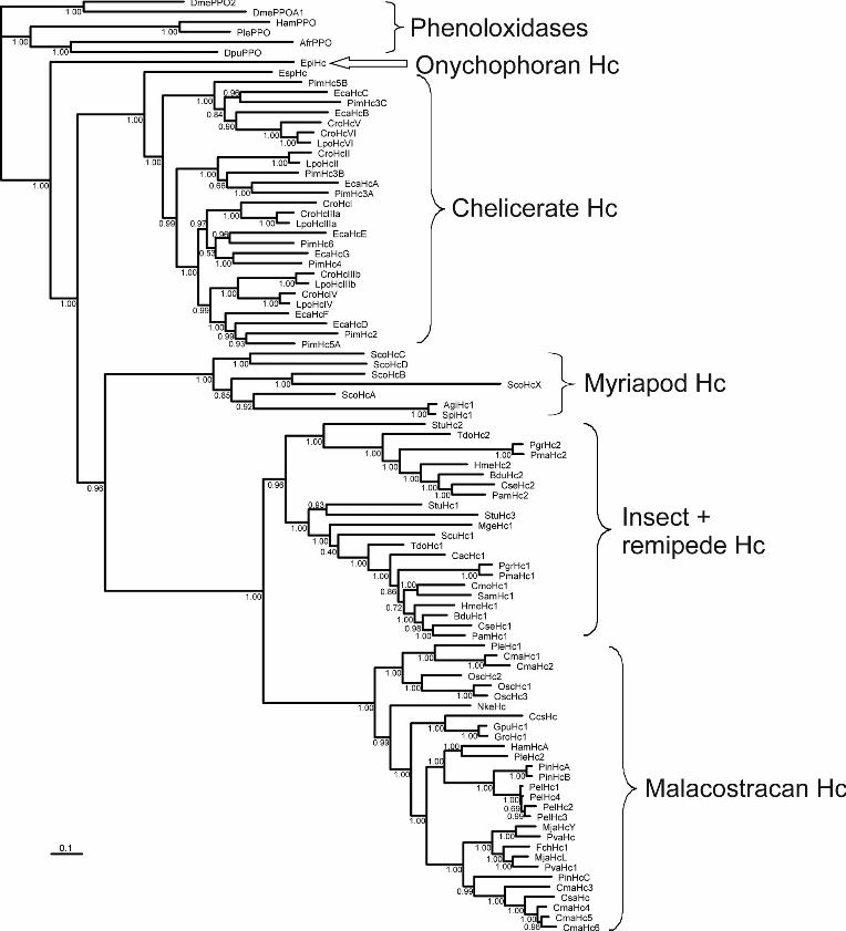
included in a multiple sequence alignment of a total of 86 other Hcs from all arthropod subphyla. Six
phenoloxidase sequences were added as outgroup. Bayesian phylogenetic analyses support the monophyly of
the myriapod (diplopod and chilopod) Hcs (posterior probability: 1.0; Fig 2). The A. gigas Hc groups with
the Spirostreptus sp. Hc. The two diplopod Hcs are a sister group of the HcA subunit of S. coleoptrata
(Kusche et.al., 2003), while the other four subunits of S. coleoptrata are basal. This demonstrates an early
divergence of distinct subunit types in the myriapods before the separation of Diplopoda and Chilopoda,
which occurred at least ~420 million years ago in the Silurian period (Wilson and Anderson, 2004). Thus the
formation of the oligohexameric Hc (probably a 36-mer) is an ancient event. The phylogeny of the Hc
subunits further supports a monophyly of the Mandibulata (Myriapoda + Crustacea; 0.96 support), but rejects
Myriochelata (Myriapoda + Chelicerata).
Oxygen binding
A. gigas whole hemolymph contains high levels of Hc, 159 mg/mL compared to centipedes (93 mg/ml in
Scutigera coleopteran) (Mangum et.al., 1985). The whole hemolymph (Fig. 3) exhibits a high O2 affinity
(P50 = 3.45 mmHg [0.46 kPa] at pH = 8.1 and 25ºC) compared to other crustacean Hcs studied under similar
temperature and pH conditions (P50 = 2.8 to 24 mmHg) (Morris and Bridges, 1994). The cooperativity
coefficients are low (~1.5 at pH <7.0 and pH >8.0) but increase to ~3.0 near pH 7.4 (Fig. 3), a pattern

The
Jou
rnal
of
Exp
erim
enta
l Bio
logy
– A
CC
EPT
ED
AU
TH
OR
MA
NU
SCR
IPT
commonly observed in other Hcs (Miller and Van Holde, 1974; Miller and Mangum, 1988). These O2
binding properties differ sharply from those of centipedal Scutigera coleoptrata Hc, despite similar
quaternary structures and subunit sequence homology in millipede and centipede Hcs (Fig. S1). The high O2
affinity and low cooperativity in A.gigas Hc predictably favor O2 binding at low O2 tensions, as expected to
prevail in myriapods with gas-impermeable exoskeletons and lacking capacities for tracheal ventilation
(Rajulu, 1970). This contrasts with the low affinity and high cooperativity observed in centipede Scutigera
coleoptrata Hc, which may be adaptive in enhancing O2 release in the highly active centipedes (Markl et.al.,
2009; Jaenicke et.al., 1999).
Interaction with allosteric cofactors
The O2 affinity of whole A. gigas hemolymph is not materially affected by pH nor by the addition of lactate
or Ca2+ at pH < 6.7 (Fig. 3A). However, at pH 7.0-8.0, a marked Bohr factor (φ = -0.73) is observed that is
further increased by effectors (φ = -0.77 and -0.85 respectively, in the presence of 10 mM (L)-lactate and 20
mM Ca2+). The marked Bohr effect implies that activity-induced acidification of tissues bathed by
the hemolymph will enhance unloading of O2 from the Hc. A similarly large Bohr effect (φ = -0.87) has been
observed in the house centipede, Scutigera coleoptrata (Mangum et.al., 1985).
In the intensively-studied tetrametric vertebrate Hbs the majority of the Bohr effect is attributable to
oxygenation-linked binding of protons to surface His residues that increase the proteins’ pK values (Lukin
and Ho, 2004; Berenbrink, 2006). The subunits of A. gigas Hc contain 34 histidine residues, including the six
copper-binding residues and four histidines in the interphase between hexamers (H152, H155, H443 and
H446) (Supplemental Fig. S1) and some of the free surface residues may undergo oxygenation-linked
proton binding under physiological conditions and transmit forces during allosteric interaction (Markl et.al.,
2009) contributing to the Bohr effect. In tarantula Eurypelma californicum Hc, salt bridges between
conserved surface histidine and glutamate residues located at particular intersubunit interfaces are considered
responsible for the observed Bohr effect (Sterner and Decker, 1994). However, the mechanism of O2 binding
and its modulation by allosteric effectors depends on interplay of many factors, including the coordination
geometry and redox potential of the copper. Studies on crustacean (Panulirus interruptus and Carcinus
aestuarii) Hc suggest that the Bohr effect of arthropod Hcs results from pH-dependent structural modulation
of the geometry of copper in the deoxygenated Hc molecules “leaving the question of identifying the role of
individual amino acids to future studies” (Hirota et.al., 2008).
Strikingly, the addition of 10 mM and 50 mM (L)-lactate to whole hemolymph slightly decreased O2
affinity in the pH range of 7 to 8 (Fig. 3), which contrasts sharply with the commonly encountered lactate-
induced increase in O2 affinity observed in crustacean Hcs, which would favor O2 binding under hypoxic
conditions (Truchot, 1980; Morris and Bridges, 1994; Bridges et.al., 1984; Paoli et.al., 2007; Hellmann et.al.,
2010).
The
Jou
rnal
of
Exp
erim
enta
l Bio
logy
– A
CC
EPT
ED
AU
TH
OR
MA
NU
SCR
IPT
The slight lactate effect observed in A. gigas supports the correlation between terrestrial life and
reduced lactate effects reported in crustaceans - and extends it to diplopods. Accordingly, lactate has no
effect on Hc-O2 affinity in the tropical land hermit crab, Coenobita clypeoatus (Morris and Bridges, 1986)
and the terrestrial, obligate air-breathing decapod, Birgus latro (Morris et.al., 1988). Compared to marine
crustaceans, Hc of the Christmas Island red landcrab, Gecarcoides natalis, even shows a reverse lactate
effect (O2 affinity falls with increasing (L)-lactate concentration) (Adamczewska and Morris, 1998) that
predictably favors O2 unloading in the tissues in terrestrial habitats where O2 loading is secured.
Addition of Ca2+ ions to whole hemolymph drastically increases Hc-O2 affinity in the pH range 7.0 -
8.0. The Ca2+ effect increases with pH (Fig. 3) suggesting that the O2-linked Ca2+-binding sites become
increasingly accessible and/or deprotonated with rising pH, as also observed in crustacean (Penaeus
monodon) Hc (Beltramini et.al., 2005).. In contrast, addition of Mg2+ to whole hemolymph does not affect
Hc-O2 affinity, indicating that the O2-linked cation binding sites are specific to Ca2+ ions or that potential O2-
linked Mg2+-binding sites are saturated in the whole hemolymph.
The pH of native hemolymph was 8.2 at 25ºC, which falls within the range measured in freshly
collected diplopod hemolymph (pH = 8.0 to 8.5) (Xylander, 2009). This implies (cf. Fig. 3) that variations in
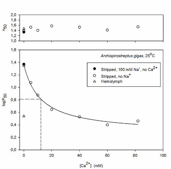
Ca2+ concentration exert a tangible effect on O2 affinity at physiological pH. The dose-response curve at pH
~7.96 for the relationship between [Ca2+] and P50 in stripped Hc (Fig. 4A), moreover, shows high sensitivity
of P50 to Ca2+ at low cation levels that characterize arthropod hemolymph (3.5 - 4.3 mM in arachnids) (Paul
et.al., 1994; Decker et.al., 1980). Fitting the logP50/[Ca2+] curve to a hyperbola (Fig. 4A) indicates that the
Ca2+ concentration required for half the maximum increase in logP50 - which reflects the apparent pKa of
ionizable groups involved in binding Ca2+ - is 12.3 mM. The slope of the double logarithmic plot (Fig. 4B)
of 0.55 ±0.05 (mean±S.E.) reflects binding of 0.5 to 0.6 Ca2+ ions per O2 molecule. In contrast to Ca2+, 100
mM Na+ - the major cation in extracellular fluids – had no detectable effect on O2 affinity of the stripped Hc
(Fig. 4A) – supporting the view that the cation binding site is highly specific for Ca2+.
The exoskeleton of arthropods contains up to 40% mineral salts, predominantly calcium carbonate,
that hardens the cuticle. Decreases in hemolymph pH associated with lactic acidoses under hypoxia may thus
be expected to mobilize calcium carbonate from exoskeleton stores, raising hemolymph Ca2+ and stabilizing
hemolymph pH. This scenario is analogous to that in turtles, whose shells are rich in calcium carbonate. In
the painted turtle, Chrysemys picta, increases in lactic acid levels (up to 200 mM) under anoxia are buffered
largely by carbonate, resulting in the accumulation of calcium and lactate in the extracellular fluid or
incorporated of calcium lactate into the shell (Jackson and Ultsch, 1982) (Jackson, 2004). Our finding that
Ca2+ increases Hc-O2 affinity thus indicates that Ca2+ released under hypoxia will increase Hc-O2 loading
under these conditions.
Extended Hill plot analysis

The
Jou
rnal
of
Exp
erim
enta
l Bio
logy
– A
CC
EPT
ED
AU
TH
OR
MA
NU
SCR
IPT
The parameters obtained by fitting the MWC model to the data (Table 1) reveal close agreement between Pm
and P50, and between nmax and n50 values, reflecting highly symmetrical O2 equilibrium curves, which permit
analysis of allosteric effects in terms of changes in P50 values. In the absence of added effectors the non-
exclusive binding coefficient c in the hemolymph (=KT/KR=0.14), indicates an approximately 7-fold higher
O2 affinity in the relaxed than in the tense state.
The q values (5.3-10.7) witness interaction between at least 11 O2 binding sites, indicating that
cooperativity is not confined within the hexameric subunits but extends between the individual hexamers.
Although the major structural elements are strictly conserved amongst arthropod Hcs (Markl and Decker,
1992; Van Holde and Miller, 1995; Burmester, 2001), identification of the salt bridges implicated in
interhexameric interactions is complicated by the large number of charged amino acid residues found in the
five types of inter-hexamer interfaces (Markl et.al., 2009). Given the large difference in cooperativity
coefficients of the sluggish millipede Spirostreptus and the swift house centipede Scutigera coleoptrata (n =
1.3 and ~10, respectively), comparison of diplopod and chilopod Hcs promises insight into structure-function
coupling.
Cooperativity between hexamers is accounted for by several salt bridges found hexamer interphases.
Two salt-bridges (Lys502 ↔ Asp575 and Asp567 ↔ Asn573) may act to connect the two 3× 6mer half-
molecules, and connections between adjacent hexamers may be stabilized by one salt-bridge (Glu395 ↔
Lys628) (Fig. S1) (Markl et.al., 2009). The observation that q values exceeding 6 were only encountered in
the hemolymph samples that had higher Hc concentration than the stripped samples (2.43 and 1.70 mM,
respectively – Table 1) is congruent with dissociation of Hc subunits at low concentration (Svedberg and
Heyroth, 1929). Analogously, the high q values found in the hemolymph at low pH and in the presence of
lactate (q = 10.7 and 8.08, respectively - Table 1) indicate that these conditions may favor the association of
the hexameric subunits. The lower q value in stripped Hc might, however, also be due to the loss of other
factors that are present in whole hemolymph, such as Ca2+, which is known to increase the stability of the
Hc quaternary structure (Van Holde and Miller, 1995). Indeed as shown (Table 1) addition of 20 mM Ca2+
increases q of the stripped Hc from 3 to 5 and n50 from 1.32 to 1.68 (Table 1). This aligns with structural
studies on diplopod Hc that show the presence of two opposing glutamate residue pairs (E410 and E411) that
are not compensated by positive charged residues, and might be responsible for the bridging of Ca2+ between
hexameric subunits (Markl et.al., 2009).
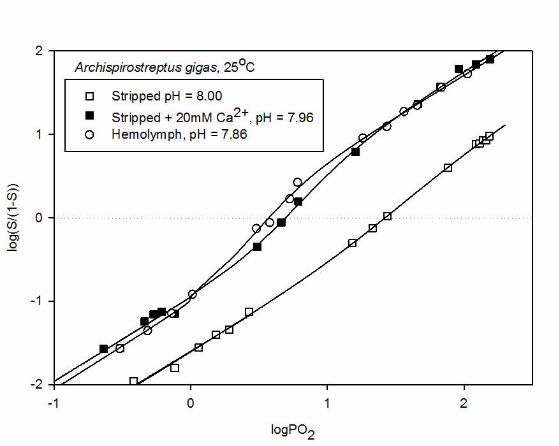
The slopes of unity seen at extremely low and high O2 saturations in extended Hill plots (Fig. 5)
reflect non-cooperative binding of the first and last O2 molecules bound by the Hc molecules. As evidenced
by the more two-fold increase in logL upon addition of lactate (Table 1), lactate increases P50 by modulating
the allosteric constant, L, without affecting KT and KR (Fig. 5A). Contrasting with this, the Bohr effect is
associated with a tangible decrease in KT as well as KR upon acidification (by >75% when hemolymph pH
falls from ~7.8 to~6.8) (Table 1; Fig. 5A). Analogously, the drastically lower Hc-O2 affinity in stripped Hc

The
Jou
rnal
of
Exp
erim
enta
l Bio
logy
– A
CC
EPT
ED
AU
TH
OR
MA
NU
SCR
IPT
compared to that in the whole hemolymph (P50 = ~26 and 4 mm Hg, respectively at pH ~7.9) results from
marked reductions in both KT and KR (Table 1; Fig. 5B). Strikingly, addition of 20 mM Ca2+ almost
completely annuls these effects resulting in left-shifting of the curve via increases in both KT and KR. The
almost exact superimposition of the plot of stripped Hc in the presence of 20 mM Ca2+ and of the whole
hemolymph indicates that Ca2+ is the main physiological regulator of O2 affinity in A. gigas Hc at constant
pH.
O2 equilibrium measurements were also carried out on stripped Hc in the presence and absence of 20
mM Ca2+ at pH 6.4. However, it was not possible to fit the MCW model to these data due to the low level of
cooperativity and almost identical KT and KR values, which suggest that the Hc molecules do not ‘switch’
between the T to the R states and that Ca2+ does not exert an affect at low pH.
The mechanisms of allosteric control in A.gigas Hc differ strikingly from those characterizing the
intensively-studied vertebrate Hbs, where the major effectors (organic phosphates, chloride ions and protons)
decrease Hb-O2 affinity by lowering KT, and other multimeric invertebrate O2-binding proteins (other Hcs
and extracellular Hbs). Thus in ~3000 kDa annelid (Arenicola marina, Perineries aibuhitensis and
Macrobdella decora) Hbs and ~1750 kDa gastropod (Biomphalaria glabrata) Hb (Weber, 1981; Tsuneshige
et.al., 1989; Weber et.al., 1995; Bugge and Weber, 1999) increased pH and divalent cation concentrations
increase O2 affinity predominantly by increasing KR. The A. gigas Hc mechanism is moreover at variance
with those encountered in other arthropod and in molluscan Hcs. Thus, as with multimeric Hbs, increased pH
and divalent cation (Ca2+) concentrations raise Hc-O2 affinity of gastropod Lymnea stagnalis Hc by
predominantly increasing KR (Dawson and Wood, 1982), whereas Mg2+ ions raise KR and lower KT of
Haliotis iris Hc (Behrens et.al., 2002). It also differs from the mechanisms observed in Crustacean Hcs,
where Mg2+ ions and increased pH increase O2 affinity by raising KR in Callianassa californiensis (Miller
and Van Holde, 1974) and (L)-lactate increases Hc-O2 affinity by increasing KT in Carcinus maenas and
Callinectes sapidus Hc (Weber et.al., 2008; Johnson et.al., 1988). Analyses of the allosteric interactions in
terms of a nesting model that reveal hierarchies of interactions based on known hierarchy of subunit structure
(Robert et.al., 1987; Decker et.al., 1988; Hellmann et.al., 2003; Menze et.al., 2005; Hellmann et.al., 2008)
will further elucidate the mechanisms regulating Hc-O2 in myriapods.
CONCLUSION
Our study of A. gigas Hc shows that millipede Hcs are a sister group to centipede HcA reflecting an early
divergence of Hc subunits, which is associated with distinct differentiation in functional properties, notably
in Hc-O2 affinity and its regulation by protons (pH) and Ca2+ and lactate concentrations. The O2 binding
properties of the Hc are compatible with a role of the circulating hemolymph in complementing the tracheal
system in supplying O2 to the respiring tissues in myriapods. The pattern of allosteric effects observed in A.
The
Jou
rnal
of
Exp
erim
enta
l Bio
logy
– A
CC
EPT
ED
AU
TH
OR
MA
NU
SCR
IPT
gigas Hc provides ideal basis for studies on the molecular basis and physiological significance of the variant
patterns of allosteric control mechanisms encountered in multimeric gas-binding proteins.
LIST OF SYMBOLS AND ABBREVIATIONS
A Absorbance
A´0 True absorbance at full deoxygenation
A´100 True absorbance at full oxygenation
A0 Apparent absorbance at full deoxygenation
A100 Apparent absorbance at full oxygenation
c non-exclusive binding coefficient KT/KR
Hb Hemoglobin
Hc Hemocyanin
KT T-state association equilibrium constant for O2 binding
KR R-state association equilibrium constant for O2 binding
L Allosteric constant ([T]/[R] at zero oxygenation
n50 Hill’s cooperativity coefficient at half saturation
nmax Maximal cooperativity coefficient
P50 Oxygen tension at half saturation
Pm Median oxygen tension
PO2 Partial pressure of oxygen
Q Number of interacting binding sites
S Apparent saturation
ΔG Free energy of cooperativity
Φ Bohr factor (Δlog P50/ΔpH).
ACKNOWLEDGEMENTS
We thank Anny Bang (Aarhus) and Christian Pick (Hamburg) for technical assistance and help with data handling. We thank Marcel Kwiatkowski (Hamburg) for the nanoLC-ESI-ion trap analysis.
The
Jou
rnal
of
Exp
erim
enta
l Bio
logy
– A
CC
EPT
ED
AU
TH
OR
MA
NU
SCR
IPT
FUNDING
The research was supported by grants from the Danish Council for Independent Research, Natural Sciences (10-084565), the German Research Foundation (DFG Bu 965/9) and the Faculty of Science and Technology of Aarhus University.

The
Jou
rnal
of
Exp
erim
enta
l Bio
logy
– A
CC
EPT
ED
AU
TH
OR
MA
NU
SCR
IPT
REFERENCES
Adamczewska, A. M. and Morris S. (1998). The functioning of the haemocyanin of the terrestrial
Christmas Island red crab Gecarcoidea natalis and roles for organic modulators. J. Exp. Biol. 201, 3233-
3244.
Behrens, J. W., Elias J. P., Taylor H. H. and Weber R. E. (2002). The archaeogastropod mollusc Haliotis iris:
tissue and blood metabolites and allosteric regulation of haemocyanin function. J. Exp. Biol. 205, 253-
263.
Beltramini, M., Colangelo N., Giomi F., Bubacco L., Di M. P., Hellmann N., Jaenicke E. and Decker H.
(2005). Quaternary structure and functional properties of Penaeus monodon hemocyanin. FEBS J.
272, 2060-2075.
Berenbrink, M. (2006). Evolution of vertebrate haemoglobins: Histidine side chains, specific buffer value
and Bohr effect. Respir. Physiol. Neurobiol. 154, 165-184.
Bonaventura, J. and Bonaventura C. (1980). Hemocyanins: Relationships in their structure, function and
assembly. Amer. Zool. 20, 7-17.
Bridges, C. R., Morris S. and Grieshaber M. K. (1984). Modulation of haemocyanin oxygen affinity in the
intertidal prawn Palaemon elegans (Rathke). Respir. Physiol. 57, 189-200.
Brouwer, M. and Serigstad B. (1989). Allosteric control in Limulus polyphemus hemocyanin: functional
relevance of interactions between hexamers. Biochemistry 28, 8819-8827.
Bugge, J. and Weber R. E. (1999). Oxygen binding and its allosteric control in hemoglobin of the pulmonate
snail, Biomphalaria glabrata. Am. J. Physiol. , Regul. Integr. Comp. Physiol. 276, R347-R356.
Burmester, T. (2001). Molecular evolution of the arthropod hemocyanin superfamily. Mol. Biol. Evol. 18,
184-195.
Burmester, T. (2002). Origin and evolution of arthropod hemocyanins and related proteins. J. Comp.
Physiol. [B] 172, 95-107.
Dawson, A. and Wood E. J. (1982). Equilibrium and kinetic studies of oxygen binding to the haemocyanin
from the freshwater snail Lymnaea stagnalis. Biochem. J. 207, 145-153.
Decker, H., Connelly P. R., Robert C. H. and Gill S. J. (1988). Nested allosteric interaction in tarantula
hemocyanin revealed through the binding of oxygen and carbon monoxide. Biochemistry 27, 6901-
6908.
Decker, H., Schmid R., Markl J. and Linzen B. (1980). Hemocyanin in spiders XII. Dissociation and
reassociation of Eurypelma hemocyanin. Hoppe-Seyler's Z. Physiol. Chem. 361, 1707-1717.
Fago, A., Bendixen E., Malte H. and Weber R. E. (1997). The anodic hemoglobin of Anguilla anguilla.
Molecular basis for allosteric effects in a Root-effect hemoglobin. J. Biol. Chem. 272, 15628-15635.
Hellmann, N., Paoli M., Giomi F. and Beltramini M. (2010). Unusual oxygen binding behavior of a 24-meric
crustacean hemocyanin. Arch. Biochem. Biophys 495, 112-121.
Hellmann, N., Weber R. E. and Decker H. (2003). Nested allosteric Interactions in extracellular hemoglobin
of the leech Macrobdella decora. J. Biol. Chem. 278, 44355-44360.
Hellmann N., Weber R. E. and Decker H. (2008). Linked Analysis of Large Cooperative, Allosteric Systems:
The Case of the Giant HBL Hemoglobins. In: Robert KP, ed. Methods in Enzymolog: Globins and Other
Nitric Oxide-Reactive Proteins, Part A, Academic Press, pp. 463-485.
Hirota, S., Kawahara T., Beltramini M., Di Muro P., Magliozzo R. S., Peisach J., Powers L. S., Tanaka N.,
Nagao S. and Bubacco L. (2008). Molecular basis of the Bohr effect in arthropod hemocyanin. J. Biol.
Chem. M803433200.
Huelsenbeck, J. P. and Ronquist F. (2001). MRBAYES: Bayesian inference of phylogenetic trees.
Bioinformatics 17, 754-755.
The
Jou
rnal
of
Exp
erim
enta
l Bio
logy
– A
CC
EPT
ED
AU
TH
OR
MA
NU
SCR
IPT
Jackson, D. C. and Ultsch G. R. (1982). Long-term submergence at 3°C of the turtle, Chrysemys picta bellii,
in normoxic and severely hypoxic water: II. Extracellular ionic responses to extreme lactic acidosis. J.
Exp. Biol. 96, 29-43.
Jackson, D. C. (2004). Surviving extreme lactic acidosis: the role of calcium lactate formation in the anoxic
turtle. Respir. Physiol. Neurobiol. 144, 173-178.
Jaenicke, E., Decker H., Gebauer W. A., Markl J. and Burmester T. (1999). Identification, structure, and
properties of hemocyanins from diplopod myriapoda. J. Biol. Chem. 274, 29071-29074.
Johnson, B. A., Bonaventura C. and Bonaventura J. (1988). Allostery in Callinectes sapidus hemocyanin:
cooperative oxygen binding and interactions with L-lactate, calcium, and protons. Biochemistry 27,
1995-2001.
Katoh, K., Kuma K., Toh H. and Miyata T. (2005). MAFFT version 5: improvement in accuracy of multiple
sequence alignment. Nucleic Acids Res 33, 511-518.
Keilin, D. and Wang Y. L. (1946). Haemoglobin of Gastrophilus larvae. Purification and properties.
Biochem. J. 40, 855-866.
Kusche, K. and Burmester T. (2001). Diplopod hemocyanin sequence and the phylogenetic position of the
Myriapoda. Mol. Biol. Evol. 18, 1566-1573.
Kusche, K., Hembach A., Hagner-Holler S., Gebauer W. and Burmester T. (2003). Complete subunit
sequences, structure and evolution of the 6 x 6-mer hemocyanin from the common house centipede,
Scutigera coleoptrata. Eur. J Biochem 270, 2860-2868.
Kusche, K., Ruhberg H. and Burmester T. (2002). A hemocyanin from the Onychophora and the emergence
of respiratory proteins. Proc. Natl. Acad. Sci. USA 99, 10545-10548.
Linzen, B., Soeter N. M., Riggs A. F., Schneider H. J., Schartau W., Moore M. D., Yokota E., Behrens P. Q.,
Nakashima H., Takagi T., et al . (1985). The structure of arthropod hemocyanins. Science 229, 519-
524.
Loewe, R. (1978). Hemocyanins in spiders. J. Comp. Physiol. A 128, 161-168.
Lukin, J. A. and Ho C. (2004). The structure--function relationship of hemoglobin in solution at atomic
resolution. Chem Rev. 104, 1219-1230.
Mangum, C. P., Scott J. L., Black R. E., Miller K. I. and Van Holde K. E. (1985). Centipedal hemocyanin: its
structure and its implications for arthropod phylogeny. Proc. Natl. Acad. Sci. U. S. A 82, 3721-3725.
Markl, J. and Decker H. (1992). Molecular structure of the arthropod hemocyanins. Adv. Comp. Environm.
Physiol. 13, 325-376.
Markl, J., Moeller A., Martin A. G., Rheinbay J., Gebauer W. and Depoix F. (2009). 10-A cryoEM structure
and molecular model of the Myriapod (Scutigera) 6x6mer hemocyanin: understanding a giant oxygen
transport protein. J. Mol. Biol. 392, 362-380.
Menze, M. A., Hellmann N., Decker H. and Grieshaber M. K. (2005). Allosteric Models for Multimeric
Proteins:ΓÇë Oxygen-Linked Effector Binding in HemocyaninΓÇá. Biochemistry 44, 10328-10338.
Meusemann, K., von Reumont B. M., Simon S., Roeding F., Strauss S., Kuck P., Ebersberger I., Walzl M.,
Pass G., Breuers S., et al . (2010). A phylogenomic approach to resolve the arthropod tree of life. Mol.
Biol. Evol. 27, 2451-2464.
Miller, K. and Van Holde K. E. (1974). Oxygen binding by Callianassa californiensis hemocyanin.
Biochemistry 13, 1668-1674.
Miller, K. I. and Mangum C. P. (1988). An investigation of the nature of Bohr, Root, and Haldane effects in
Octopus dofleini hemocyanin. J. Comp. Physiol. [B] 158, 547-552.
Monod, J., Wyman J. and Changeux J.-P. (1965). On the nature of allosteric transitions: a plausible model.
J. Mol. Biol. 12, 88-118.
Morris, S. and Bridges C. R. (1986). Oxygen binding by the hemocyanin of the terrestrial hermit crab
Coenobita clypeatus (Herbst) - The effect of physiological parameters in vitro. Physiol. Zool. 59, 606-
615.

The
Jou
rnal
of
Exp
erim
enta
l Bio
logy
– A
CC
EPT
ED
AU
TH
OR
MA
NU
SCR
IPT
Morris, S., Greenaway P. and McMahon B. R. (1988). Adaptations to a terrestrial existence by the robber
crab Birgus latro. I. an in vitro investigation of blood gas transport. J. Exp. Biol. 140, 477-491.
Morris, S. and Bridges C. R. (1994). Properties of Respiratory Pigments in Bimodal Breathing Animals: Air
and Water Breathing by Fish and Crustaceans. Am. Zool. 34, 216-228.
Nickerson, K. W. and Van Holde K. E. (1971). A comparison of molluscan and arthropod hemocyanin - I.
Circular dichroism and absorption spectra. Comp. Biochem. Physiol. [B] 39, 855-872.
O'Farrell, P. H. (1975). High resolution two-dimensional electrophoresis of proteins. J. Biol. Chem. 250,
4007-4021.
Paoli, M., Giomi F., Hellmann N., Jaenicke E., Decker H., Di M. P. and Beltramini M. (2007). The molecular
heterogeneity of hemocyanin: Structural and functional properties of the 4x6-meric protein of
Upogebia pusilla (Crustacea). Gene 398, 177-182.
Paul, R. J., Pfeffer-Seidl A., Efinger R., Pörtner H. O. and Storz H. (1994). Gas transport in the haemolymph
of arachnids. II. Carbon dioxide transport and acid-base balance. J. Exp. Biol. 188, 47-63.
Pechenik J. (2005) Biology of the Invertebrates. Boston, Massachusetts: McGraw-Hill, higher Education.
Perkins, D. N., Pappin D. J., Creasy D. M. and Cottrell J. S. (1999). Probability-based protein identification
by searching sequence databases using mass spectrometry data. Electrophoresis 20, 3551-3567.
Rajulu, G. S. (1969). Presence of haemocyanin in blood of a centipede Scutigera longicornis (chilopoda -
Myriopoda). Curr. Sci. 38, 168-169.
Rajulu, G. S. (1970). Tracheal pulsation in a marine centipede Mixophilus indicus. Curr. Sci. 39, 397-398.
Robert, C. H., Decker H., Richey B., Gill S. J. and Wyman J. (1987). Nesting: hierarchies of allosteric
interactions. Proc. Natl. Acad. Sci. U. S. A 84, 1891-1895.
Schmitz, A. and Harrison J. F. (2004). Hypoxic tolerance in air-breathing invertebrates. Respir. Physiol
Neurobiol. 141, 229-242.
Sterner, R. and Decker H. (1994). Inversion of the Bohr Effect upon oxygen-binding to 24-meric Tarantula
hemocyanin. Proc. Natl. Acad. Sci. USA 91, 4835-4839.
Svedberg, T. and Heyroth F. H. (1929). The molecular weight of the hemocyanin of Limulus polyphemus. J.
Am. Chem. Soc. 51, 539-550.
Terwilliger, N. B. (1998). Functional adaptations of oxygen-transport proteins. J. Exp. Biol. 201, 1085-
1098.
Truchot, J.-P. (1980). Lactate increases the oxygen affinity of crab hemocyanin. J. Exp. Zool. 214, 205-208.
Tsuneshige, A., Imai K., Hori H., Tyuma I. and Gotoh T. (1989). Spectrophotometric, electron
paramagnetic resonance and oxygen binding studies on the hemoglobin from the marine polychaete
Perinereis aibuhitensis (Grtibe): Comparative physiology of hemoglobin. J. Biochem. 106, 406-417.
Van Holde, K. E. and Miller K. I. (1995). Hemocyanins. Adv. Protein Chem. 47, 1-81.
Van Holde, K. E., Miller K. I. and Decker H. (2001). Hemocyanins and invertebrate evolution. J. Biol. Chem.
276, 15563-15566.
Weber, R. E. (1981). Cationic control of O2 affinity in lugworm erythrocruorin. Nature 292, 386-387.
Weber, R. E., Behrens J. W., Malte H. and Fago A. (2008). Thermodynamics of oxygenation-linked proton
and lactate binding govern the temperature sensitivity O2 binding in crustacean (Carcinus maenas)
haemocyanin. J. Exp. Biol. 211, 1057-1062.
Weber, R. E., Malte H., Braswell E. H., Oliver R. W. A., Green B. N., Sharma P. K., Kuchumov A. and
Vinogradov S. N. (1995). Mass spectrometric composition, molecular mass and oxygen binding of
Macrobdella decora hemoglobin and its tetramer and monomer subunits. J. Mol. Biol. 251, 703-720.
Wilson, H. M. and Anderson L. I. (2004). Morphology and taxonomy of paleozoic millipedes (Diplopoda:
Chilognatha: Archipolypoda) from Scotland. J. Paleontol. 78, 169-184.
Wyman, J. (1969). Possible allosteric effects in extended biological systems. J. Mol. Biol. 39, 523-538.
Xylander, W. E. R. (2009). Physico-chemical properties of haemolymph of Chilopoda and Diplopoda
(Myriapoda, Arthropoda): protein content, pH, osmolarity. Soil organisms 81, 431-439.

The
Jou
rnal
of
Exp
erim
enta
l Bio
logy
– A
CC
EPT
ED
AU
TH
OR
MA
NU
SCR
IPT
LEGENDS TO FIGURES
Fig. 1. Identification of A. gigas hemocyanin. Proteins of total hemolymph were separated on SDS-PAGE
and stained with Coomassie Brilliant Blue R-250 (A). Western blot analysis with specific anti-A. gigas Hc
antibodies identified two bands in the vicinity of 75 kDa (B). Hemolymph proteins were separated by two-
dimensional PAGE and the Hc subunits were detected by Western blotting (C). The molecular mass markers
are indicated.
Fig. 2. Bayesian phylogenetic analysis of the arthropod hemocyanins. The numbers at the branches represent
Bayesian posterior probabilities (right number). The bar represents 0.1 PAM distance. See Supplemental
Table S1 for the abbreviations.
Fig. 3. The pH dependence of O2 tensions (lower panel) and Hill’s cooperativity coefficients (upper panels)
at 50% O2-saturation (P50 and n50, respectively) of A. gigas hemolymph at 25°C in the absence of added
cofactors (filled circles) and the presence of 10 mM (L)-lactate (open circles), 20 mM Ca2+ (open triangles),
50 mM (L)-lactate (open squares), and 20 mM Mg2+ (open diamonds).
Fig. 4. Upper panel: Effects of Ca2+ concentration on P50 and n50 of stripped A. gigas Hc at 25oC and pH =
7.96 ±0.02 (mean±s.d.) in the absence of added Na+ (open circles) and the presence of 100 mM Na+ (solid
circles) compared to the P50 and n50 values of the whole hemolymph (open triangle). The dashed drop line
indicates the [Ca2+] where the O2 affinity increase induced by Ca2+ is half maximal. Hyperbolic regression,
logP50 = 1.36 – 1.10*[Ca2+]/(12.3 + [Ca2+]). Lower panel: Double logarithmic plot; linear regression: logP50
= 1.42 – (0.545*log[Ca2+]) + 1.42 (p = 0.0008).
Fig. 5. Upper panel: Extended Hill plots of A.gigas whole hemolymph measured at pH 6.85 (triangles) and
7.84 (open circles) and at pH 7.86 in the presence of 10 mM (L)-lactate (solid circles). Lower panel: Plots of
the stripped Hc at pH 8.00 in the absence (open squares) and at pH 7.96 in the presence (filled squares) of 20
mM Ca2+, compared to that of the hemolymph at pH 7.86 (open circles). The lines are MWC fits based on
true saturation values.

The
Jou
rnal
of
Exp
erim
enta
l Bio
logy
– A
CC
EPT
ED
AU
TH
OR
MA
NU
SCR
IPT
The
Jou
rnal
of
Exp
erim
enta
l Bio
logy
– A
CC
EPT
ED
AU
TH
OR
MA
NU
SCR
IPT

The
Jou
rnal
of
Exp
erim
enta
l Bio
logy
– A
CC
EPT
ED
AU
TH
OR
MA
NU
SCR
IPT
The
Jou
rnal
of
Exp
erim
enta
l Bio
logy
– A
CC
EPT
ED
AU
TH
OR
MA
NU
SCR
IPT

The
Jou
rnal
of
Exp
erim
enta
l Bio
logy
– A
CC
EPT
ED
AU
TH
OR
MA
NU
SCR
IPT
The
Jou
rnal
of
Exp
erim
enta
l Bio
logy
– A
CC
EPT
ED
AU
TH
OR
MA
NU
SCR
IPT

The
Jou
rnal
of
Exp
erim
enta
l Bio
logy
– A
CC
EPT
ED
AU
TH
OR
MA
NU
SCR
IPT

The
Jou
rnal
of
Exp
erim
enta
l Bio
logy
– A
CC
EPT
ED
AU
TH
OR
MA
NU
SCR
IPT
Table 1: The derived MWC parameters for O2 equilibria at 25oC in the presence and absence of added (L)-lactate and Ca2+, including the median O2 tension (Pm), P50, n50,
the maximal slope of the extended Hill plot (nmax), oxygen dissociation constants for the tense state (KT) and for the relaxed state (KR), c=KR/KT, the free energy of
cooperativity (ΔG), the number of interacting O2 binding sites, q, and the allosteric constant (L).
T, oC
pH
[(L)-
lactate], [Ca2+], Pm, P50,
n50 nmax KT, KR,
c ΔG,
q logL
mM mM mmHg mmHg logKT logKR mmHg-1 mmHg-1 kJ/mol
Hemolymph 25 7.841 0 0 4.00 3.76 1.77 1.82 -1.140±0.35 -0.288±0.057 0.0724 0.516 0.140 4.54 5.30±3.2 1.66±0.87
Hemolymph 25 7.857 10 0 4.68 5.02 2.21 2.25 -0.953±0.071 -0.306±0.11 0.111 0.495 0.225 3.69 10.7±6.2 3.89±1.8
Hemolymph 25 6.850 0 0 26.6 27.8 2.13 2.24 -1.74±0.026 -0.983±0.21 0.0182 0.104 0.175 4.28 8.08±3.8 3.58±0.45
Stripped Hc 25 7.995 0 0 25.6 24.7 1.32 -* -1.63±0.88 -0.714±17 0.0233 0.193 0.121 2.81 3.01±24 2.19±36
Stripped Hc 25 7.960 0 20 4.67 5.02 1.68 1.69 -0.973±0.095 -0.167±0.37 0.106 0.681 0.156 4.18 5.00±4.1 2.52±0.74
* Could not be determined in the data fitting.


![[45 ] THE QUANTITATIVE NUTRITIONAL …jeb.biologists.org/content/jexbio/33/1/45.full.pdf · Quantitative nutritional requirements of Drosophila melanogaster 47 spores, and the fluctuations](https://static.fdocuments.net/doc/165x107/5ac1f6ec7f8b9a4e7c8db233/45-the-quantitative-nutritional-jeb-nutritional-requirements-of-drosophila.jpg)