Classical conditioning of activities of salivary neurones...
14
766 Introduction Pavlov reported classical conditioning of salivation in dogs in a congress held in 1903 (Pavlov, 1927). In studying the mechanisms of digestion, Pavlov discovered that when a bell was regularly sounded just before feeding, the sound of the bell would eventually trigger salivation. He also showed lesions in several regions of the cerebral cortex affect classical conditioning of salivation (Grimsley and Windholz, 2000). Later studies on lesions and electrical stimulations of various brain regions in dogs and rats have suggested that many regions of the brain, including the orbital cortex, caudate nucleus, hypothalamus and amygdala, are involved in classical conditioning of salivation (Lagowska and Fonberg, 1975; Danilova, 1981; Danilova, 1983; Matsuo and Kusano, 1984). However, the cellular mechanisms underlying classical conditioning of salivation remain obscure because of the complexity of information processing in the mammalian brain (Kandel et al., 2000). It has been shown that classical conditioning by repeated pairing of a conditioning stimulus (CS), such as the sound of a bell, and an unconditioned stimulus (US), such as food, is very common among many vertebrates (Passe and Walker, 1985) and invertebrates (Menzel, 1999; Lechner et al., 2000). However, as far as we know, classical conditioning of salivation has so far been reported only in mammals. Since secretion of saliva to aid swallowing and digestion is a basic physiological function found in many animals, including flatworms (Orido et al., 1998) and nematodes (Zunke, 1990), the following question arises: is classical conditioning of salivation specific to mammals that are equipped with elaborated autonomous nervous systems? The control of salivary secretion has been the subject of detailed study in insects such as cockroaches and locusts (Ali, 1997). In cockroaches, salivation is regulated by the salivary duct nerve (SDN) (Whitehead, 1971; Rietdorf et al., 2003). The SDN consists of two neurones with large-diameter (3-4·m) axons (salivary neurones 1 and 2; SN1 and SN2) and several neurones with small-diameter (~1·m) axons (Whitehead, 1971), the cell bodies of the former neurones being located in the suboesophageal ganglion (SOG) (Gifford et al., 1991; Ali, 1997); the latter neurones have been reported to belong to the stomatogastric nervous system (Davis, 1985; Ali, 1997). Immunohistochemical studies suggest that SN1 is dopaminergic (Elia et al., 1994) and that small-diameter neurones are serotonergic (Davis, 1985), and in vitro application of dopamine and serotonin to salivary glands induces secretion of protein-free saliva and protein-rich saliva, respectively (Just and Walz, 1996). The neurotransmitter of SN2 has not yet been determined. In the locust, salivary neurones exhibit activity during feeding (Baines et al., 1989; Secretion of saliva to aid swallowing and digestion is a basic physiological function found in many vertebrates and invertebrates. For mammals, classical conditioning of salivation in dogs was reported by Pavlov a century ago. However, conditioning of salivation or of related neural activities in non-mammalian species has not been reported. In many species of insects, salivation is regulated by salivary neurones. In this study, we found that salivary neurones of the cockroach Periplaneta americana exhibited a strong response to sucrose solution applied to the mouth and a weak response to odours applied to an antenna, and we studied the effect of conditioning on the activities of salivary neurones. After three sets of differential conditioning trials in which an odour was presented just before the presentation of sucrose solution and the other odour was presented alone, the response of salivary neurones to sucrose-associated odour significantly increased but that to the odour presented alone was unchanged. Backward pairing trials in which an odour was presented after the presentation of sucrose solution were not effective in achieving conditioning. Our study of the change in the level of saliva secretion in response to electrical stimulation of salivary neurones suggested that the magnitude of increase in odour response of salivary neurones by conditioning is sufficient to lead to an increased level of salivation. This study suggests classical conditioning of salivation in an insect. Key words: learning, memory, olfaction, taste, salivary neurones, insect. Summary The Journal of Experimental Biology 209, 766-779 Published by The Company of Biologists 2006 doi:10.1242/jeb.02049 Classical conditioning of activities of salivary neurones in the cockroach Hidehiro Watanabe and Makoto Mizunami* Graduate School of Life Sciences, Tohoku University, Katahira 2-1-1, Sendai 980-8577, Japan *Author for correspondence (e-mail: [email protected]) Accepted 20 December 2005 THE JOURNAL OF EXPERIMENTAL BIOLOGY
Transcript of Classical conditioning of activities of salivary neurones...
766
IntroductionPavlov reported classical conditioning of salivation in dogs
in a congress held in 1903 (Pavlov, 1927). In studying themechanisms of digestion, Pavlov discovered that when a bellwas regularly sounded just before feeding, the sound of the bellwould eventually trigger salivation. He also showed lesions inseveral regions of the cerebral cortex affect classicalconditioning of salivation (Grimsley and Windholz, 2000).Later studies on lesions and electrical stimulations of variousbrain regions in dogs and rats have suggested that manyregions of the brain, including the orbital cortex, caudatenucleus, hypothalamus and amygdala, are involved in classicalconditioning of salivation (Lagowska and Fonberg, 1975;Danilova, 1981; Danilova, 1983; Matsuo and Kusano, 1984).However, the cellular mechanisms underlying classicalconditioning of salivation remain obscure because of thecomplexity of information processing in the mammalian brain(Kandel et al., 2000).
It has been shown that classical conditioning by repeatedpairing of a conditioning stimulus (CS), such as the sound ofa bell, and an unconditioned stimulus (US), such as food, isvery common among many vertebrates (Passe and Walker,1985) and invertebrates (Menzel, 1999; Lechner et al., 2000).However, as far as we know, classical conditioning ofsalivation has so far been reported only in mammals. Since
secretion of saliva to aid swallowing and digestion is a basicphysiological function found in many animals, includingflatworms (Orido et al., 1998) and nematodes (Zunke, 1990),the following question arises: is classical conditioning ofsalivation specific to mammals that are equipped withelaborated autonomous nervous systems?
The control of salivary secretion has been the subject ofdetailed study in insects such as cockroaches and locusts (Ali,1997). In cockroaches, salivation is regulated by the salivaryduct nerve (SDN) (Whitehead, 1971; Rietdorf et al., 2003). TheSDN consists of two neurones with large-diameter (3-4·�m)axons (salivary neurones 1 and 2; SN1 and SN2) and severalneurones with small-diameter (~1·�m) axons (Whitehead,1971), the cell bodies of the former neurones being located inthe suboesophageal ganglion (SOG) (Gifford et al., 1991; Ali,1997); the latter neurones have been reported to belong to thestomatogastric nervous system (Davis, 1985; Ali, 1997).
Immunohistochemical studies suggest that SN1 isdopaminergic (Elia et al., 1994) and that small-diameterneurones are serotonergic (Davis, 1985), and in vitroapplication of dopamine and serotonin to salivary glandsinduces secretion of protein-free saliva and protein-rich saliva,respectively (Just and Walz, 1996). The neurotransmitter ofSN2 has not yet been determined. In the locust, salivaryneurones exhibit activity during feeding (Baines et al., 1989;
Secretion of saliva to aid swallowing and digestion is abasic physiological function found in many vertebratesand invertebrates. For mammals, classical conditioning ofsalivation in dogs was reported by Pavlov a century ago.However, conditioning of salivation or of related neuralactivities in non-mammalian species has not beenreported. In many species of insects, salivation is regulatedby salivary neurones. In this study, we found that salivaryneurones of the cockroach Periplaneta americanaexhibited a strong response to sucrose solution applied tothe mouth and a weak response to odours applied to anantenna, and we studied the effect of conditioning on theactivities of salivary neurones. After three sets ofdifferential conditioning trials in which an odour waspresented just before the presentation of sucrose solution
and the other odour was presented alone, the response ofsalivary neurones to sucrose-associated odour significantlyincreased but that to the odour presented alone wasunchanged. Backward pairing trials in which an odourwas presented after the presentation of sucrose solutionwere not effective in achieving conditioning. Our study ofthe change in the level of saliva secretion in response toelectrical stimulation of salivary neurones suggested thatthe magnitude of increase in odour response of salivaryneurones by conditioning is sufficient to lead to anincreased level of salivation. This study suggests classicalconditioning of salivation in an insect.
Key words: learning, memory, olfaction, taste, salivary neurones,insect.
Summary
The Journal of Experimental Biology 209, 766-779Published by The Company of Biologists 2006doi:10.1242/jeb.02049
Classical conditioning of activities of salivary neurones in the cockroach
Hidehiro Watanabe and Makoto Mizunami*Graduate School of Life Sciences, Tohoku University, Katahira 2-1-1, Sendai 980-8577, Japan
*Author for correspondence (e-mail: [email protected])
Accepted 20 December 2005
THE JOURNAL OF EXPERIMENTAL BIOLOGY
767Conditioning of salivary neurone activity
Schachtner and Bräunig, 1993) that is modulated by activity ofthe mouthpart motor pattern generator (Rast and Bräunig,2001). However, responses of salivary neurones to food-associated sensory stimuli, such as taste or olfactory stimuli,have not been studied.
Cockroaches can be trained to associate olfactory CSs withgustatory USs by an operant (Sakura and Mizunami, 2001;Sakura et al., 2002) or a classical conditioning procedure(Watanabe et al., 2003). The latter procedure is effective forboth freely moving and restrained cockroaches. Here we reportthat responses of salivary neurones to an odour significantlyincreased after repeated pairing of the odour with sucrosereward. Moreover, we suggest that the observed increase inodour response of salivary neurones after conditioning(5–10·Hz) is sufficient to lead to an increased level of salivasecretion. Our results provide a unique opportunity to studycellular mechanisms of conditioning of activities of salivaryneurones in animals whose central nervous systems areaccessible to detailed electrophysiological analysis.
Materials and methodsInsects
Adult male cockroaches, Periplaneta americana L., wereobtained from a laboratory colony maintained under a12·h:12·h light:dark cycle at 26–28°C. One week before thestart of the experiment, a group of 10–20 cockroaches wasplaced in a chamber. The wall of the chamber was smearedwith liquid paraffin to prevent the cockroaches from escaping,and the floor was covered with black cardboard. There was awooden refuge and two small cups, one supplying water adlibitum and the other supplying sugar-free yeast extract, whichenhanced the motivation of the cockroaches to take up sucrose.
Metal fillings of salivary neurones
Backfills and forwardfills of the SDN were made for eachof 20 animals. Each animal was anaesthetized with ice for1–2·h. After removal of its legs and wings, it was pinnedventral-side-up on a wax-coated dish and the cuticle of theventral part of the neck was removed. One SDN was cut andits proximal or distal cut-stump was inserted into a plastic tubefilled with a solution containing 0.16·mol·l–1 NiCl2 and0.04·mol·l–1 CoCl2 (Okada et al., 2003). The preparations werekept in a moist chamber at 4°C for 4 days.
After backfilling, the ventral cuticle of the head was removedto expose the SOG. After forwardfilling, the ventral cuticle ofthe thorax was removed to expose the salivary gland. Then oneor two droplets of rubeanic acid were applied onto the SOG orthe salivary gland for 3–5·min to precipitate the metals (Okadaet al., 2003). The SOG or the salivary gland was rinsed manytimes with cockroach saline (Yamasaki and Narahashi, 1959),dissected out, fixed in 3–4% paraformaldehyde in cockroachsaline for 30–60·min, dehydrated in a graded series of ethanol,cleared in methyl salicylate, and observed as whole mountsunder a light microscope. After observation of the specimens,they were rehydrated in an ascending series of ethanol. Then
the specimens were intensified with silver (Bacon and Altman,1977) and observed as whole mounts. Digital images weretaken using a digital camera (Camedia C-3040 Zoom; Olympus,Tokyo, Japan) and were processed using Adobe Photoshop 7.0.
Extracellular recordings of activities of salivary neurones
We used two preparations for extracellular recordings from theSDN. In one preparation (called the semi-intact preparation), ananimal was anaesthetized with ice for 0.5–1·h, its wings wereremoved, and it was restrained on a wax-coated dish ventral-side-up with thin plastic plates at the neck and between the thorax andabdomen. Then the legs and antenna were fixed with low-meltingwax and staples, respectively. In another preparation (called thehighly dissected preparation), the oesophagus was punctured toprevent its expansion during chronic recording, and the neck andthe cerci were fixed with low-melting point wax. The advantageof the latter preparation is that the movement of the head and theoesophagus and also the resulting artefact in the recording wereless frequent and this facilitated reliable segregation of unitactivities. In both preparations, the restrained animal could moveits mouthparts freely.
Semi-intact preparations and highly dissected preparationswere kept in a moist chamber at 26–28°C overnight and for1–2·h, respectively, and then a small incision was made in theventrolateral sclerite of the neck to expose the salivary duct.Since the SDN runs along the surface of the salivary duct, oneSDN, as well as the salivary duct, was hooked on a pair oftungsten electrodes (Fig.·1A). To prevent drying of the SDN,the salivary duct was covered with a mixture of white Vaselineand liquid paraffin saturated with cockroach saline.
The activity of the SDN was amplified with a differentialAC amplifier (DAM80, World Precision Instruments, Sarasota,FL, USA) and displayed on an oscilloscope and a digitalrecorder (Omniace, NEC, Tokyo, Japan). Data were stored onDAT tapes (PC208AX, Sony, Tokyo, Japan). Activities ofindividual units were segregated out using a windowdiscriminator equipped with a spike counter (MET1100, NihonKohden, Tokyo, Japan).
Effects of surgical ablation of salivary neurone 1 or 2 onactivities of one SDN
To determine which of the units of the SDN reflect activitiesof salivary neurones 1 and 2 (SN1 and SN2), the SOG wasexposed by removing ventral parts of the neck and labia inhighly-dissected preparations, and the part of the SOG wherethe cell body of SN1 or SN2 was located was surgically ablatedusing a fine needle or scissors, and the resulting change inactivities of one SDN was studied. When one of the units ofthe SDN was removed by surgery, the SDN was cut andbackfilled with metal to confirm which of the neurons, SN1 orSN2, had been ablated.
Taste and olfactory stimulation
The continuous airflow system used to deliver odourstimulation to an antenna of the immobilized animal wasdescribed previously (Nishino et al., 2003). Briefly, air, which
THE JOURNAL OF EXPERIMENTAL BIOLOGY
768
was passed through a small chamber containing a piece of filterpaper soaked with 40·�l of an extract of vanilla or peppermint,could be delivered without changing the flow rate by operatinga solenoid valve. The air around the antenna was continuouslysucked out of the room through a vacuum system. Forgustatory stimulation, the mouth was gently touched with awooden stick soaked with 10% sucrose solution, 20% sodium
H. Watanabe and M. Mizunami
chloride solution or distilled water. To avoid sensoryadaptation, odour or taste stimuli were applied with an intervalof >30·s.
Classical conditioning procedures
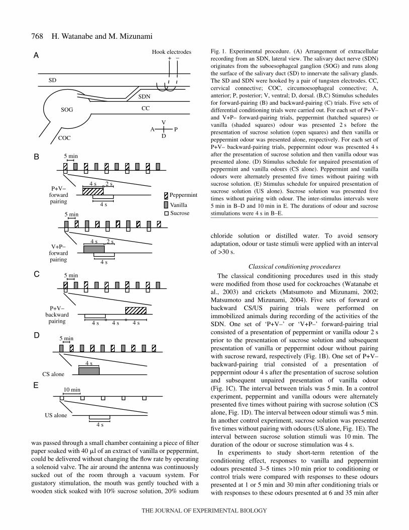
The classical conditioning procedures used in this studywere modified from those used for cockroaches (Watanabe etal., 2003) and crickets (Matsumoto and Mizunami, 2002;Matsumoto and Mizunami, 2004). Five sets of forward orbackward CS/US pairing trials were performed onimmobilized animals during recording of the activities of theSDN. One set of ‘P+V–’ or ‘V+P–’ forward-pairing trialconsisted of a presentation of peppermint or vanilla odour 2·sprior to the presentation of sucrose solution and subsequentpresentation of vanilla or peppermint odour without pairingwith sucrose reward, respectively (Fig.·1B). One set of P+V–backward-pairing trial consisted of a presentation ofpeppermint odour 4·s after the presentation of sucrose solutionand subsequent unpaired presentation of vanilla odour(Fig.·1C). The interval between trials was 5·min. In a controlexperiment, peppermint and vanilla odours were alternatelypresented five times without pairing with sucrose solution (CSalone, Fig.·1D). The interval between odour stimuli was 5·min.In another control experiment, sucrose solution was presentedfive times without pairing with odours (US alone, Fig.·1E). Theinterval between sucrose solution stimuli was 10·min. Theduration of the odour or sucrose stimulation was 4·s.
In experiments to study short-term retention of theconditioning effect, responses to vanilla and peppermintodours presented 3–5 times >10·min prior to conditioning orcontrol trials were compared with responses to these odourspresented at 1 or 5·min and 30·min after conditioning trials orwith responses to these odours presented at 6 and 35·min after
+ −
SD
SOG
COC
CC
SDN
Hook electrodes
AV
DP
Peppermint
VanillaSucrose
4 s 4 s4 s
10 min
4 s
US alone
5 min
5 min
4 s
4 s
2 s
5 min
4 s
4 s
2 s
P+V−forwardpairing
V+P−forwardpairing
P+V−backward
pairing
A
B
C
D 5 min
4 s
CS alone
E
Fig.·1. Experimental procedure. (A) Arrangement of extracellularrecording from an SDN, lateral view. The salivary duct nerve (SDN)originates from the suboesophageal ganglion (SOG) and runs alongthe surface of the salivary duct (SD) to innervate the salivary glands.The SD and SDN were hooked by a pair of tungsten electrodes. CC,cervical connective; COC, circumoesophageal connective; A,anterior; P, posterior; V, ventral; D, dorsal. (B,C) Stimulus schedulesfor forward-pairing (B) and backward-pairing (C) trials. Five sets ofdifferential conditioning trials were carried out. For each set of P+V–and V+P– forward-pairing trials, peppermint (hatched squares) orvanilla (shaded squares) odour was presented 2·s before thepresentation of sucrose solution (open squares) and then vanilla orpeppermint odour was presented alone, respectively. For each set ofP+V– backward-pairing trials, peppermint odour was presented 4·safter the presentation of sucrose solution and then vanilla odour waspresented alone. (D) Stimulus schedule for unpaired presentation ofpeppermint and vanilla odours (CS alone). Peppermint and vanillaodours were alternately presented five times without pairing withsucrose solution. (E) Stimulus schedule for unpaired presentation ofsucrose solution (US alone). Sucrose solution was presented fivetimes without pairing with odour. The inter-stimulus intervals were5·min in B–D and 10·min in E. The durations of odour and sucrosestimulations were 4·s in B–E.
THE JOURNAL OF EXPERIMENTAL BIOLOGY
769Conditioning of salivary neurone activity
control trials (presentation of US alone). The duration of thestimulation was 2·s and the interval between stimulations was>10·s. The measurement was initiated >15·min aftercompleting the set-up of electrophysiological recording tostabilize the preparation.
In an experiment to study 1-day retention of the conditioningeffect, a group of immobilized cockroaches was subjected toP+V– forward- or P+V– backward-pairing trials. Cockroacheswere kept in a moist chamber at 26–28°C for 1 day, and thenthe ventral cuticle of the neck was removed and activities ofthe salivary duct nerve were recorded to study their responsesto peppermint or vanilla odour.
Measurements of salivation in response to electricalstimulation of the SDN
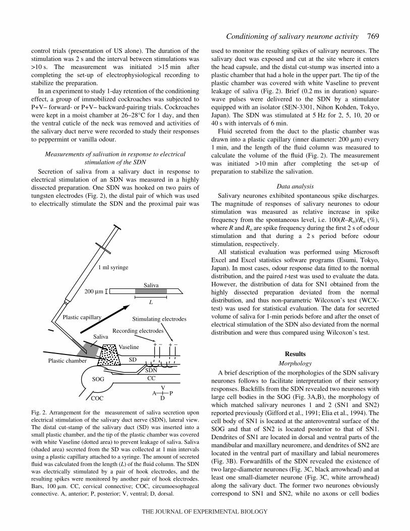
Secretion of saliva from a salivary duct in response toelectrical stimulation of an SDN was measured in a highlydissected preparation. One SDN was hooked on two pairs oftungsten electrodes (Fig.·2), the distal pair of which was usedto electrically stimulate the SDN and the proximal pair was
used to monitor the resulting spikes of salivary neurones. Thesalivary duct was exposed and cut at the site where it entersthe head capsule, and the distal cut-stump was inserted into aplastic chamber that had a hole in the upper part. The tip of theplastic chamber was covered with white Vaseline to preventleakage of saliva (Fig.·2). Brief (0.2·ms in duration) square-wave pulses were delivered to the SDN by a stimulatorequipped with an isolator (SEN-3301, Nihon Kohden, Tokyo,Japan). The SDN was stimulated at 5·Hz for 2, 5, 10, 20 or40·s with intervals of 6·min.
Fluid secreted from the duct to the plastic chamber wasdrawn into a plastic capillary (inner diameter: 200·�m) every1·min, and the length of the fluid column was measured tocalculate the volume of the fluid (Fig.·2). The measurementwas initiated >10·min after completing the set-up ofpreparation to stabilize the salivation.
Data analysis
Salivary neurones exhibited spontaneous spike discharges.The magnitude of responses of salivary neurones to odourstimulation was measured as relative increase in spikefrequency from the spontaneous level, i.e. 100(R–Ro)/Ro (%),where R and Ro are spike frequency during the first 2·s of odourstimulation and that during a 2·s period before odourstimulation, respectively.
All statistical evaluation was performed using MicrosoftExcel and Excel statistics software programs (Esumi, Tokyo,Japan). In most cases, odour response data fitted to the normaldistribution, and the paired t-test was used to evaluate the data.However, the distribution of data for SN1 obtained from thehighly dissected preparation deviated from the normaldistribution, and thus non-parametric Wilcoxon’s test (WCX-test) was used for statistical evaluation. The data for secretedvolume of saliva for 1-min periods before and after the onset ofelectrical stimulation of the SDN also deviated from the normaldistribution and were thus compared using Wilcoxon’s test.
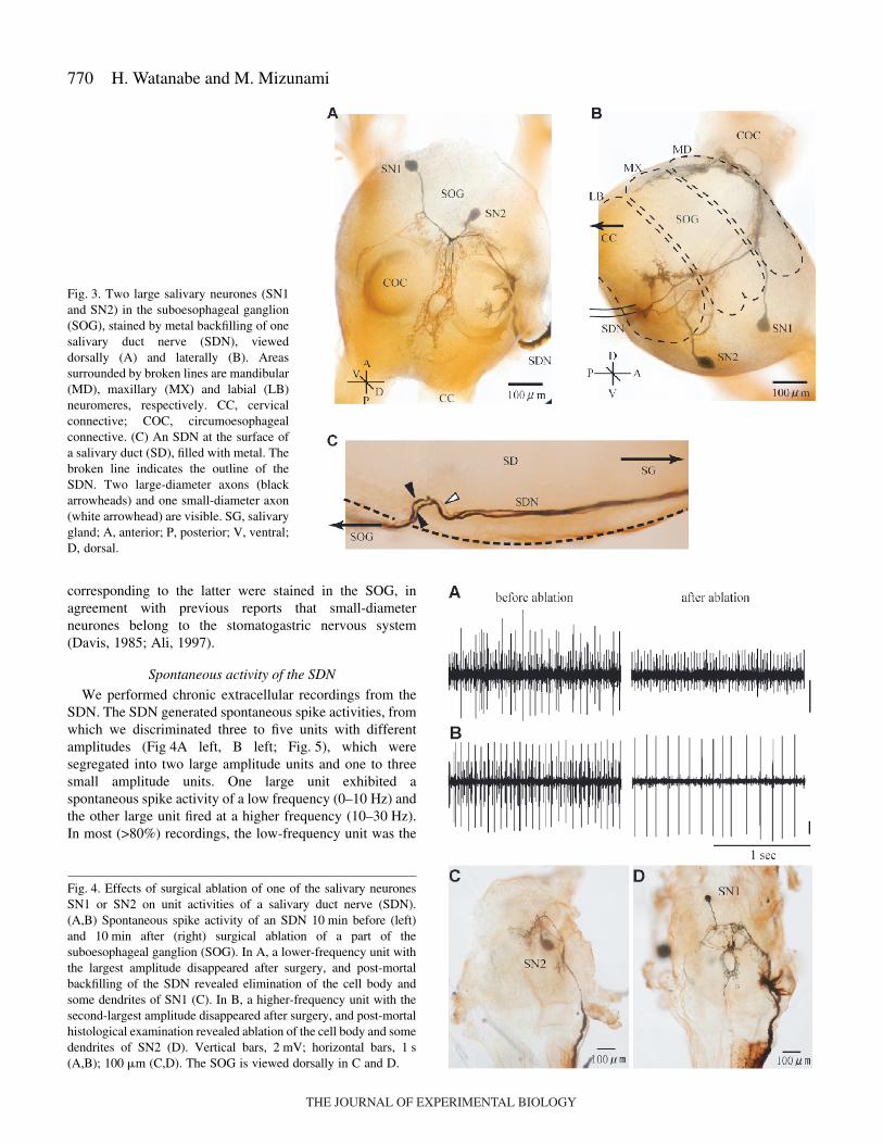
ResultsMorphology
A brief description of the morphologies of the SDN salivaryneurones follows to facilitate interpretation of their sensoryresponses. Backfills from the SDN revealed two neurones withlarge cell bodies in the SOG (Fig.·3A,B), the morphology ofwhich matched salivary neurones 1 and 2 (SN1 and SN2)reported previously (Gifford et al., 1991; Elia et al., 1994). Thecell body of SN1 is located at the anteroventral surface of theSOG and that of SN2 is located posterior to that of SN1.Dendrites of SN1 are located in dorsal and ventral parts of themandibular and maxillary neuromere, and dendrites of SN2 arelocated in the ventral part of maxillary and labial neuromeres(Fig.·3B). Forwardfills of the SDN revealed the existence oftwo large-diameter neurones (Fig.·3C, black arrowhead) and atleast one small-diameter neurone (Fig.·3C, white arrowhead)along the salivary duct. The former two neurones obviouslycorrespond to SN1 and SN2, while no axons or cell bodies
Fig.·2. Arrangement for the measurement of saliva secretion uponelectrical stimulation of the salivary duct nerve (SDN), lateral view.The distal cut-stamp of the salivary duct (SD) was inserted into asmall plastic chamber, and the tip of the plastic chamber was coveredwith white Vaseline (dotted area) to prevent leakage of saliva. Saliva(shaded area) secreted from the SD was collected at 1·min intervalsusing a plastic capillary attached to a syringe. The amount of secretedfluid was calculated from the length (L) of the fluid column. The SDNwas electrically stimulated by a pair of hook electrodes, and theresulting spikes were monitored by another pair of hook electrodes.Bars, 100·�m. CC, cervical connective; COC, circumoesophagealconnective. A, anterior; P, posterior; V, ventral; D, dorsal.
+ −
SOG
COC
CCSDN
AV
DP
Vaseline
SD
Saliva
Plastic capillary
1 ml syringe
200 μmSaliva
+ −
Stimulating electrodes
Recording electrodes
L
Plastic chamber
THE JOURNAL OF EXPERIMENTAL BIOLOGY
770
corresponding to the latter were stained in the SOG, inagreement with previous reports that small-diameterneurones belong to the stomatogastric nervous system(Davis, 1985; Ali, 1997).
Spontaneous activity of the SDN
We performed chronic extracellular recordings from theSDN. The SDN generated spontaneous spike activities, fromwhich we discriminated three to five units with differentamplitudes (Fig·4A left, B left; Fig.·5), which weresegregated into two large amplitude units and one to threesmall amplitude units. One large unit exhibited aspontaneous spike activity of a low frequency (0–10·Hz) andthe other large unit fired at a higher frequency (10–30·Hz).In most (>80%) recordings, the low-frequency unit was the
H. Watanabe and M. Mizunami
Fig.·3. Two large salivary neurones (SN1and SN2) in the suboesophageal ganglion(SOG), stained by metal backfilling of onesalivary duct nerve (SDN), vieweddorsally (A) and laterally (B). Areassurrounded by broken lines are mandibular(MD), maxillary (MX) and labial (LB)neuromeres, respectively. CC, cervicalconnective; COC, circumoesophagealconnective. (C) An SDN at the surface ofa salivary duct (SD), filled with metal. Thebroken line indicates the outline of theSDN. Two large-diameter axons (blackarrowheads) and one small-diameter axon(white arrowhead) are visible. SG, salivarygland; A, anterior; P, posterior; V, ventral;D, dorsal.
Fig.·4. Effects of surgical ablation of one of the salivary neuronesSN1 or SN2 on unit activities of a salivary duct nerve (SDN).(A,B) Spontaneous spike activity of an SDN 10·min before (left)and 10·min after (right) surgical ablation of a part of thesuboesophageal ganglion (SOG). In A, a lower-frequency unit withthe largest amplitude disappeared after surgery, and post-mortalbackfilling of the SDN revealed elimination of the cell body andsome dendrites of SN1 (C). In B, a higher-frequency unit with thesecond-largest amplitude disappeared after surgery, and post-mortalhistological examination revealed ablation of the cell body and somedendrites of SN2 (D). Vertical bars, 2·mV; horizontal bars, 1·s(A,B); 100·�m (C,D). The SOG is viewed dorsally in C and D.
THE JOURNAL OF EXPERIMENTAL BIOLOGY
771Conditioning of salivary neurone activity
largest in amplitude and the higher-frequency unit was thesecond largest (Fig.·4A left, B left; Fig.·5).
Identification of unit activities corresponding to SN1 and SN2
In order to determine which units of the SDN reflect theactivities of SN1 and SN2, we surgically ablated the part of theSOG where the cell body of SN1 or SN2 was located, and theresulting loss of unit activities of the SDN was studied. Afterrecordings, the SDN was backfilled to examine which of thesalivary neurones was ablated (Fig.·4C,D). In all preparationswhere the lower-frequency unit with the largest amplitudedisappeared after surgery (N=10), post-mortem histologicalexamination revealed that the cell body and some dendrites ofSN1 had been eliminated (Fig.·4C). In contrast, in allpreparations where the higher frequency unit with the second-largest amplitude disappeared after surgery (N=10), the cellbody and some dendrites of SN2 had disappeared (Fig.·4D). Insubsequent sections, we focus on two large units of the SDNand thus on two large salivary neurones (SN1 and SN2).
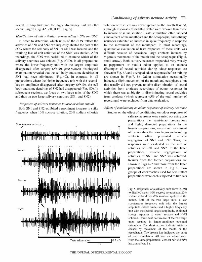
Responses of salivary neurones to taste or odour stimuli
Both SN1 and SN2 exhibited a prominent increase in spikefrequency when 10% sucrose solution, 20% sodium chloride
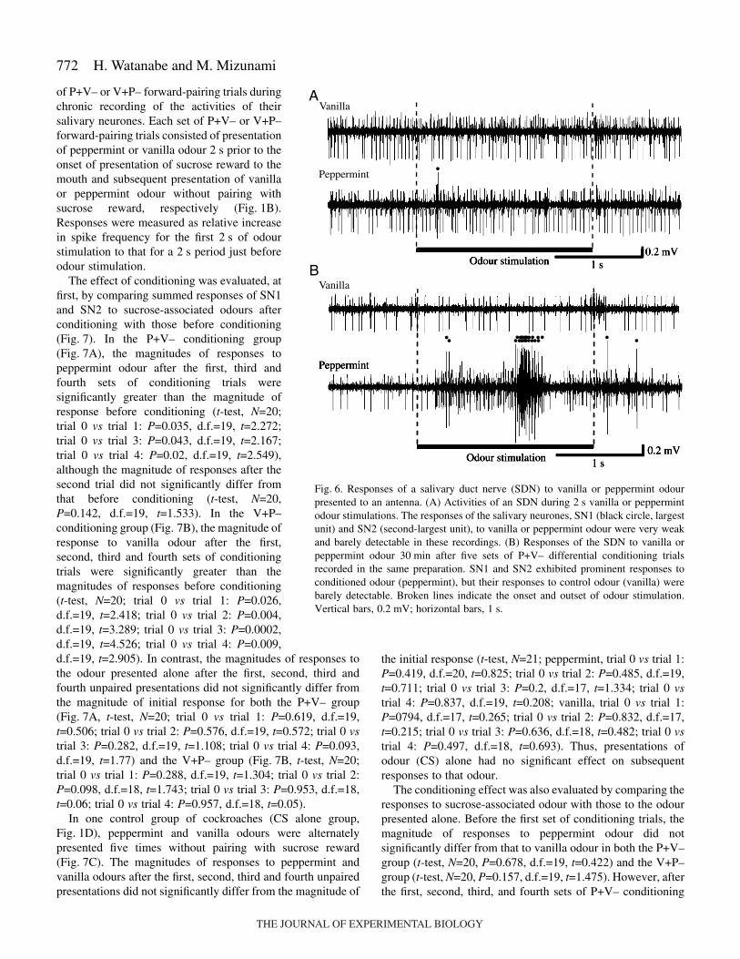
solution or distilled water was applied to the mouth (Fig.·5),although responses to distilled water were weaker than thoseto sucrose or saline solution. Taste stimulation often induceda movement of the mouthpart and the oesophagus, and salivaryneurones exhibited an increase in spike frequency in responseto the movement of the mouthpart. In most recordings,quantitative evaluation of taste responses of these units wasdifficult because of occasional large artefacts induced byvigorous movement of the mouth and the oesophagus (Fig.·5,small arrow). Both salivary neurones responded very weaklyto peppermint or vanilla odour applied to an antenna(Examples of neural activities during odour responses areshown in Fig.·6A and averaged odour responses before trainingare shown in Figs·7, 8). Odour stimulation occasionallyinduced a slight movement of the mouth and oesophagus, butthis usually did not prevent reliable discrimination of neuralactivities from artefacts; recordings of odour responses inwhich there was ambiguity in discriminating neural activitiesfrom artefacts (which represent <5% of the total number ofrecordings) were excluded from data evaluation.
Effects of conditioning on odour responses of salivary neurones
Studies on the effect of conditioning on odour responses ofsalivary neurones were carried out using twopreparations, i.e. semi-intact preparationsand highly dissected preparations. In theformer preparations, occasional movementof the mouth or the oesophagus and resultingartefacts often prevented reliablesegregation of SN1 and SN2. Thus, theresponses were evaluated as the sum ofactivities of SN1 and SN2. In the latterpreparations, reliable segregation ofactivities of SN1 and SN2 was achieved.Results from the former preparations areshown in Figs·4–7 and those from the latterpreparations are shown in Fig.·8. Twogroups of cockroaches used for semi-intactpreparations were each subjected to five sets
1 sTaste stimulation
Spontaneous activity
Water
Sucrose
NaCl
0.2 mV
Fig.·5. Responses of a salivary duct nerve (SDN)to distilled water, 10% sucrose solution and 20%sodium chloride (NaCl) solution applied to themouth. Both of the two large units, a lowspontaneous frequency unit with the largestamplitude (black circle) and a higher frequencyunit with the second-largest amplitude, exhibitedstrong responses to water, sucrose and NaClsolution. Coincident occurrence of the two largeunits resulted in larger-amplitude potential(triangles). The short arrows indicate artefactscaused by movement of the mouth or theoesophagus. The broken line indicates the onsetof taste stimulation. All four recordings werefrom the same preparation. Vertical bar, 0.2·mV;horizontal bar, 1·s.
THE JOURNAL OF EXPERIMENTAL BIOLOGY
772
of P+V– or V+P– forward-pairing trials duringchronic recording of the activities of theirsalivary neurones. Each set of P+V– or V+P–forward-pairing trials consisted of presentationof peppermint or vanilla odour 2·s prior to theonset of presentation of sucrose reward to themouth and subsequent presentation of vanillaor peppermint odour without pairing withsucrose reward, respectively (Fig.·1B).Responses were measured as relative increasein spike frequency for the first 2·s of odourstimulation to that for a 2·s period just beforeodour stimulation.
The effect of conditioning was evaluated, atfirst, by comparing summed responses of SN1and SN2 to sucrose-associated odours afterconditioning with those before conditioning(Fig.·7). In the P+V– conditioning group(Fig.·7A), the magnitudes of responses topeppermint odour after the first, third andfourth sets of conditioning trials weresignificantly greater than the magnitude ofresponse before conditioning (t-test, N=20;trial 0 vs trial 1: P=0.035, d.f.=19, t=2.272;trial 0 vs trial 3: P=0.043, d.f.=19, t=2.167;trial 0 vs trial 4: P=0.02, d.f.=19, t=2.549),although the magnitude of responses after thesecond trial did not significantly differ fromthat before conditioning (t-test, N=20,P=0.142, d.f.=19, t=1.533). In the V+P–conditioning group (Fig.·7B), the magnitude ofresponse to vanilla odour after the first,second, third and fourth sets of conditioningtrials were significantly greater than themagnitudes of responses before conditioning(t-test, N=20; trial 0 vs trial 1: P=0.026,d.f.=19, t=2.418; trial 0 vs trial 2: P=0.004,d.f.=19, t=3.289; trial 0 vs trial 3: P=0.0002,d.f.=19, t=4.526; trial 0 vs trial 4: P=0.009,d.f.=19, t=2.905). In contrast, the magnitudes of responses tothe odour presented alone after the first, second, third andfourth unpaired presentations did not significantly differ fromthe magnitude of initial response for both the P+V– group(Fig.·7A, t-test, N=20; trial 0 vs trial 1: P=0.619, d.f.=19,t=0.506; trial 0 vs trial 2: P=0.576, d.f.=19, t=0.572; trial 0 vstrial 3: P=0.282, d.f.=19, t=1.108; trial 0 vs trial 4: P=0.093,d.f.=19, t=1.77) and the V+P– group (Fig.·7B, t-test, N=20;trial 0 vs trial 1: P=0.288, d.f.=19, t=1.304; trial 0 vs trial 2:P=0.098, d.f.=18, t=1.743; trial 0 vs trial 3: P=0.953, d.f.=18,t=0.06; trial 0 vs trial 4: P=0.957, d.f.=18, t=0.05).
In one control group of cockroaches (CS alone group,Fig.·1D), peppermint and vanilla odours were alternatelypresented five times without pairing with sucrose reward(Fig.·7C). The magnitudes of responses to peppermint andvanilla odours after the first, second, third and fourth unpairedpresentations did not significantly differ from the magnitude of
H. Watanabe and M. Mizunami
the initial response (t-test, N=21; peppermint, trial 0 vs trial 1:P=0.419, d.f.=20, t=0.825; trial 0 vs trial 2: P=0.485, d.f.=19,t=0.711; trial 0 vs trial 3: P=0.2, d.f.=17, t=1.334; trial 0 vstrial 4: P=0.837, d.f.=19, t=0.208; vanilla, trial 0 vs trial 1:P=0794, d.f.=17, t=0.265; trial 0 vs trial 2: P=0.832, d.f.=17,t=0.215; trial 0 vs trial 3: P=0.636, d.f.=18, t=0.482; trial 0 vstrial 4: P=0.497, d.f.=18, t=0.693). Thus, presentations ofodour (CS) alone had no significant effect on subsequentresponses to that odour.
The conditioning effect was also evaluated by comparing theresponses to sucrose-associated odour with those to the odourpresented alone. Before the first set of conditioning trials, themagnitude of responses to peppermint odour did notsignificantly differ from that to vanilla odour in both the P+V–group (t-test, N=20, P=0.678, d.f.=19, t=0.422) and the V+P–group (t-test, N=20, P=0.157, d.f.=19, t=1.475). However, afterthe first, second, third, and fourth sets of P+V– conditioning
Odour stimulation 1 s
Vanilla
Peppermint
0.2 mV
A
1 sOdour stimulation
Vanilla
Peppermint
0.2 mV
B
Fig.·6. Responses of a salivary duct nerve (SDN) to vanilla or peppermint odourpresented to an antenna. (A) Activities of an SDN during 2·s vanilla or peppermintodour stimulations. The responses of the salivary neurones, SN1 (black circle, largestunit) and SN2 (second-largest unit), to vanilla or peppermint odour were very weakand barely detectable in these recordings. (B) Responses of the SDN to vanilla orpeppermint odour 30·min after five sets of P+V– differential conditioning trialsrecorded in the same preparation. SN1 and SN2 exhibited prominent responses toconditioned odour (peppermint), but their responses to control odour (vanilla) werebarely detectable. Broken lines indicate the onset and outset of odour stimulation.Vertical bars, 0.2·mV; horizontal bars, 1·s.
THE JOURNAL OF EXPERIMENTAL BIOLOGY

773Conditioning of salivary neurone activity
trials, the magnitudes of responses to sucrose-associatedpeppermint odour were significantly greater than themagnitudes of responses to vanilla odour presented alone (t-test, N=20; trial 1: P=0.008, d.f.=19, t=2.988; trial 2: P=0.019,
d.f.=19, t=2.571; trial 3: P=0.006 d.f.=19, t=3.091; trial 4:P=0.004, d.f.=19, t=3.307). Similarly, after the second, thirdand fourth sets of V+P– conditioning trials, the magnitudes ofresponses to sucrose-associated vanilla odour weresignificantly greater than the magnitudes of responses topeppermint odour presented alone (t-test, N=20; trial 2:P=0.01, d.f.=18, t=2.869; trial 3: P=0.024 d.f.=18, t=2.471;trial 4: P=0.049, d.f.=18, t=2.11). In the CS alone group, themagnitude of responses to peppermint odour did notsignificantly differ from that to vanilla odour (t-test, N=21; trial0: P=0.269, d.f.=18, t=1.14; trial 1: P=0.913, d.f.=18, t=0.11;trial 2: P=0.509, d.f.=17, t=0.675; trial 3: P=0.509, d.f.=16,t=0.224; trial 4: P=0.548, d.f.=19, t=0.611). We conclude thatthree sets of conditioning trials are sufficient to achieve asignificant level of conditioning.
Short-term retention and effects of backward pairing
Retention of the conditioning effect was tested at 1·min and30·min after five sets of conditioning trials in the P+V– andV+P– forward-pairing groups. Examples of responses ofsalivary neurones to sucrose-associated odour (peppermintodour) and to the odour presented alone (vanilla odour) at30·min after five sets of differential conditioning trials areshown in Fig.·6. Both SN1 and SN2 exhibited responses tosucrose-associated peppermint odour, while they exhibitedmuch less prominent responses to the vanilla odour presentedalone.
The magnitudes of summed responses of SN1 and SN2 tosucrose-associated odour at 1·min or 30·min after conditioningwere significantly greater than those before conditioning inboth the P+V– (Fig.·8A; t-test, N=20; before vs 1·min aftertraining: P=0.0003, d.f.=19, t=4.489; before vs 30·min aftertraining: P=0.009, d.f.=19, t=2.887) and V+P– forward-conditioning groups (Fig.·8B; t-test, N=20; before vs 1·minafter training: P=0.002, d.f.=19, t=3.515; before vs 30·min aftertraining: P=0.025, d.f.=19, t=2.43). Retention of theconditioning effect was also evaluated by comparing theresponses to sucrose-associated odours with those to odourspresented alone. Before conditioning, the magnitude ofresponses to peppermint odour did not significantly differ fromthe magnitude of responses to vanilla odour in both the P+V–group (Fig.·8A; t-test, N=20, P=0.992, d.f.=19, t=0.01) and theV+P– group (Fig.·8B, t-test, N=20, P=0.102, d.f.=19, t=1.72).At 1·min and 30·min after conditioning, the magnitude of theresponses to sucrose-associated odour were significantlygreater than the magnitude of responses to the odour presentedalone in the P+V– group (Fig.·8A; t-test, N=20; 1·min aftertraining: P=0.00005, d.f.=19, t=5.2; 30·min after training:P=0.000002, d.f.=19, t=6.752) and the V+P– group (Fig.·8B;t-test, N=20; 1·min after training: P=0.0005, d.f.=19, t=4.207;30·min after training: P=0.0003, d.f.=19, t=4.362). The resultsindicate that the effect of conditioning is retained for 30·minafter conditioning.
The magnitude of responses to sucrose-associatedpeppermint odour at 30·min after conditioning wassignificantly less than that 1·min after conditioning (Fig.·8A;
Fig.·7. Effects of forward-pairing trials (A,B) and unpairedpresentation of odours (C) on responses of the salivary neurones (SN1and SN2). Summed responses of SN1 and SN2 to peppermint orvanilla odour before and at 5·min after the first, second, third andfourth sets of P+V– (A) or P–V+ (B) conditioning trials or unpairedpresentations of odours (C) are shown. Relative responses, measuredas the relative increase in spike frequency for the first 2·s of odourstimulation compared to that during a 2·s period before odourstimulation, are shown as means ± s.e.m.; N=20 (A,B), N=21 (C).Asterisks indicate the results of statistical comparison with responsesto peppermint or vanilla odour before conditioning (NS, P>0.05;*P<0.05; **P<0.01; t-test).
Number of unpaired presentations of odours
0
100
180
−20
0
50
80
Peppermint (CS+)Vanilla (CS−)
Peppermint (CS−)Vanilla (CS−)
*
*
*
A
B
0
50
80
Rel
ativ
e re
spon
se (
%)
Peppermint (CS−)Vanilla (CS+)
* **
** **
C
0 1 2 3 4
0 1 2 3 4
0 1 2 3 4
Number of conditioning trials
Number of conditioning trials
THE JOURNAL OF EXPERIMENTAL BIOLOGY

774
t-test, N=20, P=0.002, d.f.=19, t=3.673). By contrast, themagnitude of the responses to sucrose-associated vanilla odourat 30·min after conditioning did not significantly differ fromthat 1·min after conditioning (Fig.·8B; t-test, N=20, P=0.885,d.f.=19, t=0.146). It was, however, uncertain whether or notthis was due to the odour-specific decay of memory, since themagnitude of responses to the odour presented alone at 30·minafter conditioning was also significantly less than that before,or 1·min after, conditioning in both the P+V– group (Fig.·8A;t-test, N=20; before vs 30·min after training: P=0.004, d.f.=19,t=3.313; 1·min vs 30·min after training: P=0.007, d.f.=19,t=3.029) and the V+P– group (Fig.·8B; t-test, N=20; before vs30·min after training: P=0.017, d.f.=19, t=2.608; 1·min vs30·min after training: P=0.015, d.f.=19, t=2.662), while themagnitude of the responses at 1·min after conditioning did notsignificantly differ from that before conditioning in the P+V–group (Fig.·8A; t-test, N=20, P=0.12, d.f.=19, t=0.12) and theV+P– group (Fig.·8B; t-test, N=20, P=0.686, d.f.=19, t=0.411).Therefore, the possibility cannot be excluded that the decay ofodour responses between 1·min and 30·min after conditioningis due to deterioration of the preparation.
We next studied the effect of five sets of backward CS/USpairing trials in another group of animals (Fig.·8C). Onebackward-pairing trial consisted of presentation of peppermintodour 4·s after the onset of presentation of sucrose reward and
H. Watanabe and M. Mizunami
subsequent unpaired presentation of vanilla odour (Fig.·1C,backward pairing). The magnitude of summed responses ofSN1 and SN2 to peppermint odour at 1·min or 30·min afterbackward-pairing trials did not significantly differ from thatbefore trials (t-test, N=23; before vs 1·min after training:P=0.906, d.f.=22, t=0.119; before vs 30·min after training:P=0.074, d.f.=22, t=1.879; 1·min vs 30·min after training:P=0.332, d.f.=22, t=0.992). The magnitude of responses tounpaired vanilla odour at 1·min and 30·min after training didnot significantly differ from that before trials (t-test, N=23;before vs 1·min after training: P=0.92, d.f.=22, t=0.102; beforevs 30·min after training: P=0.055, d.f.=22, t=2.024; 1·min vs30·min after training: P=0.143, d.f.=22, t=1.52).
The effect of backward pairing was also evaluated bycomparing the responses to backward-paired odours and thoseto odours presented alone. The magnitudes of responses tobackward-paired peppermint odour did not significantly differfrom that to unpaired vanilla odours before and at 1·min and30·min after conditioning (Fig.·8C; t-test, N=23; beforetraining: P=0.689, d.f.=22, t=0.405; 1·min after training:P=0.866 d.f.=22, t=0.17; 30·min after training: P=0.809,d.f.=22, t=0.244). The results indicate that backward pairing isnot effective in achieving conditioning of odour responses ofsalivary neurones.
In another control experiment, sucrose solution (US) was
150
100
0
50
Before 1 min after 30 min after
NS *** ******
**
N=20
A
Before 1 min after 30 min after
NS *** *****
*
150
100
0
50
N=20
B
150
100
0
50
Before 1 min after 30 min after
NS NS NS
NSNS
N=20
C150
100
0
50
BeforeUS alone
6 min afterUS alone
35 min afterUS alone
NS NS NSNS
NS
N=19
D
Rel
ativ
e re
spon
se (
%)
Peppermint Vanilla
Fig.·8. Effects of forward andbackward pairing trials and of non-associative control. (A,B) Summedresponses of salivary neurones(SN1 and SN2) to peppermint(hatched bars) or vanilla (shadedbars) odour before and at 1·min and30·min after five sets of P+V– (A)or V+P– (B) forward-pairing trials.(C) Summed responses of SN1 andSN2 to odours before and at 1·minand 30·min after five sets of P+V–backward-pairing trials. (D)Summed responses of SN1 andSN2 to odours before and at 6·minand 35·min after five presentationsof sucrose solution without pairingwith odour (US alone). Theresponses are shown as means ±s.e.m. The results of statisticalcomparison are shown above thebars (NS, P>0.05, *P<0.05,**P<0.01, ***P<0.001; t-test).
THE JOURNAL OF EXPERIMENTAL BIOLOGY
775Conditioning of salivary neurone activity
presented five times without pairing with odour (Fig.·8D; seealso Fig.·1E). The magnitudes of summed responses of SN1and SN2 to odour stimulation measured at 6 and 35·min afterpresentations of US alone did not significantly differ fromthose before presentations of US alone for both peppermintodour (t-test, N=19; before vs 6·min after US alone trials:P=0.504, d.f.=18, t=0.682; before vs 35·min after US alonetrials: P=0.222, d.f.=18, t=1.265; 6·min vs 35·min after USalone trials: P=0.176, d.f.=18, t=1.408) and vanilla odour (t-test, N=19; before vs 6·min after US alone trials: P=0.34,d.f.=18, t=0.98; before vs 35·min after US alone trials:P=0.717, d.f.=18, t=0.368; 6·min vs 35·min after US alonetrials: P=0.238, d.f.=18, t=1.221). Moreover, the magnitudesof responses to peppermint and those to vanilla did notsignificantly differ before and at 6·min and 35·min afterpresentations of sucrose solution alone (t-test, N=19; beforetrials: P=0.482, d.f.=18, t=0.81; 6·min after US alone trials:P=0.707 d.f.=18, t=0.381; 35·min after US alone trials: P=0.609,d.f.=18, t=0.521). Thus, presentations of sucrose solution alonehad no effects on odour responses of salivary neurones.
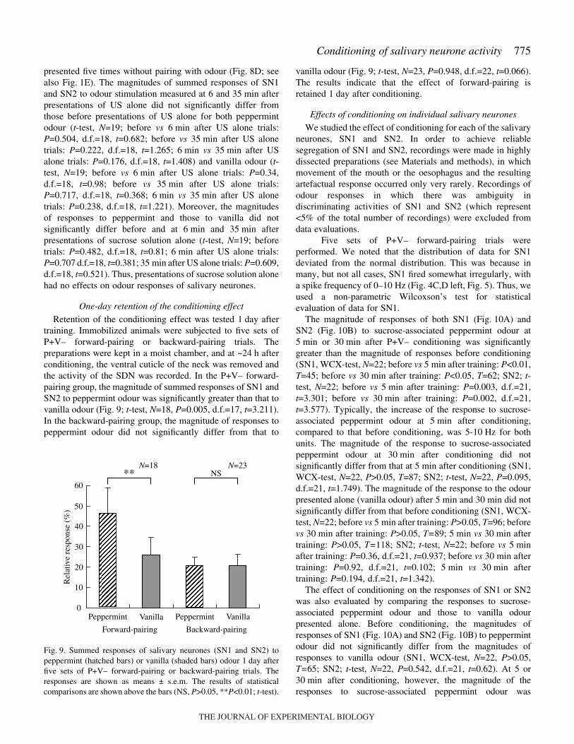
One-day retention of the conditioning effect
Retention of the conditioning effect was tested 1·day aftertraining. Immobilized animals were subjected to five sets ofP+V– forward-pairing or backward-pairing trials. Thepreparations were kept in a moist chamber, and at ~24·h afterconditioning, the ventral cuticle of the neck was removed andthe activity of the SDN was recorded. In the P+V– forward-pairing group, the magnitude of summed responses of SN1 andSN2 to peppermint odour was significantly greater than that tovanilla odour (Fig.·9; t-test, N=18, P=0.005, d.f.=17, t=3.211).In the backward-pairing group, the magnitude of responses topeppermint odour did not significantly differ from that to
vanilla odour (Fig.·9; t-test, N=23, P=0.948, d.f.=22, t=0.066).The results indicate that the effect of forward-pairing isretained 1·day after conditioning.
Effects of conditioning on individual salivary neurones
We studied the effect of conditioning for each of the salivaryneurones, SN1 and SN2. In order to achieve reliablesegregation of SN1 and SN2, recordings were made in highlydissected preparations (see Materials and methods), in whichmovement of the mouth or the oesophagus and the resultingartefactual response occurred only very rarely. Recordings ofodour responses in which there was ambiguity indiscriminating activities of SN1 and SN2 (which represent<5% of the total number of recordings) were excluded fromdata evaluations.
Five sets of P+V– forward-pairing trials wereperformed. We noted that the distribution of data for SN1deviated from the normal distribution. This was because inmany, but not all cases, SN1 fired somewhat irregularly, witha spike frequency of 0–10·Hz (Fig.·4C,D left, Fig.·5). Thus, weused a non-parametric Wilcoxson’s test for statisticalevaluation of data for SN1.
The magnitude of responses of both SN1 (Fig.·10A) andSN2 (Fig.·10B) to sucrose-associated peppermint odour at5·min or 30·min after P+V– conditioning was significantlygreater than the magnitude of responses before conditioning(SN1, WCX-test, N=22; before vs 5·min after training: P<0.01,T=45; before vs 30·min after training: P<0.05, T=62; SN2; t-test, N=22; before vs 5·min after training: P=0.003, d.f.=21,t=3.301; before vs 30·min after training: P=0.002, d.f.=21,t=3.577). Typically, the increase of the response to sucrose-associated peppermint odour at 5·min after conditioning,compared to that before conditioning, was 5-10·Hz for bothunits. The magnitude of the response to sucrose-associatedpeppermint odour at 30·min after conditioning did notsignificantly differ from that at 5·min after conditioning (SN1,WCX-test, N=22, P>0.05, T=87; SN2; t-test, N=22, P=0.095,d.f.=21, t=1.749). The magnitude of the response to the odourpresented alone (vanilla odour) after 5·min and 30·min did notsignificantly differ from that before conditioning (SN1, WCX-test, N=22; before vs 5·min after training: P>0.05, T=96; beforevs 30·min after training: P>0.05, T=89; 5·min vs 30·min aftertraining: P>0.05, T=118; SN2; t-test, N=22; before vs 5·minafter training: P=0.36, d.f.=21, t=0.937; before vs 30·min aftertraining: P=0.92, d.f.=21, t=0.102; 5·min vs 30·min aftertraining: P=0.194, d.f.=21, t=1.342).
The effect of conditioning on the responses of SN1 or SN2was also evaluated by comparing the responses to sucrose-associated peppermint odour and those to vanilla odourpresented alone. Before conditioning, the magnitudes ofresponses of SN1 (Fig.·10A) and SN2 (Fig.·10B) to peppermintodour did not significantly differ from the magnitudes ofresponses to vanilla odour (SN1, WCX-test, N=22, P>0.05,T=65; SN2; t-test, N=22, P=0.542, d.f.=21, t=0.62). At 5 or30·min after conditioning, however, the magnitude of theresponses to sucrose-associated peppermint odour was
Fig.·9. Summed responses of salivary neurones (SN1 and SN2) topeppermint (hatched bars) or vanilla (shaded bars) odour 1·day afterfive sets of P+V– forward-pairing or backward-pairing trials. Theresponses are shown as means ± s.e.m. The results of statisticalcomparisons are shown above the bars (NS, P>0.05, **P<0.01; t-test).
60
50
40
30
20
10
0 Peppermint Vanilla Peppermint Vanilla
Forward-pairing Backward-pairing
** NSN=18 N=23
Rel
ativ
e re
spon
se (
%)
THE JOURNAL OF EXPERIMENTAL BIOLOGY

776
significantly greater than the magnitude of responses tounpaired vanilla odour for SN1 (WCX-test, N=22; 5·min aftertraining: P<0.01, T=48; 30·min after training: P<0.01, T=22)and SN2 (t-test, N=22, 5·min after training: P=0.001, d.f.=21,t=3.815; 30·min after training: P=0.00003, d.f.=21, t=5.342).Therefore, conditioning is successful for both SN1 and SN2.
Saliva secretion upon electrical stimulation of one SDN
We noted that both SN1 and SN2 exhibited an increase inthe response of 5–10·spikes·s–1 for the first 2·s of odourstimulation after five sets of forward-pairing trials of theassociation of the odour with sucrose solution. Wewondered whether or not the increase in responses ofsalivary neurones by conditioning was sufficient toinduce an increased level of saliva secretion. Wetherefore measured the change in the level of salivasecretion from one salivary duct in response toelectrical stimulation of one SDN in highly dissectedpreparations. Brief (0.2·msec) square-wave pulseswere delivered to the SDN by a pair of hookelectrodes at 5·Hz for 2, 5, 10, 20 and 40·s withintervals of 6·min, and the evoked compound actionpotentials were monitored by another pair of hookelectrodes, so that the intensity of the stimulus couldbe adjusted at just above the threshold of spikes oflarge salivary neurones (~5·V). We deduced thatspikes were not evoked in smaller-diameter neuronesof the SDN, since they should have higher thresholdfor spike generation.
We found that the level of saliva secretion iscontinuously maintained and that the level increasedin response to electric stimulation of the SDN(Fig.·11). The increase was statistically significant forall 2-, 5-, 10-, 20- and 40-sec stimulations (WCX-test,N=12; 2·s stimulation: P<0.01, T=5; 5·s stimulation:
H. Watanabe and M. Mizunami
P<0.05, T=8; 10·s stimulation: P<0.01, T=3; 20·s stimulation:P<0.05, T=13; 40·s stimulation: P<0.01, T=5). The resultssuggest that increased response of salivary neurones afterconditioning is sufficient to lead to increased levels of salivation.
DiscussionMajor findings
Classical conditioning of salivation has been extensivelystudied in mammals, especially in dogs (Pavlov, 1927; Miller,1969; Harris and Brady, 1974), but, as far as we know, it has
** **NS
***
N=22
B
0
–10
10
20
30
40
50
60
Before 5 min after 30 min after0
−100
100
200
300
400
500
600
700
Before 5 min after 30 min after
Rel
ativ
e re
spon
se (
%)
Rel
ativ
e re
spon
se (
%)
** **NS
****
N=22
A
Peppermint Vanilla
Fig.·10. Responses of salivary neurones, SN1 (A) and SN2 (B), to peppermint (hatched bars) and vanilla (shaded bars) odours before and at5·min and 30·min after five sets of P+V– forward-pairing trials. The responses are shown as means ± s.e.m. The results of statistical comparisonare shown above the bars (NS, P>0.05, *P<0.05, **P<0.01, ***P<0.001; WCX-test in Fig.·8B; t-test in Fig.·8B).
2 5 10 20 40
0
100
200
300 N=12
Saliv
atio
n (n
l min
−1)
Time (min)
** * ** ** **
Stimulation time (s)
Fig.·11. Changes in the level of salivation upon electrical stimulations of onesalivary duct nerve (SDN). The amount of saliva secreted from a salivary ductwas measured every minute while brief (0.2·ms) electric pulses were delivered tothe SDN at 5·Hz for 2, 5, 10, 20 and 40·s with intervals of 6·min. Averaged datafrom 12 preparations are shown as means ± s.e.m. The amounts of secretion(broken lines) before and after the onset of electrical stimulation were statisticallycompared, and asterisks indicate the level of significance (*P<0.05; **P<0.01;WCX-test).
THE JOURNAL OF EXPERIMENTAL BIOLOGY
777Conditioning of salivary neurone activity
not been reported in any non-mammalian species. In insectssuch as cockroaches and locusts, secretion of saliva iscontrolled by salivary neurones of the SOG (Whitehead, 1973;Smith and House, 1977; House and Smith, 1978; Baines andTyrer, 1989). Here we reported that in cockroaches theresponses of two large salivary neurones (SN1 and SN2) to anodour significantly increases after repeated pairing of the odourwith sucrose solution. The increase in the response of both SN1and SN2 was 5–10·spikes·s–1 for the first 2·s of odourstimulation after conditioning, and electrical stimulation of theSDN at 5·Hz for 2·s or longer led to significantly increasedsaliva secretion. The latter finding is in accordance with resultsof previous reports on secretory response of the salivary glandto electrical stimulation of the SDN, measured for salivaryglands isolated from cockroaches, Nauphoeta cinerea (Houseand Smith, 1978) and locusts (Baines and Tyrer, 1989). Theresults suggest that the increase in odour response of salivaryneurones as a result of conditioning is sufficient to lead to anincrease in the level of salivation.
Findings in this study suggest classical conditioning ofsalivation in the cockroach, but direct behavioural evidenceneeds to be provided to prove this speculation. We arecurrently performing experiments to compare the amount ofsalivation in response to odour stimulation before and afterconditioning.
Taste and odour responses of salivary neurones
Both of the two large salivary neurones exhibitedspontaneous activity and this should lead to a spontaneouslevel of saliva secretion. Salivary neurones exhibited aprominent increase in spike frequency in response to sucroseor saline solution applied to the mouth and also exhibited avery weak response to peppermint or vanilla odour applied toan antenna. Activation of salivary neurones in response tofood-predicting odour and food-associated taste stimulation isno doubt functionally significant for effective feeding.
We also observed that both SN1 and SN2 were activeduring movement of the mouthpart. This is in accordance withan observation that activities of salivary neurones weremodulated by activity of the mouthpart motor patterngenerator in locusts (Rast and Bräunig, 2001). The presentfinding, that salivary neurones receive signals related tofeeding motor activity as well as food-predicting olfactorysignals and food-associated gustatory signals, may bereflected in the morphologies of their dendrites. The ventralpart of the SOG is thought to participate mainly in sensoryprocessing and the dorsal part of the SOG is thought toparticipate mainly in motor function (Rehder, 1988; Tyrer andGregory, 1982), and salivary neurones have dendrites in bothdorsal and ventral parts of the SOG. Notably, dendrites of SN1are mainly located in the dorsal and ventral parts ofmandibular and maxillary neuromeres, and dendrites of SN2are mainly located in the ventral part of maxillary and labialneuromeres (Fig.·3B, Fig. 4C,D). How this different dendriticmorphology reflects different functions of SN1 and SN2remains a subject of future study.
Effects of conditioning on odour response of salivaryneurones
We have shown that appetitive conditioning trials toassociate an odour with sucrose reward lead to an increasedpreference for that odour in a dual-choice test (Watanabe etal., 2003), and we found in the present study that the sameclassical conditioning leads to an increase in response ofsalivary neurones to the odour associated with sucrosereward. It should be noted, however, that salivary neuronesare activated in response to both appetitive (sucrose) andaversive (saline) taste stimuli (Fig.·5). Moreover, themagnitude of responses of salivary neurones to vanilla odourdid not differ from that to peppermint odour before training(Figs·7, 8, 10), although cockroaches innately prefer vanillaodour over peppermint odour in a dual-choice test (Sakuraand Mizunami, 2001; Watanabe et al., 2003). These resultsindicate that an increase in response of salivary neurones toan odour might not necessarily correlate with an increase inthe preference for that odour. It would be interesting todetermine whether or not classical conditioning trials toassociate an odour with saline solution lead to an increase inresponse of salivary neurones to that odour, although suchaversive conditioning trials have been shown to lead to adecrease in preference for that odour in crickets (Matsumotoand Mizunami, 2002).
Backward-pairing trials were not effective for achievingconditioning of odour responses of salivary neurones (Fig.·8C,Fig.·9). This is in accordance with previous findings thatbackward-pairing of olfactory CS with gustatory US was noteffective in achieving olfactory conditioning in insects andmammals (honeybees: Hellstern et al., 1997; crickets:Matsumoto and Mizunami, 2002; rats: Maier et al., 1976),although backward-pairing of visual CS with olfactory US wasfound to be effective for achieving conditioning in cockroaches(Lent and Kwon, 2004).
There was a significant level of memory retention 1·dayafter conditioning. This is comparable to our previous findingthat altered odour preference after three sets of classicalconditioning trials was retained for 4·days after conditioning(Watanabe et al., 2003). The time course of memory retentionafter conditioning of activities of salivary neurones was notdetermined in detail in this study. The responses of salivaryneurones to sucrose-associated vanilla odour did notsignificantly decay from 1 to 30·min after conditioning(Fig.·8B). The response of salivary neurones to sucrose-associated peppermint odour, however, significantly decayedfrom 1 to 30·min after conditioning in semi-intactpreparations (Fig.·8A), but it did not significantly decay from5 to 30·min after conditioning in highly dissectedpreparations (Fig.·10). In the former experiments, theresponse to the odour presented alone (vanilla) also decayedfrom 1·min to 30·min (Fig.·8A). Thus, the possibility that theobserved decay of odour response was due to deterioration ofthe preparation cannot be ruled out. Further improvement ofpreparations is necessary to determine in detail the timecourse of memory retention.
THE JOURNAL OF EXPERIMENTAL BIOLOGY
778 H. Watanabe and M. Mizunami
Future perspective
Cockroaches may provide model systems in which to studycellular mechanisms of classical conditioning of activities ofsalivary neurones. In mammals, many studies have suggestedthat various brain regions participate in classical conditioningof salivation. For example, electrical stimulations of the orbitalcortex (Danilova, 1983) or dorsal part of the caudate nucleus(Danilova, 1981) in dogs and the lateral hypothalamus (Matsuoand Kusano, 1984) in rats modulate salivation to conditioningstimulus. Lesions of the cerebral cortex (Grimsley andWindholz, 2000) and dorsomedial part of the amygdala(Lagowska and Fonberg, 1975) decreased salivation toconditioning stimulus in dogs. The exact cellular mechanismsof conditioning of salivation, however, remain elusive.Cockroaches are suitable materials for the study of neuralmechanisms of conditioning of activities of salivary neuronesat the level of individual neurones, since intracellularrecordings from brain neurones are feasible (Mizunami, 1990;Mizunami, 1996; Li and Strausfeld, 1997; Li and Strausfeld,1999; Strausfeld and Li, 1999; Nishino et al., 2003).
Olfactory learning in insects has been used as a pertinentmodel in which to study neural mechanisms underlyinglearning and memory (Menzel, 1999; Heisenberg, 2003; Dalyet al., 2004). In honeybees, the antennal lobe (a primaryolfactory centre) and the mushroom body (a higher olfactorycentre that processes multisensory signals) have beenimplicated in olfactory memory processing (Menzel, 1999). Inthe fruit fly, Drosophila melanogaster, mutants with defects instructure and function of the mushroom body exhibitedimpairments in olfactory learning (Heisenberg, 2003). Inmoths, Manduca sexta, olfactory conditioning produced amodulation of the ensemble representations for odours inantennal lobe neurones (Daly et al., 2004). Conditioning ofactivities of salivary neurones should provide an excellentmodel for the study of the neural basis of olfactoryconditioning, since chronic extracellular recordings fromsalivary neurones can be easily combined with intracellularrecordings from brain neurones, thereby allowing for the studyof activity changes in brain neurones during conditioning. Oneof our next steps is to investigate whether neurones in theantennal lobe and the mushroom body are involved in olfactoryconditioning of activities of salivary neurones and whetherthere is an association of olfactory CS and gustatory US forconditioning of activities of salivary neurones in the SOG.
This study was supported by grants from the Ministry ofEducation, Science, Culture, Sports and Technology of Japanand Research Fellowships of the Japan Society for thePromotion of Science for Young Scientists.
ReferencesAli, D. (1997). The aminergic and peptidergic innervation of insect salivary
glands. J. Exp. Biol. 200, 1941-1949.Bacon, J. P. and Altman, J. S. (1977). A silver intensification method for
cobalt-filled neurones in wholemount preparations. Brain Res. 138, 359-363.
Baines, R. A. and Tyrer, N. M. (1989). The innervation of locust salivarygland. II. Physiology of excitation and modulation. J. Comp. Physiol. A 165,407-413.
Baines, R. A., Tyrer, N. M. and Mason, J. C. (1989). The innervation oflocust salivary gland. I. Innervation and analysis of transmitters. J. Comp.Physiol. A 165, 395-405.
Daly, K. C., Christensen, T. A., Lei, H., Smith, B. H. and Hildebrand, J.G. (2004). Learning modulates the ensemble representations for odors inprimary olfactory networks. Proc. Natl. Acad. Sci. USA 101, 10476-10481.
Danilova, L. K. (1981). Effect of prolonged electrical stimulation of the headof the caudate nucleus on secretory reflexes to food in dogs. Neurosci.Behav. Physiol. 11, 3-9.
Danilova, L. K. (1983). Conditioned and unconditioned food reflexes in dogsduring electrical stimulation of the orbital cortex. Neurosci. Behav. Physiol.12, 337-343.
Davis, N. T. (1985). Serotonon-immunoreactive visceral nerves andneurohemal system in the cockroach Periplaneta americana (L.). CellTissue Res. 240, 593-600.
Elia, A. J., Ali, D. W. and Orchard, I. (1994). Immunochemical staining oftyrosine hydroxylase (TH)-like material in the salivary glands and ventralnerve cord of the cockroach, Periplaneta americana (L.). J. Insect Physiol.40, 671-683.
Gifford, A. N., Nicholson, R. A. and Pitman, R. M. (1991). The dopamineand 5-hydroxytryptamine content of locust and cockroach salivary neurons.J. Exp. Biol. 161, 405-414.
Grimsley, D. L. and Windholz, G. (2000). The neurophysiological aspectsof Pavlov’s theory of higher nervous activity: in honor of the 150thanniversary of Pavlov’s birth. J. Hist. Neurosci. 9, 152-163.
Harris, A. H. and Brady, J. V. (1974). Animal learning. Visceral andautonomic conditioning. Annu. Rev. Psychol. 25, 107-133.
Heisenberg, M. (2003). Mushroom body memoir: from maps to models. Nat.Rev. Neurosci. 4, 266-274.
Hellstern, F., Malaka, R. and Hammer, M. (1997). Backward inhibitorylearning in honeybees: a behavioral analysis of reinforcement processing.Learn. Mem. 4, 429-444.
House, C. R. and Smith, R. K. (1978). On the receptors involved in thenervous control of salivary secretion by Nauphoeta clinerea Olivier. J.Physiol. 279, 457-471.
Just, F. and Walz, B. (1996). The effects of serotonin and dopamine onsalivary secretion by isolated cockroach salivary glands. J. Exp. Biol. 199,407-413.
Kandel, E. R., Schwartz, J. H. and Jassell, T. M. (2000). Principles ofNeural Science, 4th edn, pp. 1227-1246. New York: McGraw-Hill.
Lagowska, J. and Fonberg, E. (1975). Salivary reactions in dogs withdorsomedial amygdalar lesions. Acta Neurobiol. Exp. 35, 17-26.
Lechner, A. H., Baxter, A. D. and Byrne, J. H. (2000). Classicalconditioning of feeding in Aplysia: I. Behavioral analysis. J. Neurosci. 20,3369-3376.
Lent, D. D. and Kwon, H. W. (2004). Antennal movements reveal associativelearning in the American cockroach Periplaneta americana. J. Exp. Biol.207, 369-375.
Li, Y. and Strausfeld, N. J. (1997). Morphology and sensory modality ofmushroom body extrinsic neurons in the brain of the cockroach, Periplanetaamericana. J. Comp. Neurol. 387, 631-650.
Li, Y. and Strausfeld, N. J. (1999). Multimodal efferent and recurrentneurons in the medial lobes of cockroach mushroom bodies. J. Comp.Neurol. 409, 647-663.
Maier, S. F., Rapaport, P. and Wheatley, K. L. (1976). Conditionedinhibition and the UCS-CS interval. Anim. Learn. Behav. 4, 217-220.
Matsumoto, Y. and Mizunami, M. (2002). Temporal determinants of long-term retention of olfactory memory in the cricket Gryllus bimaculatus. J.Exp. Biol. 205, 1429-1437.
Matsumoto, Y. and Mizunami, M. (2004). Context-dependent olfactorylearning in an insect. Learn. Mem. 11, 288-293.
Matsuo, R. and Kusano, K. (1984). Lateral hypothalamic modulation of thegustatory-salivary reflex in rats. J. Neurosci. 4, 1208-1216.
Menzel, R. (1999). Memory dynamics in the honeybee. J. Comp. Physiol. A185, 323-340.
Miller, N. E. (1969). Learning of visceral and glandular responses. Science163, 434-445.
Mizunami, M. (1990). Nonlinear signal transmission between second- andthird-order neurons of cockroach ocelli. J. Gen. Physiol. 95, 297-317.
Mizunami, M. (1996). Gain control of synaptic transfer from second- to third-order neurons of cockroach ocelli. J. Gen. Physiol. 107, 121-131.
THE JOURNAL OF EXPERIMENTAL BIOLOGY

779Conditioning of salivary neurone activity
Nishino, H., Yamashita, S., Yamazaki, Y., Nishikawa, M., Yokohari, F.and Mizunami, M. (2003). Projection neurons originating from thermo-and hygrosensory glomeruli in the antennal lobe of the cockroach. J. Comp.Neurol. 455, 40-55.
Okada, R., Sakura, M. and Mizunami, M. (2003). Distribution of dendritesof descending neurons and its implications for the basic organization of thecockroach brain. J. Comp. Neurol. 458, 158-174.
Orido, Y., Kokaze, A., Akamatsu, T., Takashima, Y. and Yoshida, M.(1998). Ultrastructure of the foregut and associated glands in the lung fluke,Paragonimus miyazakii (Digenea: Troglotrematidae), with particularreference to their functional roles. J. Morphol. 237, 43-52.
Passe, D. H. and Walker, J. C. (1985). Odor psychophysics in vertebrates.Neurosci. Biobehav. Rev. 9, 431-467.
Pavlov, I. P. (1927). Conditioned Reflexes: An Investigation of thePhysiological Activity of the Cerebral Cortex (trans. G. V. Anrep). Oxford:Oxford University Press.
Rast, G. F. and Bräunig, P. (2001). Insect mouthpart motor patterns: centralcircuits modified for highly derived appendages? Neuroscience 108, 167-176.
Rehder, V. (1988). A neuroanatomical map of the suboesophageal andprothoracic ganglia of the honey bee (Apis mellifera). Proc. R. Soc. Lond.B 235, 179-202.
Rietdorf, K., Lang, I. and Walz, B. (2003). Saliva secretion and ioniccomposition of saliva in the cockroach Periplaneta americana afterserotonin and dopamine stimulation, and effects of ouabain andbumetamide. J. Insect Physiol. 49, 205-215.
Sakura, M. and Mizunami, M. (2001). Olfactory learning and memory inthe cockroach Periplaneta americana. Zool. Sci. 18, 21-28.
Sakura, M., Okada, R. and Mizunami, M. (2002). Olfactory discrimination
of structurally similar alcohols by cockroaches. J. Comp. Physiol. A 188,787-797.
Schachtner, J. and Bräunig, P. (1993). The activity pattern of identifiedneurosecretory cells during feeding behaviour in the locust Locustamigratoria. J. Exp. Biol. 185, 287-303.
Smith, R. K. and House, C. R. (1977). Fluid secretion by isolated cockroachsalivary glands. Experientia 33, 1182-1184.
Strausfeld, N. J. and Li, Y. (1999). Organization of olfactory and multimodalafferent neurons supplying the calyx and pedunculus of the cockroachmushroom bodies. J. Comp. Neurol. 409, 603-625.
Tyrer, N. M. and Gregory, G. E. (1982). A guide to the neuroanatomy oflocust suboesophageal and thoracic ganglia. Philos. Trans. R. Soc. Lond. B297, 91-123.
Watanabe, H., Kobayashi, Y., Sakura, M., Matsumoto, Y. and Mizunami,M. (2003). Classical olfactory conditioning in the cockroach Periplanetaamericana. Zool. Sci. 20, 1447-1454.
Whitehead, A. T. (1971). The innervation of the salivary gland in theAmerican cockroach: light and electron microscopic observations. J.Morphol. 135, 483-506.
Whitehead, A. T. (1973). Innervation of the American cockroach salivarygland: neurophysiological and pharmacological investigations. J. InsectPhysiol. 19, 1961-1970.
Yamasaki, T. and Narahashi, T. (1959). The effects of potassium and sodiumions on the resting and action potentials of the cockroach giant axon. J.Insect Physiol. 3, 146-158.
Zunke, U. (1990). Observations on the invasion and endoparasitic behaviorof the root lesion nematode Pratylenchus penetrans. J. Nematol. 22, 309-320.
THE JOURNAL OF EXPERIMENTAL BIOLOGY