miR-532-5p functions as a tumor suppressor in … · miR-532-5p functions as a tumor suppressor in...
9
5842 Abstract. – OBJECTIVE: Dysregulated miR- 532-5p has been observed in epithelial ovarian cancer (EOC). However, the potential biological function and clinical significance have not been fully explained. The study aimed to investigate the prognostic value and potential role of miR- 532-5p in EOC. PATIENTS AND METHODS: MiR-532-5p and Twist homolog 1 (TWIST1) mRNA expression were examined using quantitative real-time PCR. The correlation of miR-532-5p expression with clinicopathological factors was statistically an- alyzed. Kaplan-Meier analysis and Cox propor- tional hazards regression models were explored to reveal the correlations of miR-532-5p expres- sion with survival of patients. Cell Counting Kit- 8, colony formation and transwell invasion as- says were performed to evaluate the effects of miR-532-5p on cell proliferation and invasion, respectively. MiR-532-5p target genes were con- firmed using luciferase activity, RT-PCR and Western blot assays. RESULTS: We found that miR-532-5p was sig- nificantly decreased in EOC tissue and cell lines, and its expression levels were highly correlat- ed with grade (p = 0.011), FIGO stage (p = 0.004) and distant metastasis (p = 0.008). In addition, overall patient survival for those with high miR- 532-5p expression was significantly longer than those patients with low miR-532-5p expression (p = 0.0058). Multivariate regression analysis identified miR-532-5p down-regulation as an in- dependent unfavorable prognostic factor in EOC patients. Function assays showed that overex- pression of miR-532-5p inhibited proliferation, colony formation and invasion of EOC cells. Mechanistic investigations confirmed TWIST1 as a direct target of miR-532-5p. Further in vi- tro assay indicated that restored expression of TWIST1 dampened miR-532-5p-mediated sup- pression of tumor progression. CONCLUSIONS: Our data suggested that miR- 532-5p may act not only as a novel prognostic marker, but also as a potential target for molecu- lar therapy of EOC. Key Words: MiR-532-5p, Epithelial ovarian cancer, Tumor sup- pressor, Prognosis. Introduction Ovarian cancer is the sixth common cancer in women around the world and is the leading cau- se of morbidity among malignant gynecologic diseases 1 . Epithelial ovarian carcinoma (EOC) is the most common form of ovarian cancer 2 . In China, more than 50000 new cases of this disea- se are diagnosed each year 3 . Despite advances in surgery and chemotherapy over the last few de- cades, 80% of patients diagnosed with advanced EOC relapse and only 30% of patients survive for 5 years after diagnosis 4 . The 5-year survival of the EOC patients diagnosed at FIGO I stage was around 90 %. Unfortunately, most EOC patients are diagnosed with advanced disease 5 . Therefore, major efforts have focused on the identification of the diagnostic and prognostic potential of specific and sensitive biomarkers in EOC. MicroRNAs are small (18-25nt), single stran- ded, noncoding RNAs that play important regula- tory roles in the post-transcriptional level of gene expression by binding to the 3’UTR sequence of their target mRNA, thus leading to mRNA de- gradation or translational repression 6 . More and more studies have demonstrated that miRNAs are European Review for Medical and Pharmacological Sciences 2018; 22: 5842-5850 H. WEI 1 , Q.-L. TANG 2 , K. ZHANG 3 , J.-J. SUN 4 , R.-F. DING 5 1 Department of Obstetrics and Gynecology, Linyi People’s Hospital, Linyi, Shandong, China 2 Department of Obstetrics, Women and Children’s Health Care Hospital of Linyi, Linyi, Shandong, China 3 Department of Internal Medicine, Linyi Rongjun Hospital, Linyi, Shandong, China 4 Department of Gynaecology, Lanling County Women and Children’s Hospital, Lanling, Shandong, China 5 Department of Pharmacy, Lanling County People’s Hospital, Lanling, Shandong, China Corresponding Author: Rui-fu Ding; e-mail: [email protected] miR-532-5p is a prognostic marker and suppresses cells proliferation and invasion by targeting TWIST1 in epithelial ovarian cancer
Transcript of miR-532-5p functions as a tumor suppressor in … · miR-532-5p functions as a tumor suppressor in...
5842
Abstract. – OBJECTIVE: Dysregulated miR-532-5p has been observed in epithelial ovarian cancer (EOC). However, the potential biological function and clinical significance have not been fully explained. The study aimed to investigate the prognostic value and potential role of miR-532-5p in EOC.
PATIENTS AND METHODS: MiR-532-5p and Twist homolog 1 (TWIST1) mRNA expression were examined using quantitative real-time PCR. The correlation of miR-532-5p expression with clinicopathological factors was statistically an-alyzed. Kaplan-Meier analysis and Cox propor-tional hazards regression models were explored to reveal the correlations of miR-532-5p expres-sion with survival of patients. Cell Counting Kit-8, colony formation and transwell invasion as-says were performed to evaluate the effects of miR-532-5p on cell proliferation and invasion, respectively. MiR-532-5p target genes were con-firmed using luciferase activity, RT-PCR and Western blot assays.
RESULTS: We found that miR-532-5p was sig-nificantly decreased in EOC tissue and cell lines, and its expression levels were highly correlat-ed with grade (p = 0.011), FIGO stage (p = 0.004) and distant metastasis (p = 0.008). In addition, overall patient survival for those with high miR-532-5p expression was significantly longer than those patients with low miR-532-5p expression (p = 0.0058). Multivariate regression analysis identified miR-532-5p down-regulation as an in-dependent unfavorable prognostic factor in EOC patients. Function assays showed that overex-pression of miR-532-5p inhibited proliferation, colony formation and invasion of EOC cells. Mechanistic investigations confirmed TWIST1 as a direct target of miR-532-5p. Further in vi-tro assay indicated that restored expression of TWIST1 dampened miR-532-5p-mediated sup-pression of tumor progression.
CONCLUSIONS: Our data suggested that miR-532-5p may act not only as a novel prognostic marker, but also as a potential target for molecu-lar therapy of EOC.
Key Words:MiR-532-5p, Epithelial ovarian cancer, Tumor sup-
pressor, Prognosis.
Introduction
Ovarian cancer is the sixth common cancer in women around the world and is the leading cau-se of morbidity among malignant gynecologic diseases1. Epithelial ovarian carcinoma (EOC) is the most common form of ovarian cancer2. In China, more than 50000 new cases of this disea-se are diagnosed each year3. Despite advances in surgery and chemotherapy over the last few de-cades, 80% of patients diagnosed with advanced EOC relapse and only 30% of patients survive for 5 years after diagnosis4. The 5-year survival of the EOC patients diagnosed at FIGO I stage was around 90 %. Unfortunately, most EOC patients are diagnosed with advanced disease5. Therefore, major efforts have focused on the identification of the diagnostic and prognostic potential of specific and sensitive biomarkers in EOC.
MicroRNAs are small (18-25nt), single stran-ded, noncoding RNAs that play important regula-tory roles in the post-transcriptional level of gene expression by binding to the 3’UTR sequence of their target mRNA, thus leading to mRNA de-gradation or translational repression6. More and more studies have demonstrated that miRNAs are
European Review for Medical and Pharmacological Sciences 2018; 22: 5842-5850
H. WEI1, Q.-L. TANG2, K. ZHANG3, J.-J. SUN4, R.-F. DING5
1Department of Obstetrics and Gynecology, Linyi People’s Hospital, Linyi, Shandong, China2Department of Obstetrics, Women and Children’s Health Care Hospital of Linyi, Linyi, Shandong, China3Department of Internal Medicine, Linyi Rongjun Hospital, Linyi, Shandong, China4Department of Gynaecology, Lanling County Women and Children’s Hospital, Lanling, Shandong, China5Department of Pharmacy, Lanling County People’s Hospital, Lanling, Shandong, China
Corresponding Author: Rui-fu Ding; e-mail: [email protected]
miR-532-5p is a prognostic marker and suppresses cells proliferation and invasion by targeting TWIST1 in epithelial ovarian cancer

miR-532-5p functions as a tumor suppressor in epithelial ovarian cancer
5843
involved in various cellular activities, including proliferation, apoptosis, metastasis and differentia-tion7. Recently, aberrant miRNAs expressions are frequently observed in cancers that serve as onco-genes or tumor suppressors8,9. In addition, several studies reported that aberrant miRNAs expression might be of potential use as a diagnostic and pro-gnostic biomarker for human cancer, including EOC10,11. MiR-532-5p, located at human chromoso-me Xp11.23, has been reported to be dysregulated in several tumors, including EOC12-14. However, the clinical significance of miR-532-5p and its related molecular mechanisms involved in the progression of EOC remain poorly investigated.
In this work, we detected the expression le-vels of miR-532-5p in both EOC tissues and cell lines. Then, the association of miR-532-5p with clinicopathological factors or EOC patient pro-gnosis was also statistically analyzed. Moreover, we performed gain-function assay to explore the role of miR-532-5p in EOC cells proliferation and invasion. In addition, we performed several cells experiments to explore the potential mechanism by which miR-532-5p influenced the cells beha-vior of EOC. Our findings suggest miR-532-5p as potential therapeutic candidates and prognostic biomarker for EOC.
Patients and Methods
Patient SpecimensPaired tissue specimens (tumor and adjacent
normal tissues) from 145 patients with EOC were obtained and histologically confirmed by a pa-thologist at Linyi People’s Hospital from January 2011 to December 2013. None of the patients had received chemotherapy, radiotherapy or adjuvant hormonal therapy prior to surgery. Specimens were preserved in liquid nitrogen immediately and stored at -80°C until RNA extraction. All patients from cohort 2 were taken on 5-year fol-low-up. Overall survival was defined as the date of surgery until the date of death. The clinical in-formation is summarized in Table I. Tissue sam-ple use was approved by the Ethics Committees of our hospital and written informed consent was obtained from all study participants.
Cell CultureHuman EOC (OVCAR3, SKOV3, ES‑2 and
CAOV‑3) cell lines were primary purchased from Shanghai Institute of Biochemistry and Cell Biolo-gy (Xuhui, Shanghai, China). Human normal ova-rian epithelial NOEC cell lines were purchased from
Table I. Relationship between miR-532-5p expression and clinicopathological variables in EOC patients.
miR-532-5p expression
Variable Number High Low p-value Age (y) 0.574 <50 76 35 41 ≥50 69 35 34 Tumor size (cm) 0.202 ≤ 10 68 29 39 > 10 77 41 36 CA125 level (U/ml) 0.216 < 600 92 48 44 ≥ 600 53 22 31 Ascites 0.671 < 100 74 37 37 ≥ 100 71 33 38 FIGO stage 0.004 I + II 84 49 35 III + IV 61 21 40 Grade 0.011 G1 88 50 38 G2 + G3 57 20 37 Distant metastasis 0.008 Yes 65 23 42 No 82 47 35
H. Wei, Q.-L. Tang, K. Zhang, J.-J. Sun, R.-F. Ding
5844
the American Type Culture Collection (Manassas, VA, USA). All cells were maintained in Dulbecco’s modified Eagle’s medium (DMEM) supplemented with 10% fetal bovine serum (FBS, Gibco, Jianglin Technology, Pudong, Shanghai, China), 100 U/ml penicillin (Meilunbio®, Dalian, Niaoning, China) and 100 μg/ml streptomycin (Haoran Technology, Yuzhong, Chongqing, China) at 37°C in a humidi-fied atmosphere containing 5% CO2.
Plasmid Construction And Cell Transfection
MiR-532-5p mimics and its negative control were purchased from RiboBio (Guandong, Guan-gzhou, China). For TWIST1 over-expression, the open reading frame of TWIST1 was inserted into the pcDNA3.1 vector to generate TWIST1 ove-rexpression vectors. Lipofectamine 3000 (Invitro-gen, Carlsbad, CA, USA) were used to transfect the cells once they reached 60% confluency. Tran-sfection efficiencies were detected by qRT-PCR after 48 h of transfection.
RNA Isolation And Quantitative Real-Time Reverse Transcription-PCR (qRT-PCR)
Total RNA from tissues and cell lines was ex-tracted using TRIzol reagent (Invitrogen, Carl-sbad, CA, USA) according to the manufacturer’s instructions. Complimentary DNA (cDNA) was synthesized using the PrimeScript II First Strand cDNA Synthesis kit (TaKaRa, Otsu, Shiga, Japan) according to the manufacturer’s instructions. The qPCR was performed by using the miRNA-spe-cific TaqMan MiRNA Assay Kit (Biosystems, Foster City, CA, USA) under 7900 Real-Time PCR System (Biosystems, Foster City, CA, USA). The reaction was performed in triplicate for 30 min at 16°C, 30min at 42°C, and 5 min at 85°C. TWIST1 mRNA expression was measured by RT of total RNA into complementary DNA using M‑MLV Reverse Transcriptase (Promega Cor-poration, Madison, WI, USA). For both TWIST1
mRNA and miR-532-5p detection, Glyceraldehy-de-3-phosphate dehydrogenase (GAPDH) was used as an internal control. The relative expres-sion of TWIST1 mRNA and miR-532-5p was cal-culated by the 2-ΔΔT method. The primer sequen-ces used in this study were shown in Table II.
Cell Proliferation AssayThe in vitro cell proliferation of EOC cells was
measured using the Cell Counting Kit-8 (CCK8) assay. In brief, OVCAR3 and ES-2 cells were see-ded into 96-well plates (3×10⁴/well) and incubated in RPMI 1640 at 37°C and 5% CO2 atmosphere for 96 hours. The cell viability assay was perfor-med using Cell Counting Kit-8 (CCK8; Dojindo, Pudong, Shanghai, China) according to the manu-facturer’s protocol. Absorbance levels were mea-sured at the wavelength of 490 nm using an auto-matic microplate reader (Gene, Haidian, Beijing, China). All experiments were performed at least three times in triplicate.
Colony Formation AssayApproximately 3×10⁴ cells were seeded per well
for six-well plates and were grown for 10 days with normal medium. The cells were then fixed and staining by crystal violet. All experiments were performed at least three times in triplicate.
Invasion Assay1× 105 cells in serum-free medium were placed
into the upper chamber of an insert coated with Matrigel (Sigma-Aldrich, St. Louis, MO, USA). The chambers were then incubated in culture me-dium supplemented with 10% FBS (Gibco, Jian-glin Technology, Pudong, Shanghai, China) in the bottom chambers for 24 h before examination. Following incubation, cells on the upper surface of the membrane were scraped off and the invasi-ve cells to the bottom of the membrane were fixed and stained. Then, the number of invaded cells was quantified using ImageJ (National Institutes of Health, Bethesda, MD, USA)
Luciferase Reporter AssaysA fragment of the TWIST1 3’-UTR and a mu-
tated 3’-UTR of TWIST1 that contained the pu-tative miR-532-5p binding sites were prepared to construct reporter plasmids containing the 3’-UTR regions of TWIST1. DNA fragments were cloned into subcloned into the pmirGLO-control. OVCAR3 and ES-2 cells were seeded at a densi-ty of 4000 cells in a 96-well plate. The next day, OVCAR3 and ES-2 cells were co-transfected with
Table II. The primers for PCR.
Primer name Sequences (From 5’ to 3’) miR-532-5p Fwd CGGCCATGCCTTGAGTGTAmiR-532-5p Rev GCAGGGTCCGAGGTATTCTWIST1 Fwd CCAGGTACATCGACTTCCTCTATWIST1 Rev CCATCCTCCAGACCGAGAAGAPDH Fwd CCAGGTGGTCTCCTCTGAGAPDH Rev GCTGTAGCCAAATCGTTGT
miR-532-5p functions as a tumor suppressor in epithelial ovarian cancer
5845
wild-type (mutant) reporter plasmid and miR-532-5p mimics or Ctrl using Lipofectamine 2000 (In-vitrogen, Carlsbad, CA, USA). At 48 h post-tran-sfection, the relative luciferase activities were evaluated using Dual-Luciferase Reporter Assay System (Promega, Madison, WI, USA).
Western Blot AssayTotal protein was collected using RIPA buffer
(ThermoFisher Scientific, Waltham, MA, USA). Then the protein concentration was determi-ned using the BCA Protein Assay kit (Thermo Fisher Scientific, Inc. Waltham, MA, USA). 20 mg of protein were separated by dodecyl sulfa-te, sodium salt-polyacrylamide gel electropho-resis (SDS-PAGE), and then transferred onto polyvinylidene difluoride (PVDF) membranes (Millipore, Billerica, MA, USA). The membra-nes were blocked with 5% nonfat dry milk in Tris-buffered saline and Tween 20 (TBST) for 1 h, and then incubated with anti-TWIST1 (1:500, Sigma-Aldrich, Xicheng, Beijing, China) or an-ti-GAPDH (1:1000, Sigma-Aldrich, Xicheng, Beijing, China) primary antibody overnight at 4°C before subsequent incubation with horse-radish peroxidase(HRP)-conjugated secondary antibodies (Sigma-Aldrich, Xicheng, Beijing, China) for 1 h at 37°C. Protein signals were developed using the Amersham ECL Western Blotting Detection Reagent (GE Healthcare Life Sciences, Pittsburgh, PA, USA).
Statistical AnalysisStatistical Product and Service Solutions
(SPSS) 16.0 software (SPSS Inc., Chicago, IL, USA) was applied in statistical analysis. The Stu-dent t-test was employed for comparison betwe-
en two groups. The multi-group comparison was performed using one-way analysis of variance, followed by Least-Significant Difference (LSD) test. The x2-test was used to examine the asso-ciations between miR-532-5p expression and the clinicopathological characters. Overall, survival curves were plotted according to the Kaplan-Meier method, with the log-rank test applied for comparison. Univariate and multivariate analyses were conducted to explore the independent risk factors for EOC using the Cox proportional ha-zard model. A p-value < 0.05 was considered sta-tistically significant.
Results
MiR-532-5p Expression Level Was Decreased in EOC Tissues and Cell Lines
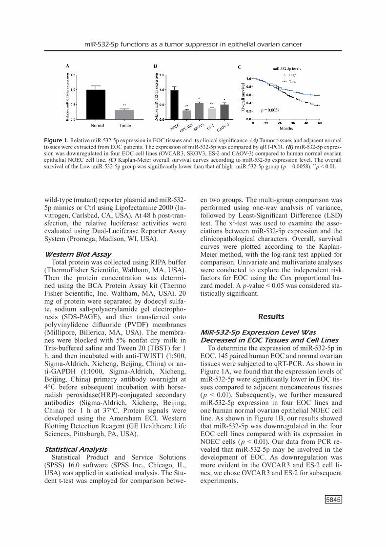
To determine the expression of miR-532-5p in EOC, 145 paired human EOC and normal ovarian tissues were subjected to qRT-PCR. As shown in Figure 1A, we found that the expression levels of miR-532-5p were significantly lower in EOC tis-sues compared to adjacent noncancerous tissues (p < 0.01). Subsequently, we further measured miR-532-5p expression in four EOC lines and one human normal ovarian epithelial NOEC cell line. As shown in Figure 1B, our results showed that miR-532-5p was downregulated in the four EOC cell lines compared with its expression in NOEC cells (p < 0.01). Our data from PCR re-vealed that miR-532-5p may be involved in the development of EOC. As downregulation was more evident in the OVCAR3 and ES‑2 cell li-nes, we chose OVCAR3 and ES‑2 for subsequent experiments.
Figure 1. Relative miR-532-5p expression in EOC tissues and its clinical significance. (A) Tumor tissues and adjacent normal tissues were extracted from EOC patients. The expression of miR-532-5p was compared by qRT-PCR. (B) miR-532-5p expres-sion was downregulated in four EOC cell lines (OVCAR3, SKOV3, ES‑2 and CAOV‑3) compared to human normal ovarian epithelial NOEC cell line. (C) Kaplan-Meier overall survival curves according to miR-532-5p expression level. The overall survival of the Low-miR-532-5p group was significantly lower than that of high- miR-532-5p group (p = 0.0058). **p < 0.01.

H. Wei, Q.-L. Tang, K. Zhang, J.-J. Sun, R.-F. Ding
5846
Association Between miR-532-5p Expression and the Clinicopathological Features of EOC
In order to further the biological significan-ce of miR-532-5p, we categorized miR-532-5p expression levels as high and low according to the mean value. The association between clini-copathological characteristics and miR-532-5p expression is summarized in Table II. We found that low miR-532-5p expression was closely rela-ted to advanced grade (p = 0.011), higher FIGO stage (p = 0.004) and positive distant metastasis (p = 0.008). However, no significant difference was observed between miR-532-5p expression and patients’ age, tumor size, CA125 level and ascites (p > 0.05). Our results indicated miR-532-5p as a potential tumor suppressor.
Relationship of miR-532-5p to Overall Survival of EOC Patients
Then, Kaplan-Meier analysis was applied to examine the prognostic value of miR-532-5p expression to overall survival of patients with EOC. As shown in Figure 1C, we found that pa-tients with lower miR-532-5p expression had a si-gnificantly shorter overall survival than patients with high miR-532-5p expression (p = 0.0058). Furthermore, univariate analysis revealed that FIGO stage (p = 0.006), grade (p = 0.028), distant metastasis (p = 0.004) and miR-532-5p expression (p = 0.006) were prognostic factors for patient’s overall survival (Table III). More importantly, the results of multivariate analysis confirmed that miR-532-5p expression (HR = 3.774, 95% CI = 1.253-4.779, p = 0.006) was an independent pre-dictor of overall survival in EOC. Overall, these findings suggested that miR-532-5p expression could be used as a powerful independent progno-stic factor in EOC patients.
Effects of miR-532-5p on EOC Cell Proliferation and Invasion
To investigate the biological role of miR-532-5p in EOC progression, the OVCAR3 and ES‑2 cell line was transfected with miR-532-5p or Ctrl. The tran-sfection efficiency was confirmed by qRT-PCR (p < 0.05, Figure 2A). Then, we performed CCK-8 assay, finding that overexpressed miR-532-5p decreased the proliferation rate of OVCAR3 and ES‑2 cells (Figure 2B and 2C). Similarly, the colony formation assay revealed that clonogenic survival was signi-ficantly suppressed in miR-532-5p -overexpressed OVCAR3 and ES‑2 cell lines (Figure 2D). In addi-tion, transwell invasion assays revealed that ectopic expression of miR-532-5p drastically suppressed OVCAR3 and ES‑2 cells invasion (Figure 2E). Ove-rall, our data suggested that miR-532-5p suppressed proliferation and invasion in EOC cells.
TWIST1 is a Direct Target of miR-532-5pTo explore the underlying molecular mecha-
nism by which miR-532-5p suppressed EOC cell proliferation and invasion, we explored its tar-get genes through two bioinformatics tools. As shown in Figure 3A, the results of TargetScan and miRanda showed that TWIST1 might be the target for miR-532-5p (Figure 2B). To validate the specific regulation of miR-532-5p on TWIST1, luciferase reporter assays were performed. As shown in Figure 3B, we observed that forced miR-532-5p expression decreased the luciferase activity of wild type TWIST1 3’ UTR construct, whereas the luciferase activity was not decrea-sed in the mutated type in both OVCAR3 and ES‑2 cell lines. Moreover, qRT-PCR and We-stern blot analyses showed that overexpression of miR-532-5p significantly downregulated the expression of TWIST1 at the mRNA and protein levels in both OVCAR3 and ES‑2 cell lines (Fi-
Table III. Univariate and multivariate analyses of overall survivor rates in 147 ovarian cancer patients by Cox regression analysis.
Variable Univariate Analysis Multivariate Analysis HR (95% CI) p-value HR (95% CI) p-value
Age 1.223(0.732-1.780) 0.562 - -Tumor size 1.562(0.894-2.321) 0.473 - -CA125 level 0.932(0.573-1.774) 0.217 - -Ascites 1.266(0.426-2.218) 0.135 - -FIGO stage 3.652(1.263-5.263) 0.002 3.253(1.093-4.263) 0.006Grade 3.251(1.445-4.273) 0.008 2.778(1.239-3.882) 0.028Distant metastasis 3.792(1.377-5.298) 0.001 3.253(1.193-4.776) 0.004miR-532-5p expression 4.263(1.557-6.372) 0.001 3.774(1.253-4.779) 0.006

miR-532-5p functions as a tumor suppressor in epithelial ovarian cancer
5847
Figure 3. TWIST1 was a direct target of miR-532-5p in EOC cells. (A) The predicted binding sites for miR-532-5p in the 3’UTR of TWIST1 and the mutations in the binding sites are shown. (B) Luciferase assay was used to confirm the direct regulation of miR-532-5p on 3’UTR of TWIST1 in both OVCAR3 and ES‑2 cells. (C) miR-532-5p decreased TWIST1 mRNA expression in OVCAR3 and ES‑2 cells. (D) miR-532-5p decreased TWIST1 proteins expression in OVCAR3 and ES‑2 cells. **p < 0.01.
Figure 2. Overexpression of miR-532-5p could inhibit cell proliferation, colony formation and invasion in EOC cell lines. (A) Upregulation of miR-532-5p in miR-532-5p -stably-overexpressing OVCAR3 and ES‑2 cells. (B-C) Cell viability was valida-ted by MTT assay in OVCAR3 and ES‑2 cells. (D) Relative colony formation rates of OVCAR3 and ES‑2 cells with indicated treatment were determined by colony formation assays. (E) Transwell matrigel assay was used to determine OVCAR3 and ES‑2 cells invasive capability. **p < 0.01.

H. Wei, Q.-L. Tang, K. Zhang, J.-J. Sun, R.-F. Ding
5848
gure 3C and 3D). Overall, these results confirm that TWIST1 is a direct target of miR-532-5p in EOC cells.
Overexpression of TWIST1 Reversed the Effect of miR-532-5p
In order to further explore whether TWIST1 is involved in the miR-532-5p-induced suppres-sion of EOC cell proliferation and invasion, we performed a rescue experiment in OVCAR3 cells. Western blotting analysis indicated that pcDNA3.1-TWIST1 transfection effectively re-plenished miR-532-5p miRNA mimics-induced TWIST1 loss (Figure 4A). In addition, transfected with pcDNA3.1-TWIST1 decreased the suppres-sive effects of miR-532-5p on cell viability (Fi-gure 4B; p < 0.01), colony formation (Figure 4C; p < 0.01) and invasion (Figure 4D; p < 0.01) of OVCAR3 cells. In summary, the data indicated that miR-532-5p could inhibit cell viability and invasion by down-regulation of TWIST1.
Discussion
At present, human EOC is still a complex malignancy, which has multiple histological subtypes15. The 5-year overall survival of EOC
patients diagnosed at an advanced stage is rela-tively low. Therefore, it is of great importance to identify novel genetic or protein biomarkers for accurate diagnosis and prediction of prognosis. In this study, we demonstrated that miR-532-5p expression was decreased in human EOC tissues and cell lines compared with matching adjacent non-tumoral tissue and normal cell lines, in ac-cordance with previous studies16. Then, we firstly analyzed its clinicopathologic and prognostic si-gnificance. Our data demonstrated that low miR-532-5p expression was closely related to advanced grade, higher FIGO stage and positively distant metastasis, indicating that miR-532-5p may act as a negative regulator in clinical progression of EOC patients. Moreover, we performed Kaplan-Meier analysis with five-year following-up, fin-ding that the patients with lower levels of miR-532-5p expression had significantly shorter survival time. This supports the hypothesis that miR-532-5p plays a potential role in EOC progres-sion. More importantly, further univariate and multivariate survival analysis showed that miR-532-5 could be used as an independent potential prognostic biomarker for EOC patients. Taken together, these findings, together with previous, highlighted the clinical significance of miR-532-5p in EOC patients and implied potentially im-
Figure 4. miR-532-5p suppressed EOC progression mainly by down-regulating TWIST1. (A) Western blotting showed TWIST1 protein expression in OVCAR3 cells transfected with Ctrl, miR-532-5p and miR-532-5p + TWIST1 plasmid, re-spectively. (B) CCK-8 assay detected the effect of miR-532-5p overexpression on miR-532-5p-induced cell growth arrest in OVCAR3 cells. (C) Colony formation assay was used to evaluate the cell viability in OVCAR3 cells. (D) The invasion of OVCAR3 cells was assessed using a transwell assay. **p < 0.01 vs. Ctrl group; &&p < 0.01 vs. miR-532-5p group.
miR-532-5p functions as a tumor suppressor in epithelial ovarian cancer
5849
portant role for miR-532-5p in predicting the prognosis of EOC patients. Previous studies have shown that miR-532-5p was involved in biological processes in cancers. For instance, Hu et al17 re-ported that miR-532 expression was significantly up-regulated in gastric cancer and its overexpres-sion promote gastric cancer cells migration and invasion by targeting NKD1. Xu et al18 further showed that miRNA-532-5p exerted its oncogenic role by directly targeting RUNX3. Of note, Song et al19 reported that miR-532-5p was significantly lowly expressed in hepatocellular carcinoma and its knockdown promoted cell proliferation and metastasis by influencing CXCL2 expression, indicating miR-532-5p as a tumor suppressor in hepatocellular carcinoma. Song et al20 found that miR-532-5p downregulation in colorectal cancer tissues and its association with aggressive clini-copathological characteristics. In vitro and in vivo revealed that up-regulation of miR-532 suppres-sed colorectal cancer cells proliferation, migra-tion and invasion by directly targeting IGF-1R and regulating the PI3K/Akt signaling pathway, supporting miR-532-5p as a tumor suppressor in colorectal cancer. The above results revealed that miR-532-5p may serve as a tumor suppressor or a tumor promoter according the types of tumors. Importantly, previously Bai et al16 reported that the expression levels of miR-532 were significant-ly down-regulated in ovarian cancer tissues and cell lines. They also observed that miR-532 served as a tumor suppressor by inhibiting ovarian can-cer cell proliferation and invasion via regulating hTERT. However, the evidence about the expres-sion pattern and biological role of miR-532-5p is limited. In this study, our in vitro experiments also confirmed that overexpression of miR-532-5p significantly suppressed EOC cells proliferation and invasion. However, the potential mechanism by which miR-532-5p exerted its tumor-suppres-sive role in EOC progression remains largely un-known. Identification of miR-532-5p target genes is critical for understanding the role of miR-532-5p in tumorigenesis, and is important for explo-ring novel therapeutic targets. Then, we screened TWIST1 (Twist homolog 1) as a potential target of miR-532-5p according to microRNA target da-tabases and published studies. As a highly con-served basic helix-loop-helix transcription factor, TWIST1 plays an essential role in many biologi-cal processes21. Increasing evidence confirms that TWIST1 function as a tumor promoter in various tumor progression, such as breast cancer, lung cancer and EOC22-24. Current findings showed that
TWIST1 was a key regulator of cancer-associated fibroblasts25. In addition, several studies reported TWIST1 was modulated by several miRNAs, such as miR-186, miR-548c and miR-32026-28. In this work, we suggested that TWIST1 is one of miR-532-5p target genes. According to the results of a luciferase reporter assay, TWIST1 was iden-tified as a target gene of miR-532-5p. In order to further confirm the association between miR-532-5p and TWIST1, we performed rescue experi-ments, finding that restoration of TWIST1 rever-sed the effects of miR-532-5p overexpression in EOC cells. Taken together, miR-532-5p/TWIST1 based targeted therapy could be new therapeutic strategies for EOC patients.
Conclusions
We found that miR-532-5p was downregulated in EOC tissues, and low miR-532-5p expression was intimately associated with poor prognosis, which is considered as an independent prognostic indicator for overall survival. Moreover, we for the first time suggested that miR-532-5p suppres-sed the proliferation and invasion of EOC cells by targeting TWIST1. Therefore, the miR-532-5p/TWIST1 axis could serve as a potential target for the treatment of EOC.
Conflict of InterestThe Authors declare that they have no conflict of interest.
References
1) Jayson GC, Kohn EC, KitChEnEr hC, LEdErmann Ja. Ovarian cancer. Lancet 2014; 384: 1376-1388.
2) BowtELL dd. The genesis and evolution of hi-gh-grade serous ovarian cancer. Nat Rev Cancer 2010; 10: 803-808.
3) wanG B, Liu sZ, ZhEnG rs, ZhanG F, ChEn wQ, sun XB. Time trends of ovarian cancer incidence in Chi-na. Asian Pac J Cancer Prev 2014; 15: 191-193.
4) dEsantis CE, Lin CC, mariotto aB, siEGEL rL, stEin Kd, KramEr JL, aLtEri r, roBBins as, JEmaL a. Cancer treatment and survivorship statistics, 2014. CA Cancer J Clin 2014; 64: 252-271.
5) GLoss Bs, samimi G. Epigenetic biomarkers in epithe-lial ovarian cancer. Cancer Lett 2014; 342: 257-263.
6) BartEL dP. MicroRNAs: genomics, biogenesis, me-chanism, and function. Cell 2004; 116: 281-297.
7) EBErt ms, sharP Pa. Roles for microRNAs in con-ferring robustness to biological processes. Cell 2012; 149: 515-524.
H. Wei, Q.-L. Tang, K. Zhang, J.-J. Sun, R.-F. Ding
5850
8) CaLin Ga, CroCE Cm. MicroRNA signatures in hu-man cancers. Nat Rev Cancer 2006; 6: 857-866.
9) yanG L, hou J, Cui Xh, suo Ln, Lv yw. MiR-133b re-gulates the expression of CTGF in epithelial-me-senchymal transition of ovarian cancer. Eur Rev Med Pharmacol Sci 2017; 21: 5602-5609.
10) Banno K, yanoKura m, iida m, adaChi m, naKamura K, noGami y, umEnE K, masuda K, Kisu i, nomura h, KataoKa F, tominaGa E, aoKi d. Application of microRNA in diagnosis and treatment of ovarian cancer. Biomed Res Int 2014; 2014: 232817.
11) Zhou Qh, Zhao ym, Jia LL, ZhanG y. Mir-595 is a significant indicator of poor patient prognosis in epithelial ovarian cancer. Eur Rev Med Pharma-col Sci 2017; 21: 4278-4282.
12) GriEsinG s, KaJino t, tai mC, Liu Z, naKatoChi m, shimada y, suZuKi m, taKahashi t. Thyroid transcrip-tion factor-1-regulated microRNA-532-5p targets KRAS and MKL2 oncogenes and induces apop-tosis in lung adenocarcinoma cells. Cancer Sci 2017; 108: 1394-1404.
13) tsai hP, huanG sF, Li CF, ChiEn ht, ChEn sC. Dif-ferential microRNA expression in breast cancer with different onset age. PLoS One 2018; 13: e0191195.
14) wanG F, ChanG Jt, Kao CJ, huanG rs. High Expres-sion of miR-532-5p, a tumor suppressor, leads to better prognosis in ovarian cancer both in vivo and in vitro. Mol Cancer Ther 2016; 15: 1123-1131.
15) hEidEmann Ln, hartwELL d, hEidEmann Ch, JoChum-sEn Km. The relation between endometriosis and ovarian cancer - a review. Acta Obstet Gynecol Scand 2014; 93: 20-31.
16) Bai L, wanG h, wanG ah, ZhanG Ly, Bai J. MicroR-NA-532 and microRNA-3064 inhibit cell prolifera-tion and invasion by acting as direct regulators of human telomerase reverse transcriptase in ova-rian cancer. PLoS One 2017; 12: e0173912.
17) hu s, ZhEnG Q, wu h, wanG C, Liu t, Zhou w. miR-532 promoted gastric cancer migration and inva-sion by targeting NKD1. Life Sci 2017; 177: 15-19.
18) Xu X, ZhanG y, Liu Z, ZhanG X, Jia J. miRNA-532-5p functions as an oncogenic microRNA in human gastric cancer by directly targeting RUNX3. J Cell Mol Med 2016; 20: 95-103.
19) sonG X, wanG Z, Jin y, wanG y, duan w. Loss of miR-532-5p in vitro promotes cell proliferation and metastasis by influencing CXCL2 expression in HCC. Am J Transl Res 2015; 7: 2254-2261.
20) sonG y, Zhao y, dinG X, wanG X. microRNA-532 suppresses the PI3K/Akt signaling pathway to inhi-bit colorectal cancer progression by directly targe-ting IGF-1R. Am J Cancer Res 2018; 8: 435-449.
21) BECK B, LaPouGE G, rorivE s, droGat B, dEsaEdELaE-rE K, dELaFaiLLE s, duBois C, saLmon i, wiLLEKEns K, marinE JC, BLanPain C. Different levels of Twist1 regulate skin tumor initiation, stemness, and pro-gression. Cell Stem Cell 2015; 16: 67-79.
22) Ai L, Kim wJ, aLPay m, tanG m, Pardo CE, hataKEyama s, may ws, KLaddE mP, hELdErmon Cd, siEGEL Em, Brown Kd. TRIM29 suppresses TWIST1 and invasive breast cancer behavior. Cancer Res 2014; 74: 4875-4887.
23) Burns tF, doBromiLsKaya i, murPhy sC, GaJuLa rP, thiyaGaraJan s, ChatLEy sn, aZiZ K, Cho yJ, tran Pt, rudin Cm. Inhibition of TWIST1 leads to activation of oncogene-induced senescence in oncoge-ne-driven non-small cell lung cancer. Mol Cancer Res 2013; 11: 329-338.
24) raFEhi s, ramos vaLdEs y, BErtrand m, mCGEE J, PréFontainE m, suGimoto a, dimattia GE, shEPhErd tG. TGFβ signaling regulates epithelial-mesen-chymal plasticity in ovarian cancer ascites-de-rived spheroids. Endocr Relat Cancer 2016; 23: 147-159.
25) LEE Kw, yEo sy, sunG Co, Kim sh. Twist1 is a key re-gulator of cancer-associated fibroblasts. Cancer Res 2015; 75: 73-85.
26) Zhu X, shEn h, yin X, LonG L, XiE C, Liu y, hui L, Lin X, FanG y, Cao y, Xu y, Li m, Xu w, Li y. miR-186 regulation of Twist1 and ovarian cancer sensitivity to cisplatin. Oncogene 2016; 35: 323-332.
27) sun X, Cui m, ZhanG a, tonG L, wanG K, Li K, wanG X, sun Z, ZhanG h. MiR-548c impairs migration and invasion of endometrial and ovarian cancer cells via downregulation of Twist. J Exp Clin Can-cer Res 2016; 35: 10.
28) Li C, duan P, wanG J, Lu X, ChEnG J. miR-320 inhi-bited ovarian cancer oncogenicity via targeting TWIST1 expression. Am J Transl Res 2017; 9: 3705-3713.