Middle Cerebral Artery Occlusion of Rats: Pathological and
7
Turkis/r Neurosurgery 9: 52 - 58, 1999 Ka/rveci: MCA Occlusioii iii Ra/s Middle Cerebral Artery Occlusion of Rats: Pathological and Neurological Evaluation of the Model Siçanlarda Orta Serebral Arter Oklüzyon Modelinin Patolojik ve Nörolojik Olarak Incelenmesi. NEVZAT KAHVECI, TÜLIN ALKAN, ENDER KORFALI, KASIM OZLÜK Departments of Physiology (NK, TA, KÖ) and Neurosurgery (EK), Uludag University School of Medicine, Bursa, Turkey Abstract: Eighty seven Sprague-Dawley rats were used to study the anatomy of the horizontal segment of middle cerebral ar tery (MCA) and infarction after occlusion of this vessel. We investigated the size and location of the lesion produced, and found a correlation between infarct size and neurological deficiL Fourty rats were used to determine the anatomical variations of MCA after intracardiac carbon black injection. Five major patterns of MCA were defined and two of them were major and constitued 88% of rats. In the experimental group (n:20) through a subtemporal burrhole MCA was exposed and after defining the anatomical variations of the ar tery according to our classification, MCA was coagulated 3-4 mm length from the origin of the lateral striate arteries to the inferior cerebral vein and divided. Control rats (n:20) underwent identical surgical procedures except for occlusion. Twenty-four hours after MCA occlusion, all animals were neurologically evaluated. In the experimental group, one rat scored 1, five rats scored 2, nine rats scored 3, five rats scored 4. On the third day after occlusion the brains were removed and stained with 2% 2,3,5 triphenyltetrazolium chloride (TIC). In each animaL, the area of infarction was assessed and graded using computer analysis method. There were no rats grade I infarcts, 4 with grade 11,10with grade III, 6 with grade iv. This study show that once the anatomical variations of the MCA and its branches in our strain of rats was determined, it was possible to achieve 80% grade III and iv infarcts. Key Word s:Focal cerebral ischemia, middle cerebral artery occlusion, middle cerebral artery variations, rat 52 Özet: Orta serebral arterin (OSA) horizontal segmentinin anatomik farkliliklarini göstermek ve oklüzyon sonrasi iskemik lezyonlarin dagilimi ve infarkt alani lokalizasyonu ve büyüklügü ile nörolojik bulgular arasindaki korelasyon için 350-400 g agirligindaki SO siçanlar (n:87) kullanildi. Bir grup siçana (n:40) OSA'in anatomik varyasyonunu saptamak amaciyla intrakardiak siyah karbon enjeksiyonu yapildi. Bes degisik OSA yapisi gözlendi ve ikisi major variyasyonu (%88) olusturuyordu. Deney grubunda (n: 20) OSA subtemporal girisimle açildi, siniflandirmamiza göre anatomik variyasyonu belirlendikten sonra arterin lateral striat arter ayrimindan inferior serebral vene kadar olan 3-4 mm uzunlugundaki kismi koagüle edildi ve kesildi. Kontrol grubu (n:20) siçanlara oklüzyon disindaki tüm cerrahi girisimler uygulandi. OSA oklüzyonundan 24 saat sonra bütün siçanlar nörolojik olarak degerlendirildi. Deney grubunda; 1 siçanin skoru 1, 5 siçanin skoru 2, 9 siçanin skoru 3, 5 siçanin skoru 4 olarak saptandi. Oklüzyon sonrasi 3. günde beyinler %2'lik 2,3,5 triphenyltetrazolium chloride ile boyandi. Infarkt alanlari kompüterize analiz metodu ile tayin edildi. Grade I'de O, grade II'de 4, grade II1'de LO, grade IV'de 6 siçan saptandi. Bu tür siçanlarda OSA ve dallarinin varyasyonunun tanimlandigi çalismamizda, %80 oraninda grade III ve iv infarkt alani saptandi. Anahtar Kelimeler: Fokal serebral iskemi, orta serebral arter oklüzyonu, orta serebral arter variyasyonlari, siçan
Transcript of Middle Cerebral Artery Occlusion of Rats: Pathological and

Turkis/r Neurosurgery 9: 52 - 58, 1999 Ka/rveci: MCA Occlusioii iii Ra/s
Middle Cerebral Artery Occlusion of Rats:Pathological and Neurological Evaluation of the Model
Siçanlarda Orta Serebral Arter Oklüzyon Modelinin Patolojik veNörolojik Olarak Incelenmesi.
NEVZAT KAHVECI, TÜLIN ALKAN, ENDER KORFALI, KASIM OZLÜK
Departments of Physiology (NK, TA, KÖ) and Neurosurgery (EK), Uludag University School of Medicine, Bursa, Turkey
Abstract: Eighty seven Sprague-Dawley rats were used tostudy the anatomy of the horizontal segment of middlecerebral ar tery (MCA) and infarction after occlusion of thisvessel. We investigated the size and location of the lesionproduced, and found a correlation between infarct sizeand neurological deficiL Fourty rats were used todetermine the anatomical variations of MCA after
intracardiac carbon black injection. Five major patterns ofMCA were defined and two of them were major andconstitued 88% of rats. In the experimental group (n:20)through a subtemporal burrhole MCA was exposed andafter defining the anatomical variations of the ar teryaccording to our classification, MCA was coagulated 3-4mm length from the origin of the lateral striate arteries tothe inferior cerebral vein and divided. Control rats (n:20)underwent identical surgical procedures except forocclusion. Twenty-four hours after MCA occlusion, allanimals were neurologically evaluated. In theexperimental group, one rat scored 1, five rats scored 2,nine rats scored 3, five rats scored 4. On the third day afterocclusion the brains were removed and stained with 2%
2,3,5 triphenyltetrazolium chloride (TIC). In each animaL,the area of infarction was assessed and graded usingcomputer analysis method. There were no rats grade Iinfarcts, 4 with grade 11,10with grade III, 6 with grade iv.This study show that once the anatomical variations ofthe MCA and its branches in our strain of rats was
determined, it was possible to achieve 80% grade III andiv infarcts.
Key Word s:Focal cerebral ischemia, middle cerebral arteryocclusion, middle cerebral artery variations, rat
52
Özet: Orta serebral arterin (OSA) horizontal segmentininanatomik farkliliklarini göstermek ve oklüzyon sonrasiiskemik lezyonlarin dagilimi ve infarkt alanilokalizasyonu ve büyüklügü ile nörolojik bulgulararasindaki korelasyon için 350-400 g agirligindaki SOsiçanlar (n:87) kullanildi. Bir grup siçana (n:40) OSA'inanatomik varyasyonunu saptamak amaciyla intrakardiaksiyah karbon enjeksiyonu yapildi. Bes degisik OSA yapisigözlendi ve ikisi major variyasyonu (%88) olusturuyordu.Deney grubunda (n: 20) OSA subtemporal girisimle açildi,siniflandirmamiza göre anatomik variyasyonubelirlendikten sonra arterin lateral striat arter ayrimindaninferior serebral vene kadar olan 3-4 mm uzunlugundakikismi koagüle edildi ve kesildi. Kontrol grubu (n:20)siçanlara oklüzyon disindaki tüm cerrahi girisimleruygulandi. OSA oklüzyonundan 24 saat sonra bütünsiçanlar nörolojik olarak degerlendirildi. Deney grubunda;1 siçanin skoru 1, 5 siçanin skoru 2, 9 siçanin skoru 3, 5siçanin skoru 4 olarak saptandi. Oklüzyon sonrasi 3.günde beyinler %2'lik 2,3,5 triphenyltetrazolium chlorideile boyandi. Infarkt alanlari kompüterize analiz metoduile tayin edildi. Grade I'de O, grade II'de 4, grade II1'de LO,
grade IV'de 6 siçan saptandi. Bu tür siçanlarda OSA vedallarinin varyasyonunun tanimlandigi çalismamizda,%80 oraninda grade III ve iv infarkt alani saptandi.
Anahtar Kelimeler: Fokal serebral iskemi, orta serebralarter oklüzyonu, orta serebral arter variyasyonlari, siçan

Turkish Neitrosiirgery 9: 52 - 58, 1999
INl'RODUCTION
The development of a reproducible and reliableanimal model for cerebral ischemia would allow the
study of pathophysiological changes that occur duringand after the event. For an occlusion method to be
optimal, it must produce a high rate of consistent,uniform, large infarcted areas. The rat is a widelystudied, readily available animal that has beenintensively investigated, and is preferred for cerebralblood flow studies. Numerous rat models of cerebralischemia have been developed. These include bilateralcarotid occlusion (33), an intracranial compressionmodel that causes cerebral ischemia due to increasedintracranial pressure (3,15), unilateral carotidocclusion plus hypoxia or hypotension (22,23),compression of the neck with a cuff (32), arterialmicroembolisation (29) and four vessel occlusionmethod s (26). While these models have been usefulfor examining various aspeets of cerebral ischemia,they cause global ischemia rather than focal lesionsand are not reliable for investigating the effeets ofvarious pharmacological drugs. For these reasons,researchers have adopted the technique of middlecerebral artery (MCA) occlusion described by Tamuraet aL.(34).The original method has been modified overthe years and has become widely accepted as the mainmodel for investigating of focal cerebral ischemia.
Recently the intraluminal thread model for MCAocclusion has gained greater acceptance (16).Numerous modifications have been reported in theliterature which indicates that the technique is not yetstandardized (l0,12,13,16,17,19,27,31).Also, problemswith subarachnoid hemorrhage and insufficient MCAocclusion are more common in the intraluminal threadmodel than the open cranieetomy model (l,13,14,16).
Our earlyexperience (30)with the model Tamuraet aL.suggested that it was not possible to achieve 100% incidence of infaretion through focal occlusion ofthe MCA, even if the site was proximal to the olfactorytract, as reported (34). Although vascular anatomyshould be similar within the same strain, inbreedingcan lead to the development of new variations ofcerebral arteries in new-generation rats. With this inmind, we investigated inbred rats in our laboratory,focusing on variations in MCA anatomy and its effeeton the production of uniform infaction site and sizeafter arterial occlusion.
MA TERIALS and METHODS
In this study, eighty seven adult male SpragueDawley rats weighting 350-400 g were used. A groupof rats (n:40) without any surgical procedures were
Kahveci: MCA Occ1ilsioli iii Ra/s
used to determine the anatomical variations ofMCA
after intracardiac carbon black injeetion (5mI). Afterthe rats' brain were removed, we carefully inspectedthe MCA and its branches us ing an operatingmicroscope, and then drew them in detaiL. Therelation between the MCA and the olfaetory bulb andinferior cerebral vein were also noted. The
experimental group of rats were anesthetized withsodium thiopental30 mg/kg i.p. A tracheotomy wasperformed and the animals were ventilated (Harvardsmall animal ventilator) with 70% nitrous oxide, 30%02 mixture containing 1.5% isoflurane. A femoralartery and vein were catheterized to allowcontinuous monitoring of arterial pressure (ProtocolPROPAQ 104) and repeated sampIing of P02' pC02,Hct, pH and for fluid administration. A rectaltemperature probe was inserted and the animalswere maintained in the normothermic range(37.4±0.4°C) using a radiant heat lamp.
On ce these procedures were done, the animalswere placed in the supine position with the ir headturned to the left, 2 cm curved vertical incision wasmade starting midway between the left orbit andexternal auditory canaL.An inci sion was made to thetempo ral is muscle and dissected from thesubtemporal bone and reflected forwards. Under theoperating microscope, temporo-mandibular joint andcoronal process of the mandible were seen and inferotemporal fossa was exposed. The pterygopalatineartery was protected and mandibular nerveoverlying on the pterygoid muscle was followedmedially to the foramen ovale was seen. Using ahigh speed drill 3-4 mm diarneter burr-hole wasopened, 3 mm anterior and 1 mm lateral to theforamen ovale. The dura was incised with a fine
needle and olfactory nerve and middle cerebralartery were located. Before the occlusion was carriedout, we identified the middle cerebral artery and itsbranches. The rats were divided into 2 groups.Control rats (n:20) had all the surgical proceduresexcept occlusion. In the experimental group (n:20)MCA was coagulated 3-4 mm length from the originof the lateral striate arteries to the inferior cerebralvein and divided.
Twenty-four hours af ter the coagulationprocedure was done, we evaluated the rats'neurological status using the system described byMenzies et aL.(l8). A scale of O to 4 was used to assessthe motor and behavioral changes after the middlecerebral occlusion (MCAO). The test consisted ofvarious maneuvers: First, the rats were suspendedby the tail approximately 30cm above the floor and
53

Tiirkis/i Neiirosiirgery 9: 52 - 5B, 1999
their forelimb posture was noted. Normal animalsextended both forelimbs toward the floor and theywere assigned a score of O. When the forelimbcontralateral to the side of the MCAO was consistentlyflexed during the suspension and there was no otherabnormality, the rat was scored 1. Rats were the nplaced on absorbent pads and they were gently heldby the tail. if animals showed an apparent decrease ingrip strength in the contralateral forelimb when pulledby the tail, then they were assigned a score of 2.Thereafter and while still being held by the tail, therats were allowed to move freely and were observedfor cireling behavior. Rats that moved spontaneouslyin all directions but established a monodireetional
cireling toward the paretic side when given a slightjerk of the tail were scored 3.Rats that showed a higherelinical score also showed all features of the lower
grades. This neurological evaluation was completedwithin a few minutes.
Kii/iveci: MCA Oeciiisioii iii Rnls
constituted the lateral striate arterial complex, whichsupplies blood to the basal ganglia. The pyriformartery usually originated several millimeters abovethe rhinal vein, and then made a right angle deviationfrom the main MCA tmnk. The pyriform branch wasseldom divided further and the area supplied by thisartery appeared isolated from other major surfacevessels. The temporal ar tery arose opposite topyriform artery and ran along the temporal lobe andrarely anastomosing with the posterior rhinal branch.Frontal branch ran superiorly and anteriorly andoften divided into several branches as it approachedthe midline. Parietal branch ran occipitallyparalleIing the midline and giying oH multiplebranches that covered much of the cortical surface
and it divided more often than the frontal artery. Thefrontal and parietal branches approached to theanterior cerebral ar tery (ACA) and posterior cerebralarteries (PCA) respectively.
rigure 1: Five major branehing pattern of MCA as see nthrough eranieetomy and their relation tointernal cerebral vein and the olfaetor bulb.
M.C.A., middle eerebral artery; P.A., pyriformartery; F.A., frontal artery; T.A., temporalartery; LCY., internal eerebral vein; O.B.,olfaetory bulb.
We observed the following 5 major MCApatterns: (Figure 1)Type A, with pyriform and frontalbranches anteriorly and temporal branch posteriorly(45%). Type B, with both pyriform and frontalbranches but lacking the temporalone (43%). TypeC, with both frontal and temporal branches butlacking the pyriform one (2,5%).Type D, with doublepyriform branches but lacking the temporal andfrontal branches (2,5%).Type E, showed fenestrationof the horizontal segment of MCA (2,5%). Type A, B(88% of rats) were the most common patterns seenin our Spraque-Dawley inbreed rat strain.
On the third day after ocelusion, animals werekilled alethal dose of Na-thiopenthal. Brains removedimmediately, a coronal slice was made 5 mm behindthe frontal pole, and the tissue was immersed in 2%solution of 2,3,5-triphenyltetrazolium chloride (TIC)(Sigma Chemical Company, England) in 0.9%phosphate buffered saline, incubated 37°C for 60minutes (4,24)and placed in formaldehyde. After TICstaining infareted brain was visualised as an area ofunstained brick red. Sections were photographedusing color film (Kodochrome ASA 100)and the infaetareas were drawn by an investigator who was blindto the rat's group identity. The area of tissue necrosisor neuronal injury, which was unstained by TIC, wascalculated using computer analysis method. Thepercent of brain tissue infarcted was calculated afterextracting the area of infaretion from the area of theentire coronal brain section. The infareted areas were
graded using the pathological grading scoredeveloped by Menzies et aL.(18).Grade I; the smallestsized lesion and ranged from 8-10 mm2, grade II; 1128 mm2, grade III; 32-60 mm2, grade iv; the largestlesions ranged from 63 to 84 mm2•
RESULTS
Seven animals of the total 87 rats used duringthe experiment died from various reasons betweenday 1 and day 3 after MCAO and exeluded from thestudy.
There was considerable variability in thenumber and location of the branches of the MCA
from its origin to its bifurcation. Several branches
54
A B
Tiirkish Neiirosiirgery 9: 52 - 5B, 1999
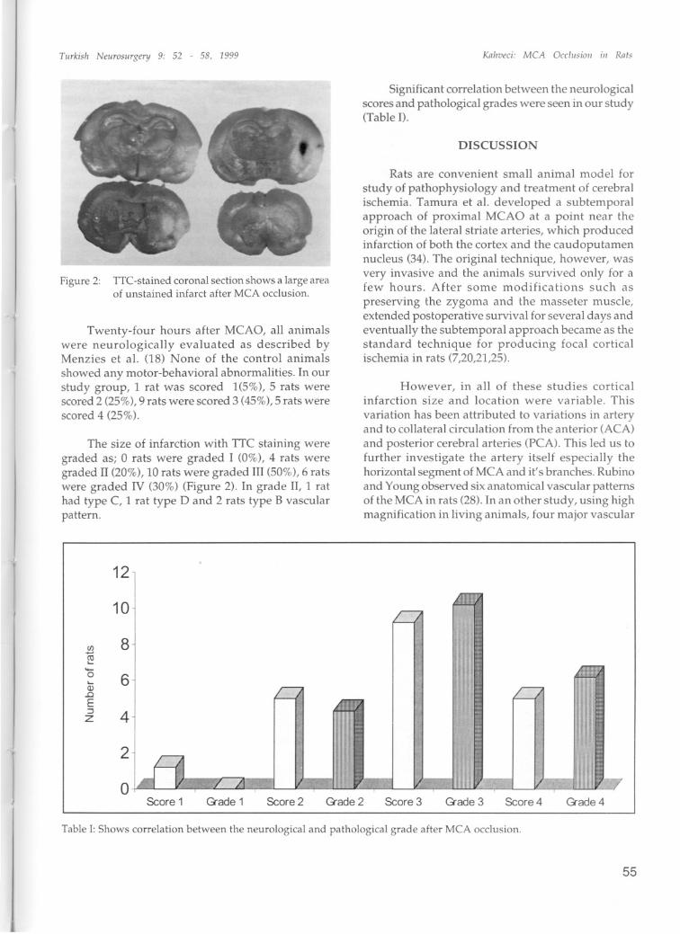
Figure 2: TTC-stained eoronal seetion shows a large areaof unstained infaret after MCA oeelusion.
Twenty-four hours after MCAO, all animalswere neurologically evaluated as described byMenzies et aL. (18) None of the control animalsshowed any motor-behavioral abnormalities. In ourstudy group,I rat was scored 1(5%), 5 rats werescored 2 (25%),9 rats were scored 3 (45%),5 rats werescored 4 (25%).
The size of infarction with TIC staining weregraded as; O rats were graded I (0%), 4 rats weregraded II (20%), 10 rats were graded III (50%), 6 ratswere graded IV (30%) (Figure 2), In grade II, 1 rathad type C, 1 rat type D and 2 rats type B vascularpattern.
Kahveci: MCA üeeliisioii iii Ral,
Significant correlation between the neurologicalscores and pathological grades were seen in our study(Table n.
DISCUSSION
Rats are convenient small animal model for
study of pathophysiology and treatment of cerebralischemia. Tamura et aL. developed a subtemporalapproach of proximal MCAO at apoint near theorigin of the lateral striate arteries, which producedinfaretion of both the cortex and the caudoputamennucleus (34). The original technique, however, wasvery invasive and the animals survived only for afew hours. Af ter some modifications such as
preserving the zygoma and the masseter muscle,extended postoperative survival for several days andeventually the subtemporal approach became as thestandard technique for producing focal corticalischemia in rats (7,20,21,25).
However, in all of these studies corticalinfarction size and location were variable. This
variation has been attributed to variations in arteryand to collateral eirculation from the anterior (ACA)and posterior cerebral arteries (PCA). This led us tofurther investigate the artery itse1f especially thehorizontal segment of MCA and it' s branches. Rubinoand Young observed six ana tomical vascular pa tternsof the MCA in rats (28). In an other study, us ing highmagnification in living animals, four major vascular
12
CIL
ro•...
"-o•...Q).oE:JZ
10
8
6
4
2
OScore 1 <?iade 1 Score2 <?iade 2 Score 3 <?iade 3 Score4 <?iade 4
Table i: Shows correlation between the neurologieal and pathological grade after MCA oeelusion.
55

Turkish Neiirosiirgery 9: 52 - 5B, 1999
patterns of the artery was demonstrated (18). In ourstudy we identified Gve vascular patterns and twoof them were major and constituted 88% of our rats.Type A (45%) and Type B (43%) resembled theMenzies et al.'s Type A (51%) and Type B (26.6%)findings, respectively. Although there was a widedifference between Type B, occurrence of Type Awere almost equal. The remaining three patternswere either not observed or less than 2.5% in our
study. Our Type B (43%) pattern resembled Type Bof Rubino and Young's (28) and our type A (45%) isType C (17%)respectively. OveralL,only 43% of theirrats' MCA pattern were similar to those we identified.Due to many variations of MCA, proximal occlusionbelow the rhinal fissure produced 13-67% lesions,whereas occlusion of larger lengths of the arteryresulted in 100 % infaretion rate (2). it was suggestedthat the latter model provided a more efficientcompromise of the collateral blood supply fromretrograde flow (18). In their model Menzies et aL.(18) occluded the longer length of the artery and inaddition the major MCA branches were alsocoagulated. In our study after determining theanatomical variations of the MCA and its branchesin our strain of rats, and added the procedure ofdividing the artery. By taking these steps, we wereable to achieve 80% grade III and IV infarcted areaswith less coagulation of the horizontal segment. Thereason we chose not to use alarger approach to theMCA is that exposure of alarger area of the brainduring craniotomy might alter blood-brain barrierpermeability and intracranial pressure af terinfaretion (5,11).Forsting et aL.,demonstrated in theirstudy that cranieetomy for cerebral ischemia in ratsnot only reduces mortaIity but also significantlyimproves outcome and reduces infaret size (9).
Using asimilar occlusion technique, Menzieset aL.,observed considerable variability in their studyand just over half of the infaret was charaeterized aslarge (18). The authors used two different type ofcoagulation in their study. In one experiment, themiddle cerebral artery was cauterized from thelateral branch to the inferior cerebral vein. In the other
which yielded more large infarets, the artery wascoagulated more extensively, including the mainbranchings for 7-9mm distance. The authors claimedthat more standardized and reproducible lesionswere produced when a longer lenght of the MCAand its branches were occluded. In our study, usingthe similar tecnique with less segmental coagulation(3-4 mm) and dividing the main trunk aftercoagulation, produced grade III-IV lesions in 80% ofthe rats. Because coagulation alone occasionally is
56
Kiiliveei: MCA Gec1iisioli iii Ra/s
not accomplish complete occlusion, we preferred todivide the main trunk to prevent collateral eirculationfrom the anterior cerebral and posterior cerebralarteries. EI-Sabban et aL.,clearly demonstrated thateleetrocoagulation produced temporary occlusiononly with embolization and in most cases partialrecanalization followed the coagulation (8). if weused this model to interpret the basis of eitherinfaretion or therapeutic efficacy of drug treatment,there would be question as to whether the infaretswere embolic or thrombotic. Thus we thought thatdividing the artery might eliminate the possibilitythat the ischemia was produced by distalembolisation.
The assesment of clinical neurological status ofthe animal accurately with stroke lesions is importantfor evaluation of the treatment. The clinical methodof neurological evaluation af ter MCAO used byBederson et aL. (2) designates three neurologicalgrades and Menzies et aL. (18) Iists four grades.According to Menzies et aL.(18), in Bederson et al'sgrading system, the application of the test is sensitiyeonly grossly paretic rats as also admitted by theauthors. This is also noticed in our studyand in ourexperiment Menzies et aL.'s, grading system was usedbecause it is appIicable to even mil d neurologicaldeficits and also easily applied. For morphologicevaluation TTC staining and computer analysismethods are widely used for deteetion of lesions. TICstaining were shown as highly correlative withhistological evaluations (24).Also 3 days after MCAOthere is known to be an assoeiation between color
differences between hemispheres and staining withTIC (18).
In our study the frontal and, indireetly, thepyriform branches were in collateral communicationwith the ACA, whereas the parietal and temporalbranches collateralized with branches of the PCA.
This explains why distal occlusion of MCA doesn'tresult in infarction. Cortical anastomoses are the keyto the extent of infarction after a MCAO. This wasclearly shown by the extent of the maximallesionsin the Menzies et aL. study (18). In addition, asobserved in our study, the margins of infaretion neverexceeded the sites of cortical interarterial
anastomoses sites. Although it was stated by Menzieset aL. (18), that none of the vascular anatomicalpatterns played a significant role in modulating theextent and severity of cortical ischemia, we foundthat the type E pattern, such as bifurcating or forkingof the arteries may change the outcome of infarct.And also in our study in grade II size of infaretion,

Turkish Neurosurgery 9: 52 - 58, 1999
rats had rarer type of anatamical patterns such astype B, C or D.
Anather important faetor in infaretian postMCAO appears to be body weight, and, thusindireetly the age of the animals (l8). In a study byCoyle (6) no young rats weighing below 150 gdeveloped infarets after proximal MCAO.
Defining of the variability in the anatamicalpatterns of MCA in the rats and alsa eoagulation ofthe horizontal segment for at least 3 mm lenght playsvery important role for sueeess of the study. And alsaeliminating the rarer vaseular patterns the incidenceand size of infaretian eould be inereased and well
predieted large size of infaretian with eorrelation ofthe infaret size with neurologie defieits eould easilybe aehieved in our study.
Our results indieate that although SpragueDawley rats are the same strain due to theirinbreeding the anatamy of the art eries considerablevaries. So any investigator planning to use MCAisehemIa model should establish normal anatomIe
pattern of theIr rats before starting the researeh.
Correspondence: Nevzat KahveciUludag UniversitySchool of Medicine
Department of PhysiologyGörükle 16059 Bursa TurkeyTel: 224 442 8855 Fax: 224 442 8034
REFERENCES
1. Bederson JB, Germano IM, Guarino L: Cortical bloodflow and cerebral perfusion pressure in a newnoncraniotomy model of subarachnoid hemorrhagein the rat. Stroke 26:1086-1091, 1995
2. Bederson JB, Pitts LH, Tsuji M, Nishimura MC, DavisRL, Bartkowski H: Rat middle cerebral arteryocdusion: evaluation of the model and developmentof aneurologic examination. Stroke 17:472-476, 1986
3. Brierley JB, Ljunggren B,Siesjo BK:Neuropathologicalalterations in rat brain after complete ischemia due toraised intracranial pressure, in Lundberg, N, Ponten,W, and Brock M (eds), Intracranial pressure, volume2, Berlin: Springer-Verlag, 167-171
4. Cole DJ, Drummond JC, Ghazal EA, Shapiro HM: Areversible component of cerebral injury as identifiedby the histochemical stain 2,3,5-triphenyltetrazoliumchloride (TTC). Acta Neuropathol (BerI) 80:152-155,1990
5. Coyle P: Arterial patterns of the rat rhinencephalonand related structures. Exp Neurol 49:671-690, 1975
Kahveci: MCA Decilisioii hi Rats
6. Coyle P: Middle cerebral artery ocdusion in the youngrat. Stroke 13:855-859, 1982
7. Duverger D, MacKenzie ET: The quantification ofcerebral infarction following focal ischemia in the rat:influence of strain, arterial pressure, blood glucoseconcentration, and age. J Cereb B100d Flow Metab8:449-461, 1988
8. EI-Sabban F, Reid KH, Zhang YP, Edmonds HL, Jr.:Stability of thrombosis induced by electrocoagulationof rat middle cerebral artery. Stroke 25:2241-2245,1994
9. Forsting M, Reith W, Schabitz WR, Heiland S, vonKummer R, Hacke W, Sartor K: Decompressivecraniectomy for cerebral infarction. An experimentalstudy in rats. Stroke 26:259-264, 1995
10. Garcia JH: A reliable method to ocdude a middlecerebral artery in Wistar rats [Ietter]. Stroke 24:14231423, 1993
11. Hudgins WR, Garcia JH: Transorbital approach to themiddle cerebral ar tery of the squirrel monkey: atechnique for experimental cerebral infarctionapplicable to ultrastructural studies. Stroke 1:107-111,1970
12. Kawamura S, Yasui N, Shirasawa M, Fukasawa H:Rat middle cerebral artery occlusion using anintraluminal thread technique. Acta Neurochir.(Wien).109:126-132, 1991
13. Kuge Y,Minematsu K, Yamaguchi T, Miyake Y:Nylonmonofilament for intraluminal middle cerebral ar teryocdusion in rats. Stroke 26:1655-7. discussion 1658,1995
14. Laing RJ, Jakubowski J, Laing RW: Middle cerebralar tery ocdusion without craniectomy in rats. Whichmethod works best? Strok e 24:294-7. discussion 2978, 1993
15. Ljunggren B, Ratcheson RA, Siesjo BK: Cerebralmetabolic state following complete compressionischemia. Brain research 73:291-307, 1974
16. Longa EZ, Weinstein PR, Carlson S, Cummins R:Reversible middle cerebral artery ocdusion withoutcraniectomy in rats. Stroke 20:84-91, 1989
17. Memezawa H, Smith ML, Siesjo BK:Penumbral tissuessalvaged by reperfusion following middle cerebralartery ocdusion in rats. Stroke 23:552-559, 1992
18. Menzies SA, Hoff JT, Betz AL: Middle cerebral ar teryocdusion in rats: a neurological and pathologicalevaiuation of a reproducible modeL. Neurosurgery31:100-6. discussion 106-7, 1992
19. Nagasawa H, Kogure K: Correlation between cerebralblood flow and histologic changes in a new rat modelof middle cerebral artery ocdusion. Stroke 20:10371043, 1989
20. Nakayama H, Dietrich WD, Watson BD, Busto R,Ginsberg MD: Photothrombotic ocdusion of ratmiddle cerebral artery: histopathological andhemodynamic sequelae of acute recanalization. JCereb Blood Flow Metab 8:357-366, 1988
21. Nakayama H, Ginsberg MD, Dietrich WD: (S)emopamil, a novel calcium channel bloeker and
57

Tiirkish Neiirosiirgery 9: 52 - 58, 1999
serotonin S2antagonist, markedly reduces infarct sizefollowing middle cerebral artery occlusion in the rat[see comments]. Neurology 38:1667-1673, 1988
22. Nordstrom CH, Rehncrona S, Siesjo BK: Effects ofphenobarbital in cerebral ischemia. Part II: restitutionof cerebral energy state, as well as of glycolyticmetabolites, citric acid cycle intermediates andassociated amino acids after pronounced incompleteischemia. Stroke 9:335-343, 1978
23. Nordstrom CH, Siesjo BK: Effects of phenobarbital incerebral ischemia. Part i: cerebral energy metabolismduring pronounced incomplete ischemia. Stroke 9:327335, 1978
24. Park CK, Mendelow AD, Graham DI, McCulloch J,Teasdale GM: Correlation of triphenyltetrazoliumchloride perfusion staining with conventionalneurohistology in the detection of early brainischaemia. Neuropathol.Appl.Neurobiol. 14:289-298,1988
25. Prado R, Ginsberg MD, Dietrich WD, Watson BD,Busto R: Hyperglycemia increases infarct size incollaterally perfused but not end-arterial vascularterritories. J Cereb Blood Flow Metab 8:186-192, 1988
26. Pulsinelli WA, Brierley JB: A new model of bilateralhemispheric ischemia in the unanesthetized rat. Stroke10:267-272,1979
27. Rabb CH: Nylon monofilament for intraluminalmiddle cerebral ar tery occlusion in rats [letter.comment]. Stroke 27:151, 1996
58
Knhveci: MCA OCc1I1Sioli in Rats
28. Rubino GJ, Young W: Ischemic corticallesions afterpermanent occlusion of individual middle cerebralartery branches in rats. Stroke 19:870-877, 1988
29. Saiford LG, Plum F, Brierley JB: Graded hypoxiaoligemia in rat brain. II. Neuropathological alterationsand their implications. Arch.Neurol. 29:234-238, 1973
30. Salgado A, Jones SC, Furlan AJ, Korfali E, MarshallSA, Little JR: Bimodal treatment with nimodipine andlow molecular weight dextran for focal cerebralischemia in the rat. Ann Neurol26: 621-627, 1989
31. Schmid-EIsaesser R, Zausinger S, Hungerhuber E,Baethmann A , Reulen HJ: A critical reevaluation ofthe intraluminal thread model of focal cerebral
ischemia: evidence of inadvertent prematurereperfusion and subarachnoid hemorrhage in rats bylaser-Doppler flowmetry. Stroke 29:2162-2170, 1998
32. Siemkowicz E, Hansen AJ: Clinical restitutionfollowing cerebral ischemia in hypo-, norm 0- andhyperglycemic rats. Acta Neurol.Scand. 58:1-8, 1978
33. Smith ML, Bendek G, Dahlgren N, Rosen i, WielochT, Siesjo BK:Models for studying long-term recoveryfollowing forebrain ischemia in the rat. 2. A 2-vesselocclusion modeL. Acta Neurol.Scand. 69:385-401, 1984
34. Tamura A, Graham DI, McCulloch J, Teasdale GM:Focal cerebral ischaemia in the rat: 1. Description oftechnique and early neuropathological consequencesfollowing middle cerebral artery occlusion. J CerebBlood Flow Metab 1:53-60, 1981