
Microsome S9 Prep Protocol
22
163 From: Methods in Pharmacology and Toxicology Optimization in Drug Discovery: In Vitro Methods Edited by: Z. Yan and G. W. Caldwell © Humana Press Inc., Totowa, NJ 11 In Vitro Drug Metabolite Profiling Using Hepatic S9 and Human Liver Microsomes Wu-Nan Wu and Linda A. McKown Summary Following oral administration to animals and humans, drugs are absorbed, transported via portal circulation to the liver, and metabolized primarily via this organ. In general, drugs are predominantly metabolized by the oxidation of parent drug, which is typically mediated by cytochrome P450 (CYP450) enzymes. To a lesser degree, flavin monooxidation (FMO), as well as the reduc- tion or cleavage of the parent drug via enzymatic (i.e., esterase and amidase) or nonenzymatic hydrolysis, forms other phase I metabolites. Subsequent conju- gation (phase II reaction) of the phase I metabolites can produce glucuronide, sulfate, glutathione, glycine, and acetate conjugated metabolites. In many cases, hepatic in vitro metabolism studies can yield valuable preliminary information on the in vivo metabolism of a compound of interest by the liver. Experimental in vitro hepatic systems using hepatocytes, 9000g supernatant (S9), and microsomal fractions are presently used to characterize the in vitro metabolism of xenobiotics. Following the incubation of drugs with either of the systems above, solvent or solid-phase extraction, radio-TLC ( 14 C/ 3 H-labeled drugs), high-performance liquid chromatography (HPLC) (radiolabeled or unlabeled), liquid chromatography/mass spectrometry (LC/MS), nuclear magnetic reso- nance (NMR), and derivatization (phenolic, alcoholic, carboxylic, and/or amino metabolites) techniques are commonly used to analyze and evaluate the meta- bolic stability of drugs (percentage of parent remaining), as well as to quantify, characterize, and identify drug metabolites and their derivatives. In this chapter, valuable in vitro methods using animal and human hepatic S9, as well as human liver microsomal fractions, and unique techniques for estimating and under- standing metabolic stability, as well as profiling and identifying metabolites,
Transcript of Microsome S9 Prep Protocol
In Vitro Drug Metabolite Profiling 163
163
From: Methods in Pharmacology and ToxicologyOptimization in Drug Discovery: In Vitro Methods
Edited by: Z. Yan and G. W. Caldwell © Humana Press Inc., Totowa, NJ
11
In Vitro Drug Metabolite Profiling Using HepaticS9 and Human Liver Microsomes
Wu-Nan Wu and Linda A. McKown
SummaryFollowing oral administration to animals and humans, drugs are absorbed,
transported via portal circulation to the liver, and metabolized primarily via thisorgan. In general, drugs are predominantly metabolized by the oxidation ofparent drug, which is typically mediated by cytochrome P450 (CYP450)enzymes. To a lesser degree, flavin monooxidation (FMO), as well as the reduc-tion or cleavage of the parent drug via enzymatic (i.e., esterase and amidase) ornonenzymatic hydrolysis, forms other phase I metabolites. Subsequent conju-gation (phase II reaction) of the phase I metabolites can produce glucuronide,sulfate, glutathione, glycine, and acetate conjugated metabolites. In many cases,hepatic in vitro metabolism studies can yield valuable preliminary informationon the in vivo metabolism of a compound of interest by the liver. Experimentalin vitro hepatic systems using hepatocytes, 9000g supernatant (S9), andmicrosomal fractions are presently used to characterize the in vitro metabolismof xenobiotics. Following the incubation of drugs with either of the systemsabove, solvent or solid-phase extraction, radio-TLC (14C/3H-labeled drugs),high-performance liquid chromatography (HPLC) (radiolabeled or unlabeled),liquid chromatography/mass spectrometry (LC/MS), nuclear magnetic reso-nance (NMR), and derivatization (phenolic, alcoholic, carboxylic, and/or aminometabolites) techniques are commonly used to analyze and evaluate the meta-bolic stability of drugs (percentage of parent remaining), as well as to quantify,characterize, and identify drug metabolites and their derivatives. In this chapter,valuable in vitro methods using animal and human hepatic S9, as well as humanliver microsomal fractions, and unique techniques for estimating and under-standing metabolic stability, as well as profiling and identifying metabolites,

164 Wu and McKown
will be discussed for use in drug discovery and drug evaluation phases of adrug’s development.
Key Words: In vitro drug metabolism; hepatic S9; liver microsomes;incubation; animals; human; metabolic stability; metabolite profiling and iden-tification, phase I and phase II metabolism; metabolic pathways; thin-layerchromatography (TLC); HPLC; LC/MS and MS/MS; NMR; derivatization.
1. IntroductionDrugs that are orally administered to animals or humans and are absorbed to
some degree from the gastrointestinal tract are subsequently transportedthrough the portal-vein circulation to the liver. These absorbed drugs are readilysubjected to potential hepatic metabolism, followed by biliary or renal elimi-nation, and excreted in the feces or urine, respectively (1–3). In general, thepredominant pathway for metabolite formation is via the oxidation of the par-ent drug and, in some cases, the reduction and cleavage of parent drug (phase Ireaction), followed by the conjugation of phase I metabolites with highly polarmolecules, such as glucuronic acid, sulfuric acid, glucose, acetic acid, glu-tathione, cysteine, glutamic acid, taurine, and so forth, to form phase II conju-gated metabolites (1–4).
In the liver, the enzymes catalyzing drug metabolism reactions are locatedmainly in the endoplasm (microsomes) and soluble fraction of the cytoplasma(cytosol), along with small amounts in lysosomes, mitochondria, and nuclei(1–4). The important enzymes responsible for phase I oxidation are primarilythe isoenzymes of the cytochrome P450 (CYP450) family (5–7) and, to a lesserdegree, flavin monooxidase (FMO) (1–5,8), alcohol dehydrogenase, aldehydedehydrogenase, and aldehyde oxidase (1–4). These enzymes are present in themicrosomal fraction. The major human CYP450 enzymes mediating drug oxi-dation reactions consist of the following seven CYP450 isoforms: 1A2, 2A6,2C9, 2C19, 2D6, 2E1, and 3A4 (5–7). Of these isoforms, the two most impor-tant ones are CYP3A4 and CYP2D6, which have been documented as beingresponsible for the formation of most oxygenated metabolites (5–7).
Enzymes mediating phase I reduction reactions such as azo, nitro, andquinone reduction are present in both microsomes and the cytosol (1–4). Like-wise, enzymes responsible for hydrolysis reactions such as epoxide hydrolaseare present in both microsomes and cytosol. However, carboxyesterase ispresent in microsomes only (1–4). The enzymes responsible for phase II reac-tions such as glucuronidation and glutathione conjugation are present prima-rily in microsomes, whereas the one responsible for sulfation conjugation islargely present in the cytosol (1–4,9–12). The major enzymes mediating phaseII reactions are UDP-dependent glucuronyltransferase (UGT), sulfotransferase

In Vitro Drug Metabolite Profiling 165
(PST), and glutathione-S-transferase (GST), which are present as multiple iso-mers (9–12).
Drug metabolism has always been an important research area for drugdiscovery and drug development. From the new drug screening programs indrug discovery to drug evaluation and drug development, where identifyingso-called “metabolically stable” drugs with better oral bioavailability is con-sidered critical, drug metabolism is now one of the pivotal factors for furtherinvestigation of new therapeutic agents. Because the liver is the major targetorgan for drug metabolism and hepatic subcellular materials are readily avail-able, high-throughput screening assays and preliminary in vitro metabolismdata generated from hepatic S9, microsomes, and hepatocytes have becomevaluable in evaluating the metabolic stability of drugs and the acquisition ofearly structural information of drug metabolites (13,14). Presently, hepatic S9,liver microsomes, and hepatocytes are the experimental in vitro systems usedfor the metabolic investigation of novel drugs. The understanding of majormetabolic pathways of xenobiotics via the identification and quantification ofmetabolites provides medicinal chemists and pharmacologists the informationnecessary to make chemical and structural modifications for increasing drugefficacy, decreasing drug toxicities, and implementing the synthesis ofmetabolites with increased biological activity (13–16). This chapter focuses onsharing the methodology used in determining the in vitro phase I metabolismof drugs using animal and human hepatic S9 and human liver microsomalincubations, thin-layer chromatography (TLC), high-performance liquid chro-matography (HPLC) and liquid chromatography/mass-spectrometric (LC/MS)profiling, and unique techniques in the identification of metabolites.
2. Materials2.1. Hepatic S9/Liver Microsomal Incubations
2.1.1. Generation of Small Animal Hepatic S9 (see Note 1)
1. Trishydroxymethylaminomethane hydrochloride (Tris-HCl) (Sigma Co., St.Louis, MO).
2. Potassium chloride (KCl) (Sigma Co., St. Louis, MO).3. Hydrochloride (HCl) (Fisher Scientific, Fair Lawn, NJ).4. Potassium hydroxide (KOH) (Fisher Scientific, Fair Lawn, NJ).5. Mouse or rat (gender and strain of choice).6. Decapitator.7. Surgical scissors.8. Top-loading balance.9. Beakers, glass-stoppered graduated cylinder.
10. Homogenizer (Brinkman Polytron®).

166 Wu and McKown
11. Plastic sorval tubes.12. High-speed refrigerated centrifuge with rotor (capable of generating 9000g at 4°C).
2.1.2. Generation of Rodent Microsomes
1. High-speed centrifuge tubes.2. 4-(2-Hydroxyethyl)-1-piperazineethanesulfonic acid (HEPES) (Aldrich Chemi-
cal Co., Milwaukee, WI).3. High-speed refrigerated centrifuge with type 40 rotor (capable of speeds
>105,000g at 4°C).
2.1.3. Human Hepatic S9 and Microsomes (see Note 2)
1. Human hepatic S9 and microsomes (XenoTech, L.L.C., Kansas City, KS, or InVitro Technologies, Baltimore, MD).
2.1.4. Incubation
1. Nicotinamide adenine dinucleotide phosphate (NADP) (Sigma Co., St. Louis, MO).2. Reduced nicotinamide adenine dinucleotide phosphate (NADPH) (Sigma Co.,
St. Louis, MO).3. Magnesium chloride (MgCl2) (Sigma Co., St. Louis, MO).4. Glucose-6-phosphate (G-6-P) (Sigma Co., St. Louis, MO).5. Beakers.6. Wide-mouth vials (Wheaton, 16 mL) or 25-mL Erlenmeyer flasks.7. Dubnoff Metabolic Shaker Incubator (Precision Scientific, Chicago, IL).8. Ethyl acetate (Burchick & Jackson Laboratories, Muskegon, MI).9. Acetone (Burchick & Jackson Laboratories, Muskegon, MI).
10. Dry ice.
2.2. Sample Preparation
1. Extraction solvents: hexane, ether, dichloromethane, ethyl acetate, acetonitrile,methanol (Burdick & Jackson Laboratories, Muskegon, MI).
2. Acetic acid (EM Science, Gibbstown, NJ).3. Ammonium hydroxide (EM Science, Gibbstown, NJ).4. Solid-phase extraction cartridges: C18, C8, C4, C2 (Whatman Inc., Clifton, NJ).5. Amberlite®-XAD2 resin (Rohm & Haas Co., Philadelphia, PA).6. Compact II centrifuge (Becton Dickinson & Co., Sparks, MD).7. Pipets (Wheaton, Millville, NJ).8. Turbo Vap® evaporator (Zymark Corp., Hopkinton, MA).
2.3. Sample Derivatization Reagents
1. Acetylation: acetic anhydride and pyridine (EM Science, Gibbstown, NJ).2. Methylation: N-methyl-N-nitroso-p-toluenesulfonamide (Diazald®) (Aldrich
Chemical Co., Milwaukee, WI).

In Vitro Drug Metabolite Profiling 167
2.4. TLC and HPLC Chromatography
1. TLC and HPLC solvents: hexane, chloroform, dichloromethane, ethyl acetate,methanol, ethanol, acetonitrile, tetrahydrofuran, water (Burdick & Jackson Labo-ratories, Muskegon, MI).
2. Acetic acid (EM Science, Gibbstown, NJ).3. Ammonium hydroxide (EM Science, Gibbstown, NJ).4. Ammonium acetate (Aldrich Chemical Co., Milwaukee, WI).5. Formic acid (Aldrich Chemical Co., Milwaukee, WI).6. Trifluoroacetic acid (TFA) (Aldrich Chemical Co., Milwaukee, WI).7. TLC plates: silica gel GF and neutral alumina (normal phase); C18, C8, C2, phenyl
(reverse phase) (Anatech, Inc., Newark, DE).8. TLC development tank or jar.9. TLC Radiochromatogram Imaging System (BID 100) (Bioscan, Inc., Washing-
ton, DC).10. HPLC system (Beckman Instrument Co., Fullerton, CA).11. HPLC Radioactive Monitor (RAM) (RAMONA, IN; US Service Corp., Fair-
field, NJ).
2.5. LC/MS and MS/MS Application
1. PE Sciex API III-Plus and API 3000 Mass Spectrometers (Perkin-Elmer SciexInstruments, Thornhill, Ontario, Canada).
2. HPLC system interfaced to MS-Hitachi HPLC solvent delivery system (L-6200AIntelligent pump) (Hitachi Co., Tokyo, Japan).
3. HPLC column: C18, C8, C2, phenyl, cyano (Agilent Technologies, Fitchburg,MA); LiChorsorb RP-2 (C2), RP-8 (C8), and RP-18 (C18) (Brownlee Laborato-ries, Inc., Santa Clara, CA).
3. Methods3.1. Hepatic S9/Microsomal Generation
3.1.1. Small Animal Hepatic S9 Preparation
3.1.1.1. BUFFER PREPARATION
The base buffer used in the preparation of any hepatic S9 is 1.15% KCl in0.05 M Tris-HCl (pH 7.4) buffer. This may be prepared ahead of time as fol-lows (see Note 3):
Tris-HCl: 6.055 gKCl: 1.15 gWater (distilled) q.s.: 1000 mLAdjust pH to 7.4 with either HCl or KOH

168 Wu and McKown
3.1.1.2. HEPATIC S9 PREPARATION
A homogenate is prepared by first euthanizing a small rodent by decapita-tion. The liver is removed (wet weight determined), minced, and homogenizedusing a Brinkmann Polytron® in cold Tris-HCl buffer to a total volume of ~4 ×the wet liver weight (see Note 4). The homogenate is divided into equal vol-umes into the sorval centrifuge tubes and spun at 9000g for 30 min. The super-natant (S9) is removed and should be used immediately or stored at –70°C tomaintain viability for use at a later date. Typically, a male SD rat is used (seeNote 5).
3.1.2. Microsomal Preparation
Microsomal suspensions are prepared by taking a measured volume of S9and centrifuging again at 105,000g (type 40 rotor) in a high-speed centrifugefor 1 h. The subsequent supernatant is discarded, and the pellet is gently resus-pended in cold 0.1 M HEPES/1.15% KCl buffer up to a volume one-half that ofthe original S9 (approx 12–16 mg of microsomal protein per milliliter withthis method). The microsomal fraction should be used immediately or storedat –70°C or lower for use at a future time.
3.2. Hepatic S9/Microsomal Incubation
3.2.1. Hepatic S9 Incubation Preparation
Prior to an incubation, each of the following cofactors is prepared fresh dailyin cold Tris-HCl buffer: (1) 5 mM MgCl2 (i.e., 127 mg MgCl2 × 6 H2O in 25 mLTris-HCl), (2) 5 mM glucose-6-phosphate (i.e., 190 mg glucose-6-phosphatein 25 mL Tris-HCl), and (3) 0.5 mM NADP (i.e., 47.5 mg in 25 mL Tris-HCl).
To maintain a 5-mL volume in each 25-mL Erlenmeyer flask or suitablevial, 1 mL each of cold Tris-HCl buffer, MgCl2, glucose-6-phosphate, andNADP solutions and the desired S9 (prepared in-house or purchased outside;~20 mg/mL protein) are placed into each flask on ice in the order given (seeNote 6). A flask containing drug but no S9 fraction, as well as one containinga compound with a documented in vitro metabolic profile (i.e., etoperidone ortramadol), is also incubated to serve as environmental and enzyme activitycontrols (see Note 7). Finally, the drug solution, typically 2.5 mg/mL, as wellas reference standards can be spiked into each vial as a small volume (5–50) inmethanol, ethanol, or dimethylsulfoxide (DMSO) or as an aqueous solutiondissolved in Tris-HCl buffer (see Note 8) so that the spike (typically 20 μL)gives a final desired concentration of drug (1–10 μg/mL or 1–20 μM/mL) (seeNote 9). Each chilled flask is placed in the Dubnoff Metabolic Shaker Incuba-tor and incubated in an open-air atmosphere for up to 120 min at 37°C. Aliquots(1 mL) may be removed and placed into prelabeled tubes at any time for analy-

In Vitro Drug Metabolite Profiling 169
sis. Equal volumes of ethyl acetate or acetonitrile are added to aliquots toterminate the reaction (see Note 10). All samples are then immediately frozenin a dry ice/acetone bath and stored at –20°C or lower pending analysis (17–27).When the incubation is complete, each remaining sample is transferred to aprelabeled storage vial, deactivated, and stored as described previously (see Note11). This hepatic S9 system primarily generates phase I metabolites.
3.2.2. Hepatic Microsomal Incubation Preparation
Incubations with microsomes are prepared and carried out using the samemethodology described for the S9 mixtures, substituting 1 mL of the microso-mal suspension for the supernatant (see Note 12) and replacing 1 mL ofNADPH solution for the NADP solution (17,22,23) (see Note 13). The hepaticmicrosomal incubation also chiefly produces phase I metabolites (see Note14). The addition of uridine 5' diphosphoglucuronic acid (UDPGA) to theincubation mixture, as well as an increased incubation duration, may formglucuronide conjugates (phase II metabolites) because of the presence of UGTin the microsomal fraction (28).
3.3. Sample Preparation for Metabolic Profiling
Acidified and or basified hepatic S9 and liver microsomal incubates can beextracted using organic solvents (i.e., ether, ethyl acetate, and dichloromethanefor organic extractable drugs), or solid-phase extraction, such as C18, C8, or C2cartridges or Amberlite®-XAD2 resin, can be used for nonorganic extractabledrugs. The extract is evaporated to dryness to yield a residue, which is recon-stituted in methanol or acetonitrile and applied on the TLC plate or injectedonto the HPLC system. For example, unchanged RWJ-34130 and its metabo-lites, generated from hepatic S9 and microsomal incubations, were profiled byHPLC, and then the drug-related peaks were individually collected from theHPLC effluents for subsequent MS analysis (Fig. 1) (17,29).
3.4. Radio-TLC Metabolic Profiling and Isolation
This method only applies to radiolabeled drugs such as the two 14C-labeleddrugs, 14C-fenoctimine and 14C-fenobam, which has an in vitro metabolismthat can be characterized by using a radio-TLC method that was previouslyreported (18,19). In general, the organic extract residue from the incubate isreconstituted in a minimal amount of organic solvent, or the aqueous incubate(nonorganic extractable) is applied directly as a band or spot on a 20 × 20-cmTLC plate, along with reference samples (parent drug, synthetic metabolites),and developed in organic solvent systems (acidic, basic, and neutral). Thedeveloped plate is radioscanned to obtain the TLC metabolic profile, followedby visualization under a short-wavelength UV light to localize the drug-related

170 Wu and McKown
Fig. 1. HPLC profiles of the rat hepatic S9 and microsomal incubation ofRWJ-34130.

In Vitro Drug Metabolite Profiling 171
zones or spots. These areas are removed by scraping and extracted with organicsolvents (a mixture of methanol and dichloromethane or methanol and ethylacetate). The extracts are filtered, followed by evaporation to yield dry resi-dues, which are then analyzed using mass spectrometry (MS) and nuclear mag-netic resonance (NMR) to gain structural information of metabolites.Derivatization using, for example, diazomethane for reacting with phenolicand carboxyl metabolites to form methyl ethers and methyl esters, respectively,or acetic anhydride/pyridine for reacting with the alcoholic, phenolic, andamino metabolites to form acetyl derivatives can be valuable for further struc-tural confirmation of metabolites (19–22,25,27,30).
3.5. Radio-HPLC and HPLC Metabolic Profiling
The typical HPLC system used is a gradient liquid chromatograph with aUV detector. A LiChrosorb RP-2 (C2) guard and analytical column (5 μm, 130× 4.6 mm) are used for sample analysis at a flow rate of 2 mL min–1 for themobile phase (see Note 15). The gradient elution is conducted from 2% to100% B in 20 min, with water (mobile phase A) and methanol (mobile phaseB) both containing 0.02% ammonium acetate. The in vitro metabolic profilingof RWJ-34130 from rat hepatic S9 fraction and liver microsomal incubationswas conducted using the HPLC conditions described above (Fig. 1) (17,29).Unchanged drug and metabolites obtained from these samples were isolated byHPLC and analyzed by MS and NMR (17,19,21,30). An estimate of the rela-tive percentages of unchanged drug and each metabolite in a given sample wasmade using the integrated peak intensity generated by the HPLC chromato-gram for the unlabeled drugs and by using the integrated radioactive peaksfrom the RAM (radioactive monitor) for the radiolabeled drugs (17,30).
3.6. LC/MS and MS/MS Metabolic Profiling
Following organic solvent/solid-phase (C2, C8, C18, Amberlite®-XAD2resin) extraction of each acidified, basified, or neutral incubate (1 mL) (seeNote 16), the residue is reconstituted in a 0.2- to 0.5-mL buffer (acetonitrile ormethanol/water [50/50, v/v] with 5 mM ammonium acetate, pH 4.0), centri-fuged, and then analyzed via a 20-μL flow-injection into a PE Sciex API III-Plus or PE Sciex API 3000. These are triple quadruple mass spectrometers,interfaced to a Hitachi HPLC (C18, C8, or C2 column) solvent delivery system(L-6200 A Intelligent pump) via an ionsprayer using nitrogen as the curtainand nebulizing gas and argon (API III Plus) or nitrogen (API 3000) as thecollision gas for MS/MS analysis. The isocratic mobile phase for this system isthe same buffer as described for residue reconstitution, delivered at a flow rateof 0.5 to 0.1 mL min–1 (see Note 17). For each sample, the relative percentageof unchanged drug and its metabolites is estimated using the integrated chro-

172 Wu and McKown
matograms generated by the Sciex API-III Plus or API 3000 Q1 scan MS (totalion chromatogram). These data are not absolutely quantitative because of thepotential differences in the degree of ionization of each analyte. However, theyare reproducible (20–22,25,27) (see Note 18).
3.7. Metabolite Derivatization
3.7.1. Methyl Derivatization (see Note 19)
Each incubate (1 mL) extract residue is dissolved in 0.2 to 0.5 mL of metha-nol, an excess amount of ethereal diazomethane (generated from Diazald with1 N methanolic KOH solution) is added, and the mixture is allowed to react atroom temperature overnight. This mixture is subsequently evaporated to dry-ness to yield a methylated residue consisting of the phenolic and carboxylicmetabolites derivatized to methyl ethers and methyl esters, respectively. Eachresidue is then further analyzed using LC/MS for the confirmation of metabo-lites (19,21,22,25,27,30).
3.7.2. Acetyl Derivatization
Each incubate (1 mL) extract residue is dissolved in 0.2 mL of acetic anhy-dride and 0.1 mL of pyridine and is allowed to react at room temperature for4 h. Then, 5 mL of cold water is added to each sample followed by organicsolvent extraction (ether, ethyl acetate, or dichloromethane). Each acetylatedextract residue is further analyzed by LC/MS for the confirmation of metabo-lites (20). Phenolic, primary and secondary alcoholic, and primary and second-ary amino metabolites can be derivatized as acetates.
3.8. Structural Elucidation of Unchanged Drugs, Metabolites,and Derivatives
The structures of unchanged drug, as well as its metabolites and derivatives,are characterized, quantified, and elucidated based on the generated MS,MS/MS, and NMR data and by comparison to synthetic samples, if available.
3.9. Proposed In Vitro Metabolic Pathways of Drugs
The proposed in vitro metabolic pathways for many investigational drugshave been established using the techniques already described. The in vitrometabolism of RWJ-34130 (17,29), RWJ-52763 (24,25), and RWJ-68025(26,27), which have previously been published, are presented as examples ofthis methodology.
3.9.1. In Vitro Metabolism of RWJ-34130
RWJ-34130, 3-[2-(1-phenyl-2-pyrrolidinylideneamino)ethylthio]indole, is apotential antiarrhythmic drug. HPLC profiling was conducted for RWJ-34130

In Vitro Drug Metabolite Profiling 173
and four synthetic putative metabolites (17,29). Rat hepatic S9, liver microso-mal, and control incubates (30 and 60 min) were profiled by HPLC, and theywere all qualitatively and nearly quantitatively identical (Fig. 1). The profilesrevealed unchanged RWJ-34130 (77% of the drug-related sample), one majormetabolite, RWJ-34130 sulfoxide (20% of the drug-related sample), and oneminor unidentified metabolite (2.5% of the drug-related sample). UnchangedRWJ-34130 and the major sulfoxide metabolite were subsequently isolated byHPLC and further confirmed by MS data in comparison with the syntheticstandard. RWJ-34130 sulfoxide was synthesized by the oxidation of RWJ-34130 with m-chloro-peroxybenzoic acid. The in vitro metabolism of RWJ-34130 in rat hepatic S9 and microsomes appeared to form substantial amountsof the sulfoxide metabolite via oxidation at the sulfur atom of the molecule(Fig. 2) (see Note 20). Cimetidine is also largely metabolized to formcimetidine sulfoxide, which is an example of an S-oxidative metabolic path-way. Further oxidation of the sulfoxide could produce a sulfone that wouldalso be synthesized, although it was not detected in these rat liver preparations.
Fig. 2. In vitro metabolic pathways for RWJ-34130.

174 Wu and McKown
3.9.2. In Vitro Metabolism of RWJ-52763
RWJ-52763, 6-N,N-dimethoxyethyl-1,2-dihydro-3-oxo-N-(2,6-difluoro-phenyl)pyrido[1,2-a]benzimidazole-4-carboxamide, is an anxiolytic agent. Thein vitro metabolism of RWJ-52763 was conducted in the human hepatic S9fraction (24,25). Unchanged RWJ-52763 (64% of the drug-related sample) anda total of six metabolites (M1 through M6) were profiled, quantified, and ten-tatively identified in 60-min incubates based on API ionspray-MS and MS/MSdata in the positive mode. The representative MS metabolic profile for the60-min human hepatic S9 incubate is shown in Fig. 3. The structures of RWJ-52763 and its metabolites, as well as their MS data, are also illustrated inFig. 4. The MS and MS/MS data revealed protonated molecular ions andprominent as well as informative product ions for the structural elucidation ofRWJ-52763 and its metabolites. The formation of RWJ-52763 metabolites inthe human hepatic S9 fraction can be explained by two metabolic pathways:N/O-dealkylation and phenylhydroxylation. Pathway 1 appeared to be the mostquantitatively important pathway, forming N-desmethyl-RWJ-52763 (M1;22% of the drug-related sample) as a major metabolite, and O-desmethyl-RWJ-52763 (M2; 2% of the drug-related sample) and N,N-didesmethoxyethyl-RWJ-52763 (M3; 3% of the drug-related sample) were two minor metabolites.Pathway 2 produced two minor phenylhydroxylated metabolites, M4 and M5,and, in combination with pathway 1, formed a trace metabolite, hydroxy-M1.The proposed in vitro metabolic pathways for RWJ-52763 in human hepaticS9 fraction are depicted in Fig. 5. RWJ-52763 is substantially metabolized inthis human hepatic in vitro system.
3.9.3. In Vitro Metabolism of RWJ-68025
RWJ-68025, 1-R-phenyl-2-R-(1-(3-methoxyphenyl)-R-ethylamino)methyl-cyclo-propane, is a calcium-mimetic agent. The in vitro metabolism of RWJ-68025 was investigated in rat and human hepatic S9 fractions (26,27).Following 60 min of incubation, unchanged RWJ-68025 (44%–48% of thesample) plus 12 metabolites were profiled, quantified, and tentatively identi-fied from 30- and 60-min incubates based on API-MS and MS/MS data col-lected in positive mode and methyl derivatization. The representative MSmetabolic profile and MS/MS spectrum of metabolite 1 from the 60-min incu-bate of human S9 are shown in Fig. 6 and Fig. 7, respectively. Formation of the12 RWJ-68025 metabolites from rat and human hepatic S9 can be explained byfour metabolic pathways: (1) O-demethylation, (2) phenyl oxidation, (3) methyloxidation, and (4) N-dealkylation. Pathway 1 appeared to be the most impor-tant pathway, forming a major metabolite, O-desmethyl-RWJ-68025 (M1;26%–16% in rat and human). Pathway 2 produced one major metabolite,
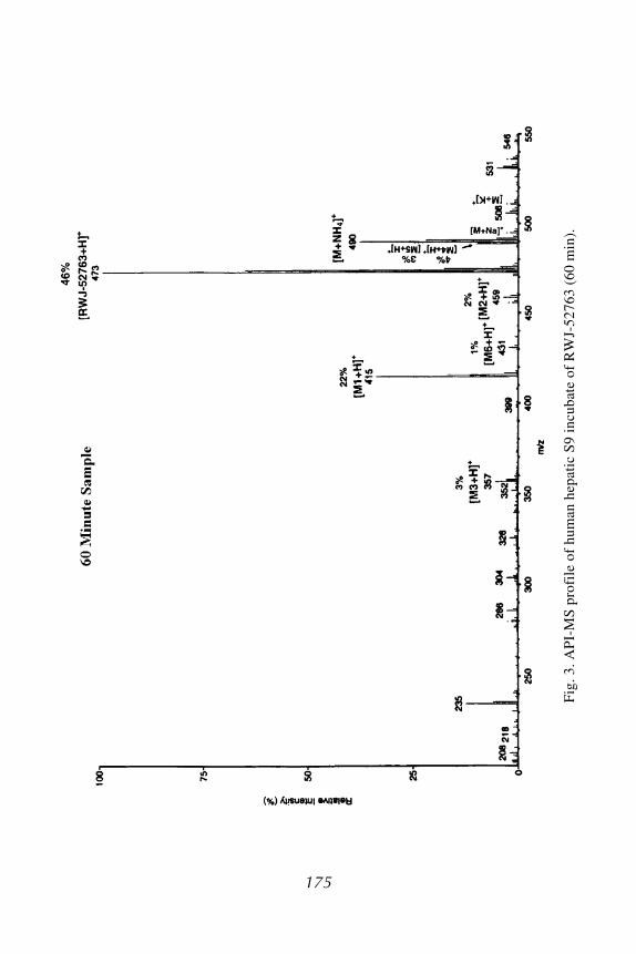
In Vitro Drug Metabolite Profiling 175
Fig
. 3. A
PI-
MS
pro
file
of
hum
an h
epat
ic S
9 in
cuba
te o
f R
WJ-
5276
3 (6
0 m
in).
175

176 Wu and McKown
Fig. 4. Structures and MS/MS product ions for RWJ-52763 and metabolites.

In Vitro Drug Metabolite Profiling 177
hydroxyphenyl-RWJ-68025 (M2; 12%–17% in both species), and two minorphenolic metabolites and, in conjunction with pathway 1, formed hydroxy-M1(M3; 4%–5% in both species). Pathways 3 and 4 produced 7 minor methyl-oxidized and N-dealkylated/acetylated metabolites. The proposed in vitro meta-bolic pathways for RWJ-68025 in rat and human hepatic S9 fractions aredepicted in Fig. 8. RWJ-68025 was rapidly and extensively metabolized inboth rat and human hepatic S9 fractions.
Fig. 5. In vitro metabolic pathways for RWJ-52763 in human hepatic S9 fraction.

178 Wu and McKown
4. Notes1. Hepatic S9 is also commercially available for a wide variety of animal species,
strains, and genders. This material usually comes with a detailed characterizationas well. A common source is In Vitro Technologies (Baltimore, MD).
2. Sources of human pooled liver preparations may vary in microsomal activitybecause of potential enzyme induction, for example, by those patients who mayhave been long-term drug users versus healthy subjects. One should try to main-tain the same source of human S9/liver microsomes to obtain reproducible results(1–3,6,7).
3. A total of 1.15% KCl in 0.05 M Tris-HCl buffer should be stored refrigeratedafter preparation and can then be used for up to ~4 mo. However, the remainingcofactors—5 mM MgCl2, 0.5 mM NADP, 0.55 mM NADPH, and 5 mM glucose-
Fig. 6. API-MS profile of human hepatic S9 incubate of RWJ-68025 (60 min).

In Vitro Drug Metabolite Profiling 179
Fig. 7. API-ionspray MS/MS spectrum of metabolite 1 from RWJ-68025 incubate.
6-phosphate, all prepared in Tris-HCl buffer—must be prepared fresh daily justprior to preparing an incubation (1).
4. The preparation of hepatic S9 should be well planned out and done quickly. Allmaterials should be chilled and procedures conducted on ice, if possible, to main-tain the viability of the enzymes during processing.
5. The hepatic S9 and microsomal fractions of male rats have higher drug-oxidizingactivity than that of females because of the higher level of CYP450 enzymespresent in males (1–5).
6. It is possible to incubate a total volume less than 5 mL. To do so, one would justadjust volumes of all components equally. The larger volume that is used in theseexperiments allows for potential isolation of metabolites of interest.
7. It is important to test your incubation conditions and the activity of each specificlot of enzyme by incubating concurrently with a control drug whose in vitro meta-bolic profile is well documented (percent disappearance of parent drug, metabo-lites formed)—that is, etoperidone (23) and tramadol (22)—used in theseexamples.
8. It is best to try to gain some solubility information about each drug or class priorto incubation. If no information is known, it is best to first try dissolving the neat

180 Wu and McKown
Fig
. 8. I
n vi
tro
met
abol
ic p
athw
ays
for
RW
J-68
025
in r
at a
nd h
uman
hep
atic
S9
frac
tion
s.
180

In Vitro Drug Metabolite Profiling 181
material in MeOH, which has a low impact on the biological system. If this is notsuccessful, then it can be evaporated down and another solvent tested. DMSO isusually the last solvent tested and is typically successful (1).
9. In general, it can be said that the lower the drug concentration of the incubationmixture, the higher the percentage of metabolite generation will be (1–4,13,20–22,25,27,30).
10. The enzymatic reactions of incubated samples need to be terminated quickly bythe addition of organic solvents, such as acetonitrile, ethyl acetate, ordichloromethane, immediately following the removal of aliquots or incubatesfrom the incubator. This should be followed by a quick freeze and storage at –20°Cor lower pending analysis (1,17–19).
11. For light-sensitive drugs, lights should be kept low and amber glass used to avoidexposure to light during incubation, storage, sample preparation, and analysis(1,17–19).
12. In many cases, there are advantages to incubating drugs with the S9 fraction vsthe microsomal fraction. The hepatic S9 is more convenient to prepare, the reac-tion is linear for a longer period of time, and the overall activity is better than thatseen with microsomes (1–4,17).
13. It is unnecessary to add NADPH to the hepatic S9 (microsomes + cytosol) incu-bation because of the presence of endogenous isocitric and glucose-6-phosphatedehydrogenases in the cytosolic fraction, which, along with NADP, are used togenerate NADPH. In contrast, NADPH is essential for the liver microsomalincubation (1).
14. Hepatic S9 and liver microsomal incubations primarily generate phase I metabo-lites only. However, in some cases, the formation of acetyl metabolites (phase II)has been documented following hepatic S9 incubation as a result of the presenceof N-acetyltransferases in cytosol (26,27).
15. Sample proteins are removed by acetonitrile precipitation and centrifugation. Thesupernatant volume injected into the LC (~20 μL) is kept as small as possible foroptimizing LC/MS separation and resolution (1–4,17,20–22,23,25,27,30).
16. Some drugs produce unstable metabolites (heat-labile, light-sensitive, andreactive) that need to be analyzed as quickly as possible after incubation andsample preparation. Avoid using CI and EI-MS for thermal-labile metabolites(i.e., N-oxides and conjugates) (1–5,8,9,17–19).
17. The formylation of a metabolite’s amino group (addition of 28 am) could occurin LC/API-MS analysis if formic acid is used. A LC mobile phase containingammonium acetate can remarkably enhance ionization efficiency in LC/MSanalysis, but it can also produce ammonium-adduct molecular ions of unchangeddrugs and its metabolites. The use of TFA to enhance ionization during LC/MSanalysis might form TFA polymers in the MS ion source; therefore, one needsto periodically refresh the LC/MS system using methanol/water (50:50, v/v)(20–22,25,27).
18. The most commonly adducted molecular ions observed in LC/API/ES-MS analy-sis are ammonium adducts ([M+18 amu]+) if ammonium acetate or ammonium

182 Wu and McKown
carbonate is used, sodium adducts ([M+23 amu]+), potassium adducts ([M+39amu]+), methanol adducts ([M+32 amu]+), acetonitrile adducts ([M+41 amu]+),acetic acid adducts ([M+60 amu]+), and TFA adducts ([M+114 amu]+). Theseadduct ions could form from the use of organic solvents, acids, and bases andfrom tubing and glassware. The methanol adduct of a drug may lead to the misin-terpretation of the formation of a dioxidized metabolite, which will not derivatizewith either diazomethane or acetic anhydride/pyridine but will fragment to forma protonated molecular ion of the parent drug via the loss of methanol, along withits product ions during MS/MS analysis (20–22,25,27).
19. Diazomethane derivatization reacts not only with phenolic and carboxylicmetabolites but also with some heterocyclic nitrogens. However, it does notderivatize N-oxide, amino, and alcoholic metabolites. Acetic anhydride/pyridineacetylation derivatizes phenolic, alcoholic (primary and secondary), and amino(primary and secondary) metabolites but not amide and tertiary alcoholicmetabolites (2,3,19–22,25,27,30).
20. N-oxide metabolites formed via cytochrome P450 or FMO may consist of twostereoisomers—for example, the cis and trans nicotine-N-oxides, which wereformed via an in vitro system (8).
References1. La Du, B. N., Mandel, H. G., and Way, E. L., eds. (1972) Fundamentals of Drug
Metabolism and Drug Disposition. Williams & Wilkins, Baltimore.2. Testa, B. and Jenner, P., eds. (1976) Drug Metabolism: Chemical and Biochemi-
cal Aspects. Marcel Dekker, New York.3. Jenner, P. and Testa, B., eds. (1980–1981) Concepts in Drug Metabolism Parts A
and B. Marcel Dekker, New York.4. Parkinson, A. (1996) Biotransformation of xenobiotics, in Casarett & Doull’s
Toxicology, (Klaassen, C. D., ed.), pp. 113–186.5. Ortiz de Montellano, P. R., ed. (1995) Cytochrome P450: Structure, Mechanism
and Biochemistry. Plenum, New York.6. Omura, T. (1999) Forty years of cytochrome P450. Biochem. Biophys. Res.
Commun. 266, 690–698.7. Rendic, S. and Di Carlo, F. J. (1997) Human cytochrome P450 enzymes: a status
report summarizing their reactions, substrates, inducers, and inhibitors. DrugMetab. Rev. 29, 413–580.
8. Cashman, J. R. (2000) Human flavin-containing monooxygenase: substrate speci-ficity and role in drug metabolism. Curr. Drug Metab. 1, 181–191.
9. Mulder, G. J., ed. (1990) Conjugation Reactions in Drug Metabolism. Taylor &Francis, London.
10. Radominska-Pandya, A., Czernik, P. J., Little, J. M., Battaglia, and Mackenzie, E.(1999) Structural and functional studies of UDP-glucuronosyltransferase. DrugMetab. Rev. 31, 817–899.

In Vitro Drug Metabolite Profiling 183
11. King, C. D., Rios, G. R., Green, M. D., and Tephly, T. R. (2000) UDP-glucuronosyltransferases. Curr. Drug Metab. 1, 143–161.
12. Banoglu, E. (2000) Current status of the cytosolic sulfotransferases in the meta-bolic activation of promutagens and procarcinogens. Curr. Drug Metab. 1, 1–30.
13. Li, A. P. (2001) Screening for human ADME/Tox drug properties in drug discov-ery. DDT 6, 357–366.
14. White, R. E. (2000) High-throughput screening in drug metabolism and pharma-cokinetic support of drug discovery. Annu. Rev. Pharmacol. Toxicol. 40, 133–157.
15. Uetrecht, J. P. (2000) Is it possible to more accurately predict which drug candi-dates will cause idiosyncratic drug reactions? Curr. Drug Metab. 1, 107–132.
16. Wu, W. N. and McKown, L. A. (2000) Recent advances in biotransformation ofcns and cardiovascular agents. Curr. Drug Metab. 1, 255–270.
17. Wu, W. N., McKown, L. A., Yorgey, K. A., and Pritchard, J. F. (1999) In vitrometabolic products of RWJ-34130, an antiarrhythmic agent, in rat liver prepara-tions. J. Pharm. Biomed. Anal. 20, 687–695.
18. McKown, L. A., Wu, W. N., and O’Neill, P. J. (1994) Characterization and iden-tification of the metabolites of fenoctimine using in vitro drug metabolizing sys-tems. J. Pharm. Biomed. Anal. 6, 771–775.
19. Wu, W. N., McKown, L. A., and O’Neill, P. J. (1995) In vitro and in vivo metabo-lism of the antianxiolytic agent fenobam in the rat. J. Pharm. Sci. 84, 185–189.
20. Wu, W. N., McKown, L. A., Moyer, M. D., Johannsen, T. B., and Takacs, A. R.(1999) In vitro metabolism of mifepristone (RU-486) in rat, monkey and humanhepatic S9 fractions: identification of three new mifepristone metabolites.Xenobiotica 31, 1089–1100.
21. Wu, W. N., McKown, L. A., Gauthier, A. D., Jones, W. J., and Raffa, R. B. (2001)Metabolism of the analgesic drug, tramadol hydrochloride, in rat and dog.Xenobiotica 31, 423–441.
22. Wu, W. N., McKown, L. A., and Liao, S. (2002) Metabolism of the analgesicdrug, ULTRAM® (tramadol hydrochloride) in humans: api-ms and ms/ms charac-terization of metabolites. Xenobiotica 32, 411–425.
23. Yan, Z., Caldwell, G. W., Wu, W. N., McKown, L. A., Rafferty, B., Jones, W. J.,et al. (2002) In vitro identification of metabolic pathways and cytochrome P450enzymes involved in the metabolism of etoperidone. Xenobiotica 32, 949–962.
24. Wu, W. N., McKown, L. A., and Reitz, A. B. (2001) In vitro metabolism of theanxiolytic agent, RWJ-52763 in human hepatic S9 fraction [abstract #243]. The6th International ISSX Meeting. Drug Metab. Rev. 33, 122.
25. Wu, W. N., McKown, L. A., and Reitz, A. B. (2003) In vitro metabolism of thenew anxiolytic agent, RWJ-52763 in human hepatic S9 fraction—api-ms/ms iden-tification of metabolites. J. Pharm. Biomed. Anal. 31, 95–102.
26. Wu, W. N., McKown, L. A., and Rybczynski, P. J. (2000) In vitro metabolism ofthe endocrine agent, RWJ-68025, in rat and human hepatic S9 fraction [abstract#230]. The 10th North American ISSX Meeting. Drug Metab. Rev. 32, 251.

184 Wu and McKown
27. Wu, W. N., McKown, L. A., Rybczynski, P. J., and Demarest, K. (2003) Hepaticbiotransformation of the new calcium-mimetic agent, RWJ-68025, in the rat andin man—api-ms/ms identification of metabolites. J. Pharm. Pharmacol. 55, 631–637.
28. Tang, C., Hochman, J. H., Ma, B., Subramanian, R., and Vyas, K. P. (2003) Acylglucuronidation and glucosidation of a new and selective endothelin ETA recep-tor antagonist in human liver microsomes. Drug Metab. Dispos. 31, 37–45.
29. McKown, L. A., Wu, W. N., and Pritchard, J. F. (1992) In vitro metabolism ofMcN-4130 (RWJ-34130) in the rat [abstract]. Presented at the AAPS EasternRegional Meeting.
30. Wu, W. N., Masucci, J. A., Caldwell, G. W., and Carson, J. R. (1998) Excretionand metabolism of the antihypertensive agent, RWJ-26240 (McN-5691) in dogs.Drug Metab. Dispos. 26, 115–125.


![[Pro] [s9] heridas](https://static.fdocuments.net/doc/165x107/55874fded8b42a8c468b4713/pro-s9-heridas.jpg)


