Efficient Drug Metabolism Strategy Based on Microsome ... · Efficient Drug Metabolism Strategy...
7
Efficient Drug Metabolism Strategy Based on Microsome− Mesoporous Organosilica Nanoreactors Xiaoni Fang, † Peng Zhang, † Liang Qiao, ‡ Xiaoyan Feng, † Xiangmin Zhang, † Hubert H. Girault, ‡ and Baohong Liu* ,† † Department of Chemistry, Institutes of Biomedical Sciences, and State Key Laboratory of Molecular Engineering of Polymers, Fudan University, Shanghai 200433, China ‡ Laboratoire d’Electrochimie Physique et Analytique, Ecole Polytechnique Fe ́ de ́ rale de Lausanne, CH-1015 Lausanne, Switzerland * S Supporting Information ABSTRACT: A rapid and accurate in vitro drug metabolism strategy has been proposed based on the design of a biomimetic nanoreactor composed of amino-functionalized periodic mesoporous organosilica (NH 2 -PMO) and microsomes. The amphiphilic nature and positive charge of NH 2 -PMO make it highly suited for the immobilization of hydrophobic and negatively charged microsomes to form nanoreactors, which can in turn extract substrates from solutions. Such nanoreactors provide a suitable environ- ment to confine multiple enzymes and substrates with high local concentrations, as well as to maintain their catalytic activities for rapid and highly effective drug metabolic reactions. Coupled with high- performance liquid chromatography−mass spectrometry analysis, the metabolites of nifedipine and testosterone were quantitatively characterized, and the reaction kinetics was evaluated. Both the metabolism conversion and reaction rate were significantly improved with the NH 2 −PMO nanoreactors compared to bulk reactions. This strategy is simple and cost-effective for promising advances in biomimetic metabolism study. I n modern drug development, assessment of safety always consists of designed bioassays for toxicology and pharma- cokinetics, animal toxicity studies, and finally human clinical trials. 1 Approximately 40% of drug candidates fail at the clinical testing stages owing to unanticipated toxicity, which drives up drug development cost significantly. 2,3 Screening of toxicity at early stages of the drug discovery process can minimize such failures by selecting drug candidates with acceptable levels of both bioactivity and toxicity. 4,5 In the past decade, the number of screenable drug targets increased dramatically with the rapid development of combinatorial chemistry and advances in bioinformatics, genomics, and proteomics. 6,7 However, the gap between the number of screenable drug candidates and the approved new drugs is widening. Therefore, development of low-cost and high-throughput toxicity screening methods, which can address early-stage toxicology and mimic human metabolism of drug candidates to predict the likelihood of drug candidate toxicity, is the major challenge in drug discovery. Human liver microsomes are enriched sources of metabolic enzymes, such as cytochrome P450 (CYP450) and uridine diphosphoglucuronosyl transferase (UGT), which together are responsible for nearly 80% of the metabolism of currently marketed drugs and are therefore widely used for in vitro metabolism and toxicity studies. 8 Carlson et al. 9,10 developed a 384-well plate assay system for high-throughput metabolism, where microsomal enzymes were dispersed in each well of the 384-well plate to form a series of parallel assays for simultaneous metabolism of many drug candidates. The automation facilitated parallel sample processing, thus enabling high-throughput drug metabolism screening. However, as a result of the relatively low concentration of microsomes and drugs, the in-solution metabolic reaction, e.g., in a well of the 384-well plate, exhibited slow kinetics. To address this issue, microsome-immobilized bioreactors have attracted wide interest and offered distinct advantages, including fast reaction kinetics, minimal consumption of microsomes, good stability, long storage time, and rapid separation of products from microsomal enzymes. Rusling et al. 11,12 designed a silica microbead bioreactor coated with DNA and enzymes to measure reactive metabolites and DNA adduct formation rates relevant to genotoxicity screening. Although this bioreactor can contribute to the efficient analysis of major and minor DNA adducts as well as the metabolites in less than 5 min of reaction time, there is still a potential to achieve higher metabolism efficiency by simultaneously concentrating enzymes and drugs in a nanoreactor. It is then desirable to design a nanoreactor with a good affinity for both multiple enzymes and multiple drugs for their enrichment, while the motion of Received: August 14, 2014 Accepted: October 14, 2014 Published: October 14, 2014 Article pubs.acs.org/ac © 2014 American Chemical Society 10870 dx.doi.org/10.1021/ac503024h | Anal. Chem. 2014, 86, 10870−10876
Transcript of Efficient Drug Metabolism Strategy Based on Microsome ... · Efficient Drug Metabolism Strategy...
Efficient Drug Metabolism Strategy Based on Microsome−Mesoporous Organosilica NanoreactorsXiaoni Fang,† Peng Zhang,† Liang Qiao,‡ Xiaoyan Feng,† Xiangmin Zhang,† Hubert H. Girault,‡
and Baohong Liu*,†
†Department of Chemistry, Institutes of Biomedical Sciences, and State Key Laboratory of Molecular Engineering of Polymers, FudanUniversity, Shanghai 200433, China‡Laboratoire d’Electrochimie Physique et Analytique, Ecole Polytechnique Federale de Lausanne, CH-1015 Lausanne, Switzerland
*S Supporting Information
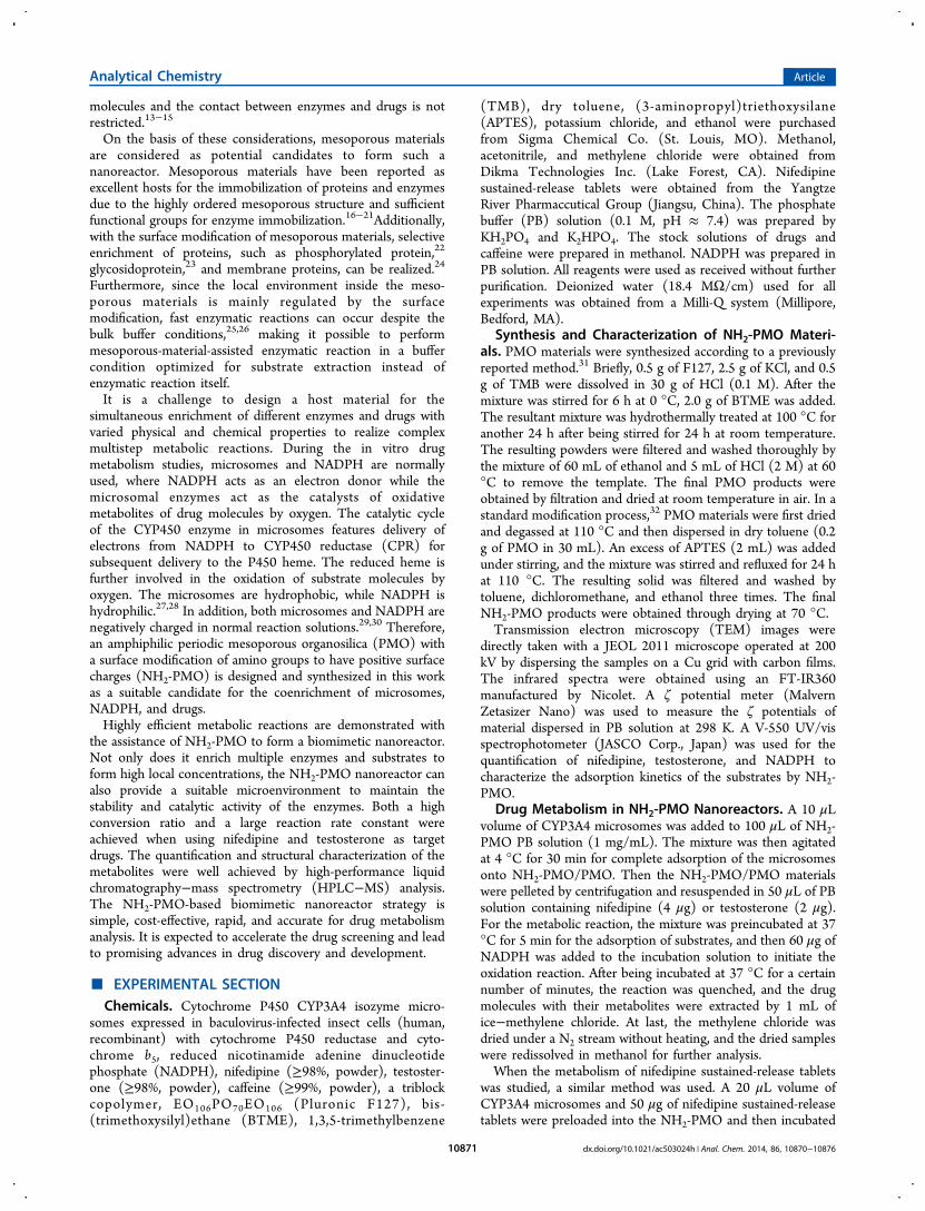
ABSTRACT: A rapid and accurate in vitro drug metabolism strategy hasbeen proposed based on the design of a biomimetic nanoreactor composedof amino-functionalized periodic mesoporous organosilica (NH2-PMO) andmicrosomes. The amphiphilic nature and positive charge of NH2-PMOmake it highly suited for the immobilization of hydrophobic and negativelycharged microsomes to form nanoreactors, which can in turn extractsubstrates from solutions. Such nanoreactors provide a suitable environ-ment to confine multiple enzymes and substrates with high localconcentrations, as well as to maintain their catalytic activities for rapidand highly effective drug metabolic reactions. Coupled with high-performance liquid chromatography−mass spectrometry analysis, themetabolites of nifedipine and testosterone were quantitatively characterized,and the reaction kinetics was evaluated. Both the metabolism conversionand reaction rate were significantly improved with the NH2−PMOnanoreactors compared to bulk reactions. This strategy is simple and cost-effective for promising advances in biomimeticmetabolism study.
In modern drug development, assessment of safety alwaysconsists of designed bioassays for toxicology and pharma-
cokinetics, animal toxicity studies, and finally human clinicaltrials.1 Approximately 40% of drug candidates fail at the clinicaltesting stages owing to unanticipated toxicity, which drives updrug development cost significantly.2,3 Screening of toxicity atearly stages of the drug discovery process can minimize suchfailures by selecting drug candidates with acceptable levels ofboth bioactivity and toxicity.4,5 In the past decade, the numberof screenable drug targets increased dramatically with the rapiddevelopment of combinatorial chemistry and advances inbioinformatics, genomics, and proteomics.6,7 However, thegap between the number of screenable drug candidates and theapproved new drugs is widening. Therefore, development oflow-cost and high-throughput toxicity screening methods,which can address early-stage toxicology and mimic humanmetabolism of drug candidates to predict the likelihood of drugcandidate toxicity, is the major challenge in drug discovery.Human liver microsomes are enriched sources of metabolic
enzymes, such as cytochrome P450 (CYP450) and uridinediphosphoglucuronosyl transferase (UGT), which together areresponsible for nearly 80% of the metabolism of currentlymarketed drugs and are therefore widely used for in vitrometabolism and toxicity studies.8 Carlson et al.9,10 developed a384-well plate assay system for high-throughput metabolism,where microsomal enzymes were dispersed in each well of the
384-well plate to form a series of parallel assays forsimultaneous metabolism of many drug candidates. Theautomation facilitated parallel sample processing, thus enablinghigh-throughput drug metabolism screening. However, as aresult of the relatively low concentration of microsomes anddrugs, the in-solution metabolic reaction, e.g., in a well of the384-well plate, exhibited slow kinetics.To address this issue, microsome-immobilized bioreactors
have attracted wide interest and offered distinct advantages,including fast reaction kinetics, minimal consumption ofmicrosomes, good stability, long storage time, and rapidseparation of products from microsomal enzymes. Rusling etal.11,12 designed a silica microbead bioreactor coated with DNAand enzymes to measure reactive metabolites and DNA adductformation rates relevant to genotoxicity screening. Althoughthis bioreactor can contribute to the efficient analysis of majorand minor DNA adducts as well as the metabolites in less than5 min of reaction time, there is still a potential to achieve highermetabolism efficiency by simultaneously concentrating enzymesand drugs in a nanoreactor. It is then desirable to design ananoreactor with a good affinity for both multiple enzymes andmultiple drugs for their enrichment, while the motion of
Received: August 14, 2014Accepted: October 14, 2014Published: October 14, 2014
Article
pubs.acs.org/ac
© 2014 American Chemical Society 10870 dx.doi.org/10.1021/ac503024h | Anal. Chem. 2014, 86, 10870−10876
molecules and the contact between enzymes and drugs is notrestricted.13−15
On the basis of these considerations, mesoporous materialsare considered as potential candidates to form such ananoreactor. Mesoporous materials have been reported asexcellent hosts for the immobilization of proteins and enzymesdue to the highly ordered mesoporous structure and sufficientfunctional groups for enzyme immobilization.16−21Additionally,with the surface modification of mesoporous materials, selectiveenrichment of proteins, such as phosphorylated protein,22
glycosidoprotein,23 and membrane proteins, can be realized.24
Furthermore, since the local environment inside the meso-porous materials is mainly regulated by the surfacemodification, fast enzymatic reactions can occur despite thebulk buffer conditions,25,26 making it possible to performmesoporous-material-assisted enzymatic reaction in a buffercondition optimized for substrate extraction instead ofenzymatic reaction itself.It is a challenge to design a host material for the
simultaneous enrichment of different enzymes and drugs withvaried physical and chemical properties to realize complexmultistep metabolic reactions. During the in vitro drugmetabolism studies, microsomes and NADPH are normallyused, where NADPH acts as an electron donor while themicrosomal enzymes act as the catalysts of oxidativemetabolites of drug molecules by oxygen. The catalytic cycleof the CYP450 enzyme in microsomes features delivery ofelectrons from NADPH to CYP450 reductase (CPR) forsubsequent delivery to the P450 heme. The reduced heme isfurther involved in the oxidation of substrate molecules byoxygen. The microsomes are hydrophobic, while NADPH ishydrophilic.27,28 In addition, both microsomes and NADPH arenegatively charged in normal reaction solutions.29,30 Therefore,an amphiphilic periodic mesoporous organosilica (PMO) witha surface modification of amino groups to have positive surfacecharges (NH2-PMO) is designed and synthesized in this workas a suitable candidate for the coenrichment of microsomes,NADPH, and drugs.Highly efficient metabolic reactions are demonstrated with
the assistance of NH2-PMO to form a biomimetic nanoreactor.Not only does it enrich multiple enzymes and substrates toform high local concentrations, the NH2-PMO nanoreactor canalso provide a suitable microenvironment to maintain thestability and catalytic activity of the enzymes. Both a highconversion ratio and a large reaction rate constant wereachieved when using nifedipine and testosterone as targetdrugs. The quantification and structural characterization of themetabolites were well achieved by high-performance liquidchromatography−mass spectrometry (HPLC−MS) analysis.The NH2-PMO-based biomimetic nanoreactor strategy issimple, cost-effective, rapid, and accurate for drug metabolismanalysis. It is expected to accelerate the drug screening and leadto promising advances in drug discovery and development.
■ EXPERIMENTAL SECTIONChemicals. Cytochrome P450 CYP3A4 isozyme micro-
somes expressed in baculovirus-infected insect cells (human,recombinant) with cytochrome P450 reductase and cyto-chrome b5, reduced nicotinamide adenine dinucleotidephosphate (NADPH), nifedipine (≥98%, powder), testoster-one (≥98%, powder), caffeine (≥99%, powder), a triblockcopolymer, EO106PO70EO106 (Pluronic F127), bis-(trimethoxysilyl)ethane (BTME), 1,3,5-trimethylbenzene
(TMB), dry toluene, (3-aminopropyl)triethoxysilane(APTES), potassium chloride, and ethanol were purchasedfrom Sigma Chemical Co. (St. Louis, MO). Methanol,acetonitrile, and methylene chloride were obtained fromDikma Technologies Inc. (Lake Forest, CA). Nifedipinesustained-release tablets were obtained from the YangtzeRiver Pharmaccutical Group (Jiangsu, China). The phosphatebuffer (PB) solution (0.1 M, pH ≈ 7.4) was prepared byKH2PO4 and K2HPO4. The stock solutions of drugs andcaffeine were prepared in methanol. NADPH was prepared inPB solution. All reagents were used as received without furtherpurification. Deionized water (18.4 MΩ/cm) used for allexperiments was obtained from a Milli-Q system (Millipore,Bedford, MA).
Synthesis and Characterization of NH2-PMO Materi-als. PMO materials were synthesized according to a previouslyreported method.31 Briefly, 0.5 g of F127, 2.5 g of KCl, and 0.5g of TMB were dissolved in 30 g of HCl (0.1 M). After themixture was stirred for 6 h at 0 °C, 2.0 g of BTME was added.The resultant mixture was hydrothermally treated at 100 °C foranother 24 h after being stirred for 24 h at room temperature.The resulting powders were filtered and washed thoroughly bythe mixture of 60 mL of ethanol and 5 mL of HCl (2 M) at 60°C to remove the template. The final PMO products wereobtained by filtration and dried at room temperature in air. In astandard modification process,32 PMO materials were first driedand degassed at 110 °C and then dispersed in dry toluene (0.2g of PMO in 30 mL). An excess of APTES (2 mL) was addedunder stirring, and the mixture was stirred and refluxed for 24 hat 110 °C. The resulting solid was filtered and washed bytoluene, dichloromethane, and ethanol three times. The finalNH2-PMO products were obtained through drying at 70 °C.Transmission electron microscopy (TEM) images were
directly taken with a JEOL 2011 microscope operated at 200kV by dispersing the samples on a Cu grid with carbon films.The infrared spectra were obtained using an FT-IR360manufactured by Nicolet. A ζ potential meter (MalvernZetasizer Nano) was used to measure the ζ potentials ofmaterial dispersed in PB solution at 298 K. A V-550 UV/visspectrophotometer (JASCO Corp., Japan) was used for thequantification of nifedipine, testosterone, and NADPH tocharacterize the adsorption kinetics of the substrates by NH2-PMO.
Drug Metabolism in NH2-PMO Nanoreactors. A 10 μLvolume of CYP3A4 microsomes was added to 100 μL of NH2-PMO PB solution (1 mg/mL). The mixture was then agitatedat 4 °C for 30 min for complete adsorption of the microsomesonto NH2-PMO/PMO. Then the NH2-PMO/PMO materialswere pelleted by centrifugation and resuspended in 50 μL of PBsolution containing nifedipine (4 μg) or testosterone (2 μg).For the metabolic reaction, the mixture was preincubated at 37°C for 5 min for the adsorption of substrates, and then 60 μg ofNADPH was added to the incubation solution to initiate theoxidation reaction. After being incubated at 37 °C for a certainnumber of minutes, the reaction was quenched, and the drugmolecules with their metabolites were extracted by 1 mL ofice−methylene chloride. At last, the methylene chloride wasdried under a N2 stream without heating, and the dried sampleswere redissolved in methanol for further analysis.When the metabolism of nifedipine sustained-release tablets
was studied, a similar method was used. A 20 μL volume ofCYP3A4 microsomes and 50 μg of nifedipine sustained-releasetablets were preloaded into the NH2-PMO and then incubated
Analytical Chemistry Article
dx.doi.org/10.1021/ac503024h | Anal. Chem. 2014, 86, 10870−1087610871
with 120 μg of NADPH. Before HPLC−MS analysis, theextracted samples were centrifuged to remove the precipitatesthoroughly.In-Solution Drug Metabolism. The control experiments
of microsomal dispersions were performed according to thepublished protocols.33,34 The same amounts of enzymes anddrugs as those used during the NH2-PMO nanoreactorcatalyzed reaction were suspended in 200 μL of PB solution.After incubation, extraction, and redispersion, the metaboliteswere analyzed by HPLC−MS.LC−MS Analysis. For rapid screening of reactive
metabolites, the reaction mixture was analyzed by HPLC−MS. Samples were first subjected to chromatographicseparation with an Agilent series 1260 HPLC system (AgilentTechnologies, Palo Alto, CA) integrated with an autosamplerand a UV detector. A 20 μL volume of the sample solution wasinjected into a VP-ODS C18 column (250 mm × 4.6 mm i.d., 5μm, Agilent) at a flow rate of 0.6 mL/min. The detailedseparation process is explained in the Supporting Information.The fractions from LC were introduced into a 6460 QQQ massspectrometer (Agilent) that was operated in positive ion modeunder the optimized parameter conditions (refer to theSupporting Information). The structure information on reactivemetabolites can be deduced from tandem MS spectra.Additionally, for quantitative analysis of the metabolites, 20μL of caffeine (0.1 μg/μL) was added as an internal standardbefore HPLC−MS analysis.
■ RESULTS AND DISCUSSIONCharacterization of the Nanoreactors. The low
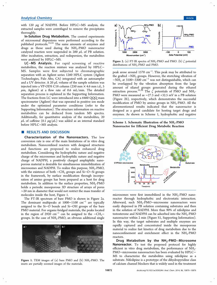
conversion rate is one of the main limitations of in vitro drugmetabolism. Nanoconfined reactors with designed structuresand functions are proposed to realize enhanced drugmetabolism. Considering the hydrophobic nature and negativecharge of the microsomes and hydrophilic nature and negativecharge of NADPH, a positively charged amphiphilic nano-porous material is desirable for simultaneous immobilization ofmicrosomes and NADPH. To realize this purpose, NH2-PMO,with the existence of both −CH2 groups and Si−O−Si groupsin the framework, by surface modification through incorpo-ration of amino groups has been prepared as a host for drugmetabolism. In addition to the surface properties, NH2-PMOholds a periodic mesoporous 3D structure of arrays of pores∼20 nm in diameter that would not restrict the mass transfer ofmolecules inside the host, Figure 1.The FT-IR spectrum of bare PMO is shown in Figure 2a.
The dominant multipeaks at 1000−1100 cm−1 are typicallyassigned to the Si−O bonds and Si−OH groups of the barePMO material. For organic-bridged materials, the peaks locatedin the region of 2920 cm−1 can be assigned to the −CH2−groups. In the case of NH2-PMO, an obvious additional single
peak arose around 1570 cm−1. This peak may be attributed tothe grafted −NH2 groups. However, the stretching vibration of−NH2 at 3100−3300 cm−1 was not distinguishable, which canbe overlapped by the vibration absorption from the largeamount of silanol groups generated during the ethanolextraction process.35,36 The ζ potentials of PMO and NH2-PMO were measured as −27.3 and +32.5 mV in a PB solution(Figure 2b), respectively, which demonstrates the successfulmodification of PMO by amino groups in NH2-PMO. All theaforementioned results indicated that the nanoreactor isdesigned as a good candidate for hosting target drugs andenzymes. As shown in Scheme 1, hydrophobic and negative
microsomes were first immobilized in the NH2-PMO nano-reactor through hydrophobic and electrostatic interaction.Afterward, such NH2-PMO−microsome nanoreactors wereeasily dispersed in PB solution containing substrates and thenin the solution of NADPH. More than 90% of nifedipine andtestosterone and NADPH can be adsorbed into the NH2-PMOnanoreactor within 2 min (Figure S1, Supporting Information).In this way, the target substrates and multiple enzymes arerapidly captured and concentrated inside the mesoporousmaterial to realize fast kinetics of drug metabolism due to thenanoconfinement and enrichment effect in the NH2-PMOreactors.
Drug Metabolism by the NH2-PMO−MicrosomeNanoreactor. To test the proposed protocol for highlyefficient in vitro drug metabolism, the performance of NH2-PMO−microsome nanoreactors has been evaluated by HPLC−MS to characterize the metabolites using nifedipine as asubstrate. Nifedipine is a prototype of the dihydropyridine classof calcium channel blockers that is widely used in the treatment
Figure 1. TEM images of (a) bare PMO and (b) NH2-PMO. Theinsets are partially zoomed images of the materials.
Figure 2. (a) FT-IR spectra of NH2-PMO and PMO. (b) ζ potentialdistributions of NH2-PMO and PMO.
Scheme 1. Schematic Illustration of the NH2-PMONanoreactor for Efficient Drug Metabolic Reaction
Analytical Chemistry Article
dx.doi.org/10.1021/ac503024h | Anal. Chem. 2014, 86, 10870−1087610872
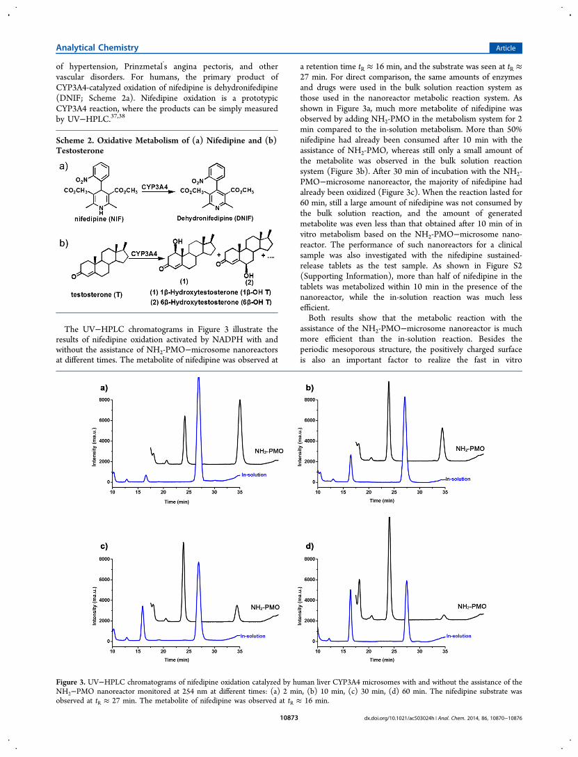
of hypertension, Prinzmetal’s angina pectoris, and othervascular disorders. For humans, the primary product ofCYP3A4-catalyzed oxidation of nifedipine is dehydronifedipine(DNIF; Scheme 2a). Nifedipine oxidation is a prototypicCYP3A4 reaction, where the products can be simply measuredby UV−HPLC.37,38
The UV−HPLC chromatograms in Figure 3 illustrate theresults of nifedipine oxidation activated by NADPH with andwithout the assistance of NH2-PMO−microsome nanoreactorsat different times. The metabolite of nifedipine was observed at
a retention time tR ≈ 16 min, and the substrate was seen at tR ≈27 min. For direct comparison, the same amounts of enzymesand drugs were used in the bulk solution reaction system asthose used in the nanoreactor metabolic reaction system. Asshown in Figure 3a, much more metabolite of nifedipine wasobserved by adding NH2-PMO in the metabolism system for 2min compared to the in-solution metabolism. More than 50%nifedipine had already been consumed after 10 min with theassistance of NH2-PMO, whereas still only a small amount ofthe metabolite was observed in the bulk solution reactionsystem (Figure 3b). After 30 min of incubation with the NH2-PMO−microsome nanoreactor, the majority of nifedipine hadalready been oxidized (Figure 3c). When the reaction lasted for60 min, still a large amount of nifedipine was not consumed bythe bulk solution reaction, and the amount of generatedmetabolite was even less than that obtained after 10 min of invitro metabolism based on the NH2-PMO−microsome nano-reactor. The performance of such nanoreactors for a clinicalsample was also investigated with the nifedipine sustained-release tablets as the test sample. As shown in Figure S2(Supporting Information), more than half of nifedipine in thetablets was metabolized within 10 min in the presence of thenanoreactor, while the in-solution reaction was much lessefficient.Both results show that the metabolic reaction with the
assistance of the NH2-PMO−microsome nanoreactor is muchmore efficient than the in-solution reaction. Besides theperiodic mesoporous structure, the positively charged surfaceis also an important factor to realize the fast in vitro
Scheme 2. Oxidative Metabolism of (a) Nifedipine and (b)Testosterone
Figure 3. UV−HPLC chromatograms of nifedipine oxidation catalyzed by human liver CYP3A4 microsomes with and without the assistance of theNH2−PMO nanoreactor monitored at 254 nm at different times: (a) 2 min, (b) 10 min, (c) 30 min, (d) 60 min. The nifedipine substrate wasobserved at tR ≈ 27 min. The metabolite of nifedipine was observed at tR ≈ 16 min.
Analytical Chemistry Article
dx.doi.org/10.1021/ac503024h | Anal. Chem. 2014, 86, 10870−1087610873
metabolism. Figure S3 (Supporting Information) shows theUV−HPLC chromatograms of bare PMO-assisted drugmetabolism after 30 min for comparison, where the surface ofthe unmodified PMO is negatively charged. The results showedthat bare PMO could also improve the efficiency of drugmetabolism compared with that of the in-solution reaction butnot as well as the NH2-PMO-assisted protocol. The existence ofpositive amino groups on the NH2-PMO surfaces contributesto the extraction of negatively charged microsomes andNADPH into the mesoporous material.The metabolites of nifedipine were characterized by tandem
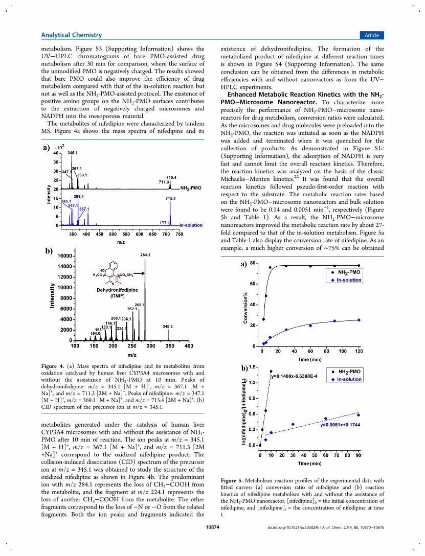
MS. Figure 4a shows the mass spectra of nifedipine and its
metabolites generated under the catalysis of human liverCYP3A4 microsomes with and without the assistance of NH2-PMO after 10 min of reaction. The ion peaks at m/z = 345.1[M + H]+, m/z = 367.1 [M + Na]+, and m/z = 711.3 [2M+Na]+ correspond to the oxidized nifedipine product. Thecollision-induced dissociation (CID) spectrum of the precursorion at m/z = 345.1 was obtained to study the structure of theoxidized nifedipine as shown in Figure 4b. The predominantion with m/z 284.1 represents the loss of CH3−COOH fromthe metabolite, and the fragment at m/z 224.1 represents theloss of another CH3−COOH from the metabolite. The otherfragments correspond to the loss of −N or −O from the relatedfragments. Both the ion peaks and fragments indicated the
existence of dehydronifedipine. The formation of themetabolized product of nifedipine at different reaction timesis shown in Figure S4 (Supporting Information). The sameconclusion can be obtained from the differences in metabolicefficiencies with and without nanoreactors as from the UV−HPLC experiments.
Enhanced Metabolic Reaction Kinetics with the NH2-PMO−Microsome Nanoreactor. To characterize moreprecisely the performance of NH2-PMO−microsome nano-reactors for drug metabolism, conversion ratios were calculated.As the microsomes and drug molecules were preloaded into theNH2-PMO, the reaction was initiated as soon as the NADPHwas added and terminated when it was quenched for thecollection of products. As demonstrated in Figure S1c(Supporting Information), the adsorption of NADPH is veryfast and cannot limit the overall reaction kinetics. Therefore,the reaction kinetics was analyzed on the basis of the classicMichaelis−Menten kinetics.22 It was found that the overallreaction kinetics followed pseudo-first-order reaction withrespect to the substrate. The metabolic reaction rates basedon the NH2-PMO−microsome nanoreactors and bulk solutionwere found to be 0.14 and 0.0051 min−1, respectively (Figure5b and Table 1). As a result, the NH2-PMO−microsomenanoreactors improved the metabolic reaction rate by about 27-fold compared to that of the in-solution metabolism. Figure 5aand Table 1 also display the conversion rate of nifedipine. As anexample, a much higher conversion of ∼75% can be obtained
Figure 4. (a) Mass spectra of nifedipine and its metabolites fromoxidation catalyzed by human liver CYP3A4 microsomes with andwithout the assistance of NH2-PMO at 10 min. Peaks ofdehydronifedipine: m/z = 345.1 [M + H]+, m/z = 367.1 [M +Na]+, and m/z = 711.3 [2M + Na]+. Peaks of nifedipine: m/z = 347.1[M + H]+, m/z = 369.1 [M + Na]+, and m/z = 715.4 [2M + Na]+. (b)CID spectrum of the precursor ion at m/z = 345.1.
Figure 5. Metabolism reaction profiles of the experimental data withfitted curves: (a) conversion ratio of nifedipine and (b) reactionkinetics of nifedipine metabolism with and without the assistance ofthe NH2-PMO nanoreactor. [nifedipine]0 = the initial concentration ofnifedipine, and [nifedipine]t = the concentration of nifedipine at timet.
Analytical Chemistry Article
dx.doi.org/10.1021/ac503024h | Anal. Chem. 2014, 86, 10870−1087610874
within 20 min when NH2-PMO−microsome nanoreactors areadded to the metabolic reaction system, whereas the in-solutionreaction achieved a conversion of only less than 15% within thesame time. Such a conversion rate derived from the NH2-PMOprotocol is very high compared with that of other developedstrategies.8 The difference in metabolism efficiency between thenanoreactor-assisted and bulk solution reactions is alwayssignificant for all the examined metabolic reactions.Both the qualitative and quantitative results validate that the
metabolic reaction based on the NH2-PMO−microsomenanoreactor is significantly enhanced compared with the in-solution method and the reaction with the assistance of purePMO nanoreactors, stemming from the amphiphilic propertyand positively charged surface of NH2-PMO for the rapidenrichment of microsomes and substrates. Furthermore,hydrophilic and negatively charged NADPH can easily partitioninto the pores. The fast enrichment not only dramaticallyincreases the concentration of multiple enzymes, drugs, anddissolved oxygen in the nanoreactor, but also provides asuitable environment for retaining their native stability andcatalytic activity. This, in fact, offers abundant catalytic sites forthe metabolic reaction while also facilitating the access ofenzymes and substrates, allowing fast charge transfer viaCYP450 reductase from NADPH to the enzyme’s iron heme
center.27,39 As a consequence, the metabolic reaction time isgreatly reduced, while the conversion rate is largely enhanced.
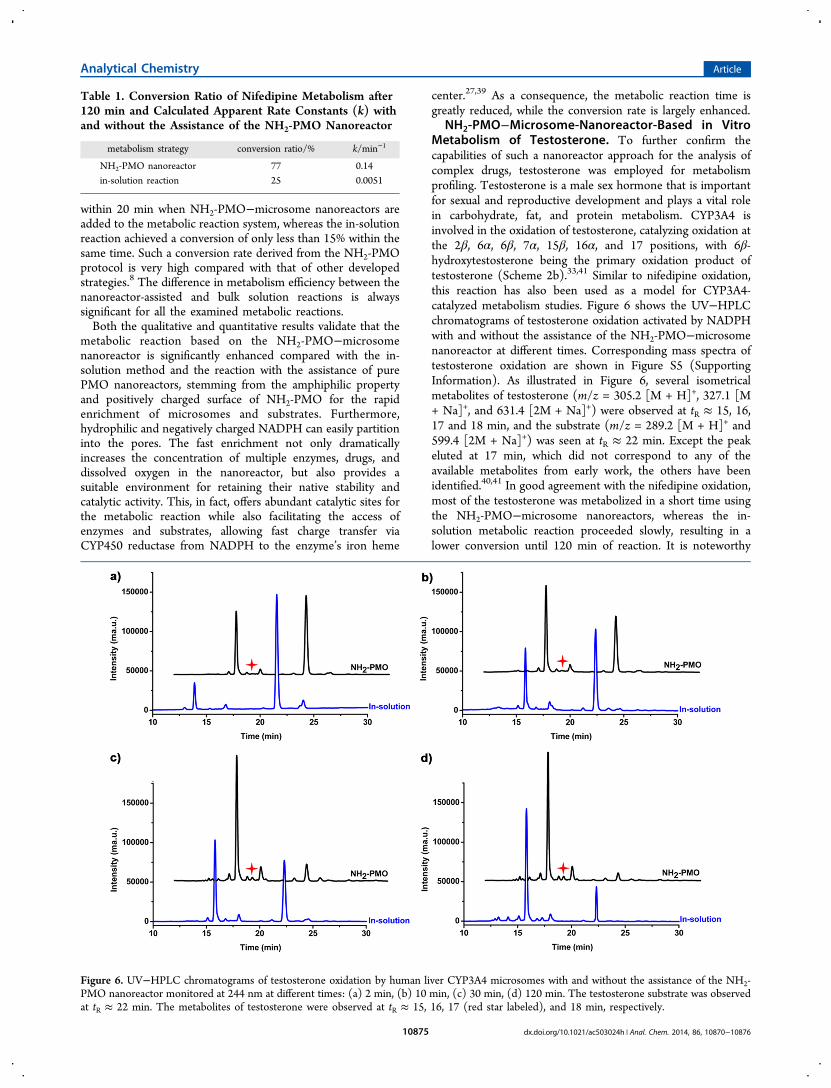
NH2-PMO−Microsome-Nanoreactor-Based in VitroMetabolism of Testosterone. To further confirm thecapabilities of such a nanoreactor approach for the analysis ofcomplex drugs, testosterone was employed for metabolismprofiling. Testosterone is a male sex hormone that is importantfor sexual and reproductive development and plays a vital rolein carbohydrate, fat, and protein metabolism. CYP3A4 isinvolved in the oxidation of testosterone, catalyzing oxidation atthe 2β, 6α, 6β, 7α, 15β, 16α, and 17 positions, with 6β-hydroxytestosterone being the primary oxidation product oftestosterone (Scheme 2b).33,41 Similar to nifedipine oxidation,this reaction has also been used as a model for CYP3A4-catalyzed metabolism studies. Figure 6 shows the UV−HPLCchromatograms of testosterone oxidation activated by NADPHwith and without the assistance of the NH2-PMO−microsomenanoreactor at different times. Corresponding mass spectra oftestosterone oxidation are shown in Figure S5 (SupportingInformation). As illustrated in Figure 6, several isometricalmetabolites of testosterone (m/z = 305.2 [M + H]+, 327.1 [M+ Na]+, and 631.4 [2M + Na]+) were observed at tR ≈ 15, 16,17 and 18 min, and the substrate (m/z = 289.2 [M + H]+ and599.4 [2M + Na]+) was seen at tR ≈ 22 min. Except the peakeluted at 17 min, which did not correspond to any of theavailable metabolites from early work, the others have beenidentified.40,41 In good agreement with the nifedipine oxidation,most of the testosterone was metabolized in a short time usingthe NH2-PMO−microsome nanoreactors, whereas the in-solution metabolic reaction proceeded slowly, resulting in alower conversion until 120 min of reaction. It is noteworthy
Table 1. Conversion Ratio of Nifedipine Metabolism after120 min and Calculated Apparent Rate Constants (k) withand without the Assistance of the NH2-PMO Nanoreactor
metabolism strategy conversion ratio/% k/min−1
NH2-PMO nanoreactor 77 0.14in-solution reaction 25 0.0051
Figure 6. UV−HPLC chromatograms of testosterone oxidation by human liver CYP3A4 microsomes with and without the assistance of the NH2-PMO nanoreactor monitored at 244 nm at different times: (a) 2 min, (b) 10 min, (c) 30 min, (d) 120 min. The testosterone substrate was observedat tR ≈ 22 min. The metabolites of testosterone were observed at tR ≈ 15, 16, 17 (red star labeled), and 18 min, respectively.
Analytical Chemistry Article
dx.doi.org/10.1021/ac503024h | Anal. Chem. 2014, 86, 10870−1087610875
that the small peak at tR ≈ 17 min was only observed in thepresence of NH2-PMO−microsome nanoreactors, demonstrat-ing that more metabolites can be generated using the NH2-PMO−microsome nanoreactor. Therefore, the metabolicreaction based on the nanoreactor approach may providemore information on the drug metabolism compared to theconventional bulk solution in vitro metabolic reaction, which isof great value in identifying reactive metabolites of drugs topredict possible toxicity in the early stage of drug development.All these features render NH2-PMO a promising host for rapidand accurate in vitro drug metabolic reaction.
■ CONCLUSIONSIn summary, we have demonstrated that drug metabolism canbe effectively carried out in the NH2-PMO−microsomenanoreactor. Due to the amphiphilic and positive property ofNH2-PMO, the resulting nanochannels can host multipleenzymes and substrates, where a highly efficient reaction canoccur. Compared with other methods, the combination of theNH2-PMO−microsome nanoreactor with HPLC−MS providesa simple, rapid, and accurate strategy for in vitro drugmetabolism. It is anticipated that the metabolic reactionbased on the nanoreactor strategy would lead to promisingadvances in drug discovery and development.
■ ASSOCIATED CONTENT*S Supporting InformationDetailed description of the LC−MS experiments, data for theadsorption kinetics of the substrates by NH2-PMO andnifedipine sustained-release tablet metabolism, and massspectra of nifedipine and testosterone metabolism. Thismaterial is available free of charge via the Internet at http://pubs.acs.org.
■ AUTHOR INFORMATIONCorresponding Author*E-mail: [email protected]. Fax: (+86) 21-6564-1740.NotesThe authors declare no competing financial interest.
■ ACKNOWLEDGMENTSThis work was supported by the National Natural ScienceFoundation of China (NSFC) (Grants 21175028 and21375022) and 863 Project 2012AA020202.
■ REFERENCES(1) Lee, M. Y.; Dordick, J. S. Curr. Opin. Biotechnol. 2006, 17, 619−627.(2) Munos, B. Nat. Rev. Drug Discovery 2009, 8, 959−968.(3) Li, A. P. Drug Discovery Today 2001, 6, 357−366.(4) Lee, M. Y.; Park, C. B.; Dordick, J. S.; Clark, D. S. Proc. Natl.Acad. Sci. U.S.A. 2005, 102, 983−987.(5) Geysen, H. M.; Schoenen, F.; Wagner, D.; Wagner, R. Nat. Rev.Drug Discovery 2003, 2, 222−230.(6) Ma, S.; Chowdhury, S. K. Anal. Chem. 2011, 83, 5028−5036.(7) Gao, D.; Li, H. F.; Wang, N. J.; Lin, J. M. Anal. Chem. 2012, 84,9230−9237.(8) Bajrami, B.; Zhao, L.; Schenkman, J. B.; Rusling, J. F. Anal. Chem.2009, 81, 9921−9929.(9) Carlson, T. J.; Fishe, M. B. J. Comb. Chem. 2008, 11, 258−264.(10) Jenkins, K. M.; Angeles, R.; Quintos, M. T.; Xu, R.; Kassel, D. B.J. Pharm. Biomed. Anal. 2004, 34, 989−1004.(11) Bajrami, B.; Hvastkovs, E. G.; Jensen, G. C.; Schenkman, J. B.;Rusling, J. F. Anal. Chem. 2008, 80, 922−932.
(12) Xue, Y.; Xiong, J.; Shi, H. L.; Liu, Y. M.; Qing, L. S.; Liao, X.Anal. Bioanal. Chem. 2013, 405, 8807−8817.(13) Yuan, Q.; Wu, Y.; Wang, J.; Lu, D. Q.; Zhao, Z. L.; Liu, T.;Zhang, X. B.; Tan, W. H. Angew. Chem., Int. Ed. 2013, 52, 13965−13969.(14) Zhao, X. H.; Gong, L.; Zhang, X. B.; Fu, T.; Hu, R.; Tan, W. H.;Yu, R. Q. Anal. Chem. 2013, 85, 3614−3620.(15) Chen, L. Q.; Xiao, S. J.; Hu, P. P.; Peng, L.; Ma, J.; Luo, L. F.; Li,Y. F.; Huang, C. Z. Anal. Chem. 2012, 84, 3099−3110.(16) Hartmann, M.; Jung, D. J. Mater. Chem. 2010, 20, 844−857.(17) Gao, P. F.; Zheng, L. L.; Liang, L. J.; Yang, X. X.; Li, Y. F.;Huang, C. Z. J. Mater. Chem. B 2013, 1, 3202−3208.(18) Wang, J.; Gao, P. P.; Yang, X. X.; Wang, T. T.; Huang, C. Z. J.Mater. Chem. B 2014, 2, 4379−4386.(19) Hartmann, M.; Kostrov, X. Chem. Soc. Rev. 2013, 42, 6277−6289.(20) Li, L. L.; Xie, M. Y.; Wang, J.; Li, X. Y.; Wang, C.; Yuan, Q.;Pang, D. W.; Lu, Y.; Tan, W. H. Chem. Commun. 2013, 49, 5823−5825.(21) Hudson, S.; Cooney, J.; Magner, E. Angew. Chem., Int. Ed. 2008,47, 8582−8594.(22) Bi, H. Y.; Qiao, L.; Busnel, J. M.; Liu, B. H.; Girault, H. H. J.Proteome Res. 2009, 8, 4685−4692.(23) Qian, K.; Wan, J. J.; Huang, X. D.; Yang, P. Y.; Liu, B. H.; Yu, C.Z. Chem.Eur. J. 2010, 16, 822−828.(24) Gan, J. R.; Zhu, J.; Yan, G. Q.; Liu, Y.; Yang, P. Y.; Liu, B. H.Anal. Chem. 2012, 84, 5809−5815.(25) Qian, K.; Wan, J. J.; Qiao, L.; Huang, X. D.; Tang, J. W.; Wang,Y. H.; Kong, J. L.; Yang, P. Y.; Yu, C. Z.; Liu, B. H. Anal. Chem. 2009,81, 5749−5756.(26) Gan, J. R.; Qian, K.; Wan, J. J.; Qiao, L.; Guo, W. C.; Yang, P. Y.;Girault, H. H.; Liu, B. H. Proteomics 2013, 13, 3117−3123.(27) Mie, Y.; Suzuki, M.; Komatsu, Y. J. Am. Chem. Soc. 2009, 131,6646−6647.(28) Krishnan, S.; Wasalathanthri, D.; Zhao, L. L.; Schenkman, J. B.;Rusling, J. F. J. Am. Chem. Soc. 2011, 133, 1459−1465.(29) Xu, X.; Wei, W.; Huang, M. H.; Yao, L.; Liu, S. Q. Chem.Commun. 2012, 48, 7802−7804.(30) Yoshioka, K.; Kato, D.; Kamata, T.; Niwa, O. Anal. Chem. 2013,85, 9996−9999.(31) Hu, Y. F.; Qian, K.; Yuan, P.; Wang, Y. H.; Yu, C. Z. Mater. Lett.2011, 65, 21−23.(32) Descalzo, A. B.; Jimenez, D.; Marcos, M. D.; Martinez-Manez,R.; Soto, J.; EI Haskouri, J.; Guillem, C.; Beltran, D.; Amoros, P.;Borrachero, M. V. Adv. Mater. 2002, 14, 966−969.(33) Sohl, C. D.; Cheng, Q.; Guengerich, F. P. Nat. Protoc. 2009, 4,1252−1257.(34) Ngui, J. S.; Chen, Q.; Shou, M.; Wang, R. W.; Stearns, R. A.;Baillie, T. A.; Tang, W. Drug Metab. Dispos. 2001, 29, 877−886.(35) Ozisik, H.; Kantarci, Z. Spectrosc. Lett. 2005, 38, 505−519.(36) Wan, J. J.; Qian, K.; Zhang, J.; Liu, F.; Wang, Y. H.; Yang, P. Y.;Liu, B. H.; Yu, C. Z. Langmuir 2010, 26, 7444−7450.(37) Wang, X. D.; Li, J. L.; Lu, Y.; Chen, X.; Huang, M.; Chowbay,B.; Zhou, S. F. J. Chromatogr., B 2007, 852, 534−544.(38) Guengerich, F. P.; Bartleson, C. J. Principles and Methods ofToxicology, 5th ed.; CRC Press: Boca Raton, FL, 2008; pp 1981−2048.(39) Aksu, Y.; Frasca, S.; Wollenberger, U.; Driess, M.; Thomas, A.Chem. Mater. 2011, 23, 1798−1804.(40) Krauser, J. A.; Voehler, M.; Tseng, L. H.; Schefer, A. B.;Godejohann, M.; Guengerich, F. P. Eur. J. Biochem. 2004, 271, 3962−3969.(41) Choi, M. H.; Skipper, P. L.; Wishnok, J. S.; Tannenbaum, S. R.Drug Metab. Dispos. 2005, 33, 714−718.
Analytical Chemistry Article
dx.doi.org/10.1021/ac503024h | Anal. Chem. 2014, 86, 10870−1087610876