Microbiology of Vinegar: from Isolation, Phenetic ... · Production João Melo Abstract Acetic acid...
10
Microbiology of Vinegar: from Isolation, Phenetic Characterization and Detection of Acetic Acid Bacteria to Microbial Profiling of an Industrial Production João Melo Abstract Acetic acid bacteria (AAB) are strictly aerobic Alphaproteobacteria known for their ability to oxidize ethanol into acetic acid. Currently, one of the limitations of the vinegar industry is the inexistence of adequate monitoring methodologies, the use of an undefined microbial community as inoculum and the unavailability of starter cultures. AAB were isolated from a variety of vinegars and were grouped into five strains based on two genomic fingerprinting techniques, GTG5-REP-PCR and PH- RAPD-PCR. Isolates belonging to all strains were selected for the amplification and sequencing of the 16S rRNA gene. Homology search and phylogenetic reconstruction positioned all strains in the genus Komagataeibacter, although the identification to the species level of any of the strains was not possible. Four strains were grown in red wine in order to evaluate the suitability of the employment of these strains as starter cultures. Strains 1 and 3 show desirable characteristics of an optimal acetic acid bacteria starter, such as a short lag phase, high cell yield, no ethanol overoxidation and no cellulose production. A molecular detection method for acetic acid bacteria was developed targeting the adhA gene. This methodology proved to be fast and reliable in the distinction of acetic acid bacteria from non-AAB isolates. Lastly, microbial community DNA was extracted from five vinegar samples, corresponding to different stages of a red wine vinegar production cycle. Two regions of the 16S rRNA gene were amplified, sequenced by Next Generation Sequencing and identified by homology search. The results showed that Komagataeibacter spp. clearly dominate this process. Keywords: acetic acid bacteria (AAB); genomic fingerprinting; starter culture; adhA detection; microbial profiling of vinegar. 1. Introduction Acetic acid bacteria (AAB) are Gram negative, rod- shaped, peritrichously or polarly flagellated when motile, mesophilic and obligate aerobes. Most are catalase positive and oxidase negative. These bacteria are capable of oxidizing sugars, sugar alcohols and alcohols to corresponding acids [8]. They also exhibit resistance to high acetic acid concentrations and low pH. AAB not only play a positive role in the production of a variety of foods and beverages, such as vinegars, but they can also occur as spoilers of other foods and beverages, such as wine, soft drinks [16]. In recent years, they have been the object of extensive research, resulting in a significant restructuration of their taxonomy and advances in understanding their physiology, metabolism and molecular biology and in methods for their isolation and identification [16]. Currently, AAB are distributed in 18 genera, with standing in nomenclature. Originally, the taxonomy of AAB was based on morphological and physiological criteria. However, phenotypic identification of strains of this group of bacteria, particularly on the species level, is not only inaccurate, but also time consuming. The main reason for this difficulty is the high frequency of spontaneous mutations, due to the presence of insertion elements in the genome of AAB, as well as the difficulty in managing these bacteria in routine laboratory techniques [16]. Nowadays, classification of AAB is particularly dependent on molecular approaches. The most common molecular techniques applied revolve around the sequencing or restriction analysis of the 16S rRNA gene and/or of the 16S-23S rRNA Internal Transcribed Spacer (ITS). However, the 16S rRNA gene sequences of AAB are very similar to each other, which may cause problems in identification when working solely with this gene [6]. Recently, their physiology has been extensively studied due to the innumerous possibilities of exploitation of their oxidation machinery [9]. AAB are biochemically quite unique since they are specialized in the incomplete oxidation of sugars, sugar alcohols and alcohols that leads to an uncommon growth behavior and response to extreme culture conditions [4]. Because these oxidative bacteria do not oxidize sugars or alcohols completely to CO2 and H2O, or at least not in their early growth phase, they accumulate the corresponding incomplete oxidation products in the growth medium, in large quantities [11]. Most of these acidic oxidation products, such as acetic acid, are detrimental to other microorganisms and thus contribute to the fitness of AAB in highly competitive environments. The respiratory chain of AAB is rather simple with respect to their arrangements of the respiratory components [11]. Oxidation reactions of sugars, sugar alcohols and alcohols are essentially carried out by specific membrane-bound dehydrogenases, directly linked to the respiratory chain, anchored in the periplasmic side of the cytoplasmic membrane of the bacteria [8]. The core system is composed of many primary membrane-bound dehydrogenases and terminal ubiquinol oxidases, both connected by a ubiquinone (UQ). Oxygen is the final electron acceptor, resulting in the formation of H2O and proton motive force necessary for the energy production through a membrane-bound ATP synthase [9].
-
Upload
nguyenhanh -
Category
Documents
-
view
217 -
download
0
Transcript of Microbiology of Vinegar: from Isolation, Phenetic ... · Production João Melo Abstract Acetic acid...

Microbiology of Vinegar: from Isolation, Phenetic Characterization and
Detection of Acetic Acid Bacteria to Microbial Profiling of an Industrial
Production
João Melo
Abstract Acetic acid bacteria (AAB) are strictly aerobic Alphaproteobacteria known for their ability to oxidize ethanol into acetic acid. Currently, one of the limitations of the vinegar industry is the inexistence of adequate monitoring methodologies, the use of an undefined microbial community as inoculum and the unavailability of starter cultures. AAB were isolated from a variety of vinegars and were grouped into five strains based on two genomic fingerprinting techniques, GTG5-REP-PCR and PH-RAPD-PCR. Isolates belonging to all strains were selected for the amplification and sequencing of the 16S rRNA gene. Homology search and phylogenetic reconstruction positioned all strains in the genus Komagataeibacter, although the identification to the species level of any of the strains was not possible. Four strains were grown in red wine in order to evaluate the suitability of the employment of these strains as starter cultures. Strains 1 and 3 show desirable characteristics of an optimal acetic acid bacteria starter, such as a short lag phase, high cell yield, no ethanol overoxidation and no cellulose production. A molecular detection method for acetic acid bacteria was developed targeting the adhA gene. This methodology proved to be fast and reliable in the distinction of acetic acid bacteria from non-AAB isolates. Lastly, microbial community DNA was extracted from five vinegar samples, corresponding to different stages of a red wine vinegar production cycle. Two regions of the 16S rRNA gene were amplified, sequenced by Next Generation Sequencing and identified by homology search. The results showed that Komagataeibacter spp. clearly dominate this process.
Keywords: acetic acid bacteria (AAB); genomic fingerprinting; starter culture; adhA detection; microbial profiling of vinegar. 1. Introduction Acetic acid bacteria (AAB) are Gram negative, rod-shaped, peritrichously or polarly flagellated when motile, mesophilic and obligate aerobes. Most are catalase positive and oxidase negative. These bacteria are capable of oxidizing sugars, sugar alcohols and alcohols to corresponding acids [8]. They also exhibit resistance to high acetic acid concentrations and low pH. AAB not only play a positive role in the production of a variety of foods and beverages, such as vinegars, but they can also occur as spoilers of other foods and beverages, such as wine, soft drinks [16]. In recent years, they have been the object of extensive research, resulting in a significant restructuration of their taxonomy and advances in understanding their physiology, metabolism and molecular biology and in methods for their isolation and identification [16]. Currently, AAB are distributed in 18 genera, with standing in nomenclature. Originally, the taxonomy of AAB was based on morphological and physiological criteria. However, phenotypic identification of strains of this group of bacteria, particularly on the species level, is not only inaccurate, but also time consuming. The main reason for this difficulty is the high frequency of spontaneous mutations, due to the presence of insertion elements in the genome of AAB, as well as the difficulty in managing these bacteria in routine laboratory techniques [16]. Nowadays, classification of AAB is particularly dependent on molecular approaches. The most common molecular techniques applied revolve around the sequencing or restriction analysis of the 16S rRNA gene and/or of the 16S-23S rRNA Internal Transcribed Spacer (ITS).
However, the 16S rRNA gene sequences of AAB are very similar to each other, which may cause problems in identification when working solely with this gene [6]. Recently, their physiology has been extensively studied due to the innumerous possibilities of exploitation of their oxidation machinery [9]. AAB are biochemically quite unique since they are specialized in the incomplete oxidation of sugars, sugar alcohols and alcohols that leads to an uncommon growth behavior and response to extreme culture conditions [4]. Because these oxidative bacteria do not oxidize sugars or alcohols completely to CO2 and H2O, or at least not in their early growth phase, they accumulate the corresponding incomplete oxidation products in the growth medium, in large quantities [11]. Most of these acidic oxidation products, such as acetic acid, are detrimental to other microorganisms and thus contribute to the fitness of AAB in highly competitive environments. The respiratory chain of AAB is rather simple with respect to their arrangements of the respiratory components [11]. Oxidation reactions of sugars, sugar alcohols and alcohols are essentially carried out by specific membrane-bound dehydrogenases, directly linked to the respiratory chain, anchored in the periplasmic side of the cytoplasmic membrane of the bacteria [8]. The core system is composed of many primary membrane-bound dehydrogenases and terminal ubiquinol oxidases, both connected by a ubiquinone (UQ). Oxygen is the final electron acceptor, resulting in the formation of H2O and proton motive force necessary for the energy production through a membrane-bound ATP synthase [9].
2
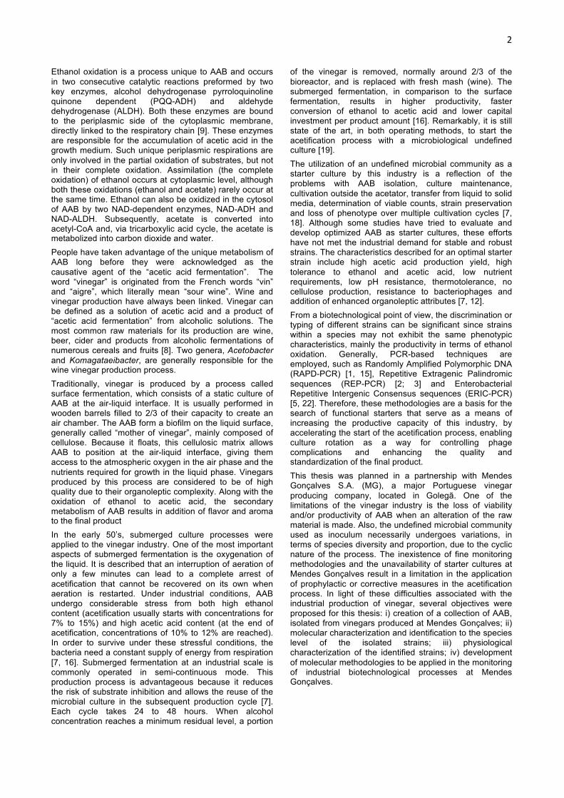
Ethanol oxidation is a process unique to AAB and occurs in two consecutive catalytic reactions preformed by two key enzymes, alcohol dehydrogenase pyrroloquinoline quinone dependent (PQQ-ADH) and aldehyde dehydrogenase (ALDH). Both these enzymes are bound to the periplasmic side of the cytoplasmic membrane, directly linked to the respiratory chain [9]. These enzymes are responsible for the accumulation of acetic acid in the growth medium. Such unique periplasmic respirations are only involved in the partial oxidation of substrates, but not in their complete oxidation. Assimilation (the complete oxidation) of ethanol occurs at cytoplasmic level, although both these oxidations (ethanol and acetate) rarely occur at the same time. Ethanol can also be oxidized in the cytosol of AAB by two NAD-dependent enzymes, NAD-ADH and NAD-ALDH. Subsequently, acetate is converted into acetyl-CoA and, via tricarboxylic acid cycle, the acetate is metabolized into carbon dioxide and water. People have taken advantage of the unique metabolism of AAB long before they were acknowledged as the causative agent of the “acetic acid fermentation”. The word “vinegar” is originated from the French words “vin” and “aigre”, which literally mean “sour wine”. Wine and vinegar production have always been linked. Vinegar can be defined as a solution of acetic acid and a product of “acetic acid fermentation” from alcoholic solutions. The most common raw materials for its production are wine, beer, cider and products from alcoholic fermentations of numerous cereals and fruits [8]. Two genera, Acetobacter and Komagataeibacter, are generally responsible for the wine vinegar production process. Traditionally, vinegar is produced by a process called surface fermentation, which consists of a static culture of AAB at the air-liquid interface. It is usually performed in wooden barrels filled to 2/3 of their capacity to create an air chamber. The AAB form a biofilm on the liquid surface, generally called “mother of vinegar”, mainly composed of cellulose. Because it floats, this cellulosic matrix allows AAB to position at the air-liquid interface, giving them access to the atmospheric oxygen in the air phase and the nutrients required for growth in the liquid phase. Vinegars produced by this process are considered to be of high quality due to their organoleptic complexity. Along with the oxidation of ethanol to acetic acid, the secondary metabolism of AAB results in addition of flavor and aroma to the final product In the early 50’s, submerged culture processes were applied to the vinegar industry. One of the most important aspects of submerged fermentation is the oxygenation of the liquid. It is described that an interruption of aeration of only a few minutes can lead to a complete arrest of acetification that cannot be recovered on its own when aeration is restarted. Under industrial conditions, AAB undergo considerable stress from both high ethanol content (acetification usually starts with concentrations for 7% to 15%) and high acetic acid content (at the end of acetification, concentrations of 10% to 12% are reached). In order to survive under these stressful conditions, the bacteria need a constant supply of energy from respiration [7, 16]. Submerged fermentation at an industrial scale is commonly operated in semi-continuous mode. This production process is advantageous because it reduces the risk of substrate inhibition and allows the reuse of the microbial culture in the subsequent production cycle [7]. Each cycle takes 24 to 48 hours. When alcohol concentration reaches a minimum residual level, a portion
of the vinegar is removed, normally around 2/3 of the bioreactor, and is replaced with fresh mash (wine). The submerged fermentation, in comparison to the surface fermentation, results in higher productivity, faster conversion of ethanol to acetic acid and lower capital investment per product amount [16]. Remarkably, it is still state of the art, in both operating methods, to start the acetification process with a microbiological undefined culture [19]. The utilization of an undefined microbial community as a starter culture by this industry is a reflection of the problems with AAB isolation, culture maintenance, cultivation outside the acetator, transfer from liquid to solid media, determination of viable counts, strain preservation and loss of phenotype over multiple cultivation cycles [7, 18]. Although some studies have tried to evaluate and develop optimized AAB as starter cultures, these efforts have not met the industrial demand for stable and robust strains. The characteristics described for an optimal starter strain include high acetic acid production yield, high tolerance to ethanol and acetic acid, low nutrient requirements, low pH resistance, thermotolerance, no cellulose production, resistance to bacteriophages and addition of enhanced organoleptic attributes [7, 12]. From a biotechnological point of view, the discrimination or typing of different strains can be significant since strains within a species may not exhibit the same phenotypic characteristics, mainly the productivity in terms of ethanol oxidation. Generally, PCR-based techniques are employed, such as Randomly Amplified Polymorphic DNA (RAPD-PCR) [1, 15], Repetitive Extragenic Palindromic sequences (REP-PCR) [2; 3] and Enterobacterial Repetitive Intergenic Consensus sequences (ERIC-PCR) [5, 22]. Therefore, these methodologies are a basis for the search of functional starters that serve as a means of increasing the productive capacity of this industry, by accelerating the start of the acetification process, enabling culture rotation as a way for controlling phage complications and enhancing the quality and standardization of the final product. This thesis was planned in a partnership with Mendes Gonçalves S.A. (MG), a major Portuguese vinegar producing company, located in Golegã. One of the limitations of the vinegar industry is the loss of viability and/or productivity of AAB when an alteration of the raw material is made. Also, the undefined microbial community used as inoculum necessarily undergoes variations, in terms of species diversity and proportion, due to the cyclic nature of the process. The inexistence of fine monitoring methodologies and the unavailability of starter cultures at Mendes Gonçalves result in a limitation in the application of prophylactic or corrective measures in the acetification process. In light of these difficulties associated with the industrial production of vinegar, several objectives were proposed for this thesis: i) creation of a collection of AAB, isolated from vinegars produced at Mendes Gonçalves; ii) molecular characterization and identification to the species level of the isolated strains; iii) physiological characterization of the identified strains; iv) development of molecular methodologies to be applied in the monitoring of industrial biotechnological processes at Mendes Gonçalves.

3
2. Materials and Methods 2.1 Vinegar Samples and Bacterial Strains Forty-one samples of vinegars produced by MG were brought to the Lab Bugworkers | M&B-BioISI and were screened for AAB using two cultural methods. Five additional red wine vinegar samples were obtained from MG. These five samples were collected from the same acetator, throughout 36 hours, corresponding to different stages of a red wine vinegar production cycle. A total of 31 AAB strains were used in this study, belonging to different genera and corresponding to 22 wild strains and 9 strains obtained from the German Collection of Microorganisms and Cell Cultures (DSMZ) and from the Spanish Type Culture Collection (CECT). The wild strains were isolated both in the MG microbiology laboratory and in the Lab Bugworkers | M&B-BioISI, from different types of vinegar. 2.2 Isolation of Acetic Acid Bacteria The vinegar samples were analyzed for AAB using two cultural methods, a direct approach and/or an enrichment approach. The direct approach consisted in directly plating 100 µl of each vinegar sample in GYC medium (5% glucose, 0.5% yeast extract, 0.3% peptone, 2% calcium carbonate and 1.5% agar). For the enrichment approach, 15 ml of each sample was centrifuged at 3220xg for 15 min and the cellular pellet was inoculated in 20 ml of GYP medium (5% glucose, 0.5% yeast extract, 0.3% peptone), supplemented with 3% ethanol (v/v). The liquid cultures were incubated at 28ºC and 160 rpm for 5 days. Then, the total volume of each culture was centrifuged in the same conditions described above and the cellular pellet was plated in GYC plates. All plates were incubated at 28ºC. 2.3 DNA extraction Genomic DNA was extracted using an adapted Guanidium Thiocyanate method described by Pitcher et al. (1989). These modifications were made primarily in the first steps of the method. Bacterial cells were ressuspended in 250 µl of lysis buffer (50 mM Tris; 250 mM NaCl; 50 mM EDTA; 0.3% SDS; pH 8.0) and 100 µl of microspheres. After 2 min of homogenization in a vortex, the cells were incubated in 65ºC for 30 min, followed by another 2 min of homogenization. Afterwards, the GES reagent (5 M guanidium thiocyanate; 10 mM EDTA; 0.5% sarkosyl; pH 8,0) was added and the original method was followed but using an equal volume of isopropanol. 2.4 Genomic Fingerprinting: RAPD-PCR, REP-PCR The RAPD-PCR was performed using the PH primer (5’ AAGGAGGTGATCCAGCCGCA ‘3) [10] and the REP-PCR was performed using the GTG5 primer (5’ GTGGTGGTGGTGGTG ‘3) [3]. Both amplification reactions were carried out in a total volume of 25 µl, containing 1x PCR reaction buffer, 3 mM of MgCl2, 25 pmol of primer, 0.2 mM of each of the four dNTP’s, 1 U of Taq polymerase and 1 µl of template DNA per reaction. All reagents used were acquired from Invitrogen (USA). This assay was performed in a UNO II thermal cycler (Biometra), with the following PCR conditions: 5 min of initial denaturation at 95ºC, followed by 40 cycles of denaturation at 95ºC for 1 min, annealing at 50ºC for 2 min and extension at 72ºC for 2 min, and a final extension at 72ºC for 5 min.
The banding patterns were analyzed with Bionumerics software (version 6.6, Applied Maths) and a composite dendrogram was created based on the genomic profiles obtained with the primers GTG5 and PH. This dendrogram was constructed using the Pearson correlation coefficient as a similarity measure and the unweighted pair group method with the arithmetic average clustering algorithm (UPGMA). A reproducibility assay was performed to determine the percentage of similarity necessary for strain discrimination. For each type of genomic fingerprinting, 10% of the isolates were randomly chosen and the amplification reaction was performed in duplicate. A dendrogram was built for these three isolates and their repeats and the optimization parameters were adjusted until each isolate was grouped with its repeat. The reproducibility of each type of genomic fingerprinting was determined as the average of the levels of similarity observed between repeats. The reproducibility of the composite dendrogram was defined as the average of the reproducibilities determined for each type of genomic fingerprinting. 2.5 Molecular Identification by 16S rRNA Gene Sequencing DNA from bacterial isolates (DNA extraction described in section 2.3) was used as a template for the amplification of a portion of the 16S rRNA gene, using the universal primers PA (27f) (5’ AGAGTTTGATCCTGGCTCAG 3’) [10] and 907r (5’ CCGTCAATTCMTTTRAGTTT 3’) [14]. The amplification reaction was carried out in a total volume of 50 µl, containing 1x PCR reaction buffer, 2 mM of MgCl2, 25 pmol of each primer, 0.2 mM of each of the four dNTP’s, 1 U of Taq polymerase and 1 µl of template DNA per reaction. All reagents used were acquired from Invitrogen (USA). This assay was performed in a UNO II thermal cycler (Biometra), with the following PCR conditions: 5 min of initial denaturation at 95ºC, followed by 35 cycles of denaturation at 95ºC for 1 min, annealing at 55ºC for 1 min and extension at 72ºC for 2 min, and a final extension at 72ºC for 5 min. The PCR products were purified using the kit Jet Quick PCR Product Purification Spin Kit (Genomed) and sequenced by Biopremier (Portugal). The algorithm BLAST (Basic Local Alignment Search Tool) was used to determine the closest known relative(s) of the partial 16S rRNA sequence obtained. Additionally, a phylogenetic reconstruction was generated using the MEGA software (version 7.0.20). The 16S rRNA gene sequences obtained were aligned (ClustalW 1.6) with the sequences of the same gene of the type strains of all species of the genus Komagataeibacter and were clustered with the neighbor-joining algorithm. 2.6 Multiplex-PCR: Primer Design and PCR Conditions Four different degenerate primers were designed based on DNA sequences available in GenBank for the subunit I of the PQQ-dependent ADH gene, adhA. The multiple alignment was done using the algorithm ClustalW (version 1.6) and the primers were designed based on the conserved regions shown by the alignment. All primers are composed of 20 nucleotides, consisting in two forward primers ADH-F1 (5’ ACMGCNACATACTGCTTGCC 3’) and ADH-F2 (5’ GCGTCRTARGCRTGGAATTC 3’) and two reverse primers ADH-R1 (5’ TGGTACGGCATKCCSGGKGA 3’) and ADH-R2 (5’ TKGGYCTSGACATGAACAAG 3’). The final amplification reaction was performed in a total volume of 25 µl, containing 1x PCR reaction buffer, 1.5 mM of MgCl2, 25 pmol of each of the adhA directed

4
primers (ADH-F1, ADH-F2 and ADH-R2), 6.25 pmol of each of the 16S rRNA gene directed primers (PA and 907r), 0.2 mM of each of the four dNTP’s, 1 U of Taq polymerase and 1 µl of template DNA per reaction. All reagents used were acquired from Invitrogen (Massachusetts, USA). This assay was performed in a T Gradient thermal cycler (Biometra, Germany), with the following PCR conditions: 5 min of initial denaturation at 95ºC, followed by 30 cycles of denaturation at 95ºC for 1 min, annealing at 57ºC for 1 min and extension at 72ºC for 1 min, and a final extension at 72ºC for 5 min. 2.7 Gel Electrophoresis The PCR products were visualized by electrophoresis in a 1.2% (w/v) agarose gel using a 1kb Plus DNA ladder (Invitrogen). The electrophoresis was performed in 0.5x TBE buffer with a constant voltage of 4.5 V/cm. The gel was stained in an ethidium bromide solution and photographed in an Alliance 4.7 UV transiluminador (UVItec) with Alliance software (version 15.15). 2.8 Growth Analysis and Quantification of Acetic Acid AAB strains were grown in red wine (14.4% ethanol). The wine was diluted in autoclaved ultrapure water in a proportion of 1:2, respectively (the final ethanol concentration was expected to be around 4.8%). Pre-cultures were grown (28ºC, 160 rpm) during four days, in 100 ml Erlenmeyers with 50 ml of GYP medium, supplemented with 2% ethanol and inoculated with a loopfull (10 µl) from frozen (-80ºC) preparations of the strains. Each pre-culture was then centrifuged at 3220xg for 15 min and the cellular pellet was ressuspended in 300 µl of GYP medium. For each strain, three 250 ml Erlenmeyers with 100 ml of the diluted wine were inoculated with 100 µl of the pre-culture ressuspended pellet. The cultures were incubated for several days at 28ºC and 160 rpm and samples were collected at several time-points. At each time-point, 1 ml of the culture was collected to a cuvette to measure the optical density (OD). Additionally, another culture sample of 1 ml was collected and centrifuged at 17968xg for 10 min. Subsequently, 900 µl were collected to another tube and frozen at -20ºC until they were used to measure the pH and the acetic acid concentration. OD600nm was measured using a UV1101 Biotech Photometer (WPA) and the pH was measured using a BioTrode lab pH microelectrode (Hamilton) coupled with a model 15 pH meter (Denver Instruments). Acetic acid concentration was determined enzymatically, using the Acetic Acid Assay Kit from Megazyme (Ireland). All reactions were performed in microplate assay. The specific growth rate (µ) was calculated by regression of logOD vs time during exponential growth phase. The acetic acid production rate (%day-1) was calculated by linear regression. the maximal bioconversion efficiency and productivity (gl-1day-1) of acetic acid was calculated as described by [13]. 2.9 Microbial Profiling Analysis Community DNA was extracted from the five red wine vinegar samples. About 250 ml of each vinegar sample was centrifuged at 15000xg for 10 min and the supernatant was discarded. The cellular pellet was transferred directly to the DNA Isolation Kit PowerMax™Soil (MO BIO Laboratories). The DNA extracts were provided to Biopremier (Portugal) where the Microbial Profiling analysis was performed. Two regions of the 16S rRNA gene were amplified by PCR (region 1 and region 2). The amplified fragments were sequenced by
NGS technology (Ion Torrent) and the resulting DNA sequences were identified by a BLAST analysis. 3. Results and Discussion 3.1 Isolation of Acetic Acid Bacteria The direct approach led to the isolation of bacteria from 8 of the 41 vinegars analyzed, totaling 12 different isolates. Of these 12 bacterial isolates, 3 belonged to the AAB group, isolated from vinegars samples 31 and 41. The enrichment approach led to the isolation of bacteria from 13 of the 40 vinegars analyzed, totaling 19 different isolates. Of these 19 bacterial isolates, 4 belonged to the AAB group, isolated from vinegars samples 14, 22, 31 and 32 (Figure 1). The isolation and cultivation of AAB has always been described as problematic. This is especially true in the isolation from a high acetic acid level source [16]. Additionally, a viable but not cultivable (VBNC) state as been described for AAB, mainly in oxygen privation conditions [9]. When comparing the forty vinegar samples used in both approaches, it is possible to state that the enrichment approach was more effective in the isolation of AAB than the direct approach, since it led to the isolation of 4 AAB isolates in comparison to one. It is possible that the aeration from the shaking culture helped the bacteria recover from the VBNC state. Also, the enrichment approach seems to have not been specific to AAB, as it was intended, since the number of non-AAB isolates also increased. A possible way to overcome this could be the removal of glucose from the liquid medium, with ethanol as the only carbon source. Overall, the enrichment approach was fairly successful, since it led to the isolation of three AAB isolates that otherwise wouldn’t be isolated (samples 14, 22 and 32). 3.2 Typing and Identification of Acetic Acid Bacteria Isolates A dendrogram-based identification using type strains was applied to 22 AAB isolates and four reference strains belonging to the genus Komagataeibacter. These strains were grouped based on REP-PCR and RAPD-PCR genomic profiles. The reproducibility analysis established a discrimination threshold (97%) below which patterns were deemed different, indicated by the red dotted line. The dendrogram divided the 22 AAB isolates into five distinct strains, with strain 1 having the isolates AAB 001, 002, 003, 004, 010, 011, 012, 015, 016, 017, 025, 026, 029 and 030; strain 2 having the isolates AAB 027 and 028; strain 3 having the isolates AAB 023, 024, 031 and 032; and lastly, strains 4 and 5, having the isolates AAB 034 and 033, respectively (Figure 1). The grey bar shown in the dendrogram represents the cutoff level for species separation. The range of similarity between the two different species more closely related (strain 3 and 5) determines this delimitation. Thus, depending on the level of similarity chosen for the cutoff level for species separation, strains 1 and 2 may or may not belong to Km. europaeus. Still, these strains show a high level of similarity with this species. Concerning the isolates of the strain 3, they do not show any meaningful similarity with any of the reference strains. The same happens with isolates AAB 033 and 034 (strains 5 and 4, respectively). Therefore, this analysis is not capable of identifying these strains.

5
0
1
2
3
1 2 3 4 5 6 7 8 9 10 11 12 13 14 15 16 17 18 19 20 21 22 23 24 25 26 27 28 29 30 31 32 33 34 35 36 37 38 39 40 41
Num
ber o
f diff
eren
t iso
late
s
Vinegar Samples
Direct Approach AAB Not AAB
39
0
1
2
3
1 2 3 4 5 6 7 8 9 10 11 12 13 14 15 16 17 18 19 20 21 22 23 24 25 26 27 28 29 30 31 32 33 34 35 36 37 38 39 40
Num
ber o
f diff
eren
t iso
late
s
Vinegar Samples
Enrichment Approach AAB Not AAB
4115
Figure 1. Number of different isolates obtained from each vinegar sample, with the direct and enrichment approaches. The dark blue represents AAB isolates while the light blue represents bacterial isolates not belonging to
the AAB group. The pie charts show the total number of isolates for each approach. Only the direct approach was performed for vinegar sample 41.
Figure 2. REP-PCR (GTG5) and RAPD-PCR (PH) fingerprinting patterns from AAB isolates and four reference
strains belonging to the genus Komagataeibacter. The dendrogram was constructed using the Pearson
correlation coefficient as a similarity measure and the unweighted pair group method with the arithmetic average
clustering algorithm (UPGMA). The red dotted line represents the cutoff level (97%) determined by the reproducibility
analysis and the grey bar shows the cutoff level for species separation. DSMZ 5602: Km. hansenii; DSMZ 6160: Km.
europaeus; DSMZ 6513: Km. xylinus; DSMZ 11804: Km. intermedius.

6
GTG5-REP-PCR was reported to be a useful fingerprinting technique for identification and classification of AAB to the species level [3]. The RAPD-PCR methodology with the primer PH has been reported as providing suitable fingerprints, with well defined amplification patterns, appropriate for the identification of Listeria spp. [1]. As far as we know, it was never applied to AAB. This methodology seems to be promising for the discrimination and classification of this group of bacteria, since it is able to distinguish strain 1 from strain 2 and strain 4 from strain 5, contrarily to GTG5-REP-PCR. However, several other type strains would have to be tested, along with a larger collection of isolates to truly unveil the discriminatory power of the method. The sequences obtained with the amplification and sequencing of a portion of the 16S rRNA gene of bacterial isolates were used in a BLAST analysis in search of their closest known relative. However, this analysis was inconclusive since the identification to the species level was not possible, for any of the isolates. So, these sequences were used in a phylogeny reconstruction (neighbor-joining algorithm), along with the 16S rRNA gene sequences of the type strains of all the species of the genus Komagataeibacter, collected from the GenBank database (Figure 3). The phylogenetic tree cannot distinguish between the isolates AAB 016, 025, 026 and 030 (representatives of strain 1), AAB 027 and 028 (strain 2), AAB 023, 024 and 032 (strain 3) and the type strains of Km. europaeusT and Km. swingsiiT. This result is in agreement with the results obtained with the genomic fingerprints, where isolates from strains 1 and 2 were shown to be closely related to each other and with Km. europaeusT. However, the type strain of Km. swingsiiT was not used in this study, resulting in its absence from the genomic fingerprinting analysis. Regarding the isolates of strain 3, it was surprising to see how they align with the isolates from strains 1 and 2. This result is in clear contradiction with the difference demonstrated by the analysis of the genomic fingerprints. Additionally, the isolates AAB 033 (strain 5), AAB 034 (strain 4) also align with each other and with the type strains of Km. nataicolaT and Km. sucrofermentansT. Again, this result does not correspond to the interpretation of the dendrogram, where strain 4 and 5 are clearly separated from each other, belonging to different species. Overall, there is a strong suggestion that the strains 1 and 2 belong to Km. europaeus. Still, the possibility of them belonging to Km. swingsii cannot be discarded. Interestingly, the two strains show phenotypic traits both agreeing and disagreeing with those described for these species. Regarding strains 4 and 5, they both show cellulose production, which is a characteristic shared with Km. nataicola and Km. sucrofermentans. Concerning strain 3, the results of both analyses are incoherent since the dendrogram clearly separates this strain from strains 1 and 2 and the phylogeny reconstruction does not distinguish these three strains. Thus, the identification to the species level of any of the strains remains to be confirmed. 3.3 Development of a Molecular Detection Method for Acetic Acid Bacteria Different amplification conditions and primer combinations were tested in order to optimize the PCR amplification with the designed primers. Initially, a PCR assay was planned where two concentrations of MgCl2 (1.5 mM and 2.5 mM) and primers (25 pmol and 50 pmol) and a range of
annealing temperatures (46.5ºC to 57.5ºC) were tested for the four possible primer combinations. The selection of 1.5 mM of MgCl2, 25 pmol of primer and annealing temperature at 57ºC led to the sequential experimentation on a variety of AAB reference stains, AAB isolates and non-AAB isolates, as well as the combination of all primers in the same reaction (multiplex-PCR approach). Figure 8 shows the Multiplex-PCR profile of AAB, as well as of bacterial isolates not belonging to this group. The PCR with the primers ADH-F1, ADH-F2 and ADH-R2 results in the amplification of two fragments in AAB, 240 bp (primers ADH-F2 and ADH-R2), 336 bp (primers ADH-F1 and ADH-R2). Sometimes, the unspecific amplification of a third fragment occurs. In Figure 4, the Multiplex-PCR amplification reaction is shown, in an electrophoretic separation in agarose gel (1.2%), for all the reference strains used in this study and for 2 isolates already confirmed to be AAB (AAB 003, 016; Komagataeibacter sp.) by 16S rRNA sequencing. Additionally, 17 isolates brought to Lab Bugworkers | M&B-BioISI from the MG lab as potential AAB were also screened. Lastly, M. morganii and E. coli ATCC 25922 are shown here as exclusivity controls, where amplification with the adhA primers was not expected to occur and a negative control (—) where no DNA was added to the PCR mix and no amplification was expected to occur with any of the five primers.
Km. europaeusT
Km. swingsiiT
Km. intermediusT
Km. nataicolaT
Km. sucrofermentansT
Km. oboediensT
Km. xylinusT
Km. medellinensisT
Km. saccharivoransT
Km. rhaeticusT
Km. hanseniiT
Km. kombuchaeT
Km. maltacetiT
Km. kakiacetiT
A. acetiT
Figure 3. Phylogenetic relationships amongst all species of the genus Komagataeibacter and AAB isolates. The phylogenetic
tree is based on the sequences of the 16S rRNA gene. The tree was constructed with neighbor-joining algorithm and numbers at the
nodes indicate bootstrap values (%) derived from 1000 replications. Acetobacter aceti was used as an outgroup.

7
Since AAB are involved not only in the production, but also in the spoilage of foods and beverages, the monitoring of their presence is essential in the different stages of an industrial bioprocess, especially in the final product, after the manufacturing process [16]. Since identification methodologies based on phenotypic characteristics of AAB are not only unreliable, but also time-consuming, the application of molecular detection and/or identification methods could provide a fast and accurate solution [20]. The discrimination power of a molecular detection method is extremely dependent on its molecular target. The unique ability of this group of bacteria to oxidize ethanol to acetic acid is due to two periplasmic proteins, PQQ-ADH and ALDH [12]. Therefore, the gene adhA, which encodes for the subunit I of the PQQ-ADH was evaluated as a potential molecular target. A multiple alignment of sequences of this gene belonging to different AAB species showed the presence of variable and conserved segments, ideal for the design of oligonucleotide primers. The gene adhA has already been studied and was shown to be more and less discriminatory for AAB when compared with the 16S rRNA gene and the 16S-23S rRNA ITS, respectively. Also, A. aceti specific primers were designed and the gene adhA was reported to be a promising target for the construction of species-specific oligonucleotides for quick molecular identification of AAB [20]. Here, the designed primers were shown to be specific for AAB, since all the non-AAB isolates tested were negative (only showed amplification of the internal control). Besides, all these non-AAB strains were isolated from vinegar samples. Still, one of the reference strains, A. cerevisiae CECT 824, showed a negative result, meaning that the primer ADH-R2 does not hybridize in the adhA sequence of this strain and that the designed primers were not 100% inclusive in the tested conditions. Additionally, strains Km. xylinus DSMZ 6513T, A. aceti DSMZ 3508T and A. pasteurianus DSMZ 3509T all show amplification problems with at least one of the designed primers. However, if the primer does not hybridize in a certain type strain, sequence differences may exist to open the road for the use of adhA gene to build species-specific probes. Taking everything into account, these results show that the designed primers, and the optimized amplification reaction, are effective in the molecular detection of AAB and that once again, the gene adhA has been successfully used for this purpose, even though different regions were explored. This methodology was applied as a routine
detection method to several unidentified bacterial isolates brought to the Lab Bugworkers | M&B-BioISI from the MG lab and it proved to be a fast and reliable methodology in the distinction of AAB from non-AAB isolates. 3.4 Growth Performance of Acetic Acid Bacteria in Red Wine AAB isolates AAB 023, 030, 033 and 034, corresponding to strains 3, 1, 5 and 4, respectively, were inoculated in diluted wine (around 4.8% ethanol concentration) and their growth was followed for several days. Figure 5 shows the growth characteristics of these four strains, as well as the pH of the medium and the concentration of acetic acid. All strains were grown in triplicates and all OD, pH and acetic acid concentration measures were obtained for each triplicate. The isolate that showed the shortest lag phase, around one day, was AAB 030, with isolates AAB 023 and 033 showing a lag phase of around two days. Regarding the cell yield, isolates AAB 023 and 030 showed the highest values, reaching OD values of 0.9 and 0.75, respectively, and the highest growth rates, 0.28 day-1 and 0.34 day-1, respectively, while isolates AAB 033 and 034 showed very low cell yield values and growth rates. The highest acetic acid concentration, around 4% (w/v), was obtained with isolate AAB 023, while around 3.2% (w/v) acetic acid was obtained with isolates AAB 030 and 033 and around 2.8% (w/v) acetic acid was obtained with isolate AAB 034. Isolate AAB 023 showed the highest bioconversion efficiency, 78.3%. Isolates AAB 033 and 034 showed lower values, 64.2% and 55.2%, respectively, since they must redirect carbon and energy for the production of cellulose. As expected, isolate AAB 023 shows a high productivity, 2.96 gl-1day-1. Still, isolate AAB 033 achieved the highest productivity, 3.11 gl-1day-1. This could be explained by the fact that isolate AAB 033 reached the maximum acetic acid concentration in 10 days while isolate AAB 023 reached the maximum acetic acid concentration in 13 days. In general, these four isolates show two distinct growth types that seem to be associated to the production of cellulose. Isolates AAB 033 and 034 are cellulose producers. This is quite possibly the explanation to the low cell yield measured, since cells of these strains grow primarily on the cellulose matrix. It is interesting to see that although isolates AAB 023 and 030 show a diauxic growth curve, the second exponential growth phase does not correspond to the assimilation of acetic acid, as is
Mul$plex(PCR,
62#A16#
57#A16#
63#A16#A#
67#A16#
64#A16#
65#A16#
66#A16#
68#A16#
71#A16#
74#A16#
75#A16#
77#A16#
78#A16#
83#A16#
85#A16#
86#A16#
63#A16#B#
DSMZ
#3503#T#
DSMZ
#3508#T #
DSMZ
#3509#T #
DSMZ
#5602#T #
DSMZ
#6513#T #
DSMZ
#11804#T#
CECT#824#
CECT#4009#
DSMZ
#6160#T #
M.#morg
anii#
E.#coli#(A
TCC#25922)#
AAB#003#
AAB#016#
M#M#
Figure 4. Typical Multiplex-PCR profile of AAB, as well as of bacterial isolates not belonging to this group. The black arrow shows the expected amplification bands for an AAB isolate. The white arrow shows the band corresponding to the
internal control (primers PA and 907r). The (M) refers to 1kb Plus DNA ladder (Invitrogen).

8
Figure 5. Growth characteristics of strains of AAB in diluted wine. A: non-cellulose producing isolates AAB 023, 030, corresponding to strains 3, 1, respectively. B: cellulose producing isolates AAB 033 and 034, corresponding to strains 5 and 4, respectively. All strains were grown in triplicates, at 28ºC and shaking conditions. Error bars represent the standard deviation at
each time-point. OD: optical density measured at 600 nm (u); pH (n); % AcH: acetic acid concentration (m/v) (p).
91.1% 97.5% 83.9%
100.0% 84.7%
8.9% 2.6%
16.2%
0.0%
15.3%
Microbial Profiling - Region 1
Komagataeibacter sp. Other bacteria sp.p.
t1 (0h)
t2 (19h)
t3 (23h)
t4 (29h)
t5 (36h)
89.9% 97.8%
87.8% 100.0% 93.9%
10.1% 2.1%
12.2% 0.0%
5.9%
Microbial Profiling - Region 2
Komagataeibacter sp. Other bacteria sp.p.
t1 (0h)
t2 (19h)
t3 (23h)
t4 (29h)
t5 (36h)
Figure 6. Representation of the proportion of acetic acid bacteria in each time-point. The pie charts show the relative abundance of non-AAB in each time-point. Percentages relative to the analysis of the region 1 of the 16S rRNA gene were
calculated from a total of 39 093 reads. Percentages relative to the analysis of the region 2 of the 16S rRNA gene were calculated from a total of 45 997 reads.
9
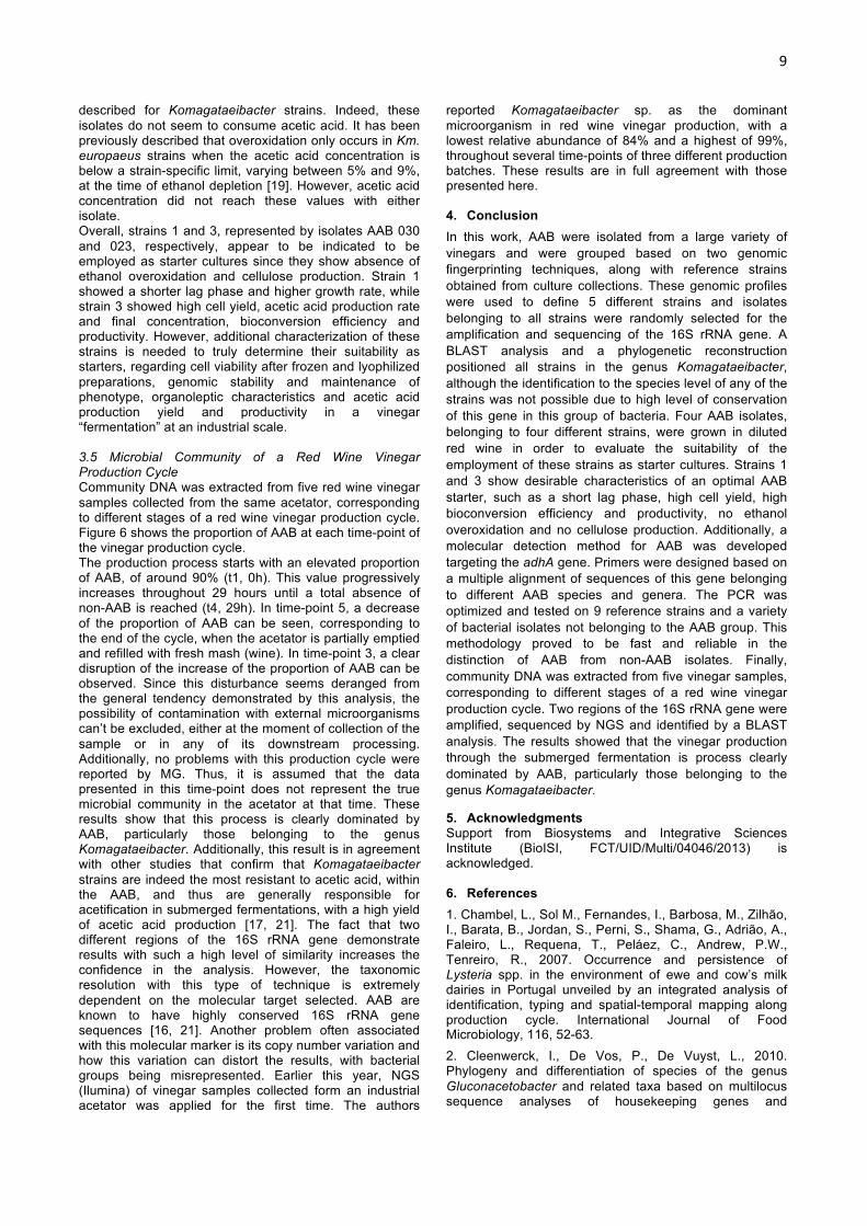
described for Komagataeibacter strains. Indeed, these isolates do not seem to consume acetic acid. It has been previously described that overoxidation only occurs in Km. europaeus strains when the acetic acid concentration is below a strain-specific limit, varying between 5% and 9%, at the time of ethanol depletion [19]. However, acetic acid concentration did not reach these values with either isolate. Overall, strains 1 and 3, represented by isolates AAB 030 and 023, respectively, appear to be indicated to be employed as starter cultures since they show absence of ethanol overoxidation and cellulose production. Strain 1 showed a shorter lag phase and higher growth rate, while strain 3 showed high cell yield, acetic acid production rate and final concentration, bioconversion efficiency and productivity. However, additional characterization of these strains is needed to truly determine their suitability as starters, regarding cell viability after frozen and lyophilized preparations, genomic stability and maintenance of phenotype, organoleptic characteristics and acetic acid production yield and productivity in a vinegar “fermentation” at an industrial scale. 3.5 Microbial Community of a Red Wine Vinegar Production Cycle Community DNA was extracted from five red wine vinegar samples collected from the same acetator, corresponding to different stages of a red wine vinegar production cycle. Figure 6 shows the proportion of AAB at each time-point of the vinegar production cycle. The production process starts with an elevated proportion of AAB, of around 90% (t1, 0h). This value progressively increases throughout 29 hours until a total absence of non-AAB is reached (t4, 29h). In time-point 5, a decrease of the proportion of AAB can be seen, corresponding to the end of the cycle, when the acetator is partially emptied and refilled with fresh mash (wine). In time-point 3, a clear disruption of the increase of the proportion of AAB can be observed. Since this disturbance seems deranged from the general tendency demonstrated by this analysis, the possibility of contamination with external microorganisms can’t be excluded, either at the moment of collection of the sample or in any of its downstream processing. Additionally, no problems with this production cycle were reported by MG. Thus, it is assumed that the data presented in this time-point does not represent the true microbial community in the acetator at that time. These results show that this process is clearly dominated by AAB, particularly those belonging to the genus Komagataeibacter. Additionally, this result is in agreement with other studies that confirm that Komagataeibacter strains are indeed the most resistant to acetic acid, within the AAB, and thus are generally responsible for acetification in submerged fermentations, with a high yield of acetic acid production [17, 21]. The fact that two different regions of the 16S rRNA gene demonstrate results with such a high level of similarity increases the confidence in the analysis. However, the taxonomic resolution with this type of technique is extremely dependent on the molecular target selected. AAB are known to have highly conserved 16S rRNA gene sequences [16, 21]. Another problem often associated with this molecular marker is its copy number variation and how this variation can distort the results, with bacterial groups being misrepresented. Earlier this year, NGS (Ilumina) of vinegar samples collected form an industrial acetator was applied for the first time. The authors
reported Komagataeibacter sp. as the dominant microorganism in red wine vinegar production, with a lowest relative abundance of 84% and a highest of 99%, throughout several time-points of three different production batches. These results are in full agreement with those presented here. 4. Conclusion In this work, AAB were isolated from a large variety of vinegars and were grouped based on two genomic fingerprinting techniques, along with reference strains obtained from culture collections. These genomic profiles were used to define 5 different strains and isolates belonging to all strains were randomly selected for the amplification and sequencing of the 16S rRNA gene. A BLAST analysis and a phylogenetic reconstruction positioned all strains in the genus Komagataeibacter, although the identification to the species level of any of the strains was not possible due to high level of conservation of this gene in this group of bacteria. Four AAB isolates, belonging to four different strains, were grown in diluted red wine in order to evaluate the suitability of the employment of these strains as starter cultures. Strains 1 and 3 show desirable characteristics of an optimal AAB starter, such as a short lag phase, high cell yield, high bioconversion efficiency and productivity, no ethanol overoxidation and no cellulose production. Additionally, a molecular detection method for AAB was developed targeting the adhA gene. Primers were designed based on a multiple alignment of sequences of this gene belonging to different AAB species and genera. The PCR was optimized and tested on 9 reference strains and a variety of bacterial isolates not belonging to the AAB group. This methodology proved to be fast and reliable in the distinction of AAB from non-AAB isolates. Finally, community DNA was extracted from five vinegar samples, corresponding to different stages of a red wine vinegar production cycle. Two regions of the 16S rRNA gene were amplified, sequenced by NGS and identified by a BLAST analysis. The results showed that the vinegar production through the submerged fermentation is process clearly dominated by AAB, particularly those belonging to the genus Komagataeibacter.
5. Acknowledgments Support from Biosystems and Integrative Sciences Institute (BioISI, FCT/UID/Multi/04046/2013) is acknowledged. 6. References 1. Chambel, L., Sol M., Fernandes, I., Barbosa, M., Zilhão, I., Barata, B., Jordan, S., Perni, S., Shama, G., Adrião, A., Faleiro, L., Requena, T., Peláez, C., Andrew, P.W., Tenreiro, R., 2007. Occurrence and persistence of Lysteria spp. in the environment of ewe and cow’s milk dairies in Portugal unveiled by an integrated analysis of identification, typing and spatial-temporal mapping along production cycle. International Journal of Food Microbiology, 116, 52-63. 2. Cleenwerck, I., De Vos, P., De Vuyst, L., 2010. Phylogeny and differentiation of species of the genus Gluconacetobacter and related taxa based on multilocus sequence analyses of housekeeping genes and

10
reclassification of Acetobacter xylinus subsp. sucrofermentans as Gluconacetobacter sucrofermentans (Toyosaki et al. 1996) sp. nov., comb. nov. International Journal of Systematic and Evolutionary Microbiology, 60, 2277–2283. 3. De Vuyst, L., Camu, N., De Winter, T., Vandemeulebroecke, K., Van de Perre, V., Vancanneyt, M., De Vos, P., Cleenwerck, I., 2008. Validation of the (GTG)5-rep-PCR fingerprinting technique for rapid classification and identification of acetic acid bacteria, with a focus on isolates from Ghanaian fermented cocoa beans. International Journal of Food Microbiology 125, 79–90. 4. Deppenmeier, U., Hoffmeister, M., Prust, C., 2002. Biochemistry and Biotechnological Applications of Gluconobacter strains. Applied Microbiology Biotechnology, 60:233–242. 5. Fernández-Pérez, R., Torres, C., Sanz, S., Ruiz-Larrea, F., 2010. Strain typing of acetic acid bacteria responsible for vinegar production by the submerged elaboration method. Food Microbiology 27, 973-978. 6. Guillamón, J.M., Mas, A., 2009. Acetic Acid Bacteria. In Biology of Microorganisms on Grapes, in Must and in Wine (pp. 31-46). König, H., Unden, G., Fröhlich, J. (Eds.) Springer, Heidelberg. 7. Gullo, M., Verzelloni, E., Canonico, M., 2014. Aerobic submerged fermentation by acetic acid bacteria for vinegar production: Process and biotechnological aspects. Process Biochemistry 49, 1571-1579. 8. Komagata, K., Iino, T., Yamada, Y., 2014. The Family Acetobacteraceae. In The Prokaryotes, (pp. 3-78) Rosenberg, E., et al. (Eds.) Springer, New York. 9. Mamlouk, D., Gullo, M., 2013. Acetic Acid Bacteria: Physiology and Carbon Sources Oxidation. Indian Journal of Microbiology, 53:377–384. 10. Massol-Deya, A.A., Odelson, D.A., Hickey, R.F., Tiedje, J.M., 1995. Bacterial community fingerprinting of amplified 16S and 16-23 ribossomal DNA gene sequences and restriction endonuclease analysis (ARDRA). Molecular Microbial Ecology Manual, 3.3.2: 1-8. 11. Matsushita, K., Toyama, H., Adachi, O., 2004. Diversity of Prokaryotic Respiratory Systems. In Respiration in Archaea and Bacteria. (pp. 81-99). Davide Zannoni, (Ed.) Springer, The Netherlands. 12. Matsushita, K., Toyama, H., Tonouchi, N., Okamoto-Kainuma, A., 2016. Acetic Acid Bacteria, Ecology and Physiology. Springer, Japan. 13. Mounir, M., Shafiei, R., Zarmehrkhorshid, R., Hamouda, A., Thonart, P., Delvigne, F., Alaoui, M.I., 2016. Optimization of biomass production of Acetobacter pasteurianus KU710511 as a potential starter for fruit vinegar production. African Journal of Biotechnology, Vol. 15(27), pp. 1429-1441. 14. Muyzer, G., Brinkhoff, T., Nübel, U., Santegoeds, C., Schäfer, H., Wawer, C., 1998. Denaturating gradient gel electrophoresis (DGGE) in microbial ecology. Molecular Microbial Ecology Manual, 3.4.4: 1-27. 15. Nanda, K., Taniguchi, M., Ujike, S., Ishihara, N., Mori, H., Ono, H., Murooka, Y., 2001. Characterization of Acetic Acid Bacteria in Traditional Acetic Acid Fermentation of Rice Vinegar (Komesu) and Unpolished Rice Vinegar
(Kurosu) Produced in Japan. Applied And Environmental Microbiology, p. 986–990. 16. Raspor, P., Goranovič, D., 2008. Biotechnological Applications of Acetic Acid Bacteria. Critical Reviews in Biotechnology, 28:101-124. 17. Sievers, M., Sellmer, S., Teuber, M., 1992. Acetobacter europaeus sp. nov., a Main Component of Industrial Vinegar Fermenters in Central Europe. Systematic and Applied Microbiology, 15, 386-392. 18. Sokollek, S.J., Hammes, W.P., 1997. Description of a Starter Culture Preparation for Vinegar Fermentation. Systematic and Applied Microbiology, 20: 481–491. 19. Sokollek, S.J., Hertel, C., Hammes, W.P., 1998. Cultivation and Preservation of Vinegar Bacteria. Journal of Biotechnology, 60, 195-206. 20. Trček, J., 2005. Quick identification of acetic acid bacteria based on nucleotide sequences of the 16S–23S rDNA internal transcribed spacer region and of the PQQ-dependent alcohol dehydrogenase gene. Systematic and Applied Microbiology 28, 735–745. 21. Trček, J., Mahnič, A., Rupnik, M., 2016. Diversity of the microbiota involved in wine and organic apple cider submerged vinegar production as revealed by DHPLC analysis and next-generation sequencing. International Journal of Food Microbiology, 223, 57–62. 22. Vegas, C., Mateo, E., González, Á., Jara, C., Guillamón, J.M., Poblet, M., Torija, M.J., Mas, A., 2010. Population dynamics of acetic acid bacteria during traditional wine vinegar production. International Journal of Food Microbiology 138, 130–136.