UNIT 2: Chemistry of the Human Body Compounds Compounds Metabolism Metabolism Nutrition Nutrition.

METABOLISM OF ORGANOSULFUR COMPOUNDS IN RUEGERIA POMEROYI DSS-3
by
WARREN CRABB
(Under the Direction of William B. Whitman)
ABSTRACT
Dimethylsulfoniopropionate (DMSP) accounts for up to 10% of the carbon fixed by
marine phytoplankton, most of which, is released as dissolved organic matter available for
degradation and assimilation by bacterioplankton. Bacterial metabolism of DMSP proceeds via
two competing pathways: demethylation or cleavage. The latter releases dimethyl sulfide (DMS),
a climatically relevant gas and a major source of atmospheric sulfur. The demethylation pathway
was found to be highly abundant in marine bacteria and accounts for a majority of DMSP
degradation. In addition to being a source of energy and sulfur for marine bacteria, the
demethylation pathway directs transformation of DMSP away from formation of DMS. A
number of techniques were utilized to elucidate the pathways and enzymology of DMSP
metabolism in the marine bacteria, Ruegeria pomeroyi DSS-3.
INDEX WORDS: Dimethylsulfoniopropionate; DMSP; Methanethiol; MeSH; Acrylate; 3-
Hydroxypropionate; Metabolism; Assimilation; Sulfur; Ruegeria
pomeroyi

METABOLISM OF ORGANOSULFUR COMPOUNDS IN RUEGERIA POMEROYI DSS-3
by
WARREN CRABB
B.S, Armstrong Atlantic State University, 2010
A Thesis Submitted to the Graduate Faculty of The University of Georgia in Partial Fulfillment
of the Requirements for the Degree
MASTER OF SCIENCE
ATHENS, GEORGIA
2013

© 2013
Warren Crabb
All Rights Reserved
METABOLISM OF ORGANOSULFUR COMPOUNDS IN RUEGERIA POMEROYI DSS-3
by
WARREN CRABB
Major Professor: William B. Whitman
Committee: Mary Ann Moran
Robert J. Maier
Electronic Version Approved:
Maureen Grasso
Dean of the Graduate School
The University of Georgia
August 2013

iv
ACKNOWLEDGEMENTS
I would like to acknowledge the generous support and mentorship given by my thesis advisor
Dr. William B. Whitman. I would also like to acknowledge Dr. Mary Ann Moran for her support
and limitless enthusiasm as well as Dr. Robert Maier for his help and guidance. Finally, I would like
to thank the members of the Whitman lab for their support and friendship.

v
TABLE OF CONTENTS
Page
ACKNOWLEDGEMENTS ........................................................................................................... iv
LIST OF TABLES ........................................................................................................................ vii
LIST OF FIGURES ..................................................................................................................... viii
CHAPTER
1 Literature Review ..........................................................................................................1
DMSP Demethylation .............................................................................................3
Methanethiol incorporation and utilization ..............................................................4
DMSP Cleavage .......................................................................................................5
References ..............................................................................................................10
2 Crystal structure of DmdD, a crotonase superfamily enzyme that catalyzes the
hydration and hydrolysis of methylthioacryloyl-CoA .................................................16
Introduction ............................................................................................................16
Materials and Methods ...........................................................................................17
Results and Discussion ..........................................................................................19
References ..............................................................................................................23

vi
3 Assimilation of dimethylsulfoniopropionate carbon by Ruegeria pomeroyi DSS-3 ...31
Introduction ............................................................................................................31
Materials and Methods ...........................................................................................33
Results and Discussion ..........................................................................................38
References ..............................................................................................................51
4 Metabolism of One-Carbon Compounds .....................................................................62
Introduction ............................................................................................................62
Materials and Methods ...........................................................................................63
Results ....................................................................................................................63
References ..............................................................................................................66
APPENDICES
A. Isotope labeled DMSP Synthesis .................................................................................70

vii
LIST OF TABLES
Page
Table 1: Summary of kinetic parameters .......................................................................................27

viii
LIST OF FIGURES
Page
Figure 1.1: Overview of DMSP degradation pathways in the marine environment ........................7
Figure 1.2: Pathways of DMSP metabolism in Ruegeria pomeroyi DSS-3 ....................................8
Figure 1.3: End point partition of sulfur moiety derived from DMSP ............................................9
Figure 2.1: Reactions of DmdD with crotonyl-CoA and 3-hydroxybutyryl-CoA .........................29
Figure 2.2: A proposed catalytic mechanism for the hydration and hydrolysis of MTA-CoA by
DmdD .................................................................................................................................30
Figure 3.1: Proposed DMSP cleavage and acrylate assimilation pathway in R. pomeroyi ...........58
Figure 3.2: Growth of wild-type R. pomeroyi and mutant strains .................................................59
Figure 3.3: Carbon fluxes during DMSP assimilation and oxidation ............................................60
Figure 3.4: Growth phenotype of wild-type R. pomeroyi and the pdhA::Tn5 mutant pdh1 ..........61
Figure 4.1: Growth of wild-type R. pomeroyi and ccr- (SPO0370::tet) .........................................68
Figure 4.2: Growth of the wild-type cells with various C-1 compounds .......................................69

1
CHAPTER 1
Literature Review
The ubiquitous phytoplankton metabolite dimethylsulfoniopropionate (DMSP) is a major
source of reduced sulfur and carbon for marine microbes. The concentration of dissolved DMSP
ranges from less than 1 nM up to 50 nM in surface waters with intracellular concentrations
reaching 1.0 M in some phytoplankton [14]. DMSP accounts for up to 10% of the carbon fixed
by marine phytoplankton, where it functions primarily as an osmoprotectant [23] with proposed
secondary roles as an antioxidant [33], predator deterrent [44], and cryoprotectant [15]. Much of
the interest in DMSP stems from the hypothesis that it is a precursor for the climatically active
gas dimethyl sulfide (DMS) [25]. Oxidation of atmospheric DMS results in the formation of
sulfate, sulfur dioxide, and other sulfur gases that may act as cloud condensation nuclei, thus
increasing solar backscatter and effectively lowering the amount of solar radiation reaching the
ocean surface. Therefore, the release of DMS acts as a negative feedback loop by decreasing
phytoplankton growth and reducing DMSP production (Figure 1). This hypothesis, referred to as
CLAW for the surnames of authors who proposed it, has recently been called into question [29].
Regardless of the impact of DMS on cloud formation, it still remains a significant source of
gaseous sulfur to the marine troposphere [2]. It is estimated that 40 Teragrams/year of DMS are
released from marine systems globally from non-anthropogenic sources, where it accounts for
42% of the atmospheric sulfur burden [32].
While some DMSP is degraded by the marine algae that synthesized it, most of the 12-
100 Tmol DMSP-S released each year from phytoplankton is accumulated and metabolized by

2
marine bacteria through two competing pathways (Figure 2). Referred to as the demethylation
and cleavage pathways, these pathways result in the release of methanethiol (MeSH) or DMS,
respectively. By examining size, fractionation, and chemical fate of [35S]DMSP incubations,
Vila-Costa et al. [43] estimated 10–50% of DMSP synthesized from algae is released as
dissolved matter. The estimated fates of the sulfur from this released DMSP were 20–40%
transformation to nonvolatile sulfur (DMSO, sulfate or other compounds), 5–30% assimilation
by bacteria into the protein fraction, and 2–5% transformation to DMS.
Using incubations of oceanic and coastal waters amended with 35S radiolabeled DMS,
MeSH and DMSP, Kiene and Linn [21] measured the end product partitioning of the sulfur
moiety (Figure 3). Approximately 15% of the 35S accumulated intracellularly, was found as un-
degraded DMSP. This pool of DMSP reached concentrations of roughly 70 mM and is
hypothesized to function as an osmoprotectant [30]. In coastal waters nearly 60% of the
[35S]DMSP was incorporated into cellular material, possibly into methionine via assimilation of
MeSH. In contrast, only 16% was incorporated in oceanic waters. The higher assimilation rates
of DMSP sulfur in coastal waters is likely due to increased growth rates; suggesting a higher
sulfur demand for organisms inhabiting these systems. MeSH that was not incorporated was
oxidized to nonvolatile dissolved sulfur. DMS turnover was much lower than the flux from
DMSP to MeSH. Small amounts of the sulfur from DMS were converted to cell protein, but most
of it was converted to sulfate.
Following incubations of coastal and oceanic samples with [35S]DMS, 70% of DMS was
converted to DMSO, 3% was incorporated into macromolecules, and the remainder was oxidized
to SO2– [43]. Samples pre-incubated with DMS formed less DMSO. The authors speculate that
there was a population of bacteria, perhaps in the roseobacter clade or a Hypomicrobium, that

3
converts DMS to MeSH and then to HS– by heterotrophic oxidation of the methyl groups. This
HS can then be oxidized to SO2–. In total, up to 90% of the DMSP sulfur enters the
demethylation pathway, forming MeSH, which is then incorporated into cells or degraded to
sulfate. Of the DMSP that is cleaved to DMS, 60–99% of the sulfur in DMS is not released,
being transformed to sulfate or DMSO. [43]
DMSP Demethylation
The model organism Ruegeria pomeroyi (formerly Silicibacter) is capable of metabolizing
DMSP through either the demethylation or cleavage pathway (Figure 2). dmdA, the gene
responsible for initiating demethylation of DMSP, was identified in 2006 by transposon
mutagenesis [14]. Subsequent characterization revealed that it encodes a tetrahydrofolate (THF)-
dependent enzyme that produces 5-methyl-THF and methylmercaptopropionate (MMPA) [30].
Homologues to R. pomeroyi dmdA are abundant in genomic databases, and the discovery of a
homologue in the ubiquitous SAR11 clade bacterium Pelagibacter ubique solidified the
importance of DMSP demethylation in the global carbon and sulfur cycles.
Most of the MMPA produced by DMSP demethylation is ultimately converted to MeSH,
CO2, and acetaldehyde through a series of coenzyme-A (CoA) mediated reactions [31]. Like
dmdA, the genes involved in the next two steps of MMPA catabolism (dmdB and dmdC) are
abundant in marine surface waters. Analysis of the GOS metagenomic dataset suggests that
dmdB and dmdC are present in 61% of marine bacteria [27]. Interestingly, homologues to these
genes are widely distributed in non-marine bacteria such as Myxococcus xanthus, Burkholderia
spp., and Deinococcus [31], indicating that some steps in the demethylation pathway may play a
wider role than initially thought. In contrast to the abundance of dmdB and dmdC homologues,

4
the final gene in the pathway, dmdD, is relatively rare with only 16 homologues identified in the
GOS compared to the 6,200 of dmdB and dmdC. Recently, a non-orthologous isofunctional
enzyme (DmdD’) has been identified in Ruegeria lacuscaerulensis that may provide clues to the
‘missing’ enzymes in the GOS dataset.
MeSH incorporation and utilization
As seen above, a significant percent of sulfur derived from DMSP is first converted to MeSH
and ultimately assimilated by bacteria. However, the route by which MeSH is assimilated is
poorly understood. One possibility is that the entire MeSH molecule is incorporated into
methionine. The enzyme cystathionine γ-synthase (EC 2.5.1.48) is thought to be responsible for
this activity [19]. The typical function of this enzyme is to catalyze the release of succinate from
O-succinyl-L-homoserine and stimulate the formation of a C-S bond between cysteine and
homoserine to form cystathionine. This enzyme, however, has broad substrate specificity. When
methanethiol is the substrate instead of cysteine, it will form a C-S bond between methanethiol
and homoserine to form methionine [10]. The sulfur in methanethiol is at the same redox state as
the sulfur in methionine; as such, this reaction would allow the conservation of reducing
equivalents during the synthesis of methionine. Moreover, DMSP was found to be an essential
source of reduced sulfur in P. ubique and, by extension, the highly abundant SAR11 clade; thus,
direct assimilation of MeSH by this reaction may be an obligatory method of methionine
synthesis [40].
A second possible fate of MeSH is complete oxidation to sulfide. MeSH may be used as a
source of energy by autotrophic thiobacilli and diverse methyl-oxidizing aerobic bacteria and by
methylotrophic hyphomicrobia as a source of carbon and energy [3; 6; 16]. MeSH is degraded in

5
Hyphomicrobium and Thiobacillus by methanethiol oxidase (E.C. number 1.8.3.4), producing
formaldehyde and sulfide by the following reaction: methanethiol + O2 + H2O → formaldehyde
+ H2S + H2O2. The enzyme that catalyzes this reaction was purified from Rhodococcus
rhodochrous [22], but no genes encoding this function are known.
DMSP Cleavage
Ruegeria pomeroyi expresses three enzymes, DddP, DddQ, and DddW, capable of cleaving
DMSP to DMS and acrylate [37; 38; 39]. A mutation in dddQ reduces DMS production by 95%
in R. pomeroyi, suggesting that this is the dominant enzyme under certain conditions [40]. In
comparison to wild-type R. pomeroyi, DddP and DddW mutants decrease DMS production by
55% and 50%, respectively. All three enzymes share predicted structural similarity in the form of
a cupin-binding domain, but lack significant sequence similarity.
Three additional DMSP cleavage enzymes have been identified in addition to the enzymes
shown to have physiological activity in R. pomeroyi. The first, DddD, was shown to produce 3-
hydroxypropionate instead of acrylate [35; 36]. dddD negative R. pomeroyi produced wild-type
levels of DMS under standard growth conditions. The role of DddD in this organism remains
unclear. The gene encoding DddL was identified in a cosmid library of Sulfitobacter sp. EE-36,
and a dddL deletion mutant was unable to produce DMS from DMSP [5]. DddY was first
purified and characterized in 1995 from a marine isolate Alcaligenes faecalis M3A. However,
only recently was the gene encoding this enzyme identified [4]. DddY has no homology to the
other DMSP lyases.
In contrast to the genes encoding steps involved in the demethylation pathway, the genes
encoding the cleavage pathway are less abundant in the GOS metagenome database [31]. dddP,

6
the most widespread of the DMSP lyases is present in approximately 6% of marine bacteria. The
remaining known DMSP lyases are present at <0.5%. The low abundance of these genes may be
one contributing factor for the observed lower flux of DMSP to DMS in environmental samples
(Fig. 3).
Significant progress has been made to elucidate the biochemical pathways by which DMSP is
degraded; however, the biological factors that control which pathway is utilized are effectively
unknown. The work being carried out in our lab will attempt to shed new light on the
environmental and physiological conditions that regulate these pathways.

7
DMSP
CH3-S-CH2-CH2-COO-
CH3
DMS CH3-S-CH3
MMPA
CH3-S-CH2-CH2-COO-
Methanethiol CH3-SH
5-methyl-THF
Demethylation Bacteria Only
Cleavage Bacteria + Phytoplankton
SO4
Methionine
Cloud Condensation
Nuclei
Central Carbon Metabolism
THF
Acrylate
CH2=CH-COO-
3-Hydroxypropionate OH-CH2-CH2-COO-
DddD DddL DddP DddQ DddW DddY
DmdA
Produced by Marine Phytoplankton
+
DmdB DmdC DmdD
Figure 1.1 Overview of DMSP degradation pathways in
the marine environment

8
Figure 1.2 Pathways of DMSP metabolism in Ruegeria pomeroyi DSS-3

9
DMSP Uptake and retention by
bacteria
15%
MeSH DMS
75% 10%
Coastal
Oceanic
60% 20% 20%
16% 42%
42%
Assimilation Oxidation to sulfate
Reaction with DOM
Assimilation Dissolved Non-volatile
sulfur
Loss to atmosphere
10% 90%
?%
Figure 1.3 End point partition of sulfur moiety derived from
DMSP. Adapted from ref. 21

10
1.4 References
[1] Bürgmann, H., E. C. Howard, W. Ye, F. Sun, S. Sun, S. Napierala, and M. A. Moran.
2007. Transcriptional response of silicibacter pomeroyi dss-3 to
dimethylsulfoniopropionate (dmsp). Environ Microbiol, 9(11):2742–55, 11
[2] Covert, D. S., Kapustin, V. N., Quinn, P. K., & Bates, T. S. 1992. New particle formation
in the marine boundary layer. Journal of Geophysical Research D, 97, 20581–20589.
[3] Bentley, R. and T. G. Chasteen. 2004. Environmental VOSCs–formation and degradation
of dimethyl sulfide, methanethiol and related materials. Chemosphere, 55:291–317.
[4] Curson, A. R., M. J. Sullivan, J. D. Todd, and A. W. Johnston. 2011. DddY, a
periplasmic dimethylsulfoniopropionate lyase found in taxonomically diverse species of
Proteobacteria. ISME J.
[5] Curson, A. R. J., R. Rogers, J. D. Todd, C. A. Brearley, and A. W. B. Johnston. 2008.
Molecular genetic analysis of a dimethylsulfoniopropionate lyase that liberates the climate-
changing gas dimethylsulfide in several marine alpha-proteobacteria and Rhodobacter
sphaeroides. Environ Microbiol 10:1099-1099.
[6] de Zwart, J. M. M. and J. G. Kuenen. 1992. C1-cycle of sulfur compounds.
Biodegradation, 3:37–59.
[7] Dias, B. and B. Weimer. 1998. Purification and characterization of l-methionine gamma-
lyase from brevibacterium linens bl2. Applied and environmental microbiology,
64(9):3327,

11
[8] Erb, T.J., G. Fuchs, and B.E. Alber. 2009. (2s)-methylsuccinyl-coa dehydrogenase closes
the ethylmalonyl-coa pathway for acetyl-coa assimilation. Molecular microbiology,
73(6):992–1008.
[9] Erb, T. J., I. A. Berg, V. Brecht, M. Müller, G. Fuchs, and B. E. Alber. 2007. Synthesis
of c5-dicarboxylic acids from c2-units involving crotonyl-coa carboxylase/reductase: the
ethylmalonyl-coa pathway. Proc Natl Acad Sci U S A, 104(25):10631–6, 6.
[10] Flavin, M. and C. Slaughter. 1967. Enzymatic synthesis of homocysteine or methionine
directly from O-succinyl-homoserine. Biochimica et Biophysica Acta, 132:400–5.
[11] González, J. M., R. P. Kiene, and M. A. Moran. 1999. Transformation of sulfur
compounds by an abundant lineage of marine bacteria in the alpha-subclass of the class
proteobacteria. Appl Environ Microbiol, 65(9):3810–9, 9.
[12] Gould, W. D. and T. Kanagawa. 1992. Purification and properties of methyl mercaptan
oxidase from thiobacillus thioparus tk-m. Journal of General Microbiology, 138(1):217.
[13] Henriksen, J. R. H., 2008. Physiology of Dimethylsulfoniopropionate Metabolism in a
Model Marine Roseobacter, Silicibacter pomeroyi. PhD thesis, The University of Georgia.
[14] Howard, E. C. , J. R Henriksen, A. Buchan, C. R. Reisch, H. Bürgmann, R. Welsh, W.
Ye, J. M. González, K. Mace, S. B. Joye, R. P. Kiene, W. B. Whitman, and M. A.
Moran. 2006. Bacterial taxa that limit sulfur flux from the ocean. Science, 314(5799):649–
52, 10.
[15] Karsten, U., K. Kuck, C. Vogt, and G.O. Kirst. 1996. Dimethylsulfoniopropionate
production in phototrophic organisms and its physiological function as a cryoprotectant.
Biological and environmental chemistry of DMSP and related sulfonium compounds.
Plenum Press, New York, pages 143–153.

12
[16] Kelly, D. P. and S. C. Baker. 1990. The organosulphur cycle: aerobic and anaerobic
processes leading to turnover of C1-sulphur compounds. FEMS Microbiology Letters, 87:241–
246.
[17] Kerr, D.S. 1971. O-acetylhomoserine sulfhydrylase from neurospora. Journal of Biological
Chemistry, 246(1):95,
[18] Kiene, R. P.1996. Production of methanethiol from dimethylsulfoniopropionate in marine
surface waters. Marine chemistry, 54(1-2):69–83.
[19] Kiene, R. P., L. J. Linn, J. González, M. A. Moran, and J. A. Bruton. 1999.
Dimethylsulfoniopropionate and methanethiol are important precursors of methionine and
protein-sulfur in marine bacterioplankton. Appl Environ Microbiol, 65(10):4549–58, 10.
[20] Kiene, R. P., & Linn, L. J. 2000. The fate of dissolved dimethylsulfoniopropionate
(DMSP) in seawater: Tracer studies using 35S-DMSP. Geochimica Et Cosmochimica Acta,
64(16), 2797-2810.
[21] Kiene, R. P. and L. J. Linn. 2000b. The fate of dissolved dimethylsulfoniopropionate
(DMSP) in seawater: tracer studies using 35S-DMSP. Geochimica et Cosmochimica Acta,
64:2797–2810
[22] Kim, S. J., H. J. Shin, Y. C. Kim, D. S. Lee, and J. W. Yang. 2000. Isolation and
purification of methyl mercaptan oxidase from rhodococcus rhodochrous for mercaptan
detection. Biotechnology and Bioprocess Engineering, 5(6):465–468.
[23] Kirst, G. O. 1996. Osmotic adjustment in phytoplankton and macroalgae: the use of
dimethylsulfoniopropionate (dmsp). Biological and environmental chemistry of DMSP and
related sulfonium compounds. Plenum Press, New York, NY, pages 121–129.

13
[24] Liu, Y., W.B. Whitman, and R.H. White. 2010. Cysteine is not the sulfur source for iron-
sulfur cluster and methionine biosynthesis in the methanogenic archaeon methanococcus
maripaludis. Journal of Biological Chemistry, 285(42):31923.
[25] Lovelock, J. E., R. J. Maggs, and R. A. Rasmussen. 1972. Atmospheric dimethyl sulphide
and the natural sulphur cycle. Nature, 237(5356):452–453.
[26] McAllan, D.T, T.V. Cullum, R. A. Dean, and F. A. Fidler. 1951. The preparation and
properties of sulfur compounds related to petroleum. The dialkyl sulfides and disulfides.
Journal of the American Chemical Society, 73(8):3627–3632.
[27] Moran, M. A., C. R. Reisch, R. P. Kiene, and W. B. Whitman. 2012. Genomic Insights
into Bacterial DMSP
Transformations. Annu Rev Mar Sci 4
[28] Murooka, Y., K. Kakihara, T. Miwa, K. Seto, and T. Harada. 1977. O-alkylhomoserine
synthesis catalyzed by o-acetylhomoserine sulfhydrylase in microorganisms. Journal of
Bacteriology, 130(1):62.
[29] Quinn, P.K. and Bates, T.S. 2011. The case against climate regulation via oceanic
phytoplankton sulphur emissions. Nature, 480(7375):51-56.
[30] Reisch, C. R., Mary Ann Moran, and William B. Whitman. 2008.
Dimethylsulfoniopropionate-dependent demethylase (dmda) from pelagibacter ubique and
silicibacter pomeroyi. J Bacteriol, 190(24):8018–24.
[31] Reisch, C. R., M. J. Stoudemayer, V. A. Varaljay, I. J. Amster, M. A. Moran, and W.
B. Whitman. 2011. Novel pathway for assimilation of dimethylsulphoniopropionate
widespread in marine bacteria. Nature, 473(7346):208–11.

14
[32] Simó, R., C. Pedros-Alio, G. Malin, and J. O. Grimalt. 2000. Biological turnover of
DMS, DMSP and DMSO in contrasting open-sea waters. Marine Ecology Progress Series,
203:1–11.
[33] Sunda, W., D. J. Kieber, R. P. Kiene, and S. Huntsman. 2007. An antioxidant function
for dmsp and dms in marine algae. Nature, 418(6895):317–20.
[34] Suylen, G. M. H., P. J. Large, J. P. Van Dijken, and J. G. Kuenen. 1987. Methyl
mercaptan oxidase, a key enzyme in the metabolism of methylated sulphur compounds by
hyphomicrobium eg. Journal of general microbiology, 133(11):2989.
[35] Taylor, K. A. 1997. A colorimetric formaldehyde assay. Applied Biochemistry and
Biotechnology, 68(1),81-93.
[36] Todd, J. D., A. R. Curson, N. Nikolaidou-Katsaraidou, C. A. Brearley, N. J.
Watmough, Y. Chan, P. C.
Page, L. Sun, and A. W. Johnston. 2010. Molecular dissection of bacterial acrylate
catabolism - unexpected links with dimethylsulfoniopropionate catabolism and dimethyl
sulfide production. Environ Microbiol. 12:327-343.
[37] Todd, J. D., R. Rogers, Y. G. Li, M. Wexler, P. L. Bond, L. Sun, A. R. Curson, G.
Malin, M. Steinke, and
A. W. Johnston. 2007. Structural and regulatory genes required to make the gas dimethyl
sulfide in bacteria. Science 315:666-669.
[38] Todd, J. D., A. R. J. Curson, C. L. Dupont, P. Nicholson, and A. W. B. Johnston. 2009.
The dddp gene, encoding a novel enzyme that converts dimethylsulfoniopropionate into
dimethyl sulfide, is widespread in ocean metagenomes and marine bacteria and also occurs
in some ascomycete fungi. Environmental Microbiology, 11(6):1624–1625.

15
[39] Todd, J.D., M. Kirkwood, S. Newton-Payne, and A.W.B. Johnston. 2011. Dddw, a third
dmsp lyase in a model roseobacter marine bacterium, ruegeria pomeroyi dss-3. The ISME
Journal.
[40] Todd, J. D., R. J. Curson, M. Kirkwood, M. J. Sullivan, R. T. Green, and A. W. B.
Johnston. 2011. Dddq, a novel, cupin-containing, dimethylsulfoniopropionate lyase in
marine roseobacters and in uncultured marine bacteria. Environ Microbiol, 13(2):427–38.
[41] Tripp, H. J., J. B. Kitner, M. S. Schwalbach, J. W. H. Dacey, L. J. Wilhelm, and S. J.
Giovannoni. 2008. SAR11 marine bacteria require exogenous reduced sulphur for growth.
Nature.
[42] van Duyl, F. C., W. W. C. Gieskes, A. J. Kop, and W. E. Lewis. 1998. Biological control
of short-term variations in the concentration of dmsp and dms during a phaeocystis spring
bloom. Journal of Sea Research, 40(34):221–231.
[43] Vila-Costa, M., D. A. D. Valle, J. M. González, D. Slezak, R. P. Kiene, O. Sánchez, and
R. Simó 2006. Phylogenetic identification and metabolism of marine dimethylsulfide-
consuming bacteria. Environmental Microbiology, 8:2189–200.
[44] Wolfe, G. V., M. Steinke, and G. O. Kirst. 1997. Grazing-activated chemical defence in a
unicellular marine
alga. Nature, 387(6636):894–897.

16
Chapter 2
Crystal structure of DmdD, a crotonase superfamily enzyme that catalyzes the hydration and
hydrolysis of methylthioacryloyl-CoA
2.1 Introduction
Dimethyl-sulphoniopropionate (DMSP) is produced in abundant quantities by marine surface-
water phytoplankton, including coccolithophores, dinoflagellates, and diatoms, as well as some
plants common in salt marshes. In these organisms, the compound plays a role in
osmoregulation, stress control, detoxification, and other functions [1,2,3,4]. DMSP is an
important source of carbon and reduced sulfur for marine bacteria. It is catabolized through two
competing pathways, releasing either the climatically active gas dimethylsulphide (DMS) or the
highly reactive volatile gas methanethiol (MeSH). For the DMS pathway, a lyase catalyzes the
cleavage of DMSP, producing DMS and acrylate (or occasionally 3-hydroxypropionate in some
bacteria). For the MeSH pathway, DMSP is first demethylated to produce 3-
methylmercaptopropionate (MMPA), which is then esterified to coenzyme A (CoA). MMPA-
CoA is demethiolated through a set of reactions analogous to b-oxidation of fatty acids—a
dehydrogenation reaction produces methylthioacryloyl-CoA (MTA-CoA) followed by a
hydration reaction that ultimately converts MMPA to acetaldehyde, CO2, and MeSH.
The enzyme DmdD catalyzes the last step of the MeSH pathway, the hydration and
hydrolysis of MTA-CoA [2]. DmdD belongs to the crotonase superfamily of enzymes,
homologous to the equivalent enzyme in the fatty acid b-oxidation pathway. For example,

17
Ruegeria pomeroyi DmdD shares 32% amino acid sequence identity with rat liver enoyl-CoA
hydratase (ECH), a canonical crotonase. Of special interest, the two acidic residues that are
important for the catalysis of ECH (Glu144 and Glu164) are also conserved in DmdD (Glu121
and Glu141). Enzymes in the crotonase superfamily are mechanistically diverse and catalyze
many different types of reactions on CoA esters [5], including hydration [6], hydrolysis
[7,8,9,10], isomerization [11], dehalogenation [12], decarboxylation [13], and others.
To understand the molecular basis for the unique catalytic activities of DmdD, we have
determined the crystal structures of R. pomeroyi wild-type DmdD free enzyme and the E121A
mutant in complex with MTA-CoA or MMPA-CoA at 1.5, 1.8, and 1.8 Å resolution,
respectively. Our structures reveal conformational differences for the C-terminal loop of DmdD
compared to canonical crotonases, which affect the organization of the active site. MTA-CoA
and MMPA-CoA have similar binding modes in the active site. However, MMPA-CoA cannot
be hydrated and is only hydrolyzed slowly by DmdD. Replacement of the sulfur atom in MMPA-
CoA with a methylene group abolishes hydrolysis, suggesting that the unique property of the
substrate is a major determinant of the hydrolysis activity of DmdD.
2.2 Materials and Methods
Sources and synthesis of CoA thioesters
Butyryl-CoA, isobutyryl-CoA, malonyl-CoA, acetyl-CoA, 3-hydroxybutyryl-CoA and crotonyl-
CoA were purchased from Sigma-Aldrich. MTA-CoA and MMPA-CoA were synthesized
enzymatically as describe by Reisch et. al. [2]. Pentanoyl-CoA was prepared from the acid
anhydride using the methods described by Stadtman [25]. Acryloyl-CoA was synthesized from
acryloyl chloride (Sigma-Aldrich) as describe by Kuchta & Abeles [26]. CoA thioesters that

18
were synthesized were purified by reverse phase chromatography using an Ultrasphere ODS
preparative column (10 x 250 mm). With the exception of acryloyl-CoA, the column was
developed with 50 mM ammonium acetate (pH 6) and a gradient of 2-20% acetonitrile. The CoA
thioesters were detected at 254 nm. Fractions containing the CoA thioester were lyophilized, re-
suspended in dH2O and again lyophilized. For acryloyl-CoA, the column was developed with 50
mM sodium phosphate (pH 7) and a gradient of 2-20% acetonitrile. The fractions containing
acryloyl-CoA were then pooled and concentrated 3-4-fold under a stream of N2 gas.
Enzyme assays and kinetic analyses
Enzyme assays were performed in 100 mM HEPES (pH 7.4) with substrate concentrations of 1,
2.5, 5.0, 10, 25, 50, and 150 µM. Reactions were initiated with the addition of enzyme,
incubated for 2-10 minutes, quenched by addition of H3PO4, and briefly centrifuged to remove
denatured proteins. Coenzyme-A release and the formation of 3-hydroxybutyryl-CoA from
crotonyl-CoA were determined by HPLC using a 4.6 x 150 mm, 3 mm, Hypersil Gold column
(Thermo-Fisher) developed with a linear gradient of 2-20% acetonitrile in 50 mM ammonium
acetate (pH 6) over 10 min. A Waters model 2487 UV detector was used at 260 nm. 3-
Hydroxybutyryl-CoA was identified by coelution with authentic standard. MeSH release was
determined in sealed vials. The headspace was sampled, and MeSH was measured by gas
chromatography on an SRI 8610-C gas chromatograph with a Chromosil 330 column (Supelco)
with N2 carrier gas at a flow rate of 60 ml min–1, an oven temperature of 60°C, and a flame
photometric detector. A standard curve for MeSH was obtained by suspending sodium
methanethiolate (Sigma-Aldrich) in H2O. The final values of MeSH produced were then
calculated from the sum of the MeSH in the headspace plus the MeSH dissolved in the assay

19
solution. This latter value was calculated from a Henry’s constant of 0.1447 at 25 °C [27]. In
control experiments, the amount of MeSH produced was equal to the amount of HS-CoA
detected by HPLC, indicating that MeSH was measured quantitatively.
All enzyme assays were performed with an acid-killed enzyme controls or T0, in which
acid was added to the assay buffer prior to the addition of enzyme. Any observed product was
then subtracted from the enzymatic rates. However, only very low rates of nonenzymatic
hydration of MTA-CoA were observed. Kinetic data were analyzed using SigmaPlot 10.0 with
the Enzyme Kinetics module (Systat Software Inc.).
2.3 Results and Discussion
Kinetic studies of DmdD
We characterized the catalytic activities of wild-type DmdD and the active site mutants (E121A,
E141A, and E121A/E141A) toward MTA-CoA, MMPA-CoA and other CoA analogs—acryloyl-
CoA, crotonyl-CoA, and pentanoyl-CoA. We monitored the hydrolysis of the CoA ester as well
as the release of MeSH from MTA-CoA and MMPA-CoA. Wild-type DmdD has strong activity
toward MTA-CoA, both for MeSH release (equivalent to hydration of MTA-CoA) and for CoA
ester hydrolysis (Table 1). The kcat/Km value for wild-type DmdD is 5×10⁶ M–1s–1, ~10-fold less
than that for ECH, which is diffusion limited. This result suggests that DmdD is well adapted for
MTA-CoA hydration, which is its physiological activity. In contrast, no MeSH release could be
detected with the MMPA-CoA substrate, and CoA ester hydrolysis is also much weaker (~400-
fold lower kcat/Km) with this substrate.
The E121A and E121A/E141A mutation abolished MeSH release and CoA ester
hydrolysis activities against all the substrates tested (Table 1). Interestingly, the E141A mutant

20
showed very weak (~88,000-fold lower kcat/Km compared to the wild-type enzyme) but
detectable MeSH release activity, although it had no CoA ester hydrolysis activity.
Crotonyl-CoA is an analog of MTA-CoA. Wild-type DmdD catalyzed the hydration of
this substrate to produce 3-hydroxybutyryl-CoA, although the kcat for this reaction, 42 s–1 (Table
1), was much lower than that of the canonical crotonases, which are in the range of 2,000-6,000
s–1 [16,17,18]. Although the kinetics could not be determined because of competition with the
hydration reaction, DmdD also catalyzed the release of CoA from crotonyl-CoA (Fig. 1A).
Moreover, DmdD catalyzed the hydrolysis of 3-hydroxylbutyryl-CoA with a kcat of 13 s–1 (Table
1). Because these hydrolysis reactions compete with the hydration of crotonyl-CoA, it is unclear
if they are part of the physiological reactions of this enzyme.
Acryloyl-CoA is one carbon shorter than crotonyl-CoA, but wild-type DmdD showed no
detectable activity (hydration or CoA ester hydrolysis) toward this substrate. Pentanoyl-CoA is
an isosteric analog of MMPA-CoA, with the replacement of the sulfur atom of MMPA by a
methylene group. However, wild-type DmdD and the mutants displayed no detectable CoA ester
hydrolysis activity toward this substrate. Likewise, we did not observe any hydrolysis activity for
wild-type DmdD against acetyl-CoA, malonyl-CoA, butyryl-CoA, and isobutyryl-CoA either.
Catalytic mechanism of DmdD
Our studies show that DmdD catalyzes the efficient hydration as well as hydrolysis of MTA-
CoA. The hydration reaction is analogous to the canonical crotonase enzymes, and likely
employs a similar mechanism, with Glu121 as the general base and Glu141 as the general acid
(Fig. 2). The MeSH release activity of DmdD is a spontaneous outcome after the hydration of

21
MTA-CoA, and does not require enzymatic catalysis. This is also supported by the fact that
DmdD cannot release MeSH from MMPA-CoA.
Hydroxycinnamoyl-CoA hydratase-lyase (HCHL) is another crotonase that possesses
CoA ester hydrolysis activity [7]. The enzyme proceeds through an anhydride mechanism, where
an active site carboxylate group attacks the carbonyl carbon of the CoA ester. This produces an
enzyme-substrate anhydride, which is then hydrolyzed by a solvent water molecule. Besides the
anhydride mechanism, an elimination-addition mechanism has also been observed for the
hydrolysis of CoA esters [19]. This involves the abstraction of the a proton of the CoA ester,
which gives rise to an enolate. Elimination of CoA produces a ketene intermediate, which is
followed by the addition of a hydroxide to generate the acid product. Finally, hydrolysis of the
CoA ester by the enzyme CarB most likely proceeds through the attack of the carbonyl carbon by
a water molecule activated by an active site glutamate side chain [9].
For DmdD, the Glu141 residue either directly attacks the carbonyl carbon of the CoA
ester (the anhydride mechanism) or activates a water molecule for the attack to catalyze CoA
ester hydrolysis (Fig. 2). This is supported by our observation that the E141A mutant has weak
hydration activity (MeSH release) but no hydrolysis activity (Table 1). At the same time, the
strongest hydrolysis activity is observed for MTA-CoA, while MMPA-CoA and crotonyl-CoA
are hydrolyzed at much lower rates and pentanoyl-CoA is not hydrolyzed. Therefore, the
presence of both the double bond and the sulfur atom in the substrate is important for the
hydrolysis activity. MTA-CoA is converted to malonate semialdehyde-CoA after hydration and
MeSH release, and the 3-aldehyde group may also be an important factor in stimulating the
hydrolysis reaction.

22
With hydration followed by release of MeSH and hydrolysis to eliminate CoA, MTA-
CoA is converted to malonyl semialdehyde by DmdD (Fig. 2). It is expected that this compound
can spontaneously decompose, producing CO2 and acetaldehyde, thereby explaining the
observed products of the reaction.

23
2.4 References
1. Otte ML, Wilson G, Morris JT, Moran BM (2004) Dimethylsulphoniopropionate (DMSP)
and related compounds in higher plants. J Exp Botany 55: 1919-1925.
2. Reisch CR, Stoudemayer MJ, Varaljay VA, Amster IJ, Moran MA, et al. (2011) Novel
pathway for assimilation of dimethylsulphoniopropionate widespread in marine bacteria.
Nature 473: 208-211.
3. Reisch CR, Moran MA, Whitman WB (2011) Bacterial catabolism of
dimethylsulfoniopropionate. Front Microbiol 2: 172.
4. Curson ARJ, Todd JD, Sullivan MJ, Johnston AWB (2011) Catabolism of
dimethylsulphoniopropionate: microorganisms, enzymes and genes. Nat Rev Microbiol
9: 849-859.
5. Hamed RB, Batchelar ET, Clifton IJ, Schofield CJ (2008) Mechanisms and structures of
crotonase superfamily enzymes-how nature controls enolate and oxyanion reactivity. Cell
Mol Life Sci 65: 2507-2527.
6. Bahnson BJ, Anderson VE, Petsko GA (2002) Structural mechanism of enoyl-CoA
hydratase: three atoms from a single water are added in either an E1cb stepwise or
concerted fashion. Biochem 41: 2621-2629.
7. Wong BJ, Gerlt JA (2003) Divergent function in the crotonase superfamily: an anhydride
intermediate in the reaction catalyzed by 3-hydroxyisobutyryl-CoA hydrolase. J Amer
Chem Soc 125: 12076-12077.

24
8. Eberhard ED, Gerlt JA (2004) Evolution of function in the crotonase superfamily: the
stereochemical course of the reaction catalyzed by 2-ketocyclohexanecarboxyl-CoA
hydrolase. J Amer Chem Soc 126: 7188-7189.
9. Batchelar ET, Hamed RB, Ducho C, Claridge TDW, Edelmann MJ, et al. (2008)
Thioester hydrolysis and C-C bond formation by carboxymethyl-proline synthase from
the crotonase superfamily. Angew Chem Int Ed 47: 9322-9325.
10. Hamed RB, Batchelar ET, Mecinovic J, Claridge TDW, Schofield CJ (2009) Evidence
that thienamycin biosynthesis proceeds via C-5 epimerization: ThnE catalyzes the
formation of (2S,5S)-trans-carboxymethylproline. Chembiochem 10: 246-250.
11. Modis Y, Filppula SA, Novikov DK, Norledge B, Hiltunen JK, et al. (1998) The crystal
structure of dienoyl-CoA isomerase at 1.5 Å resolution reveals the importance of
aspartate and glutamate sidechains for catalysis. Structure 6: 957-970.
12. Benning MM, Taylor KL, Liu R-Q, Yang G, Xiang H, et al. (1996) Structure of 4-
chlorobenzoyl coenzyme A dehalogenase determined to 1.8 Å resolution: an enzyme
catalyst generated via adaptive mutation. Biochem 35: 8103-8109.
13. Benning MM, Haller T, Gerlt JA, Holden HM (2000) New reactions in the crotonase
superfamily: Structure of methylmalonyl CoA decarboxylase from Escherichia coli.
Biochem 39: 4630-4639.
14. Holm L, Kaariainen S, Rosenstrom P, Schenkel A (2008) Searching protein structure
databases with DaliLite v.3. Bioinformatics 24: 2780-2781.
15. Engel CK, Mathieu M, Zeelen JP, Hiltunen JK, Wierenga RK (1996) Crystal structure of
the enoyl-coenzyme A (CoA) hydratase at 2.5 Å resolution: a spiral fold defines the
CoA-binding pocket. EMBO J 15: 5135-5145.

25
16. Waterson RM, Hill RL (1972) Enoyl coenzyme A hydratase (crotonase). Catalytic
properties of crotonase and its possible regulatory role in fatty acid oxidation. J Biol
Chem 247: 5258-5265.
17. Kiema TR, Engel CK, Schmitz W, Filppula SA, Wierenga RK, et al. (1999) Mutagenic
and enzymological studies of the hydratase and isomerase activities of 2-enoyl-CoA
hydratase-1. Biochem 38: 2991-2999.
18. Feng Y, Hofstein HA, Zwahlen J, Tonge PJ (2002) Effect of mutagenesis on the
stereochemistry of enoyl-CoA hydratase. Biochem 41: 12883-12890.
19. Douglas KT (1986) Elimination-addition pathways for thiol esters. Acc Chem Res 19: 186-
192.
20. Otwinowski Z, Minor W (1997) Processing of X-ray diffraction data collected in oscillation
mode. Method Enzymol 276: 307-326.
21. Jogl G, Tao X, Xu Y, Tong L (2001) COMO: A program for combined molecular
replacement. Acta Cryst D57: 1127-1134.
22. Brunger AT, Adams PD, Clore GM, DeLano WL, Gros P, et al. (1998) Crystallography
& NMR System: A new software suite for macromolecular structure determination. Acta
Cryst D54: 905-921.
23. Emsley P, Cowtan KD (2004) Coot: model-building tools for molecular graphics. Acta
Cryst D60: 2126-2132.
24. Adams PD, Grosse-Kunstleve RW, Hung L-W, Ioerger TR, McCoy AJ, et al. (2002)
PHENIX: building a new software for automated crystallographic structure
determination. Acta Cryst D58: 1948-1954.

26
25. Stadtman ER (1957) Preparation and assay of acyl coenzyme-A and other thiol esters - use
of hydroxylamine. Methods Enzymol 3: 931-941.
26. Kuchta RD, Abeles RH (1985) Lactate reduction in Clostridium propionicum. Purification
and properties of lactyl-CoA dehydratase. J Biol Chem 260: 13181-13189.
27. Staudinger J, Roberts PV (2001) A critical compilation of Henry's law constant
temperature dependence relations for organic compounds in dilute aqueous solutions.
Chemosphere 44: 561-576.

27
Table 1.
Summary of kinetic parameters
Reaction Substrate Wild-type DmdD
E121A E141A E121A/E141A
Km (mM) 1
CoA ester hydrolysis
MTA-CoA 8.2 ± 2.0 N.A. 2 N.A. N.A.
MMPA-CoA 69.0 ± 16.4 N.A. N.A. N.A.
3-hydroxybutyryl-CoA
119 ± 20
MeSH release
MTA-CoA 9.4 ± 2.1 N.A. 63.3 ± 12.0 N.A.
MMPA-CoA N.A.
Hydration Crotonyl-CoA 19.0 ± 3.1
kcat (s–1) 1
CoA ester hydrolysis
MTA-CoA 44 ± 4 N.A. N.A. N.A.
MMPA-CoA 0.9 ± 0.1 N.A. N.A. N.A.
3-hydroxybutyryl-CoA
13.2 ± 1.6
MeSH release
MTA-CoA 47 ± 3 N.A. 0.0036 ± 0.0004
N.A.
MMPA-CoA N.A.
Hydration Crotonyl-CoA 42 ± 3

28
1. Values shown are the average and standard deviation from triplicate experiments. Acetyl-
CoA, acryloyl-CoA, butyryl-CoA, isobutyryl-CoA, malonyl-CoA and pentanoyl-CoA were also
tried as substrates for CoA ester hydrolysis, but no activity was observed. In addition, acryloyl-
CoA was not hydrated.
2. N.A. – No activity was detected. The limits for detection were 1.4x10⁻⁴ s–1 and 4.8x10⁻⁵ s–1
for CoA ester hydrolysis and MeSH release, respectively.

29
Figure 2.1. Reactions of DmdD with crotonyl-CoA and 3-hydroxybutyryl-CoA.
Chromatography the DmdD reaction products following incubation with 160 µM crotonyl-
CoA for 0, 10, and 50 min (A) or 3-hydroxybutyryl-CoA (3-HB-CoA) for 0 and 30 min (B).
AU is absorbance units. (C). Reaction time course for the hydration/hydrolysis of crotonyl-
CoA by DmdD. Crotonyl-CoA (∆) is consumed at an initial rate of 90 µmol min–1 mg–1. The
formation of 3-hydroxybutyryl-CoA (□) and HS-CoA (○) occurs at initial rates of 72 µmol
min–1 mg–1 and 12 µmol min–1 mg–1, respectfully. After 2 min the rate of consumption of
crotonyl-CoA proceeds at 0.94 µmol min–1 mg–1. Consumption of 3-hydroxybutyryl-CoA
occurs at 2.5 µmol min–1 mg–1, while formation of free CoA proceeds at 3.3 µmol min–1 mg–1.

30
Figure 2.2. A proposed catalytic mechanism for the hydration and hydrolysis of MTA-
CoA by DmdD. The products of the reaction are shown in red. For the hydrolysis of the
anhydride in the anhydride mechanism, the hydroxyl group can attack either of the
carbonyl carbons, as indicated by the two arrows.

31
Chapter 3
Assimilation of dimethylsulfoniopropionate carbon by Ruegeria pomeroyi DSS-3
3.1 Introduction
The marine phytoplankton metabolite dimethylsulfoniopropionate (DMSP) is ubiquitous in
marine surface waters, making it one of the most abundant low molecular weight sources of
carbon and reduced sulfur in the marine environment (7, 30, 33). Marine bacteria consume
DMSP through two competing biochemical pathways, the demethylation pathway resulting in
the release of methanethiol (MeSH) or the cleavage pathway producing dimethylsulfide (DMS).
While some marine bacteria only possess one of these pathways (15), the model organism
Ruegeria pomeroyi DSS-3 utilizes both. Recently, the biochemical pathway and genes
responsible for the demethylation pathway were elucidated (35). This pathway starts with
demethylation of DMSP by a tetrahydrofolate (THF)-dependent enzyme, DmdA, producing 5-
methyl-THF and methylmercaptopropionate (MMPA). MMPA is then catabolized in a series of
coenzyme-A mediated reactions analogous to fatty acid β-oxidation. The terminal step of the
pathway results in the release of MeSH, CO2, acetaldehyde, and free CoA. Acetaldehyde is
further oxidized to acetate by an acetaldehyde dehydrogenase. Thus, the carbon from this
pathway enters central carbon metabolism as acetate. R. pomeroyi is missing isocitrate lyase, the
key enzyme in the glyoxylate shunt, but it does possess homologs for all the genes of the
ethylmalonyl-CoA pathway (13). Therefore, R. pomeroyi was hypothesized to use the

32
ethylmalonyl-CoA pathway to assimilate the DMSP carbon that is routed through the
demethylation pathway.
R. pomeroyi has four genes; dddD, dddP, dddQ, and dddW, that encode for proteins that
catalyze the cleavage of DSMP, the initial step in the cleavage pathway. However, only
mutations in dddP, dddQ, and dddW decreased DMSP cleavage by whole cells, while a mutation
in dddD had no effect (44, 45). dddP, dddQ, and dddW all form acrylate in addition to DMS (27,
44, 45). However, the assimilation of acrylate in R. pomeroyi and other marine bacteria is poorly
understood. Acrylate metabolism in a strain of Halomonas was extensively investigated by
recombinant expression of several genes in E. coli (42). This work proposed a scheme in which
acrylate is hydrated to 3-hydroxypropionate, which is further oxidized to malonate-
semialdehyde. Malonate-semialdhyde is then decarboxylated, and acetyl-CoA is formed.
Whether or not the first three steps are CoA- mediated reactions was not determined as these
investigations were carried out in whole cells of E. coli and the enzymes were not purified.
Acryloyl-CoA is also part of the 3-hydroxypropionate pathway for CO2 fixation
described in the green nonsulfur phototrophic bacterium Clhroroflexus auranticus and the
thermoacidophilic Archaea (1, 2). In this pathway, hydroxypropionate is converted to its CoA
thioester, hydroxypropionyl-CoA, and then dehydrated to acryloyl-CoA before reduction to
propionyl-CoA. In C. auranticus, these reactions are catalyzed by a trifunctional fusion protein.
In contrast, members of the thermoacidophilic archaea Sulfolobales possess individual enzymes
capable of catalyzing each of the three reactions.
Recently it was proposed that Rhodobacter sphaeroides assimilates 3-hydroxypropionate
through a CoA-mediated pathway involving the dehydration of 3-hydroxypropionyl-CoA to
acryloyl-CoA and then reduction to propionyl-CoA (38). The enzyme that catalyzes the

33
reduction of acryloyl-CoA in R. pomeroyi was recently identified by its ability to confer
resistance to acrylate toxicity (43).
In this report the pathways used to assimilate DMSP in R. pomeroyi were investigated.
DMSP carbon routed through the cleavage pathway was assimilated through acryloyl-CoA and
propionyl-CoA (Figure 1). Two of the three enzymes that constitute this pathway were identified
by purification from cell extracts and confirmed by recombinant expression. The fate of DMSP
methyl groups was also investigated by using a 13C tracer. This label was only incorporated into
compounds biosynthesized via tetrahydrofolate-dependent pathways, indicating that cells do not
have a robust C-1 metabolism when grown on DMSP.
3.2 Materials and Methods
Substrate synthesis
DMSP was synthesized as described previously (5) using 99% [1-13C] acrylic acid (Sigma-
Aldrich, St. Louis, MO) and dimethylsulfide or [13C2]dimethylsulfide (Cambridge Isotopes,
Cambridge, MA) and acrylic acid. Acryloyl-CoA was synthesized with acryloyl-chloride and
free coenzyme-A as described previously (28). The acryloyl-CoA was purified by reverse-phase
chromatography using an Ultrasphere ODS preparative column (10 × 250 mm). The column was
developed with 20 mM potassium phosphate (pH 6.8) and a gradient of 2–25% acetonitrile.
Acryloyl-CoA was detected by its absorbance at 254 nm. Fractions containing acryloyl-CoA
were lyophilized, resuspended in dH2O, and again lyophilized. 3-Hydroxypropionyl-CoA was
synthesized enzymatically from acryloyl-CoA with purified SPO0147 as described below. The
product was purified by reverse phase chromatography as described above except that the buffer
was 20 mM ammonium acetate, pH 6.0.

34
Growth of cultures
R. pomeroyi was grown at 30 °C in a carbon-limited chemostat with marine basal medium as
described previously (34) using 2 mM DMSP at a flow rate of 0.1 ml min-1 and a dilution rate of
0.0416 h-1. For labeling experiments, after five volumetric exchanges the outflow was collected
into 100% ethanol, with the final concentration of ethanol being kept above 50%. Outflow was
harvested daily by centrifugation at 10000 x g for 10 min, and the pellet was stored at -20 °C.
For microarray experiments, cells were grown using the same conditions. Culture, 50 mL, was
combined on ice with 5 mL of 95% ethanol/5% phenol and immediately centrifuged at 10000 x
g for 5 min at -20 oC. The supernatant was decanted, and the cell pellet was stored at -80 oC
until processing. For growth in batch cultures, R. pomeroyi wild-type and mutant strains were
grown in batch culture using a marine basal medium as described previously (35). Cell material
used for protein purifications was grown in a 1 L chemostat with a flow rate of 0.7 mL min-1 and
a dilution rate of 0.042 hr-1 with 2 mM DMSP and 3 mM sodium acetate as the sources of
carbon. After establishing steady state, approximately 900 mL of cell material was collected
each day for three days. Collected cell material was harvested by centrifugation at 10000 x g for
10 min, washed with ice cold 50 mM Tris-HCl (pH 7.5), and then frozen at -20 oC. Cell material
from three collections was resuspended in 2 mL buffer and lysed by bead beating for with 0.1
mm zirconia beads for 5 min using a vortex genie bead beating adapter (MoBio Laboratories).
Cell lysate was centrifuged for 10 min at 10000 x g and then used for protein purifications.
Calculations for the minimum enzymatic specific activity required to consume carbon entering
the chemostat were performed as described previously (35), yielding a value of 57 nmol min-1
mg of protein-1 for the conditions used in these experiments. Since 40% of DMSP is routed

35
through the cleavage pathway in the DMSP-limited chemostat (35), the minimum specific
activity of enzymes in the cleavage pathway was 23 nmol min-1 mg of protein-1.
Methanethiol and dimethylsulfide measurements
Methanethiol and dimethylsulfide were measured in the culture headspace by gas
chromatography on an SRI 8610-C gas chromatograph with a Chromosil 330 column with
nitrogen carrier gas at a flow rate of 60 ml min-1, an oven temperature of 60 oC, and a flame
photometric detector (8).
Enzyme Assays
Acrylate-CoA ligase was assayed in 50 mM HEPES (pH 7.5), 2 mM ATP, 2 mM MgCl2, 0.05
mM CoA, and 2 mM acrylate. Reactions were initiated by the addition of cell extract. After 2-5
min, they were quenched by the addition of 4 µl H3PO4. After centrifugation to remove
denatured proteins, the remaining CoA was analyzed by HPLC. Acryloyl-CoA hydratase activity
was measured in 50 mM HEPES (pH 7.5) and 0.05 mM acryloyl-CoA. Reactions were initiated
by the addition of protein and processed as described above. Activity was measured as the
production of 3-hydroxypropionyl-CoA. Acryloyl-CoA reductase activity was measured in 50
mM HEPES (pH 7.5), 0.05 mM acryloyl-CoA or 3-hydroxypropionyl-CoA, 1 mM NADPH, and
1 mM MgCl2. Reactions were initiated with the addition of protein. After 2-5 min, they were
quenched and analyzed as described above. Activity was measured as the production of
propionyl-CoA. Propionyl-CoA carboxylase activity was measured in 50 mM HEPES (pH 7.5),
0.05 mM propionyl-CoA, 2 mM ATP, 2 mM MgCl2, and 10 mM NaHCO3. Reactions were
initiated with the addition of protein. After 2-5 min, they were quenched and analyzed. Activity

36
was measured by the disappearance of propionyl-CoA. Acetyl-CoA carboxylase activity was
measured similarly except that acetyl-CoA was substituted for propionyl-CoA. Acetyl- and
propionyl-CoA transferase activity was assayed in 50 mM HEPES (pH 7.5), 0.05 mM acetyl- or
propionyl-CoA, and 2 mM sodium acrylate. Reactions were initiated by the addition of protein
and quenched after 30 min. Activity was measured by the disappearance of either acetyl- or
propionyl-CoA.
Genetic Modifications
Gene disruptions of SPO0370 and SPO1914 were made by homologous recombination of suicide
plasmids as described previously (35). For the pdh mutant, random transposon mutagenesis was
performed using an EZ-Tn5<KAN-2> Tnp Transposome kit (Epicentre), and the mutants were
screened for their ability to reduce Ellman’s reagent during growth with DMSP. One strain
which grew poorly on DMSP, was identified, and the transposon insertion was mapped to
position 83 of SPO2240 (pdhA) by Sanger sequencing at the Georgia Genomics Facility. For this
reason, the strain was named pdh1.
Recombinant protein expression
Genes SPO1914, SPO0147, and SPO2934 were PCR amplified from R. pomeroyi genomic DNA
and cloned into the pTrcHisA (Invitrogen) vector by standard techniques.
Protein purifications
For purification of the acryloyl-CoA hydratase (SPO0147) from R. pomeroyi, cell extract was
applied to a Mono-Q HR anion exchange column (GE Healthcare, 1.6 x 10 cm) equilibrated with

37
50 mM Tris-HCl (pH 8.0) at a flow rate of 2 mL min-1. Protein was eluted with a gradient from
0-1 M NaCl over 8 column volumes. Activity eluted between 18-28 mS/cm. Active fractions
from the Mono-Q chromatography were pooled and made 1.7 M (NH4)2SO4 by addition of solid
(NH4)2SO4. After centrifugation, the supernatant was applied to a phenyl-Superose HR
hydrophobic interaction column (GE Healthcare, 1 x 10 cm) at a flow rate of 1 mL min-1. The
column was washed with one column volume of 1.7 M (NH4)2SO4 in 50 mM Tris-HCl (pH
7.5). Protein was eluted with a gradient of 1.7-0 M (NH4)2SO4 in 50 mM Tris-HCl (pH 7.5) over
7 column volumes. Activity eluted at 62-48 mS cm-1. Active fractions were pooled and
concentrated with an Amicon Ultra centrifugal filter (10 kD) to a final volume of about 0.2ml,
then diluted to 5 mL with 5 mM potassium phosphate buffer (pH 7.5), and again concentrated to
0.2 mL. The final concentrate was then diluted to 1 ml in 5 mM potassium phosphate buffer (pH
7.5). The concentrated protein solution was then applied to a type-II hydroxyapatite column
(1mL, BioRad) that was equilibrated with 5 mM potassium phosphate (pH 7.5) containing 1 mM
CaCl2. The column was washed with four column volumes of buffer, and protein was eluted
with a 5-500 mM gradient of potassium phosphate buffer with 1 mM CaCl2 over six column
volumes. Activity eluted just after start of the gradient. The two 1 mL fractions containing the
highest activity were concentrated using an Amicon Ultra centrifugal filter (10 kD). The acrylyl-
CoA reductase (SPO1914) was purified as described above for the acryloyl-CoA hydratase.
Activity co-eluted with acryloyl-CoA hydratase during anion-exchange chromatography.
Activity eluted at 75-61 mS cm-1 after hydrophobic interaction chromatography. Active
fractions were pooled, concentrated, and chromatographed with the hydroxyapatite column.
Activity eluted after the start of the gradient. The two 1 mL fractions with the highest activity
were concentrated as described above.

38
3.3 Results and Discussion
Acrylate-CoA ligase
Assuming that free acrylate was the product of DMSP cleavage by DddP, DddQ, or DddW (44),
it was hypothesized that acryloyl-CoA was the next intermediate in the pathway of acrylate
assimilation. To test this hypothesis, acrylate-CoA ligase activity was assayed in crude cell
extracts of R. pomeroyi grown in a chemostat with DMSP as the sole source of carbon. Cell-free
extracts provided with acrylate, HS-CoA, and ATP produced acryloyl-CoA at a rate of 24 nmol
min-1 mg of protein-1, which was sufficient to consume all of the substrate expected to pass
through the cleavage pathway (see methods section for calculation). In contrast, acyl-CoA
transferase activities from acetyl- or propionyl-CoA to acrylate were <1 nmol min-1 mg of
protein-1. Therefore, CoA ligase was the likely source of acryloyl-CoA.
It was hypothesized that the enzyme catalyzing the ligase reaction was encoded by the
gene annotated as propionate-CoA ligase (prpE, EC# 6.2.1.17). This enzyme functions in the
methylmalonyl-CoA pathway of propionate assimilation, and the enzymes from Ralstonia
solanacearum and Salmonella choleraesuis possessed activity with both acrylate and propionate
(32). To investigate the prpE from R. pomeroyi, the gene (SPO2934) was cloned and expressed
in E. coli. Cell-free extracts of the recombinant E. coli had activity with both propionate and
acrylate, while the host strain alone did not, supporting the hypothesis that the enzyme catalyzed
both reactions in vivo. In addition, the microarray analysis showed that the prpE gene was up-
regulated when grown on DMSP.
A mutant strain of R. pomeroyi was constructed in which a tet resistance cassette replaced
most of the prpE gene. This mutant strain grew on propionate similarly to wild-type (Fig 2A).

39
This phenotype was also observed after mutation of the prpE gene in Salmonella typhimurium.
In S. typhimurium a second mutation in the acetyl-CoA synthetase gene impaired the ability to
grown on propionate, indicating that the acetyl-CoA synthetase was capable of complementing
prpE (22). Likewise, R. pomeroyi has an acetyl-CoA synthetase as well as two forms of DmdB,
which also possesses propionyl-CoA ligase activity (H. Bullock and C. Reisch, unpublished
data). Thus, several enzymes may contribute to the ability of the prpE mutant to grow on
propionate. The R. pomeroyi prpE mutant was also able to grow on acrylate and DMSP,
although the growth rates were much decreased as compared to wild-type (Figure 2A). Again,
the presence of several additional CoA-ligases may have contributed to the ability of this mutant
to grow on acrylate. However, the diminished growth rate of the prpE mutant supports the
hypothesis that acrylate-CoA ligases initiate the pathway of acrylate assimilation and are part of
the DMSP-cleavage pathway.
Acryloyl-CoA hydratase
In cell-free extracts, acryloyl-CoA was rapidly converted to an unknown CoA-containing
intermediate. To identify this compound, it was collected after HPLC separation and analyzed
by Fourier Transformed Ion Cyclotron Resonance mass spectrometry (FTICR). The molecular
mass was 839.14 Da, which was equal to the exact mass of acryloyl-CoA plus one water
molecule. This datum suggested that acryloyl-CoA was hydrated to either 2- or 3-
hydroxypropionyl-CoA. Since standards for these two compounds were neither commercially
available nor easily synthesized, 1H NMR was used to distinguish between them. Upon 1H NMR
analysis, the product of acryloyl-CoA hydration contained doublets at 2.6 and 3.8 ppm (data not
shown), consistent with 3-hydroxypropionyl-CoA. If the product had been 2-hydroxypropionyl-

40
CoA, a distinctive doublet corresponding to the C-3 methyl group would have been located at 1.3
ppm. Thus, it was concluded that the product of acryloyl-CoA hydratase was 3-
hydroxypropionyl-CoA. The specific activity of 3-hydroxypropionyl-CoA synthesis in cell
extracts was >8 µmol min-1 mg of protein-1, far exceeding the minimum activity required to
support chemostat growth. This exceedingly high rate was consistent with the enzymatic
efficiency of enoyl-CoA hydratases, which have been reported to be limited only by the rate of
substrate diffusion (18).
The enzyme catalyzing the acryloyl-CoA hydration was identified by purification from
cell extracts. A three-step purification, consisting of anion exchange, hydrophic interaction, and
hydoxyapaptite chromatography, yielded a protein purified to electrophoretic homogeneity. The
protein-encoding gene was identified by in-gel trypsin digestion and MALDI-TOF mass
fingerprinting as SPO0147 and annotated as an enoyl-CoA hydratase. To confirm that this gene
encoded for a protein with the correct catalytic function, the gene was cloned and expressed in E.
coli. Cell extracts of the recombinant E. coli possessed acryloyl-CoA hydratase activity, while
the host strain alone did not.
Acrolyl-CoA reductase
The fate of 3-hydroxypropionyl-CoA was next investigated in enzyme assays using cell-free
extracts. In the absence of exogenous cofactors, cell free extracts did not consume 3-
hydroxypropionyl-CoA. Upon the addition of NADH or NADPH, there was a quantitative
conversion of 3-hydroxypropionyl-CoA to propionyl-CoA. This activity could be due to either
an unprecedented 3-hydroxypropionyl-CoA reductase activity or coupling of the acryloyl-CoA
hydratase with an acryloyl-CoA reductase activity (Figure 1). To clarify these results, the 3-

41
hydroxypropionyl-CoA reductase activity was partially purified from extracts of chemostat-
grown cells. One of the three proteins remaining on a SDS-PAGE gel was identified by peptide
mass fingerprinting as a zinc-dependent oxidoreductase encoded by gene SPO1914. To confirm
the function of this gene product, SPO1914 was cloned and expressed in E. coli. The partially
purified recombinant protein had activity for acryloyl-CoA reductase but not 3-
hydroxypropionyl-CoA reductase. Thus, the 3-hydroxypropionyl-CoA reductase activity
observed in cell extracts resulted from the coupling of the acryloyl-CoA hydratase with an
acryloyl-CoA reductase activity.
To confirm the physiological significance of this activity, a mutant strain of R. pomeroyi
was constructed in which SPO1914 was disrupted. The mutant was incapable of growth on
acrylate or 3-hydroxypropionate (Figure 2B). In contrast, it grew similarly to wild-type when
provided with propionate as the sole source of carbon. These results were consistent with the
role of this enzyme in catalyzing the reduction of acryloyl-CoA to propionate during acrylate
assimilation. The mutant strain also grew poorly on DMSP, with only 30% of inoculations
capable of yielding growth similar to wild-type (data not shown). This result was unexpected as
growth on DMSP should be possible since the demethylation pathway was uninterrupted. While
the reason for this irregular growth phenotype was unclear, one possibility was that a build-up of
acryloyl-CoA or 3-hydroxypropionyl-CoA in these cells caused a metabolic collapse due to
shortage of free CoA. Lastly, the transcriptional response of gene SPO1914 was consistent with
it being involved in DMSP metabolism, and in microarray experiments this gene was up-
regulated 14-fold during growth on DMSP (Table 2).
Based in part upon its location, SPO1914 had previously been implicated in conferring
acrylate resistance in R. pomeroyi as well as other proteobacteria (40). In R. pomeroyi, SPO1914

42
is adjacent to and predicted to be within the same transcriptional unit as dmdA, which encodes
the enzyme that catalyzes the first step of the demethylation pathway. Candidatus
Puniceispirillum marinum IMC1322, a member of the SAR116 clade of Alphaproteobacteria,
also possessed an acryloyl-CoA reductase homolog with a protein identity of 62%. Interestingly,
in this bacterium the gene was positioned immediately upstream of a dddP homolog, which
encoded for a DMSP-cleavage enzyme (31), providing circumstantial evidence for a role in
DMSP metabolism in this bacterium as well. Similarly, the R. sphaeroides homolog, called acuI
for acrylate incorporation, was coexpressed with the dddL gene during growth on DMSP (39).
Although this bacterium does not grow on acrylate, this gene was implicated in increased
resistance to acrylate toxicity and acrylate degradation by resting cells.
Assimilation of the propionyl carbons from DMSP
Only a small portion of the label from [1-13C] DMSP was assimilated during chemostat growth.
During growth with 99% enriched [1-13C] DMSP, the 13C/12C of the cells and carbonate (sum of
CO2 + HCO3- + H2CO3) produced were 9.2% and 28%, respectively. Based upon total cellular
and carbonate production rates of 300 and 540 nmol C min-1, respectively, the 13C-cellular and
13C-carbonate production rates were 28 and 171 nmol min-1 (Table 3). The sum of these values,
199 nmol min-1, was close to the expected value of 206 nmol min-1, verifying the nearly
complete metabolism of DMSP and the accuracy of the measurements. The small amount of
label appearing in cells suggested that most of the C-1 carbon of DMSP was oxidized to CO2.
Based upon the expected fluxes through the demethylation and cleavage pathways, the
fluxes of individual intermediates were solved algebraically to yield the estimated levels of CO2
from respiration and intermediates needed for growth. During growth in the chemostat, the flux

43
through the cleavage pathway was equal to the amount of DMS produced or 80 nmol min-1 and
yielded acryloyl-CoA. The remaining 120 nmol min-1 was routed through the demethylation
pathway and produced acetaldehyde, which would be metabolized to acetate and acetyl-CoA.
Acetyl-CoA was assumed to be assimilated by the ethylmalonyl-CoA pathway. The fluxes of
intermediates necessary to support growth of 300 nmol min-1 cellular C were 21 nmol min-1 of
acetyl-CoA, 20 nmol min-1 of pyruvate, 50 nmol min-1 of oxaloacetate, and 7 nmol min-1 of α-
ketogluturate. Given these constraints, it was not possible to solve pathways that did not include
a significant flux through the pyruvate dehydrogenase complex (Fig. 3). Moreover, pathways
could not be solved that included a significant role for malate and α-ketogluturate oxidation via
the TCA cycle or the serine cycle enzymes for the transformation of glyoxylate to either
phosphoenolpyruvate for carbon assimilation or malyl-CoA for oxidation in the TCA cycle.
Based upon this predicted pathway, the 13C-labeling of whole cells was expected to be
8.8% or close to the measured value of 9.2%. The labeling of the internal carbonate pool was
assumed not to be in equilibrium with the external carbonate pool. For the labeling of the internal
pool, 195 nmol min-1 of C-1 DMSP carbons were estimated to be oxidized to carbonate, and the
total carbonate production from DMSP was estimated to be 592 nmol min-1. After accounting for
the natural abundance, the internal pool carbonate pool was estimated to be enriched by 34%.
Given the dilution of the net cellular production of carbonate of 540 nmol min-1 by 70 nmol min-
1 carbonate from aeration, the enrichment of the external carbonate pool was estimated to be 30%
or close to the observed value of 28%. These comparisons between the estimated and observed
enrichments for cells and carbonate provided a further test for the proposed pathway.
The labeling patterns of key amino acids from [1-13C] DMSP supported for this pathway
of carbon assimilation. The C-1 and C-4 carbons of aspartate were enriched by 40% and 32%,

44
respectively (Table 4). This pattern was consistent with the formation of aspartate from malate
via oxaloacetate and two sources of malate in the DMSP-grown cells (Fig. 3). Part of the malate
would be formed from succinyl-CoA via succinate. Because succinate is symmetrical, the
enrichment of the C-1 and C-4 carbons formed via this route would be identical. The remaining
malate would be formed from malyl-CoA via the ethylmalonyl-CoA pathway. For this malate,
the C-1 would be enriched due to the incorporation of enriched CO2, but the C-4 would not be
enriched. For the fluxes calculated in Figure 4, the theoretical enrichments for the C-1 and C-4
carbons of aspartate were 37% and 26%, respectively, or close to the observed enrichments.
Similarly, the theoretical enrichment for the C-1 of pyruvate, 37%, was close to the observed
value of 39%.
The labeling patterns of leucine and valine were consistent with their formation from
pyruvate and acetyl-CoA as predicted by the canonical pathway for branched-chain amino acid
biosynthesis (data not shown). However, isoleucine possessed no highly enriched carbons,
which indicated that it was not derived from threonine (data not shown). This observation was
consistent with the alternative pathway for isoleucine synthesis in which acetyl-CoA and
pyruvate form citramalate (11, 36). Isoleucine synthesized by this pathway would not contain
any highly enriched carbons.
Phenotype of a pyruvate dehydrogenase mutant
To verify the role of the pyruvate dehydrogenase complex in DMSP metabolism, a mutant with a
transposon insertion in the gene encoding the α-subunit of pyruvate dehydrogenase was
characterized. The mutant grew poorly on DMSP, supporting the proposed role of pyruvate
dehydrogenase in DMSP metabolism (Fig. 4). Growth on MMPA and acetate was

45
indistinguishable from wild type, suggesting that cells were able to overcome the mutation
during growth on these substrates. In contrast, the mutant was unable to grow with propionate as
the sole carbon source. When both propionate and acetate were present, the mutant exhibited
growth identical to the wild-type (Fig. 4). Similarly, the mutant grew poorly on succinate, and
growth was restored to wild-type levels by acetate (data not shown). Carbonate had no effect on
growth of the mutant on propionate, indicating that poor growth was not due to CO2 limitation
for succinate biosynthesis. These results suggested that pyruvate dehydrogenase played an
important oxidative role during growth on electron-rich substrates such as propionate, succinate
and DMSP (Fig. 3).
Discussion
Acrylate assimilation
R. pomeroyi possesses two routes of DMSP catabolism. The first route, known as the
demethylation pathway, is initiated by the enzyme DmdA, which transfers a methyl group from
DMSP to THF, producing 5-methyl-THF and methylmercaptopropionate (MMPA). MMPA is
then catabolized in a series of coenzyme-A- mediated reactions, releasing MeSH, CO2, and
acetate (35, 42). The second route is the DMSP cleavage pathway and results in the production
of DMS and a three carbon moiety identified as acrylate or 3-hydroxypropionate. Four gene
products in R. pomeroyi catalyze the cleavage reaction for DMS formation (41, 44, 46).
Mutations in three of these genes, dddP, dddQ, and dddW affected DMS production during
growth on DMSP and were functional under the conditions tested. In contrast, a mutation in the
fourth gene, dddD, had no effect, and its physiological importance is not clear. Upon
purification, DddP was shown to produce acrylate in addition to DMS. Cell free extracts of E.

46
coli expressing DddW also formed acrylate, but the enzyme has not been purified in R.
pomeroyi. Likewise, DddQ has not been characterized in-vitro, but in whole cells experiments
E. coli expressing DddQ produced acrylate in the presence of DMSP, suggesting that acrylate
was in fact the product of DddQ. The purpose of the investigations here was to establish the
pathway for DMSP and acrylate assimilation in R. pomeroyi.
The genome of R. pomeroyi encodes three enzymes which carboxylate C3 substrates to
form a C4 moiety that could enter the TCA cycle and possibly be involved in metabolism of the
C-3 moiety formed in DMSP cleavage: pyruvate carboxylase, phosphoenolpyruvate carboxylase,
and propionyl-CoA carboxylase, (29). Of these genes, only propionyl-CoA carboxylase was up-
regulated in the microarray experiments during growth on DMSP. Furthermore, the other
enzymes in the methylmalonyl-CoA pathway for C3 assimilation were up-regulated during
growth on DMSP. These microarray results were consistent with previous experiments which
found that DMSP caused a significant up-regulation of propionate assimilation genes (3, 48).
Thus, these results are consistent with the role of the methylmalonyl-CoA pathway in DMSP
assimilation.
Transcriptional response studies and bioinformatics analysis are complicated by the fact
that a number of metabolic pathways share common intermediates and enzymes. For example,
the ethylmalonyl-CoA pathway for C2 assimilation includes components of the methylmalonyl-
CoA pathway for propionyl-CoA assimilation. Thus, observations of increased expression of C3
metabolic genes may be a physiological response to C2 compounds. Given the recent
identification of the MMPA-CoA pathway, which results in acetate production, up-regulation of
propionate assimilation genes is expected regardless of whether the DMSP demethylation or
cleavage pathway is being utilized. However, the propionate-CoA ligase gene, which is

47
proposed to physiologically function as an acrylate-CoA ligase as well, is not part of the
ethylmalonyl-CoA pathway. Thus, the up-regulation of this gene during growth with DMSP is
strong evidence for its participation in acrylate assimilation.
To determine the pathway for assimilation of carbon routed through the cleavage
pathway, enzyme assays were used to reconstruct the metabolic pathway. Enzyme assays
revealed that cell extracts were capable of catalyzing the production of acryloyl-CoA, which is
then reduced to propionyl-CoA. In a side reaction, acryloyl-CoA is rapidly hydrated to 3-
hydroxypropionyl-CoA. This side reaction may be a mechanism for protecting the cell against
the toxic effects of acryloyl-CoA.
The recently described ethylmalonyl-CoA pathway is used for acetate assimilation in
many isocitrate lyase-negative bacteria. The diagnostic gene in this pathway, ccr, encodes for
the crotonyl-CoA carboxylase/reductase, which carboxylates crotonyl-CoA to ethymalonyl-CoA
(12). In the isocitrate lyase negative bacteria Methylobacter extorquens and Streptomyces
coelicor, a mutation in ccr yielded strains incapable of growth on acetate (6, 19). Accordingly, a
strain of R. pomeroyi with a disruption of the ccr gene was also incapable of growth on acetate
and had a serious defect with growth on MMPA. These results confirm that the ethylmalonyl-
CoA pathway is used for acetate assimilation in R. pomeroyi and support the hypothesis that
MMPA is assimilated as acetate. While the mutant strain was able to grow on MMPA after an
extended lag phase, the growth yield was greatly reduced compared to wild-type. The reason for
this leaky phenotype is currently under investigation.

48
Methyl carbon assimilation
R. pomeroyi grown in a chemostat with [13C-methyl] DMSP resulted in cell material with only a
few carbons highly enriched in 13C. Each of these enrichments can be attributed to methyl group
donations from THF single carbon carriers. Serine was enriched by 30% in the C-3 position.
This labeling was consistent with the transfer of an enriched methylene group from methylene-
THF to glycine, in a reaction catalyzed by serine hydroxymethyltransferase. The remaining 70%
of serine is probably biosynthesized via 3-phosphoglycerate derived from gluconeogenesis. Not
unexpectedly, the serine hydroxymethyltransferase gene was significantly up-regulated in
microarray experiments.
There were two possible sources for the enriched C1 unit that appeared in serine. First,
5-methyl-THF produced by DmdA during the initial demethylation of DMSP may be oxidized to
5-methylene-THF, which may then be directly utilized by serine hydroxymethyltransferase. The
genome sequence of R. pomeroyi contains a metF homolog, which reduces methylene-THF to 5-
methyl-THF. This reaction is reversible, so it is possible that the physiological reaction under
these conditions was to oxidize 5-methyl-THF. Alternatively, the methyl group of methanethiol
may be oxidized to formaldehyde, which spontaneously reacts with THF to form methylene-
THF. Several enzymes that catalyze the oxidation of methanethiol to formaldehyde, hydrogen
sulfide, and hydrogen peroxide have been purified and characterized (16, 26, 40). However, the
genes encoding these enzymes have not been identified, and this activity has not yet been
examined in R. pomeroyi.
The methyl group of methionine was enriched to 99% 13C under these conditions. There
are two possible explanations for this high enrichment. Previous experiments have shown that
35S sulfur from DMSP is assimilated into the cellular amino acids of marine bacteria (25). It was

49
hypothesized that a direct incorporation of methanethiol into homoserine was catalyzed by
cystathionine γ-synthase and may be responsible for this production of methionine (23).
However, the gene for this enzyme is absence from R. pomeroyi, so there may be an alternative
source of this activity. R. pomeroyi possesses the gene for methionine γ-lyase, which catalyzes
the release of methanethiol from methionine. Growth of R. pomeroyi on methionine also results
in the production of methanethiol (C.R. Reisch unpublished observation), indicating that this
activity is present in whole cells under these conditions. It is possible that this enzyme works in
the reverse direction to catalyze the direct incorporation of methanethiol. The second possible
source of highly enriched methyl groups is from 5-methyl-THF. The last step of the canonical
pathway of methionine biosynthesis transfers a methyl group from 5-methyl-THF to
homocysteine. Since the initial demethylation of DMSP produces 5-methyl-THF, there should
be an abundance of highly enriched 5-methyl-THF available for methionine biosynthesis.
The biosynthesis of purine nucleosides is the third reaction for which a THF derivative
provides a single carbon unit. Of the five carbons present in the purines, only the carbon derived
from formyl-THF is highly enriched. Formyl-THF may be derived from two different sources.
First, 10-methylene-THF is oxidized to 5-10-methenyl-THF and subsequently hydrated to
formyl-THF. Two enzymes, MtdA or MtdB, that catalyze the first reaction have been identified
in some Alphaproteobacteria, although not in R. pomeroyi (17, 49). R. pomeroyi does have two
FolD homologs, which are annotated as methylenetetrahydrofolate
dehydrogenase/methenyltetrahydrofolate cyclohydrase (DHCH) proteins. Functional DHCH
proteins oxidize 5-10-methylene-THF to 5-10-methenyl-THF and hydrolyze the latter to form
10-formyl-THF (9). Second, formyl-THF may be synthesized by formate-THF ligase. This
ATP-dependent enzyme forms 10-formyl-THF from free formate and THF. The genome

50
sequence of R. pomeroyi contains two formate-THF ligase homologs with identical protein
sequences. The 90% enrichment in carbons derived from formyl-THF suggests that both
pathways of formyl-THF synthesis may be active. If all of the formyl-THF was derived directly
from methylene-THF, an enrichment of 99% would be expected, consistent with the enrichment
of methionine. However, the data suggests that there is a source of formate that is not derived
from the methyl groups of DMSP and would therefore dilute the enriched pool to 90%. R.
pomeroyi possesses several genes annotated as formate dehydrogenases which could provide this
unenriched carbon.

51
3.5 References
1. Alber, B. E., and G. Fuchs. 2002. Propionyl-coenzyme A synthase from Chloroflexus
aurantiacus, a key enzyme of the 3-hydroxypropionate cycle for autotrophic CO2
fixation. J Biol Chem 277:12137-12143.
2. Berg, I. A., D. Kockelkorn, W. Buckel, and G. Fuchs. 2007. A 3-hydroxypropionate/4-
hydroxybutyrate autotrophic carbon dioxide assimilation pathway in archaea. Science
318:1782-1786.
3. Burgmann, H., E. C. Howard, W. Ye, F. Sun, S. Sun, S. Napierala, and M. A.
Moran. 2007. Transcriptional response of Silicibacter pomeroyi DSS-3 to
dimethylsulfoniopropionate (DMSP). Environ Microbiol 9:2742-2755.
4. Carter, H. E., and C. M. Stevens. 1941. Benzyoylation of amino acids. J Biol Chem
138:627-629.
5. Chambers, S. T., C. M. Kunin, D. Miller, and A. Hamada. 1987. Dimethylthetin can
substitute for glycine betaine as an osmoprotectant molecule for Escherichia coli. J
Bacteriol 169:4845-4847.
6. Chistoserdova, L. V., and M. E. Lidstrom. 1996. Molecular characterization of a
chromosomal region involved in the oxidation of acetyl-CoA to glyoxylate in the
isocitrate-lyase-negative methylotroph Methylobacterium extorquens AM1. Microbiol-
Uk 142:1459-1468.
7. Curson, A. R., J. D. Todd, M. J. Sullivan, and A. W. Johnston. 2011. Catabolism of

52
dimethylsulphoniopropionate: microorganisms, enzymes and genes. Nature Rev
Microbiol 9:849-859.
8. de Souza, M. P., and D. C. Yoch. 1995. Purification and characterization of
dimethylsulfoniopropionate lyase from an Alcaligenes-like dimethyl sulfide-producing
marine isolate. Appl Environ Microbiol 61:21-26.
9. Dev, I. K., and R. J. Harvey. 1978. A complex of N5,N10-methylenetetrahydrofolate
dehydrogenase and N5,N10-methenyltetrahydrofolate cyclohydrolase in Escherichia coli.
Purification, subunit structure, and allosteric inhibition by N10-formyltetrahydrofolate. J
Biol Chem 253:4245-4253.
10. Eisenreich, W., G. Strauss, U. Werz, G. Fuchs, and A. Bacher. 1993.
Retrobiosynthetic analysis of carbon fixation in the phototrophic eubacterium
Chloroflexus aurantiacus. Eur J Biochem 215:619-632.
11. Ekiel, I., I. C. Smith, and G. D. Sprott. 1983. Biosynthetic pathways in
Methanospirillum hungatei as determined by 13C nuclear magnetic resonance. J Bacteriol
156:316-326.
12. Erb, T. J., I. A. Berg, V. Brecht, M. Muller, G. Fuchs, and B. E. Alber. 2007.
Synthesis Of C-5-dicarboxylic acids from C-2-units involving crotonyl-CoA
carboxylase/reductase: The ethylmalonyl-CoA pathway. P Natl Acad Sci USA
104:10631-10636.
13. Erb, T. J., G. Fuchs, and B. E. Alber. 2009. (2S)-Methylsuccinyl-CoA dehydrogenase
closes the ethylmalonyl-CoA pathway for acetyl-CoA assimilation. Mol Microbiol
73:992-1008.
14. Friedrich, C. G., D. Rother, F. Bardischewsky, A. Quentmeier, and J. Fischer. 2001.

53
Oxidation of reduced inorganic sulfur compounds by bacteria: Emergence of a common
mechanism? Appl Environ Microbiol 67:2873-2882.
15. Gonzalez, J. M., R. P. Kiene, and M. A. Moran. 1999. Transformation of sulfur
compounds by an abundant lineage of marine bacteria in the alpha-subclass of the class
Proteobacteria. Appl Environ Microbiol 65:3810-3819.
16. Gould, W. D., and T. Kanagawa. 1992. Purification and properties of methyl mercaptan
oxidase from Thiobacillus thioparus TK-m. J Gen Microbiol 138:217-221.
17. Hagemeier, C. H., L. Chistoserdova, M. E. Lidstrom, R. K. Thauer, and J. A.
Vorholt. 2000. Characterization of a second methylene tetrahydromethanopterin
dehydrogenase from Methylobacterium extorquens AM1. Eur J Biochem 267:3762-3769.
18. Hamed, R. B., E. T. Batchelar, I. J. Clifton, and C. J. Schofield. 2008. Mechanisms
and structures of crotonase superfamily enzymes - How nature controls enolate and
oxyanion reactivity. Cell Mol Life Sci 65:2507-2527.
19. Han, L., and K. A. Reynolds. 1997. A novel alternate anaplerotic pathway to the
glyoxylate cycle in streptomycetes. J Bacteriol 179:5157-5164.
20. Henriksen, J. R. 2008. Physiology of dimethylsulfoniopropionate metabolism in a model
marine Roseobacter, Silicibacter pomeroyi. University of Georgia, Athens.
21. Hetzel, M., M. Brock, T. Selmer, A. J. Pierik, B. T. Golding, and W. Buckel. 2003.
Acryloyl-CoA reductase from Clostridium propionicum. An enzyme complex of
propionyl-CoA dehydrogenase and electron-transferring flavoprotein. Eur J Biochem
270:902-910.
22. Horswill, A. R., and J. C. Escalante-Semerena. 1999. The prpE gene of Salmonella
typhimurium LT2 encodes propionyl-CoA synthetase. Microbiol-Uk 145:1381-1388.

54
23. Kanzaki, H., M. Kobayashi, T. Nagasawa, and H. Yamada. 1987. Purification and
characterization of cystathionine gamma-synthase type-II from Bacillus sphaericus. Eur J
Biochem 163:105-112.
24. Kiene, R. P., L. J. Linn, and J. A. Bruton. 2000. New and important roles for DMSP in
marine microbial communities. J. Sea. Res 43:209-224.
25. Kiene, R. P., L. J. Linn, J. Gonzalez, M. A. Moran, and J. A. Bruton. 1999.
Dimethylsulfoniopropionate and methanethiol are important precursors of methionine
and protein-sulfur in marine bacterioplankton. Appl Environ Microbiol 65:4549-4558.
26. Kim, S. J., H. J. Shin, Y. C. Kim, D. S. Lee, and J. W. Yang. 2000. Isolation and
purification of methyl mercaptan oxidase from Rhodococcus rhodochrous for mercaptan
detection. Biotechnol Bioloproc E 5:465-468.
27. Kirkwood, M., N. E. Le Brun, J. D. Todd, and A. W. B. Johnston. 2010. The dddP
gene of Roseovarius nubinhibens encodes a novel lyase that cleaves
dimethylsulfoniopropionate into acrylate plus dimethyl sulfide. Microbiol-SGM
156:1900-1906.
28. Kuchta, R. D., and R. H. Abeles. 1985. Lactate reduction in Clostridium propionicum -
purification and properties of lactyl-Coa dehydratase. J. Biol. Chem. 260:3181-3189.
29. Moran, M. A., A. Buchan, J. M. Gonzalez, J. F. Heidelberg, W. B. Whitman, R. P.
Kiene, J. R. Henriksen, G. M. King, R. Belas, C. Fuqua, L. Brinkac, M. Lewis, S.
Johri, B. Weaver, G. Pai, J. A. Eisen, E. Rahe, W. M. Sheldon, W. Ye, T. R. Miller,
J. Carlton, D. A. Rasko, I. T. Paulsen, Q. Ren, S. C. Daugherty, R. T. Deboy, R. J.
Dodson, A. S. Durkin, R. Madupu, W. C. Nelson, S. A. Sullivan, M. J. Rosovitz, D.
H. Haft, J. Selengut, and N. Ward. 2004. Genome sequence of Silicibacter pomeroyi

55
reveals adaptations to the marine environment. Nature 432:910-913.
30. Moran, M. A., C. R. Reisch, R. P. Kiene, and W. B. Whitman. 2012. Genomic
insights into bacterial DMSP transformations. Annu Rev Mar Sci 4:523-542.
31. Oh, H. M., K. K. Kwon, I. Kang, S. G. Kang, J. H. Lee, S. J. Kim, and J. C. Cho.
2010. Complete genome sequence of "Candidatus Puniceispirillum marinum"
IMCC1322, a representative of the SAR116 clade in the Alphaproteobacteria. J Bacteriol
192:3240-3241.
32. Rajashekhara, E., and K. Watanabe. 2004. Propionyl-coenzyme A synthetases of
Ralstonia solanacearum and Salmonella choleraesuis display atypical kinetics. FEBS
Lett 556:143-147.
33. Reisch, C. R., M. A. Moran, and W. B. Whitman. 2011. Bacterial catabolism of
dimethylsulfoniopropionate (DMSP). Frontiers Microbiol 2:172.
34. Reisch, C. R., M. A. Moran, and W. B. Whitman. 2008. Dimethylsulfoniopropionate-
dependent demethylase (DmdA) from Pelagibacter ubique and Silicibacter pomeroyi. J
Bacteriol 190:8018-8024.
35. Reisch, C. R., M. J. Stoudemayer, V. A. Varaljay, I. J. Amster, M. A. Moran, and
W. B. Whitman. 2011. Novel pathway for assimilation of dimethylsulphoniopropionate
widespread in marine bacteria. Nature 473:208-211.
36. Risso, C., S. J. Van Dien, A. Orloff, D. R. Lovley, and M. V. Coppi. 2008. Elucidation
of an alternate isoleucine biosynthesis pathway in Geobacter sulfurreducens. J Bacteriol
190:2266-2274.
37. Roberts, R. B. 1955. Studies of biosynthesis in Escherichia coli. Carnegie Institution of
Washington, Washington,.

56
38. Schneider, K., M. Asao, M. S. Carter, and B. E. Alber. 2012. Rhodobacter
sphaeroides uses a reductive route via propionyl coenzyme A to assimilate 3-
hydroxypropionate. J Bacteriol 194:225-232.
39. Sullivan, M. J., A. R. Curson, N. Shearer, J. D. Todd, R. T. Green, and A. W.
Johnston. 2011. Unusual regulation of a leaderless operon involved in the catabolism of
dimethylsulfoniopropionate in Rhodobacter sphaeroides. PLoS One 6:e15972.
40. Suylen, G. M. H., P. J. Large, J. P. Van Dijken, and J. G. Kuenen. 1987. Methyl
mercaptan oxidase, a key enzyme in the metabolism of methylated sulphur compounds
by Hyphomicrobium EG. J Gen Microbiol 133:2989-2997.
41. Todd, J. D., A. R. Curson, C. L. Dupont, P. Nicholson, and A. W. Johnston. 2009.
The dddP gene, encoding a novel enzyme that converts dimethylsulfoniopropionate into
dimethyl sulfide, is widespread in ocean metagenomes and marine bacteria and also
occurs in some Ascomycete fungi. Environ Microbiol 11:1376-1385.
42. Todd, J. D., A. R. Curson, N. Nikolaidou-Katsaraidou, C. A. Brearley, N. J.
Watmough, Y. Chan, P. C. Page, L. Sun, and A. W. Johnston. 2009. Molecular
dissection of bacterial acrylate catabolism - unexpected links with
dimethylsulfoniopropionate catabolism and dimethyl sulfide production. Environ
Microbiol.
43. Todd, J. D., A. R. Curson, M. J. Sullivan, M. Kirkwood, and A. W. Johnston. 2012.
The Ruegeria pomeroyi acuI gene has a role in DMSP catabolism and resembles yhdH of
E. coli and other bacteria in conferring resistance to acrylate. PloS one 7:e35947.
44. Todd, J. D., A. R. J. Curson, M. Kirkwood, M. J. Sullivan, R. T. Green, and A. W.
B. Johnston. 2011. DddQ, a novel, cupin-containing, dimethylsulfoniopropionate lyase

57
in marine roseobacters and in uncultured marine bacteria. Environ Microbiol 13:427-438.
45. Todd, J. D., M. Kirkwood, S. Newton-Payne, and A. W. Johnston. 2011. DddW, a
third DMSP lyase in a model Roseobacter marine bacterium, Ruegeria pomeroyi DSS-3.
ISME J.
46. Todd, J. D., R. Rogers, Y. G. Li, M. Wexler, P. L. Bond, L. Sun, A. R. Curson, G.
Malin, M. Steinke, and A. W. Johnston. 2007. Structural and regulatory genes required
to make the gas dimethyl sulfide in bacteria. Science 315:666-669.
47. Tusher, V. G., R. Tibshirani, and G. Chu. 2001. Significance analysis of microarrays
applied to the ionizing radiation response. P Natl Acad Sci USA 98:5116-5121.
48. Vila-Costa, M., J. M. Rinta-Kanto, S. Sun, S. Sharma, R. Poretsky, and M. A.
Moran. 2010. Transcriptomic analysis of a marine bacterial community enriched with
dimethylsulfoniopropionate. ISME J 4:1410-1420.
49. Vorholt, J. A., L. Chistoserdova, M. E. Lidstrom, and R. K. Thauer. 1998. The
NADP-dependent methylene tetrahydromethanopterin dehydrogenase in
Methylobacterium extorquens AM1. J Bacteriol 180:5351-5356.

58
Figure 3.1. Proposed DMSP cleavage and acrylate assimilation pathway
in R. pomeroyi.

59
Figure 3.2. Growth of wild-type R. pomeroyi and mutant strains. (A) Growth of
wild-type R. pomeroyi and the prpE mutant SPO2934::tet. Wild-type cells grown
with 3 mM acrylate (□), DMSP (○), and propionate (∆). SPO2934::tet grown with
3 mM acrylate (■), DMSP (●), and propionate (▲). (B) Growth of wild-type R.
pomeroyi and the acuI mutant SPO1914::tet. Wild-type cells grown with 3 mM
propionate (∆), acrylate (□), and 3-hydroxypropionate (○). SPO1914::tet grown
with 3 mM propionate (▲), acrylate (■), 3-hydroxypropionate (●).

60
Figure 3.3. (A). Carbon fluxes during DMSP assimilation and oxidation. Only
key intermediates of the demethylation and cleavage pathways are shown. (B.)
Oxidative ethylmalonyl-CoA pathway for growth on electron-rich substrates.

61
Figure 3.4. Growth phenotype of wild-type R. pomeroyi and the pdhA::Tn5 mutant
pdh1. (A) Growth of the wild-type cells with 5 mM MMPA (∆) and 3 mM DMSP
(○) and pdh1 mutant with 5 mM MMPA (▲) and 3 mM DMSP (●). (B) Growth of
the wild-type cells with 3 mM acetate (□), propionate (∆), and 3 mM acetate + 3 mM
propionate (◊) and the pdh1 mutant with 3 mM acetate (■) propionate (▲) and 3
mM acetate + 3 mM propionate (u)
62
Chapter 4
Metabolism of One-Carbon Compounds
4.1 Introduction
C-1 compounds, such as methanol, formaldehyde, formate and methylamine, play a central role
in microbial metabolism. Methylotrophs, the best studies of these organisms, oxidize methyl
groups and C-1 compounds to derive energy and cellular carbon via the ribulose monophosphate
(RuMP) or serine cycle pathways (2,3,6). However, some organisms have C-1 oxidation
pathways for energy production, but lack pathways for the net synthesis of biomass from C-1
precursors (1).
Ruegeria pomeroyi is a member of the roseobacter clade of marine bacteria. This group
of organism is highly abundant in coastal water systems (10).
R. pomeroyi was found to lack a robust C-1 pathway when grown on DMSP [see chapter 3].
However, results herein suggest the potential for growth on C-1 compound. Previous studies
have shown the ability for bacteria to utilize TRIS buffer as a potential carbon source (5). It is
unclear if R. pomeroyi is merely deriving energy from C-1 metabolism and utilizing the organic
buffer HEPES as a carbon source or if it is consuming the C-1 compounds for both energy and
cellular carbon. Future work is necessary to elucidate the mechanism of C-1 metabolism in
Ruegeria pomeroyi.

63
4.2 Materials and Methods
Generation of SPO0370 Mutant
Targeted gene replacement of SPO0370 was made by introduction of tetAR by homologous
recombination of a suicide plasmid. Up and downstream regions of homology 1000-1500 bp in
length and the tetAR genes from pRK415 were PCR amplified and cloned into the pCR2.1
vector by SLIC cloning (8). Plasmid DNA was methylated by CpG methyltransferase as
recommended by New England Biolabs and then introduced into R. pomeroyi DSS-3 cells by
electroporation. Mutants were selected for ability to grow on tetracycline but not kanamycin, and
confirmed by PCR.
Media preparation
Marine basal media (MBM) was prepared as described in Henrisken, 2008 (7).
Growth curves
Three mL cultures of wild-type R. pomeroyi DSS-3 and mutant strains were grown in MBM
with 4 mM DMSP or MMPA; 25mM methylamine, trimethylamine or methanol as the sole
source of carbon. Acetate and formate were added to a concentration of 6mM. Growth was
recorded at an optical density of 600 nm.
4.3 Results
Ethylmalonyl-CoA pathway for acetate and MMPA metabolism
To fully understand growth of R. pomeroyi on DMSP, it is also important to know how carbon is
metabolized during the demethylation pathway. Previously, the demethylation pathway was

64
found to form acetaldehyde (9). Because cell extracts contained high specific activities of
acetaldehyde dehydrogenase, acetate was hypothesized to be the first common intermediate of
central metabolism. R. pomeroyi does not possess an ortholog of isocitrate lyase. Thus, this
organism appears to be incapable of metabolizing acetate through the glyoxylate cycle.
However, R. pomeroyi possesses homologs for all known genes in the ethylmalonyl-CoA
pathway, which is an alternative means for acetate metabolism. To confirm that the
ethylmalonyl-CoA pathway was required for growth on acetate and MMPA, a mutation in the
ccr gene (SPO0370) was constructed. This gene encodes the crotonyl-CoA
carboxylase/reductase enzyme, which is indicative of the ethylmalonyl-CoA pathway (4). The
mutant strain was unable to grow on acetate as the sole source of carbon (Figure 1), confirming
that acetate was metabolized through the ethylmalonyl-CoA pathway. When MMPA was
provided as the sole source of carbon, the mutant displayed an extended lag phase, and the
growth yield was diminished by two-thirds (Figure 1). This result was unexpected, as growth on
MMPA was expected to behave the same as acetate. In other experiments, slow growth of the
wild type was supported by trimethylamine and other C-1 compounds (Figure 2). Currently, it is
not known if R. pomeroyi is able to grow with C-1 compounds as sole carbon sources. While
many of the genes for oxidation of C-1 compounds can be identified in the genome, the presence
of the genes for formaldehyde assimilation that are required for growth on these compounds as
sole carbon sources are difficult to confirm bioinformatically. An alternative is that the organic
HEPES buffer in the medium served as a carbon source and the C-1 compounds served as
electron donors under these conditions. If this were true, growth of the ccr mutant on MMPA
would be primarily supported by oxidation of the methanethiol derived from MMPA and carbon
from HEPES. While it is not possible to choose between these explanations at this time, the

65
severe growth phenotype of the ccr mutant provided strong evidence for the role of the
ethylmalonyl-CoA pathway in metabolism of carbon from DMSP demethylation. In contrast to
the poor growth on MMPA, the mutant was able to grow on DMSP as the sole source of carbon,
demonstrating that the cleavage pathway was still functional. These results also indicated that
the product of the cleavage pathway does not enter central carbon metabolism as acetate, which
was consistent with the acrylate pathway.

66
4.4 Reference:
1. Bicknell B., Owens J.D. 1980. Utilization of methylamines as nitrogen sources by non-
methylotrophs. J Gen Microbiol 117: 89–96.
2. Chistoserdova L, Vorholt J.A., Thauer R.K., Lidstrom M.E. 1998. C1 transfer
enzymes and coenzymes linking methylotrophic bacteria and methanogenic Archaea.
Science 281: 99–102.
3. Chistoserdova L, Chen S.W., Lapidus A, Lidstrom M.E. 2003. Methylotrophy in
Methylobacterium extorquens AM1 from a genomic point of view. J Bacteriol 185:
2980–2987.
4. Erb, T.J., I. Berg A., Brecht V., Muller M., Fuchs G., and Alber B.E. 2007. Synthesis
Of C-5-dicarboxylic acids from C-2-units involving crotonyl-CoA carboxylase/reductase:
The ethylmalonyl-CoA pathway. P Natl Acad Sci USA 104:10631-10636.
5. Fabregas, J., Vazquez, V., Cabezas, B., and Otero, A. 1993. TRIS not only controls
the pH in microalgal cultures, but also feeds bacteria. J. Appl. Phycol. 5:543–5.
6. Giovannoni, S.J., Hayakawa, D.H., Tripp, H.J., Stingl, U., Givan S.A., et al. 2008.
The small genome of an abundant coastal ocean methylotroph. Environ Microbiol 10:
1771–1782.
7. Henriksen, J.R. 2008. Physiology of dimethylsulfoniopropionate metabolism in a model
marine Roseobacter, Silicibacter pomeroyi. University of Georgia, Athens.

67
8. Li, M.Z. and Elledge, S.J. 2007. Harnessing homologous recombination in vitro to
generate recombinant DNA via SLIC. Nat. Methods 4:251-256.
9. Roberts, R.B. 1955. Studies of biosynthesis in Escherichia coli. Carnegie Institution of
Washington, Washington,.
10. Wagner-Döbler, I., & Biebl, H. 2006. Environmental biology of the marine roseobacter
lineage. Microbiology, 60(1), 255.
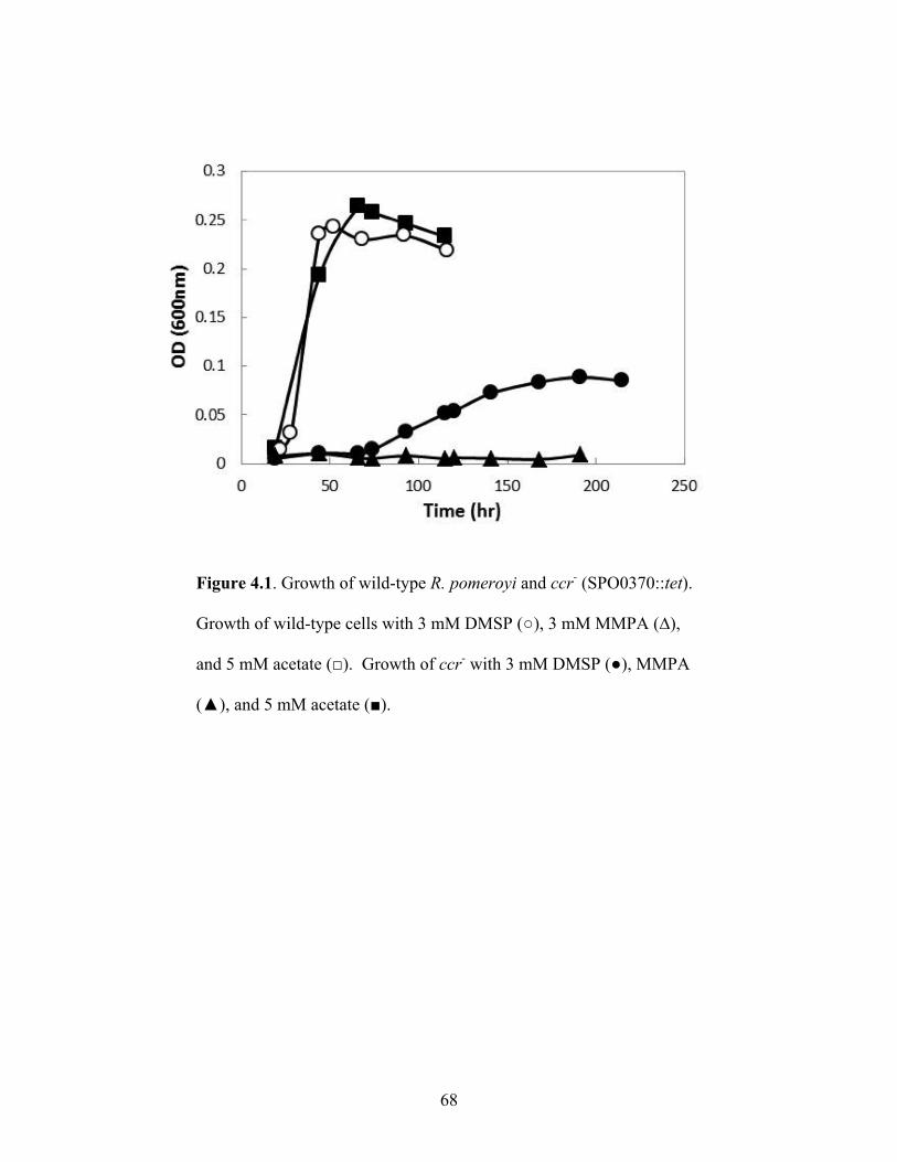
68
Figure 4.1. Growth of wild-type R. pomeroyi and ccr- (SPO0370::tet).
Growth of wild-type cells with 3 mM DMSP (○), 3 mM MMPA (∆),
and 5 mM acetate (□). Growth of ccr- with 3 mM DMSP (●), MMPA
(▲), and 5 mM acetate (■).

69
Figure 4.2. Growth of the wild-type cells with 10 mM Formate (●); 25
mM methylamine (■) trimethylamine (▲) and methanol (u).

70
APPENDICES
A. Isotope labeled DMSP Synthesis protocol
Preparation of 35S and 13C labeled dimethylsulfide to be used in the synthesis of DMSP.
100mg of elemental 35S (Sigma-Aldrich) was added to the small bottle of a sealed
double bottle connected via a side arm. To the small bottle, 5 mL of 0.5M NaOH containing
1g of dithiothreitol was added. The mixture was heated to 120°C in a sand bath until all of
the sulfur had dissolved. After cooling, 10mL of 1M HCL was added to the solution to
volatilize the reduced 35S. 5mL of 0.5M NaOH was added via syringe to the larger bottle
containing a stir bar. The apparatus was positioned to allow stirring overnight to trap the
volatilized sulfur in NaOH. After trapping, the solution was removed by syringe and added to
a 50 mL round bottom flask. Sulfide concentration was determined upon reaction of DTNB
(Ellman’s reagent) and an extinction coefficient of ε412 = 14.15 mM-1 cm-1 was used for
calculations. The stoppered flask was incubated in an ice bath for 30 min after which time,
[13C] methyl iodide was added at a concentration approximately 2 times the molar
concentration of the sulfide. Mixture was vigorous stirred for 4 hours under ice bath
conditions. After stirring, 1 mL of 2.5M NaOH soln. and 1mL of 0.1M Na2S203 soln. were
added to stop the reaction. The flask was connected to a receiving flask of the same
dimensions via a U-shaped glass tube to give a closed system. The receiving glass was
cooled with dry ice while the mixture was allowed to warm to room temperature. After 2
hours the receiving flask containing the condensed [13C][35S]DMS was removed and
stoppered. This labeled DMS was utilized in the standard DMSP synthesis protocol outlined

71
by Henrisken 2008 (1) adjusting acrylic acid addition for synthesized DMS concentration.
The final product formed is a small amount of crystallized DMSP, however, the yield is often
very low and synthesis is not consistent. This protocol will need to be improved to generate
better yields and consistency.
1. Henriksen, J.R. 2008. Physiology of dimethylsulfoniopropionate metabolism in a model
marine Roseobacter, Silicibacter pomeroyi. University of Georgia, Athens.