MEMBRANE ANCHORING AND INTERACTION BETWEEN TRANSMEMBRANE ... · MEMBRANE ANCHORING AND INTERACTION...
16
1 MEMBRANE ANCHORING AND INTERACTION BETWEEN TRANSMEMBRANE DOMAINS IS CRUICAL FOR K + CHANNEL FUNCTION Manuela Gebhardt 1 , Franziska Hoffgaard 2 , Kay Hamacher 2 , Stefan M. Kast 3 , Anna Moroni 4 , Gerhard Thiel 1 Institute of Botany 1 , Computational Biology Group 2 , Technische Universität Darmstadt, D-64287 Darmstadt, Germany; Physikalische Chemie III, Technische Universität Dortmund, Germany 3 ; Department of Biology and CNR IBF-Mi, Università degli Studi di Milano, Italy 4 Running title: K + channel transmembrane domain architecture Address correspondence to: Gerhard Thiel, Schnittspahnstrasse 3-5, D-64287 Darmstadt, Germany, Tel. +49 6151 166050; Fax +49 6151 163202; E-mail: [email protected] Summary The small viral channel Kcv is a Kir like K + channel of only 94 amino acids. The tetramer of Kcv represents with this simple structure the pore module of all complex K + channels. In order to examine the structural contribution of the transmembrane domains (TMDs) to channel function we performed Ala scanning mutagenesis of the two domains and tested the functionality of the mutants in a yeast complementation assay. The data reveal in combination with computational models that the upper halves of both TMDs, which face towards the external medium, are rather rigid while the inner parts are more flexible. The rigidity of the outer TMD is conferred by a number of essential aromatic amino acids, which face the membrane and probably anchor this domain in the bilayer. The inner TMD is intimately connected with the rigid part of the outer TMD via π···π interactions between a pair of aromatic amino acids. This structural principle is conserved within the viral K + channels and also present in Kir2.2 implying a general importance of this architecture for K + channel function. Introduction K + channels are transmembrane proteins, which catalyze the selective and regulated flux of potassium ions across membranes. A breakthrough in understanding of structure/function correlates in these important proteins occurred with the high-resolution structures determined for several K + channels (1- 4). Many functional properties such as selectivity and gating of K + channels are now understood on the basis of the specific architecture of these channel proteins. Still many aspects of function and regulation cannot be explained only on the basis of the protein structure; the performance of the protein depends also on the lipid environment and on the organization of the protein in this environment. Indeed, there is increasing evidence that many different properties of K + channels are depending on the interaction between the protein and the surrounding host membranes (5-7). As a consequence of this dependence, many amphiphilic drugs, which target ion channels have dual effects, one effect being directly related to an interaction with the protein and a secondary effect being mediated by modification of the bilayer properties (8). A good model system for understanding the interplay of membrane proteins with the membrane is provided by the viral potassium channel Kcv (9-10). This Kir like potassium channel with two transmembrane domains is with only 94 amino acids very small. The protein is almost completely embedded in the membrane. Only a small 15 aa long domain at the n-terminus and a short turret domain between the two transmembrane domains stick out of the membrane into aqueous solution (11). It can be assumed that in this arrangement any protein/membrane interaction is strongly reflecting back on function. Unbiased information on the structural significance of the two transmembrane domains (TMDs) and their importance for channel function can be obtained by an alanine-scan of the relevant domains. In this approach, each amino acid in a primary sequence is individually replaced by the amino acid alanine and the effect of this mutation is tested in a functional assay. In this way, it is possible to eliminate all side chain interactions, except for the C β atom, without altering the main chain conformation or the http://www.jbc.org/cgi/doi/10.1074/jbc.M110.211672 The latest version is at JBC Papers in Press. Published on February 10, 2011 as Manuscript M110.211672 Copyright 2011 by The American Society for Biochemistry and Molecular Biology, Inc. by guest on February 15, 2019 http://www.jbc.org/ Downloaded from
Transcript of MEMBRANE ANCHORING AND INTERACTION BETWEEN TRANSMEMBRANE ... · MEMBRANE ANCHORING AND INTERACTION...
1
MEMBRANE ANCHORING AND INTERACTION BETWEEN TRANSMEMBRANE DOMAINS IS CRUICAL FOR K+ CHANNEL FUNCTION
Manuela Gebhardt1, Franziska Hoffgaard2, Kay Hamacher2, Stefan M. Kast3,
Anna Moroni4, Gerhard Thiel1
Institute of Botany1, Computational Biology Group2, Technische Universität Darmstadt, D-64287 Darmstadt, Germany; Physikalische Chemie III, Technische Universität Dortmund, Germany3;
Department of Biology and CNR IBF-Mi, Università degli Studi di Milano, Italy4
Running title: K+ channel transmembrane domain architecture Address correspondence to: Gerhard Thiel, Schnittspahnstrasse 3-5, D-64287 Darmstadt, Germany, Tel. +49 6151 166050; Fax +49 6151 163202; E-mail: [email protected]
Summary The small viral channel Kcv is a Kir like K+ channel of only 94 amino acids. The tetramer of Kcv represents with this simple structure the pore module of all complex K+ channels. In order to examine the structural contribution of the transmembrane domains (TMDs) to channel function we performed Ala scanning mutagenesis of the two domains and tested the functionality of the mutants in a yeast complementation assay. The data reveal in combination with computational models that the upper halves of both TMDs, which face towards the external medium, are rather rigid while the inner parts are more flexible. The rigidity of the outer TMD is conferred by a number of essential aromatic amino acids, which face the membrane and probably anchor this domain in the bilayer. The inner TMD is intimately connected with the rigid part of the outer TMD via π···π interactions between a pair of aromatic amino acids. This structural principle is conserved within the viral K+ channels and also present in Kir2.2 implying a general importance of this architecture for K+ channel function.
Introduction K+ channels are transmembrane proteins, which catalyze the selective and regulated flux of potassium ions across membranes. A breakthrough in understanding of structure/function correlates in these important proteins occurred with the high-resolution structures determined for several K+ channels (1-4). Many functional properties such as selectivity and gating of K+ channels are now understood on the basis of the specific architecture of these channel proteins. Still many
aspects of function and regulation cannot be explained only on the basis of the protein structure; the performance of the protein depends also on the lipid environment and on the organization of the protein in this environment. Indeed, there is increasing evidence that many different properties of K+ channels are depending on the interaction between the protein and the surrounding host membranes (5-7). As a consequence of this dependence, many amphiphilic drugs, which target ion channels have dual effects, one effect being directly related to an interaction with the protein and a secondary effect being mediated by modification of the bilayer properties (8). A good model system for understanding the interplay of membrane proteins with the membrane is provided by the viral potassium channel Kcv (9-10). This Kir like potassium channel with two transmembrane domains is with only 94 amino acids very small. The protein is almost completely embedded in the membrane. Only a small 15 aa long domain at the n-terminus and a short turret domain between the two transmembrane domains stick out of the membrane into aqueous solution (11). It can be assumed that in this arrangement any protein/membrane interaction is strongly reflecting back on function. Unbiased information on the structural significance of the two transmembrane domains (TMDs) and their importance for channel function can be obtained by an alanine-scan of the relevant domains. In this approach, each amino acid in a primary sequence is individually replaced by the amino acid alanine and the effect of this mutation is tested in a functional assay. In this way, it is possible to eliminate all side chain interactions, except for the Cβ atom, without altering the main chain conformation or the
http://www.jbc.org/cgi/doi/10.1074/jbc.M110.211672The latest version is at JBC Papers in Press. Published on February 10, 2011 as Manuscript M110.211672
Copyright 2011 by The American Society for Biochemistry and Molecular Biology, Inc.
by guest on February 15, 2019http://w
ww
.jbc.org/D
ownloaded from
2
insertion of steric effects (12-14). Alanine is a common natural amino acid in all kinds of secondary structures including TMDs (12). For these reasons, alanine is the amino acid of choice in the mutagenesis scan. Hence, the replacement of a residue through alanine should reveal the contribution of the replaced residue to the overall stability and fold of the protein (15). In the present study, we use alanine-scanning mutagenesis of the two transmembrane domains of Kcv in conjunction with a structural Kcv model (11,16). The data provide an comprehensive picture of the functional architecture of the channel. The upper part of the outer transmembrane domain is anchored via aromatic side chains with the membrane. A mutual interaction between a pair of amino acids on the two TMDs attaches the inner TMD to the outer TMD and immobilizes in this way also the upper part of TMD2. The application of an elastic network model supports the structural significance of a mutual interaction of these amino acids (17, 18).
Experimental Procedures For the yeast complementation assay the Kcv gene was cloned into pYES2 (Invitrogen GmbH, Karlsruhe, Germany) into the EcoRI and XhoI site or as a chimera with EGFP into the BamHI and XhoI site of the plasmid (19). The QuikChange Site-directed Mutagenesis method (Stratagen) was used to insert the point mutations. The point mutations were designed on the primer level or randomized on the primer level by using the wobble base codon NNK (N = AGCT, K = GT), which codes for all possible amino acids. The resulting constructs were checked by DNA sequencing. Yeast complementation experiments were done with the yeast strain SGY1528 (Mat a ade 2–1 can 1–100 his 3–11,15 leu 2–3,112 trp 1–1 ura 3–1 trk 1::HIS3 trk 2::TRP1) (19). This strain, kindly provided by Dr. Minor (UCSF, USA), lacks an endogenous potassium uptake system and is not able to grow on media < 10 mM potassium. For the complementation assay yeasts were grown in parallel under selective (1mM and 0.5 mM KCl agar plates) and non-selective (100 mM KCl agar plates) conditions for three days at 30 °C.
For analyzing the spatial distribution of amino acids in the channel protein the MD model of Kcv based on the tetrameric form of the KirBac1.1 (PDB-Code: 1P7B) x-ray template structure was used (11). The programs PyMOL (http://www.pymol.org/) and MOLCAD (20) were used for studying the 3D structure and the spatial relationships of the amino acid residues. Elastic Network Models (18) have already been successfully applied to determine vibrational dynamics and kinetically hot residues avoiding time-consuming molecular dynamics simulations (21). Such network models use a coarse-grained representation for amino acids by substituting the complex amino acid by a bead on the respective Cα position. Interactions between residues are modeled as harmonic springs for contacts within a cutoff distance. Anisotropic Network Models (17) additionally invoke anisotropy of positional fluctuations to assess the directions of collective motions. Eigenmodes and -frequencies are derived from the so-called Hessian matrix by diagonalization techniques. The approach to quantify the impact of thought-experiment-style mutations, e.g. altering intramolecular forces up to the full deletion of contacts, is described elsewhere (22). We expanded this approach by computing overlaps of corresponding eigenvectors belonging to eigenvalues greater than zero. This allows us to qualitatively assess the effect the presence or absence of an amino acid pair interaction has on the functional mechanics of the protein.
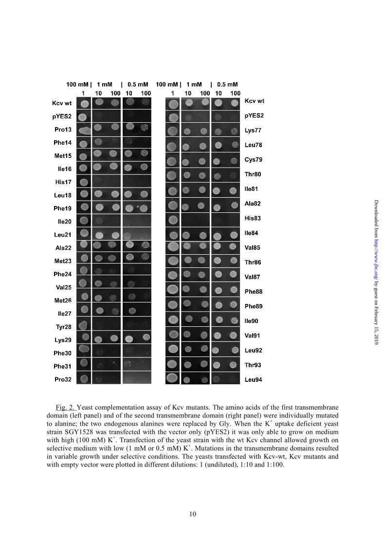
RESULTS In order to detect functionally important side chains in the amino acid composition of the transmembrane domains of Kcv, the respective amino acids were replaced one by one, by alanine; the two alanines (A22, A82), which are already present in the TMDs of the wild type (wt) channel (Fig. 1A), were replaced by glycine. All channel mutants were expressed individually in a yeast strain, which lack a functional K+ uptake system. These yeasts are only able to survive in medium with a high K+ concentration. They do not grow on a selective low K+ medium unless they are expressing a heterologous K+ uptake system (19, 23). Fig. 2 shows representative data from a growth assay of the yeast mutants expressing only the vector (pYES2), the Kcv-wt channel or its mutants. All yeasts transfected with either of the constructs were able to grow on high (100 mM) K+
by guest on February 15, 2019http://w
ww
.jbc.org/D
ownloaded from
3
medium, meaning that the expression of the mutations was deleterious for the cells. The two upper rows show that the yeast strain transfected with the vector only is as expected not able to grow under selective conditions with only 1 mM or 0.5 mM K+. Growth under selective conditions can be rescued by expressing the Kcv-wt channel. Hence an active viral K+ channel in the plasma membrane provides sufficient K+ influx for yeast growth under selective pressure. A scrutiny of the yeasts transfected with Kcv mutants shows that many mutants also grew under the selective conditions (Fig. 2). This implies that a large number of amino acid positions in the TMDs tolerate a mutation into alanine without loss of function. The data also highlight several important amino acids, which are crucial for channel function in yeast. The substitutions of nearly all phenylalanines (Phe14, Phe24, Phe30 and Phe31), histidine (His17), isoleucine (Ile20), tyrosine (Tyr28) and proline (Pro32) in the first, outer transmembrane (TMD1) domain showed the most dramatic effects; in these cases, the substitution results in a near or complete loss of channel function. The picture is different in the second, inner transmembrane domain (TMD2). In this domain alanine scanning revealed that nearly all of the amino acid side chains were no essential for channel function. Only the substitution of histidine (His83) caused a total loss of function while substitution of leucine (Leu94) leads only to a significantly reduced function of yeast growth (Fig. 2). To understand the spatial organization of the sensitive amino acids in the context of the three-dimensional structure of the channel we localized their position in the simulation model of Kcv (11). Fig. 1B shows the Kcv structure with the respective amino acids highlighted. It occurs that the relevant amino acids of TMD1 nearly all present their side chains to the membrane. Fig. 1A illustrates that nearly all of these sensitive amino acids shown in Fig. 2 with the exception of Phe31 and Ile20 are also conserved (24-26) within the sequences of all viral potassium channels. This emphasizes that the overall architecture of the channel is maintained throughout this family of channels.
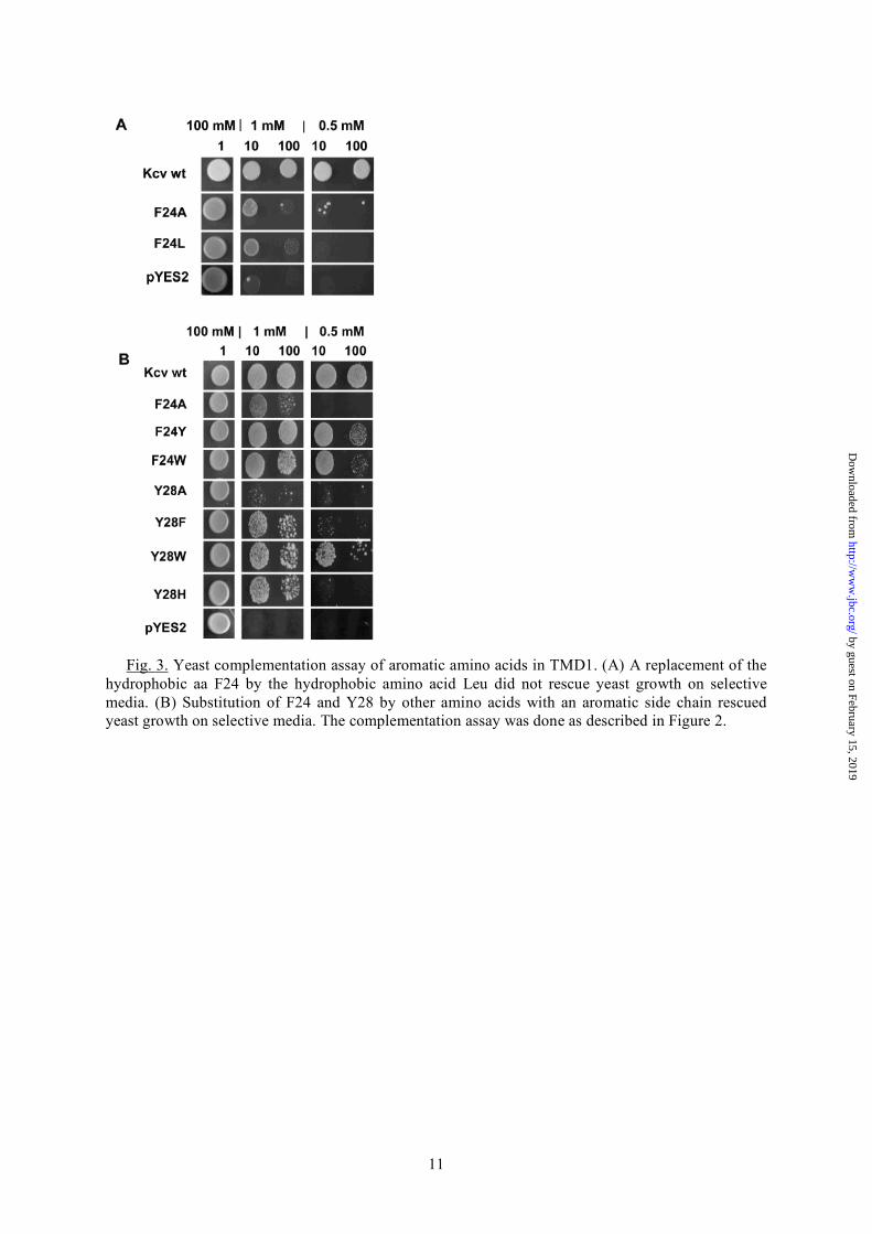
The conservation and orientation together with the aromatic side chain character suggests that these amino acids are involved in an anchoring of TMD1 in the lipid. An alternative explanation for the loss of function in the mutants could be the reduced hydrophobicity, which is introduced with the alanine. To test the importance of a hydrophobic flavor of the critical amino acids, one of them, Phe24, was mutated to Leu. This eliminates the aromatic side chain but preserves the hydrophobic character of this amino acid. A test of the mutant in the yeast system revealed that the substitution of Phe24 to leucine failed to recover channel function (Fig. 3A); hence, the hydrophobic nature of the amino acid in this position is not the only requirement. To test the alternative hypothesis, namely the importance of the aromatic side chains, the amino acids Phe24 and Tyr28 were replaced by tryptophan or tyrosine for position 24 or to tryptophan, histidine or phenylalanine for position 28 respectively. The results of the yeast complementation assay in Fig. 3B imply that the presence of an aromatic side chain in these positions is indeed sufficient for channel function. All aromatic substitutions were able to rescue channel function with varying degree. The results of these experiments stress that proper channel function requires the presence of aromatic amino acids in TMD1, which are oriented towards the membrane for anchoring of the protein in the bilayer. In further experiments we also tested the significance of another aromatic amino acid His17 in TMD1. In this case we replaced His17 by a randomization approach, meaning that in principle any of the 20 possible amino acids could occur at this position. Twelve out of the 60 tested yeast colonies transformed with the randomized plasmids showed growth on selective media with 0.5 mM KCl. After eliminating false positives and wt-like channels, we found that a mutant in which histidine was substituted by tryptophan was able to rescue channel function. The results of these experiments imply that the amino acid tryptophan with its aromatic side chain can replace the aromatic histidine in a functional channel. The results of these experiments are in agreement with the findings of Fig. 1B and underscore the importance of the aromatic side chains for anchoring of the protein in the membrane. Hence the essential features of the
by guest on February 15, 2019http://w
ww
.jbc.org/D
ownloaded from
4
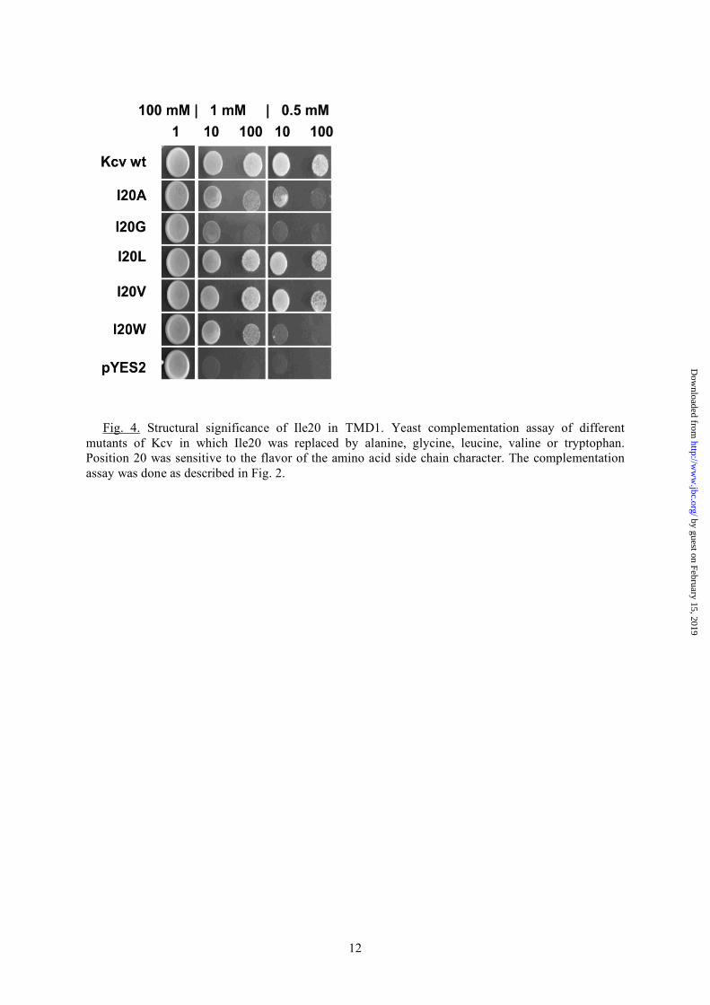
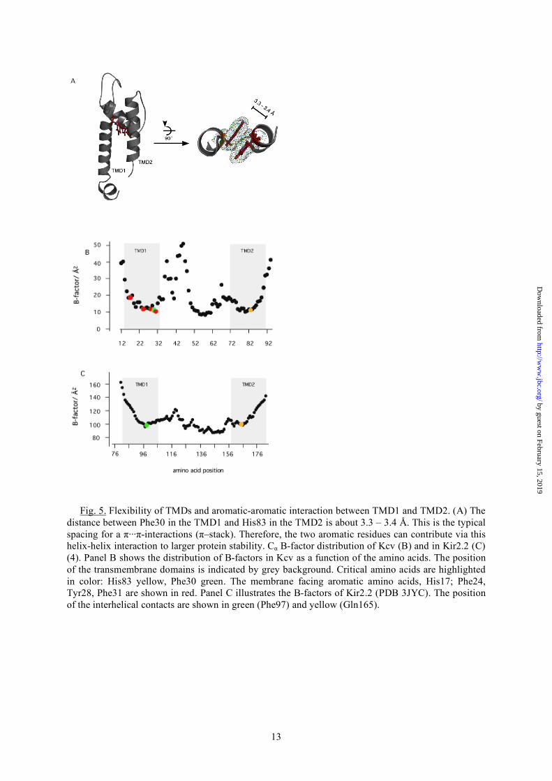
critical amino acids are exposure and geometric orientation of the side chains towards the lipid bilayer. This kind of anchoring of transmembrane domains occurs to be a general feature and has been described for several transmembrane domains (27-29). Among the critical amino acids in TMD1 Ile20 is an exception in that it does not carry an aromatic side chain. Yeasts expressing a channel with an Ala substitution are barely growing (Fig. 2). Previous studies already revealed that this position in TMD1 influences functional properties of the Kcv channel via long distance interactions with the pore (30). This mutant is, for example, more sensitive to cesium than the wild type channel in voltage clamp measurements in Xenopus oocytes (30). Because of this critical role of Ile20 further mutations were made to test the influence of the position on channel function and behavior. Fig. 4 reports the complementation assay of a series of different mutants. The data support a critical role of this position in channel function in the sense that channel performance is sensitive to the amino acid flavor in this position. The Kcv-I20V mutant is, as expected from previous experiments (30), functional; it is able to rescue yeast growth on selection media with 0.5 mM and 1 mM K+. However, the exchange to Trp or Gly modifies channel function. The substitution of Ile to Trp leads to a mutant, which can still support yeast growth on 1 mM K+ selective media but not on 0.5 mM KCl. The second mutant Kcv-I20G completely fails to rescue yeast growth under selective conditions. An important feature, which is highlighted with the help of the MD model, is the interaction of the two TMDs of Kcv. Although TMD2 is very tolerant to exchanges in amino acids, the substitution of His83 caused a total loss of function (Fig. 2). The reason for the critical role of this aa becomes apparent from the MD model, which shows that this residue is in close contact with another essential amino acid in the TMD1, namely Phe30. The structure reveals that Phe30 and His83 are interacting via a π-stack over a distance of only 3.3 – 3.4 Å (Fig. 5A). Such a stack of π-electron-rich residues occurs if two or more aromatic molecules are oriented in a parallel manner over a distance of about 3.3 Å. London dispersion forces, which dominate this π···π-interactions play important roles for correct folding and stability of many proteins (31) and
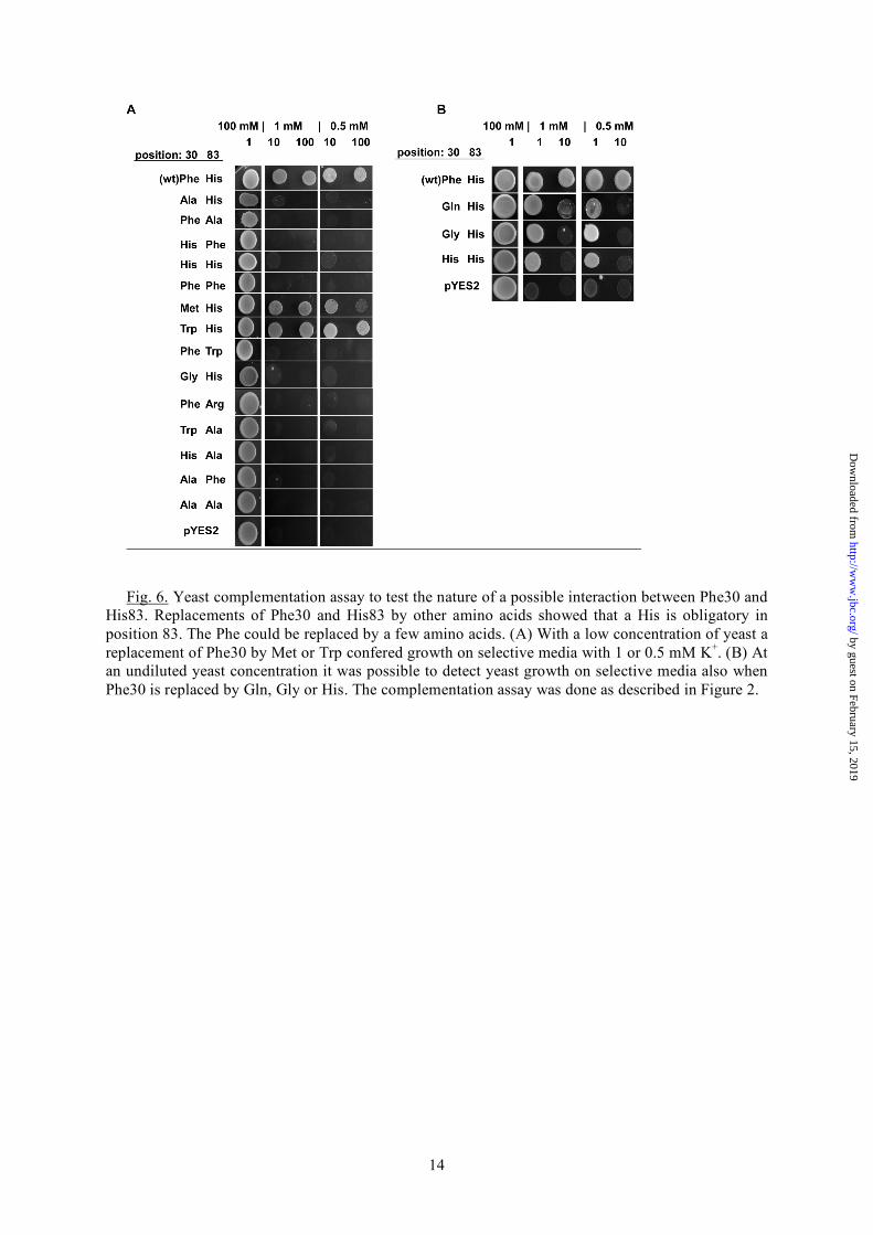
for mutual interactions between helices in many membrane proteins (32, 33). Evidence for the contribution of these aromatic side chain interactions were already found for acid-sensing ion channels (ASICs) where the π -π-stacking between the extracellular and transmembrane domain is essential for proton gating (34, 35). Also the architecture of synthetic ion channels relies on this kind of π -π-stacking interactions (36). It is hence possible that the aromatic-aromatic interaction between Phe30 and His83 is an essential factor for structure and function also in the Kcv channel. The presumed interaction between Phe and His in Kcv is not a frequently occurring pairing in membrane proteins for interhelical contact points (37). An alignment of viral potassium channels (Fig. 1A) however shows that the His in TMD2 is highly conserved throughout all viral potassium channels. Even the distantly related potassium channel Kesv from Ectocarpus siliculosus Virus (EsV-1) (38) is conserved at this position. The complementary amino acid in TMD1 e.g. Phe30 in Kcv is only semi-conserved (Fig. 1A). In Kesv, we find a glutamine in this position i.e. an amino acid, which is uncharged and aliphatic. All other viral ion channels contain in this position a phenylalanine or methionine. In the case of methionine, it seems possible that the long and extended thiomethyl group is able to stabilize the channel structure through C-H···π-interactions, where methionine is the aliphatic C-H donor and histidine is the aromatic π-acceptor (39). To test the importance of π···π- or C-H···π-interactions for channel structure and function different substitutions of both positions Phe30 and His83 with Ala, Gly, Phe, His, Met, Trp and Gln were made. Note that the Phe30Gln mutant mimics the amino acid pair in the Kesv channel (Fig. 1A). Fig. 6A shows that only two out of the 14 substitutions were able to rescue yeast growth on selection media. The substitutions, which generate a functional channel, agree with the idea of a π···π-interaction in this site. One of the two positive substitutions of Phe30 is tryptophan i.e. an amino acid with an aromatic side chain. As already expected from the alignments (Fig. 1A), Phe30 could also be replaced by methionine. C-H···π-interactions, like the Met-His interaction, can contribute to the stability of proteins with the overall stabilization energy of about 0.5 to 1.0 kcal mol-1 per interaction. They
by guest on February 15, 2019http://w
ww
.jbc.org/D
ownloaded from
5
are therefore an important factor for protein stability (39). Also almost all tryptophan residues in proteins are involved in C-H···π- or π···π-interactions (39, 40). The Trp-His interaction can stabilize a structure by about 1.0 to 4 kcal mol-1 (41). This is in the same range as the Met-His interaction. Hence, it is quite reasonable that the substitution of either methionine or tryptophane can rescue channel function in yeast. This finding highlights the concept of a functional conservation in proteins. This means the conservation in a protein is not on the level of the specific amino acid but rather on the maintenance of a structural interaction. In the present case the important factor is the interaction between the two TMDs, which can be brought about either by a π···π-interaction or alternatively also by the weaker C-H···π-interactions (42). Additional data show that three other mutations namely F30H, F30Q and F30G rescued yeast growth on selective media. However, in these cases yeast growth could only be detected at high, undiluted yeast concentration on the selective plates (Fig. 6B). The data imply that these channel mutants are in principle also functional. They may provide a lower conductance for K+ uptake and hence a reduced rescue efficiency. Important to note is that the amino acids Gln and His in position 30 are potential partners for a side chain pairing with His83 (43, 44). Only the mutant F30G does not follow this pattern. Collectively the results show that the two positions Phe30 and His83 are essential for a proper channel function. The structural importance is given by the formation of intra-subunit interactions between the two TMDs. Furthermore these positions are highly conserved throughout the viral K+ channels and are susceptible to changes. Moreover, the results emphasize an even higher complexity because there is no reciprocity between the two positions as the mutual exchange of Phe and His leads to a non-functional mutant (Fig. 6A). Additionally, these results demonstrate that the MD model of Kcv (11) reproduces the real conditions in the protein quite well; delicate interactions such as the π -π-stacking would not have occurred in an inappropriate model which differs from the more realistic one for instance by a small helix rotation.
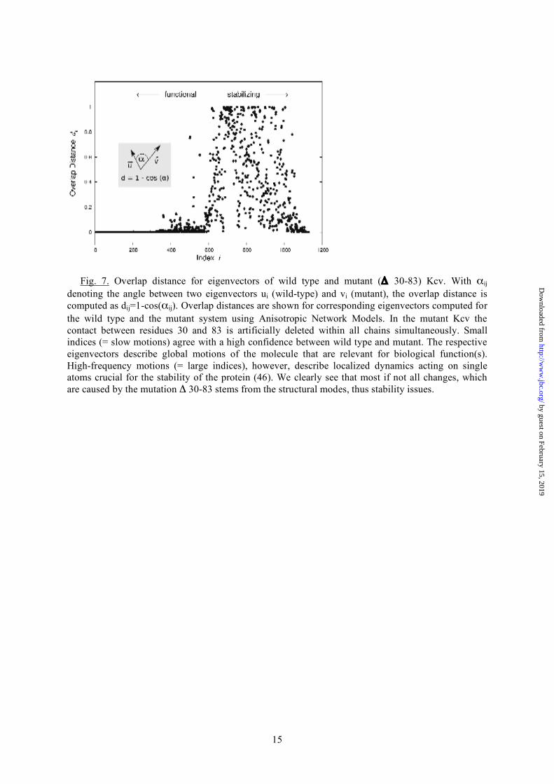
The information from the alanine scanning mutagenesis is together with the structural model of Kcv able to provide details on the three-dimensional model of the Kcv channel. More information can be gained by analyzing thermal B-factors originating from the MD simulations (11) that measure the average atomic fluctuations around the equilibrium position and therefore the flexibility of each amino acid in the Kcv protein. Fig. 5B shows Cα atom B-factors that indicate a pronounced dichotomy in both TMDs. Both membrane-spanning domains exhibit towards the cytosolic side a high degree of flexibility. The upper halves, which face to the extracellular side, are far less flexible. The present data imply that the more or less rigid nature of TMD1 is achieved by an anchoring of this part of the protein in the membrane. Most of the aforementioned critical aromatic amino acids-, which face the membrane-, are located in this rigid part of TMD1 (Fig. 5B). TMD2 on the other hand does not have such a direct anchor in the membrane. The rigid character of the upper part of TMD2 can therefore be best explained by the contact between the two TMDs via the π···π-interactions between Phe30 and His83. This hypothesis is supported by the fact that the mobility of TMD2 indeed increases downstream of His83 e.g. the point at which the two transmembrane domains are in intimate contact (Fig. 5B). The fact that any disturbance of the architecture, which affects the rigidity of the TMDs, results in a loss of channel function implies a certain importance for channel function. It is reasonable to speculate that the maintenance of the rigid nature of the TMDs in this part of the protein is connected to the function of the selectivity filter. The delicate structure of the selectivity filter, which has to maintain its structure in order to select between cations, probably requires this suspension by the two TMDs. The relevance of the π···π-interactions between the two TMDs for channel structure and stability is further supported by an application of the theory of anisotropic network models to the Kcv model (11). By using a homogeneous parameterization of the network model, we perform a purely structural-topological sensitivity analysis of the channel: we artificially delete the mutual contact of amino acids 30-83 within each subunit at the same time. This mutation has no experimental analog and can therefore be performed in silico only. To assess
by guest on February 15, 2019http://w
ww
.jbc.org/D
ownloaded from
6
the effect of this deletion on the mechanics of the potassium channel, we compute the difference between the wild type and mutant covariance matrix using the Frobenius norm. The covariance matrix contains information on correlated motions of all the residues in the tetramer. To avoid obscuration effects, that may emerge by summing up the absolute differences of the whole matrix, we define distinct channel elements, and thus by extension different subparts of the above mentioned covariance matrix, which describe local mechanics for relevant parts of the channel (11). The Frobenius norm of a mutant with respect to the wild-type increases whenever a mutation has a significant impact on the mechanics of the region under consideration. We detected here a major influence of the contact deletion on both TMDs and the turret region (data not shown). Another measure of the biomechanical effect that is invariant under rotation is the overlap distance. It is derived for all mechanical modes of the channel. The similarity of corresponding eigenvectors, denoting distinct fluctuations of each residue, of wild type and mutant system can be quantified by the angle between the vectors. We define the overlap distance dij as dij=1-cos(αij) with αij being the angle between each two eigenvectors (i in the wild type, j in the mutant). A pairing with similar fluctuation vectors shows a distance close to zero whereas a distance close to one indicates large differences between the eigenvectors. In the present case we identified corresponding modes from wild type and mutant by using the frequencies of the mechanical motions. The final results for mechano-dynamical similarity of modes of motions upon mutation of the 30-83 contacts are depicted in Fig. 7. Clearly, large deviations (dij) between wild type and mutant eigenvectors are present for large indices only. Small indices denote small frequencies and represent functional motions of the channel. Such global mechanics are mainly due to function, whereas high-frequency motions, which are populated with only a small probability describe localized motions having effects mainly on the structure and stability of the protein (45). The data show that the contact between residues 30 and 83 has a major impact on local dynamics as expressed by high-frequency modes and, hence, influences the
structure rather than the functionality of the Kcv channel.
DISCUSSION The present data support a model for the functional architecture of the small viral K+ channels in which the upper part of the outer transmembrane domain must be anchored via aromatic side chains with the surrounding membrane. Such an anchoring is typical for many membrane proteins (27-29). Mutual interaction of an amino acid pair attaches the inner TMD of the channels to the outer TMD and immobilizes in this way also the upper part of the former TMD. An elastic network model of the Kcv channel underscores the structural significance of this amino acid pairing and reveals that its disruption results in modified protein stability. The experimental data further imply that the respective interaction between the two TMDs supports optimal function of the channel, while it does not seem to be essential. Loss of channel function was induced hy the disruption of the pair-wise interaction in most of the mutants, which were tested in the rescue assay. The fact that mutants, which maintained the stronger π···π-interaction or the weaker C-H···π-interaction between the TMDs still support proper rescue efficiency, implies that the amino acid pairing motive, which connects the two TMDs, is beneficial for channel function. Interesting to note is that the F30Q mutant, that mimics the amino acid sequence of the Kesv channel, is able to rescue, albeit with a low efficiency, growth of yeast mutants. This observation implies that a general motive of interactions between the two transmembrane domains is indeed conserved within the small viral K+ channels. One mutant, F30G, in which G30 does not interact with His83, is also able to rescue yeast growth with a low efficiency. The results of these experiments suggest that a close interaction between position 30 and 83 is not bligatory for basic channel function. The structural connection between the two TMDs may in the case of the F30G mutant with the small and flexible Gly be taken over by other interactions between the TMDs e.g. between Phe31 and His83. The overall architecture and dynamics, which we find here for the TMDs in Kcvs is not restricted to the small viral K+ channel; the same bipartite organization of the two TMDs, which is
by guest on February 15, 2019http://w
ww
.jbc.org/D
ownloaded from

7
characteristic for Kcvs (Fig. 5B), is also present in the Kir2.2 channel. In the latter channel (4) the B-factors imply that those parts of the TMDs that face the external medium are fairly rigid (Fig. 5C). Like in Kcv also the parts of the Kir2.2 TMDs, which are directed towards the cytosol, are more flexible (compare Fig. 5B,C). A scrutiny of its crystal structure shows that the amino acid pair Phe97 and Gln165 can in Kir2.2 generate the contact between the two TMDs and hence a mutual fixation of the two helices in Kir2.2. The XH··π interaction between the two amino acids over a distance of 3 Å is in the same range as the π-stacking interaction in Kcv.
Moreover the two critical amino acids of Kir2.2 are located like in Kcv in the rigid parts of the TMDs at the transition to the flexible parts (Fig. 5C). The mutual interaction between the two amino acids together with their location is consistent with the idea that they generate a close contact between the two transmembrane domains in Kir2.2. The finding of a similar architectural principle in the Kcv and Kir channels implies that the interhelical contact between the two TMDs and the rigid character of the outer part of the TMDs is an important feature for channel function.
REFERENCES 1. Doyle, D.A., Cabral, J.M., Pfuetzner, R.A., Kuo, A., Gulbis, J.M., Cohen, S.L., Chait, B.T.,
and MacKinnon, R. (1998) Science 280, 69–76 2. Jiang, Y., Lee, A., Chen, J., Cadene, M., Chait, B.T., and MacKinnon, R. (2002) Nature 417,
515-522 3. Kuo, A., Gulbis, J.M., Antcliv, J.F., Rahman, T., Lowe, E.D., Zimmer, J., Cuthbertson, J.,
Ashcroft, F.M., Ezaki, T., and Doyle, D.A. (2003) Science 300, 1922–1926 4. Tao, X., Avalos, J.L., Chen, J., and MacKinnon, R. (2009) Science 326, 1668-1674 5. Marius, P., Zagnoni, M., Sandison, M.E., East, J.M., Morgan, H., and Lee, A.G. (2008)
Biophys. J. 94, 1689-1698 6. Valiyaveetil, F.I., Zhou, Y., and MacKinnon, R. (2002) Biochemistry 41, 10771-10777 7. Barrera, F.N., Renart, M.L., Poveda, J.A., de Kruijff, B., Killian, J.A., and González-Ros,
J.M. (2008) Biochemistry 47, 2123–2133 8. Lundbaek, J.A. (2008) J. Gen Physiol. 131, 421-429 9. Plugge, B., Gazzarrini, S., Nelson, M., Cerana, R., Van Etten, J.L., Derst, C., DiFrancesco,
D., Moroni, A., and Thiel, G. (2000) Science 287, 1641-1644. 10. Thiel, G., Baumeister, D., Schroeder, I., Kast, S.M., Van Etten, J.L., and Moroni, A. (2010)
Biochim. Biophys. Acta, Biomembr., in press 11. Tayefeh, S., Kloss, T., Kreim, M., Gebhardt, M., Baumeister, D., Hertel, B., Richter, C.,
Schwalbe, H., Moroni, A., Thiel, G., and Kast, S.M. (2009) Biophys. J. 96, 485–498 12. Cunningham, B.C., and Wells, J.A. (1989) Science 244, 1081-1085 13. Holst, B., Zoffmann, S., Elling, C.E., Hjorth, S.A., and Schwartz, T.W. (1998) Mol.
Pharmacol. 53, 166–175 14. DiCera, E. (1998) Chem. Rev. 98, 1565-1591 15. Clackson, T., and Wells, J.A. (1995) Science 267, 383-386 16. Tayefeh, S., Kloss, T., Thiel, G., Hertel, B., Moroni, A., and Kast, S.M. (2007) Biochemistry
46, 4826–4839 17. Atilgan, A.R., Durell, S.R., Jernigan, R.L., Demirel, M.C., Keskin, O., and Bahar, I. (2001)
Biophys. J. 80, 505-515 18. Tozzini, V. (2010) Q. Rev. Biophys. 43, 333-371 19. Minor, D.L., Masseling, S.J., Jan, Y.N., and Jan, L.Y. (1999) Cell 96, 879-891 20. Brickmann, J., Goetze, T., Heiden, W., Moeckel, G., Reiling, S., Vollhardt, H., Zachmann,
C.-D. (1995) Interactive visualization of molecular scenarios with MOLCAD/SYBYL, in: Bowie, J.E. (ed.), Data Visualisation in Molecular Science: Tools for Insight and Innovation, Addison-Wesley, pp. 83–97
21. Hamacher, K., and McCammon, J.A. (2006) J. Chem. Theory Comput. 2, 873-878 22. Hamacher, K. (2008) Gene 422, 30-36
by guest on February 15, 2019http://w
ww
.jbc.org/D
ownloaded from
8
23. Balss, J., Papatheodorou, P., Mehmel, M., Baumeister, D., Hertel. B., Delaroque, N., Chatelain, F.C., Minor, D.L., Van Etten, J.L., Rassow, J., Moroni, A., and Thiel, G. (2008) Proc. Natl. Acad. Sci. U.S.A. 105, 12313-12318
24. Ashkenazy, H., Erez, E., Martz, E., Pupko, T., and Ben-Tal, N. (2010) Nucl. Acids Res. doi:10.1093/nar/gkq399
25. Landau, M., Mayrose, I., Rosenberg, Y., Glaser, F., Martz, E., Pupko, T., and Ben-Tal, N. (2005) Nucl. Acids Res. 33, W299-W302
26. Glaser, F., Pupko. T., Paz, I., Bell, R.E., Bechor, D., Martz, E., and Ben-Tal, N. (2003) Bioinformatics 19, 163-164
27. Yau, W.M., Wimley, W.C., Gawrisch, K., and White, S.H. (1998) Biochemistry 37, 14713-14718
28. Killian JA (2003) FEBS Lett. 555, 134-138 29. Planque, M.R.R., and Killian, J.A. (2003) Mol. Membr. Biol. 20, 271-284 30. Gazzarrini, S., Kang, M., Van Etten, J.L., Tayefeh, S., Kast, S.M., DiFrancesco, D., Thiel, G.,
and Moroni, A. (2004) J. Biol. Chem. 279, 28443-28449 31. Shoemaker, K.R., Fairman, R., Schultz, D.A., Robertson, A.D., York, E.J., Stewart, J.M., and
Baldwin, R.L. (1990) Biopolymers 29, 1-11 32. Popot, J.L., and Engelman, D.M. (1990) Biochemistry 29, 4031–4037 33. Bowie, J.U. (2005) Nature 438, 581-589 34. Li, T., Yang, Y., and Canessa, C.M. (2008) J. Biol. Chem. 284, 4689-4694 35. Yang, H., Yu, Y., Li, W.G., Yu, F., Cao, H., Xu, T.L., and Jiang, H. (2009) PLoS Biol. 7(7):
e1000151. doi:10.1371/journal.pbio.1000151 36. Bhosale, S., Sisson, A.L., Sakai, N., and Matile, S. (2006) Org. Biomol. Chem. 4, 3031-3039 37. Adamian, L., and Liang, J. (2001) J. Mol. Biol. 311, 891-907 38. Delaroque, N., Müller, D.G., Bothe, G., Pohl, T., Knippers, R., and Boland, W. (2001)
Virology 287, 112-132 39. Brandl, M., Weiss, M.S., Jabs, A., Sühnel, J., and Hilgenfeld, R. (2001) J. Mol. Biol. 1, 357-
377 40. Wang, L., Sun, N., Terzyan, S., Zhang, X., and Benson, D.R. (2006) Biochemistry 45, 13750-
13759 41. Fernández-Recio, J., Vázquez, A., Civera, C., Sevilla, P., and Sancho, J. (1997) J. Mol. Biol.
1, 184-197 42. Nishio, M., Hirota, M., and Umezawa, Y. (1998) The CH/π Interaction. Evidence, Nature and
Consequences, Wiley-VCH, New York 43. Bhattacharyya, R., Saha, R.P., Samanta, U., Chakrabarti, P. (2003) J Prot. Res. 2, 255-263 44. Heyda, J., Mason, P.E., Jungwirth, P. (2010) J. Phys. Chem. 114, 8744-8749 45. Bahar, I., Atilgan, A.R., Demirel, M.C., and Erman, B. (1998) Phys. Rev. Lett. 80, 2733-2736 46. Berezin, C., Glaser, F., Rosenberg, J., Paz, I., Pupko, T., Fariselli, P., Casadio, R., and Ben-
Tal, N. (2004) Bioinformatics 20, 1322-1324
FOOTNOTES *We are grateful to Profs. Adam Bertl (TU Darmstadt) and Dan Minor (UCSF) for help with the yeast rescue assay and to Prof. Mike Blatt (Uni. Glasgow) for help with the manuscript. This work was supported in part by the Deutsche Forschungsgemeinschaft (to G.T. and S.M.K), the Adolf-Messer-Stiftung and the Fonds der Chemischen Industrie (to S.M.K.), and the European Drug Initiative on Channels and Transporters (EDICT) project EU FP7 (201924) to A.M. Computer time was provided on the IBM Regatta system (Hessischer Hochleistungsrechner) at the Hochschulrechenzentrum Darmstadt.
by guest on February 15, 2019http://w
ww
.jbc.org/D
ownloaded from
9
FIGURE LEGENDS
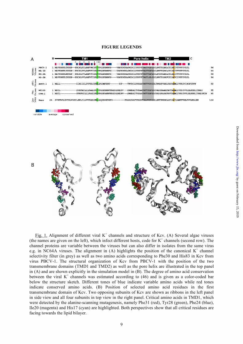
Fig. 1. Alignment of different viral K+ channels and structure of Kcv. (A) Several algae viruses
(the names are given on the left), which infect different hosts, code for K+ channels (second row). The channel proteins are variable between the viruses but can also differ in isolates from the same virus e.g. in NC64A viruses. The alignment in (A) highlights the position of the canonical K+ channel selectivity filter (in grey) as well as two amino acids corresponding to Phe30 and His83 in Kcv from virus PBCV-1. The structural organization of Kcv from PBCV-1 with the position of the two transmembrane domains (TMD1 and TMD2) as well as the pore helix are illustrated in the top panel in (A) and are shown explicitly in the simulation model in (B). The degree of amino acid conservation between the viral K+ channels was estimated according to (46) and is given as a color-coded bar below the structure sketch. Different tones of blue indicate variable amino acids while red tones indicate conserved amino acids. (B) Position of selected amino acid residues in the first transmembrane domain of Kcv. Two opposing subunits of Kcv are shown as ribbons in the left panel in side view and all four subunits in top view in the right panel. Critical amino acids in TMD1, which were detected by the alanine-scanning mutagenesis, namely Phe31 (red), Tyr28 (green), Phe24 (blue), Ile20 (magenta) and His17 (cyan) are highlighted. Both perspectives show that all critical residues are facing towards the lipid bilayer.
by guest on February 15, 2019http://w
ww
.jbc.org/D
ownloaded from
10
Fig. 2. Yeast complementation assay of Kcv mutants. The amino acids of the first transmembrane
domain (left panel) and of the second transmembrane domain (right panel) were individually mutated to alanine; the two endogenous alanines were replaced by Gly. When the K+ uptake deficient yeast strain SGY1528 was transfected with the vector only (pYES2) it was only able to grow on medium with high (100 mM) K+. Transfection of the yeast strain with the wt Kcv channel allowed growth on selective medium with low (1 mM or 0.5 mM) K+. Mutations in the transmembrane domains resulted in variable growth under selective conditions. The yeasts transfected with Kcv-wt, Kcv mutants and with empty vector were plotted in different dilutions: 1 (undiluted), 1:10 and 1:100.
by guest on February 15, 2019http://w
ww
.jbc.org/D
ownloaded from
11
Fig. 3. Yeast complementation assay of aromatic amino acids in TMD1. (A) A replacement of the
hydrophobic aa F24 by the hydrophobic amino acid Leu did not rescue yeast growth on selective media. (B) Substitution of F24 and Y28 by other amino acids with an aromatic side chain rescued yeast growth on selective media. The complementation assay was done as described in Figure 2.
by guest on February 15, 2019http://w
ww
.jbc.org/D
ownloaded from
12
Fig. 4. Structural significance of Ile20 in TMD1. Yeast complementation assay of different
mutants of Kcv in which Ile20 was replaced by alanine, glycine, leucine, valine or tryptophan. Position 20 was sensitive to the flavor of the amino acid side chain character. The complementation assay was done as described in Fig. 2.
by guest on February 15, 2019http://w
ww
.jbc.org/D
ownloaded from
13
Fig. 5. Flexibility of TMDs and aromatic-aromatic interaction between TMD1 and TMD2. (A) The distance between Phe30 in the TMD1 and His83 in the TMD2 is about 3.3 – 3.4 Å. This is the typical spacing for a π···π-interactions (π–stack). Therefore, the two aromatic residues can contribute via this helix-helix interaction to larger protein stability. Cα B-factor distribution of Kcv (B) and in Kir2.2 (C) (4). Panel B shows the distribution of B-factors in Kcv as a function of the amino acids. The position of the transmembrane domains is indicated by grey background. Critical amino acids are highlighted in color: His83 yellow, Phe30 green. The membrane facing aromatic amino acids, His17; Phe24, Tyr28, Phe31 are shown in red. Panel C illustrates the B-factors of Kir2.2 (PDB 3JYC). The position of the interhelical contacts are shown in green (Phe97) and yellow (Gln165).
by guest on February 15, 2019http://w
ww
.jbc.org/D
ownloaded from
14
Fig. 6. Yeast complementation assay to test the nature of a possible interaction between Phe30 and His83. Replacements of Phe30 and His83 by other amino acids showed that a His is obligatory in position 83. The Phe could be replaced by a few amino acids. (A) With a low concentration of yeast a replacement of Phe30 by Met or Trp confered growth on selective media with 1 or 0.5 mM K+. (B) At an undiluted yeast concentration it was possible to detect yeast growth on selective media also when Phe30 is replaced by Gln, Gly or His. The complementation assay was done as described in Figure 2.
by guest on February 15, 2019http://w
ww
.jbc.org/D
ownloaded from
15
Fig. 7. Overlap distance for eigenvectors of wild type and mutant (Δ 30-83) Kcv. With αij
denoting the angle between two eigenvectors ui (wild-type) and vi (mutant), the overlap distance is computed as dij=1-cos(αij). Overlap distances are shown for corresponding eigenvectors computed for the wild type and the mutant system using Anisotropic Network Models. In the mutant Kcv the contact between residues 30 and 83 is artificially deleted within all chains simultaneously. Small indices (= slow motions) agree with a high confidence between wild type and mutant. The respective eigenvectors describe global motions of the molecule that are relevant for biological function(s). High-frequency motions (= large indices), however, describe localized dynamics acting on single atoms crucial for the stability of the protein (46). We clearly see that most if not all changes, which are caused by the mutation Δ 30-83 stems from the structural modes, thus stability issues.
by guest on February 15, 2019http://w
ww
.jbc.org/D
ownloaded from
and Gerhard ThielManuela Gebhardt, Franziska Hoffgaard, Kay Hamacher, Stefan M. Kast, Anna Moroni
for K+ channel functionMembrane anchoring and interaction betweentransmembrane domains is cruical
published online February 10, 2011J. Biol. Chem.
10.1074/jbc.M110.211672Access the most updated version of this article at doi:
Alerts:
When a correction for this article is posted•
When this article is cited•
to choose from all of JBC's e-mail alertsClick here
by guest on February 15, 2019http://w
ww
.jbc.org/D
ownloaded from