Lawrence-Douglas County Fire Medical Treatment Protocols (PDF)
Upload
nguyenduongCategory
view
223download
1
The Maryland
Medical Protocols for Emergency Medical Services Providers Effective July 1, 2015
Maryland Institute for Emergency Medical Services Systems

ii © 2015 ii
The complete “Maryland Medical Protocols for Emergency Medical Services Providers” is also available on the Internet. Check out the MIEMSS
website www.MIEMSS.org.
This text was produced from materials from the Div. of State Documents, Office of the Secretary of State, State of Maryland. Only text obtained directly from the DSD is enforceable under Maryland law.
For copies of the official text, call the DSD at 1-800-633-9657.
Copyright 2015.This document may not be amended or altered;however, it may be individually reproduced and distributed for free to
Maryland EMS Providers without permission.

iii © 2015
To All Health Care Providers in the State of Maryland:
Re: 2015 revisions, updates, and additions to The Maryland Medical Protocols for EMS Providers
EMS providers will be able to download the replacement pages from the MIEMSS website at www.miemss.org and will be receiving a single copy of the 2015 pocket protocols.
The EMS Board has approved these protocols for implementation on July 1, 2015. Prior to July 1, all EMS providers must complete the protocol update “Meet the Protocols” (visit the Online Training Center) that will highlight the new material.
Some major protocol additions, deletions, and changes have been made this year. A spreadsheet of those changes is included in this year’s packet of updates. The spreadsheet is for reference only and the information located in the full protocol book is the official medical reference for EMS providers. It is the responsibility of each EMS Provider to be familiar with all of the Maryland Medical Protocols for EMS Providers.
Protocol Changes:
• Many hospitals have changed their names to reflect their EMS Board designations.
• A requirement for patient demographic information has been placed in the 12 lead procedure section.
• BLS providers whose jurisdiction participates in an option supplemental program may admin-ister IM epinephrine manually with a prefilled syringe or a syringe and single dose ampule or vial.
• Midazolam- Intranasal (IN) has been added as an acceptable route of administration.
• The ability to capture waveform capnography is now required for all ALS transport units.
• Magnesium Sulfate has been added to the ALS formulary in multiple sections of the protocol including asthma, pregnant seizure patients, Torsades de Pointes and patients with refractory VF/VT after administration of lidocaine
• Neuroprotective hypothermia- Iced IV fluid has been removed as a method of cooling. Neu-roprotective Hypothermia ill still be implemented with the use Ice/cold packs as the ONLY method of cooling that is acceptable.
• Surgical Cricothyroidotomy is now available as a procedure for approved Paramedics whose jurisdiction has been MIEMSS approved and participates in the new Pilot Program.
• Spinal Protection- This new protocol replaces the Spinal cord trauma protocol. Providers will now assess patients to determine the level of spinal protection treatment (only a properly sized cervical collar ) or spinal Immobilization treatment ( a properly sized cervical collar and long backboard), that is warranted based on EMS provider assessment, signs and symptoms and not simply a mechanism of injury.
• Research protocols- A new section has been added to the Maryland Medical Protocols. This year two jurisdiction specific programs have been placed in this section.

iv © 2015
• Calcium Chloride- The dose of calcium chloride has been increased to 500 mg for the rever-sal of Diltiazem or Magnesium Sulfate side effects or toxicity.
• RSI age definition- Adult patients are now defined as patients who are 15 years of age or older.
• Potentially Volatile Environments -Life Sustaining Interventions- The Governor’s Active Assail-ant Task force developed this new protocol in consultation with Maryland State Police, FBI, and federal law enforcement partners, tactical operators, TEMS medics, EMS operational leadership and MIEMSS to address incidents where implementation of standard Maryland Medical Protocol for EMS Providers is not practical due to provider/ victim safety concerns and in an effort to maximize patient survival.
Richard L. Alcorta, MD, FACEP Kevin Seaman, MD, FACEPState EMS Medical Director, MIEMSS Executive Director, MIEMSS

v © 2015
TABLE OF CONTENTS
I. GENERAL INFORMATION A. General Provisions 1B. Important Numbers 3C. Health Care Facility Codes 5D. Maryland Trauma and Specialty Referral Centers 11E. Protocol Key 15F . Protocol Usage Flow Diagram 16G. Protocol Variation Procedure 17 H. Inability to Carry Out Physician Order 19 I . Physician Orders for Extraordinary Care 21J. Quality Review Procedure for Pilot Programs 23
II. GENERAL PATIENT CARE 25
III. TREATMENT PROTOCOLSAbuse/NeglectA. Abuse/Neglect 35Altered Mental StatusB. Seizures 37C. Unresponsive Person 39Apparent Life-Threatening Event (ALTE) D. Apparent Life-Threatening Event (ALTE) 41Behavioral EmergenciesE. Behavioral Emergencies 42Cardiac Emergencies F. Cardiac Guidelines 44 Universal Algorithm for Adult Emergency Cardiac Care for BLS 45 Universal Algorithm for Adult Emergency Cardiac Care for ALS 46 Universal Algorithm for Pediatric Emergency Cardiac Care for BLS 47 Universal Algorithm for Pediatric Emergency Cardiac Care for ALS 48 G. Bradycardia 49 Adult Bradycardia Algorithm 50 Pediatric Bradycardia Algorithm 51 G1. Tachycardia 51-1 Adult Tachycardia Algorithm 51-3 Pediatric Tachycardia Algorithm 51-4H. Cardiac Arrest 52 Adult Asystole Algorithm 53 Pediatric Cardiac Arrest Algorithm 54 Pulseless Electrical Activity (PEA) Algorithm 55 VF Pulseless VT Algorithm 56H1. Termination of Resuscitation 56-1 H2. Pronouncement of Death in the Field 56-3

vi © 2015
TABLE OF CONTENTS
H3. EMS DNR/MOLST 56-4 H4. EMS/DNR Flowchart 56-14 I. Chest Pain/Acute Coronary Syndrome 57 J. Hyperkalemia: Renal Dialysis/Failure or Crush Syndrome 59 J1. Implantable Cardioverter Defibrillator (ICD) Malfunction 60-1 K. Intentionally Omitted L. Premature Ventricular Contractions (PVCs) 64 M. ST Elevation Myocardial Infarction (STEMI) 65 N. Sudden Infant Death Syndrome (SIDS) 67 O. Intentionally Omitted P. Intentionally Omitted Environmental Emergencies Q. Cold Emergencies (Frostbite) 74 R. Cold Emergencies (Hypothermia) 76 S. Depressurization 78 T . Hazardous Materials Exposure 79 U. Heat-Related Emergencies 81 V. Near-Drowning 82 W. Overpressurization 83Hyperbaric Emergencies X. Hyperbaric Therapy 84Nausea and Vomiting X1. Nausea and Vomiting 85-1Non-Traumatic Shock Y. Hypoperfusion 86Obstetrical/Gynecological Emergencies Z. Childbirth Algorithm 88 AA. Newly Born 90 Universal Algorithm for Newly Born for BLS 90 Universal Algorithm for Newly Born for ALS 90-1 BB. Vaginal Bleeding 91Overdose/Poisoning CC. Absorption 92 DD. Ingestion 94 EE. Inhalation 97 FF. Injection 99 FF1. Stimulant Toxicity 100-1Pain Management GG. Pain Management 101Respiratory Distress HH. Allergic Reaction/Anaphylaxis 103 I I. Asthma/COPD 106 JJ. Croup 108 KK. Pulmonary Edema/Congestive Heart Failure 110 Universal Algorithm for Pediatric Respiratory Distress for BLS 114 Universal Algorithm for Pediatric Respiratory Distress for ALS 115

vii © 2015
TABLE OF CONTENTS
Stroke LL. Stroke: Neurological Emergencies 116Trauma Protocol
MM. Burns 118NN. Eye Trauma 120OO. Hand/Upper/Lower Extremity Trauma 122PP. Multiple/Severe Trauma 124
Glasgow Coma Scale 126QQ. Sexual Assault 127RR. Spinal Protection 128 SS. Trauma Arrest 130TT . Trauma Decision Tree 132
IV. APPENDICES A. Glossary 137 B. Procedures, Medical Devices, and Medications for EMS
and Commercial Services 144 C. Rule of Nines 148 D. Normal Vital Signs and APGAR Chart 149 E. Intentionally Omitted F. Intentionally Omitted G. Intentionally Omitted G1. Proposed Protocol Submission Request Policy 163 G2. Proposed Protocol Submission Template 164-1 G3. Protocol Concept/Sponsor Request 164-2 H. Procedures 165
Accessing Central Venous Catheters and Devices 165Airway Management
Bag-Valve-Mask Ventilation 168Continuous Positive Airway Pressure (CPAP) 170-1
Latex-Free Dual Lumen Tube 170-3Airway Management (continued)
Gastric Tube 171Nasotracheal Intubation 172Needle Decompression Thoracostomy (NDT) 174Obstructed Airway Foreign Body Removal: Direct Laryngoscopy 175Orotracheal Intubation 176Tracheostomy Change 179Tracheostomy Suctioning 181 Ventilatory Difficulty Secondary to Bucking or Combativeness 181-1

viii © 2015
TABLE OF CONTENTS
Electrical Therapy Automated External Defibrillation 182 Cardioversion 184 Defibrillation 186 External Transcutaneous Cardiac Pacing 187 Go-Team Activation 189 External Jugular (EJ) 191 Glucometer Protocol 192 Intraosseous Infusion (IO) 194 Intravenous Maintenance Therapy for EMT 196 Medevac Utilization 198-1 Patient-Initiated Refusal of EMS 198-7 Peripheral Intravenous Access for CRT-(I) and Paramedic, and IV Access Option for EMT Approved by the EMS Operational Program 199Physical and Chemical Restraints 202Neuroprotective Induced Hypothermia 204-112-Lead Electrocardiogram 204-3Acupressure 204-4Multiple Casualty Incident/Unusual Event 204-5Potentially Volatile Environments with Life Sustaining Interventions 204-8I. BLS Pharmacology Acetaminophen 205 Activated Charcoal (Without Sorbitol) 205-1 Albuterol 205-2 Aspirin 206 Epinephrine (1:1,000) 206-1 Epinephrine Auto-Injector 207 Naloxone 207-1 Nitroglycerin 208 Oral Glucose 209 Oxygen 210J. ALS Pharmacology Acetaminophen 210-1 Activated Charcoal (Without Sorbitol) 211 Adenosine 212 Albuterol 213 Aspirin 215 Atropine Sulfate 216 Atrovent 218 Calcium Chloride 220
Dexamethasone 222

ix © 2015
TABLE OF CONTENTS
J. ALS Pharmacology (continued) Dextrose 50% 222-1
Diazepam 223 Diltiazem 224 Diphenhydramine Hydrochloride 226 Dopamine Hydrochloride 227 Epinephrine 229 Fentanyl 232 Glucagon 233 Haloperidol (Haldol) 234 Lactated Ringer’s 236 Lidocaine 237 Magnesium Sulfate 238-1 Midazolam 239 Morphine Sulfate 240 Naloxone 242 Nitroglycerin 243 Nitroglycerin Paste 244 Ondansetron 244-1 Oxygen 245 Sodium Bicarbonate 247 Terbutaline Sulfate 247-2
InterfacilityK. Lidocaine Infusion for Interfacility Transport 249L. Morphine Sulfate Infusion for Interfacility Transport 250Pilot ProgramsM. Adult Rapid Sequence Intubation RSI Pilot Program 253
Ventilatory Difficulty Secondary to Bucking or Combativeness in Intubated Patients 255 Protocol for Cricothyroidotomy (Surgical and Needle) 257 RSI Quality Assurance Process 259 N. Pediatric Rapid Sequence Intubation RSI Pilot Program 260 Ventilatory Difficulty Secondary to Bucking or Combativeness in Intubated Patients 262 Protocol for Cricothyroidotomy 264 RSI Quality Assurance Process 265N1. Rapid Sequence Intubation Pharmacology 265-1 Etomidate 265-1 Ketamine 265-2 Midazolam 266 Succinylcholine 267 Vecuronium 268

x © 2015
TABLE OF CONTENTS
Pilot Programs (continued)N2. EMT Acquisition of 12-Lead Electrocardiography 268-7N3. Pelvic Stabilization Binder Device 268-10N4. On-Scene Protocol and Alternative Dispatch Protocol 268-12N5. Video Laryngoscopy for Orotracheal Intubation 268-15N6. Transport to Shore Emergency Center at
Queenstown Freestanding Medical Facility 268-18N7. Adult Surgical Cricothyroidotomy 268-19N8. Mobile Integrated Community Health Program 268-21
V. JURISDICTIONAL OPTIONAL PROTOCOLS O. Cyanide Poisoning 269 P . Glycoprotein IIb/IIIa Antagonist Infusions 271 Glycoprotein IIb/IIIa Antagonist 272P1. Intranasal Naloxone: EMR & Commercial BLS 272-1 Q. Heparin Infusion for Interfacility Transport 273 Heparin 274Q1. Impedance Threshold Device 274-1Q2. Laryngeal Tube Airway Device 274-3 Q3. Bi-Level Positive Airway Pressure (BiPAP) 274-5Q4. BLS Glucometer 274-6Q5. High Performance CPR 274-7Q6. Antimicrobial Infusion for Interfacility Transport 274-11 R. MARK I / DuoDote Kits (Atropine and 2-PAM Auto-injectors) 275 S. Specialty Care Paramedic 279 T. Tactical EMS 283 U. Transport of Acute Ventilated Interfacility Patients 300 V. Transport of Chronic and Scene Ventilated Patients 302 W. Transport to Freestanding Medical Facility 305 X. Wilderness Emergency Medical Services Protocols 306 Y. Maryland Vaccination & Testing Program 322
VI. RESEARCH PROTOCOLS Z. EMS Linkage to Addiction Treatment 327Z1. Prehospital Point of Care Testing for Shock 331

© 2015
I. GENERAL INFORMATION
A. GENERAL PROVISIONS The goal of prehospital emergency medical services is to deliver a viable patient to appropriate definitive care as soon as possible. Optimal prehospital care results from a combination of careful patient assessment, essential prehospital emergency medical services, and appropriate medical consultation.
The Maryland Medical Protocols were developed to standardize the emergency patient care that EMS providers, through medical consultation, deliver at the scene of illness or injury and while transporting the patient to the closest appropriate hospital. These pro-tocols will help EMS providers anticipate and be better prepared to give the emergency patient care ordered during the medical consultation.
Maryland has highly trained and dedicated basic and advanced life support personnel who may need on-line medical consultation only for complicated or extended resuscita-tive patient care. These protocols are a form of “standing orders” for emergency patient care intervention in a patient who has a life-threatening illness or injury. It remains the responsibility of the EMT, CRT-(I), or Paramedic to obtain on-line medical consultation when appropriate. If it is genuinely impossible or inappropriate (i.e., when rendering emergency care to a patient who has a life-threatening injury or medical condition) to obtain on-line medical consultation, the EMT/CRT-(I)/Paramedic may render emergency patient care in accordance with these protocols in an effort to save a patient’s life or limb. Whenever such emergency life-saving patient care is rendered, the EMT/CRT-(I)/Paramedic must document the treatment rendered and the reason on-line medical con-sultation could not be obtained on the Patient Care Report (PCR), the equivalent of the MAIS runsheet, and on an additional narrative. In addition, the “exceptional call” area on the PCR must be marked, and the provider must immediately notify the EMS Jurisdic-tion. The EMS Jurisdiction must notify the State EMS Medical Director within 5 days of the incident. This general provision applies throughout these protocols.
Requests for additions, deletions, or exceptions must be submitted through the State EMS Medical Director’s Office of the Maryland Institute for Emergency Medical Services Systems.
Unless otherwise specified, a mandate with a stated year but no date shall be interpreted as taking effect on the protocol implementation date for that year.
THE GENERAL PATIENT CARE SECTION AND THE ALGORITHMS MUST BE FOLLOWED IN THE SPECIFIC SEQUENCE NOTED. FOR ALL OTHER TREATMENT PROTOCOLS, THE LETTER AND NUMERICAL OUTLINE FORMAT IS STRICTLY FOR RAPID AND UNIFORM REFERENCE AND DOES NOT IMPLY OR DIRECT A MANDATORY SEQUENCE FOR PATIENT CARE.
1

2 © 2015
IF AN EMERGENCY MEDICAL RESPONDER IS DISPATCHED AS AN EMS UNIT, OR FOR PURPOSES RELATED TO MEDICAL ASSISTANCE, OXYGEN AND AED TREATMENT MAY BE UTILIZED, WHEN APPROPRIATE AND APPLICABLE, PROVIDED THE EMERGENCY MEDICAL RESPONDER IS JURISDICTIONALLY AUTHORIZED TO USE AN AED AND/OR THE EMERGENCY MEDICAL RESPONDER HAS BEEN EDUCATED AND TRAINED TO PROVIDE OXYGEN AND/OR AED THERAPY.
THE EMERGENCY MEDICAL RESPONDER SHALL DOCUMENT ALL PATIENT CARE.

3 © 2015
B. IMPORTANT NUMBERS
1. Commercial Ambulance Licensing and Regulation Office (410) 706-8511 Fax (410) 706-8552
2. Critical Incident Stress Management (800) 648-3001
3. Office of Licensure and Certification Office (800) 762-7157 Fax (410) 706-2367
4. Regional Programs a) Region I (Allegany and Garrett Counties) Office (301) 895-5934
Fax (301) 895-3618
b) Region II (Washington and Frederick Counties) Office (301) 791-2366 (301) 416-7249 Fax (301) 791-9231
c) Region III (Baltimore City, Anne Arundel, Office (410) 706-3996 Baltimore, Carroll, Harford, and Howard Counties) Fax (410) 706-8530 d) Region IV (Caroline, Cecil, Dorchester, Kent, Office (410) 822-1799 Queen Anne’s, Somerset, Talbot, Wicomico, Fax (410) 822-0861 and Worcester Counties) e) Region V (Calvert, Charles, Montgomery, Office (301) 474-1485 Prince George’s, and St. Mary’s Counties) Fax (301) 513-5941 5. State EMS Medical Director Office (410) 706-0880 Fax (410) 706-0853
6. SYSCOM (Administrative) 800-648-3001
7. EMRC a) Consult Line (Region I) (301) 722-0494b) Consult Line (Region III) (800) 492-3805c) Consult Line (Region IV) (877) 963-6963d) Consult Line (Region V) (877) 840-4245

4 © 2015
IMPORTANT NUMBERS (Continued)
POISON INFORMATION CENTER RECOMMENDATIONS SHOULD BE SOLICITED IN CONJUNCTION WITH MEDICAL CONSULTATION, BUT MEDICATION ORDERS CAN ONLY BE ACCEPTED FROM AN APPROVED BASE STATION.
8. Poison Control Centers a) Maryland Poison Center/University of Maryland
School of Pharmacy, Baltimore (800) 222-1222
b) National Capital Poison Center, Washington, DC (800) 222-1222
9. In-Patient Hospice Facilities a) Hospice of Baltimore–Gilchrist Center (443) 849-8200 b) Joseph Richey Hospice–Joseph Richey House (410) 523-2150 c) Stella Maris Hospice (410) 560-9695

5 © 2015
C. HEALTH CARE FACILITY CODES
Code Health Care Facility Name 345 10th Street Medical Center, Ocean City, MD 346 26th Street Medical Center, Ocean City, MD 379 63rd Street Medical Center, Ocean City, MD 380 75th Street Medical Center, Ocean City, MD 347 93rd Street Medical Center, Ocean City, MD 409 126th Street Medical Center, Ocean City, MD 527 Adventist Behavioral Health - Rockville 529 Adventist Rehabilitation Hospital - Rockville 492 Alleghany General Hospital, Alleghany, PA (former facility code 422) 397 Altoona Rehabilitation Hospital 231 Andrew Rader Clinic, VA 221 Anne Arundel Medical Center (Base Station, Cardiac Interventional, Primary Stroke)382 Anne Arundel Medical Park 550 Annie M. Warner Hospital 381 Atlantic General Hospital (Base Station, Primary Stroke) 590 Baltimore City Public Service Infirmary (former facility code 520) 222 Baltimore Washington Medical Center (UM) (Base Station, Cardiac Interventional, Primary Stroke)350 Bayhealth Kent General (formerly Kent General) (Cardiac Interventional)359 Bayhealth Medical Center, Milford Hospital (formerly Milford Memorial Hospital) 234 Beebe Medical Center Millville Center, DE 358 Beebe Medical Center Sussex County, DE 208 Bon Secours Hospital 353 Bowie Health Center 235 Brooke Lane Psychiatric Center 236 Brunswick Medical Center 553 Bryn Mawr Hospital 752 Bryn Mawr Rehabilitation Hospital 754 Bryn Mawr Rehabilitation Hospital at Midtown Campus (UM) (formerly Maryland General Hospital)771 Calvert County Nursing Home Center 266 Calvert Memorial Hospital (Base Station, Primary Stroke) 554 Carlisle Regional Medical Center, PA 555 Carpenter’s Clinic 219 Carroll Hospital Center (Base Station, Cardiac Interventional) 755 Central Industrial Medical Center 276 Chambersburg Hospital, PA 291 Charles Regional (UM) (formerly Civista Medical Center) 284 Charlestown Area Medical Center 241 Chemtrec Chemical Manufacturers Association Chemical Transportation Emergency Center, Washington, DC296 Chestertown (UMSRH) (Base Station) (formerly Chester River Health) 243 Chestnut Lodge Hospital 225 Children’s Hospital & Center for Reconstructive Surgery - Baltimore, MD 756 Children’s Hospital of Philadelphia, PA317 Children’s National Health System, DC (Neonatal, Pediatric Base Station,
Pediatric Burn, Pediatric Trauma) 304 Christiana Care Health System, Christiana Hospital (Cardiac Interventional)

6 © 2015
Code Health Care Facility Name299 Christiana Care Health System, Wilmington Hospital 341 City Hospital, Martinsburg, WV245 Columbia Hospital for Women Medical Center, Washington, DC383 Columbia Medical Plan757 Cooper Trauma Center, NJ248 Crownsville State Hospital252 Cullen Center342 DC General Hospital (Neonatal)293 Deer’s Head State Hospital556 Delaware Memorial Hospital, DE256 DeWitt Army Hospital, VA329 Doctor’s Community Hospital (formerly Doctor’s Hospital of P.G. Co.) (Base Station) 257 Dominion Hospital, VA 294 Dorchester (UMSRH)310 Dover U.S. Air Force Clinic (formerly Dover U.S. Air Force Hospital)302 DuPont Memorial Hospital 491 Eastern Neurological Rehabilitation Hospital (formerly facility code 421)331 Eastern Shore State Hospital297 Easton (UMSRH) (Base Station, Primary Stroke)557 Elizabethtown Children’s Hospital306 Ellsmere Veteran’s Administration Hospital, DE558 Emmitsburg Hospital258 Finan Center279 Fort Dietrick Medical Center247 Fort Howard Veteran’s Administration Hospital522 Fort Washington Hospital 203 Franklin Square (MedStar) (Base Station, Cardiac Interventional, Primary Stroke)239 Frederick Memorial Hospital (Base Station, Cardiac Interventional, Perinatal)253 Freeman Hospital286 Fulton County Medical Center, PA322 Garrett County Memorial Hospital (Base Station)580 Geisinger Medical Center, PA335 George Washington University Hospital, DC337 Georgetown University (MedStar), DC240 Gettysburg Hospital, PA759 Gladys Spellman Nursing Center226 Good Samaritan Hospital (MedStar) (Base Station, Primary Stroke)559 Grant Memorial Hospital217 Greater Baltimore Medical Center (Base Station, Primary Stroke, Neonatal)261 Greater Northeast Medical Center, DC (see also Northeast Georgetown #313)316 Greater Southeast Community Hospital, DC760 The Greenery348 Groupe Memorial Hospital263 Gundry Hospital363 Hadley Memorial Hospital, DC560 Hagerstown State Hospital561 Hampshire Memorial Hospital, WV

7 © 2015
Code Health Care Facility Name 242 Hanover Hospital, PA211 Harbor Hospital (MedStar) (Base Station, Primary Stroke)220 Harford Memorial Hospital (UMUCH) (Base Station, Primary Stroke)562 Harryon State Hospital399 Health South Chesapeake Rehabilitation Center (formerly Chesapeake
Rehabilitation Hospital)490 Health South Rehabilitation Hospital of Altoona (former facility code 420)267 Highland State Health Facility Psychiatric Unit444 Holy Cross Germantown Hospital (New ’15)244 Holy Cross Hospital (Base Station, Cardiac Interventional, Primary Stroke)450 Hospice of Baltimore - Gilchrist Center - Baltimore, MD268 HSC Pediatric Center, DC223 Howard County General Hospital (JHM) (Base Station, Cardiac Interventional, Primary Stroke)270 Howard University Hospital, DC230 Inova Alexandria Hospital, VA340 Inova Fair Oaks Hospital, VA305 Inova Fairfax Hospital, VA326 Inova Loudoun Hospital, VA287 Inova Mount Vernon Hospital, VA349 Isle of Wight Medical Center273 Jefferson Memorial Hospital, Arlington, VA314 Jefferson Memorial Hospital, Ranson, WV201 Johns Hopkins Bayview (Adult Burn, Adult Trauma, Base Station,
Cardiac Interventional, Neonatal, Perinatal, Primary Stroke)766 Johns Hopkins Bayview Medical Center Transitional Care Unit761 Johns Hopkins Comprehensive Geriatric Center204 Johns Hopkins Hospital (Adult Trauma, Base Station, Cardiac Interventional, Eye Trauma,
Neonatal, Pediatric Base Station, Pediatric Burn, Pediatric Trauma, Perinatal, Primary Stroke)706 Johns Hopkins Hospital Inpatient Rehabilitation Center451 Joseph Richey Hospice - Joseph Richey House, Baltimore, MD274 Kennedy-Krieger Institute (formerly John F. Kennedy Institute for Handicapped Children)277 Keswick Multi-Care Center (Formerly Keswick Home for the Incurables of Baltimore City)262 Kimbrough Army Hospital563 King’s Daughters Hospital, WV259 Kirk Army Hospital403 Lancaster General Hospital, PA564 Lancaster Osteopathic Hospital, PA582 Laurel Highlands Specialized Acute Care, PA (New ’15)352 Laurel Regional Hospital (formerly Greater Laurel Beltsville Hospital)773 Laurel Regional Hospital–Rehabilitation565 Leesburg Hospital, VA278 Levindale Hebrew Geriatric Center & Hospital209 Liberty Medical Center (formerly Provident Hospital)205 Liberty Medical Center Psychiatric Center (formerly Lutheran Hospital)255 Lincoln Memorial Hospital354 Malcolm Grow U.S. Air Force Medical Center280 Mary Washington Hospital, VA281 Maryland Penitentiary Hospital

8 © 2015
Code Health Care Facility Name 300 Maryland Poison Information Center at UMAB285 Masonic Eastern Star Home, DC566 McConnellsburg Hospital332 McCready Memorial Hospital (Base Station)339 McGuire Veteran’s Administration Hospital, VA398 Mechanicsburg Rehabilitation Hospital774 Medlink, DC327 MedStar Washington Hospital Center, DC (Adult Trauma, Burn, Cardiac Interventional)404 Memorial Hospital, PA567 Memorial Osteopathic Hospital, PA207 Mercy Medical Center (Base Station, Neonatal, Perinatal, Primary Stroke)389 Meritus Medical Center (formerly listed as Washington County Health System #289)
(Adult Trauma, Base Station, Cardiac Interventional, Primary Stroke)799 Meritus Medical Center, Comprehensive Inpatient Rehabilitation Services
(formerly listed as Washington County Health System, Comprehensive Inpatient Rehabilitation Services #789)
499 Meritus Medical Center, Psychiatric Unit (formerly listed as Washington County Health System, Psychiatric Unit #456)
798 Meritus Medical Center, Skilled Nursing Facility (formerly listed as Washington County Health System, Skilled Nursing Facility #764)
206 Midtown (UM) (Base Station, Primary Stroke) (formerly Maryland General Hospital)271 Monongalia General Hospital, WV228 Montebello Center - Baltimore, MD264 Montgomery Medical Center (MedStar) (Base Station, Primary Stroke)292 Mount Washington Pediatric Hospital400 Myersdale Medical Center, PA351 Nanticoke Memorial Hospital, DE (Cardiac Interventional)295 National Capital Poison Center, Washington, DC 334 National Hospital for Orthopedics & Rehabilitation, VA308 National Institute of Mental Health356 National Institutes of Health Clinical Center309 National Rehabilitation (MedStar) at Irving Street, Washington, DC751 Nemours/Alfred I. DuPont Hospital for Children (formerly Alfred I. DuPont Institute)307 Newark Emergency Center, Newark, DE568 Newark Hospital, NJ762 Newmedico Rehabilitation753 Northampton-Accomac Memorial Hospital313 Northeast Georgetown Medical Center (see also Greater Northeast # 261)315 Northern Virginia Doctor’s Hospital, VA218 Northwest Hospital Center (Base Station)408 Peninsula Regional Medical Center
(Adult Trauma, Base Station, Cardiac Interventional, Primary Stroke)
454 Peninsula Regional Medical Center, Transitional Care Unit419 Penn State Children’s Hospital, Hershey, PA301 Penn State Milton Hershey Medical Center, PA318 Perkins State Hospital357 Perry Point Veteran’s Administration Hospital569 Pittsburgh Institute for Rehabilitation

9 © 2015
Code Health Care Facility Name362 Pocomoke City Medical Center361 Pocomoke Family Health Center338 Police & Fire Clinic, Washington, DC325 Potomac Hospital, VA401 Potomac Valley Hospital, WV232 Prince George’s Hospital Center (Adult Trauma, Cardiac Interventional, Base Station, Neonatal)344 Prince William Hospital, VA288 Providence Hospital, DC378 Psychiatric Institute of DC364 Psychiatric Institute of Montgomery County634 R Adams Cowley Shock Trauma Center (UM) (Adult Trauma, Base Station,
Hyperbaric, Neurotrauma)570 Reading Medical Center227 Rehabilitation & Orthopaedic Institute (UM) (formerly Kernan Hospital)571 Riverside Hospital, DE311 Riverside Hospital, VA365 Rosewood Center461 Ruby Hospital Morgantown, WV572 Sacred Heart Hospital, PA573 Saint Agnes Burn Center, PA (formerly listed as a Delaware facility)212 Saint Agnes Hospital (Base Station, Cardiac Interventional, Neonatal, Perinatal, Primary Stroke)366 Saint Elizabeth’s Hospital, Washington, DC303 Saint Francis Hospital, WV460 Saint Francis Hospital, Wilmington, DE213 Saint Joseph Medical Center (UM), MD (Base Station, Cardiac Interventional, Primary Stroke)405 Saint Joseph Hospital, PA367 Saint Luke Institute333 Saint Mary’s Hospital (MedStar) (Base Station, Primary Stroke)455 Salisbury Genesis Center384 Shady Grove Adventist Emergency Center at Germantown265 Shady Grove Adventist Hospital (Base Station, Cardiac Interventional, Primary Stroke)368 Sheppard & Enoch Pratt Hospital387 Shore Emergency Center at Queenstown (UMSRH) (Base Station)
(formerly Queenstown Emergency Center) (New ’15)324 Sibley Memorial Hospital (JHM), Washington, D.C.750 Sinai Head Injury Rehabilitation Hospital210 Sinai Hospital of Baltimore (Adult Trauma, Base Station, Cardiac Interventional,
Neonatal, Perinatal, Primary Stroke)770 Sinai Rehabilitation Hospital772 Solomon’s Nursing Home Center360 Southern Chester County Medical Center, PA343 Southern Maryland Hospital (MedStar) (Base Station, Cardiac Interventional, Primary Stroke)369 Spring Grove State Hospital406 Springfield State Hospital370 Springwood Psychiatric Institute, VA521 State Post Mortem Examiner’s (Morgue)452 Stella Maris Hospice - Dulaney Valley Road - Timonium, MD453 Stella Maris Hospice at Mercy Medical Center - Baltimore, MD249 Suburban Hospital (JHM) (Adult Trauma, Base Station, Cardiac Interventional, Primary Stroke)
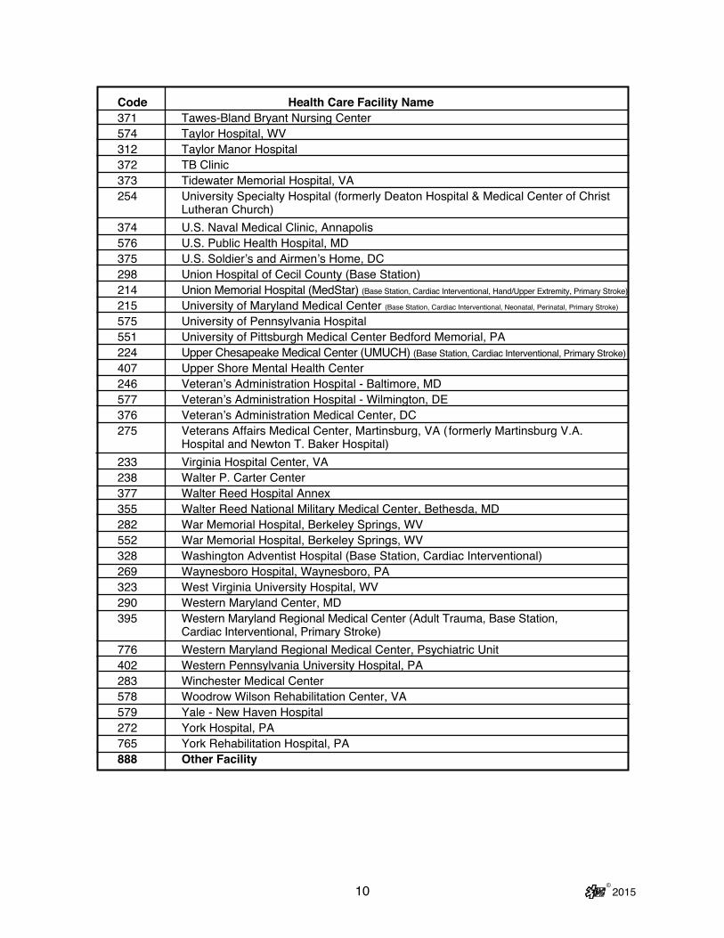
10 © 2015
Code Health Care Facility Name371 Tawes-Bland Bryant Nursing Center574 Taylor Hospital, WV312 Taylor Manor Hospital372 TB Clinic373 Tidewater Memorial Hospital, VA254 University Specialty Hospital (formerly Deaton Hospital & Medical Center of Christ
Lutheran Church)374 U.S. Naval Medical Clinic, Annapolis576 U.S. Public Health Hospital, MD375 U.S. Soldier’s and Airmen’s Home, DC298 Union Hospital of Cecil County (Base Station)214 Union Memorial Hospital (MedStar) (Base Station, Cardiac Interventional, Hand/Upper Extremity, Primary Stroke)215 University of Maryland Medical Center (Base Station, Cardiac Interventional, Neonatal, Perinatal, Primary Stroke)575 University of Pennsylvania Hospital551 University of Pittsburgh Medical Center Bedford Memorial, PA224 Upper Chesapeake Medical Center (UMUCH) (Base Station, Cardiac Interventional, Primary Stroke)407 Upper Shore Mental Health Center246 Veteran’s Administration Hospital - Baltimore, MD577 Veteran’s Administration Hospital - Wilmington, DE376 Veteran’s Administration Medical Center, DC275 Veterans Affairs Medical Center, Martinsburg, VA ( formerly Martinsburg V.A.
Hospital and Newton T. Baker Hospital)233 Virginia Hospital Center, VA238 Walter P. Carter Center 377 Walter Reed Hospital Annex 355 Walter Reed National Military Medical Center, Bethesda, MD282 War Memorial Hospital, Berkeley Springs, WV552 War Memorial Hospital, Berkeley Springs, WV 328 Washington Adventist Hospital (Base Station, Cardiac Interventional)269 Waynesboro Hospital, Waynesboro, PA323 West Virginia University Hospital, WV290 Western Maryland Center, MD395 Western Maryland Regional Medical Center (Adult Trauma, Base Station,
Cardiac Interventional, Primary Stroke) 776 Western Maryland Regional Medical Center, Psychiatric Unit 402 Western Pennsylvania University Hospital, PA283 Winchester Medical Center578 Woodrow Wilson Rehabilitation Center, VA579 Yale - New Haven Hospital272 York Hospital, PA765 York Rehabilitation Hospital, PA888 Other Facility

© 2015 11
D. MARYLAND TRAUMA AND SPECIALTY REFERRAL CENTERS
Trauma Centers
Primary Adult Resource Center l R Adams Cowley Shock Trauma Center (UM), BaltimoreLevel I Trauma Center l The Johns Hopkins Hospital Adult Trauma Center, BaltimoreLevel II Trauma Centers l Johns Hopkins Bayview Medical Center, Baltimore l Prince George’s Hospital Center, Cheverly l Sinai Hospital of Baltimore l Suburban Hospital (JHM), BethesdaLevel III Trauma Centers l Meritus Medical Center, Hagerstown l Peninsula Regional Medical Center, Salisbury l Western Maryland Regional Medical Center, CumberlandOut-of-State Centers l Christiana Care Health System, Wilmington, DE l MedStar Washington Hospital Center, Washington, DC
Specialty Referral Centers
Eye Trauma l Wilmer Eye Institute at The Johns Hopkins Hospital, BaltimoreHand/Upper Extremity Trauma l The Curtis National Hand Center for Treatment of the Hand and Upper Extremity/Union Memorial Hospital (MedStar), BaltimoreHyperbaric Medicine l Center for Hyperbaric Medicine/R Adams Cowley Shock Trauma Center (UM),
BaltimoreNeurotrauma (Head and Spinal Cord Injuries) l Neurotrauma Center/R Adams Cowley Shock Trauma Center (UM), BaltimorePediatric Trauma l Pediatric Trauma Center at The Johns Hopkins Children’s Center, Baltimore l Pediatric Trauma Center at Children’s National Health System,
Washington, DCBurns l Baltimore Regional Burn Center at Johns Hopkins Bayview Medical
Center, Baltimore l Burn Center at MedStar Washington Hospital Center, Washington, DC l Pediatric Burn Center at The Johns Hopkins Children’s Center, Baltimore l Pediatric Burn Center at Children’s National Health System, Washington, DC

12 © 2015
Specialty Referral Centers
Perinatal Referral Centers l Anne Arundel Medical Center, Annapolis l Franklin Square Medical Center (MedStar), Baltimore l Frederick Memorial Hospital, Frederick l Greater Baltimore Medical Center, Towson l Holy Cross Hospital, Silver Spring l Howard County General Hospital (JHM), Columbia l Johns Hopkins Bayview Medical Center, Baltimore l Mercy Medical Center, Baltimore l Prince George’s Hospital Center, Cheverly l Saint Agnes Hospital, Baltimore l Saint Joseph Medical Center (UM), Baltimore l Shady Grove Adventist Hospital, Gaithersburg l Sinai Hospital of Baltimore l The Johns Hopkins Hospital, Baltimore l University of Maryland Medical Center, Baltimore
Primary Stroke l Anne Arundel Medical Center, Annapolis l Atlantic General Hospital, Berlin l Baltimore Washington Medical Center (UM), Glen Burnie l Calvert Memorial Hospital, Prince Frederick l Charles Regional Medical Center.(UM), La Plata l Franklin Square Medical Center (MedStar), Baltimore l Frederick Memorial Hospital, Frederick l Good Samaritan Hospital (MedStar), Baltimore l Greater Baltimore Medical Center, Baltimore l Harbor Hospital (MedStar), Baltimore l Harford Memorial Hospital (UMUCH), Havre De Grace l Holy Cross Hospital, Silver Spring l Howard County General Hospital (JHM), Columbia l Johns Hopkins Bayview Medical Center, Baltimore l Mercy Medical Center, Baltimore l Meritus Medical Center, Hagerstown l Midtown Campus (UM), Baltimore l Montgomery Medical Center (MedStar), Olney l Northwest Hospital, Baltimore l Peninsula Regional Medical Center, Salisbury l Saint Agnes Hospital, Baltimore l Saint Joseph Medical Center (UM), Baltimore l Saint Mary’s Hospital (MedStar), Leonardtown l Shady Grove Adventist Hospital, Rockville l Shore Medical Center at Easton (UMSRH)
MARYLAND TRAUMA AND SPECIALTY REFERRAL CENTERS (Continued)

13 © 2015
MARYLAND TRAUMA AND SPECIALTY REFERRAL CENTERS (Continued)
Primary Stroke (Continued) l Sinai Hospital of Baltimore l Southern Maryland Hospital (MedStar), Clinton l Suburban Hospital (JHM), Bethesda l The Johns Hopkins Hospital, Baltimore l Union Hospital of Cecil County, Elkton l Union Memorial Hospital (MedStar), Baltimore l University of Maryland Medical Center, Baltimore l Upper Chesapeake Medical Center (UMUCH), Bel Air l Washington Adventist Hospital, Takoma Park l Western Maryland Regional Medical Center, Cumberland
Comprehensive Stroke l The Johns Hopkins Hospital, Baltimore l University of Maryland Medical Center, Baltimore
Cardiac Interventional l Anne Arundel Medical Center, Annapolis l Baltimore Washington Medical Center (UM), Glen Burnie l Bayhealth Kent General, Dover, DE l Carroll Hospital Center, Westminster l Christiana Care Health System, Newark, DE l Franklin Square Medical Center (MedStar), Baltimore l Frederick Memorial Hospital, Frederick l Holy Cross Hospital, Silver Spring l Howard County General Hospital (JHM), Columbia l Johns Hopkins Bayview Medical Center, Baltimore l MedStar Washington Hospital Center, Washington, DC l Meritus Medical Center, Hagerstown l Nanticoke Memorial Hospital, Seaford, DE l Peninsula Regional Medical Center, Salisbury l Prince George’s Hospital Center, Cheverly l Saint Agnes Hospital, Baltimore l Saint Joseph Medical Center (UM), Baltimore l Shady Grove Adventist Hospital, Rockville l Sinai Hospital of Baltimore l Southern Maryland Hospital (MedStar), Clinton l Suburban Hospital (JHM), Bethesda l The Johns Hopkins Hospital, Baltimore l Union Memorial Hospital (MedStar), Baltimore l University of Maryland Medical Center, Baltimore l Upper Chesapeake Medical Center (UMUCH), Bel Air l Washington Adventist Hospital, Takoma Park l Western Maryland Regional Medical Center, Cumberland

THIS PAGE IS INTENTIONALLY BLANK
14 © 2015

15 © 2015
E. PROTOCOL KEY
1. Basic Life Support Level Care
2. Advanced Life Support Level Care
3. Requires Medical Consultation
4. Pediatric CareNOTE: ALL PROVIDERS (BLS and ALS) SHOULD CHECK ALL PEDIATRIC SECTIONS FOR NECESSARY CARE.
5. Caution/Warning/Alert

16 © 2015
F. PROTOCOL USAGE FLOW DIAGRAM
General Patien tCare Sectio n
Refer to Specifi cProtocols
LEGENDResponse
Scene Arrival+
Size Up
PatientApproach
PersonalProtectiveEquipment
InitialAssessment
Histor y+
Physical Exam
Presumed Dead On Arrival
DNR/MOLSTPalliative
CareProtocol
Detailed +Ongoing
Assessment
Determine and Provide Care Accordingto Treatment Protocol
Disposition: Determine ReceivingFacility + Mode of T ransportation
Transport the Patient whenAppropriate
Communications:Consult / Notify Receiving Facility
Transfer of Care / Rendezvous:Transfer Patient to Receiving Facility
Complete Documentation
YES
NO
Withhol dResuscitation
OPTION
A/B
A
B
YES
Assign Clinical Priority
Procedures
Pharmacology
Inability toCarry Out
Physician’sOrders
ExtraordinaryCare
Termination ofResuscitation
Efforts
NO
Pronouncement of Death

17 © 2015
If an error or variance occurs (i.e., any act or failure to act, in practice or judg-ment, involving patient care that is not consistent with established protocol, whether or not it results in any change in the patient’s status or condition):
1. The EMS Provider must: a) Notify the consulting physician via radio as soon as the error or
variance is discovered, if prior to arrival at the receiving hospital, b) Monitor the patient’s condition very closely for any changes, c) Notify the receiving physician upon arrival, and d) Notify the local EMS jurisdiction or licensed commercial ambu-
lance service and Program Medical Director within 24 hours of the incident.
2. The EMS Operational Program Quality Assurance Officer in accor-
dance with COMAR 30.03.04.02 B(6) must: a) Within 5 days of being made aware of the incident, submit written
notification of the incident to the: (1) Local EMS jurisdiction, (2) Program Medical Director, (3) MIEMSS Compliance Office, and (4) State EMS Medical Director.
b) Within 14 days of the written notification of the incident, initiate a Medical Review Committee QA investigation.
c) Within 30 days of the written notification of the incident, forward to MIEMSS’ Compliance Office and State EMS Medical Director the written results of the Medical Review Committee QA investigation and recommendations.
G. PROTOCOL VARIATION PROCEDURE:

THIS PAGE IS INTENTIONALLY BLANK
18 © 2015

19 © 2015
H. INABILITY TO CARRY OUT PHYSICIAN ORDER: Occasionally a situation may arise in which a physician’s order cannot be carried out; e.g., the provider feels the administration of an ordered medication would endanger the patient, a medication is not available, or a physician’s order is outside the protocol. If this occurs:
1. The EMS Provider must: a) Immediately notify the consulting physician as to the reason the order cannot
be carried out. b) Document on the patient care report what was ordered, the time it was
ordered, and the reason the order could not be carried out. c) As soon as practical following the call, notify the local EMS
jurisdiction of the incident. 2. Public Service EMS Operational Programs must:
a) Within 5 days of being made aware of the incident, submit written notification
of the incident through the local EMS jurisdiction and Program Medical Direc-tor to the Regional Medical Director with the MIEMSS Regional EMS Admin-istrator being notified at the discretion of the Regional Medical Director, and a copy to the State EMS Medical Director.
b) Within 14 days of the written notification of the incident, initiate a QA investi-gation under the authority of the Medical Review Committee.
c) Within 30 days of the written notification of the incident, forward to MIEMSS’ Compliance Office and State EMS Medical Director written results of the Medical Review Committee QA investigation and recommendations.
3. Licensed Commercial Programs must:
a) Within 5 days of being made aware of the incident, submit written notification
of the incident through the commercial Program Medical Director to the Direc-tor of the State Office of Commercial Ambulance Licensing and Regulation and a copy to the State EMS Medical Director.
b) Within 14 days of the written notification of the incident, initiate a QA investi-gation under the authority of the Medical Review Committee.
c) Within 30 days of the written notification of the incident, forward to the Pro-gram Medical Director and to the Director of the State Office of Commercial Ambulance Licensing and Regulation and State EMS Medical Director written results of the Medical Review Committee QA investigation and recommenda-tions.

THIS PAGE IS INTENTIONALLY BLANK
20 © 2015

21 © 2015
I. PHYSICIAN ORDERS FOR EXTRAORDINARY CARE NOT COVERED BY MARYLAND PROTOCOL: Rarely, a physician providing on-line medical consulta-tion may direct a prehospital provider to render care that is truly life-saving and is not explicitly listed within the protocols.
1. ALL of the following criteria MUST be present for prehospital providers to proceed with an order under this section: a) During the consultation, both the consulting physician and the provider must
acknowledge and agree that the patient’s condition and extraordinary care are not addressed elsewhere within these medical protocols and that the order is absolutely necessary to maintain the life of the patient.
b) The provider must feel capable of correctly performing the care directed by the consulting physician, based on the instructions given by the consulting physician.
c) When such an order is carried out, the consulting physician and the provider must immediately notify the State EMS Medical Director (via SYSCOM, 800-648-3001) of the extraordinary care situation. In addition, the provider must fax documentation of the rationale for extraordinary care within 24 hours to the State EMS Medical Director at (410) 706-0853. Attendance at a subse-quent review meeting shall be required.
d) The prehospital provider must inform the consulting physician of the effect of the treatment and notify the receiving physician of the treatment upon arrival at the hospital (if the receiving physician is different than the consulting physi-cian). The prehospital provider must also notify his/her BLS/ALS program medical director within 24 hours.
e) The public service local EMS jurisdiction and the Program Medical Director must then submit written notification of the incident to the Regional Medical Director with a copy to the State EMS Medical Director within 5 days of the incident.
f) The commercial ambulance company and the Program Medical Director must submit written notification of the incident to the Director of the State Office of Commercial Ambulance Licensing and Regulation and the State EMS Medi-cal Director within 5 days of the incident.

22 © 2015
g) The State EMS Medical Director shall conduct a review conference to include when appropriate: the prehospital provider, the on-line physician who provided the medical consultation, the appropriate local jurisdictional official(s), the Pro-gram Medical Director, and the Regional Medical Director.
h) Reports of incidents shall be submitted by the State EMS Medical Director to the Incident Review Committee and, when appropriate, to the Board of Physi-cian Quality Assurance.
2. If a prehospital provider receives an order for care that is not covered by Mary-land protocols, but does not feel comfortable with it or does not agree that it is absolutely necessary to maintain the life of the patient, he/she shall proceed with the “Inability to Carry Out a Physician’s Order” section.
3. Protocols provide a safe basis for prehospital intervention and transport and provide both prehospital providers and on-line physicians with parameters for this care. Extraordinary care situations not within the protocols may occur a handful of times over a span of years. The extraordinary care protocol is intended to ad-dress the potential moral/ethical dilemma which may arise in unanticipated or un-foreseen situations not specifically addressed within protocols. The extraordinary care protocol is neither a carte blanche for any and all actions nor a device to avoid or circumvent protocols. In all situations, emergency health care providers, both prehospital providers and on-line physicians providing medical direction, are accountable for their actions in discharging their patient care responsibilities.
EXTRAORDINARY CARE CHECKLIST
• Identify the need for extraordinary care with physician consult and EMS provider acceptance. º Care is not covered elsewhere in the protocols. º Care is absolutely necessary to maintain the life of the patient.
• Immediately upon delivery of patient, EMS provider must notify the receiving physician and the State EMS Medical Director via SYSCOM.
• Fax (410-706-0853) the rationale to the State EMS Medical Director within 24 hours.
• Notify the Program Medical Director within 24 hours.• Submit written notification of event to Regional Medical Director or SOCALR and the State EMS Medical Director within 5 days.

23 © 2015
J. QUALITY REVIEW PROCEDURE FOR PILOT PROGRAMS (Old Class B)
1. Through a quality assurance review process directly involving the Program Medical Director (PMD), developed by the local program and approved by the PMD, the respective Regional Medical Director (RMD) and the State EMS Medical Director, the local program will review the runsheet and patient outcome records to determine the appropriateness of each individual use of the skill or administration of the medication. If the pilot procedure or medica-tion is judged to be an appropriate intervention, the occurrence is added to the jurisdictional database and forwarded to the RMD and the State EMS Medical Director.
2. If a variance or question arises from the review of the case, a case review conference will be held with the provider, the PMD, and, if indicated, the on- line medical consultant with the summary of the findings to be reported to the RMD and the State EMS Medical Director.
Quality Assurance Mechanism for PILOT Programs and Procedures
Data to — PMD — RMD — State EMS Medical Director
Case Review Conference with — Prehospital Provider — PMD — Consulting Physician
EMS ResponsePCR Documentation
QA Review Processwith PMD *
Appropriate ?
YES NO
* — Approved by PMD, RMD, MIEMSS State EMS Medical Director

THIS PAGE IS INTENTIONALLY BLANK
24 © 2015

25 © 2015
II. GENERAL PATIENT CARE (GPC)
A. RESPONSE Review the dispatch information and select appropriate response.
B. SCENE ARRIVAL AND SIZE-UP 1. Consider Body Substance Isolation (BSI).
2. Consider Personal Protective Equipment (PPE). 3. Evaluate the scene safety. 4. Determine the number of patients. 5. Consider the need for additional resources.
C. PATIENT APPROACH 1. Determine the Mechanism of Injury (MOI)/Nature of Illness (NOI). 2. If appropriate, begin triage and initiate Mass Casualty Incident (MCI)
procedures.
D. INITIAL ASSESSMENT
CORRECT LIFE-THREATENING PROBLEMS AS IDENTIFIED.STABILIZE CERVICAL SPINE WHEN APPROPRIATE.
FOR PEDIATRIC PATIENTS, CONSIDER USING THE PEDIATRIC ASSESSMENT TRIANGLE. 1. Assess mental status
a) Alertb) Responds to Verbal stimulic) Responds to Painful stimulid) Unresponsive
2. Airway a) Open and establish airway using appropriate adjunct. b) Place patient in appropriate position. c) Suction airway as needed, including tracheostomy tubes.
Appearance Work ofBreathing
Circulation to Skin

26 © 2015
IF A PATENT AIRWAY CANNOT BE ESTABLISHED, THE PATIENT MUST BE TRANSPORTED TO THE NEAREST APPROPRIATE HOSPITAL-BASED EMERGENCY DEPARTMENT OR DESIGNATED FREESTANDING MEDICAL FACILITY. ONCE THE PATIENT PRESENTS TO THE HOSPITAL OR DESIGNATED FREESTANDING MEDICAL FACILITY FOR TREATMENT OF AN EMERGENCY CONDITION, TREATMENT AND TRANSFER DECISIONS ARE THE RESPONSIBILITY OF THE HOSPITAL UNDER APPLICABLE LAW. THE PROVIDER SHOULD STAND BY TO BE AVAILABLE FOR AND ASSIST WITH TRANSFER OF THE PATIENT IF THE HOSPITAL DETERMINES SUCH A TRANSFER IS APPROPRIATE.
IN INFANTS AND YOUNG CHILDREN, INSPIRATORY STRIDOR IS AN INDICATION OF UPPER AIRWAY FOREIGN BODY OR PARTIAL AIRWAY OBSTRUCTION. REQUEST ALS RENDEZVOUS. TRANSPORT THE PATIENT RAPIDLY AND CAUTIOUSLY AND HAVE FOREIGN BODY AIRWAY REMOVAL EQUIPMENT READY FOR IMMEDIATE USE IN CASE THE PATIENT’S AIRWAY BECOMES OBSTRUCTED.
3. Breathing a) Determine if breathing is adequate. Assess oxygen saturation (SpO2) with
portable pulse oximeter (required on all transport units since 2012). (1) If patient’s ventilations are not adequate, provide assistance with 100%
oxygen using Bag-Valve-Mask (BVM).(i) For all ages except neonates, 1 breath every 5 seconds (8–12
breaths/min) (manually-activated positive pressure oxygen delivery device is not recommended for this group) (NEW ’15)
(ii) For a neonate, 1 breath every 3 seconds (higher rates may be required) (NEW ’15)
(2) The decision to oxygenate will be based upon the patient’s clinical condition. (i) SpO2 ≥ 94% is considered normoxia in adults and children. Supplemental
oxygen is not needed if SpO2 ≥ 94% unless the patient is in respiratory distress, acutely dyspneic, or suffering from suspected CO poisoning. Patients in severe respiratory distress may benefit from high flow oxygen from a nonrebreather. Note: Respiratory distress is present if the patient has retractions, nasal flaring, wheezing, stridor, or difficulty speaking.
(ii) Unless in respiratory distress, avoid administration of high flow oxygen to patients presenting with the following conditions: (a) STEMI/Angina (b) CVA/stroke (c) Post arrest
(iii) CO Exposure: Apply 100% oxygen via NRB mask. Maintain SpO2 at 100%.
INACCURATE OR MISLEADING SpO2 READINGS MAY OCCUR IN THE FOLLOWING PATIENTS: HYPOTHERMIC, HYPOPERFUSION (SHOCK), CO POISONING, HEMOGLOBIN ABNORMALITY, ANEMIA, AND VASOCONSTRICTION.

27 © 2015
(3) If available, utilize end-tidal CO2 waveform monitoring in intubated pa-tients (required on all ALS transport units for advanced airway manage-ment by 2015).
(4) Consider carbon monoxide measurement, if available.
b) Hyperventilate the head injured patient only if signs/symptoms of herniation are present, including posturing, loss of pupillary light response, dilation of one or both pupils, vomiting, hypertension, bradycardia, and/or irregular res-pirations. (1) If hyperventilating, use the following rates
Adult: 20 breaths per minute Child: 30 breaths per minute Infant: 35 breaths per minute
(2) If hyperventilating, use EtCO2 monitoring if available.
NEVER WITHHOLD OXYGEN FROM A PATIENT IN RESPIRATORY DISTRESS!
Percent O2Saturation Ranges General Patient Care94–100% Normal Give Oxygen as necessary91–93% Mild Hypoxia Give Oxygen as necessary86–90% Moderate Give 100% Oxygen Hypoxia Assisting Ventilations if necessary ≤ 85% Severe Give 100% Oxygen Hypoxia Assist Ventilations If indicated, IntubateFalse SpO 2 readings may occur in the following patients:Hypothermic, Hypoperfusion (Shock), Carbon Monoxide Poisoning,Hemoglobin Abnormality, Anemic, and Vasoconstriction.
DEVICE FLOW RATE CONCENTRATION Nasal Cannula 2–6 lpm 24–44% Venturi Mask Variable 24–50%Partial Rebreather Mask 6–10 lpm 35–60%Simple Face Mask 6–10 lpm 35–60%Pocket Mask 12–15 lpm 50–60%Non-Rebreather Mask 12–15 lpm 80–100%Bag-Valve-Mask 12–15 lpm 90–100%

28 © 2015
4. Circulation ONCE CONFIRMED PULSELESS, HIGH-QUALITY CONTINUOUS CPR WITH FREQUENT PROVIDER ROTATION IS AN ESSENTIAL COMPONENT IN THE SUCCESSFUL RESUSCITATION OF THE AR-RESTED PATIENT. THIS MAY BE ACCOMPLISHED THROUGH MANUAL OR MECHANICAL MEANS AS APPROPRIATE. PERFORM CPR WHILE PREPARING FOR RHYTHM ANALYSIS AND DEFIBRILLATION.
a) Assess pulse.(1) Patients from birth up to those who have not reached their 12th birthday: (a) If pulse is absent, use AED/manual defibrillator or begin CPR. (b) If patient is symptomatic with poor perfusion (unresponsive or
responds only to painful stimuli) and pulse is less than 60 bpm:(i) Ventilate for 30 seconds. (ii) If after 30 seconds, the pulse is less than 60 bpm, begin CPR.
(c) If pulse greater than 60 bpm, continue assessment. (2) Patients 12 year of age or older:
(a) If pulse is absent, use AED/manual defibrillator or begin CPR. (b) If pulse is present, continue assessment.
b) Assess for and manage profuse bleeding. c) Assess skin color, temperature, and capillary refill.
5. Disability a) Perform Mini-Neurologic Assessment (Pulse/Motor/Sensory). b) Spinal protection (NEW ’15)
(1) The provider shall determine the appropriate method for use in spinal pro-tection of the patient. Infant or child car seats may NOT be used as a spinal immobilization device for the pediatric patient.
(2) Patients who have a blunt trauma with a high-energy mechanism of injury that has potential to cause spinal cord injury or vertebral instability and one or more the following should receive spinal protection. (NEW ’15) (a) Midline spinal pain, tenderness, or deformity (b) Signs and symptoms of new paraplegia or quadriplegia (c) Focal neurological deficit (d) Altered mental status or disorientation (e) Distracting injury
In addition to the above indicators for adults, the below apply to children that have not yet reached their 15th birthday
(f) High impact diving incident or high risk MVC - head on collision, rollover, ejected from the vehicle, death in the same crash, or speed > 55 mph
(g) Substantial torso injury (h) Conditions predisposing to spine injury (3) If NO to all of the above, transport as appropriate.
IF PATIENT IS UNABLE TO COMMUNICATE OR APPROPRIATELY RESPOND TO THE ABOVE QUESTIONS, APPLY SPINAL PROTECTION PROTOCOL. (NEW ’15)
6. Exposure To assess patient’s injuries, remove clothing as necessary, considering condition
and environment.

© 2015
7. Assign Clinical Prioritya) Priority 1 — Critically ill or injured person requiring immediate attention; un-
stable patients with life-threatening injury or illness. b) Priority 2 — Less serious condition yet potentially life-threatening injury or illness,
requiring emergency medical attention but not immediately endangering the patient’s life.
c) Priority 3 — Non-emergent condition, requiring medical attention but not on an emergency basis.
d) Priority 4 — Does not require medical attention. e) In the event of a multiple casualty incident, the Simple Triage And Rapid Treat-
ment (START and/or JumpSTART) technique will be instituted for rapid tagging and sorting of patients into priority categories for both treatment and transport.
28-1

THIS PAGE IS INTENTIONALLY BLANK
© 201528-2

29 © 2015
HIS
TOR
Y A
ND
PH
YS
ICA
L E
XA
MIN
AT
ION
TR
AU
MA
PA
TIE
NT
ME
DIC
AL
PA
TIE
NT
CO
NS
IDE
R A
LS,
PE
RF
OR
M I
NT
ER
VE
NT
ION
S,
AN
D T
RA
NS
PO
RT.
D C A P B T L S
Sig
nific
ant M
OI
Rap
id T
raum
aA
sses
smen
t
H
ead
Cre
pita
tion
C
hest
Cre
pita
tion
Res
pira
tion
Par
adox
ical
Mot
ion
Bre
ath
Sou
nds
A
bdom
enR
igid
ityD
iste
ntio
n
Pel
vis/
GU
Pai
n on
Mot
ion
Blo
od,U
rine,
Fec
es
Ext
rem
ities
Pul
se/M
otor
/Sen
sory
P
oste
rior
Bas
elin
e V
ital S
igns
Obt
ain
SA
MP
LE H
isto
ry
Sig
ns &
Sym
ptom
s
A
llerg
ies
Med
icat
ions
Per
tinen
t His
tory
Las
t Ora
l Int
ake
Eve
nts
Prio
r
Non
-Sig
nific
ant M
OI
Det
erm
ine
Chi
efC
ompl
aint
Per
form
Foc
used
Exa
min
atio
n o
f the
Inju
red
Site
and
Are
asC
ompa
tible
with
Giv
enM
OI
Bas
elin
e V
ital S
igns
Obt
ain
SA
MP
LE H
isto
ry
Sig
ns &
Sym
ptom
s
A
llerg
ies
Med
icat
ions
Per
tinen
t His
tory
Las
t Ora
l Int
ake
Eve
nts
Prio
r
Unr
espo
nsiv
e P
atie
nt
Rap
id P
hysi
cal
Exa
min
atio
n
H
ead
N
eck
JVD
Med
ical
Ale
rt D
evic
e
Che
stB
reat
h S
ound
s
Abd
omen
Rig
idity
Dis
tent
ion
P
elvi
s/G
UB
lood
,Urin
e,F
eces
E
xtre
miti
esM
SP
Med
ical
Ale
rt D
evic
e
Pos
terio
r
Bas
elin
e V
ital S
igns
Obt
ain
His
tory
of E
piso
de
O
nset
Pro
voca
tion
Qua
lity
Rad
iatio
n
S
ever
ity
T
ime
Obt
ain
SA
MP
LE H
isto
ry
Res
pons
ive
Pat
ient
Obt
ain
His
tory
of E
piso
de
O
nset
Pro
voca
tion
Qua
lity
Rad
iatio
n
S
ever
ity
Tim
e
Bas
elin
e V
ital S
igns
Obt
ain
SA
MP
LE H
isto
ry
S
igns
& S
ympt
oms
A
llerg
ies
M
edic
atio
ns
Per
tinen
t His
tory
La
st O
ral I
ntak
e
Eve
nts
Prio
r
Foc
used
Phy
sica
l Exa
mD
CA
PB
TLS
Che
ck a
reas
sug
gest
edby
MO
I and
SA
MP
LE.
D C A P B T L S
D C A P B T L S

30 © 2015
DE
TAIL
ED
A
ND
ON
GO
ING
AS
SE
SS
ME
NT
S
DE
TAIL
ED
EX
AM
INA
TIO
NO
NG
OIN
G A
SS
ES
SM
EN
T
HE
AD
Sca
lp &
Cra
nium
Cre
pita
tion
Eye
s Dis
colo
ratio
nE
qual
ityF
orei
gn B
odie
sB
lood
in A
nter
ior
Cha
mbe
r E
ars
& N
ose
Flu
id D
rain
age
or B
leed
ing
Dis
colo
ratio
n M
outh Te
eth
& F
orei
gn B
odie
sS
wel
ling
or L
acer
atio
nsB
reat
h O
dor
Dis
colo
ratio
nN
EC
K Jugu
lar
Vei
n D
iste
ntio
nTr
ache
a P
ositi
onC
repi
tatio
nC
HE
ST
Par
adox
ical
Mot
ion
Bre
ath
Sou
nds
Cre
pita
tion
AB
DO
ME
NR
igid
ityD
iste
ntio
nP
ELV
IS/G
UP
ain
on M
otio
nE
XT
RE
MIT
IES
Pul
se, M
otor
, Sen
sory
Cap
illar
y R
efill
PO
ST
ER
IOR
ME
DIC
AL
PA
TIE
NT
RE
PE
AT
IN
ITIA
L A
SS
ES
SM
EN
T
R
eass
ess
AV
PU
R
eass
ess
Airw
ay
Mon
itor
Bre
athi
ng
Rea
sses
s C
ircul
atio
n
M
onito
r S
kin
C
onfir
m C
linic
al P
riorit
y
RE
PE
AT
& R
EC
OR
D V
ITA
L S
IGN
S
RE
PE
AT
FO
CU
SE
DA
SS
ES
SM
EN
TE
spec
ially
Chi
ef C
ompl
aint
or
Inju
ries
CH
EC
K A
LL I
NT
ER
VE
NT
ION
S
A
ssur
e O
xyge
n A
dequ
acy
C
heck
Ble
edin
g
Che
ck In
terv
entio
ns
Che
ck fo
r Tr
endi
ng
Sta
ble
Pt.-
Eve
ry 1
5 M
in.
U
nsta
ble
Pt.-
Rec
omm
end
E
very
5 M
in.
TR
AU
MA
PA
TIE
NT
RE
PE
AT
IN
ITIA
L A
SS
ES
SM
EN
T
R
eass
ess
AV
PU
R
eass
ess
Airw
ay
Mon
itor
Bre
athi
ng
Rea
sses
s C
ircul
atio
n
M
onito
r S
kin
C
onfir
m C
linic
al P
riorit
y
RE
PE
AT
& R
EC
OR
D V
ITA
L S
IGN
S
RE
PE
AT
RA
PID
TR
AU
MA
AS
SE
SS
ME
NT
CH
EC
K A
LL I
NT
ER
VE
NT
ION
S
A
ssur
e O
xyge
n A
dequ
acy
C
heck
Ble
edin
g
Che
ck N
eck
Sta
biliz
atio
n
Che
ck In
terv
entio
ns
Che
ck fo
r Tr
endi
ng
Sta
ble
Pt.-
Eve
ry 1
5 M
in.
U
nsta
ble
Pt.-
Rec
omm
end
E
very
5 M
in.
D C A P B T L S
CO
NS
IDE
R A
LS,
PE
RF
OR
M I
NT
ER
VE
NT
ION
S,
AN
D T
RA
NS
PO
RT.

© 2015 30-1

THIS PAGE IS INTENTIONALLY BLANK
© 201530-2

31 © 2015
E. HISTORY AND PHYSICAL EXAMINATION/ASSESSMENT 1. Conduct a Focused Examination/Detailed Examination/Ongoing Assessment. 2. Collect and transport documentation related to patient’s history (example: Emer-
gency Information Form, Medic Alert, EMS DNR/MOLST, or jurisdictional form).
3. Obtain an EKG when appropriate.
F. TREATMENT PROTOCOLS1. Refer to ALL appropriate protocols. 2. Patients who have had an impaled conducted electrical weapon used on them will
be transported to the nearest appropriate facility without dart removal (Exception Tactical EMS).
3. Providers may assist the patient or primary caregiver in administering the pa-tient’s prescribed rescue medication. a) BLS providers may assist with the administration of the patient’s fast-acting
bronchodilator MDI and sublingual nitroglycerin. b) ALS providers may administer the patient’s prescribed benzodiazepine for
seizures, Factor VIII or IX for Hemophilia A or B, or reestablish IV access for continuation of an existing vasoactive medication.
c) Providers should obtain on-line medical direction to administer other prescribed rescue medications not specifically mentioned in The Maryland Medical Proto-cols for EMS Providers (e.g., Solucortef for Adrenal Insufficiency). The rescue medication must be provided by the patient or caregiver and the label must have the patient’s name and the amount of medication to be given.
DO NOT ADMINISTER ORAL MEDICATIONS (EXCEPT GLUCOSE PASTE) TO PATIENTS WITH AN ALTERED MENTAL STATUS.
4. For pediatric patientsa) Pediatric section of the treatment protocol will be used for children who have
not reached their 15th birthday (trauma and medical), except as otherwise stated in the treatment protocol.
b) Medication dosing (1) Pediatric doses apply to patients weighing less than 50 kg. (2) For pediatric patients equal to or greater than 50 kg, utilize adult dosing.
c) The developmental age of the infant/child must be considered in the commu-nication and evaluation for treatment.
d) Destination consideration: For those patients who are 15 years of age or older who receive spe-
cialized care at a pediatric facility, consider medical consultation with a pediatric base station for patient destination.
e) Infants and children must be properly restrained prior to and during transport. f) When appropriate, family members should remain with pediatric patients.

32 © 2015
G. COMMUNICATIONS 1. All Priority 1 patients require on-line medical consultation.
2. All Priority 2 patients who have persistent symptoms or need further therapeutic
intervention(s) require on-line medical consultation.
3. Notification (“information only call” that can be through EOC or EMS communica-tion system following local standard operating procedures) should be made to the receiving hospital for Priority 2 or Priority 3 patients whose symptoms have resolved and whose vital signs are within normal limits.
ON-LINE MEDICAL CONSULTATION MAY BE OBTAINED AT ANY TIME FOR ANY PATIENT, IF DESIRED BY THE PREHOSPITAL EMS PROVIDER. PEDIATRIC AND SPECIALTY CONSULTATION IS ENCOURAGED FOR TRAUMA AND MEDICAL PATIENTS. CONSULTATION WITH PEDIATRIC AND SPECIALTY CENTERS SHALL OCCUR SIMULTANEOUSLY WITH A BASE STATION CONSULT.
4. If medical consultation is genuinely unavailable, or if the time necessary to initiate consultation significantly compromises patient care, the provider shall proceed with additional protocol directed care, so long as transport will not be significantly de-layed. “Exceptional Call” must be indicated on the Patient Care Report (PCR).
5. Trauma Communications The following information must be communicated to the appropriate Trauma Center and/or Local Hospital: a) Patient’s age, injuries, ETA b) Number of victims c) Detailed description of the incident d) Provide patient Trauma Decision Tree Category (Alpha, Bravo, Charlie, Delta) e) Provide assigned patient priority (1 to 4) f) Pertinent patient signs and symptoms (e.g., HR, RR, BP, Pulse Ox and GCS)
CONSIDER ACTIVATION OF THE GO-TEAM FOR SERIOUSLY INJURED PATIENTS WHO REQUIRE A PROLONGED EXTRICATION AND WHO MEET THE INDICATIONS FOR GO-TEAM ACTIVATION.
6. Mass Casualty Incident (MCI) Communications a) When a local jurisdiction declares an MCI, it is extremely important to maximize
patient care resources and reserve EMS communications for emergent situa-tions. Except for extraordinary care interventions, EMS providers may perform all skills and administer medications within protocol, during a declared MCI. When the MCI condition is instituted, the Exceptional Call box must be checked on the PCR.
b) During an MCI, the EMS Officer-in-Charge (OIC) shall designate an EMS Com-municator who shall establish appropriate communications.
c) Reference the Multiple Casualty Incident/Unusual Incident Protocol.
7. Communications with and through EMRC/SYSCOM are recorded. In addition, as part of the quality assurance and quality improvement process, communications with hospitals are frequently recorded. Therefore, you should assume that all your communications among EMS providers, hospitals, public safety communica-tions centers, and EMRC/SYSCOM are being recorded.

33 © 2015
H. REASSESSMENT1. Reassess unstable patients frequently (recommended every 5 minutes).
2. Reassess stable patients at a minimum of every 15 minutes. 3. Reassess patients being discharged to home or long-term care at the beginning
and end of the transport or more frequently, at the provider’s discretion.
I. DISPOSITION1. Destination
a) Priority 1 patients shall be triaged according to Maryland Medical Protocols to the closest appropriate hospital-based emergency department, designated trauma, or designated specialty referral center. Critically unstable patients in need of immediate life-saving interventions that cannot be provided in the field shall, with the approval of EMS System medical consultation, be diverted to the closest facility (including freestanding medical facility) capable of immedi-ately providing those interventions.
b) Priority 2 patients shall be triaged according to the Maryland Medical Protocols to the closest appropriate hospital-based emergency department, designated trauma or designated specialty referral center unless otherwise directed by EMS System medical consultation.
c) Stable priority 3 or 4 patients who do not need a time-critical intervention may also be transported to the local emergency department or freestanding medi-cal facility.
2. Mode of transport (air, land, water, etc.) a) Medevac patients with indications for specialty referral center should be flown
to the appropriate type of specialty center if not more than 10–15 minutes further than the closest trauma center. (Patients with an airway, breathing, or circulatory status who would be jeopardized by going an additional 10–15 minutes should go to the closest trauma center.)
b) Consider utilization of a helicopter when the patient’s condition warrants transport to a trauma or specialty referral center and the use of a helicopter would result in a clinically significant reduction in time compared with driving to a trauma/specialty center.
ALL REQUESTS FOR SCENE HELICOPTER TRANSPORTS SHALL BE MADE THROUGH SYSCOM. FOR TRAUMA DECISION TREE CATEGORY CHARLIE OR DELTA, RECEIVING TRAUMA CENTER MEDICAL CONSULTATION REQUIRED WHEN CONSIDERING WHETHER HELICOPTER TRANSPORT IS OF CLINICAL BENEFIT.
c) If the time of arrival at the trauma or specialty referral center via ground unit is less than 30 minutes, there will generally not be a benefit in using the helicopter, especially for Trauma Decision Tree classes Charlie and Delta.
d) Refer to the Trauma Decision Tree when considering use of aeromedical trans-port. Provide SYSCOM with the patient’s Category (Alpha, Bravo, Charlie, or Delta).
e) On-line medical direction should be obtained from the local trauma center and the specialty referral center when transport to the specialty center would require more than 10–15 minutes additional transport time. (1) Pediatric Trauma Patients: Indications as per the pediatric section of the trauma protocols.

34 © 2015
(2) Spinal Trauma Patients: Indications as per Spinal Protection protocol. (3) Burn Patients: Indications as per burn protocol. Special note:
Isolated burn patients without airway injury or other associated trauma should normally be flown to a burn center, regardless of the location of the closest trauma center.
(4) Hand Injury Patients: Indications as per hand protocol. Special note: Medevac patients with appropriate indications for hand center referral should normally be flown to the hand center, regardless of the location of the closest trauma center.
3. Status Evaluate the need for emergent versus non-emergent transportation.
DO NOT WAIT ON-SCENE FOR ADVANCED LIFE SUPPORT. ATTEMPT TO RENDEZVOUS EN ROUTE TO THE HOSPITAL.
J. TRANSFER OF CARE/RENDEZVOUS AND TRANSITION OF PATIENT CARE ALS TO BLS The ALS provider-patient relationship is established when the ALS provider
initiates patient assessment and 1. ALS medication(s)* is/are administered or
2. ALS procedure(s)* is/are performed or 3. Upon ALS provider assessment of the patient there is potential risk of deterio-
ration. * Based on the medication or procedure as listed in the protocol pages 144–147
ALS providers may only terminate their EMS provider-patient relationship when they are assured that the patient will continue to receive care at the same or greater levels, or when they have documented with on-line medical direction that the patient’s condition has improved and that patient care may be transferred safely to an EMS Provider with a lower scope of practice. BLS providers have the right to decline the transition of patient care. When consensus between the providers cannot be gained, ALS shall get on-line medical direction. Providers will relay assessment findings and treatment provided to the individual(s) assuming responsibility for the patient(s).
K. DOCUMENTATION A Patient Care Report (PCR) will be completed and delivered to the receiving facility
as soon as possible, ideally upon transfer of care. If this is not immediately possible, providers must provide documentation of the patient’s prehospital care on a template and in a format provided or approved by MIEMSS for inclusion in the patient care record before leaving the receiving facility, then deliver the completed PCR within 24 hours after transfer of care, in compliance with COMAR 30.03.04.04. Only the unit that pronounces death will select the “Dead on Scene” option in the Patient Care Report (eMEDS®) and thus all other units will report “Operational Sup-port Only.” If no interventions are performed, the highest level EMS provider on scene

© 2015
will pronounce death and document “Dead on Scene.” If BLS care was rendered by a BLS unit and then termination of resuscitation and pronouncement of death occurred, the BLS unit will select “Dead at Scene with BLS Intervention” option on the eMEDS® PCR. If ALS care was rendered by an ALS unit and then termination of resuscitation and pronouncement of death occured, the ALS unit will select “Dead at Scene with ALS Intervention” option on the eMEDS® PCR.
L. CONFIDENTIALITY Patient confidentiality must be maintained at all times.
M. PROFESSIONAL CONDUCT All patients should be treated with dignity and respect in a calm and reassuring
manner.
34-1

THIS PAGE IS INTENTIONALLY BLANK
© 201534-2

35 © 2015
III. TREATMENT PROTOCOLS
FOR ALL TREATMENT PROTOCOLS, THE LETTER AND NUMERICAL OUTLINE FORMAT IS STRICTLY FOR RAPID AND UNIFORM REFERENCE AND DOES NOT IMPLY OR DIRECT A MANDATORY SEQUENCE FOR PATIENT CARE. HOWEVER, THE GENERAL PATIENT CARE SECTION AND THE ALGORITHMS DO HAVE A SPECIFIC SEQUENCE TO BE FOLLOWED.
A. ABUSE/NEGLECT1. Initiate General Patient Care.
ALL HEALTH CARE PROVIDERS ARE OBLIGATED BY LAW TO REPORT CASES OF SUSPECTED CHILD OR VULNERABLE ADULT ABUSE OR NEGLECT TO EITHER THE LOCAL POLICE OR SOCIAL SERVICE AGENCIES. DO NOT INITIATE REPORT IN FRONT OF THE PATIENT, PARENT, OR CAREGIVER. DO NOT CONFRONT OR BECOME HOSTILE TO THE PARENT OR CAREGIVER.
2. Presentation The patient may present with patterned burns or injuries suggesting intentional
infliction, such as injuries in varying stages of healing, injuries scattered over multiple areas of the body, fractures, or injuries inconsistent with stated cause of injury. The patient, parent, or caregiver may respond inappropriately to the situa-tion. Malnutrition or extreme lack of cleanliness of the patient or environment may indicate neglect. Signs of increased intracranial pressure (bulging fontanels and altered mental status in an infant) may also be seen.
3. Treatmenta) Stabilize injuries according to protocol. b) Discourage patient from washing if sexual abuse is suspected. c) Document the following information on the PCR:
(1) All verbatim statements made by the patient, the parent, or caregiver shall be placed in quotation marks, including statements made about the manner of the injuries.
(2) Any abnormal behavior of the patient, parent, and/or caregiver
(3) The condition of the environment and other residents present

36 © 2015
ABUSE/NEGLECT (Continued)
(4) The time the police/welfare agency was notified and the name of the per-son notified
(5) The name of the receiving health care provider (RN, PA, MD) and any statements made
d) Treat injuries according to presentation.
4. Continue General Patient Care.

37 © 2015
B. ALTERED MENTAL STATUS: SEIZURES
1. Initiate General Patient Care. 2. Presentation
Seizures are a neuromuscular response to an underlying cause such as: epi-lepsy, hypoxia, hypoglycemia, hypoperfusion, head injury, CVA, alcohol or drug abuse. Consider recent history of possible illness, infection, fever, or stiff neck.
DO NOT ATTEMPT TO FORCE ANY DEVICE INTO THE PATIENT’S MOUTH IF THE PATIENT IS STILL SEIZING.
3. Treatmenta) If the patient is still seizing:
(1) DO NOT RESTRAIN. (2) Protect patient from further injury. (3) Consider cause of seizure activity.
b) When seizure activity has stopped (1) Identify and treat injuries. (2) If patient is a known diabetic, glucose paste (10–15 grams) should be
administered between the gum and cheek. Consider single additional dose of glucose paste if not improved after 10 minutes.
c) Use glucometer and treat accordingly. d) Consider midazolam (Paramedic may perform without consult for pa-
tients with active seizures). If patient has no IV or IO in place, administer midazolam 5 mg IN or IM. (NEW ’15)0.1 mg/kg in 2 mg increments slow IV push over one to two minutes per increment with maximum single dose 5 mg.
REDUCE BY 50% FOR PATIENTS 69 YEARS OR OLDER.
If IV unavailable, 5 mg IM may be administered. Additional doses up to a maximum total dose 10 mg require medical con-sultation for all providers. If patient seizures are refractory to treatment, consider IO administration of midazolam. If midazolam is not available, consider diazepam (Paramedic may per-form without consult for patients with active seizures). 2.5 mg increments slow IVP/IM (IM requires all providers to obtain medical consultation).

38 © 2015
ALTERED MENTAL STATUS: SEIZURES (Continued)
Maximum total dose 10 mg If patient is in status, consider IO administration of diazepam. If suspected severe nerve agent exposure, providers may administer midazolam 5 mg IM or diazepam (CANA) without medical consultation.
e) Establish IV access with LR.
f) If patient is pregnant, actively seizing, consider magnesium sulfate 4 gm IV/IO over 10 minutes. (NEW ’15)
g) If seizures persist, consult for second dose of magnesium sulfate. (NEW ’15)
IF PATIENT IS PREGNANT, USE MIDAZOLAM FOLLOWED BY MAGNESIUM SULFATE. MEDICAL CONSULTATION REQUIRED FOR PREGNANT PATIENTS WHO MAY REQUIRE LARGER DOSES OF MIDAZOLAM TO CONTROL SEIZURES. (NEW ’15)
IF, FOLLOWING ADMINISTRATION OF MAGNESIUM SULFATE, PATIENT EXHIBITS SIGNS OF TOXICITY, CONSIDER ADMINISTRATION OF CALCIUM CHLORIDE. CONSIDER CALCIUM CHLORIDE 500 MG IVP FOR RESPIRATORY DEPRESSION, DECREASED REFLEXES, FLACCID PARALYSIS, AND APNEA FOLLOWING MAGNESIUM SULFATE ADMINISTRATION. MEDICAL CONSULTATION REQUIRED. (NEW ’15)
h) If the patient is still seizing: (1) DO NOT RESTRAIN. (2) Protect from further injury. (3) Consider underlying cause of seizure.
i) When seizure activity has stopped: (1) Identify and treat any injuries. (2) If patient is a known diabetic, glucose paste (10–15 grams) should be
administered between the gum and cheek. Consider single additional dose of glucose paste if not improved after 10 minutes.
j) Use glucometer and treat accordingly. k) The paramedic may assist patients with the administration of their prescribed
benzodiazepine.

© 2015 38-1
ALTERED MENTAL STATUS: SEIZURES (Continued)
l) Consider midazolam for seizures lasting greater than 10 minutes (Paramedic may perform without consult for patients with active seizures). If patient has no IV or IO in place: Administer midazolam 0.1 mg/kg IN or IM. Maximum total dose 5 mg. (NEW ’15) If IV or IO is already in place: Administer midazolam 0.1 mg/kg in 2 mg increments slow IV push over one to two minutes. Maximum total dose 5 mg.
m) Establish IV/IO access with LR. n) Administer fluid bolus, if appropriate
20 mL/kg of LR IV/IO.
FOR A CHILD ACTIVELY SEIZING, ADMINISTER MIDAZOLAM IN/IM AND RESERVE IO FOR LIFE-THREATENING ILLNESS. (NEW ’15)
o) Additional doses up to a maximum total dose 5 mg require medical consulta-tion for all providers.
If patient’s seizures are refractory to treatment, consider IO administra-tion of midazolam. (NEW ’15)If midazolam is not available, consider diazepam for seizures lasting greater than 10 minutes (Paramedic may perform without consult for patients with active seizures). Up to 0.2 mg/kg rectal Maximum total dose 10 mg OR0.1 mg/kg in 2.5 mg increments SLOW IVP/IO/IM (IM requires all providers to obtain medical consultation). Maximum total dose 5 mg If suspected severe nerve agent exposure, providers may administer midazolam as above or diazepam (CANA) without medical consultation.
4. Continue General Patient Care.

THIS PAGE IS INTENTIONALLY BLANK
© 201538-2

39 © 2015
C. ALTERED MENTAL STATUS: UNRESPONSIVE PERSON
1. Initiate General Patient Care. 2. Presentation Patients may exhibit confusion, focal motor sensory deficit, unusual behavior,
unresponsiveness to verbal or painful stimulus.
ALCOHOL CAN CAUSE ALTERED MENTAL STATUS BUT IS NOT COMMONLY A CAUSE OF TOTAL UNRESPONSIVENESS TO PAIN.
3. Treatmenta) Obtain pulse oximetry, if available. b) Administer glucose paste (10–15 grams) between the gum and cheek. Con-
sider single additional dose of glucose paste if not improved after 10 minutes.
c) If patient has respiratory depression with decreased LOC, constricted pupils, and provider strongly suspects an opioid/narcotic overdose, Administer naloxone. 2 mg intranasal atomizer (Divide administration of the dose equally between the nostrils to a maximum of 1 mL per nostril.)
d) If patient has respiratory depression with decreased LOC, constricted pupils, and provider strongly suspects an opioid/narcotic overdose, Administer naloxone. 0.4–2 mg SLOW IVP/IO/IM/Intranasal (If delivery device is available - divide administration of the dose equally between the nostrils to a maximum of 1 mL per nostril.) Titrate to adequate respiratory effort.
e) Establish IV access with LR.
Administer fluid bolus, if appropriate.20 mL/kg of LR IV
f) Titrate to a systolic pressure of 100 mmHg.
g) Consider obtaining blood sample using closed system.
h) Use glucometer and treat accordingly. i) Consider an additional dose of naloxone.
j) Consider additional fluid administration Maximum 2,000 mL without medical consultation.

40 © 2015
ALTERED MENTAL STATUS: UNRESPONSIVE PERSON (Continued)
k) Obtain pulse oximetry if available.
l) Administer glucose paste (10–15 grams) between the gum and cheek. Con-sider single additional dose of glucose paste if not improved after 10 minutes.
m) If patient has respiratory depression with decreased LOC, constricted pupils, and provider strongly suspects an opioid/narcotic overdose, Administer naloxone 28 days to 8 years: Administer naloxone 0.8–1 mg intranasal atomizer (Divide administration of the dose equally between the nostrils to a maximum of 1 mL per nostril.) 8 years to adult: Administer naloxone 2 mg intranasal atomizer (Divide ad-ministration of the dose equally between the nostrils to a maximum of 1 mL per nostril.)
n) If patient has respiratory depression with decreased LOC, constricted pupils, and provider strongly suspects an opioid/narcotic overdose, Administer naloxone.
0.1 mg/kg SLOW IVP/IO/IM/Intranasal (If delivery device is available—divide administration of the dose equally between the nostrils to a maximum of 1 mL per nostril.)
Maximum dose 0.4–2 mg
o) Consider repeating naloxone.
p) Establish IV/IO access with LR. (1) If age-related vital signs and patient’s condition indicate hypoperfusion,
administer initial fluid bolus of 20 mL/kg LR IV/IO. If patient’s condition does not improve, administer the second bolus of fluid at 20 mL/kg LR IV/IO. ORFor volume-sensitive children administer initial fluid bolus of 10 mL/kg LR IV/IO. If patient’s condition does not improve, administer the second bolus of fluid at 10 mL/kg LR IV/IO. Volume-sensitive children include: neonates (0–28 days), children with congenital heart disease, chronic lung disease, or chronic renal failure.
(2) Consider obtaining blood sample using closed system.
q) Use glucometer and treat accordingly.
r) Third and subsequent fluid boluses at 20 mL/kg IV/IO except in volume-sensitive children, then bolus at 10 mL/kg.
4. Continue General Patient Care.

41 © 2015
D. APPARENT LIFE-THREATENING EVENT (ALTE)
1. Initiate General Patient Care. 2. Presentation An episode in an infant or child less than 2 years old that is frightening to the
observer and is characterized by some combination of the following: a) Apnea (central or obstructive) b) Skin color change: cyanosis, erythema (redness), pallor, plethora (fluid
overload) c) Marked change in muscle tone
d) Choking or gagging not associated with feeding or a witnessed foreign body aspiration
MOST PATIENTS WILL APPEAR STABLE AND EXHIBIT A NORMAL PHYSICAL EXAM UPON ASSESSMENT BY RESPONDING FIELD PERSONNEL. HOWEVER, THIS EPISODE MAY BE THE SIGN OF UNDERLYING SERIOUS ILLNESS OR INJURY. FURTHER EVALUATION BY MEDICAL STAFF IS REQUIRED AND IT IS ESSENTIAL TO TRANSPORT ALL PATIENTS WHO EXPERIENCED ALTE.
3. Treatmenta) Perform an initial assessment utilizing the Pediatric Assessment Triangle. b) Obtain a description of the event including nature, duration, and severity. c) Obtain a medical history with emphasis on the following conditions:
(1) Known chronic diseases (2) Evidence of seizure activity (3) Current or recent infections (4) Gastroesophageal reflux (5) Recent trauma (6) Medications (current or recent)
d) Apply oxygen. e) Be prepared to assist with ventilation if this type of episode occurs again
during transport. f) Assess environment for possible causes.
g) Place patient on cardiac monitor. h) Consider establishing IV/IO access with LR.
IF THE PARENT OR GUARDIAN REFUSES MEDICAL CARE OR TRANSPORT, PROVIDER SHALL CONTACT A PEDIATRIC BASE STATION PHYSICIAN.
4. Continue General Patient Care.

42 © 2015
E. BEHAVIORAL EMERGENCIES
1. Initiate General Patient Care. 2. Presentation Behavior or actions that indicate the patient’s mental function is disturbed
and may pose a threat to oneself or to others (suicide, threat of violence, or psychosis).
THE PROVIDER SHOULD RECOGNIZE CRITICAL INCIDENT STRESS AS A STATE OF EMOTIONAL DISTRESS THAT DOES NOT NECESSARILY POSE A THREAT TO ONESELF OR OTHERS (E.G., DEATH IN THE FAMILY, BYSTANDERS AT A CRASH SCENE, OR REACTION TO VIOLENCE). THE PREHOSPITAL CARE PROVIDER SHOULD NOT BE PLACED IN ANY PHYSICAL JEOPARDY OR ASSUME ANY LAW ENFORCEMENT FUNCTIONS, ESPECIALLY WHEN WEAPONS AND/OR ACTS OF VIOLENCE ARE INVOLVED! LAW ENFORCEMENT SHOULD BE REQUESTED ON ALL CALLS INVOLVING POTENTIALLY VIOLENT PATIENTS.
3. Treatmenta) When considering the prehospital use of restraints, a law enforcement
officer should apply the device and accompany the provider and the patient in the ambulance.
b) For interfacility transport, a physician order must be obtained for physical restraint.
c) Implement SAFER model. (1) Stabilize the situation by containing and lowering the stimuli. (2) Assess and acknowledge the crisis. (3) Facilitate the identification and activation of resources (chaplain, family,
friends, or police).
(4) Encourage patient to use resources and take actions in his/her best interest.
(5) Recovery or referral—leave patient in care of responsible person or professional or transport to appropriate facility.

43 © 2015
BEHAVIORAL EMERGENCIES (Continued)
d) Establish IV access with LR, if appropriate. e) Consider Chemical Restraint.
4. Continue General Patient Care.

44 © 2015
F. CARDIAC EMERGENCIES: CARDIAC GUIDELINES
1. The following algorithmic and standard formatted sections pertain to cardiac emergencies. Several guidelines apply to all algorithms when assessing and treating cardiac patients. These guidelines are: a) When the patient’s condition changes, indicating the transition to a new
treatment algorithm, the new treatment shall take into account prior therapy (e.g., previously administered medications).
b) As BLS/ALS guidelines indicate, definitive airway control is preferable; if this can be achieved, along with other initial interventions, then the earlier the better. However, defibrillation is more important initially if the patient can be ventilated without intubation.
c) Cardiac Arrest: Immediately start CPR and apply AED or manual defibrillator as soon as possible; shock if indicated. The goal is to defibrillate as soon after stopping CPR as possible (ideally for manual defibrillator, in less than 5 seconds). After single shock, immediately restart CPR (do not perform pulse or EKG rhythm check) for 2 minutes, then assess for pulse and rhythm and apply single shock if indicated. Repeat this sequence of single shocks and 2 minutes of CPR.
SOME AEDs MAY NOT CURRENTLY DELIVER THE REFERENCED JOULE SETTING NOR ALLOW FOR SINGLE SHOCK DELIVERY. THE AED INDUSTRY WILL BE IMPLEMENTING THESE CHANGES OVER TIME, WHICH WILL BE A MULTI-YEAR PROCESS. WHEN IN DOUBT FOLLOW THE AED PROMPT.
d) If unable to initiate an IV or perform endotracheal intubation within 5 min-utes, continue with appropriate care and transport the patient as soon as possible to the appropriate hospital. Further attempts to initiate IV therapy or endotracheal intubation should be accomplished while en route to the receiving hospital.
e) Only in a pediatric or neonatal arrest situation, naloxone, atropine, epi-nephrine, and lidocaine can be administered via the ET route. Medica-tions administered for pediatric patients via the endotracheal tube route shall be 2–2.5 times the IV dose for naloxone, atropine, and lidocaine and ten times the IV dose for epinephrine (1:1,000). All ET medications shall be diluted in 5 mL of Lactated Ringer’s for pediatric patients.

45 © 2015
2. UNIVERSAL ALGORITHM FOR ADULTEMERGENCY CARDIAC CARE FOR BLS
UnresponsiveNot Breathing
Pulse?
SupportVentilation
ALS &Transport
Begin CPRAttach AED ASAP
AnalyzeShockable rhythm?
Defibrillate 1 timeResume CPR Immediatelyfor 2 minutes
YES NO
YES NO
Resume CPR Immediatelyfor 2 minutes

46 © 2015
3. UNIVERSAL ALGORITHM FOR ADULTEMERGENCY CARDIAC CARE FOR ALS
Assess Responsiveness
Breathing
Pulse
Begin CPR
Too Slow Too Fast
VF/VT Present on Monitor
Not Responsive:Call for DefibrillatorAssess Breathing
IntubateConfirm Tube PlacementConfirm Ventilations
GO TOTACHYCARDIA
ALGORITHM
GO TOBRADYCARDIA
ALGORITHM
GO TO
VT/VF
ALGORITHM
Oxygen as needed (NEW ’15)VENTILATE as needed
IV with LRCardiac Monitor
Vital SignsHistory & Physical
Detailed Assessment
Determine Rhythm &Possible Cause
GO TO ASYSTOLEALGORITHM
GO TO PEAALGORITHM
Electrical Activity?
Suspected Cause
Pulmonary Edema/CHFSee Protocol
Chest PainSee Protocol
Dysrhythmia
Assess Circulation If unconscious and no trauma, place in Recovery Position
Responsive:Observe
Treat as Indicated
YES
YES
YES
YES
NO
NO
NO
NO

47 © 2015
4. UNIVERSAL ALGORITHM FOR PEDIATRIC (LESS THAN 12 YEARS OF AGE) EMERGENCY CARDIAC CARE FOR BLS (If newborn, refer to Newly Born Protocol)
UnresponsiveNot Breathing
Pulse?
SupportVentilation
ALS &Transport
Begin CPRAttach AED with
Pediatric capability
AnalyzeShockable rhythm?
Defibrillate 1 timeResume CPR Immediatelyfor 2 minutes
YES NO
YES NO
Resume CPR Immediatelyfor 2 minutes

48 © 2015
5. UNIVERSAL ALGORITHM FOR PEDIATRIC (LESS THAN 12 YEARS OF AGE) EMERGENCY CARDIAC CARE FOR ALS (If newborn, refer to Newly Born Protocol)
Assess Responsiveness
Breathing
Pulse
Begin CPRAttach AED with pediatric capability
100 compressions/minute
100% oxygen
GO TO PEDIATRIC CARDIAC ARREST ALGORITHM
Too FastToo Slow
Not Responsive: Call for Defibrillator Assess Breathing
GO TO PEDIATRICTACHYCARDIA
ALGORITHM
GO TO PEDIATRICBRADYCARDIA
ALGORITHM
Oxygen as needed (NEW ’15)VENTILATE as needed
Cardiac monitorVital signsIV with LR
History & PhysicalDetailed Assessment
Suspected Cause
Altered Mental Status: See Protocol
Respiratory DistressAllergic Reaction/Anaphylaxis: See
ProtocolAsthma/COPD: See Protocol
Pulmonary Edema/CHF: See Protocol
Dysrhythmia
Assess CirculationIf unconscious with adequate
respiratory rate and effort and no trauma, place in recovery position
Responsive:Observe
Treat as Indicated
YES
YES
NO
NO

49 © 2015
G. CARDIAC EMERGENCIES: BRADYCARDIA
1. Initiate General Patient Care.
2. Presentation Patient may present with a slow heart rate and chest pain, shortness of
breath, decreased level of consciousness, hypotension, hypoperfusion, pulmonary congestion, congestive heart failure, and/or acute myocardial infarction.
3. Treatment a) Place patient in position of comfort.
b) Assess and treat for shock, if indicated.
c) Constantly monitor airway and reassess vital signs every 5 minutes.
d) Establish IV access with LR.
e) If patient is hemodynamically unstable: Initiate Transcutaneous Pacing. f) If transcutaneous pacing is unsuccessful or not available,
administer atropine: 0.5–1 mg IVP Atropine should be given in repeat doses in 3–5 minute intervals up to a total of 0.04 mg/kg.
g) Consider dopamine 2–20 mcg/kg/min
h) If patient is hemodynamically stable and in Type II, second-degree AV Block or third-degree AV Block:
(1) Consider/Prepare for Transcutaneous Pacing.
(2) If patient develops discomfort with TCP Administer opioid per Pain Management protocol. OR Consider midazolam 0.1 mg/kg in 2 mg increments slow IV push over one
to two minutes per increment with maximum single dose 5 mg. (Reduce by 50% for patients 69 years or older.)
i ) Refer to appropriate algorithm.

© 2015 50
(a) - Serious signs and symptoms must be related to the slow rate. Signs and symptoms may include chest pain, shortness of breath, decreased level of consciousness, hypotension, hypoperfusion, pulmonary congestion, CHF, and/or AMI.
(b) - Do not delay TCP while awaiting IV or atropine to take effect if the patient is symptomatic.
(c) - Denervated transplanted hearts will not respond to atropine. Go at once to TCP.
(d) - Atropine shall be given in repeat doses in 3–5 minute intervals up to a total of 0.04 mg/kg. Consider shorter intervals in severe clinical conditions. Medical consultation required to administer atropine in AV block at the His-Purkinje level (Type II AV block and new third-degree block with wide QRS complexes).
(e) - Never treat third-degree AV block or ventricular escape beats with lidocaine.
(f) - In the presence of Mobitz II and third-degree AV block, medical consultation is required for atropine administration.
(g) - Requires medical consultation for administration of dopamine. Adults: titrate to systolic BP 100 mmHg or medical consultation directed BP. IV infusion pump is preferred.
GENERALPATIENT CARE
Type II, Second-degree AV Blockor
Third-degree AV Block (e,f)
Bradycardia - (Less than 60 bpm)
Serious Signs or Symptoms (a,b)
Prepare for possible Transcutaneous Pacing should patientdevelop serious signs or symptoms.
Observe
Transcutaneous Pacemaker (b)
Atropine 0.5–1 mg IVP (c,d,f)
Dopamine 2–20 mcg/kg/min. (g)Medical Consultation Required
4. ADULT BRADYCARDIA ALGORITHM
YES
YES
NO
NO

© 2015 51
(a) - Hemodynamically unstable is defined as a systolic blood pressure less than 60 in neonates (patients less than 28 days old), less than 70 in infants (patients less than 1 year of age), and less than [70 + (2 x years) = systolic BP] for patients greater than 1 year of age.
(b) - Neonates (0–28 days), Epinephrine ET 0.03 mg/kg (1:10,000) dilute with 1 mL. (c) - Volume infusion for neonates and volume-sensitive children 10 mL/kg; for infant and child 20 mL/kg.(d) - Sodium Bicarbonate, 1mEq/kg with medical consultation. See Sodium Bicarbonate.(e) - Calcium chloride, 20 mg/kg (0.2 mL/kg) slow IVP/IO (50 mg/min). Max dose 1 gram.
5. PEDIATRIC BRADYCARDIA ALGORITHM
ObserveSupport ABCs
Hemodynamically unstable? (a)
Begin CPR if HR < 60 with poor perfusion despite oxygenation and ventilation
Bradycardia persists?
Identify and treat underlying causes
Possible causes of bradycardia(Parenthesis) = Possible Therapies and Treatments
Hypovolemia (Volume Infusion) (c)Hypoxia (Ventilation) Hydrogen ion (acidosis) (d)Hypo-/hyperkalemia (d,e)Hypoglycemia (Glucometer Protocol)Hypothermia (Warming)Toxins (d,e)Tamponade, cardiac Tension pneumothorax (NDT) Thrombus Trauma
Pacer Age-Related Rates Start pacemaker at age appropriate heart rate: Infant (less than 1 year): 120 beats per minute Child (1 through 11 years): 100 beats per minute Adult (12 years and greater): 80 beats per minute
YESNO
NO YES
Epinephrine (b)IV/IO 0.01 mg/kg (1:10,000)ET 0.1 mg/kg (1:1,000),
Dilute in 5 mL; Repeat every 3–5 minutes
AtropineIV/IO 0.02 mg/kg,
Minimum dose 0.1 mg,Maximum single dose 0.5 mg,
ET 0.04–0.06 mg/kg, Dilute in 5 mLRepeat once
Consider Transcutaneous Pacing
If pulseless arrest developsgo to Cardiac Arrest Algorithm

© 2015 51-1
G1. CARDIAC EMERGENCIES: TACHYCARDIA
1. Initiate General Patient Care. 2. Presentation
Patient may present with chest pain, shortness of breath, decreased level of consciousness, low blood pressure, hypoperfusion, pulmonary congestion, congestive heart failure, and/or acute myocardial infarction.
3. Treatmenta) Place patient in position of comfort. b) Assess and treat for shock, if indicated. c) Constantly monitor airway and reassess vital signs every 5 minutes.
d) Establish IV access with LR. e) Verify presence of pulse. f) If no pulse present, treat as pulseless VF/VT. g) If patient is hemodynamically unstable with a ventricular rate greater than
150, prepare for immediate cardioversion.
h) If patient is hemodynamically stable, identify rhythm and proceed to appropriate algorithm.
i) Place patient in position of comfort. j) Assess and treat for shock, if indicated. k) Constantly monitor airway and reassess vital signs every 5 minutes.
l) Establish IV access with LR. m) Verify presence of pulse. n) If no pulse present, treat as pulseless VF/VT.

© 2015 51-2
CARDIAC EMERGENCIES: TACHYCARDIA (Continued)
o) If patient is hemodynamically unstable with a ventricular rate greater than 220 for an infant or 180 for a child, prepare for immediate cardioversion.
p) If patient is hemodynamically stable, identify rhythm and proceed to appropriate algorithm.
4. Continue General Patient Care.

© 2015 51-3
(a) - Unstable condition must be related to the tachycardia. Signs and symptoms may include chest pain, shortness of breath, decreased level of consciousness, hypotension, hypoperfusion, pulmonary congestion, CHF, and/or AMI.
(b) - Consider sedation (midazolam with medical consultation). However, overall patient status, including BP, may affect ability to administer sedative.
(c) - Consider calcium chloride 500 mg IVP for hypotension induced by diltiazem. Medical consultation required. If rate does not slow in 15 minutes, administer a second dose of diltiazem (15–25 mg over 2 minutes). Medical consultation required. (New ’15)
(d) - Be prepared for up to 40 seconds of asystole. (Paramedic may administer without consult.)
5. ADULT TACHYCARDIA ALGORITHM
GENERALPATIENT CARE
SVT
Valsalva Maneuvers
Medical Consultation
Narrow
BP?
Normal or Elevated Low or
UnstableMonitor & Transport
Wide
Complex Width?
Medical ConsultationAdenosine (d)6 mg Rapid IVP
Adenosine (d)6 mg Rapid IVP
Adenosine (d)12 mg Rapid IVP
Repeat X 1 in 1–2 Min.
Adenosine 12 mg Rapid IVP (d)Repeat X 1 in 1–2 Min.
Wide complex tachycardiaof uncertain type
Lidocaine 0.5–0.75 mg/kg IVPMaximum 3 mg/kg
Lidocaine 1–1.5 mg/kg IVP Lidocaine 1–1.5 mg/kg IVP
Lidocaine 1–1.5 mg/kg IVP
Lidocaine 0.5–0.75 mg/kg IVPMaximum 3 mg/kg
Ventricular Tachycardia
Atrial Fibrillationor
Atrial Flutter
MedicalConsultation
Diltiazem10–20 mg over
2 min. (c)
Unstable with serious signs and symptoms and ventricular rate greater than 150 bpm? (a)
PREPARE FOR IMMEDIATE CARDIOVERSION (b)
SYNCHRONIZED CARDIOVERSION (b)
YESNO

© 2015 51-4
6. PEDIATRIC TACHYCARDIA ALGORITHM
Identify and treat underlying causes
Evaluate QRS duration
Narrow(≤ 0.09 seconds)
Wide(> 0.09 seconds)
Possible VT
Hemodynamically unstable? (b)
Cardiovert0.5 J/kg (c) (d)
ProbableSupraventriculartachycardia (a)
Probable sinustachycardia
Cardiovert 1 J/kg
Consider Vagal manuevers
Consider adenosine (e)
Consider (c) (d)cardioversion
Identify and treat underlying
cause
Cardiovert 2 J/kg
IV/IO access
Lidocaine (f)
YES NO
Consider adenosine (e)
Lidocaine (f)
(c) - If calculated joules setting is lower than cardioversion device is able to deliver, use the lowest joules setting possible or obtain medical consultation.
(d) - Consider sedation (midazolam with medical consultation). However, overall patient status, including BP, may affect ability to administer sedative.
(e) - Adenosine: 0.1 mg/kg rapid IV/IO, maximum 6 mg. Second and third doses 0.2 mg/kg rapid IV/IO, maximum single dose 12 mg. Be prepared for up to 40 seconds of asystole. (Paramedic may administer without consult.) (Contraindicated in polymorphic or irregular wide complex tachycardia)
(f) - Lidocaine: With medical consult, 1 mg/kg IV/IO bolus, followed by 0.5 mg/kg at 8 minute intervals until a maximum dose of 3 mg/kg has been administered or rhythm conversion has occurred. A paper tracing must be obtained prior to each administration. ET dose is 2–2.5 times the above dose.
(a) - Ventricular Heart Rates in excess of: Infant 220 bpm or Pediatric 180 bpm
(b) - Hemodynamically unstable is defined as a systolic blood pressure less than 60 in neonates (patients less than 28 days old), less than 70 in infants (patients less than 1 year of age), less than [70 + (2 x years) = systolic BP] for patients greater than 1 year of age, altered mental status with hypoperfusion evidenced by delayed capillary refill, pallor, or peripheral cyanosis.

© 2015 52
H. CARDIAC EMERGENCIES: CARDIAC ARREST
1. Initiate General Patient Care. 2. Presentation
Patient must be unconscious, apneic, and pulseless.
3. Treatmenta) Perform CPR.
HIGH-QUALITY CONTINUOUS CPR WITH FREQUENT PROVIDER ROTATION IS AN ESSENTIAL COMPONENT IN THE SUCCESSFUL RESUSCITATION OF THE ARRESTED PATIENT. THIS MAY BE ACCOMPLISHED THROUGH MANUAL OR MECHANICAL MEANS AS APPROPRIATE. PERFORM CPR WHILE PREPARING FOR RHYTHM ANALYSIS AND DEFIBRILLATION.
b) Utilize AED as appropriate.
c) Transport (1) If no shock indicated, consider Termination of Resuscitation Protocol or transport immediately. (2) If shock indicated, defibrillate and resume CPR. Consider Termination of Resuscitation Protocol or transport ASAP. (3) If ROSC, transport to a Cardiac Interventional Center via medevac or ground. (4) If no ROSC, consider Termination of Resuscitation Protocol or transport to the closest appropriate facility.
d) Identify rhythm and treat according to appropriate algorithm. e) If no ROSC, consider Termination of Resuscitation Protocol or transport to the closest appropriate facility. f) If ROSC, perform 12-Lead EKG and, if necessary, initiate neuroprotective
hypothermia. Transport the patient to the nearest Cardiac Interventional Center by ground as long as the transport time is not more than 30 minutes greater than transport to the nearest ED that can perform neuroprotective hypothermia. Consider helicopter transport for prolonged transports.
g) When indicated and based on the EMS provider’s report, the Base Station physician at the receiving Cardiac Interventional Center will activate its Car-diac Interventional Team.
h) Perform CPR. i) Utilize AED as appropriate (see AED Procedure). j) If no shock indicated, continue CPR and transport ASAP. k) If shock indicated, defibrillate, continue CPR, and transport ASAP.
For patients who have not reached their 18th birthday: l) Identify rhythm and treat according to appropriate algorithm. m) If no ROSC, transport to the closest appropriate facility. n) If ROSC, perform 12-Lead EKG and transport the patient to Children’s National
Medical Center or Johns Hopkins Children’s Center by ground or medevac. If arrival time is greater than 30 minutes to either of these destinations, transport to the closest appropriate ED.

53 © 2015
4. ADULT ASYSTOLE ALGORITHM
- Continue CPR - Intubate O2 (90–100%) - Establish IV access with LR - Confirm asystole in more than one lead
Consider possible causes of asystole.(Parenthesis) = Possible Therapies and Treatments
Hypovolemia (Volume Infusion) (c)Cardiac Tamponade (Volume Infusion) (c)Tension Pneumothorax (Needle Decompression Thorocostomy–NDT) Massive Pulmonary EmbolismMassive AMIDrug Overdose (a,b)Hypoxia (Ventilation)Hypothermia (Warming)Acidosis (a)Hyperkalemia (a,b)
Consider PossibleCauses
Epinephrine 1 mg IVP Repeat every 3–5 minutes
(a) - Sodium bicarbonate 1 mEq/kg, with medical consultation. See sodium bicarbonate.
(b) - Calcium chloride, 0.5–1 gram IVP, with medical consultation. See calcium chloride.
(c) - Volume infusion is 20 mL/kg.

54 © 2015
5. PEDIATRIC CARDIAC ARREST ALGORITHM
(a) - Continue cycle of epinephrine, defibrillation (at 4 J/kg), then lidocaine. Defibrillate at increasing dosage: 6 J/kg, 8 J/k g, 10 J/kg.
(b) - Neonates (0–28 days), epinephrine ET 0.03 mg/kg (1:10,000) dilute with 1 mL.(c) - Sodium bicarbonate, 1 mEq/kg, with medical consultation. See sodium bicarbonate. (d) - Calcium chloride, 20 mg/kg (0.2 mL/kg) slow IVP/IO (50 mg/min). Max dose 1 gram.(e) - Volume infusion for neonates and volume-sensitive children, 10 mL/kg; for infant and child 20 mL/kg.
Begin CPRAttach monitor
Asystole/PEA
Consider possible causes
IV/IO AccessIV/IO Access
VF/VT
Lidocaine 1 mg/kg IV/IO/ET (a)
Epinephrine (b)IV/IO 0.01 mg/kg (1:10,000)ET 0.1 mg/kg (1:1,000),
dilute with 5 mL Repeat every 3–5 minutes
Epinephrine (b)IV/IO 0.01 mg/kg (1:10,000)ET 0.1 mg/kg (1:1,000),
dilute with 5 mLRepeat every 3–5 minutes
Consider possible causes of asystole.(Parenthesis) = Possible Therapies and Treatments
Hypovolemia (Volume Infusion) (e)Hypoxia (Ventilation) Hydrogen ion (acidosis) (c)Hypo-/hyperkalemia (c,d)Hypoglycemia (Glucometer Protocol)Hypothermia (Warming)Toxins (c,d)Tamponade, cardiac Tension pneumothorax (NDT) Thrombus Trauma
Defibrillate 2 J/kgResume CPR Immediately
for 2 minutes
Defibrillate 4 J/kgResume CPR Immediately
for 2 minutes
Defibrillate 4 J/kgResume CPR Immediately
for 2 minutes

55 © 2015
Epinephrine 1 mg IVP. Repeat every 3–5 minutes.
Consider possible causes of PEA.(Parenthesis) = Possible Therapies and Treatments
Hypovolemia (Volume Infusion) (c)Cardiac Tamponade (Volume Infusion) (c)Tension Pneumothorax (Needle Decompression Thorocostomy–NDT) Massive Pulmonary EmbolismMassive AMIDrug Overdose (a,b)Hypoxia (Ventilation)Hypothermia (Warming)Acidosis (a)Hyperkalemia (a,b)
Continue CPR
Intubate
IV with LR
Consider PossibleCauses
Includes:- EMD - Idioventricular Rhythms- Pseudo EMD - Ventricular Escape Rhythms- Brady-asystolic Rhythms - Post-defibrillation Idioventricular Rhythms
6. PULSELESS ELECTRICAL ACTIVITY(PEA) ALGORITHM
(a) - Sodium bicarbonate 1 mEq/kg, with medical consultation. See sodium bicarbonate.
(b) - Calcium chloride, 0.5–1 gram IVP, with medical consultation. See calcium chloride.
(c) - Volume infusion is 20 mL/kg.

56 © 2015
7. VENTRICULAR FIBRILLATIONPULSELESS VENTRICULAR TACHYCARDIA
(a) - Sodium bicarbonate 1 mEq/kg, if medical consult directed. See sodium bicarbonate.(b) - If Torsades dePointes is present, give magnesium sulfate 1–2 grams IV/IO over 2 minutes before
lidocaine, with medical consult. (NEW ’15)
Perform CPR until defibrillator is attachedVF/VT present on monitor
Defibrillate 1 timeResume CPR Immediately
for 2 minutes
Confirm Rhythm
Assess Vital Signs
Support Airway
Support Breathing
IV with LR
If lidocaine has not previously been given and is not contraindicated,give lidocaine 1.5 mg/kg IVP
Provide medications appropriate forBP, heart rate, and rhythm as per
appropriate protocol
Defibrillate 1 timeResume CPR Immediately
for 2 minutes
Intubate
IV with LR
Epinephrine1 mg IVP
Repeat every 3–5 minutes
Defibrillate 1 timeResume CPR Immediately
for 2 minutes
Lidocaine1.5 mg/kg IVP
Repeat in 3–5 min.Max. 3 mg/kg (a) (b)
Defibrillate 1 timeResume CPR Immediately
for 2 minutes
Persistent orRecurrentVF/VT
Return ofSpontaneousCirculation
PEAGO TO PEAALGORITHM
AsystoleGO TO ASYSTOLE
ALGORITHM

© 2015 56-1
H1. TERMINATION OF RESUSCITATION (Medical and Traumatic)
IF ANY DOUBT EXISTS, INITIATE RESUSCITATION AND TRANSPORT
1. PURPOSE This protocol is designed to guide the provider in determining a futile resusci-
tation and managing the patient after this determination.
2. PROCEDURE (a) Exclusions to this protocol.
(1) If arrest is believed to be secondary to hypothermia or submersion, treat according to appropriate protocol and transport to the nearest appropriate facility.
(2) If patient is pregnant, treat according to appropriate protocol and transport to the nearest appropriate facility.
(3) If patient has not reached their 18th birthday, treat according to appropri-ate protocol and transport to the nearest appropriate facility.
b) Medical Arrest (1) EMS providers may terminate resuscitation without medical consult when
all three criteria are met. a. The arrest was not witnessed by an EMS provider (and patient is un-
responsive, pulseless, and apneic). AND b. There is no shockable rhythm identified by an AED or there is
asystole or PEA on a manual cardiac monitor. AND c. There is no return of spontaneous circulation (ROSC) prior to decision
to terminate resuscitation despite appropriate field EMS treatment that includes 15 minutes of minimally-interrupted EMS CPR. OR
(2) EMS providers may terminate resuscitation with medical consult when there is no ROSC prior to decision to terminate resuscitation despite appropriate field EMS treatment that includes 15 minutes of minimally-interrupted CPR in the presence of an arrest witnessed by an EMS provider or the presence of a shockable rhythm.
c) Trauma Arrest (1) EMS providers may terminate resuscitation without medical consult when
both criteria are met. (If medical etiology is suspected, use “Medical Ar-rest” above.) a. There are no signs of life. AND
b. The patient is in asystole. OR (2) EMS providers may terminate resuscitation with medical consult
when both criteria are met in either blunt or penetrating trauma. a. Blunt i. There are no signs of life. AND ii. The patient is in a rhythm other than asystole and there is no ROSC
despite 15 minutes of appropriate treatment which includes 15 min-utes of minimally-interrupted CPR.

© 2015
H1. TERMINATION OF RESUSCITATION (Medical and Traumatic) (Continued)
b. Penetrating i. There are no signs of life. AND ii. The patient is in a rhythm other than asystole and there is no ROSC.
If less than 15 minutes from a trauma center, transport the patient. If transport time exceeds 15 minutes, consult.
THERE ARE SOME CAUSES OF TRAUMATIC CARDIOPULMONARY ARREST (E.G., PENETRATING TRAUMA) THAT MAY BE REVERSED IF APPROPRIATELY AND EMERGENTLY MANAGED. THEREFORE, EMS PROVIDERS SHOULD FOLLOW APPROPRIATE PROTOCOLS FOR TRAUMATIC ARREST INCLUDING APPROPRIATE AIRWAY MANAGEMENT AND CONSIDERATION FOR BILATERAL NEEDLE DECOMPRESSION THORACOSTOMY. HOWEVER, EVEN WITH THE APPLICATION OF THESE MANEUVERS, ASYSTOLE AND PULSELESSNESS FOR GREATER THAN 10 MINUTES ARE INDEPENDENT PREDICTORS OF MORTALITY.
d) Pronouncement of Death in the Field protocol.
56-2

© 2015 56-2A
TERMINATION OF RESUSCITATION (NEW ’15)

THIS PAGE IS INTENTIONALLY BLANK
© 201556-2B

© 2015
H2. PRONOUNCEMENT OF DEATH IN THE FIELD
1. PURPOSE This protocol is designed to guide the EMS provider in pronouncing death in
the field. Health General Article §5-202 provides that: (a) An individual is dead if, based on ordinary standards of medical practice,
the individual has sustained either: (1) Irreversible cessation of circulatory and respiratory functions; or (2) Irreversible cessation of all functions of the entire brain, including the
brain stem.
2. INDICATIONSEMS providers may pronounce the death of a patient when one or more of the following criteria has been met. a) Decapitation b) Rigor mortis c) Decomposition d) Dependent lividity e) Pulseless, apneic patient in a multi-casualty incident where system
resources are required for the stabilization of living patients f) Pulseless, apneic patient with an injury not compatible with life (with
the exception of an obviously pregnant female where resuscitation attempts should be initiated and the patient transported to the near-est appropriate facility)
g) The EMS provider has terminated resuscitation per the Termination of Resuscitation protocol.
3. PROCEDUREa) Confirm that the patient is unresponsive, pulseless, and apneic. b) The patient who meets criteria in 2.e may be “black” tagged during triage (by a BLS or ALS provider), but asystole must be confirmed by ALS pro-vider before a formal pronouncement of death.
c) The patient who meets criteria in 2.f must be confirmed to be in asystole by ALS provider before a formal pronouncement of death. If the condition of the remains precludes obtaining a cardiac rhythm to confirm asystole (e.g., incineration, severe disruption of the torso, etc.), this must be documented on the patient care report.
d) Document the exact time and location of the pronouncement of death. e) Notify law enforcement and follow local jurisdictional policies or, if death is
pronounced during transport, deliver patient to emergency department and follow hospital policies.
56-3

© 2015 56-4
H3. EMS DNR/MOLST
THE FOLLOWING SECTION IS ABSTRACTED FROM THE ORIGINAL MARYLAND EMERGENCY MEDICAL SERVICES DO NOT RESUSCITATE PROGRAM 2ND REVISION (07/01/98). THE PAGE (pg.) AND THE CHAPTER (ch.) NUMBERS HAVE BEEN APPENDED TO THE FOLLOWING CHAPTER TITLES FOR EASY REFERENCE. BECAUSE THIS ABSTRACT IS CONDENSED FROM THE ORIGINAL DOCUMENT, SOME CHAPTER NUMBERS OR LETTERS WERE INTENTIONALLY LEFT OUT. PLEASE REFER TO THE ORIGINAL MARYLAND EMS/DNR DOCUMENT FOR FURTHER INFORMATION. AS OF JANUARY 1, 2002, A COPY OF THE MARYLAND EMS DNR ORDER FORM CAN BE ACCEPTED IN LIEU OF THE ORIGINAL. AS OF OCTOBER 1, 2011, THE MARYLAND MOLST FORM CAN BE ACCEPTED IN LIEU OF THE MARYLAND EMS/DNR FORM.
1. PREFACE EMS/DNR Order or MOLST forms, bracelets, and necklaces will recognize three patient options for care prior to arrest: (pg. 15 ch. A) a) Option A (ALS) (MOLST A1)—Maximal (Restorative) Care (with intuba-
tion) Before Arrest, then DNR b) Option A (DNI) (MOLST A2)—Comprehensive Efforts to Prevent Arrest
But Do Not Intubate, then DNR c) Option B (BLS) (MOLST B)—Limited (Palliative) Care Only Before
Arrest, then DNR
2. VALID EMS/DNR or MOLST BRACELET WITH INSERT or AUTHORIZED METAL EMBLEM HAS THE SAME EFFECT AS THE FORM. (pg. 17 ch. D) a) Typically only one EMS/DNR device is needed to initiate the EMS/DNR
protocol. b) EMS providers should only request a second instrument (e.g., a bracelet
when a form has already been presented) if there is reason to question the validity of the first produced notification device.
3. RECIPROCITY (pg. 19 ch. E) a) A standardized EMS/DNR Order from another state may be honored. b) Treat out-of-state EMS/DNR Orders as Option “B” EMS/DNR patients. c) See chart in “EMS/DNR Program” booklet for how other states will treat
Maryland devices.
4. ORAL EMS/DNR ORDERS (pg. 19 ch. G) a) EMS providers may follow an oral EMS/DNR Order directly from a Mary-
land-licensed physician (MD or DO), physician assistant, or nurse prac-titioner that is physically present “on-site.” EMS shall not accept orders from private physician attendings or nurse practitioner by telephone.
b) EMS providers may follow an oral EMS/DNR Order from a Maryland- licensed physician “on-line” via the EMS Communications System (e.g., radio or telephone consult that is routed through a public service access point (PSAP) for audio recording).
5. ACCEPTABLE AND UNACCEPTABLE EMS/DNR ORDERS (pg. 19 ch. H) a) The following are acceptable for implementing the EMS/DNR protocol:
(1) Original Maryland EMS/DNR Order Form

© 2015 56-5
H3. EMS DNR/MOLST (Continued) (2) Copy of the Maryland EMS/DNR Order Form
(3) Other State EMS/DNR Order Form (4) Maryland EMS/DNR Bracelet Insert (5) Medic Alert DNR Bracelet or Necklace (6) Oral DNR Order from EMS System Medical Consultation (7) Oral DNR Order from other on-site physician or nurse practitioner (8) Maryland MOLST Form (9) Maryland MOLST Bracelet
b) The following are not acceptable for implementing the EMS/DNRprotocol: (1) Advance directives without an EMS/DNR Order (2) Facility-specific DNR orders (3) Notes in medical records (4) Prescription pad orders (5) DNR stickers (6) An oral request from someone other than a physician or nurse
practitioner (7) An oral order from an attending physician or nurse practitioner who is
not on site (8) Any other device or instrument not listed above as acceptable
6. VALIDITY OF EARLIER VERSIONS OF EMS/DNR ORDERS (pg. 22 ch. K) a) Older versions of EMS/DNR Orders — i.e., initial version (1995 and first
revision, 4/1/96) — continue to be valid and need not be updated un-less the patient or authorized decision maker wishes to take advantage of new features available in the newer forms.
b) EMS providers should treat older versions of EMS/DNR order (pre 7/1/98) as “Option B (BLS) - Limited (Palliative) Care Only Before Arrest, Then DNR.”
7. REVOCATION OF AN EMS/DNR ORDER (pg. 24 ch. M) a) An EMS/DNR Order may be revoked at any time by:
(1) Physical cancellation or destruction of all EMS/DNR Order devices; or
(2) An oral statement by the patient made directly to emergency medical services personnel requesting only palliative care or resuscitation. If the patient revokes an EMS/DNR order orally, the EMS/DNR Order notification devices do not need to be destroyed. EMS providers should thoroughly document the circumstances of the revocation. An oral revocation by a patient is only good for the single response or transport for which it was issued.
b) An authorized decision-maker, other than the patient, cannot revoke an EMS/DNR Order orally. Because of the difficulty in identifying authorized decision makers in emergent situations, it is incumbent upon an autho-rized decision maker who has authority to revoke an EMS/DNR Order to either destroy or withhold all EMS/DNR Order devices, if they wish resus-citation for the patient.

© 2015 56-6
H3. EMS DNR/MOLST (Continued)
c) Section 5-610 of the Health Care Decision Act (Health General Article, Annotated Code of Maryland) makes willful concealment, cancellation, defacement, obliteration, or damage of an advance directive (including EMS/DNR Orders), without the patient’s or authorized decision maker’s consent, a misdemeanor subject to a fine not exceeding $10,000, imprisonment not exceeding one year, or both.
8. ANTICIPATED LOCATIONS FOR EMS/DNR ORDER FORMS: (pg. 25 ch. N) EMS personnel shall be directed to look for an EMS/DNR Order in the following places: a) About a patient’s wrist, hung from a necklace, or safety-pinned to a
patient’s clothing. b) At medical facilities, in the patient’s chart.
c) In residences and domicile facilities, by the bedside, behind the patient’s bedroom door, or on the refrigerator door.
d) In schools and educational institutions, in the nurse’s office, health room, or with the student’s attendant caregiver/aide.
e) Family or caregivers will be expected to retrieve the original EMS/DNR Order prior to the ambulance’s arrival.
9. IDENTIFICATION OF PATIENT (pg. 25 ch. O) a) If the patient is able, the patient can self-identify during the initial
assessment. b) If the patient is unable to communicate, then family, caregivers, or
bystanders can identify the patient for EMS providers. c) If an EMS/DNR vinyl bracelet with insert or metal emblem (bracelet or
necklace) is attached to a patient (on wrist, pendant from neck, pinned to clothing, etc.) the patient’s identity can be reasonably assumed by EMS providers.
d) If an EMS/DNR vinyl bracelet insert or metal emblem (bracelet or necklace) is found detached from the patient, EMS personnel must treat it as an EMS/DNR Order form and identify the subject of the EMS/DNR Or-der as the patient. A valid bracelet insert alone, without the vinyl bracelet, is a valid EMS/DNR Order so long as EMS providers confirm the patient’s identity (pg. 17 ch. D).
e) If EMS personnel are unable to ascertain with reasonable certainty, when required to do so, that the subject of the EMS/DNR Order is the patient, they may resuscitate the patient.
10. HEALTH PROVIDER/EMS PERSONNEL IMMUNITY (pg. 26 ch. R) a) General immunity provisions, such as Good Samaritan immunity for
volunteers and sovereign immunity for government employees, may apply under specific circumstances.
© 2015 56-7
H3. EMS DNR/MOLST (Continued)
b) In addition to other immunity that may be provided for in law, the Health Care Decisions Act provides the following specific immunity in cases involving the provision, withdrawal, or withholding of care that may be life-sustaining in nature:
(1) EMS providers are not subject to criminal prosecution or civil liability or deemed to have engaged in unprofessional conduct as determined by the appropriate licensing, registering, or certifying authority as a result of withholding or withdrawing any health care under autho-rization obtained in accordance with the Health Care Decisions Act. See HG (5-609(a)(1)).
(2) EMS providers providing, withholding, or withdrawing treatment under authorization obtained under the Health Care Decisions Act do not incur liability arising out of any claim to the extent the claim is based on lack of consent or authorization for the action.
See HG (5-609(a)(2)). (3) EMS providers providing treatment because they reasonably believe
that an EMS/DNR order, other than a bracelet, is not valid, do not incur liability arising out of any claim to the extent the claim is based on lack of consent or authorization for the action.
See HG (5-608(d)).
11. EMS/DNR MEDICAL PROTOCOLS (pg. 29 ch. T) a) DISPATCH
(1) Option B EMS/DNR patients (7/98 version) or patients with older version EMS/DNR orders (pg. 22 ch K) only require a BLS response. Once the on-scene BLS provider has determined the need for additional pain control, an ALS Rendezvous may be requested. Medevac requests are not appropriate for these patients.
(2) Option A or A (DNI) EMS/DNR patients (7/98 version) who are not in arrest may require a range of responses from BLS through the highest echelon of response available. This will depend on the information available to dispatch and the service requested. The response complement in these cases will be dictated by local standard operating procedures (SOP).
(3) If a dispatch center is unclear whether the DNR order is an EMS/DNR order or is unclear about the pre-arrest patient care option selected (A, A (DNI), or B), the dispatch center shall dispatch the appropriate resources based on the information available.
(4) In the absence of knowledge to the contrary, information from medical professionals at a health care facility about the EMS/DNR status of a patient may be presumed to be reliable.
© 2015 56-8
H3. EMS DNR/MOLST (Continued)
b) PERFORM LIMITED PATIENT ASSESSMENT Vital signs: (1) Check for absence of a palpable pulse. (2) Check for absence of spontaneous respirations in an unresponsive
patient. (3) Check for a valid EMS/DNR Order or MOLST form; vinyl bracelet
insert worn either on the wrist, as a necklace, or pinned to clothing; or for a metal emblem (bracelet or necklace).
c) RESUSCITATE/DO NOT RESUSCITATE CRITERIA (1) If an EMS /DNR Order is not present, revoked, or otherwise void, the
EMS provider shall treat and, if necessary, transport the patient. (2) If an EMS/DNR Order is not present, but the EMS provider believes
that resuscitation or further resuscitation is futile, they may initiate the termination of resuscitation protocol.
(3) If a valid EMS/DNR order is found and the patient is in cardiac or respiratory arrest, no resuscitative measures shall be initiated.
(4) If the patient is conscious and able to communicate that he/she re-vokes the EMS/DNR orally directly to EMS providers, EMS providers shall treat and, if necessary, transport the patient.
(5) If the EMS/DNR patient (Option A, A (DNI), or B) arrests, withhold or withdraw further resuscitation and provide support to the family and caregivers. Consider notifying appropriate personnel.
d) OPTION A (MOLST A1) – MAXIMAL (RESTORATIVE) CARE PROTOCOL(1) When Option A - “Maximal (Restorative) Care (with intubation) Before
Arrest, then DNR” is selected on an EMS/DNR Order or MOLST form, the patient shall receive the full scope of restorative interventions permissible under the Maryland EMS Medical Protocols (including Continuous Positive Airway Pressure (CPAP), cardiac monitoring, synchronized cardioversion for pulse-present ventricular or supra-ventricular tachycardia, cardiac pacing for pulse-present symptom-atic bradycardia, insertion of IVs, and drug therapy), in an attempt to forestall cardiac or respiratory arrest.
(2) This option was requested primarily by long-term care facilities for their patients who are on DNR orders for potentially prolonged periods of time. Many of these patients are less concerned about palliation of pain and more concerned about the quality of life after a stroke or heart attack. The primary medical conditions seen in the field neces-sitating this option have been the desire to administer dextrose for diabetic emergencies and epinephrine for anaphylactic reactions in patients who, upon arrest, are not to be resuscitated.

© 2015 56-9
H3. EMS DNR/MOLST (Continued)
(3) If, despite these efforts, the patient becomes pulseless or stops breathing spontaneously, EMS providers shall then withhold or with-draw cardiopulmonary resuscitation including, but not limited to, no CPR, no cardiac pacing, no defibrillation, withdrawal of active ventila-tory assistance upon cardiac arrest, and withholding or withdrawal of drug therapy (e.g., chemical resuscitation).
e) OPTION A (DNI) (MOLST A2) – COMPREHENSIVE EFFORTS TO PRE-VENT ARREST BUT DO NOT INTUBATE, THEN DNR(1) Option A (DNI) is exactly the same as Option A which may include limited ventilatory support by CPAP or BiPAP, but Do Not Intubate. (2) Therefore, inappropriate care for “Option A (DNI) – Comprehensive Efforts to Prevent Arrest but Do Not Intubate, then DNR” would be
nasal or oral intubation.
IF MAXIMAL CARE IS SELECTED AND THE PATIENT’S CONDITION REQUIRES ALS, AN ALS UNIT SHOULD BE REQUESTED IF FEASIBLE GIVEN THE LOCATION OF THE INCIDENT RELATIVE TO THE NEAREST APPROPRIATE FACILITY, THE AVAILABILITY OF AN ALS UNIT, AND ITS ABILITY TO ARRIVE OR RENDEZVOUS IN A MEDICALLY APPROPRIATE PERIOD OF TIME.
f) OPTION B (MOLST B)– PALLIATIVE CARE PROTOCOL (1) Supportive Care for Control of Signs and Symptoms
(a) Respiratory distress (i) Open the airway using non-invasive means (e.g., chin lift, jaw
thrust, finger sweep, nasopharyngeal airway, oropharyngeal airway, and Heimlich maneuver, but no laryngoscopy, no Magill forceps, no cricothyroidotomy, and no tracheostomy).
(ii) Administer O2 as follows:a. If the patient is not on a ventilator and would benefit from
oxygen therapy, provide passive oxygen via nasal cannula or non-rebreather mask (but no positive pressure oxygen via ambu bag, demand valve, or ventilator).
b. If the patient is found on an outpatient ventilator and is not in cardiac arrest, maintain ventilatory support during transport to the hospital.
c. If the patient is found on an outpatient ventilator and is in cardiac arrest, contact on-line medical direction to consult about disconnecting the ventilator.
(iii) Maintain an open airway by non-invasive means (e.g., chin lift, jaw thrust, finger sweep, nasopharyngeal airway, oropha-ryngeal airway, and Heimlich maneuver, but no laryngoscope, no Magill forceps, no cricothyroidotomy, and no tracheostomy).
(iv) Suction as necessary. (v) Position for comfort.

© 2015 56-10
H3. EMS DNR/MOLST (Continued)
(b) External bleeding (i) Standard treatment (direct pressure with dressing, tourniquet).
(ii) No IVs. (c) Immobilize fractures using skills and devices that minimize pain.
(d) Uncontrolled pain or other symptoms (e.g., severe nausea) (i) Allow patient, family, or health care providers (other than the
prehospital provider) to administer patient’s prescribed medi-cations. Such health care providers administering medication will not have to accompany the patient to the hospital.
(ii) Patient controlled analgesia (PCA) systems for pain medica-tion delivery and other patient-controlled medication (PCM) systems shall be left in place in DNR patients and monitored to the extent possible according to the provider’s level of certification or licensure.
(iii) For the patient with significant pain and/or pain with a pro-longed transport, opioid may be administered.
(e) Existing IV lines may be in place and if so, shall be monitored to the extent possible according to the provider’s level of certification and licensure.
(2) Inappropriate Care for a Palliative Care Patient (a) Cardiac monitoring, including 12-lead EKG, pacing, cardioversion,
and defibrillation (b) Initiation of IV therapy (except when directed by online physician
for morphine administration for pain control as in 1 (d) (iii)) (c) EMS-Initiated Medications (except oxygen and morphine adminis-
tration for pain control as in 1 (d) (iii)) (d) CPR (e) Intubation (alternative airway device, endotracheal, nasotracheal,
or gastric tube) (f) Active ventilatory assistance, unless on an outpatient ventilator (pg. 32 ch. 5)
g) TRANSPORT (1) Upon request of the patient, family, or caregivers and in lieu of trans-
port to a hospital-based emergency department, EMS providers may transport Option B EMS/DNR patients who require transportation for pain control or symptom management or respite care to a specified inpatient hospice facility.
(2) A current list of those facilities is available from the MIEMSS Program Development Office (410) 706-4367 (4DNR). The receiv-ing status of a particular facility can be ascertained from EMRC (24 hours a day) by EMS radio, EMSTEL, or red phone, or by calling 1 (800) 492-3805.

© 2015 56-11
H3. EMS DNR/MOLST (Continued)
(3) The State EMS Board may authorize additional facilities under 6.2.2 or 6.2.4 (pp. 35-36), if recognized in the future by DHMH in accor-dance with 42 CFR 418.98 and 42 CFR 418.100. EMS jurisdictions and commercial ambulance services will be notified by MIEMSS of any facilities that become eligible and elect to receive patients by am-bulance, become ineligible, or elect to discontinue their participation.
(4) Take a copy of EMS/DNR Order or MOLST form, vinyl bracelet with insert, or metal emblem (bracelet or necklace) to the hospital with the patient. If returning the patient from a previous transport, be sure to request a copy of the EMS/DNR Order form, vinyl bracelet with insert, or metal emblem (bracelet or necklace) from the staff (see pg. 20 ch H2 and the “EMS/DNR Order Retrieval Strategies” on pg. 58 of the EMS/DNR program booklet).
h) COMMUNICATIONS (1) Consultation requirements for Option A EMS/DNR patients shall be
dictated by the Maryland EMS Medical Protocols in accordance with the patient’s medical needs. EMS providers shall notify the hospital of the patient’s EMS/DNR status (i.e., Option A) and the identity of patient’s physician or nurse practitioner.
(2) No consultation is required for the Option B EMS/DNR patients. The receiving hospital or inpatient hospice facility should be notified to expect the patient and prepare accordingly. Also make the hospital or inpatient facility aware of the patient’s EMS/DNR status (i.e., Option B) and the identity of the patient’s physician or nurse practitioner.
(3) If there is misunderstanding with family members or others present at the scene or if there are other concerns about following the EMS/DNR Order and the patient’s condition permits, contact the physician or nurse practitioner signing the order, or the patient’s hospice program, or on-line medical direction for assistance.
i) DOCUMENTATION (1) If possible, make or retain a copy of the EMS/DNR Order or MOLST
form and attach it to the official copy of the call runsheet that is kept by the EMS service. Having a copy of the EMS/DNR Order or MOLST form can significantly reduce documentation require-ments. Encourage sending facilities to provide you with a copy of the EMS/DNR order or MOLST form, in addition to an original of the order, with the patient’s transfer documents.

© 2015 56-12
H3. EMS DNR/MOLST (Continued)
(2) If the EMS/DNR protocol is initiated: (a) Document, in the narrative section:
(i) Who gave you the EMS/DNR Order or MOLST form (as an ap-plicable person physically providing the written order, name of on-site physician or nurse practitioner, physician assistant, or name of on-line medical direction physician) or
(ii) Where the EMS/DNR Order or MOLST form was found; (b) Document the EMS/DNR order number, the effective date of the
order, the name of the patient, the patient’s date of birth, and the name of the physician, nurse practitioner, or physician assistant signing the order;
(c) Document the time the EMS/DNR protocol was initiated; (d) Document any care rendered; (e) If the patient arrests while under your care, document the time the
patient lost spontaneous respirations or palpable pulse, if able to determine, and
(f) If the patient arrests while under your care, document the chain of custody until the body is out of custody of EMS.
(3) If resuscitation protocols are initiated, document: (a) Care rendered as per normal practice; (b) The reason the EMS/DNR protocol was not initiated, if relevant
(e.g., unable to find EMS/DNR Order, EMS/DNR is not or does not appear to be valid, patient request, etc.);
(c) If resuscitation was started because there was reasonable doubt as to the validity of an EMS/DNR Order; (i) The EMS/DNR Order number, the effective date of the order,
the name of the patient, the patient’s date of birth, and the name of the physician, nurse practitioner, or physician assis-tant signing the order; and
(ii) Who gave you the EMS/DNR or where the EMS/DNR Order or MOLST form was found.
(4) Transfer any EMS/DNR Order or MOLST form to the appropriate authorities (e.g., to hospital or in-patient hospice personnel of the facility where the patient was transferred or, if the patient is deceased, to the physician/police/medical examiner). If possible at the receiving facility, and if not already done, make a copy of the EMS/DNR Order or MOLST form. DO NOT RETAIN an original EMS/DNR Order or MOLST form.

© 2015 56-13
H3. EMS DNR/MOLST (Continued)
(5) If a copy of the EMS/DNR Order or MOLST form is available to EMS providers, it shall be attached to the official copy of the call runsheet that is retained by the EMS service.
(6) A vinyl bracelet with insert or metal emblem (bracelet or necklace) shall be left where found on the patient. Bracelets or metal emblems shall not be removed without the permission of the patient or the patient’s authorized decision maker and, when possible, shall be returned with the patient to the sending facility (see pg.16 ch. C of the EMS/DNR Program booklet).
j) PATIENT DISPOSITION IF NOT TRANSPORTED If the EMS/DNR Protocol is implemented and the patient is not trans-ported because the patient arrested at the response site, EMS personnel shall:
(1) Follow local operational procedures for handling deceased patients (see “How to Best Tell the Worst News” on pp. 105–106 of the EMS/DNR program booklet);
(2) Do not remove an EMS/DNR vinyl bracelet or metal emblem (bracelet or necklace) from the deceased patient;
(3) Law enforcement personnel or a representative of the medical exam-iner’s office needs to be notified only in the case of sudden or unan-ticipated death that occurs: (a) By violence (b) By suicide (c) As a result of an accident (d) Suddenly, if the deceased was in apparent good health, or (e) In any suspicious or unusual manner.

© 2015 56-14
H4. EMS DNR Flowchart
EMS/DNR Order Presented: 1. Maryland EMS/DNR Order Form 2. Other State EMS/DNR Order Form 3. Maryland EMS/DNR Bracelet Insert 4. Medic Alert DNR Bracelet or Necklace 5. Oral DNR Order from medical consultation 6. Oral DNR Order from other on-site physician
or nurse practitioner 7. Maryland MOLST form 8. Maryland MOLST Bracelet Insert
If spontaneous respirations are ABSENT, OR palpable pulse is ABSENT, OR patient meets “Pronouncement of Death” criteria: DO NOT ATTEMPT RESUSCITATION
If OPTION “A” or “A (DNI)”: Treat in accordance with all Maryland Protocols
If patient loses spontaneous res-pirations or palpable pulse,withdraw resuscitative
efforts.
If OPTION “B”: Treat in accordance with Maryland Palliative Care
Protocol
If spontaneous respirations AND palpable pulse are PRESENT:DETERMINE DNR CARE OPTION “A” OR “B”

57 © 2015
I. CARDIAC EMERGENCIES: CHEST PAIN/ACUTE CORONARY SYNDROME
1. Initiate General Patient Care. 2. Presentation
Chest discomfort that may radiate to the arm, shoulders, jaw, or back. Generally described as a crushing pain or toothache. May be accompanied by shortness of breath, sweating, nausea, or vomiting.
ACUTE CORONARY SYNDROME (ACS) IS DEFINED AS PATIENTS PRESENTING WITH ANGINA OR ANGINAL EQUIVALENTS SUCH AS SHORTNESS OF BREATH; CHEST, EPIGASTRIC, ARM, OR JAW PAIN OR DISCOMFORT; DIAPHORESIS; AND/OR NAUSEA. (NEW ’15)
3. Treatmenta) Place patient in position of comfort. b) Assist patient with administration of patient’s own prescribed nitroglycerin.
May be repeated in 3–5 minutes if chest pain persists, blood pressure is greater than 90 mmHg, and pulse is greater than 60 bpm. Maximum three doses total (patient and EMT assisted).
c) Assess and treat for shock if indicated. d) Constantly monitor airway and reassess vital signs every 5 minutes.
e) Consider aspirin 324 mg or 325 mg chewed, if acute myocardial infarction is suspected.
NITROGLYCERIN IS CONTRAINDICATED FOR ANY PATIENT HAVING TAKEN MEDICATION FOR PULMONARY ARTERY HYPERTENSION (E.G., ADCIRCA™ OR REVATIO™) OR ERECTILE DYSFUNCTION (E.G., VIAGRA™, LEVITRA™, OR CIALIS™) WITHIN THE PAST 48 HOURS. MEDICAL CONSULTATION IS REQUIRED TO OVERRIDE THIS CONTRAINDICATION. IF THE PATIENT’S BLOOD PRESSURE DROPS MORE THAN 20 mmHg AFTER ADMINISTRATION OF NITROGLYCERIN, OBTAIN MEDICAL CONSULTATION BEFORE
FURTHER ADMINISTRATION.
f) Additional doses of nitroglycerin require medical consultation.
g) Establish IV access with LR. h) Shall perform a 12-lead EKG for patients with ACS.
(If trained, providers may perform a 15-lead EKG.)
i) If patient has a prescription or previous history of nitroglycerin use, administer nitroglycerin: 0.4 mg SL. May be repeated if symptoms persist, and BP is greater than 90 mmHg and pulse is greater than 60 bpm, to a maximum dose of 1.2 mg.

58 © 2015
CARDIAC EMERGENCIES: CHEST PAIN/ACUTE CORONARY SYNDROME (Continued)
j) If patient does not have a prescription or previous history of nitroglycerin use, an IV must be established prior to administration; then administer nitroglyc-erin as above.
k) If IV cannot be established, nitroglycerin may be administered with medical consultation.
l) Identify rhythm and treat according to appropriate algorithm.
m) Administer additional doses of nitroglycerin.
n) Administer opioid per Pain Management protocol.
CONSULT A PEDIATRIC BASE STATION FOR CHILDREN (WHO HAVE NOT REACHED THEIR 18TH BIRTHDAY) WITH CHEST PAIN WITH ASSOCIATED DYSRHYTHMIAS, CARDIAC DISEASE, OR BLUNT CHEST TRAUMA.
4. Continue General Patient Care.

59 © 2015
J. CARDIAC EMERGENCIES: HYPERKALEMIA (RENAL DIALYSIS/FAILURE OR CRUSH SYNDROME)
1. Initiate General Patient Care. 2. Presentation Certain conditions may produce an elevated serum potassium level that can
cause hemodynamic complications.
3. Treatment a) Patients must meet the following criteria:
(1) Suspected hyperkalemia patient
(a) Renal dialysis/failure with poor or non-functioning kidneys or (b) Crush syndrome or patients with functional kidneys by history AND
(2) Hemodynamically unstable renal dialysis patients or patients suspected of having an elevated potassium with bradycardia and wide QRS com-plexes.
b) Place patient in position of comfort. c) Assess and treat for shock, if indicated. d) Constantly monitor airway and reassess vital signs every 5 minutes.
e) Establish IV access with LR. f) Initiate Bradycardia protocol.
g) Consider calcium chloride 0.5–1 gram slow IVP over 3–5 minutes. Maximum dose 1 gram or 10 mL.
h) Consider sodium bicarbonate 50 mEq IV over 5 minutes.
i) Consider albuterol 20 mg (high dose) via nebulizer (if available).
FLUSH IV WITH 5 ML OF LACTATED RINGER’S BETWEEN CALCIUM AND SODIUM BICARBONATE ADMINISTRATION.
j) Crush syndrome or patients with functional kidneys by history
Consider sodium bicarbonate 50 mEq SLOW IV over 5 minutes and then initiate drip of sodium bicarbonate 100 mEq in 1,000 mL to run over 30–60 minutes (Reserve for patient suspected of crush syndrome or patients with functional kidneys by history).

60 © 2015
CARDIAC EMERGENCIES: HYPERKALEMIA (Continued)
k) Place patient in position of comfort.
l) Assess and treat for shock, if indicated. m) Constantly monitor airway and reassess vital signs every 5 minutes.
n) Establish IV access with LR. o) Initiate Bradycardia protocol. p) Administer calcium chloride 20 mg/kg (0.2 mL/kg) slow IVP/IO
(50 mg/min) Maximum dose 1 gram or 10 mL.
q) Consider albuterol via nebulizer (1) For patients 2 years of age or greater, administer albuterol 2.5 mg (2) For patients less than 2 years of age, administer albuterol 1.25 mg.
FLUSH IV WITH 5 ML OF LACTATED RINGER’S BETWEEN CALCIUM AND BICARBONATE ADMINISTRATION.
r) Crush syndrome or patients with functional kidneys by history
Consider sodium bicarbonate 1 mEq/kg IV over 5 minutes. Maximum dose 50 mEq. (Reserve for patient suspected of crush syndrome or patients with functional kidneys by history.)
4. Continue General Patient Care.

© 2015
J1. CARDIAC EMERGENCIES: IMPLANTABLE CARDIOVERTER DEFIBRILLATOR (ICD) MALFUNCTION
1. Initiate General Patient Care. 2. Presentation An implantable cardioverter defibrillator (ICD) is a device that delivers an
internal defibrillation (shock) whenever the patient’s heart rhythm/rate ex-ceeds defined limits. EMS providers may encounter ICD devices that are appropriately or inappropriately delivering shock therapy. Internal shocks cause patient discomfort but DO NOT pose a danger to EMS personnel even when in direct contact with patient receiving an internal shock.
3. Treatment a) Place patient in position of comfort.
b) Assess and treat for shock if indicated. c) Constantly monitor airway and reassess vitals every 5 minutes.
IF PATIENT IS IN CARDIAC ARREST, PERFORM CPR AND USE AED AS APPROPRIATE DESPITE THE PATIENT’S ICD, WHICH MAY OR MAY NOT BE DELIVERING SHOCKS.
d) Establish IV access with LR. e) Monitor cardiac rhythm and treat according to appropriate algorithm(s). f ) ICD deactivation: Patient must meet the following criteria:
(1) Three or more distinct shocks and (2) Obvious device malfunction with an EMS provider-witnessed inappro-
priate shock (e.g., alert patient in atrial fibrillation with rapid ventricular rate or SVT)
g) Place an EMS donut magnet directly over device. Magnet placed directly over will deactivate device and shocks will not be delivered. After defibril-lator is deactivated, tape magnet firmly in place and treat according to the appropriate algorithm(s).
IF THE PATIENT HAS A COMBINATION ICD AND PACEMAKER, DEACTIVATING THE ICD MAY OR MAY NOT DEACTIVATE THE PACEMAKER.
h) Regardless of the decision to deactivate the ICD device, be prepared to manage the underlying rhythm (e.g., treat wide complex tachycardia with cardioversion or lidocaine per protocol as appropriate).
60-1

© 2015
CARDIAC EMERGENCIES: IMPLANTABLE CARDIOVERTER DEFIBRILLATOR (ICD) MALFUNCTION (Continued)
IF PATIENT BECOMES UNSTABLE OR IN THE EVENT OF A RHYTHM CHANGE WHERE A SHOCK IS DESIRED, REMOVE THE MAGNET TO REACTIVATE THE ICD. IF REACTIVATION DOES NOT OCCUR, USE MANUAL DEFIBRILLATOR IN ACCORDANCE WITH TACHYCARDIA PROTOCOL. CONTINUE CHEST COMPRESSIONS FOR PEDIATRIC PATIENTS WHO REMAIN POORLY PERFUSED DESPITE PACEMAKER CAPTURE.
i) If ICD deactivation indications are questionable or deactivation is unsuc-cessful (or a donut magnet is not available) and undesired shocks continue, medications may be administered for patient comfort.
(1) Administer opioid per Pain Management protocol. OR
(2) Midazolam 0.1 mg/kg slow IVP/IN/IM/IO. Maximum single dose is 5 mg.
(Paramedic may perform without consult.) IN administration max 1 mL per nostril IM administration requires all providers to obtain consultation.
j) Transport to the closest appropriate facility.
Consult a pediatric base station for children (who have not reached their 18th birthday) with an ICD device delivering shock therapy or malfunctioning.
k) If ICD deactivation indications are questionable or deactivation is unsuc-cessful (or a donut magnet is not available) and undesired shocks continue, medications may be administered for patient comfort.
(1) Administer opioid per Pain Management protocol. OR
(2) Midazolam 0.1 mg/kg slow IV/IO over 1–2 minutes. Maximum single
IV/IN/IO dose 2 mg. Maximum total dose 5 mg. IN administration max 1 mL per nostril. If IV cannot be established, administer 0.2 mg/kg IM. Max single IM dose is 5 mg. (IM requires all providers to obtain medi-cal consultation.) Maximum total dose 5 mg.
l) Transport to the closest appropriate facility. 4. Continue General Patient Care.
60-2

THIS PAGE IS INTENTIONALLY BLANK
61 © 201561

THIS PAGE IS INTENTIONALLY BLANK
62 © 2015

THIS PAGE IS INTENTIONALLY BLANK
63 © 2015

64 © 2015
L. CARDIAC EMERGENCIES: PREMATURE VENTRICULAR CONTRACTIONS (PVCs)
1. Initiate General Patient Care. 2. Presentation Irregular heart beat of ventricular origin.
3. Treatment: a) Patients must meet the following criteria
1) PVCs in the presence of cardiac symptoms that are: (a) Near the “T” wave (b) Multifocal (different shape) (c) Sequential or closely coupled
OR2) Runs of Ventricular Tachycardia (5 or more consecutive beats) or
ventricular tachycardia with a pulse OR
3) After successful electrical conversion from ventricular tachycardia or ven-tricular fibrillation to a supraventricular rhythm
b) Place patient in position of comfort. c) Assess and treat for shock, if indicated. d) Constantly monitor airway and reassess vital signs every 5 minutes.
e) Establish IV access with LR. f) Patients meeting the above criteria:
(1) Initial Dose: lidocaine 1–1.5 mg/kg IVP (2) Follow-up Doses: lidocaine 0.5–0.75 mg/kg IVP every 5–10 minutes (3) Maximum Dose: 3 mg/kg IVP
MAY BE MODIFIED BY MEDICAL CONSULTATION.
g) Medical consultation must be obtained for treatmentof asymptomatic patients.
4. Continue General Patient Care.

65 © 2015
M. CARDIAC EMERGENCIES: ST ELEVATION MYOCARDIAL INFARCTION (STEMI)
1. Initiate General Patient Care. 2. Presentation
ACUTE CORONARY SYNDROME (ACS) IS DEFINED AS PATIENTS PRESENTING WITH ANGINA OR ANGINAL EQUIVALENTS SUCH AS SHORTNESS OF BREATH; CHEST, EPIGASTRIC, ARM, OR JAW PAIN OR DISCOMFORT; DIAPHORESIS; AND/OR NAUSEA. (NEW ’15)
Inclusion Criteria: Patient presents with Acute Coronary Syndrome (ACS) symptoms and has one of the following in a diagnostic quality EKG: a) Greater than 1 mm of ST elevation in two or more contiguous limb leads b) Greater than 1.5 mm of ST elevation in two or more precordial leads (in
women)c) Greater than 2 mm of ST elevation in two or more precordial leads (in men) d) Anterior, Inferior, or Lateral MI: ST elevation greater than 1 mm in two or
more contiguous leads andQRS complex is narrower than 0.12 seconds; (if wider than 0.12, you are unable to diagnose as STEMI)
OR e) Posterior MI: ST depression greater than 1 mm in V1 and V2 with an R/S
ratio of greater than or equal to one andQRS complex is narrower than 0.12 seconds; (if wider than 0.12, you are unable to diagnose as STEMI)
IF PATIENT MEETS ABOVE STEMI CRITERIA, THIS PATIENT IS A PRIORITY 1 PATIENTAND REQUIRES NOTIFICATION OF THE NEAREST DESIGNATED CARDIAC INTERVENTIONAL CENTER AS SOON AS POSSIBLE TO ALLOW FOR HOSPITAL PREPARATION. DURING THE CONSULTATION WITH THE RECEIVING FACILITY, THE PROVIDER SHALL USE THE VERBIAGE, “STEMI ALERT” AS THE UNIVERSAL METHOD OF NOTIFYING THE FACILITY THAT THE PATIENT MEETS THE STEMI INCLUSION CRITERIA. (NEW ’15) DETECTION OF RIGHT VENTRICULAR AND POSTERIOR WALL INFARCTION IS IMPORTANT, AS APPROXIMATELY 40% OF PATIENTS WITH INFERIOR WALL INFARCTIONS HAVE RIGHT VENTRICULAR AND/OR POSTERIOR WALL INVOLVEMENT, WHICH PREDISPOSES THEM TO MORE COMPLICATIONS AND INCREASED MORTALITY.

66 © 2015
CARDIAC EMERGENCIES: ST ELEVATION MYOCARDIAL INFARCTION (STEMI) (Continued)
Consider the following presentations as indicative of increased cardiovascular risk and request guidance from the closest appropriate EMS Base Station or Cardiac lnterventional Facility.
a) Left bundle branch block (LBBB): LBBB is rare in the setting of acute myocardial infarction and often indicates underlying cardiovascular disease. LBBB is more likely to signal a myocardial infarction if the one of the following conditions are met:1) Patient presents in cardiogenic shock2) EKG shows excessive ST segment elevation > 5 mm3) EKG shows ST segment deviation (elevation or depression) in the same direction as the QRS complex. This concept is known as inappropriate concordance.
b) Wellens’ Wave: Biphasic T waves or deeply inverted T waves in precordial leads. (V2-V3, +/-V4)
c) ST segment elevation in Lead aVR: Multilead ST segment depression with coexisting ST segment elevation in lead aVR.
d) Hyperacute T waves: Peaked, broad based T waves
3. Treatment
a) Follow Chest Pain Protocol for nitrate, aspirin, and pain management. b) If patient meets above STEMI criteria, this patient is a priority 1 patient and
requires a medical consult. c) If a patient meets one of the above condition sets for STEMI inclusion criteria,
the patient shall be transported to the closest Cardiac Interventional Center by air or ground as long as the delivery time is not more than 45 minutes greater than transport to the nearest ED. (1) When indicated and based on the EMS provider’s report, the Base Sta-
tion physician at the receiving Cardiac Interventional Center will activate its Cardiac Interventional Team.
(2) The receiving ED physician will determine if the patient can bypass the ED and go directly to the cardiac catheterization lab to meet the cardiac interventional team.
(3) If the patient cannot be delivered to a Cardiac Interventional Center within the allotted time, complete the Fibrinolytic Therapy Checklist for STEMI. (a) If the patient meets all of the criteria for fibrinolytic therapy, transport
to the nearest ED. (b) If the patient does not meet all of the criteria for fibrinolytic therapy,
consult with the nearest Cardiac Interventional Center and the nearest ED to determine the most appropriate receiving facility.
(NEW ’15)

© 2015 66-1
CARDIAC EMERGENCIES: ST ELEVATION MYOCARDIAL INFARCTION (STEMI) (Continued)
d) If patient does not have EKG ST elevations greater than 1 mm in two contigu-ous leads, the patient shall be transported to the closest appropriate facility.
e) If a patient presents with IWMI, obtain a tracing of V4R to rule out right ventricular involvement. If ST elevation noted in V4R, withhold nitrates. The triad of RVMI often includes clear lung sounds, hypotension, and JVD. 40% of IWMI have right ventricular involvement. If hypotensive with clear lung sounds, administer 250–500 mL of Lactated Ringer’s. For additional bolus, perform medical consultation.
CONSULT A PEDIATRIC BASE STATION FOR CHILDREN WITH ST ELEVATIONS WHO HAVE NOT REACHED THEIR 18TH BIRTHDAY.
Fibrinolytic Therapy Checklist for STEMI
Use this checklist if a STEMI patient cannot be delivered to a Cardiac Interventional Center within 45 minutes greater than transport to the nearest ED. All of the “YES” boxes and all of the “NO” boxes must be checked
before a patient should be transported to the nearest emergency department.
INCLUSION CRITERIA(All of the “YES” boxes must be checked)
YES q 18 years of age or older q Signs and symptoms of STEMI q Patient cannot be delivered to a Cardiac Interventional Center within 45 minutes greater than transport to
the nearest ED
EXCLUSION CRITERIA(If any of the“NO” are unchecked, provider must consult with a Cardiac Interventional Center and
nearest ED to determine most appropriate receiving facility.) PATIENT HAS NO: q Active internal bleeding (e.g., GI or urinary
bleeding within the last 21 days) q Known bleeding disorder q Within 3 months of intracranial surgery, serious
head trauma, or stroke
q Within 14 days of major surgery or serious trauma q History of intracranial hemorrhage q Witnessed seizure at onset q History of cancer of the brain

THIS PAGE IS INTENTIONALLY BLANK
© 201566-2

67 © 2015
N. SUDDEN INFANT DEATH SYNDROME (SIDS)
1. Initiate General Patient Care. 2. Presentation
The unexpected arrest of an apparently healthy infant in which resuscitation is unsuccessful and there is no attributable cause of death. The infant is often discovered by a caretaker in the early morning hours after having been uneventfully laid down to sleep the night before.
3. Treatmenta) Perform an initial patient assessment, assign a treatment priority, and
perform CPR, if indicated.
RIGOR MORTIS MAY BE PRESENT (SEE PRONOUNCEMENT OF DEATH IN THE FIELD PROTOCOL).
b) Move patient to the transport unit. c) Establish communications and obtain medical direction.
d) If physician consultation is genuinely unavailable, monitor cardiac rhythm and treat according to the appropriate algorithm(s).
e) Transport quickly to the closest appropriate facility.
SIDS IS ONE OF THE LEADING CAUSES OF DEATH IN THE 1–12 MONTH AGE GROUP AND SEEMS TO PEAK AT 2 TO 4 MONTHS OF AGE. HOW YOU INTERACT WITH THE FAMILY MAY HAVE A SIGNIFICANT IMPACT ON HOW THEY DEAL WITH THE LOSS OF THE INFANT. BE CAUTIOUS OF STATEMENTS OR ACTIONS THAT MAY BE JUDGMENTAL. SPECIAL ATTENTION SHOULD BE PAID TO THE CONDITION OF THE INFANT, INCLUDING THE PRESENCE OF ANY MARKS OR BRUISES, AND TO PRESERVATION OF THE ENVIRONMENT, INCLUDING ANY BED CLOTHING AND THE CONDITION OF THE ROOM. RIGOR MORTIS MAY BE PRESENT (SEE PRONOUNCEMENT OF DEATH IN THE FIELD PROTOCOL).
4. Continue General Patient Care.

THIS PAGE IS INTENTIONALLY BLANK
68 © 2015

THIS PAGE IS INTENTIONALLY BLANK
69 © 2015

THIS PAGE IS INTENTIONALLY BLANK
70 © 2015

THIS PAGE IS INTENTIONALLY BLANK
71 © 2015

THIS PAGE IS INTENTIONALLY BLANK
72 © 2015

THIS PAGE IS INTENTIONALLY BLANK
73 © 2015

74 © 2015
Q. ENVIRONMENTAL EMERGENCIES: COLD EMERGENCIES (FROSTBITE)
1. Initiate General Patient Care. 2. Presentation
Exposure to cold environment (not necessarily outdoors). Frostbite usually af-fects the feet first followed by the hands, face, and/or ears. The skin initially appears reddened, then turns mottled, bluish, white and/or gray with continued freezing of the flesh. Pain persists during initial stages followed by numbness.
3. Treatmenta) Remove patient from cold environment. b) Handle potential frostbitten areas gently. c) Cover lightly with gauze. d) Protect from further heat loss.
DO NOT RUB THE AFFECTED AREAS, AS THIS WILL CAUSE MORE DAMAGE TO THE FROZEN TISSUE.
e) Establish IV access with LR.
f) Administer opioid per Pain Management protocol.
PEDIATRIC SECTION ON NEXT PAGE

75 © 2015
ENVIRONMENTAL EMERGENCIES: COLD EMERGENCIES (FROSTBITE) (Continued)
g) Remove patient from cold environment.
h) Handle potential frostbitten areas gently.
i ) Cover lightly with gauze.
j ) Protect from further heat loss
k) Establish IV/IO access with LR, if appropriate. l) Administer opioid per Pain Management protocol.
4. Continue General Patient Care.

76 © 2015
R. ENVIRONMENTAL EMERGENCIES: COLD EMERGENCIES (HYPOTHERMIA)
1. Initiate General Patient Care. 2. Presentation a) Mild to moderate hypothermia (90°–95° F) Core body temperature (if available) less than 95° F but greater than 90°
F. Patient may present with a history of exposure to cold, altered level of consciousness, shivering, stiffness of muscles, stumbling or staggering gait, cool or cold skin, mottled or pale skin, absent or difficult to detect re-spiratory effort and/or peripheral pulses, respiratory and/or cardiac arrest.
b) Severe hypothermia (less than 90° F) Core body temperature (if available) less than 90° F. Patient may present
with any of the symptoms listed above except shivering.
HANDLE ALL HYPOTHERMIC PATIENTS CAREFULLY. ROUGH HANDLING MAY PRECIPITATE CARDIAC ARREST. IF HYPOTHERMIA IS SUSPECTED AND THE PATIENT DOES NOT HAVE INJURIES INCOMPATIBLE WITH LIFE, THE PATIENT SHOULD BE RESUSCITATED.
3. Treatmenta) Remove the patient from the cold environment. b) Avoid further heat loss by removing wet clothing, replacing with dry
blankets and insulating material. Use a thermal type blanket and special attention to covering the patient’s head.
c) PASSIVELY re-warm patient within a warm environment. d) If available, administer warmed oxygen.
ADMINISTER SHOCK(S) WITH THE AED IF INDICATED.
e) For further AED shocks, obtain medical consultation.

77 © 2015
ENVIRONMENTAL EMERGENCIES: COLD EMERGENCIES (HYPOTHERMIA) (Continued)
f) Monitor EKG closely. g) Establish IV access with LR, if appropriate. h) Identify rhythm and treat according to appropriate algorithm.
CONSIDER WITH MEDICAL CONSULTATION CONTINUED CARDIOPULMONARY ARREST PROTOCOLS WITH LONGER MEDICATION INTERVALS.
4. Continue General Patient Care.

78 © 2015
S. ENVIRONMENTAL EMERGENCIES: DEPRESSURIZATION
1. Initiate General Patient Care. 2. Presentation
History of SCUBA, breathing in a pressurized environment, or altitude cham-ber usage with sudden depressurization. Patients may present with any of the following symptoms: fatigue and itching, pain, vertigo, focal weakness, visual disturbances, speech difficulty, marbled rash, numbness, tingling, confusion, seizure, and/or cardiac arrest.
CONSIDER TRANSPORT TO HYPERBARIC MEDICINE SPECIALTY CENTER. AEROMEDICAL TRANSPORT MAY BE APPROPRIATE FOR PATIENTS WITH BAROTRAUMA. FOR ADDITIONAL INFORMATION CONCERNING SCUBA INJURIES, CONTACT THE DIVING ALERT NETWORK VIA EMRC 1-800-648-3001.
3. Treatmenta) Remove patient from water. b) Protect patient from and/or treat for hypothermia.
c) Establish IV access with LR.
4. Continue General Patient Care.

79 © 2015
T. ENVIRONMENTAL EMERGENCIES: HAZARDOUS MATERIALS EXPOSURE
1. Initiate General Patient Care. 2. Presentation Exposure to a known or unknown hazardous material. Patient may present
with a wide array of signs and symptoms due to the variables of substance exposure. Any patient who is exposed to a hazardous material is considered contaminated until the patient is decontaminated thoroughly.
3. Treatment
DO NOT ENTER THE SCENE UNLESS PROPERLY TRAINED AND EQUIPPED TO DO SO. PROPER LEVELS OF PERSONAL PROTECTIVE EQUIPMENT (PPE) ARE TO BE WORN BY ALL PERSONNEL, DEPENDING ON THE MATERIAL INVOLVED AND THE ZONE OCCUPIED. IT IS ESSENTIAL TO HAVE THE EMS PROVIDER IN CHARGE NOTIFY EMRC AND POTENTIAL RECEIVING HOSPITALS OF A HAZARDOUS MATERIALS EVENT IN WHICH THEY MAY BE CONSULTED. NOTIFY EMRC/RECEIVING HOSPITALS ABOUT THE FIRST PATIENT’S ETA, THE NUMBER OF VICTIMS, AND THE TYPE OF HAZARDOUS MATERIAL AS SOON AS INFORMATION BECOMES AVAILABLE.
a) Transport of patients even after decontamination will be by ground units only.
THE USE OF AEROMEDICAL TRANSPORT IS CONTRAINDICATED FOR ANY POTENTIALLY CONTAMINATED PATIENT.
b) Triage and decontaminate if indicated. c) Protect the patient from the environment and ensure the patient is not/does
not become hypothermic.
d) Establish IV access with LR in a clean area if medication administration is anticipated.
e) Consider antidote to specific agent if available.
f) Consider antibiotic specific to agent in mass casualty incident, if available.

80 © 2015
ENVIRONMENTAL EMERGENCIES: HAZARDOUS MATERIALS EXPOSURE (Continued)
g) Medical Follow -Up All public safety personnel who come into close contact with hazardous
materials should receive an appropriate medical examination, post-incident, based on information from the designated poison control center. This should be completed within 48 hours of the incident and compared with the findings of any recent pre-incident examination. Personnel who routinely respond to hazardous materials emergencies should have periodic pre-incident exami-nations. Personnel should be advised of possible latent symptoms at the time of their exams.
4. Continue General Patient Care.

81 © 2015
U. ENVIRONMENTAL EMERGENCIES: HEAT-RELATED EMERGENCIES
1. Initiate General Patient Care 2. Presentation
a) Heat Cramps: Moist, cool skin temperature, cramps, normal to slightly elevated temperature
b) Heat Exhaustion: Moist, cool skin, cramps, weakness, dizziness, normalto elevated temperature, nausea
c) Heat Stroke: Hot, dry skin (25% of patients will still be moist), seizures,altered mental status, dilated pupils, rapid heart rate, or arrhythmia
3. Treatment
a) Remove patient from hot environment.
b) Cool patient as appropriate.
DO NOT GIVE ANYTHING BY MOUTH TO A PATIENT WITH AN ALTERED MENTAL STATUS.
c) If patient is fully conscious and not nauseated, give electrolyte-rich fluid if available.
d) If heat stroke, aggressively cool patient and place patient in semi-fowler’s position.
e) Establish IV access with LR.
f) Administer fluid bolus, if appropriate. 20 mL/kg of LR IV Titrate to a systolic pressure of 100 mmHg.
4. Continue General Patient Care.

82 © 2015
V. ENVIRONMENTAL EMERGENCIES: NEAR-DROWNING
1. Initiate General Patient Care. 2. Presentation Confirmed or suspected near drowning, altered level of consciousness,
dyspnea, cyanosis, vomiting, seizures, or cardiopulmonary arrest.
3. Treatment a) Remove patient from water.
ABDOMINAL THRUSTS ARE CONTRAINDICATED, UNLESS THE PATIENT HAS A FOREIGN BODY AIRWAY OBSTRUCTION. ALL NEAR-DROWNING VICTIMS SHOULD BE TRANSPORTED EVEN IF THEY APPEAR UNINJURED OR APPEAR TO HAVE RECOVERED. ENTER WATER ONLY IF TRAINED AND AS A LAST RESORT. (REACH, THROW, ROW, GO WITH ASSISTANCE)
b) Protect from and/or treat for hypothermia.
c) Establish IV access with LR. d) Identify rhythm and treat according to appropriate algorithm.
e) Protect from and/or treat for hypothermia.
f ) Establish IV/IO access with LR. g) Identify rhythm and treat according to appropriate algorithm.
4. Continue General Patient Care.

83 © 2015
W. ENVIRONMENTAL EMERGENCIES: OVERPRESSURIZATION
1. Initiate General Patient Care. 2. Presentation History of SCUBA, breathing in a pressurized environment and altitude cham-
ber or exposure to blast concussion waves. Patients may present with any of the following symptoms: fatigue and itching, pain, vertigo, visual disturbanc-es, dyspnea, bleeding from any body orifice, hearing difficulty, speech difficulty, numbness, tingling, confusion, seizure, and/or cardiac arrest.
ASSOCIATED INJURIES MAY MAKE ASSESSMENT AND COMMUNICATION DIFFICULT. SYMPTOMS MAY BE SLOW TO PRESENT. AEROMEDICAL TRANSPORT MAY BE APPROPRIATE FOR PATIENTS WITH BAROTRAUMA. FOR ADDITIONAL INFORMATION CONCERNING SCUBA INJURIES, CONTACT THE DIVING ALERT NETWORK VIA EMRC 1-800-648-3001.
3. Treatment a) Treat associated trauma.
b) Establish IV access with LR.
c) Administer fluid bolus, if appropriate. 20 mL/kg of LR IV Titrate to a systolic pressure of 100 mmHg.
4. Continue General Patient Care.

84 © 2015
X. HYPERBARIC THERAPY PROTOCOL
1. Initiate General Patient Care. 2. Presentation
a) Patients involved in a closed space fire and/or explosion incident with expo-sure to products of combustion or toxic gas inhalation are more likely to have carbon monoxide toxicity.
b) Patients with a recent history of scuba diving exhibiting signs of decompres-sion complications.
3. INDICATIONS FOR REFERRAL TO A HYPERBARIC MEDICINE SPECIALTY CENTER a) Patients presenting with altered mental status or nausea with vomiting, sei-
zures, loss of consciousness, or marked dyspnea in the face of suspected carbon monoxide or toxic inhalation with or without minor burns should be considered for transport to the hyperbaric specialty center. Patients in closed space incidents are more likely to manifest these symptoms.
b) Patients experiencing pain, paralysis, respiratory distress, altered mental status with a history of scuba diving in the last 48 hours.
4. CONTRAINDICATIONS FOR REFERRAL TO A HYPERBARICMEDICINE SPECIALTY CENTER a) Patients who meet the criteria for referral to a burn center. b) Patients with injuries that meet the criteria for a trauma center.
PATIENTS WITH BURNS AND TRAUMA SHOULD BE REFERRED TO THE NEAREST APPROPRIATE TRAUMA CENTER, NOT A BURN CENTER. WHILE TIME, DISTANCE, AND PROXIMITY ARE ALL FACTORS TO BE CONSIDERED IN THE TRIAGE DECISION, THE TRAUMA DECISION TREE SHOULD BE USED TO DETERMINE WHO SHOULD BE TRANSPORTED TO THE NEAREST APPROPRIATE TRAUMA CENTER AND WHEN THE TRANSPORT SHOULD OCCUR. CHILDREN WHO MEET INCLUSION BASED ON THE TRAUMA DECISION TREE AND WHO HAVE NOT REACHED THEIR 15TH BIRTHDAY SHOULD BE TRANSPORTED TO A PEDIATRIC TRAUMA CENTER.
5. Treatmenta) Remove patient from toxic environment or eliminate source of toxic gas. b) Administer as high a concentration of oxygen as possible.

85 © 2015
HYPERBARIC THERAPY PROTOCOL (Continued)
c) Establish IV access with LR. (1) If hypoperfusion exists, initiate IV LR fluid therapy 20 mL/kg bolus in
unburned area, if possible. Titrate to a systolic pressure of 100 mmHg.
(2) Obtain medical consultation to initiate an IV in an area of burn,if unable to obtain an IV in unburned area.
(3) Consider additional fluid administration.(Max 2,000 mL without medical consultation)
d) Establish IV/IO access with LR. (1) If age-related vital signs and patient’s condition indicates hypoperfusion,
administer initial fluid bolus of 20 mL/kg LR IV/IO in unburned area, if pos-sible. If patient’s condition does not improve, administer the second bolus of fluid at 20 mL/kg LR IV/IO.
(2)
Obtain medical consultation to initiate an IV in an area of burn,if unable to obtain an IV in unburned area.
6. Transportation a) Priority 1 Patients (immediate threat to life)
(1) Consider air transportation if the patient will ARRIVE at theappropriate receiving facility more quickly than could be accomplished by ground transportation.
(2) The provider should consider all of the following: (a) Time for helicopter response (b) Patient turnover (loading time) (c) Flight time to appropriate facility (d) Weather conditions
b) Priority 2 Patients (no immediate threat to life) Consider air transport if drive time is greater than 30 minutes.
7. Continue General Patient Care.

© 2015
X1. NAUSEA AND VOMITING 1. Initiate General Patient Care.
2. Presentation
Patients presenting with nausea and/or vomiting due to underlying injury, medical condition, active motion sickness, or medication side effect/complica-tion.
Under certain injury or medical conditions, vomiting or intense nausea can complicate the existing injury or medical condition. Preventative administra-tion of an anti-nausea/anti-emetic should be considered and approved with medical consultation (e.g., penetrating eye injury, high risk for aspiration, side effects of opioid administration).
3. Treatment
a) Place patient either in position of comfort or in left lateral position if not prevented by spinal protection or packaging. (NEW ’15)
b) Perform acupressure on P6 point either digitally or with commercial wrist band.
c) Establish IV access with LR, if appropriate. d) Administer fluid bolus, if appropriate.
20 mL/kg of LR IV Titrate to a systolic pressure of 100 mmHg.
e) Administer ondansetron. Adult: 4 mg slow IV over 2–5 minutes or 4 mg IM;
May repeat once with medical consultation.Preventative administration of an anti-nausea/anti-emetic
f) Establish IV access with LR, if appropriate. g) If age-related vital signs and patient’s condition indicate
hypoperfusion, administer initial fluid bolus of 20 mL/kg LR IV/IO.
h) Administer ondansetron:For patients who weigh less than 40 kg: 0.1 mg/kg slow IV over 2–5 minutes For patients who weigh 40 kg or greater: 4 mg slow IV over 2–5 minutes ORIf no IV: 0.1 mg/kg IM (with max single dose of 4 mg); May repeat once with medical consultation. Preventative administration of an anti-nausea/anti-emetic
4. Continue General Patient Care.
85-1

THIS PAGE IS INTENTIONALLY BLANK
© 201585-2

© 2015
Y. NON-TRAUMATIC SHOCK: HYPOPERFUSION
1. Initiate General Patient Care.
2. Presentation The body responds in various ways to a state of inadequate blood flow to
meet the oxygen demands of the cells. A patient may exhibit an altered men-tal status; cool, clammy skin; diaphoresis; dilated pupils; a rapid, weak pulse; shallow, labored respirations; general weakness; and/or a decreasing pulse pressure.
3. Treatment a) Continue General Patient Care.
b) Establish IV access with LR.
(1) If lungs are clear, administer fluid bolus. 20 mL/kg of LR IV Titrate to a systolic pressure of 100 mmHg.
(2) If rales are present, administer fluid bolus. Maximum of 250 mL of LR IV Titrate to a systolic pressure of 100 mmHg. More fluid requires medical consultation.
c) Consider dopamine (2–20 mcg/kg/min).Titrate to a systolic pressure of 100 mmHg.
d) Consider additional fluid administration. Maximum Dose 2,000 mL without medical consultation.
86

87 © 2015
NON-TRAUMATIC SHOCK: HYPOPERFUSION (Continued)
e) The pediatric patient may present hemodynamically unstable or with hypoperfu-sion evidenced by hypotension and signs such as altered mental status, delayed capillary refill greater than 2 seconds, pallor, and/or peripheral cyanosis. Hypoten-sion is defined as a systolic blood pressure less than 60 in neonates (patients less than 28 days old), less than 70 in infants (patients less than 1 year of age), less than [70 + (2 x years) = systolic BP] for patients greater than 1 year of age.
f) Continue General Patient Care.
g) Establish IV/IO access with LR.
If age-related vital signs and patient’s condition indicates hypoperfusion, administer initial fluid bolus of 20 mL/kg LR IV/IO. If patient’s condition does not improve, administer the second bolus of fluid at 20 mL/kg LR IV/IO. ORFor volume-sensitive children administer initial fluid bolus of 10 mL/kg LR IV/IO. If patient’s condition does not improve, administer the second bolus of fluid at 10 mL/kg LR IV/IO. Volume-sensitive children include: neonates (0–28 days), children with congenital heart disease, chronic lung disease, or chronic renal failure.
h) Third and subsequent fluid boluses at 20 mL/kg IV/IO.
i) Consider dopamine.2–20 mcg/kg/min IVP/IO Titrate to age-specific vital signs.
4. Continue General Patient Care.

88 © 2015
Z. OBSTETRICAL/GYNECOLOGICAL EMERGENCIES: CHILDBIRTH ALGORITHM
1. Initiate General Patient Care. 2. Presentation
Patient presents pregnant, with contractions and/or pain, accompanied by bleed-ing or discharge, crowning during contraction, the feeling of an impending bowel movement, and/or a rock-hard abdomen.
3. Treatment
(Continued on page 89)
Pre-Arrival Information
Excessive Bleeding?
Seizures
Baby’s Head Presents?
Amniotic Sac Broken?
Support Head
Suction mouth then nose; if meconium present, multiple suction attempts should be made.
Hand/Foot Presents?
Puncture Sac
Left Lateral Position
Absorb BleedingTreat for Shock
TransportLeft Lateral PositionMaintain Body Temp.
Have Suction Ready (d)
Deliver BodySupport Baby’s Wt.Form V to Open
Airway
Feetor
Butt Presents?
Cord Presents? Position Mother Face Down & Butt Up
Wrap CordKeep Moist
Insert Gloved Hand to Lift Baby (a,b)
YES
YES
YES
YES
YES
YES
YES
NO
NONO
NO

89 © 2015
OBSTETRICAL/GYNECOLOGICAL EMERGENCIES: CHILDBIRTH ALGORITHM (Continued)
(a) - Keep presenting part of baby off the cord. Monitor and attempt to maintain the pulse in the cord.
(b) - Position of mother:
(c) - Uterine massage is performed with the heel of the hand applying firm pressure from the pubis toward the umbilicus only. This massage is continued until bleeding diminishes. Transport rapidly.
(d) - Go to Seizure Protocol: Consider midazolam.
4. Continue General Patient Care.
Deliver Remainder of Baby
Breathing?
Multiple Birth?
Consider UterineMassage (c)
GO TO BEGINNING
GO TO NEWLY BORN
PROTOCOL
Clamp and Cut Cord
Deliver Placenta
Excessive Bleeding?
Transport to
Medical Facility
Reconsider ALS,Priority Transport
& Notify Receiving Facility
Shoulders Delivered? Assist Upper then Lower Shoulder
YES
YES
YES
YES
YES
YES
NO NO
NO
NO
NO
NO

90 © 2015
AA. NEWLY BORN PROTOCOL
1. Initiate General Patient Care.
2. Presentation This protocol applies to the infant who has just been delivered.
Dry, Warm, Position, Stimulate (a)
If cyanoticOxygen
If Apnea/Gasping, HR is less than 100 or central cyanosis
Ventilate with BVM @ 40–60 breaths/min
HR less than 60 after 30 seconds of BVM120 compressions/minute
with 3:1 compressions:ventilations
MEDICATIONS(ALS ONLY) (b)
(a) - Suction only if the newly born infant has an obvious obstruction.
(b) - Identify rhythm and treat according to appropriate algorithm.
UNIVERSAL ALGORITHM FOR THE NEWLY BORN FOR BLS

© 2015 90-1
3. UNIVERSAL ALGORITHM FOR NEWLY BORN FOR ALS
Deliver headDeliver body
Clamp cord, cut when possible
IV/IO with LREpinephrine IV/IO 0.01 mg/kg
(1:10,000) Neonates (0–28 days),
Epinephrine ET 0.03 mg/kg (1:10,000) dilute with 1 mL Repeat every 3–5 minutes
Reassess
Administer supplemental O2
MonitorIV/IO with LR
Medical consult
Monitor and maintain body temperature
Transport
Monitor and maintain body temperature
Transport
Meconium Absent
Respiratory Rate Slow/Gasping, Absent
Position airwayVentilate with BVM 100% O2
Give 20–30 breaths in 30 seconds
Respirations Spontaneous with Good Effort
Evaluate Heart Rate
Dry, Warm, Position, Stimulate
Heart Rate 60–100Heart Rate < 60
Perform CPR 120 compressions/minute
with 3:1 compressions:ventilations
Intubate
Support ventilations with BVM at a rate of 40–60 breaths/min
Reassess respiratory rate and effort
Evaluate skin color (a)APGAR at 1 min, repeat at 5 mins
Heart Rate > 100
Meconium PresentNot Vigorous
Meconium PresentVigorous
Visualize and suction hypopharynx, IntubatePerform deep suction
Repeat until returns are free of meconiumVentilate between suctioning attempts as indi-
cated to stabilize
Blue
Blue
Pink
Pink
Assess respirations

© 2015 90-2
NEWLY BORN PROTOCOL (Continued)
Consider possible causes of depressed newborn.(Parenthesis) = Possible Therapies and Treatments
Respiratory depression (b,c)Hypoglycemia (d)Hypothermia (Warming)Hypovolemia Volume Infusion (e)
(a) - Just prior to birth, oxygen saturation is approximately 60%. At 10 minutes postpartum, saturation should be between 85–95%.
(b) - Premature infants less than 32 weeks gestation will likely require ongoing BVM ventilations due to immature lungs.
(c) - Naloxone 0.1 mg/kg ET/IV/IO.
(d) - D10W 5–10 mL/kg IV/IO (D10W is prepared by mixing one part of D50W with four parts LR).
(e) - Volume infusion is 10 mL/kg.

© 2015 91
BB. OBSTETRICAL/GYNECOLOGICAL EMERGENCIES: VAGINAL BLEEDING
1. Initiate General Patient Care. 2. Presentation
Unusually heavy vaginal bleeding as a result of possible pregnancy, miscarriage, postpartum bleeding, or sexual assault. Patient may exhibit the signs and symptoms of hypoperfusion.
3. Treatmenta) Place absorbent pads underneath patient. b) Treat for hypoperfusion. c) If post-partum bleeding, consider uterine massage from pubis toward
umbilicus only.
d) Reconsider ALS.
PRODUCTS OF CONCEPTION SHOULD BE BROUGHT TO THE HOSPITAL! DO NOT PULL CONCEPTUAL PRODUCTS FROM VAGINAL OPENING WITHOUT MEDICAL CONSULTATION!
e) Establish IV access with LR, if appropriate.
f) Administer fluid bolus, if appropriate. 20 mL/kg of LR IV Titrate to a systolic pressure of 100 mmHg.
g) Consider additional fluid administration. Maximum dose 2,000 mL without medical consultation.
4. Continue General Patient Care.

92 © 2015
CC. OVERDOSE/POISONING: ABSORPTION
1. Initiate General Patient Care. 2. Presentation Patient may exhibit any of the following: nausea, vomiting, diarrhea, altered
mental status, abdominal pain, rapid heart rate, dyspnea, seizures, arrhyth-mias, sweating, tearing, defecation, constricted/dilated pupils, rash, or burns to the skin.
3. Treatmenta) Remove patient from the toxic environment by appropriately trained
personnel using proper level PPE.
b) Identify agent and mechanism of exposure. c) Decontaminate as appropriate.
d) If patient has respiratory depression with decreased LOC, constrict-ed pupils, and provider strongly suspects an opioid/narcotic over-dose, Administer naloxone. 2 mg intranasal atomizer (Divide administration of the dose equally be-tween the nostrils to a maximum of 1 mL per nostril.)
Consider additional doses of naloxone.
e) If patient has respiratory depression with decreased LOC, constricted pupils, and provider strongly suspects an opioid/narcotic overdose,
Administer naloxone 0.4–2 mg SLOW IVP/IO/IM/Intranasal (If delivery device is available - divide administration of the dose equally between the nostrils to a maximum of 1 mL per nostril.) Titrate to adequate respiratory effort.
f) Consider repeating naloxone.
g) Establish IV access with LR in a clean area, if appropriate. h) If organophosphate poisoning, consider atropine
2–4 mg IV or IM every 5–10 minutes.
i) Consider antidote to specific agent if available.
j) Consider antibiotic specific to agent in mass casualty incident, if available.

93 © 2015
OVERDOSE/POISONING: ABSORPTION (Continued)
k) If patient has respiratory depression with decreased LOC, constrict-
ed pupils, and provider strongly suspects an opioid/narcotic over-dose, Administer naloxone. 28 days to 8 years: Administer naloxone 0.8–1 mg intranasal atomizer (Divide administration of the dose equally between the nostrils to a maxi-mum of 1 mL per nostril.) 8 years to adult: Administer naloxone 2 mg intranasal atomizer (Divide administration of the dose equally between the nostrils to a maximum of 1 mL per nostril.)
Consider additional doses of naloxone.
l) Remove patient from the toxic environment by appropriately trained personnel using proper level PPE.
m) Identify agent and mechanism of exposure. n) Decontaminate as appropriate.
o) Establish IV access with LR in a clean area, if appropriate.
p) If patient has respiratory depression with decreased LOC, constrict-ed pupils, and provider strongly suspects an opioid/narcotic over-dose, Administer naloxone. 0.1 mg/kg slow IVP/IO/IN (Divide administration of the IN dose equally between nostrils to a maximum of 1 mL per nostril.) Maximum single dose 2 mg ET dose 0.2–0.25 mg/kg
q) If organophosphate poisoning, consider atropine0.02 mg/kg IV/IO or IM every 5–10 minutes.
r) Consider antidote to specific agent if available.
s) Consider antibiotic specific to agent in mass casualty incident,if available.
4. Continue General Patient Care.

94 © 2015
DD. OVERDOSE/POISONING: INGESTION
1. Initiate General Patient Care. 2. Presentation Patient may exhibit any of the following: nausea, vomiting, diarrhea,
altered mental status, abdominal pain, rapid or slow heart rate, dyspnea, seizures, arrhythmias, chemical burns around or inside the mouth, or abnormal breath odors.
3. Treatment
DO NOT GIVE ANYTHING BY MOUTH WITHOUT MEDICAL CONSULTATION! POISON INFORMATION CENTER RECOMMENDATIONS SHOULD BE SOLICITED IN CONJUNCTION WITH MEDICAL CONSULTATION, BUT MEDICATION ORDERS CAN ONLY BE ACCEPTED FROM AN APPROVED BASE STATION.
a) Identify substance and amount ingested. b) Consider activated charcoal without Sorbitol 1 gram/kg PO. c) If patient has respiratory depression with decreased LOC, constrict-
ed pupils, and provider strongly suspects an opioid/narcotic over-dose, Administer naloxone. 2 mg intranasal atomizer (Divide administration of the dose equally be-tween the nostrils to a maximum of 1 mL per nostril.)
Consider additional doses of naloxone.
d) If patient has respiratory depression with decreased LOC, constricted pupils, and provider strongly suspects an opioid/narcotic overdose,
Administer naloxone. 0.4–2 mg SLOW IVP/IO/IM/Intranasal (If delivery device is available - divide administration of the dose equally between the nostrils to a maximum of 1 mL per nostril.) Titrate to adequate respiratory effort.
e) Establish IV access with LR in a clean area, if appropriate.
f) If dystonic, extrapyramidal, or mild allergic reaction,consider diphenhydramine. 25 mg IV or IM

95 © 2015
OVERDOSE/POISONING: INGESTION (Continued)
g) If beta-blocker overdose, consider glucagon.1 mg every 5 minutes IVP
h) If calcium channel blocker overdose, consider calcium chloride.0.5–1 gram slow IVP (50 mg/min)
CALCIUM CHLORIDE IS CONTRAINDICATED IN A CALCIUM CHANNEL BLOCKER OVERDOSE PATIENT TAKING DIGOXIN.
i) If organophosphate poisoning, consider atropine.2–4 mg IVP or IM every 5–10 minutes
j) If tricyclic overdose, consider sodium bicarbonate.1 mEq/kg IVP bolus initially with 0.5 mEq/kg at 10 minute intervals
k) Consider antidote to specific agent if available. l) Consider antibiotic specific to agent in mass casualty incident, if
available.
m) Identify substance and amount ingested.
n) Consider activated charcoal without Sorbitol 1 gram/kg PO.
o) If patient has respiratory depression with decreased LOC, constrict-
ed pupils, and provider strongly suspects an opioid/narcotic over-dose, Administer naloxone. 28 days to 8 years: Administer naloxone 0.8–1 mg intranasal atomizer (Divide administration of the dose equally between the nostrils to a maxi-mum of 1 mL per nostril.) 8 years to adult: Administer naloxone 2 mg intranasal atomizer (Divide administration of the dose equally between the nostrils to a maximum of 1 mL per nostril.)
Consider additional doses of naloxone.

96 © 2015
OVERDOSE/POISONING: INGESTION (Continued)
p) If patient has respiratory depression with decreased LOC, constrict-ed pupils, and provider strongly suspects an opioid/narcotic over-dose, Administer naloxone. 0.1 mg/kg slow IVP/IO/IN (Divide administration of the IN dose equally between nostrils to a maximum of 1 mL per nostril.) Maximum single dose 2 mg ET dose 0.2–0.25 mg/kg
q) Establish IV/IO access with LR in a clean area, if appropriate. r) If dystonic, extrapyramidal, or mild allergic reaction,
consider diphenhydramine 1 mg/kg IVP/IO or IM. Maximum single dose 25 mg
s) If beta-blocker overdose, consider glucagon.1 mg IVP (25–40 kg) 0.5 mg IVP (less than 25 kg) Every 5 minutes as necessary
t) If calcium channel blocker overdose, consider calcium chloride.20 mg/kg (0.2 mL/kg) slow IVP/IO (50 mg/mL) Maximum dose 1 gram or 10 mL
CALCIUM CHLORIDE IS CONTRAINDICATED IN A CALCIUM CHANNEL BLOCKER OVERDOSE PATIENT TAKING DIGOXIN.
u) If organophosphate poisoning, consider atropine.0.02 mg/kg IVP/IO or IM Maximum single dose 2 mgMay be repeated every 5–10 minutes
v) If tricyclic overdose, consider sodium bicarbonate.1 mEq/kg diluted 1:1 slow IVP/IO
w) Consider antidote to specific agent if available.
x) Consider antibiotic specific to agent in mass casualty incident, if available.
4. Continue General Patient Care.

97 © 2015
EE. OVERDOSE/POISONING: INHALATION
1. Initiate General Patient Care. 2. Presentation Presentation may vary depending on the concentration and duration of
exposure. Symptoms may include, but are not limited to, the following: nausea, vomiting, diarrhea, altered mental status, abnormal skin color, dyspnea, seizures, burns to the respiratory tract, stridor, sooty sputum, known exposure to toxic or irritating gas, sweating, tearing, constricted/dilated pupils, and/or dizziness.
PULSE OXIMETRY MAY NOT BE ACCURATE FOR TOXIC INHALATION VICTIMS! PATIENTS PRESENTING WITH ALTERED MENTAL STATUS OR NAUSEA WITH VOMITING, SEIZURES, LOSS OF CONSCIOUSNESS, OR MARKED DYSPNEA IN THE FACE OF SUSPECTED CARBON MONOXIDE OR TOXIC INHALATION WITH OR WITHOUT MINOR BURNS SHOULD BE CONSIDERED FOR TRANSPORT TO THE HYPERBARIC SPECIALTY CENTER. PATIENTS IN CLOSED SPACE INCIDENTS ARE MORE LIKELY TO MANIFEST THESE SYMPTOMS.
3. Treatment a) Remove patient from the toxic environment by appropriately trained
personnel using proper level PPE.
b) Identify agent and mechanism of exposure. c) Decontaminate as appropriate.
d) Consider obtaining blood sample using closed system, if indicated.
e) Establish IV access with LR in a clean area, if appropriate.
f) If organophosphate poisoning, consider atropine2–4 mg IVP or IM every 5–10 minutes.
g) Consider antidote to specific agent if available. h) Consider antibiotic specific to agent in mass casualty incident, if
available.

98 © 2015
OVERDOSE/POISONING: INHALATION (Continued)
i) Remove patient from the toxic environment by appropriately trained person-
nel using proper level PPE.
j) Identify agent and mechanism of exposure. k) Decontaminate as appropriate.
l) Establish IV/IO access with LR in a clean area, if appropriate.
m) If organophosphate poisoning, consider atropine.0.02 mg/kg IV/IO or IM every 5–10 minutes.
n) Consider antidote to specific agent if available.
o) Consider antibiotic specific to agent in mass casualty incident, if available.
4. Continue General Patient Care.

99 © 2015
FF. OVERDOSE/POISONING: INJECTION
1. Initiate General Patient Care. 2. Presentation Patient may exhibit any of the following: local pain, puncture wounds,
reddening skin, local edema, numbness, tingling, nausea, vomiting, diarrhea, altered mental status, seizures, muscle twitching, hypoperfusion, metallic or rubbery taste.
3. Treatmenta) Identify markings (insects, bites, needlestick, etc.). b) Do not apply distal and/or proximal constricting bands for a poisonous
snakebite to an extremity. Do remove any jewelry on the affected extremity. c) Assist patient experiencing moderate to severe allergic reaction symp-
toms or mild symptoms with a history of life-threatening allergic reaction with the patient’s prescribed or EMS service’s Epinephrine (1:1000) 0.3 mg in 0.3 mL IM or patient’s prescribed fast-acting bronchodilator. (NEW ’15)
IF THE SNAKE IS DEAD, AND IF IT IS PRACTICAL, DELIVER IT WITH ITS HEAD INTACT. DEAD SNAKES STILL BITE!
d) Immobilize extremity. e) Apply cool packs for relief of pain only. f) If patient has respiratory depression with decreased LOC, constrict-
ed pupils, and provider strongly suspects an opioid/narcotic over-dose, Administer naloxone. 2 mg intranasal atomizer (Divide administration of the dose equally be-tween the nostrils to a maximum of 1 mL per nostril.)
Consider additional doses of naloxone.
g) Establish IV access with LR; administer 20 mL/kg bolus in uninjured extremity. Titrate to a systolic pressure of 100 mmHg.
h) If patient has respiratory depression with decreased LOC, constricted pupils, and provider strongly suspects an opioid/narcotic overdose,
Administer naloxone. 0.4–2 mg SLOW IVP/IO/IM/Intranasal (If delivery device is available - divide administration of the dose equally between the nostrils to a maximum of 1 mL per nostril.) Titrate to adequate respiratory effort.

100 © 2015
OVERDOSE/POISONING: INJECTION (Continued) i) If organophosphate poisoning, consider atropine.
2–4 mg IVP or IM every 5–10 minutes.
j) Consider antidote to specific agent if available.
k) Consider antibiotic specific to agent in mass casualty incident, if available.
l) Identify markings (insects, bites, needlestick, etc.).
m) Do not apply distal and/or proximal constricting bands for a poisonous
snakebite to an extremity. Do remove any jewelry on the affected extremity. n) Assist patient experiencing moderate to severe allergic reaction symptoms
or mild symptoms with a history of life-threatening allergic reaction with the patient’s prescribed or EMS service’s Epinephrine (1:1,000) 0.15 mg in 0.15 mL IM or patient’s prescribed fast-acting bronchodilator. (NEW ’15)
o) If patient has respiratory depression with decreased LOC, constricted pupils,
and provider strongly suspects an opioid/narcotic overdose,Administer naloxone. 28 days to 8 years: Administer naloxone 0.8–1 mg intranasal atomizer (Divide administration of the dose equally between the nostrils to a maximum of 1 mL per nostril.) 8 years to adult: Administer naloxone 2 mg intranasal atomizer (Divide administration of the dose equally between the nostrils to a maximum of 1 mL per nostril.)
Consider additional doses of naloxone.
p) Establish IV access with LR; administer 20 mL/kg bolus in uninjured extremity. Titrate to a systolic pressure of 100 mmHg.
q) I f patient has respiratory depression with decreased LOC, constricted pupils, and provider strongly suspects an opioid/narcotic overdose, Administer naloxone. 0.1 mg/kg slow IVP/IO/IN (Divide administration of the IN dose equally be-tween nostrils to a maximum of 1 mL per nostril.) Maximum single dose 2 mg ET dose 0.2–0.25 mg/kg
r) If organophosphate poisoning, consider atropine.0.02 mg/kg IV/IO or IM every 5–10 minutes
s) Consider antidote to specific agent if available.
t) Consider antibiotic specific to agent in mass casualty incident, if available.
4. Continue General Patient Care.

© 2015
FF1. OVERDOSE/POISONING: STIMULANT TOXICITY
1. Initiate General Patient Care. 2. Presentation
a) Moderate toxicity: - Patient exhibits chest pain, hypertension, supraventricular tachycardia, moderate anxiety, respiratory distress, and/or hallucinations
b) Moderate to severe toxicity: - Includes the symptomatology described above along with severe agita-tion, seizures, and hyperthermia
3. Treatmenta) Ensure scene is secure and safe from paraphernalia. b) Initiate patient care.
c) Identify amount, route, and time the stimulant was introduced into the body if possible.
d) Establish IV access with LR. Consider blood draw if possible.
e) Consider midazolam.0.1 mg/kg in 2 mg increments slow IV push over one to two minutes per increment with maximum single dose 5 mg (Reduce by 50% for patients 69 years or older) If IV unavailable, 2 mg IN or 5 mg IM may be administered. IN administration max 1 mL per nostril Larger doses may be needed to treat stimulant toxicity. Additional doses require medical consultation.
f) Initiate “Chest Pain” protocol and treat accordingly with unstable angina or suspected MI.
SUPRAVENTRICULAR TACHYCARDIA (SVT) MAY RESOLVE WITH THE ADMINISTRATION OF MIDAZOLAM. TREATING SVT DUE TO STIMULANT TOXICITY WITH ADENOSINE WILL NOT WORK SINCE THE SUBSTANCE CAUSING THE SVT WILL STILL BE IN THE SYSTEM AND CAUSE REFRACTORY SVT AFTER THE ADENOSINE HAS WORN OFF.
100-1

© 2015
OVERDOSE/POISONING: STIMULANT TOXICITY (Continued)
g) Establish IV access with LR.
h) Consider midazolam 0.1 mg/kg in 2 mg increments slow IV push over one to two minutes with maximum single dose of 5 mg. If IV unavailable, administer 0.2 mg/kg IN to a maximum single dose of 2 mg. or 0.2 mg/kg IM to maximum single dose of 5 mg. IN administration max 1 mL per nostrilAdditional doses require medical consultation.
4. Continue General Patient Care.
100-2

101 © 2015
GG. PAIN MANAGEMENT
1. Initiate General Patient Care.
2. Presentation Pain may be present in many different conditions. Management of pain in the field can help to reduce suffering, make transport easier, and allow the emer-gency department personnel to initiate specific treatment sooner.
3. Treatment Indications a) Measure level of pain. Ask adults to rate their pain on a scale from 0 (no
pain) to 10 (worst pain imaginable). Young children can be asked to rate their pain using the FACES scale, which provides 5 levels of pain perception.
HurtsLittle W orse
HurtsLittle Bit
NoHurt
HurtsEven More
HurtsWhole Lot
HurtsWorse
Pain Rating Scale10 - Worst Pain Possible
Unbearable(Unable to do any activitiesbecause of pain)
9
8 - Intense/Dreadful/Horrible(Unable to do most activitiesbecause of pain)
7 - Severe Pain
6 - Miserable/Distressing(Unable to do some activitiesbecause of pain)
4 - Nagging/Uncomfortable(Can do most activities withrest periods)
2 - Mild PainAnnoying(Pain is present but does notlimit activity)
5 - Moderate Pain
3
1
0 - No Pain

102 © 2015
PAIN MANAGEMENT (Continued)
b) Allow patient to remain in position of comfort unless contraindicated. c) Monitor airway and vitals signs every 5 minutes for unstable patients.
d) Mild pain (1) Indications for pain management (a) Isolated musculoskeletal injuries such as sprains and strains
(b) Pain related to childhood illnesses such as headache, ear infection, and pharyngitis
(2) Contraindications for pain management with acetaminophen (a) Head injury (b) Hypotension (c) Administration of acetaminophen or medications containing
acetaminophen within the previous four hours (d) Inability to swallow or take medications by mouth
(e) Respiratory distress (f) Persistent vomiting (g) Known or suspected liver disease (h) Allergy to acetaminophen
(3) Administer acetaminophen to patients ages 3 years and above judged to be in mild to moderate discomfort. (2–5 on FACES scale) by child or parent. (a) Standard unit dosing of liquid preparation:
(1) Less than 3 years of age: Not indicated (2) 3–5 years: Unit dose 160 mg/5 mL (3) 6–9 years: TWO unit doses of 160 mg/5 mL each for a total of
320 mg/10 mL (4) 10 years and above: FOUR unit doses of 160 mg/5 mL each for a total of
640 mg/20 mL (b) Obtain on-line medical direction for appropriate dosing for patients
who are significantly underweight or overweight.
ADMINISTRATION OF ACETAMINOPHEN FOR MILD TO MODERATE PAIN DOES NOT ELIMINATE THE NEED FOR TRANSPORT OF THE PATIENT TO THE HOSPITAL TO RECEIVE A COMPREHENSIVE EVALUATION OF THE CAUSE OF HIS/HER PAIN AND APPROPRIATE DEFINITIVE TREATMENT.
e) Moderate to severe pain (1) Indications for pain management
(a) The patient reports moderate to severe pain. (b) In the provider’s judgment, the patient will benefit from treatment with an opioid analgesic, including patients that are MOLST and/or EMS/DNR patients.

© 2015 102-1
PAIN MANAGEMENT (Continued)
(2) Contraindications for pain management (a) Hypersensitivity or known allergy to the medication
(morphine or fentanyl) (b) Uncorrected respiratory distress or hypoxemia refractory to
supplemental oxygen (c) Uncorrected hypotension, defined as a persistent systolic pres-
sure < 90 mmHg (3) Administer agent (a)
Morphine IV/IM
(i) Administer 0.1 mg/kg maximum single dose of 20 mg. (ii) Reassess in 5–10 minutes. If pain remains moderate to severe,
then administer a second dose of morphine 0.05 mg/kg to a maximum additional dose of 10 mg.
(iii) Obtain on-line medical direction for additional doses, if required. OR (b)
Fentanyl IV/IN/IM. IN administration max 1 mL per nostril
(i) Administer 1 mcg/kg to a maximum initial dose of 200 mcg. (ii) Reassess in 5–10 minutes. If pain remains moderate to severe,
then administer a second dose of fentanyl 1 mcg/kg to a maxi-mum dose of 200 mcg.
(iii) Obtain on-line medical direction for additional doses, if required.
(c)
Morphine IV/IM
(i) Administer 0.1 mg/kg to a maximum initial dose of 20 mg. (ii) Reassess in 5–10 minutes. If pain remains moderate to severe,
then administer a second dose of morphine 0.05 mg/kg to a maximum additional dose of 10 mg.
(iii) Obtain on-line medical direction for additional doses, if required. OR (d)
Fentanyl IV/IN/IM. IN administration max 1 mL per nostril
(i) Administer 1 mcg/kg to a maximum initial dose of 200 mcg. Administer at a rate of 0.5 mcg/kg/min.
(ii) Reassess in 5–10 minutes. If pain remains moderate to severe, then administer a second dose of fentanyl 1 mcg/kg to a maxi-mum dose of 200 mcg.
(iii) Obtain on-line medical direction for additional doses, if required.

© 2015
PAIN MANAGEMENT (Continued)
CHEST PAIN WHICH IS THOUGHT TO BE DUE TO ACUTE CORONARY SYNDROME SHOULD INITIALLY BE MANAGED WITH NITROGLYCERIN. IF PAIN REMAINS REFRACTORY TO NITROGLYCERIN, CONSIDER THE USE OF OPIOID ANALGESIA. AVOID OPIOIDS FOR PATIENTS WITH SUSPECTED EXACERBATION OF CONGESTIVE HEART FAILURE. USE OPIOID ANALGESIA WITH CAUTION IN THE MANAGEMENT OF THE MULTIPLE TRAUMA PATIENT. OBSERVE FOR EVIDENCE OF HYPOTENSION AND CORRECT AS NEEDED WITH FLUID BOLUSES. REASSESS VITAL SIGNS AFTER ADMINISTRATION OF THE MEDICATION. USE OPIOID ANALGESIA WITH CAUTION IN THE MANAGEMENT OF PATIENTS WITH ALTERED MENTAL STATUS. OBSERVE FOR RESPIRATORY DEPRESSION AND TAKE STEPS AS NEEDED TO ENSURE A STABLE AIRWAY.
4. Repeat - Measure level of pain and monitor the patient’s level of pain during
subsequent treatment and transport. 5. Transport
PATIENTS RECEIVING A NEW OPIOID (EITHER WITHIN 1 HOUR OR GREATER THAN 1 DOSE WITHIN ANY TIME FRAME) FROM ALS OR BY THE SENDING FACILITY MUST BE TRANSPORTED BY ALS.
6. Continue General Patient Care.
102-2

© 2015
HH. RESPIRATORY DISTRESS: ALLERGIC REACTION/ANAPHYLAXIS
1. Initiate General Patient Care. 2. Presentation
a) An allergic reaction is an exaggerated response of the body’s immune system to any substance.
b) Allergic reactions may range from mild to severe life-threatening anaphy-lactic reactions. (1) MILD: Local swelling and itching at the site (2) MODERATE: Hives and mild wheezing (3) SEVERE: Diffuse wheezing, pharyngeal swelling, dyspnea, hypoper-
fusion, abnormal skin color, stridor, and/or loss of peripheral pulses
3. Treatmenta) Assist patient experiencing moderate to severe symptoms or mild symp-
toms with a history of life-threatening allergic reaction with the patient’s prescribed or EMS service’s epinephrine (1:1,000) 0.3 mg in 0.3 mL IM or patient’s prescribed fast-acting bronchodilator. (NEW ’15)
b) Albuterol inhaler (2 puffs) may be repeated once within 30 minutes. c) Consider additional doses of epinephrine (1:1,000) 0.3 mg in 0.3 mL
IM or prescribed fast-acting bronchodilator. (NEW ’15)
d) Moderate to Severe Distress (1) Administer epinephrine 1:1,000.
0.01 mg/kg IM Maximum single dose 0.5 mg May repeat every 5 minutes for total of 3 doses for severe reactions. Additional doses of epinephrine require medical consultation.
(2) Establish IV access with LR; administer 20 mL/kg bolus.Titrate to a systolic pressure of 100 mmHg.
(3) Administer diphenhydramine. 50 mg slow IVP or IM Additional doses of diphenhydramine require medical consultation.
(4) Patients with moderate to severe respiratory distress may require high flow oxygen via non-rebreather mask, CPAP, or BVM while receiving medication via nebulizer.
(5) Administer a combination of albuterol/Atrovent via nebulizer. Albuterol 2.5 mg and Atrovent 500 mcg
(6) If further treatments are indicated, an additional albuterol-only nebulizer may be given.
103

104 © 2015
RESPIRATORY DISTRESS: ALLERGIC REACTION/ANAPHYLAXIS (Continued)
e) For anaphylactic shock only (hypotension or severeairway/respiratory distress), consider epinephrine 1:10,000 (concentration is 0.1 mg per mL) with medical consultation. 0.01 mg/kg slow IVP (give 1 mL increments) Maximum dose 1 mg
f) Mild Allergic Reaction
(1) Consider diphenhydramine. 25 mg slow IVP or IM ORConsider epinephrine 1:1,000. 0.01 mg/kg IM Maximum single dose 0.5 mg
(2) Consider additional fluid administration. Maximum dose 2,000 mL without medical consultation
g) Assist patient experiencing moderate to severe symptoms or mild symptoms
with a history of life-threatening allergic reaction with the patient’s prescribed or EMS service’s epinephrine (1:1,000) 0.15 mg in 0.15 mL IM or patient’s prescribed fast-acting bronchodilator. (NEW ’15)
h) Albuterol inhaler (2 puffs) may be repeated once within 30 minutes. i) Consider additional doses of epinephrine (1:1,000) 0.15 mg in 0.15 mL IM
or fast-acting bronchodilator. (NEW ’15)
j) Moderate to Severe Distress (1) Administer epinephrine 1:1,000.
0.01 mg/kg IM Maximum single dose 0.5 mgMay repeat every 5 minutes for total of 3 doses for severe reactions. Additional doses of epinephrine require medical consultation.
(2) Establish IV/IO access with LR.

105 © 2015
RESPIRATORY DISTRESS: ALLERGIC REACTION/ANAPHYLAXIS (Continued)
(3) If age-related vital signs and patient’s condition indicate hypoperfusion,
administer initial fluid bolus of 20 mL/kg LR IV/IO. If patient’s condition does not improve, administer the second bolus of fluid at 20 mL/kg LR IV/IO.
(4) Administer diphenhydramine. 1 mg/kg slow IVP/IO or IM Maximum single dose 50 mg Additional doses of diphenhydramine require medical consultation.
(5) Patients with moderate to severe respiratory distress may require high flow oxygen via non-rebreather mask, CPAP, or BVM while receiving medication via nebulizer.
(6) Administer a combination of albuterol/Atrovent via nebulizer:
• For an infant less than 1 year of age, administer albuterol 1.25 mg via nebulizer; Atrovent is contraindicated. • For a child 1 year of age or greater, but less than 2 years of age, administer albuterol 1.25 mg and Atrovent 250 mcg.
• For a patient 2 years of age or greater, administer albuterol 2.5 mg and Atrovent 500 mcg.
(7) If further treatments are indicated, an additional albuterol-only nebulizer may be given.
k) For anaphylactic shock only (hypotension or severeairway/respiratory distress), consider epinephrine 1:10,000 (concentration is 0.1 mg per mL) with medical consultation. 0.01 mg/kg slow IVP/IO (give 1 mL increments) Maximum dose 1 mg
l) Mild Allergic Reaction Consider diphenhydramine. 1 mg/kg slow IVP or IM Maximum single dose 25 mg ORConsider epinephrine 1:1,000. 0.01 mg/kg IM Maximum single dose 0.5 mg
4. Continue General Patient Care.
106 © 2015
II. RESPIRATORY DISTRESS: ASTHMA/COPD
1. Initiate General Patient Care. 2. Presentation Patient may exhibit any of the following: wheezing and/or crackles, abnormal
respiratory rate, rapid heart rate, stridor, grunting, cyanosis, mottled skin, altered mental status, nasal flaring, retractions, accessory muscle use, dys-pnea, diminished or absent breath sounds, and/or tripod positioning.
3. Treatment
CONSIDER MEDICAL CONSULTATION FOR PATIENTS GREATER THAN 45 YEARS OFAGE OR PATIENTS WITH A CARDIAC HISTORY.
a) Assist patient experiencing moderate to severe symptoms or mild symptoms with a history of life-threatening allergic reaction with the patient’s prescribed fast-acting bronchodilator or prescribed epinephrine auto-injector.
b) Use of the EMS service’s epinephrine (1:1,000) 0.3 mg in 0.3 mL IM requires medical consultation. (NEW ’15)
c) Albuterol inhaler (2 puffs) may be repeated once within 30 minutes.
d) Consider additional doses of patient’s prescribed fast-acting bronchodilator or epinephrine (1:1,000) 0.3 mg in 0.3 mL IM. (NEW ’15)
e) Establish IV access with LR on all Priority 1 or 2 patients and all patients with a history of cardiac disease.
f) Patients with moderate to severe respiratory distress may require high flow oxygen via non-rebreather mask, continuous positive airway pressure (CPAP), or BVM while receiving medication via nebulizer.
g) Administer a combination of albuterol/Atrovent via nebulizer. Albuterol 2.5 mg and Atrovent 500 mcg
h) If further treatments are indicated, an additional albuterol-only nebulizer may be given.
i) Consider CPAP if patient continues to deteriorate in spite of above nebulized treatments. Continue inline nebulizations.
j) Consider the administration of epinephrine 1:1,000. 0.01 mg/kg IM Maximum single dose 0.5 mg May repeat every 5 minutes for a total of 3 doses for severe reactions. OR
k) Consider the administration of terbutaline. 0.25 mg IM
l) For moderate to severe exacerbations, consider the administration of dexamethasone 10 mg IV/PO.

107 © 2015
RESPIRATORY DISTRESS: ASTHMA/COPD (Continued)
m) For moderate to severe exacerbations, consider the administration of magnesium sulfate 1–2 grams in 50–100 mL Lacted Ringer’s IV/IO over 10–20 minutes. (NEW ’15)
n) Consider additional doses of epinephrine, albuterol, or terbutaline.
o) Assist patient(s) experiencing moderate to severe symptoms or mild symptoms with a history of life-threatening allergic reaction with the patient’s prescribed or EMS service’s epinephrine (1:1,000) 0.15 mg in 0.15 mL IM or patient’s pre-scribed fast-acting bronchodilator. (NEW ’15)
MEDICAL CONSULTATION IS REQUIRED IF THE PATIENT HAS CONGENITAL HEART OR CHRONIC LUNG DISEASE.
p) Fast-acting bronchodilator (2 puffs) may be repeated once within 30 minutes.
q) Consider additional doses of patient’s prescribed fast-acting bronchodila-tor or epinephrine (1:1,000) 0.15 mg in 0.15 mL IM. (NEW ’15)
r) Patients with moderate to severe respiratory distress may require high flow oxygen via non-rebreather mask, CPAP, or BVM while receiving medication via nebulizer.
s) Administer a combination of albuterol/Atrovent via nebulizer: • For an infant less than 1 year of age, administer albuterol 1.25 mg via nebulizer; Atrovent is contraindicated. • For a child 1 year of age or greater, but less than 2 years of age, administer albuterol 1.25 mg and Atrovent 250 mcg.• For a patient 2 years of age or greater, administer albuterol 2.5 mg and Atrovent 500 mcg.
t) If further treatments are indicated, an additional albuterol-only nebulizer may be given.
AND/OR MEDICAL CONSULTATION IS REQUIRED IF THE PATIENT HAS CONGENITAL HEART OR CHRONIC LUNG DISEASE.
u) Administer epinephrine 1:1,000. 0.01 mg/kg IM Maximum single dose 0.5 mg May repeat every 5 minutes for a total of 3 doses for severe reactions.
v) Consider magnesium sulfate 50 mg/kg IV/IO to a max of 2 grams given over 10–20 minutes. (NEW ’15)
w) For moderate to severe exacerbations, consider the administration of dexameth-asone 0.5 mg/kg PO/IV up to a maximum dose of 10 mg.
x) Consider additional doses of albuterol or epinephrine.
y) Establish IV/IO access with LR in a clean area, if appropriate. 4. Continue General Patient Care.

108 © 2015
JJ. RESPIRATORY DISTRESS: CROUP
1. Initiate General Patient Care. 2. Presentation
Forms of Croup: Mild - Barky cough exhibited without stridor at rest (Priority 2)Moderate - Barky cough with stridor at rest without agitation, may exhibit mild respiratory distress (Priority 2) Severe - Stridor at rest, signs of severe respiratory distress that is associated with agitation or decresed level of consciousness (Priority 1)
IF EPIGLOTTITIS IS SUSPECTED, I.E., DROOLING WITH ABOVE SIGNS AND SYMPTOMS, DO NOT INITIATE THIS PROTOCOL WITHOUT APPROPRIATE MEDICAL DIRECTION.
3. Treatmenta) Ensure that the patient has a patent airway and adequate respiratory effort.
Assess respiratory status looking specifically for signs and/or symptoms of respiratory distress (nasal flaring, retractions, increased/decreased respira-tions, skin color, change in level of consciousness).
b) Place patient on cardiac monitor and record vital signs. (This may be done concurrently with medication administration if patient is unstable.)
c) MILD: For children exhibiting symptoms of a mild croup presentation, administer dexamethasone 0.5 mg/kg PO up to a maximum dose of 10 mg.
d) MODERATE: For children who exhibit symptoms of a moderate croup presentation, administer dexamethasone 0.5 mg/kg PO up to a maximum dose of 10 mg. If no change in patient’s condition, then administer 2.5 mL of epinephrine 1:1,000 via nebulizer.
e) SEVERE: If respiratory distress is so severe that respiratory arrest is imminent:i) First, administer 0.01 mg/kg of epinephrine 1:1,000 IM (max single dose of 0.5 mg). ii) Then administer dexamethasone 0.5 mg/kg IV up to a maximum dose of
10 mg AND 2.5 mL of epinephrine 1:1,000 via nebulizer. If IV not estab-lished, give IM dexamethasone.
f) Establish communications with the appropriate facility and obtain medical direction if patient is less than 1 year of age, if additional nebulized epineph-rine is needed due to level of distress, or if other interventions or directions are needed.
ALL PATIENTS WHO RECEIVE NEBULIZED EPINEPHRINE MUST BE TRANSPORTED BY AN ADVANCED LIFE SUPPORT UNIT TO THE APPROPRIATE MEDICAL FACILITY.
4. Continue General Patient Care.

THIS PAGE IS INTENTIONALLY BLANK
109 © 2015

110 © 2015
KK. RESPIRATORY DISTRESS: PULMONARY EDEMA/CONGESTIVE HEART FAILURE
1. Initiate General Patient Care. 2. Presentation
Accurate diagnosis of congestive heart failure (CHF)/acute pulmonary ede-ma (APE) as the cause of respiratory distress can be challenging. The most accurate identification of CHF/APE is made using the medical history, risk factors, medications, and physical exam with interpretation of blood pressure. CHF/APE is difficult to distinguish, at times, from other respiratory causes. Factors most associated with a short-of-breath patient having CHF include: a history of CHF, exam features of jugular venous distension and EKG evidence of Atrial Fibrillation. CHF patients are commonly on anti-hyper-tensive and cardiac medicines. Orthopnea (use of additional pillows to prop the head up during sleep), Dyspnea on Exertion and Paroxysmal Noctur-nal Dyspnea (PND) are symptoms associated with CHF/APE. Blood pres-sure is frequently elevated, usually greater than 160/100 but not uncommonly greater than 180/120. EMS providers should strongly consider CHF/APE in patients possessing the factors above, presenting with acute respiratory distress, tachypnea, hypoxia, rales, or wheezing and marked hypertension, even in the absence of periph-eral edema.
GERIATRIC PATIENTS DEMONSTRATING MARKED HYPERTENSION IN ASSOCIATION WITH SHORTNESS OF BREATH/RESPIRATORY DISTRESS AND WHEEZING (IN THE AB-SENCE OF ASTHMA OR INFECTION) STRONGLY SUGGESTS CHF/APE. Acute Respiratory Distress from CHF may range from mild to severe,
life-threatening cases of Acute Pulmonary Edema. This classification is for patients with Systolic BP greater than 110 mmHg.
(1) Asymptomatic – dyspnea on exertion but no symptoms at rest. (2) Mild – mild dyspnea at rest, despite O2 treatment. Able to speak
in full sentences. (3) Moderate – moderate dyspnea. O2 saturation less than 93% on oxy-
gen. Systolic BP usually greater than 150. Unable to speak in full sentences. Normal mental status.
(4) Severe – severe dyspnea, respiratory failure, hypoxia (O2 saturation less than 90% on oxygen), diaphoresis, Systolic BP commonly greater than 180. One word sentences, altered con-sciousness.
The goals of treatment are to reduce the pressure of blood returning to the heart (preload) and the resistance that the left ventricle must pump against (afterload). The most effective and safe medication for these goals is nitroglycerin (NTG).

111 © 2015
RESPIRATORY DISTRESS: PULMONARY EDEMA/CONGESTIVE HEART FAILURE (Continued)
3. Treatment a) Position patient in high Fowler’s position.
b) Rate the patient’s difficulty breathing on a scale where 0 is “no trouble breathing” and 10 is “the worst trouble breathing.”
c) Continuous positive airway pressure (CPAP) should be considered for
moderate dyspnea and must be implemented in severe dyspnea. (Use early; attempt to administer 3 doses of NTG while setting up, acclimatiz-ing the patient, and applying CPAP.)
PERFORM 12-LEAD EKG (IF AVAILABLE), AND IF INFERIOR WALL WITH POSTERIOR WALL EXTENSION MI IS PRESENT, WITHOLD NTG. CONSULT FOR FURTHER ADMINISTRATION.
d) Establish IV access with LR.
e) Identify rhythm and treat according to appropriate algorithm. f) For patients with hypertension and moderate to severe symptoms, admin-
ister NTG (does not require IV before administration). If SBP drops below 90 mmHg, treat with medical fluid bolus: initial bolus 250–500 mL, may repeat once. (i) Asymptomatic - apply oxygen per GPC to maintain O2 saturation greater than 93%. (ii) Mild - administer low dose NTG 0.4 mg SL at 3–5 minute intervals to a maximum dose of 1.2 mg. (iii) Moderate and severe - CPAP is preferred therapy. Until CPAP is applied, administer high dose NTG. Assess BP before each administration.
CPAP IS THE PREFERRED THERAPY. DO NOT REMOVE CPAP TO CONTINUE ADMINISTERING NTG.
High Dose NTG until CPAP is applied or if CPAP is not tolerated. (Dose at 3–5 minute intervals.) (i) Administer 1 dose of 0.4 mg NTG and apply 1 inch of NTG paste. (ii) Administer 1 dose of 0.8 mg NTG. (iii) Continue 0.8 mg NTG dosing to achieve a 20% reduction in SBP.
IF BLOOD PRESSURE IS LOW, CONSIDER MEDICAL FLUID BOLUS(ES) FOLLOWED BY DOPAMINE.
g) Consider dopamine 2–20 mcg/kg/min. Titrate to SBP 100 mmHg or medical-consultation-directed BP. IV infusion pump preferred.

112 © 2015
RESPIRATORY DISTRESS: PULMONARY EDEMA/CONGESTIVEHEART FAILURE (Continued)
MEDICAL CONSULTATION IS REQUIRED IF THE PATIENT HAS CONGENITAL HEART OR CHRONIC LUNG DISEASE.
h) Position patient in semi-Fowler’s position.
i) Establish IV access with LR.
j) Identify rhythm and treat according to appropriate algorithm.
k) Patients with moderate to severe respiratory distress may require high flow oxygen via non-rebreather mask, CPAP, or BVM while receiving medication via nebulizer.
l) Consider albuterol.For children less than 2 years, albuterol 1.25 mg For children greater than or equal to 2 years, albuterol 2.5 mg
m) Consider morphine.0.1 mg/kg slow IVP/IO/IM (1–2 mg/min) Maximum dose 5 mg
n) Consider dopamine.2–20 mcg/kg/min Titrate to pediatric medical consultation directed BP. IV infusion pump preferred.
4. Continue General Patient Care.
5. Consider transport to the pediatric specialty center that follows patient.

THIS PAGE IS INTENTIONALLY BLANK
113 © 2015

114 © 2015
6. UNIVERSAL ALGORITHM FOR PEDIATRICRESPIRATORY DISTRESS FOR BLS
Assess Responsiveness
If respiratory with inadequate rate and effort: (a)
BVM with 100% oxygen at 12–20 breaths/min
If respiratory with adequate (b) rate and effort:
Oxygen 90–100% via nonrebreather mask
Go to Universal Algorithm for Pediatric Emergency Cardiac
Care for BLS
Not ResponsiveAssess ABCs
ResponsiveAssess Breathing
Suspected Cause
Acute onset of upper airway symptoms:
Stridor, head bobbing, drooling
Assess/treat for foreign body obstruction
See GPC D. 2. AirwaySee Croup Protocol
History of life-threaten-ing allergic reaction or severe symptoms
See Allergic Reaction/Anaphylaxis Protocol
History of asthma/chronic lung disease
See Asthma/COPD Protocol
History of congenital or acquired heart disease
See Pulmonary Edema/Congestive
Heart Failure Protocol
Transport to nearest appropriate medical facility
Consider ALS Rendezvous
(a) Inadequate RR: Infant less than 20, Child less than 16, Adolescent less than 12. Inadequate effort: Poor chest rise, shallow respirations/poor air movement, cyanosis, severe retractions, paradoxical breathing.
(b) For children with chronic lung disease or congenital heart disease: Maintain or increase home oxygen to maintain patient’s target saturations.

115 © 2015
7. UNIVERSAL ALGORITHM FOR PEDIATRICRESPIRATORY DISTRESS FOR ALS
Assess Responsiveness
If respiratory with inadequate rate and effort: (a)
BVM with 100% oxygen at 12–20 breaths/min
If respiratory with adequate (b) rate and effort:
Oxygen 90–100% via nonrebreather mask
Go to Universal Algorithm for Pediatric Emergency Cardiac
Care for ALS
Not ResponsiveAssess ABCs
ResponsiveAssess Breathing
Suspected Cause
Acute onset of upper airway symptoms:
Stridor, head bobbing, drooling
Assess/treat for foreign body obstruction:
See GPC D. 2. AirwaySee Croup Protocol
History of life-threaten-ing allergic reaction or severe symptoms
See Allergic Reaction/Anaphylaxis Protocol
History of asthma/ chronic lung disease oracute onset of lower airway symptoms:
Wheezing, retractions, nasal flaring
See Asthma/COPD Protocol
History of congenital or acquired heart disease oracute onset of heart failure:
Wheezing/crackles, edema, poor perfusion
See Pulmonary Edema/Congestive Heart Failure
Protocol
Transport to nearest appropriate medical facility
(a) Inadequate RR: Infant less than 20, Child less than 16, Adolescent less than 12. Inadequate effort: Poor chest rise, shallow respirations/poor air movement, cyanosis, severe retractions, paradoxical breathing.
(b) For children with chronic lung disease or congenital heart disease: Maintain or increase home oxygen to maintain patient’s target saturations.

© 2015 116
LL. STROKE: NEUROLOGICAL EMERGENCIES
1. Initiate General Patient Care.
2. Presentation Patient may present with numbness or weakness (often on one side only),
difficulty speaking, blurred vision, dizziness, or a severe, unexplained head-ache. May be accompanied by seizures or altered mental status.
3. Treatment a) Position patient with head elevated at 30 degrees. b) Complete the Fibrinolytic Therapy Checklist for Ischemic Stroke. c) If the patient is a candidate for fibrinolytic therapy AND can be delivered to
the hospital within 3.5 hours* of when patient was last known well, transport the patient to the closest Designated Stroke Center. If there is not one within 30 minutes, then go to the nearest hospital.
IF PATIENT MEETS ABOVE STROKE CRITERIA, THIS PATIENT IS A PRIORITY 1 PATIENT AND REQUIRES NOTIFICATION OF THE NEAREST DESIGNATED STROKE CENTER AS SOON AS POSSIBLE TO ALLOW HOSPITAL PREPARATION. DURING THE CONSULTATION WITH THE RECEIVING FACILITY, THE PROVIDER SHALL USE THE VERBIAGE, “STROKE ALERT” AS THE UNIVERSAL METHOD OF NOTIFYING THE FACILITY THAT THE PATIENT MEETS THE STROKE INCLUSION CRITERIA. *STROKE TREATMENTS ARE TIME SENSITIVE. REDUCTION IN TIME OF SYMPTOM ONSET TO TREATMENT IMPROVES OUTCOMES WHILE STROKES DURING PREGNANCY OR SHORTLY AFTER GIVING BIRTH ARE RARE, THERE HAS BEEN A SIGNIFICANT RISE REPORTED IN THE LITERATURE. MOTHERS-TO-BE AND POSTPARTUM MOTHERS HAVE AN INCREASED RISK.
d) Use glucometer and treat if glucose less than 70 mg/dl. e) Establish IV access with LR. f) If the patient is hypotensive, obtain medical consultation. g) Consider obtaining blood sample using closed system. h) Do not treat hypertension in the field.
The Cincinnati Prehospital Stroke Scale(Kothari R, et al. Acad Emerg Med 1997; 4:9866-990.)
Facial Droop (have patient show teeth or smile): • Normal – both sides of face move equally • Abnormal – one side of face does not move as well as the other side
Arm Drift (patient closes eyes and holds both arms straight out for 10 seconds): • Normal – both arms move the same or both arms do not move at all (other findings, such as strength of
grip, may be helpful) • Abnormal – one arm does not move or one arm drifts down compared with the other
Abnormal Speech (have the patient say “you can’t teach an old dog new tricks”): • Normal – patient uses correct words with no slurring • Abnormal – patient slurs words, uses the wrong words, or is unable to speak

117 © 2015
STROKE: NEUROLOGICAL EMERGENCIES (Continued)
THE CAUSES OF STROKES IN CHILDREN ARE DIFFERENT FROM ADULTS. WHILE STROKES ARE UNCOMMON IN CHILDREN, THEY DO OCCUR AND ARE MOST OFTEN CAUSED BY ONE OF THE FOLLOWING CONDITIONS: CONGENITAL HEART DEFECTS, INFECTIONS (INCLUDING CHICKEN POX, MENINGITIS, OR ENCEPHALITIS), BRAIN INJURY, OR BLOOD DISORDERS (SUCH AS SICKLE CELL DISEASE). STROKES IN CHILDREN ARE MOST OFTEN SEEN IN INFANTS BUT DO OCCUR IN CHILDREN OF ANY AGE. CHILDREN WITH STROKE SYMPTOMS THAT HAVE NOT REACHED THEIR 18TH BIRTHDAY SHALL BE TREATED UNDER THE PEDIATRIC PROTOCOL. CONSULT WITH A LOCAL BASE STATION AND A PEDIATRIC BASE STATION TO ARRANGE TRANSPORT TO A MARYLAND PEDIATRIC TRAUMA CENTER.
i) Administer oxygen at 2–6 liters via nasal cannula (unless hypoxic or in respiratory distress).
j) Position patient with head elevated at 30 degrees.
k) If a child presents with a SUSPECTED stroke (e.g., sickle cell patient), consult with the nearest pediatric base station and local base station.
l) Use glucometer and treat accordingly.
(See Section IV, Glucometer Protocol.) m) Establish IV access with LR.
n) If the patient is hypotensive, obtain medical consultation. o) Consider obtaining blood sample using closed system. p) Do not treat hypertension in the field.
4. Continue General Patient Care.
Fibrinolytic Therapy Checklist for Ischemic Stroke
All of the “YES” boxes and all of the “NO” boxes must be checked before a patient should be transported to a “Designated Stroke Center.”
INCLUSION CRITERIA(All of the “YES” boxes must be checked)
YES q 18 years of age or older q Signs and symptoms of stroke with neurologic deficit (abnormal Cincinnati Stroke Scale) q Patient can be delivered to a Stroke Center within 3.5 hours of when patient was last known well
EXCLUSION CRITERIA(All of the “NO” boxes must be checked)
NO q Active internal bleeding (e.g., GI or urinary bleed-
ing within the last 21 days) q Known bleeding disorder q Within 3 months of intracranial surgery, serious
head trauma, or previous stroke
q Within 14 days of major surgery or serious trauma q History of intracranial hemorrhage q Witnessed seizure at stroke onset q History of cancer of the brain

118 © 2015
MM. TRAUMA PROTOCOL: BURNS
1. Initiate General Patient Care. 2. Presentation a) The primary objectives in burn care by EMS providers are to stop the burn-
ing process, establish IV access, avoid hypothermia, and transport patients quickly and safely to a burn center. While patients with large burns (> 20%), facial burns, and/or significant smoke inhalation often require endotracheal intubation and mechanical ventilation during their resuscitation and care, airway compromise in the first few hours following a burn is uncommon. (1) In adults, prehospital tracheal intubation following acute burns is gener-
ally unnecessary unless signs of respiratory failure are present (symp-tomatic airway obstruction, shock, altered mental status, hypoxemia while receiving supplemental oxygen, or dyspnea, etc.).
(2) Pediatric airways are smaller than adult airways and require frequent and thorough assessment for signs of respiratory distress. Intubate if necessary.
b) Burns are the body’s response to injuries to the skin, muscles, bone, nerves, and blood vessels caused by thermal, chemical, electrical, radia-tion, or light source. Patients may exhibit any of the following: reddening of the skin, deep and intense pain, blisters, mottled appearance, and/or charred black or brown areas with severe or no pain.
c) Indications for Referral to a Burn Center (1) All third degree burns (full thickness) (2) Second degree burns (partial thickness) greater than 10% total body surface area (3) Burns of the face, hands, feet, major joints, genitalia, or perineum (4) Electrical burns, including lightning or contact with high voltage
(greater than 120 volts) (5) Suspected inhalation injury of toxic smoke (Monitor the patients with
suspected inhalation injury for delayed airway obstruction, respiratory distress, or oxygen desaturation as the patient may need emergent airway management.)
(6) Circumferential burns involving the extremities or torso (7) Chemical burns should be transported to the closest appropriate
hospital for decontamination prior to referral to a burn center
PATIENTS WITH BURNS AND TRAUMA SHOULD BE REFERRED TO THE NEAREST APPROPRIATE TRAUMA CENTER FOR INITIAL CARE. CHILDREN WHO MEET BURN INCLUSIVE CRITERIA WHO HAVE NOT REACHED THEIR 15TH BIRTHDAY SHOULD BE TRANSPORTED TO A PEDIATRIC BURN CENTER. PATIENTS PRESENTING WITH ALTERED MENTAL STATUS OR NAUSEA WITH VOMITING, SEIZURES, LOSS OF CONSCIOUSNESS, OR MARKED DYSPNEA IN THE FACE OF SUSPECTED CARBON MONOXIDE OR TOXIC INHALATION WITH OR WITHOUT MINOR BURNS SHOULD BE CONSIDERED FOR TRANSPORT TO THE HYPERBARIC SPECIALTY CENTER. PATIENTS IN CLOSED SPACE INCIDENTS ARE MORE LIKELY TO MANIFEST THESE SYMPTOMS.
3. Treatmenta) Extract the patient from burning vehicles or buildings if safe to do so and
move patient to a place of relative safety.

119 © 2015
TRAUMA PROTOCOL: BURNS (Continued)
b) Do what is necessary to stop the burning process. If water is used to extinguish the fire, remove wet clothing and dry the patient to prevent hypothermia. c) Administer oxygen in as high a concentration of oxygen as possible (note: pulse oximetry is not reliable in the presence of carbon monoxide or cyanide exposure).
d) Determine percent of body surface area (BSA) and depth. e) Treat associated trauma. f) For burns greater than 10%, follow hypothermia protocol as well. g) Remove all rings, bracelets, and other jewelry. h) Cover wounds appropriately (with a clean sheet or Mylar blanket—sterile dressings no longer recommended). i) For chemical burns, brush off dry chemical, remove clothing, flush with water.
DO NOT GIVE ANYTHING BY MOUTH. DO NOT PLACE ICE OR ICE PACKS ON ANY PATIENT WITH BURNS GREATER THAN 5% TOTAL BODY SURFACE AREA. CONSIDER UTILIZING AEROMEDICAL RESOURCE IF PATIENT IS MORE THAN 30 MINUTES FROM A BURN CENTER/HYPERBARIC MEDICINE SPECIALTY CENTER BY GROUND.
j) Establish IV access with LR, if appropriate.
(1) 10 mL/kg bolus. (2) For shock patients, administer a fluid bolus of 20 mL/kg LR followed by a sec-
ond 20 mL/kg LR if needed. Titrate to a systolic pressure of 100 mmHg. k)
Administer opioid per Pain Management protocol.
l) Consider additional fluid administration. Maximum dose 2,000 mL without medical consultation.
m) Establish IV access with LR, if appropriate.
(1) 10 mL/kg bolus. (2) If age-related vital signs and patient’s condition indicate hypoperfusion, ad-
minister initial fluid bolus of 20 mL/kg LR IV/IO. If patient’s condition does not improve, administer the second bolus of fluid at 20 mL/kg LR IV/IO.
n) Third and subsequent fluid boluses at 20 mL/kg LR IV/IO.
o) Administer opioid per Pain Management protocol.
4. Continue General Patient Care.

120 © 2015
NN. TRAUMA PROTOCOL: EYE TRAUMA
1. Initiate General Patient Care. 2. Presentation The patient may present with profuse bleeding, avulsions, lacerations,
foreign objects, impaled objects, and/or soft tissue damage to the eye(s) and/or surrounding facial areas.
3. Treatment
NEVER APPLY PRESSURE TO THE EYEBALL OR GLOBE! IF THE PATIENT HAS OTHER ASSOCIATED TRAUMA OR BURNS, TRANSPORT THE PATIENT TO THE APPROPRIATE TRAUMA OR BURN CENTER; OTHERWISE, TRANSPORT THE PATIENT TO THE NEAREST EYE TRAUMA CENTER, IF APPROPRIATE. DO NOT USE CHEMICAL COLD PACKS ON THE FACE.
a) Foreign objects NOT embedded in the eye(s): Flush with copious amounts of water (preferably sterile), normal saline, or LR from the bridge of the nose outward.
b) Injury to orbits (area around the eye): Consider head stabilization and spinal protection protocol. (NEW ’15)
c) Lacerations/injuries to the eyeball or globe: Shield affected eye-ball and dress other eye to reduce movement and protect loss of fluids; consider head stabilization and spinal protection and elevate the head to decrease intraocular pressure. (NEW ’15)
d) Impaled objects: Stabilize object, shield affected eyeball, and dress other eye to reduce movement.
e) Establish IV access with LR, if appropriate. f) Administer opioid per Pain Management protocol.

121 © 2015
TRAUMA PROTOCOL: EYE TRAUMA (Continued)
g) Foreign objects NOT embedded in the eye(s): Flush with copious amounts of water (preferably sterile), normal saline, or LR from the bridge of the nose outward.
h) Injury to orbits (area around the eye): Consider head stabilization and spinal protection protocol. (NEW ’15)
i ) Lacerations/injuries to the eyeball or globe: Shield affected eye-ball and dress other eye to reduce movement and protect loss of fluids; consider head stabilization and spinal protection and elevate the head to decrease intraocular pressure. (NEW ’15)
j ) Impaled objects: Stabilize object, shield affected eyeball, and dress other eye to reduce movement.
k) Establish IV/IO access with LR, if appropriate. l )
Administer opioid per Pain Management protocol.
4. Continue General Patient Care.

122 © 2015
OO. TRAUMA PROTOCOL: HAND/UPPER/LOWER EXTREMITY TRAUMA
1. Initiate General Patient Care. 2. Presentation a) Patient may exhibit injuries to skeletal or soft tissue components of the
hand or upper extremity at or below the level of the mid-humerus, including complete or incomplete amputations of the elements of the hand or upper extremity, crush or degloving injuries, and other trauma resulting in loss of perfusion or suspected nerve injury (e.g., compartment syndrome).
Upper Extremity b) Indications for
Referral of adult patients to the Curtis National Hand Center at Union Memorial Hospital or Referral of pediatric patients to the nearest Pediatric Trauma Center (children who have not reached their 15th birthday) Stable patients with an isolated upper extremity injury at or below the mid-humerus
(Hand Center and/or nearest appropriate trauma center) (1) Complete or incomplete hand or upper extremity amputation (2) Partial or complete finger or thumb amputation (3) Degloving, crushing, or devascularization injuries of hand or upper
extremity (4) High-pressure injection injuries to hand or upper extremity
(5) Complicated nerve, vessel, or compartment syndrome (excessive swelling and pain of extremity with possible evolving nerve deficit) injury of the forearm and hand
Lower Extremity c) Indications for Referral to Pediatric or Adult Trauma Center: Patient may
exhibit injuries to skeletal or soft tissue components with complete or incomplete amputation of ankle/foot lower extremity, complicated nerve, vessel, or compartment syndrome (excessive swelling and pain of ex-tremity with possible evolving nerve deficit injury).
LIFE BEFORE LIMB.
TOE INJURIES FROM LAWN MOWER ARE NOT CANDIDATES FOR REIMPLANTATION ANDPATIENTS SHOULD GO TO THEIR LOCAL MEDICAL FACILITY.
d) Contraindications for referral to a Hand Center: (1) Patients with unstable or abnormal vital signs (2) Patients with major and/or multiple system trauma
e) Contraindication for referral to Pediatric or Adult Trauma Center Patients with toe amputation (partial or complete)

123 © 2015
TRAUMA PROTOCOL: HAND/UPPER/LOWER EXTREMITY TRAUMA (Continued)
3. Treatmenta) Package amputated extremity in sealed plastic bag (keep dry) and place
on top of ice to keep cool. DO NOT FREEZE.
DO NOT SUBMERGE IN WATER OR FREEZE AMPUTATED PART. USE TIME, DISTANCE, WEATHER, AND PROXIMITY TO DESIGNATED TRAUMA CENTER TO DETERMINE MODE OF TRANSPORT. IF ESTIMATED TRANSPORT TIME TO DESIGNATED HAND CENTER IS LESS THAN 30 MINUTES, USE GROUND TRANSPORT.
b) Establish IV access with LR, if appropriate. c) Administer fluid bolus, if appropriate.
20 mL/kg of LR IV Titrate to a systolic pressure of 100 mmHg.
d) Administer opioid per Pain Management protocol.
e) Consider additional fluid administration.Maximum dose 2,000 mL without medical consultation
f) Establish IV/IO access with LR, if appropriate.
g) If age-related vital signs and patient’s condition indicate hypoperfusion,
administer initial fluid bolus of 20 mL/kg LR IV/IO. If patient’s condition does not improve, administer the second bolus of fluid at 20 mL/kg LR IV/IO.
h) Third and subsequent fluid boluses at 20 mL/kg LR IV/IO.
i) Administer opioid per Pain Management protocol.
4. Continue General Patient Care.

124 © 2015
PP. TRAUMA PROTOCOL: MULTIPLE/SEVERE TRAUMA
1. Initiate General Patient Care. 2. Presentation
The patient may present with hypovolemic or neurogenic shock, hypoten-sion, hypertension, rapid or slow heart rate, unequal pupils, shallow or ab-sent respirations, decreased distal pulses, decreased motor and sensory function in extremities, internal or external bleeding, fractures, or lacera-tions.
WHILE TIME, DISTANCE, AND PROXIMITY ARE ALL FACTORS TO BE CONSIDERED IN THE TRIAGE DECISION, THE TRAUMA DECISION TREE SHOULD BE USED TO DETERMINE WHO SHOULD BE TRANSPORTED TO THE NEAREST APPROPRIATE TRAUMA CENTER AND WHEN THE TRANSPORT SHOULD OCCUR. CHILDREN WHO MEET INCLUSION BASED ON THE TRAUMA DECISION TREE AND WHO HAVE NOT REACHED THEIR 15TH BIRTHDAY SHOULD BE TRANSPORTED TO A PEDIATRIC TRAUMA CENTER.
3. Treatment a) Apply spinal protection protocol for blunt trauma patients. Patients with iso-
lated penetrating trauma should not have spinal immobilization performed. (NEW ’15)
b) Control bleeding and immobilize patient, if blunt mechanism indicates. Spinal immobilization should not be performed on patients with isolated penetrating mechanism. If mechanism includes both blunt and penetrat-ing trauma, apply spinal protection protocol. Backboard may be used for patient transfer maneuvers. (NEW ’15)
c) Hyperventilate the head-injured patient as follows: Adult: 20 breaths per minute Child: 30 breaths per minute Infant: 35 breaths per minute (1) Who has signs of herniation such as unequal pupils, posturing,
or paralysis or (2) Who is manifesting a rapidly decreasing GCS or (3) With on-line medical consultation
d) Consider pelvic stabilization technique if indicated.
e) Establish IV access with LR; administer 20 mL/kg bolus.
Titrate to a systolic pressure of 100 mmHg.
f) Consider additional fluid administration.Maximum dose 2,000 mL without medical consultation

125 © 2015
TRAUMA PROTOCOL: MULTIPLE/SEVERE TRAUMA (Continued)
g) Apply spinal protection protocol for blunt trauma patients. Patients with isolated penetrating trauma should not have spinal immobilization performed. (NEW ’15) h) Control bleeding and immobilize patient, if blunt mechanism indicates. Spinal
immobilization should not be performed on patients with isolated penetrating mechanism. If mechanism includes both blunt and penetrating trauma, apply spinal protection protocol. Backboard may be used for patient transfer ma-neuvers. (NEW ’15)
i ) Hyperventilate the head-injured patient as follows: Adult: 20 breaths per minute Child: 30 breaths per minute Infant: 35 breaths per minute (1) Who has signs of herniation such as unequal pupils, posturing,
or paralysis or (2) Who is manifesting a rapidly decreasing GCS or (3) With on-line medical consultation
j) Establish IV/IO access with LR. k) If age-related vital signs and patient’s condition indicate hypoperfusion,
administer initial fluid bolus of 20 mL/kg LR IV/IO. If patient’s condition does not improve, administer the second bolus of fluid at 20 mL/kg LR.
l) Third and subsequent fluid boluses at 20 mL/kg LR IV/IO.
4. Continue General Patient Care.

126 © 2015
GLASGOW COMA SCALE
Eye Opening Spontaneously 4
To Voice 3To Pain 2No Response 1
Motor Response To Verbal Command - Obeys 6 To Painful Stimulus - Localizes Pain 5 Flexion - Withdraw 4 Flexion - Abnormal 3 Extension 2 No Response 1 Verbal Response
Less than 2 years old 2–5 years old Greater than 5 years old5 SMILES/COOS/CRIES APPROPRIATE WORDS ORIENTED AND CONVERSES 54 CRIES INAPPROPRIATE WORDS DISORIENTED AND CONVERSES 43 INAPPROPRIATE CRIES/SCREAMS CRIES/SCREAMS INAPPROPRIATE WORDS 32 GRUNTS GRUNTS INCOMPREHENSIBLE SOUNDS 21 NO RESPONSE NO RESPONSE NO RESPONSE 1
Glasgow Coma Score Total (3–15)

127 © 2015
QQ. TRAUMA PROTOCOL: SEXUAL ASSAULT
1. Initiate General Patient Care. 2. Presentation Patient may present with no overt evidence of trauma, or may present
with bruising, bleeding, or associated physical and/or emotional trauma.
ALL HEALTH CARE PROVIDERS ARE OBLIGATED BY LAW TO REPORT CASES OF SUSPECTED CHILD OR VULNERABLE ADULT ABUSE AND/OR NEGLECT TO EITHER THE LOCAL POLICE OR ADULT/CHILD PROTECTIVE SERVICE AGENCIES. DO NOT INITIATE REPORT IN FRONT OF THE PATIENT, PARENT, OR CAREGIVER.
3. Treatmenta) Patient may feel more comfortable talking to someone of the same gender. b) Maintain non-judgmental, but caring attitude. c) Preserve crime scene and clothing articles, if practical. d) Maintain strict confidentiality. e) Do not perform a genital examination. f) Dress wounds (do not attempt to clean). g) Discourage any self-treatment (shower, washing, changing clothes). h) Treat injuries according to presentation.
4. Continue General Patient Care.

128 © 2015
RR. TRAUMA PROTOCOL: SPINAL PROTECTION (NEW ’15)
1. Initiate General Patient Care. 2. Presentation
Indications for initiating spinal protection: “Spinal protection” refers to the act of protecting the spinal cord from further injury. “Spinal immobilization” is the act of placing a patient on a backboard with cervical collar for the purpose of trying to prevent excessive movement of the spinal column.a) Patients who have a blunt trauma with a high-energy mechanism of injury
that has potential to cause spinal cord injury or vertebral instability and one or more the following should receive spinal protection.(1) Midline spinal pain, tenderness, or deformity(2) Signs and symptoms of new paraplegia or quadriplegia(3) Focal neurological deficit(4) Altered mental status or disorientation(5) Distracting injury: Any injury (e.g., fracture, chest, or abdominal
trauma) associated with significant discomfort that could potentially distract from a patient’s ability to accurately discern or define spinal column pain or tenderness. (NEW ’15)
Indications for referral to an Adult Specialty Spinal Center.(1) 15 years of age or older AND(2) Signs and symptoms of new paraplegia or quadriplegia in the pres-
ence of trauma AND(3) Patent airway AND(4) Hemodynamically stable
If considering referral to Adult Specialty Spinal Center, consult with both the nearest Trauma Center and the Adult Spinal Specialty Center, when possible.
3. Treatmenta) Initiate general patient care.
b) All patients meeting the spinal protection protocol shall have manual in-line cervical spine stabilization and application of a correctly sized cervical collar.
c) Minimize flexion, extension, and rotation of the spinal column.
d) Patients meeting the spinal protection protocol that are with neurological deficit, or not able to ambulate on their own accord, shall be immobilized with cervical collar and a backboard.

129 © 2015
TRAUMA PROTOCOL: SPINAL PROTECTION (Continued)
e) The following patients only need application of a cervical collar and do not need to be placed in full immobilization with a backboard:(1) Patients that are found by EMS providers to be standing or
ambulatory,(2) Patients that have a GCS of 15 and are able to safely extricate them-
selves from the environment (e.g., vehicle seat) without gross move-ment (flexion, extension, or rotation) of the spinal column, and
(3) Patients that do not have evidence of a neurological deficit.
f) Patients that are placed in a cervical collar without a need for immobiliza-tion on a backboard should be assisted in minimal movement to the EMS stretcher and allowed to lie down supine on their own accord.
g) Patients meeting spinal protection protocol and not requiring immobiliza-tion with a backboard should be secured to the EMS stretcher in a supine position with the head elevated at 30 degrees.
h) Backboards may be used for patient extrication and patient transfer for patients not meeting spinal protection protocol; however, other devices are preferred (e.g., sheet, Reeves sleeve, or scoop stretcher).
i) If the backboard is used for extrication from the scene to an ambulance, the patient should be removed from the backboard as soon as possible. The stretcher mattress will provide support in place of the backboard.
j) Interfacility transport patients that have already been removed from a backboard should not be placed back on the backboard prior to transport.
k) Helmet Removal(1) If patient is wearing a helmet, the goals are assessment and manage-
ment of the airway, breathing, and circulation followed by protection of the spinal column by maintaining neutral alignment of the spinal column.
(2) If patient is wearing helmet and no shoulder pads, removal of the helmet is indicated.
(3) If patient is wearing helmet with shoulder pads, removal of the helmet is acceptable only with concurrent removal of shoulder pads. Under these conditions, removal of the helmet is indicated for management of the airway or other facial trauma.
l) Patients found with backboard applied before EMS arrival(1) If EMS providers find patient immobilized on a backboard applied prior
to arrival, the principles of the spinal protection protocol still apply.

© 2015 129-1
TRAUMA PROTOCOL: SPINAL PROTECTION (Continued)
m) Establish IV/IO access with LR, if appropriate.
n) Administer fluid bolus, if appropriate. 20 mL/kg of LR IV Titrate to a systolic blood pressure of 100 mmHg.
o) Consider dopamine. 2–20 mcg/kg/min IV/IO Titrate to a systolic blood pressure of 100 mmHg.
p) Consider additional fluid administration. Maximum dose 2,000 mL without medical consultation.
In children that have not reached their 15th birthday:Indications for initiating spinal protection: q) Patients who have a blunt trauma with a high-energy mechanism of injury
that has potential to cause spinal cord injury or vertebral instability and the presence of or inability to assess one or more of the following should receive spinal protection.(1) Midline spinal pain, tenderness, or deformity(2) Signs and symptoms of new paraplegia or quadriplegia(3) Focal neurological deficit(4) Altered mental status or disorientation(5) Distracting injury(6) Neck pain or torticollis(7) High impact diving incident or high risk MVC (i.e., head on collision,
rollover, ejected from the vehicle, death in the same crash, or speed > 55 mph)
(8) Substantial torso injury(9) Conditions predisposing to spine injury
Indications for referral to a Pediatric Trauma Center:(1) Patient is less than 15 years of age AND(2) Signs and symptoms of new paraplegia or quadriplegia in the
presence of trauma AND(3) Patent airway AND(4) Hemodynamically stable
Consult with nearest Trauma Center and, when possible, the nearest Pediatric Trauma Center. r) Initiate general patient care. s) All patients meeting the spinal protection protocol shall have manual in-
line cervical spine stabilization and application of a correctly sized cervical collar.

© 2015 129-2
TRAUMA PROTOCOL: SPINAL PROTECTION (Continued)
t) Minimize flexion, extension, and rotation of the spinal column.
u) Patients meeting the spinal protection protocol that are with neurological deficit, not able to ambulate on their own accord, or who are unable to respond during assessment shall be immobilized with cervical collar and a backboard.
v) The following patients only need application of a cervical collar and do not need to be placed in full immobilization with a backboard: (1) Patients that are found by EMS providers to be standing or
ambulatory(2) Patients that have a GCS of 15 and are able to safely extricate them-
selves from the environment (e.g., vehicle seat) without gross move-ment (flexion, extension, or rotation) of the spinal column, and
(3) Patients that do not have evidence of a neurological deficit.
w) Patients that are placed in a cervical collar without a need for immobiliza-tion on a backboard should be assisted in minimal movement to the EMS stretcher and allowed to lie down supine on their own accord.
x) Patients meeting spinal protection protocol and not requiring immobiliza-tion with a backboard should be secured to the EMS stretcher in a supine position with the head elevated at 30 degrees.
y) Backboards may be used for patient extrication and patient transfer for patients not meeting spinal protection protocol; however, other devices are preferred (e.g., sheet, Reeves sleeve, or scoop stretcher).
z) If the backboard is used for extrication from the scene to an ambulance, the patient should be removed from the backboard as soon as possible. The stretcher mattress will provide support in place of the backboard.
aa) Interfacility transport patients that have already been removed from a backboard should not be placed back on the backboard prior to transport.
bb) Helmet Removal(1) If patient is wearing a helmet, the goals are assessment and manage-
ment of the airway, breathing, and circulation followed by protection of the spinal column by maintaining neutral alignment of the spinal column.
(2) If patient is wearing helmet and no shoulder pads, removal of the hel-met is indicated.
(3) If patient is wearing helmet with shoulder pads, removal of the helmet is acceptable only with concurrent removal of shoulder pads. Under these conditions, removal of the helmet is indicated for management of the airway or other facial trauma.

TRAUMA PROTOCOL: SPINAL PROTECTION (Continued)
cc) Patients found with backboard applied before EMS arrival(1) If EMS providers find patient immobilized on a backboard applied prior
to arrival, the principles of the spinal protection protocol still apply.
dd) Establish IV/IO access with LR, if appropriate.
ee) Administer fluid bolus, if appropriate. 20 mL/kg of LR IV Titrate to a systolic blood pressure of 100 mmHg.
ff) Consider dopamine. 2–20 mcg/kg/min IV/IO Titrate to a systolic blood pressure of 100 mmHg.
gg) Consider additional fluid administration. Maximum dose 2,000 mL without medical consultation
4. Continue General Patient Care.
129-3 © 2015

THIS PAGE IS INTENTIONALLY BLANK
© 2015129-4

130 © 2015
SS. TRAUMA PROTOCOL: TRAUMA ARREST
1. Initiate General Patient Care. 2. Presentation Early cardiac arrest secondary to trauma is usually due to severe hypox-
ia, neurologic injury, or massive hemorrhage. The patient is unresponsive, pulseless, and apneic.
3. Treatment
a) Rapid assessment and extrication b) Determine if patient meets the criteria for termination of resuscitation for a
patient in traumatic arrest. If patient meets criteria, discontinue resuscita-tion. If criteria are not met, continue resuscitation.
c) Perform spinal immobilization for blunt trauma patients only. Patients with isolated penetrating trauma should not have spinal immobilization per-formed. If mechanism includes both blunt and penetrating trauma, per-form spinal immobilization. (NEW ’15)
d) CPR e) Consider AED if arrest is believed to be medical in nature and the patient
meets the criteria.
IF TRAUMATIC ARREST IS SECONDARY TO PENETRATING TRAUMA, PATIENT IS IN A RHYTHM OTHER THAN ASYSTOLE, AND THE TRAUMA CENTER IS WITHIN 15 MINUTES, TRANSPORT THE PATIENT. IF TRANSPORT TIME EXCEEDS 15 MINUTES, CONSULT.
f) Establish IV access with LR, if appropriate.
g) Administer fluid bolus, if appropriate. 20 mL/kg of LR IV Titrate to a systolic pressure of 100 mmHg.
h) Identify rhythm and refer to appropriate algorithm. i) If traumatic arrest is suspected due to multi-system blunt trauma, or
due to penetrating neck, chest, or abdominal trauma, bilateral needle decompressions should be performed. Once catheters are placed do not remove.

131 © 2015
TRAUMA PROTOCOL: TRAUMA ARREST (Continued)
j) Rapid assessment and extrication k) Perform spinal immobilization for blunt trauma patients only. Patients with
isolated penetrating trauma should not have spinal immobilization per-fomed. If mechanism includes both blunt and penetrating trauma, perform spinal immobilization. (NEW ’15)
l) CPR m) Consider AED if arrest is believed to be medical in nature.
(See Section IV, AED.)
A PATIENT IN CARDIOPULMONARY ARREST SECONDARY TO TRAUMA SHOULD BE TAKEN TO THE NEAREST APPROPRIATE PEDIATRIC TRAUMA CENTER. CONSIDERATION SHOULD BE GIVEN TO TRANSPORTING THE PATIENT TO THE NEAREST EMERGENCY DEPARTMENT OR ADULT TRAUMA CENTER IF THE PEDIATRIC TRAUMA CENTER IS MORE THAN 10 MINUTES ADDITIONAL TRANSPORT TIME!
n) Establish IV/IO access with LR.
o) If age-related vital signs and patient’s condition indicate hypoperfusion, administer initial fluid bolus of 20 mL/kg LR IV/IO. If patient’s condition does not improve, administer the second bolus of fluid at 20 mL/kg LR IV/IO.
p) If traumatic arrest is suspected due to multi-system blunt trauma, or due to penetrating neck, chest, or abdominal trauma, bilateral needle decom-pressions should be performed. Once catheters are placed do not re-move.
4. Continue General Patient Care.

132 © 2015
q Older adults • Risk of injury/death increases after age 55 • SBP less than 110 may indicate shock after age 65 • Low-impact mechanisms (e.g., ground-level falls) may result in severe injury
q Children (Should be triaged to Pediatric Trauma Center)
q 2 or more proximal long-bone fracturesq Amputation proximal to wrist or ankleq Chest wall instability or deformity
(e.g., flail chest)
q Crushed, degloved, mangled, or pulseless extremityq Open or depressed skull fractureq Penetrating injuries to head, neck, torso, or
extremities proximal to elbow and knee
Measure vital signs and level of consciousness and assess for major injury
Category Alpha
Assess for other injuries.
Evaluate for evidence of mechanism of injury and high-energy impact.
Transport to trauma center or specialty center per protocol; alert trauma team; consider helicopter transport if quicker and of clinical benefit (refer to GPC Section I).
When in doubt, take patient to an appropriate Trauma Center
Transport to trauma center or specialty center per protocol; alert trauma team; consider helicopter transport if quicker and of clinical benefit (refer to GPC Section I).
Evaluate for other considerations.Transport to Trauma Center; alert trauma team. Patients within a 30-minute drive time of the closest appropriate trauma/specialty center shall go by ground unless there are extenuating circumstances. Receiving Trauma Center Medical Consultation required when considering whether helicopter transport is of clinical benefit (refer to GPC Section I).
q GCS less than or equal to 13q Systolic BP less than 90 mmHg (Adult) less than 60 mmHg (Peds)q Respiratory rate less than 10 or greater than 29 (less than 20 in infant age less than one year) or need for ventilatory support
q Pelvic fractureq Paralysis (spine)
Category Bravo
Transport according to protocol.Consider medical direction and transport to trauma center. Patients within a 30-minute drive time of the closest appropriate trauma/specialty center shall go by ground unless there are extenuating circumstances. Receiving Trauma Center Medical Consultation required when considering whether helicopter transport is of clinical benefit (refer to GPC Section I).
Category Delta
Category Charlieq High Risk Auto Crash• Intrusion (including roof) greater than 12 in. occupant site; greater than 18 in. any site• Ejection (partial or complete) from vehicle• Death in same passenger compartment• Vehicle telemetry data consistent with high risk of injury
q Falls q Exposure to blast or explosion • Adult: greater than 20 feet (one story is equal to 10 feet) • Pediatric: greater than 10 feet or 3 times the child’s height
• Rollover without restraint• Auto v. pedestrian/bicyclist thrown, run over, or with significant (greater than 20 mph) impact
• Motorcycle crash greater than 20 mph
TT. TRAUMA DECISION TREE
YES NO
NO
NO
NO
YES
YES
YES
q Burns • Without trauma mechanism, triage to Burn Center • With trauma mechanism, triage to Trauma Centerq Pregnancy greater than 20 weeksq EMS provider judgmentq Anticoagulants and bleeding disorders(Patients with head injury are at high risk for rapid deterioration)

THIS PAGE IS INTENTIONALLY BLANK
133 © 2015

THIS PAGE IS INTENTIONALLY BLANK
134 © 2015

THIS PAGE IS INTENTIONALLY BLANK
135 © 2015

THIS PAGE IS INTENTIONALLY BLANK
136 © 2015

137 © 2015
IV. APPENDICES
A. GLOSSARY
AED: Automated External Defibrillation or Automated External Defibrillator Alternative Airway Device: An airway adjunct other than an endotracheal tube that may include dual lumen airways (e.g., EasyTube®) or the laryngeal tube airway device (e.g., King LTS-D™) AMI: Acute Myocardial Infarction APGAR score: An acronym and method of scoring to determine the condition of a newborn (see APGAR chart on page 149) Apnea: An absence of spontaneous respirations Aspiration: The act of taking fluid (e.g., vomitus, mucus, or blood) from the body via a suction device or the act of taking foreign material or vomit into the lungs Asymptomatic: The lack of any evidence or indication of illness, disease, or physi-cal disturbance of patient’s condition AVPU: A method of determining and recording a patient’s mental status or level of consciousness where “A” stands for Alert, “V” stands for responsive to Verbal stimuli, “P” stands for responsive to Painful stimuli, and “U” stands for Unresponsive Barotrauma: Injury sustained as a result of exposure to excessive environmental pressure changes (e.g., blast injury or underwater pressure injury) BSI: Body Substance Isolation BVM: Bag-Valve-Mask Carte blanche: Full discretionary power Children with Special Healthcare Needs (CSHN): Children with chronic illness or conditions requiring specialized assessment, treatment, technology, or transport destination CISM: Critical Incident Stress Management Commercial ambulance: Ambulance licensed by the State Office of Commercial Ambulance Licensing and Regulation Continuous CPR: Chest compressions asynchronous with ventilation and infrequent, minimal interruptions (less than 10 seconds each)

138 © 2015
COPD: Chronic Obstructive Pulmonary Disease (e.g., asthma, emphysema, bronchitis) Cricothyroidotomy (needle or surgical): A syringe with a needle attached or a scal-pel is used to make a puncture hole or surgical incision through the cricothyroid mem-brane that overlies the trachea. A needle catheter or ET tube is passed into the tra-chea and then attached to a jet insufflation device or bag-valve device to ventilate the patient. Critical: Approaching death or having the nature of a crisis (e.g., time-critical, critical injury) CRT-(I): Cardiac Rescue Technician-Intermediate Cyanotic: Bluish color of the skin or mucus membranes caused by lack of oxygen to the tissue DCAP BTLS: Acronym for signs of injuries to assess during a physical examination of patients: D = Deformity, C = Contusions, A = Abrasions, P = Punctures/penetrations, B = Burns, T = Tenderness, L = Lacerations, S = Swelling Defibrillation: Administration of electrical current(s) to the heart in an effort to normal-ize rhythm Defibrillation set (stacked shocks): Includes a set of three successive shocks either biphasic or monophasic standard 200 J, 300 J, 360 J, or peds 2–4 J/kg Distracting Injury: Any injury (e.g., fracture, chest, or abdominal trauma) associated with significant discomfort that could potentially distract from a patient’s ability to accu-rately discern or define spinal column pain or tenderness (NEW ’15) DNR: Do Not Resuscitate Dystonic: Any impairment of muscle tone, which may be manifested by prolonged muscle contractions that may cause twisting and repetitive movements or abnormal posture. These movements may be in the form of rhythmic jerks. Symptoms that “ap-pear” to be of a focal seizure-like nature with an awake and alert person and no history of seizures but who probably has a recent history of anticholenergic medication use (e.g., anti-psychotic, anti-vomiting). EJ: External Jugular vein of the neck; peripheral IV access site Emergency Information Form: A two-page form, designed by the American Academy of Pediatrics and American College of Emergency Physicians (AAP and ACEP), that pro-vides a brief summary of special health care needs including: diagnosis, usual pattern of disease, emergency action plan, primary and specialty doctors and hospitals. Can be downloaded and data entered at http://www.aap.org/advocacy/eif.doc. Emetic: Referring to a substance that causes vomiting

139 © 2015
eMEDS®: electronic Maryland EMS Data System (a patient care reporting system) EMR: Emergency Medical Responder EMS: Emergency Medical Services EMT: Emergency Medical Technician EOC: Emergency Operations Center Erythema: Redness or inflammation of the skin or mucous membranes that is the result of dilatation and congestion of superficial capillaries ETA: Estimated Time of Arrival EtCO2: Non-invasive measurement (numeric and/or waveform) of carbon dioxide levels in exhaled breaths Extrapyramidal: Pertaining to tissues and structures outside of the cerebrospinal pyramidal tracts of the brain that are associated with movement of the body, excluding stimulation from the motor neurons, the motor cortex, and the corticospinal and corti-cobulbar tracts. Symptoms that “appear” to be of a focal seizure-like nature with an awake and alert person and no history of seizures but who probably has a recent history of anticholenergic medication use (e.g., anti-psychotic, anti-vomiting). Fluid Bolus: The administration of a fluid dose as rapidly as possible, usually over five to twenty minutes, to a patient with clinical signs of shock GCS: Glasgow Coma Scale (a tool to evaluate injury and illness severity) Hemodynamically Stable: When a patient’s vital signs (including pulse oximeter or EKG if available) are all within normal for the patient’s age range, the patient does not have active bleeding, and there are no signs of distress (skin conditions or capillary refill are normal) as observed over time Hemodynamically Unstable: When a patient exhibits any of the following: abnormal vitals signs for age range (including pulse oximeter or EKG if available), active bleeding, or there are signs of distress (skin conditions or capillary refill are abnormal) Hemostatic Dressing: A bandage or gauze with impregnated hemostatic agent that hastens the hemostasis/clotting process HTN: Hypertension Hypoxia: Too little oxygen in the cells IM: Intramuscular injection IN: Intranasal administration IV: Intravenous line or administration of medication through IV

140 © 2015
IVP: Intravenous Push IWMI: Inferior Wall Myocardial Infarction J: Joules or watts-seconds of electrical energy for defibrillation or cardioversion JVD: Jugular Vein (external) Distention kg: Kilogram, metric measure of weight equal to 1,000 grams (1 kg = 2.2 pounds) KVO: Keep Vein Open. A slow IV flow rate. Laryngectomy: The removal of the larynx and separation of the airway from the mouth, nose, and esophagus. Patients with a laryngectomy breathe through an opening in the neck called a stoma. Patients with a laryngectomy are not able to breathe or be intubat-ed through the mouth or nose. Lividity: Venous pooling in dependent body parts LOC: Level of Consciousness LR: Lactated Ringer’s (a type of isotonic IV solution) MAIS: Maryland Ambulance Information System for recording confidential patientcare data (a patient care report). MCI: Mass Casualty Incident. Occurs when the number of victims exceeds the number of medical personnel or resources immediately available and is declared by the local jurisdiction. Meconium: The first feces of an infant Medical Consultation: With an atmosphere of courtesy and respect, direct voice/data communication between a provider and an EMS base-station physician, or a jurisdictionally affiliated physician, or with an “on-scene physician.” This communication is bi-directional and provides the provider with medical direction while providing the physi-cian or the receiving hospital with valuable information on the patient. Medical Protocol: A guideline for the provision of patient care mL: Milliliter (the symbol for a metric measure of volume) MOI: Mechanism of Injury MOLST: Medical Orders for Life-Sustaining Treatment NDT: Needle Decompression Thoracostomy Near Drowning: A short duration of submersion under water with possible short-term loss of consciousness

141 © 2015
Neonatal (also neonate): A term that describes an infant from birth through the first 28 days of life Newly Born (also called newborn): A term that describes an infant during the first few hours after birth NOI: Nature of Illness Notification: An “information only call” directly to the receiving hospital through the jurisdictional EOC or EMS communication system not requiring medical consultation and may follow local standing operational procedures NRB: Non-Rebreather Mask NTG: Nitroglycerin Nurse Practitioner: An individual who has been licensed as a Registered Nurse and certified as a Nurse Practitioner by the Maryland Board of Nursing. This does not include individuals who are only Registered Nurses or Licensed Practical Nurses. OIC: Officer in Charge On-Line Medical Direction: The direct voice/data communication between a provider and an EMS base station physician, a jurisdictionally affiliated physician, or with an “on-scene physician.” This communication is bi-directional and provides the provider with medical direction while providing the physician or receiving hospital with valuable information on the patient. This exchange can take place on-scene, over a telecommuni-cations device, or in the hospital setting. On-Scene Physician: On-Scene physician may be the patient’s identified private physician or a bystander physician who is physically on location. Care rendered or orders given by the on-scene physician should be documented, including the identifica-tion of the physician. All on-scene medical direction shall be consistent with The Mary-land Medical Protocols for EMS Providers. Any medical procedure that is not consistent with the protocols shall only be rendered by the on-scene physician who shall accom-pany the patient to the hospital. Any extraordinary care by EMS providers pursuant to the protocols may be approved only by the EMS base station physician or a system medical director (based on COMAR 30.02.03.02A.). OPQRST: Used to recall pertinent questions (Onset, Provocation, Quality, Radiation, Severity, Time) to ask when obtaining a patient history for medical emergencies Optional Supplemental Program (OSP): A voluntary jurisdictional program that re-quires MIEMSS approval Pallor: An unnatural paleness or absence of color in the skin

142 © 2015
PCM: Patient Controlled Medications (a medication delivery system under a patient’s control) PCR: Patient Care Report (equivalent to MAIS) document used to record pertinentpatient information regarding assessment, treatment, and transport (this is a confidential medical record) Pilot Program (PP): A program designed to test a new project or procedure in order to determine its effect on EMS (requires MIEMSS approval and reporting all uses to MIEMSS) Plethora: A term applied to the beefy red coloration of a newborn PMD: Program Medical Director PO: By mouth PPE: Personal Protective Equipment Provider: Includes EMR, EMT, CRT-(I), and Paramedic Pulse Oximetry: A non-invasive measurement of arterial oxygen saturation usinginfrared absorption frequencies PVC: Premature Ventricular Contraction Recovery Position: The position (patient flat on left lateral side) or placement ofpatients to reduce risk of aspiration RMD: Regional Medical Director RVMI: Right Ventricular Myocardial Infarction SAFER: Stabilize, Assess and acknowledge, Facilitate, Encourage, and Recovery OR Referral SAMPLE: Used to aid in obtaining pertinent patient history (S = Symptoms and signs patient is exhibiting, A = patient Allergies, M = patient Medications (prescription and non-prescription), P = Past medical history, L = what and when was the patient’s Last oral intake, E = Events prior to arrival, or simply, the history of the current emergency) SC: Subcutaneously Sign: Any objective evidence or indication of illness, disease, or physical disturbance of patient’s condition SL: Sublingual (under the tongue)SMOI: Significant Mechanism Of Injury

143 © 2015
SOP: Standard Operational Procedure (defined by local jurisdiction or region) Spinal Immobilization: The act of placing a patient on a backboard with cervical collar for the purpose of trying to prevent excessive movement of the spinal column (NEW ’15) Spinal Protection: The act of protecting the spinal cord from further injury (NEW ’15) Standing Orders: Orders, rules, regulations, or procedures prepared as guidelines in the preparation and carrying out of medical and surgical procedures Sublingually: Under the tongue Symptom: Any subjective evidence of disease or of a patient’s condition (such asevidence perceived by the patient) Symptomatic: The subjective evidence or indication of illness, disease, or physicaldisturbance of patient’s condition Syncope: A fainting spell. It usually follows a feeling of lightheadedness and mayoften be prevented by lying down. Syncope may also result from any number of heart, neurologic, or lung disorders. System Medical Director: Means any of the following: Executive Director of MIEMSS, State EMS Medical Director, Associate State Medical Director for Pediatrics, Regional Medical Directors, Associate Regional Pediatric Medical Directors, EMS Operational Program Medical Directors, and Assistant EMS Operational Program Medical Directors TOI: Type Of Incident to which EMS providers may be called upon to respond (e.g., ill and/or injured patients, hazardous materials incidents, fires, mass casualty incidents) Tracheostomy: An incision into the trachea (windpipe) that forms a temporary or permanent opening called a stoma. A tube is inserted through the opening to allow passage of air and removal of secretions. Vagal: Pertaining to the vagus nerve (the tenth cranial nerve, which is essential forspeech, swallowing, and slowing of the heart rate) VF: Ventricular Fibrillation Volume-Sensitive Children: Children that need smaller fluid bolus volumes due to special needs including: neonates (birth to 28 days), congenital heart diseases, chronic lung disease, or chronic renal failure VT: Ventricular Tachycardia Vulnerable Adult: An adult who lacks the physical or mental capacity to provide forhis or her daily needs (Digest of Criminal Law)

© 2015 144
ADMINISTRATION OF MEDICATIONS Buccal, Oral, Sublingual – SO SO SO SC, IV, Rectal, Nebulizer – – SO SO Intramuscular – OSP SO SO Intranasal OSP SO SO SO Intraosseous – – SO SO Intradermal PPD (Public Safety Personnel only) – – – OSPAIRWAY MANAGEMENT Alternative Airway Device (e.g., EasyTube®) – – SO SO BiPAP – – – OSP Carbon Dioxide Detector (ALS required) – SO SO SO Capnograph (ALS required by 2015) – – SO SO CPAP – – SO SO Cricothyroidotomy – – – PP Direct Laryngoscopy – – SO SO Gastric Tube (BLS “Burp,” ALS insert) – – SO SO Impedance Threshold Device (ITD) – OSP OSP OSP Laryngeal Tube Airway (King LTS-D™) – – OSP OSP Nasotracheal Intubation – – – SO Oropharyngeal/Nasopharyngeal Airway SO SO SO SO Orotracheal Intubation – – SO SO Needle Decompression Thoracostomy (NDT) – – SO/MC SO/MC Pulse Oximeter – SO SO SO Suction SO SO SO SO Ventilator – – – OSP Video Laryngoscopy (NEW ’15) – – PP PPCHEMICAL RESTRAINT – – SO/MC SO ELECTROCARDIOGRAM Standard Limb Leads – – SO SO 12-Lead – PP SO SOELECTRICAL THERAPY Automated External Defibrillator SO SO SO SO Cardioversion – – SO SO Defibrillation – – SO SO Transcutaneous Cardiac Pacing – – SO SOGLUCOMETER – OSP SO SOINTRAVENOUS THERAPY External Jugular Access & Maintenance – – SO SO Intraosseous Infusion & Maintenance – – SO SO Peripheral IV Access/Saline Lock/Blood Drawn – OSP SO SO Peripheral IV Maintenance – SO SO SOPRONOUNCEMENT OF DEATH – SO SO SOSKELETAL STABILIZATION/IMMOBILIZATION SO SO SO SOBLEEDING MANAGEMENT: TOURNIQUET / HEMOSTATIC DRESSING SO SO SO SOVALSALVA MANEUVER – – SO SO
PROCEDURE EMR EMT CRT-(I) PM
B. PROCEDURES, MEDICAL DEVICES, AND MEDICATIONS FOR EMS AND COMMERCIAL SERVICES
SO Standing Order MC Medical Consultation Required OSP Optional Supplemental Program PP Pilot Program REA Research

145 © 2015
PROCEDURE EMR EMT CRT-(I) PMApnea Monitors – SO SO SOArterial Lines and Cardiac Sheaths – – – –Chemotherapy Administration/Drip – – – –Chest tubes with Chest Drainage System – – – –Chest tubes with Heimlich Valve – – SO SOColostomy bag – SO SO SOExternal Orthopedic Fixators – SO SO SOFoley catheter – SO SO SOFoley catheter with irrigation – SO SO SOGastrostomy and jejunal feeding tubes (Non-infusing) – SO SO SOHALO Cervical Immobilization – SO SO SOIABP InterAortic Balloon Pump – – – –Ileostomy tube (Non-infusing) – SO SO SOiStat (NEW ’15) – – – REAPICC–peripherally inserted central catheter or – SO SO SOCVA–central venous access line, capped onlyPICC–peripherally inserted central catheter or – – SO SOCVA–central venous access line, subclavian/ femoral orinternal jugular may be monitored if fluid/medication beingadministered meets protocol. The ALS provider may access the line in a life-threatening emergency. Intraventricular / Intracranial Monitor – – – –Left Ventricular Assist Device (LVAD) Scene (BLS & ALS) – SO SO SOLeft Ventricular Assist Device (LVAD) Interfacility – – – –Nasogastric and Orogastric tubes – SO SO SO(Existing, Non-infusing, or Capped)Nephrostomy Tubes – SO SO SOPeak Expiratory Flow Meter – – SO SOPelvic Binder Device – PP PP PPPortable Outpatient Fixed Medication Pump/PCA Pump – SO SO SOPeritoneal Dialysis (Non-active, Capped) – SO SO SOPhysical Restraint – SO SO SO Sengstaken-Blakemore tube – – – –Suprapubic catheter – SO SO SOSurgical drains – SO SO SOSwan-Ganz – – – –Tracheostomy (Existing) – SO SO SOTranstracheal O2 (Outpatient/Existing) – SO SO SOTransvenous Pacemaker (Temporary Transvenous) – – – –Ventilators (Acute, Chronic, Scene) – – – OSPVentricular Peritoneal Shunt – SO SO SO Wound vacuum device – SO SO SO
B. PROCEDURES, MEDICAL DEVICES, AND MEDICATIONS FOR EMS AND COMMERCIAL SERVICES (Continued)
SO Standing Order MC Medical Consultation Required OSP Optional Supplemental Program PP Pilot Program REA Research

© 2015
Acetaminophen – SO SO SO Activated Charcoal (Without Sorbitol) – MC MC MC Adenosine – – MC SO Albuterol/Fast-acting bronchodilator MDI (Patient’s Prescribed) – SO/MC SO/MC SO/MC Albuterol Sulfate Nebulizer – – SO/MC SO/MC Antimicrobial (Pre-established interfacility only) (NEW ’15) – – – OSP Aspirin – SO SO/MC SO Atropine Sulfate – – SO/MC SO/MC Atrovent – – SO SO Calcium Chloride (10% Solution) – – MC MC Dexamethasone – – SO SO Dextrose 50% – – SO SO Diazepam – – MC SO/MC Diltiazem – – MC MC Diphenhydramine Hydrochloride – – SO/MC SO/MC Dopamine Hydrochloride – – MC MC Epinephrine Auto-Injector OSP SO/MC SO SO Epinephrine Nebulizer – – MC MC Epinephrine (1:1,000) Vial or Syringe (NEW ’15) – OSP SO SO Epinephrine 1:10,000 (NEW ’15) – – SO SO Etomidate (Amidate) – – – PP Fentanyl – – OSP OSP Glucagon – – SO/MC SO/MC Glycoprotein IIb/IIIa – – – OSP Haldol – – SO SO Hemophilia Blood Factor (VIII or IX) – – SO SO Heparin (Interfacility transport only) – – – OSP Hydroxocobalamin – – OSP OSP Lidocaine – – SO SO Magnesium Sulfate (NEW ’15) – – SO/MC SO/MC MARK I/DuoDote (Atropine & 2 PAM) OSP OSP OSP OSP Midazolam (Versed) – – MC SO/MC Morphine Sulfate – – SO/MC SO/MC Morphine Sulfate (Infusion) – – – MC Naloxone (IN) Public Safety OSP SO SO SO Naloxone (IV, IM, ET) – – SO SO Nitroglycerin Paste – – SO SO Nitroglycerin (tablet /spray) – SO SO SO (Patient’s Prescribed) Nitroglycerin (tablet/spray) – – SO SO Ondansetron – – SO/MC SO/MC
146
SO Standing Order MC Medical Consultation Required OSP Optional Supplemental Program PP Pilot Program REA Research
B. PROCEDURES, MEDICAL DEVICES, AND MEDICATIONS FOR EMS AND COMMERCIAL SERVICES (Continued)
MEDICATIONS EMR EMT CRT-(I) PM

147 © 2015
Oral Glucose – SO SO SO Oxygen SO SO SO SO Purified Protein Derivative – – – OSP (Public Safety Personnel only) Sodium Bicarbonate – – MC MC Sodium Bicarbonate (Infusion) – – MC MC Succinylcholine (Anectine) – – – PP Terbutaline Sulfate – – SO SO Vaccines (Hepatitis and Influenza) – – – OSP (Public Safety Personnel only) Vecuronium (Norcuron) – – – PP
B. PROCEDURES, MEDICAL DEVICES, AND MEDICATIONS FOR EMS AND COMMERCIAL SERVICES (Continued)
SO Standing Order MC Medical Consultation Required OSP Optional Supplemental Program PP Pilot Program REA Research
MEDICATIONS EMR EMT CRT-(I) PM

148 © 2015
C. RULE OF NINES
9 9
18
18 18
9
1
1
18
18
99
13.5 13.5
12
1
18
9 9
16.5 16.5 ADOLESCENT/ ADULT
INFANT
CHILD
Note: The surface of the patient’s palm equals 1% of his/her body surface area.
Back: 18
Back: 18
Back: 18

149 © 2015
D. NORMAL VITAL SIGNS AND APGAR CHART
AGE
PrematureNewborn
3 mo.6 mo.1 yr.
2 yrs.3 yrs.4 yrs.6 yrs.8 yrs.10 yrs.12 yrs.14 yrs.ADULT
ESTIMATEDWEIGHT
Less than 3 kg3.5 kg6 kg8 kg10 kg12 kg15 kg17 kg20 kg25 kg35 kg40 kg50 kg
Greater than 50 kg
HEARTRATE
1601301301301201151101001009085858080
RESPIRATORYRATE
Greater than 4040303026262424202020201818
SYSTOLICB/P
60709090909090909595
100100110120
Normal Vital Signs
APGAR ChartSIGN 0 1 2
MUSCLE TONE LIMP SOME FLEXION ACTIVE,(ACTIVITY) GOOD FLEXION
PULSE ABSENT LESS THAN GREATER THAN 100/MIN 100/MIN
REFLEX IRRITABILITY* NO RESPONSE SOME GRIMACE COUGH, CRY(GRIMACE) OR AVOIDANCE OR SNEEZE
COLOR BLUE, PALE PINK BODY, BLUE PINK(APPEARANCE) HANDS/FEET
RESPIRATIONS ABSENT SLOW/IRREGULAR, CRYING, RHYTHMIC INEFFECTIVE EFFECTIVE
*Nasal or Oral Suction Catheter Stimulus

150 © 2015
THIS PAGE IS INTENTIONALLY BLANK

151 © 2015
THIS PAGE IS INTENTIONALLY BLANK

THIS PAGE IS INTENTIONALLY BLANK
152 © 2015

THIS PAGE IS INTENTIONALLY BLANK
153 © 2015

THIS PAGE IS INTENTIONALLY BLANK
154 © 2015

THIS PAGE IS INTENTIONALLY BLANK
155 © 2015

THIS PAGE IS INTENTIONALLY BLANK
156 © 2015

THIS PAGE IS INTENTIONALLY BLANK
157 © 2015

THIS PAGE IS INTENTIONALLY BLANK
158 © 2015

THIS PAGE IS INTENTIONALLY BLANK
159 © 2015

THIS PAGE IS INTENTIONALLY BLANK
160 © 2015

THIS PAGE IS INTENTIONALLY BLANK
161 © 2015

THIS PAGE IS INTENTIONALLY BLANK
162 © 2015

163 © 2015
G1. PROPOSED PROTOCOL SUBMISSION REQUEST POLICY
MIEMSS is open to Protocol Concept/Sponsor Request and Proposed Protocol Submis-sions from any health care provider or interested party. 1. PROTOCOL APPLICATION PROCESS
a) Comple the attached “Proposed Protocol Submission Template.” b) Each application will need a sponsoring “System Medical Director” (someone
from the following groups: Executive Director of MIEMSS, State EMS Medical Director, Associate State Medical Director for Pediatrics, Regional Medical Directors, Associate Regional Pediatric Medical Directors, EMS Operational Pro-gram Medical Directors, or Assistant EMS Operational Program Medical Direc-tors).
c) Proposed Protocol Submission Template will be delivered to the State EMS Medical Director.
d) If you do not have a sponsoring System Medical Director, a “Protocol Concept/ Sponsor Request” submission may be submitted to the Protocol Review Committee for a straw vote on the concept and to acquire a sponsoring “System Medical Director” before the formal Proposed Protocol Submission Template submission.
2. ESSENTIAL CRITERIA FOR PROPOSED PROTOCOL SUBMISSIONa) Clearly defined indication(s) for the proposed protocol b) An explanation providing the advantages and disadvantages that the Proposed
Protocol will have on patients encountered by EMS and how it will impact the delivery of EMS within Maryland
c) Strong evidence supporting the implementation of the Proposed Protocol (as noted on the template)
d) Fiscal impact statement e) A System Medical Director sponsor
3. PROTOCOL EVALUATION BEFORE SUBMISSION TO THE PROTOCOL REVIEW COMMITTEE
a) The Proposed Protocol Submission Template will be evaluated by the State EMS Medical Director with input from subject matter experts and appropriate standing committees within MIEMSS when indicated.
b) Once the Proposed Protocol submission has been appropriately formatted and reviewed, it will be forwarded to the Protocol Review Committee.
c) With the approval of the Proposed Protocol Submission by the Protocol Review Committee, the Proposed Protocol will then be forwarded for comment to the State EMS Advisory Council followed by approval of the EMS Board for implementation based on the current protocol printing and implementation cycle.
d) Following EMS Board approval of Optional Supplemental, Pilot, and Research Proposed Protocols, the EMS Operational Programs may apply for and implement these types of proposed protocols with the approval of the State EMS Medical Director through a separate application and approval process.

164 © 2015
Date submitted to State EMS Medical Director: ______________________________
Submitted by Name (print): _____________________________________________ Signature: __________________________________________________________ Jurisdiction: _________________________________________________________ Contact Phone: _______________________________________________________ Email: ____________________________________________________________
Sponsoring System Medical Director (print): _______________________________ Signature: __________________________________________________________ Contact Phone: _______________________________________________________ Email: _____________________________________________________________
Forward Proposed Protocol Submission to: MIEMSS State EMS Medical Director 653 West Pratt St., Room 405 Baltimore, MD 21201 Or email: [email protected]
___________________________________________________________________ Official Use Only Date received by OMD: ____________________ Review Date: ______________________________________ Approved / Denied Protocol Review Committee hearing date: _______________ Approved / Denied

© 2015 164-1
G2. PROPOSED PROTOCOL SUBMISSION TEMPLATE
I. EXPLANATION
II. INDICATION
III. SUPPORTING EVIDENCE AND LITERATURE
IV. SUPPORTING MARYLAND AND/OR NATIONAL DATA
V. FORMATTED PROTOCOL TO MEET The Maryland Medical Protocols for EMS Providers Patient Care Presentation
Treatment Basic Life Support
Advanced Life Support Adult Pediatric Where indicated, Geriatric Where indicated, online Medical Consultation Where indicated, Algorithm Where indicated, Alerts
Procedure/ Skill Purpose
Indication Contraindications Potential Adverse Effects/Complications Precautions Procedure
Medication Indication
Pharmacokinetics Adverse Effects Precautions Contraindications Preparations Dosage Adult
Pediatric Where indicated, Geriatric Where indicated, online Medical Consultation
VI. FISCAL IMPACT STATEMENT COVERING THE START-UP AND MAINTE-NANCE COST OF THE MEDICATION, DEVICE, REPLACEMENT PARTS, AND ANY UNIQUE REQUIREMENTS TO IMPLEMENT THE PROTOCOL
VII. IMPACT ON THE EXISTING Maryland Medical Protocols for EMS Providers

© 2015 164-2
G3. PROTOCOL CONCEPT/SPONSOR REQUEST
The Protocol Concept/Sponsor Request is to allow for the submission of an idea, medication, or skill to the Protocol Review Committee as a sounding board
before completing the “Proposed Protocol Submission Template.” The Protocol Concept/Sponsor Request also provides an opportunity for the author of the concept to recruit a System Medical Director to champion and sponsor the formal Proposed Protocol Submission Template. Requirements for submission Provide a paragraph describing the concept in as much detail as possible cover-ing the idea, medication, or skill and the following demographics.
Date submitted to State EMS Medical Director: _______________________________
Submitted by Name (print): _______________________________________________Signature: ____________________________________________________________Contact Phone: ________________________________________________________ Email: ______________________________________________________________
Forward Protocol Concept/Sponsor Request Submission to:MIEMSSState EMS Medical Director653 West Pratt St., Room 405Baltimore, MD 21201Or email:[email protected]
_____________________________________________________________________Official Use OnlyDate received by OMD: ____________________
Review Date: ______________________________________ Approved / DeniedProtocol Review Committee hearing date: _______________ Approved / DeniedAcquired Sponsoring System Medical Director (print): __________________________ Signature: ____________________________________________________________Contact Phone: ________________________________________________________ Email: ________________________________________________________________

© 2015
H. PROCEDURES 1. ACCESSING CENTRAL VENOUS CATHETERS AND DEVICES
a) PURPOSEAccessing a preexisting central venous catheter or device may be required for fluid volume resuscitation and/or medication administration for critically ill/injured patients when peripheral IV access cannot be established.
b) INDICATIONS Life-Threatening EmergencyA preexisting central venous access catheter or device may be accessed by a Paramedic for resuscitation medication administration or fluid volume administration.
A CRT-I may access these devices WITH MEDICAL CONSULTATION.
Non–Life-Threatening EmergencyMedical consultation is required for all ALS (CRT-I and Paramedic) providers.
c) CONTRAINDICATIONSNone
d) POTENTIAL ADVERSE EFFECTS/COMPLICATIONS(1) Infection (local site and in the central bloodstream)(2) Air in the catheter line (air embolism)(3) Damage to catheter line(4) Obstruction in the line(5) Dislodge the catheter
e) PROCEDURE: PORTS (e.g., Port-a-Cath®, Mediport®, Bard®, Infuse-a-Port®)A port (reservoir) is a disc about an inch in diameter that is just under the skin, usually on the upper chest. Under the skin, it is connected to a catheter line that lies in a large vein just above the heart. (1) Explain the procedure to the patient whenever possible.(2) Obtain assistance as needed.(3) Position the patient supine.(4) Using a 10mL syringe or larger, draw up TWO 5mL flushes with NS/RL.
NOTE: 10mL syringes are used because they have lower pressure when flushing fluids than smaller volume syringes (1mL, 3mL, or 5mL). The smaller volume syringes may deliver enough pressure to break the catheter.
(5) Open the right-angle, non-coring (Huber® or Gripper®) needle package and flush with NS/RL. Be sure there are no air bubbles in the tubing.
(6) Clean the skin site at the port with cleaning material from patient/family, or use alcohol or other approved antibacterial agent (e.g., ChloraPrep®), using a circular motion.
165

166 © 2015
(7) Use sterile latex-safe gloves. Using the non-dominant hand, palpate the area over the port to stabilize the port and locate the center.
(8) With other hand, insert the non-coring needle into the center of the port with firm, steady pressure until you feel the needle reach the back of the port. Do not rock the non-coring needle back and forth in the port.
(9) Aspirate 5mL of blood and/or heparinized solution and discard. If unable to aspirate blood, verify needle position by gently pushing the needle farther against the backstop of the port. If you are still unable to aspirate blood or fluid, contact MEDICAL CONSULTATION prior to use.
(10) Flush with 5mL NS/RL while assessing for swelling at the site. Be sure there are no air bubbles in the syringe or tubing. Do not force flush if resistance is met. Verify the non-coring needle position by gently pushing the needle further against the backstop of the port, and attempt to flush again.
(11) After assessing patency, clamp the tubing, and remove the syringe. (12) Apply needleless injection cap, if available, and cleanse with alcohol. (13) IV fluids, tubing, and connectors must be assembled and primed in
the cleanest area possible with all air eliminated prior to connecting to the patient.
(14) Attach the completely flushed IV line, unclamp the needle tubing, and begin infusion of fluid/medication. NOTE: IV fluids may not infuse by gravity.
(15) Secure the non-coring needle with sterile 2x2 or 4x4 and tape or oc-clusive dressing, being careful not to tape over the insertion site.
(16) Tape or loop extension tubing to outside of dressing.
f) PROCEDURE: TUNNELED AND NON-TUNNELED LINES TUNNELED LINES (e.g., Hickman®, Groshong®, Broviac®, Cook®)A tunneled central line is a catheter that is inserted under the skin of the chest, and the tip of the catheter is in a large vein just above the heart. A tunneled catheter has a cuff below the skin that the soft tissue grows into, reducing the risk of dislodgement and infection. These can be single or multiple-lumen catheters. NON-TUNNELED LINES: PICC and MLC (e.g., Cook®, Neo-PICC®)A PICC (Peripherally Inserted Central Catheter) line is a thin catheter that is inserted into one of the large veins, usually in the arm near the bend of the elbow, but may be in the neck or a lower extremity, and is threaded in a large vein just above the heart. A MLC (Mid-Line Catheter) is a thin peripheral catheter that is inserted into a large vein in the elbow and ends in the vein before the shoulder. Both of these catheters have a very small lumen and are considered “low volume lines” and not appropriate for volume resuscitation.

167 © 2015
(1) Explain the procedure to the patient whenever possible. (2) Obtain assistance as needed. (3) Position the patient supine. (4) Using a 10 mL syringe or larger, draw up 5 mL flushes with NS/RL.
Be sure there are no air bubbles in the syringe. Attach a stopcock if available. NOTE: 10mL syringes are used because they have lower pressure when flushing fluids than smaller volume syringes (1 mL, 3 mL, or 5mL). The smaller volume syringes may deliver enough pressure to break the catheter.
(5) Use sterile latex-safe gloves. (6) If multiple lumens or ports, determine from patient/family which cath-
eter is most appropriate for use, if possible, or refer to the EIF Form. This is usually the white port.
(7) Clean the existing cap on catheter with alcohol for 30 seconds. (8) Clamp all lines with special clamps that do not have teeth, which
might damage the catheter. (9) Access the appropriate catheter port with a 10 mL syringe. (10) Unclamp the catheter line to be accessed and aspirate 5 mL of blood/
heparinized solution and discard to confirm placement and access patency. Delete this step if less than 2 Fr PICC catheter, as this may damage the catheter (the lumen is very small and the catheter wall may collapse and any blood in the catheter will form a clot). NOTE: Contact MEDICAL CONSULTATION if unable to aspirate blood/fluid, or less than 2 Fr catheter.
(11) Reclamp the catheter any time you are changing lines or syringes.Remember that regular clamps may damage the central line tubing.
(12) Attach the flush syringe and unclamp. (13) Flush with 5 mL NS/RL. Be sure there are no air bubbles in the
syringe or tubing. (14) Clamp this line again with the special clamp.

168 © 2015
2. AIRWAY MANAGEMENT: BAG-VALVE-MASK VENTILATION
a) PURPOSE
(1) Bag-valve-mask ventilation (BVM) is the technique of providing rescue breathing for patients with inadequate respiratory effort or cardiac arrest. Patients in respiratory failure may respond to BVM ventilation and not require endotracheal intubation.
(2) A BVM may also be used to administer inhaled medications for patients with severe respiratory failure.
b) INDICATIONS
(1) Inadequate respiratory rate (a) Adult less than 8 (b) Adolescent less than 12 (c) Child less than 16 (d) Infant/Toddler less than 20
(2) Inadequate respiratory effort (a) Absent or diminished breath sounds (b) Paradoxical breathing (chest and abdomen moving in opposite
directions) (c) Cyanosis or oxygen saturation less than 90% on 100% oxygen
by nonrebreather with the exception of patients with chronic hypoxemia
(3) Symptomatic Bradycardia (a) Adult Heart rate less than 60 (b) Child Heart rate less than 80 (c) Infant Heart rate less than 100
(4) Cardiac arrest (5) Altered mental status
Glasgow coma scale 8 or less
c) CONTRAINDICATIONS
None
d) POTENTIAL ADVERSE EFFECTS / COMPLICATIONS
(1) Gastric distension (2) Vomiting (3) Increased intracranial pressure as a result of increased vagal
stimulation if mask applied over the patient’s eyes

169 © 2015
e) PRECAUTIONS
(1) Have suction available since vomiting may occur. (2) Use an appropriate size airway adjunct with BVM. (3) Use an appropriate size mask to avoid pressure over the eyes
(pediatric patient), which may cause vagal stimulation. (4) For single provider BVM use the “E-C clamp” technique to achieve
an adequate seal and avoid pressure on the soft tissues of the face or neck: Place the third, fourth, and fifth fingers along the jaw to pro-vide a chin lift (forming an E); use the thumb and index finger to hold the mask on the child’s face (forming a C).
(5) If the patient does not have adequate chest rise and breath sounds with BVM, consider the following interventions: (a) Use 2-hand jaw lift and oral airway to relieve tongue obstruction. (b) Use a larger bag to increase the volume of air delivered into the
patient. (c) Evaluate and treat the patient for gastric distension.
Providers may manually decompress the stomach and/or open an existing gastric tube or button.
f ) SUGGESTED SIZES FOR RESUSCITATION MASKS
g) SUGGESTED SIZES FOR RESUSCITATION BAGS
AgePremature infantsNewborn to 1 year1–4 years4–10 years10–14 yearsAdult
Mask SizeNeonatal Infant Toddler Pediatric Small adult Adult
Age/WeightNewborn to 3 monthsChild less than 30 kgChild 30 kg or greater Adult
Bag SizeNeonatal 450–500 mLPediatric 750 mLAdult 1,000–1,200 mLAdult 1,000–1,200 mL

170 © 2015
THIS PAGE IS INTENTIONALLY BLANK
© 2015
3. AIRWAY MANAGEMENT: CONTINUOUS POSITIVE AIRWAY PRESSURE (CPAP)
CONTINUOUS POSITIVE AIRWAY PRESSURE (CPAP) 1. INDICATIONS
a) Respiratory distress or failure, due to cardiogenic pulmonary edema or COPD/asthma in which the patient demonstrates spontaneous respira-tions and a patent, self-maintained airway
b) Patients who are 15 years of age or older
2. CONTRAINDICATIONS
a) Circumstances in which endotracheal intubation or a surgical airway is preferred or necessary to secure a patent airway
b) Circumstances in which the patient does not improve or continues to deteriorate despite CPAP administration
3. PROCEDURE
a) Assure patent airway. b) Administer 100% O2 via appropriate delivery system.c) Perform appropriate patient assessment, including obtaining vital
signs, pulse oximeter (SpO2) reading, and cardiac rhythm. d) Apply CPAP device per manufacturer’s instructions.
e) Continuously reassess the patient. f ) Monitor continuous pulse oximetry. g) Monitor continuous EtCO2 with nasal prongs (if available).h) Follow the appropriate set of standing orders for continued treatment. i ) Contact the medical control as soon as possible to allow for prompt
availability of hospital CPAP equipment and respiratory personnel.
FOR CIRCUMSTANCES IN WHICH THE PATIENT DOES NOT IMPROVE OR CONTINUES TO DETERIORATE DESPITE CPAP AND/OR MEDICATIVE THERAPY, TERMINATE CPAP ADMINISTRATION AND PERFORM BVM VENTILATION AND ENDOTRACHEAL INTUBATION IF NECESSARY. CPAP MAY BE CONSIDERED FOR NON-CARDIOGENIC PULMONARY EDEMA.
170-1

THIS PAGE IS INTENTIONALLY BLANK
© 2015170-2

© 2015
3A. AIRWAY MANAGEMENT: LATEX-FREE DUAL LUMEN TUBE (E.G., EASYTUBE®)
1. INDICATIONInability to place an endotracheal tube in a patient who has no gag reflex (including patients who cannot be intubated following the administration of succinylcholine)
2. CONTRAINDICATIONS a) Responsive patients with an intact gag reflexb) Patients under 35.5 inches (90 cm) c) Known esophageal disease or ingestion of caustic substances
3. PROCEDURE
a) Inspect all components of the EasyTube® for damage.b) Select appropriate size EasyTube®
(1) EasyTube® 28 Fr (Small): Patients 35–51 inches (90–130 cm) in height(2) EasyTube® 41 Fr (Large): Patients over 51 inches (130 cm) in height
c) Test cuffs and lubricate with water soluble jelly. d) Maintain cervical immobilization (if indicated) and lift tongue and jaw
upward with one hand. e) Insert EasyTube® to the indicated depth; DO NOT FORCE. f ) Inflate cuffs. g) Ventilate through primary tube #1 and evaluate lung ventilation
(breath sounds, gastric sounds, chest rise, end tidal carbon dioxide, oxygen saturation).
h) If lung ventilation is absent, immediately ventilate through secondary tube (#2) and re-evaluate (breath sounds, gastric sounds, chest rise, end tidal carbon dioxide, oxygen saturation).
i ) If no lung ventilation, then deflate the cuff #1, withdraw EasyTube® 2–3 cm, re-inflate cuff, and re-evaluate ventilation through tube #1 (as in #7 and #8 of this section).
j ) Once effective ventilation is confirmed, continue to monitor oxygen saturation and ventilate to desired end tidal carbon dioxide level.
k) If unable to achieve adequate ventilation using EasyTube®, remove device, reinsert, and attempt again. If unable to ventilate, re-attempt bag-valve-mask ventilation, consider obstructed airway maneuvers (if not yet performed), and refer to cricothyroidotomy protocol.
170-3

THIS PAGE IS INTENTIONALLY BLANK
© 2015170-4

© 2015
4. AIRWAY MANAGEMENT: GASTRIC TUBE
a) PURPOSE
A naso/orogastric tube is passed to relieve the gastric distension or pressure in an effort to reduce the risk of aspiration and increase the intrathoracic volume.
b) INDICATIONS
(1) All pediatric intubated patients (2) Intubated adult patients exhibiting signs and symptoms of gastric
distension that compromise ventilation or circulation (3) Although there are other indications for the use of gastric tubes
(e.g., gastric lavage and feeding), none appear to be appropriate for use in the prehospital phase of treatment in Maryland.
c) CONTRAINDICATIONS
(1) History of esophageal varices (2) Esophageal or gastric surgery within the past 6 weeks (3) Anatomical deformity complicating nasal passage of the tube
(nasogastric) (4) Suspected basilar skull fracture
d) POTENTIAL ADVERSE EFFECTS/COMPLICATIONS
(1) Tracheal intubation with gastric tube (2) Epistaxis (3) Coiling or knotting of tube in the stomach or esophagus (4) Trauma to the nose, esophagus, or stomach (5) Triggering vomiting (6) Intracranial placement of gastric tube in patients with unidentified
skull fractures
e) PRECAUTIONS
Have suction available since vomiting may be induced.
171

172 © 2015
Paramedic ONLY
5. AIRWAY MANAGEMENT: NASOTRACHEAL INTUBATION
a) PURPOSE
Nasal intubation is the technique of passing an endotracheal tube through the nose and pharynx into the trachea. This is done without using a laryngoscope to visualize the vocal cords (blind technique). The procedure is limited to breathing patients in whom oral intubation is difficult.
b) INDICATIONS
(1) Use is primarily for hypoxemic CHF and COPD patients and is allowed for closed head injury patients with clenched teeth
(2) An oxygen saturation of less than or equal to 90% in a patient on 100% oxygen by face mask and respiratory distress
(3) A respiratory rate of 8 or less per minute or 35 or greater per minute (4) A Glasgow Coma Score of 8 or less, or(5) Loss of gag reflex
c) CONTRAINDICATIONS
(1) Patient receiving anticoagulants, such as coumadin (warfarin) (2) Patient with upper airway hemorrhage, significant mid-facial trauma,
or laryngeal trauma (3) Patient with cerebral spinal fluid leakage or evidence of basilar skull
fracture (4) Patient less than 12 years of age
d) POTENTIAL ADVERSE EFFECTS/COMPLICATIONS
(1) Epistaxis (2) Intubation of the esophagus (3) Trauma to the oral pharynx, vocal cords, esophagus, or trachea (4) Right mainstem bronchus intubation (5) Vomiting (6) Increased intracranial pressure, as result of increased vagal
stimulation

173 © 2015
(7) Pneumothorax/ tension pneumothorax from high pressure ventilation
or underlying pre-existing trauma (8) Intracranial tube placement through basal skull fracture
e) PRECAUTIONS
(1) Topical anesthesia (lidocaine 4% spray or gel) should be applied to both nostrils to minimize discomfort.
(2) Confirmation of ET placement (a) Utilization of the Beck Airway Airflow Monitor (BAAM) device when
available (b) Auscultation of all lung fields to confirm air exchange
(c) Auscultation of the epigastrium to deny disturbance of gastric fluids upon ventilation
(d) Observation of bilateral expansion of the thorax (e) End-tidal CO2 detection device required. At a minimum, use
colorimetric devices. (f) The esophageal detection device
(g) Documentation of tube depth at the nares (h) Other clinical signs of improved perfusion and ventilation
(e.g., pupillary response, skin color, etc.) (3) Nasal intubation may require facilitation with sedation.
When hypovolemia is unlikely, morphine or midazolam, or a combination of both may be given by direct medical consultation to achieve mild sedation.

174 © 2015
6. AIRWAY MANAGEMENT: NEEDLE DECOMPRESSION THORACOSTOMY
(NDT) a) PURPOSE
Needle Decompression Thoracostomy is a procedure of introducing a needle/catheter (with flutter valve attached) into the pleural space of the chest to provide temporary relief for the patient suffering from a tension pneumothorax.
b) INDICATIONS
MEDICAL CONSULTATION IS REQUIRED UNLESS THE DELAY WOULD COMPROMISE PATIENT CARE.
(1) Patients who are assessed to have a life-threatening tension pneumo-
thorax in extremis with diminished/absent lung sounds, hypotension, and/or arrest.
(2) If traumatic arrest is suspected due to multi-system blunt trauma, or due to penetrating neck, chest, or abdominal trauma, bilateral needle decompression should be performed. Once catheters are placed, do not remove.
(3) Allowable Site: Second intercostal space anterior midclavicular line
c) CONTRAINDICATIONS
(1) Patients with suspected simple pneumothorax
(2) Patients whose tension pneumothorax can be relieved by the removal of an occlusive dressing from an open chest wound
d) POTENTIAL ADVERSE EFFECTS / COMPLICATIONS
(1) Intercostal vascular or nerve injury (2) Pneumo/hemothorax (3) Direct damage to the lung (4) Pericardial/cardiac injury (5) Infection
e) PRECAUTIONS
(1) Reassessment of catheter patency
(2) Second decompression may need to be performed if reaccumulation, catheter occlusion, or dislocation is evident.

175 © 2015
7. OBSTRUCTED AIRWAY FOREIGN BODY REMOVAL: DIRECT
LARYNGOSCOPY a) PURPOSE
The attempted correction of a foreign-body airway obstruction through direct laryngoscopy should be accomplished only by a Maryland licensed CRT-(I) or Paramedic. This is accomplished after the ALS provider has determined (by noting repeated unsuccessful attempts at dislodging the object by applying the standard basic method of foreign body removal by BLS providers or the ALS provider) that the object cannot be dislodged by these means. The patient must be uncon-scious and supine before this method is attempted.
b) INDICATIONS
Patient must be unconscious due to foreign body upper airway obstruction that has not resolved with standard basic methods for foreign body removal.
c) CONTRAINDICATIONS
None
d) POTENTIAL ADVERSE EFFECTS/COMPLICATIONS
Trauma to the oral pharynx, vocal cords, esophagus, or trachea
e) PRECAUTIONS
It is important to distinguish the foreign body from portions of the patient’s anatomy.

176 © 2015
8. AIRWAY MANAGEMENT: OROTRACHEAL INTUBATION
a) PURPOSE (1) Endotracheal intubation involves the passage of an endotracheal
tube with direct visualization or digital manipulation through the larynx and into the trachea to provide direct maximum ventilatory support for a patient.
(2) Blind digital intubation is accomplished without the laryngoscope.
b) INDICATIONS
(1) Cardiac arrest (2) Respiratory arrest, patient without gag reflex (3) Deep coma, patient without gag reflex (4) Patient in extremis, in severe respiratory distress with extremely poor
air exchange, or agonal respirations (gag reflex may be present)
c) CONTRAINDICATIONS
Upper airway obstruction due to foreign objects
d) POTENTIAL ADVERSE EFFECTS/COMPLICATIONS
(1) Intubation of the esophagus (2) Trauma to the oral pharynx, vocal cords, esophagus, or trachea (3) Right mainstem bronchus intubation (4) Vomiting (5) Increased intracranial pressure as a result of increased vagal
stimulation (6) Pneumothorax/ tension pneumothorax from high pressure ventilation
or underlying pre-existing trauma
e) PRECAUTIONS
(1) When the patient cannot be intubated (following no more than two tracheal intubation attempts), avoid future intubation attempts until the patient reaches the hospital, unless otherwise directed by the physician.

177 © 2015
(2) Confirmation of ET placement As it has been determined that no single method of assessment is 100% reliable, the position of the endotracheal tube must be as-sessed to be properly in the trachea by all means available to the EMS provider. The following methods may be used to confirm proper placement of the endotracheal tube: (a) Visualization of the ET Tube protruding adequately past the vocal
cords and into the trachea (b) Auscultation of all lung fields to confirm adequate air exchange
(c) Auscultation of the epigastrium to deny disturbance of the gastric fluids upon ventilation
(d) Observation of the bilateral expansion of the thorax (e) End-tidal CO2 detection device. At a minimum, utilize
colorimetric devices (required for all intubated patients). (f ) The esophageal detection device
(g) Documentation of tube depth at the lip (h) Other clinical signs of improved perfusion and ventilation
(e.g., pupillary response, skin color, etc.) (3) Once initial placement is confirmed:
(a) The tube must be adequately secured (b) The patient must be prepared for transport in such a fashion as to
minimize movement of the head and neck. This may include the use of a long backboard, and cervical collar, or other means of stabilization of the head and neck.
(4) Placement of the tube should be verified by all means possible (as in “(2)” above) and as often as possible as part of the providers’ ongoing assessments. It has been further noted that flexion of the neck can cause 3–5 cm displacement of the ET tube dislodging the tube from the trachea. At a minimum this reconfirmation should occur
(a) Once the patient is prepared for transport, (b) Anytime the patient is moved, (c) Anytime dislodgment of the tube is suspected, and (d) When responsibility for care is transferred to any other provider.
(5) During routine reporting procedures, documentation of proper placement should include which methods were utilized and at which points, in the care of the patient, verification was accomplished.
(6) Maintain neutral alignment of head and neck with cervical stabilization when intubating trauma patients.
(7) The Blind Digital method may be utilized for intubation of a patient in whom hyperextension of the cervical spine may be contraindicated. It may also benefit patients with severe facial trauma. However, it must be emphasized that this can be a difficult procedure, and the provider must be certain that the patient cannot bite.

178 © 2015
f) SUGGESTED SIZES FOR ENDOTRACHEAL TUBES AND SUCTION CATHETERS
ENDOTRACHEAL TUBE SELECTION FOR A CHILD SHOULD BE BASED ON 16 PLUS CHILD’S AGE DIVIDED BY FOUR [ (16 + YEAR) / 4 = TUBE SIZE ]. UNCUFFED ENDOTRACHEAL TUBES ARE RECOMMENDED FOR CHILDREN LESS THAN 8 YEARS OF AGE OR LESS THAN 25 KG. AGE IN THE CHART IS A QUICK REFERENCE. ONE SIZE LARGER AND ONE SIZE SMALLER SHOULD BE ALLOWED FOR INDIVIDUAL VARIATIONS. USE A LENGTH-BASED TAPE IF AVAILABLE.
AGE
Premature
Newborn
3 mo.6 mo.1 yr.
2 yrs.3 yrs.4 yrs.6 yrs.8 yrs.10 yrs.12 yrs.
14 yrs.ADULT
ORALAIRWAY
0
0
1112234455
55
BAG-VALVE-MASK
NEONATAL
NEONATAL
INFANTINFANTINFANTCHILDCHILDCHILDCHILDCHILDCHILDADULTSMALLADULTADULT
ETTSIZE
2.5–3.0
3.0–3.5
3.53.5–4.0
4.04.0–4.5
4.54.5–5.05.0–5.55.5–6.05.5–6.56.5–7.0
6.5–7.57.0–10.0
ETTBLADE
0
0–1
111
1–2222233
34
SUCTIONCATHETER
6F
6F
6–8F8F8F
8–10F10F10F10F
10–12F12F12F
12–14F12–14F
GASTRIC TUBE
5F
5–8F
5–8F8F8F
8–10F10F
10–12F12–14F
14F14F
14–18F
16–18F16–18F
Equipment Sizes

179 © 2015
9. AIRWAY MANAGEMENT: TRACHEOSTOMY CHANGE
a) PURPOSEChanging a tracheostomy tube may be required to reestablish a patent airway in patients who present with respiratory distress secondary to tracheostomy tube occlusion or obstruction that has not been relieved through suctioning.
b) INDICATIONS (1) Inability to ventilate with BVM (2) Ineffective spontaneous ventilations (poor chest rise, decreased breath sounds bilaterally) (3) Hypoxia, cyanosis, or decreased O2 saturation levels, not relieved by suctioning (4) Increased work of breathing (5) Altered mental status secondary to hypoxia
c) CONTRAINDICATIONSNone
d) POTENTIAL ADVERSE EFFECTS/COMPLICATIONS (1) Inability to re-insert a tracheostomy tube(2) Edema at stoma site (3) Inability to maintain adequate chest rise and fall with assisted
ventilations due to air leak around uncuffed tracheostomy tube
PATIENTS GREATER THAN EIGHT YEARS OF AGE WHO REQUIRE ASSISTED VENTILATIONS WILL NEED TO HAVE A CUFFED TUBE INSERTED TO PREVENT AIR LEAK AROUND THE TUBE AND ENSURE ADEQUATE CHEST RISE. IF AN APPROPRIATE SIZED CUFFED TRACHEOSTOMY TUBE IS NOT AVAILABLE, THEN ALS PROVIDERS MAY USE AN ET TUBE.
e) PROCEDURE (1) Two providers or provider and trained family member(2) Use latex-safe sterile gloves and equipment. (3) Position patient with the head and neck hyperextended to expose the tracheostomy site. (4) Explain procedure to patient/family. (5) Have new tracheostomy tube nearby. (6) To remove the tracheostomy tube:
(a) If a double cannula tracheostomy tube is in place, attempt to change inner cannula first and reassess the patient to see if the obstruction is relieved. If the patient continues to have respiratory distress, change the entire tracheostomy tube. If cuffed, deflate using a 10 mL syringe.
(b) Carefully cut the tracheostomy ties.

180 © 2015
(c) Remove the tracheostomy tube, outward and backward towards the chest.
(d) Lubricate the new tracheostomy tube with lubricating jelly or saline/water.
(e) Insert new tracheostomy tube into stoma, inward and downward towards the lungs. NOTE: STOP IF YOU MEET RESISTANCE (see (7) below).
(f) If cuffed tracheostomy tube is used, once the tube has been inserted, inflate the cuff with an appropriate amount of air to avoid air leak around the tube (1–3 mL for pediatric tubes and 5–10 mL for adult tubes).
(g) Reassess the patient. (h) With good chest rise and fall and improved skin color, secure
the tracheostomy tube with ties or Velcro at the back of the neck, so only one fingertip fits between the neck and the ties.
(7) If you meet resistance inserting the tracheostomy tube, do NOT force the tube into the stoma. Request ALS rendezvous, if appropriate. As-sess the patient: (a) Reposition the patient, hyperextend the neck area. (b) Re-oxygenate using BVM to stoma site, with infant mask and appropriate
size reservoir bag for the patient’s size. Assess for chest rise and fall. (c) If inadequate rise and fall of the chest, AND the patient has not had a
laryngectomy, attempt BVM orally while placing an occlusive dressing over the stoma site. If a laryngectomy patient, you will only be able to ventilate with BVM at the stoma site.
(d) Attempt to insert a half-size smaller tracheostomy tube after lubricating with lubricating jelly or saline/water.
(e) Proceed with (6) f-g-h above. (f) If you meet resistance, reassess the patient. Re-oxygenate as needed.
(g) Insert a suction catheter through the tracheostomy tube, and use the suc-tion catheter as a guide to insert the tracheostomy tube.
(h) Proceed with (6) f-g-h above. (i) If ALS, attempt to insert a similar sized endotracheal tube into the stoma.
If cuffed endotracheal tube is used, inflate the cuff with an appropriate amount of air to avoid air leak around the tube (1–3 mL for pediatric tubes and 5–10 mL for adult tubes).
(j) If ALS and unable to insert the ET tube into the stoma, AND the patient has not had a laryngectomy, attempt to intubate orally and apply an oc-clusive dressing over the stoma site.
(k) If you continue to have problems, STOP, consult the base station and con-tinue BVM ventilations orally, or BVM to tracheostomy site ventilations if a laryngectomy patient, while en route to the closest appropriate hospital.

181 © 2015
10. AIRWAY MANAGEMENT: TRACHEOSTOMY SUCTIONING
a) PURPOSE Tracheostomy suctioning may be required to maintain a patent airway in patients who present with respiratory distress secondary to tracheostomy tube occlusion or obstruction.
b) INDICATIONS (1) Increased secretions from tracheostomy site or a mucous plug (2) Hypoxia, cyanosis, or decreased oxygen saturation levels (3) Increased work of breathing (4) Altered mental status secondary to hypoxia
c) CONTRAINDICATIONS None
d) POTENTIAL ADVERSE EFFECTS/COMPLICATIONS (1) Bleeding at tracheal stoma site (2) Dislodgment of tracheostomy tube (3) Exaggerated cough reflex with introduction of saline (4) Increased hypoxia/respiratory distress (5) Infection
e) PROCEDURE (1) Two providers or provider and trained family member (2) Use latex-safe sterile gloves and equipment. (3) Position patient with the head and neck hyperextended to expose the
tracheostomy site. (4) Pre-oxygenate patient at the tracheostomy site:
(a) NRB mask if patient has adequate effective spontaneous respirations (b) BVM if ventilator-dependent or there are ineffective spontaneous respirations
(5) Select appropriately sized suction catheter (2 x internal diameter of tracheostomy tube).
(6) Insert suction catheter: (a) Measure from the tracheostomy site to the sternal notch. OR(b) Insert until there is a cough reflex.
(7) Apply suction ONLY as the catheter is withdrawn, rotating the catheter in a twisting motion between thumb and finger.
(8) Suction for maximum of 10 seconds. (9) Re-oxygenate and re-evaluate patient.
(10) Repeat suction procedure as needed (for thick secretions instill 3–5 cc sterile saline/water prior to repeat suctioning).

© 2015
10A. AIRWAY MANAGEMENT: VENTILATORY DIFFICULTY SECONDARY TO BUCKING OR COMBATIVENESS IN INTUBATED PATIENTS a) INDICATIONS
Patients successfully intubated with an endotracheal tube, an approved alternative airway device, or cricothyroidotomy, for whom the ability to provide manual or mechanical ventilation is impaired secondary to bucking or combativeness
b) CONTRAINDICATIONS Unsecured airway
c) PROCEDURE (1) Midazolam up to 0.05 mg/kg IVP over 1–2 minutes, titrated to
abate bucking and relax ventilation while maintaining systolic BP greater than 90 mmHg. Maximum single dose is 5 mg.
(2) If ventilatory difficulty is thought to be the result of pain response, opioid may be used per Pain Management protocol in addition to or instead of midazolam: Titrate to abate bucking and relax ventilation while maintaining systolic BP greater than 90 mmHg.
(3) Continue to monitor oxygen saturation and ventilate to desired end tidal carbon dioxide level.
(4) Obtain on-line medical direction if further problems present.
(5) Midazolam up to 0.05 mg/kg IVP over 1–2 minutes, titrated to abate bucking and relax ventilation while maintaining systolic BP: greater than 60 in neonates, 70 in infants, and [70 + (2 x years) = systolic BP] for patients greater than 1 year of age. Maximum total dose is 5 mg.
(6) If ventilatory difficulty is thought to be the result of pain response, opioid may be used per Pain Management protocol in addition to or instead of midazolam: Titrate to abate bucking and relax ventilation while maintaining systolic BP: greater than 60 in neonates, 70 in infants, and [70 + (2 x years) = systolic BP] for pa-tients greater than 1 year of age.
(7) Continue to monitor oxygen saturation and ventilate to desired end tidal carbon dioxide level.
(8) Obtain on-line medical direction if further problems present.
181-1

© 2015
10B. VENTILATORY MANAGEMENT (NEW ’15)
a) PURPOSE(1) Manual ventilation using a bag-valve-mask (BVM) or mechanical (ma-
chine) ventilation can be an effective method for managing a patient in the prehospital environment when performed correctly. Ventilatory man-agement is important at both the BLS and ALS levels.
(2) Special considerations such as etiology of respiratory failure and method of achieved airway management, including intubation (e.g., rapid se-quence intubation), may require the advanced life support provider to provide additional care.
b) INDICATIONS(1) Any condition requiring assisted or artificial ventilation with a bag-valve-
mask or mechanical (machine) ventilation (2) All patients will require manual ventilation after the placement of an ad-
vanced airway. Inadequate respiratory rate may be secondary to under-lying respiratory pathology or the result of pharmacologic intervention secondary to medications used in rapid sequence intubation.
c) CONTRAINDICATIONS None
d) POTENTIAL ADVERSE EFFECTS/COMPLICATIONS(1) Gastric distension, vomiting, and/or aspiration(2) Hypoxemia(3) Secretions and tube/bag obstruction(4) Barotrauma (5) Patient agitation(6) Equipment failure
e) PROCEDURE/PRECAUTIONS:(1) Have suction available and ensure a patent airway using a BLS airway
adjunct (OPA or NPA).(2) Rate of initial ventilation by single hand bag-valve technique should gen-
erally be the following:a) For all ages except neonates, 1 breath every 5 seconds
(8–12 breaths/min)b) For a neonate, 1 breath every 3 seconds (higher rates may be re-
quired) (3) AVOID hyperventilating unless patient exhibits signs of brainstem hernia-
tion (e.g., unequal pupils, posturing). Hyperventilation is associated with increased mortality.
181-2

© 2015 181-3
(4) In the absence of contraindications (e.g., CPR or spinal trauma), consider elevating the head of the bed to 30 degrees.
(5) Continuous pulse oximetry shall be used. If a sudden drop in SpO2 is observed, assess airway patency and consider obstruction (e.g., tongue, vomitus, blood), poor seal around BVM, and flow of oxygen being admin-istered (LPM).
(6) A gastric tube should be considered for gastric decompression whenever
distention is caused by BVM ventilation. Gastric distention can reduce ef-fectiveness of ventilations.
(7) Waveform capnography and patient-specific considerations:a) Continuous end-tidal carbon dioxide (EtCO2) shall be used whenever
an advanced airway has been placed.b) Continuous EtCO2 monitoring is encouraged for all other manually-
ventilated patients.c) The waveform shape and reading can contribute to an understanding
of the underlying pathology.d) Waveform capnography is utilized to optimize manual ventilation.
Deliver ventilations to achieve a target EtCO2 level of 35–40 mmHg if patient has a pulse.
e) EtCO2 can be used to assess trends during a cardiac arrest and may contribute to understanding the pathology. A sudden substantial in-crease in EtCO2 may indicate ROSC.
f) Hypercapnia is seen in patients experiencing respiratory failure as a result of obstructive disease, such as asthma and COPD. Chronic baseline hypercapnia should be considered when ventilating to a tar-get EtCO2.
g) A target EtCO2 of 30–35 mmHg should be used for the rare patient who exhibits signs of brainstem herniation. Lower EtCO2 has been as-sociated with increased mortality.
(8) If advanced airway is placed and patient does not have adequate chest rise, absent or significantly diminished breath sounds, or decreased SpO2 or abnormal EtCO2 levels, consider the DOPES mnemonic:
“D”: Is the tube displaced? Assess for bilateral breath sounds and reassess tube depth and compare to initial depth noted after insertion.
“O”: Is an obstruction present? Suction the tube with a flexible suc-tion catheter.
“P”: Are there signs of a tension pneumothorax? If present, per-form needle decompression thoracotomy.
“E”: Is there an equipment malfunction? Check oxygen flow in tub-ing and level in portable cylinder, determine whether SpO2 and EtCO2 devices are working correctly, and ensure the cuff is adequately inflated.
“S”: If history of asthma or COPD is known, consider extending the interval between ventilations to avoid stacked ventilations.

© 2015 181-4
(9) Consider using a positive end expiratory pressure (PEEP) valve on the BVM, especially if the patient is hypoxemic (start at 5 cm H2O).
(10) If combativeness or bucking prevents the delivery of adequate ven-tilations, management shall be guided by the “Ventilatory Difficulty Secondary to Bucking” protocol.

© 2015
11. ELECTRICAL THERAPY: AUTOMATED EXTERNAL DEFIBRILLATION (AED)
a) INDICATIONS
Sudden cardiac arrest (patients with no pulse and not breathing). Birth - less than 1 of age Manual defibrillator preferred. (If unavailable, an AED with pediatric capability is preferred
over an adult AED.) 1 year of age - 8 years of age AED with pediatric capability using the pediatric pad is preferred over an adult AED. Child 8 years of age or greater Adult AED
b) CONTRAINDICATIONS Patient exhibiting signs of life
USE OF THE AED IN THE MANUAL MODE IS RESERVED FOR ALS.
c) POTENTIAL ADVERSE EFFECTS/COMPLICATIONS (1) Burns to skin (2) Deactivation of patient’s implanted pacemaker (3) Injury to patient, self, and/or bystanders
d) PRECAUTIONS
(1) Make sure the patient and the environment are dry. (2) Avoid placing pads over cardiac pacemakers/defibrillators or
nitroglycerin patches. (3) DO NOT touch the patient while the AED is analyzing the patient or
discharging energy. (4) ENSURE that no one is touching the patient when the shock button
is pushed. (5) Never defibrillate while moving the patient or when in a moving
ambulance.
e) PROCEDURE
(1) Initiate analysis of rhythm. (2) If shock is indicated:
(a) Ensure all individuals are clear of the patient. (b) Initiate shock to the patient.
(c) Immediately perform 5 cycles of CPR between shocks, then initiate analysis of rhythm.
(d) If patient remains pulseless, continue this cycle of CPR and shocks until the patient regains a pulse, the AED prompt states “no shock advised,” or ALS arrives.
182

183 © 2015
(3) No more than 3 stacked shocks (9) or 4 single new device shocks via AED without medical consultation.
(4) If shock is not indicated and the patient remains in cardiac arrest: (a) Perform 5 cycles of CPR. (b) Initiate analysis of rhythm. (c) If shock is indicated, see “If shock is indicated” section above. (d) If shock is not indicated, continue CPR and transport.
(5) If shock is not indicated and patient regains pulse, treat per Altered Mental Status protocol.
f) SPECIFIC DOCUMENTATION (1) Document the number of analyses and shocks delivered, times of
assessments and treatments, and the patient’s response to shocks/CPR. Specify the type of AED, location of AED, bystander and provider contact, and the triggering event.
(2) If using an AED with EKG strip recorder, generate 2 recordings. (3) Give one to the ALS provider or hospital and attach the other to your
patient care report. (4) Record the name of the contact for accessing AED data download
summary. (5) Consider bringing the AED to the hospital for downloading.

184 © 2015
12. ELECTRICAL THERAPY: CARDIOVERSION
a) PURPOSE
Emergency cardioversion involves the delivery of a synchronized electric current to the myocardium of a patient who is exhibiting supra-ventricular or ventricular tachydysrhythmias that results in hemody-namic compromise (i.e., a systolic BP less than 80 mmHg with shock-like signs and symptoms). Emergency cardioversion is appropriate in the field only in those patients where there is hemodynamic com-promise or where it is evident that the patient’s condition may further deteriorate.
b) INDICATIONS
Symptomatic Rate-Related Tachycardia (age-specific) with serious signs and symptoms related to tachycardia. Signs and symptoms may include chest pain, shortness of breath, decreased level of consciousness, low blood pressure, shock, pulmonary edema, congestive heart failure, and/or acute myocardial infarction.
c) DOSAGE
(1) Adult (a) For symptomatic PSVT or atrial flutter:
(i) Initial 50 J (ii) Subsequent 100 J, 200 J, 300 J, 360 J
(b) For symptomatic atrial fibrillation: (i) Initial 200 J (ii) Subsequent 200 J, 300 J, 360 J
(c) For other symptomatic tachydysrhythmias (i) Initial 100 J (ii) Subsequent 200 J, 300 J, 360 J
(2) Pediatric Symptomatic tachydysrhythmias
(a) Initial 0.5 J/kg; if the calculated joules setting is lower than the defibrillation device is able to deliver, use the lowest joules setting possible or obtain medical consultation.
(b) Subsequent 1 J/kg; repeat at 2 J/kg (3) If the patient exhibits ventricular fibrillation following emergency
cardioversion, immediately turn off the synchronizer and defibrillate with appropriate delivered energy (200 to 360 J for adults and 2 to 4 J/kg for pediatric patients) and refer to defibrillation and/or other appropriate protocol.

185 © 2015
d) CONTRAINDICATIONS
Tachydysrhythmias due to digitalis toxicity
e) POTENTIAL ADVERSE EFFECTS/COMPLICATIONS
An unsynchronized shock can result in ventricular fibrillation.
f ) PRECAUTIONS
(1) If the calculated joules setting is lower than the cardioversion device is able to deliver, use the lowest joules setting possible or obtain medical consultation.
(2) Administer midazolam 0.1 mg/kg in 2 mg increments slow IV push over one to two minutes per increment with maximum single dose 5 mg. (Reduce by 50% for patients 69 years or older.)
(3) Administer midazolam 0.1 mg/kg in 2 mg increments slow IV push over one to two minutes per increment to a maximum total dose of 5 mg.

186 © 2015
13. ELECTRICAL THERAPY: DEFIBRILLATION a) PURPOSE Defibrillation involves the delivery of non-synchronized direct electric
current (mono or biphasic) to the myocardium of a patient exhibit-ing ventricular fibrillation or ventricular tachycardia without palpable pulses/blood pressure. The objective of defibrillation is to depolarize the entire myocardium, which, it is hoped, will result in allowing a single reliable pacemaker site to assume pacemaker control at a rate capable of producing an adequate cardiac output.
b) INDICATIONS (1) Ventricular fibrillation (2) Ventricular tachycardia without palpable pulse or BP
c) DOSAGE (1) Adult
(a) Initial delivered energy monophasic 360 J or biphasic 120–200 J (b) Subsequent delivered energy monophasic 360 J or biphasic in-
creasing joules setting if device allows (2) Pediatric
(a) Initial delivered energy 2 J/kg (monophasic or biphasic) (b) Subsequent delivered energy 4 J/kg (monophasic or biphasic) (c) If refractory after 4 shocks, increase dosage to 6 J/kg, 8 J/kg, then
10 J/kg.
d) CONTRAINDICATIONS
None
e) POTENTIAL ADVERSE EFFECTS/COMPLICATIONS
(1) Burns to the skin (2) Deactivation of patient’s implanted pacemaker
f) PRECAUTIONS (1) Patients who are fully digitalized may require less than the normal
recommended delivered energy. (2) If the calculated joules setting is lower than the defibrillation device
is able to deliver, use the lowest joules setting possible or obtain medical consultation.

187 © 2015
14. ELECTRICAL THERAPY: EXTERNAL TRANSCUTANEOUS CARDIAC
PACING a) PURPOSE
Non-invasive cardiac pacing, also referred to as external or
transcutaneous pacing, involves the temporary application of externally applied electrodes to deliver an adjustable electrical impulse directly across an intact chest wall for the purpose of rhythmically stimulating the myocardium to increase the mechanical heart rate.
b) INDICATIONS (1) It is indicated for the treatment of hemodynamically compromised
patients in settings where cardiac output is compromised due either to the complete failure of cardiac rhythm or to an insufficient rate of the patient’s intrinsic pacemaker.
(2) Bradycardia (EKG other than second-degree Mobitz Type II or third- degree AV Block)
(3) Second-degree Mobitz Type II and third-degree AV block with a sys-tolic BP of less than 80 mmHg, or 80–100 mmHg with shock-like signs or symptoms
In the presence of Mobitz II and third-degree AV block, medical consultation is required for atropine administration.
(4) Pacing may be indicated in certain instances in which the heart rate is 60–75 BPM and shock-like symptoms persist. Pacing in these instances requires medical consultation from a physician.
(5) Pediatric patients with profound symptomatic bradycardia unresponsive to optimal airway management, oxygenation, epinephrine, and atropine

188 © 2015
c) DOSAGE
Start pacemaker at age appropriate heart rate: Infant (less than 1 year): 120 beats per minute Child (1 through 11 years): 100 beats per minute Adult (12 years and greater): 80 beats per minute Start milliamperes (m.a.) as low as possible and gradually increase m.a. until palpable pulse to confirm capture or 200 m.a.
CONTINUE CHEST COMPRESSIONS FOR PEDIATRIC PATIENTS WHO REMAIN POORLY PERFUSED DESPITE PACEMAKER CAPTURE.
d) CONTRAINDICATIONS (1) Non-witnessed cardiopulmonary arrest with asystole (2) Patient not meeting blood pressure criteria
e) POTENTIAL ADVERSE EFFECTS/COMPLICATIONS
(1) Patient may experience mild to moderate discomfort. Administer opioid per Pain Management protocol.
OR Midazolam 0.1 mg/kg in 2 mg increments slow IV push
over one to two minutes per increment with maximum single dose 5 mg (Reduce by 50% for patients 69 years or older.) Administer midazolam 0.1 mg/kg in 2 mg increments
slow IV push over one to two minutes per increment to a maximum total dose of 5 mg.
(2) Musculoskeletal twitching in upper torso may occur during pacing.
f) PRECAUTIONS
When properly applied, chest compressions can be performed directly over the insulated electrodes while the pacer is operating.

189 © 2015
15. GO-TEAM ACTIVATION a) PURPOSE The University of Maryland Medical System, R Adams Cowley Shock
Trauma Center (STC) maintains a deployable advanced surgical team (Go-Team) that includes an attending physician with surgical skills and an anesthetist capable of assisting EMS providers with the care of seriously injured patients when extrication times are anticipated to be more than 1 hour. On-scene incident commanders may request the Go-Team by contacting SYSCOM.
b) INDICATIONS The on-scene incident commander may contact SYSCOM and request the Go-Team for seriously injured patients with potentially life or limb threatening injuries when extrication times are anticipated to be more than 1 hour and who may require advanced resuscitative or surgical services that are beyond the scope of prehospital emergency services. Examples include:(1) During a prolonged extrication, assist rescue personnel with planning
the type and pace of the rescue by assessing the extent of injury and determine potential consequences that delays in time to definitive care might have on patient outcome.
(2) A patient trapped in heavy machinery requiring anesthesia/pain management to perform extrication
(3) A patient surviving a building collapse requiring an amputation to enable extrication
(4) A patient with a prolonged extrication requiring advanced fluid resuscitation including the administration of blood products
(5) Insertion of chest tubes or gastric and urinary catheters during the course of prolonged extrication
c) PROCEDURE(1) On-scene incident commander will request the Go-Team by
contacting SYSCOM. SYSCOM will coordinate the Go- Team’s transport to and from the scene with Maryland Express Care.
(2) If the Go-Team is dispatched by air, then SYSCOM will notify the Go-Team when the aircraft is landing on the STC helipad. If the Go-Team is dispatched by land, then Maryland Express Care will coordinate the Team’s response.
(3) Prior to the Go-Team’s departure to the scene, SYSCOM will notify the on-scene incident commander for the Go-Team’s ETA and re-confirm the need for the Go-Team.

190 © 2015
(4) If the Go-Team is dispatched, the EMS medical commander will contact them using the “Trauma Line” (or other radio) to update them about the circumstances of the entrapment and the patient’s condition.
(5) When the Go-Team arrives on the scene, they are to report to the on-scene incident commander and operate within the Incident Command System.
(6) Once the patient is extricated, the EMS system will transport the patient to the appropriate facility under established EMS guidelines with consultation by the Go-Team physician.
(7) The Go-Team will document the care they provide and file a patient care report with the State EMS Medical Director at MIEMSS.

191 © 2015
16. IV ACCESS AND MAINTENANCE: EXTERNAL JUGULAR (EJ)
INTRAVENOUS ACCESS a) PURPOSE
The external jugular vein is a large vessel in the neck that may be used by a CRT-(I) or Paramedic for intravenous cannulation.
b) INDICATIONS
EJs are appropriate when IV access is emergently indicated, but an extremity vein cannot be catheterized.
c) CONTRAINDICATIONS (1) Inability to visualize the vein (2) Suspected spinal trauma
d) POTENTIAL ADVERSE EFFECTS/COMPLICATIONS
Hematoma, pain, infiltration, infection, dislodged catheter, nerve injury, thrombosis, air embolism, airway occlusion, and pneumothorax
e) PRECAUTIONS
Carefully secure EJ catheter and tubing.

192 © 2015
17. GLUCOMETER PROTOCOL
a) PURPOSE
The glucometer should be utilized by ALS providers to determine the blood glucose level in an attempt to determine the etiology of the patient’s condition and provide treatment tailored to the needs of the patient.
b) INDICATIONS
The glucometer should be utilized for any patient presenting with an altered mental status, seizure activity, or unresponsiveness.
c) TREATMENT
(1) ADULT (a) If blood glucose is less than 70 mg/dl, administer 25 grams 50%
dextrose IVP. (b) If unable to initiate an IV and blood glucose is less than
70 mg/dl, administer glucagon 1 mg IM. (c) If blood glucose is greater than 300 mg/dl, administer 10 mL/kg
LR bolus unless rales, wheezing, pedal edema, or history of renal failure or CHF is present.
(d) If blood glucose is less than 40 mg/dl, obtain medical consultation for authorization to administer a second dose of D50W.
(2) PEDIATRIC(a) Patient 2 months of age or less - If blood glucose is less than 30
mg/dl, administer 5–10 mL/kg of 10% dextrose IV/IO (D10W is prepared by mixing one part of D50W with four parts LR).
(b) Patient greater than 2 months but less than 2 years of age - If blood glucose is less than 70 mg/dl, administer 2–4 mL/kg of 25% dextrose IV/IO (D25W is prepared by mixing D50W with an equal volume of LR).
(c) Patient 2 years of age or greater - If blood glucose is less than 70 mg/dl, administer 1–2 mL/kg of 50% dextrose IV. For IO, 2–4 mL/kg of D25W (prepared by mixing D50W with an equal volume of LR’s). Maximum dose 25 grams

193 © 2015
(d) If unable to initiate an IV and blood glucose is less than 70 mg/dl, administer glucagon 1 mg IM (if over 25 kg) or 0.5 mg IM (if less than 25 kg).
(e) If blood glucose is greater than 300 mg/dl, administer 10 mL/kg LR bolus unless rales, wheezing, pedal edema, or history of renal failure or CHF is present.
(f) If blood glucose is less than 40 mg/dl, obtain medical consultation for authorization to administer second dose of D25W or D10W.

194 © 2015
18. INTRAOSSEOUS INFUSION (IO)
a) PURPOSE
The administration of fluids and medications via intraosseous infusion has long been known to be a relatively safe and effective procedure in the treatment of critically ill patients.
b) INDICATIONS Patients in which the following conditions are present:
(1) Cardiac arrest, OR(2) Profound hypovolemia, OR(3) No available vascular access, or following two unsuccessful
peripheral IV attempts for patients with any other life-threatening illness or injury requiring immediate pharmacological or volume intervention OR
(4) In pediatric patients in cardiac arrest, go directly to IO if no peripheral sites are obvious and without having to attempt peripheral access.
c) PROCEDURES Allowable sites for IO:
(1) Sites for manual placement of IO needle (a) IO needle with 18 G should be used in patients less than 3 kg
(b) Patients 6 years of age or less, use the proximal tibial site: locate the preferred site 1–3 cm distal to the tibial tuberosity on the anteromedial surface of the tibia.
(c) Patients greater than 6 years of age, use the distal tibial site: locate the medial surface of the distal tibia just proximal to the medial malleolus.
(2) Sites for mechanical placement of IO needle (a) Select appropriate site:
(i) Patients 3–39 kg: use the proximal tibial site as in manual placement above. (ii) Patients 40 kg and greater: a. Preferred site: the proximal tibial site: 1–3 cm distal to the tibial tuberosity on the anteromedial surface of the tibia b. If proximal tibial site is not available, use the distal tibial site: locate the medial surface of the distal tibia just proximal to the medial malleolus. c. If lower extremity site is not available, use the proximal humerus site: adduct the humerus, position the elbow posteriorly to the back of the stretcher or floor, and the patient’s forearm at the waistline with hand toward the umbilicus. Go two finger breaths below the tip of the

195 © 2015
acromion to locate the tuberosity. Insert at 90 degree angle to lateral surface of the tuberosity.
(b) Select the appropriate needle: (i) There are three lengths of 15 G mechanical IO needles. (ii) Estimate tissue depth at selected site and select appropri- ate needle (15 mm, 25 mm, or 45 mm). Always use the 45 mm needle for the proximal humerus site. (iii) Insert so needle is touching bone. (iv) Check the IO needle hub to assure that 5 mm of the needle is visible when the tip of the needle touches the bone. The black line closest to the hub should be visible.
TWO ATTEMPTS WITHIN FIVE MINUTES ARE PERMITTED. MEDICAL CONSULTATION SHOULD BE OBTAINED FOR FURTHER ATTEMPTS.
(3) Pain due to infusion via IO (a) To prevent or treat pain during an IO infusion for adults, administer
20–40 mg of 2% (only 1–2 mL preservative free/cardiac) lidocaine IO.
(b) To prevent or treat pain during an IO infusion for pediatric patients equal to or greater than 40 kg, administer 20–40 mg of 2% (only 1–2 mL preservative free/cardiac) lidocaine IO.
(c) Medical consultation is required for pediatric patients under 40 kg.
d) CONTRAINDICATIONS (1) Conscious patient with stable vital signs (2) Peripheral vascular access readily available (3) Suspected or known fractures in the extremity targeted for IO infusion (4) Previous attempt in the same bone (5) Cellulitis at the intended site of the procedure (6) Patient with known bone disorder (7) Prior knee or shoulder joint replacement
e) POTENTIAL ADVERSE EFFECTS/COMPLICATIONS (1) Extravasation of fluid (2) Infection (3) Fat emboli (4) Compartment syndrome
f) PRECAUTIONS Humeral site: Stabilize the needle prior to any attempt at removing
the driver. The humeral cortex can be considerably less dense and failure to stabilize the needle may cause inadvertent dislodgement. Also, as patients advance in age, bone density continues to decrease and the proximal humeral needle’s stability must be routinely assessed.

196 © 2015
19. INTRAVENOUS MAINTENANCE THERAPY FOR EMT a) Provider-controlled IV solutions
(1) The EMT is authorized to be the primary caregiver for patients with
established intravenous (IV) therapy ONLY when the reason for transport is not related to complications associated with the IV line, and: (a) The IV Solution DOES NOT contain:
(i) MEDICATIONS,(ii) WHOLE BLOOD, or(iii) BLOOD PRODUCTS (such as plasma, platelets, or packed red blood cells)
(b) The IV catheter is placed in a PERIPHERAL LIMB VEIN, or(c) The IV catheter is a capped (e.g., heparin-locked) peripheral or
central line, and (d) No other ALS interventions are required.
(2) IV fluids The EMT is authorized to perform IV maintenance of NON-MEDICATED IV solutions that contain only:(a) Lactated Ringer’s solution (b) 2.5%–10.0% dextrose in water (c) 0.25%–0.9% saline solution (d) Potassium chloride (KCL) added to the solution. The amount of
KCL in solution shall not exceed 20 milli-equivilants (mEq)/ liter OR
(e) Peripheral Parenteral Nutrition (PPN) (NEW ’15)
IF IV FLUIDS OR PPN ARE BEING ADMINISTERED VIA INFUSION PUMP AND NOT PATIENT-CONTROLLED, THE PATIENT MUST BE ACCOMPANIED BY A NURSE OR APPROPRIATELY TRAINED ALS PROVIDER. (NEW ’15)
b) Patient-controlled medications or IV solutions
The EMT is authorized to be the primary caregiver for patients with established intravenous (IV) therapy ONLY when the reason for transport is not related to complications associated with the IV line or the medications being infused and the patient has been caring for the line, IV fluids, and/or IV medications at home without the assistance of a health care provider.

197 © 2015
UNDER NO CIRCUMSTANCES SHALL THE EMT PROVIDER ATTEMPT TO MAKE ANY ADJUSTMENTS TO IV INFUSION PUMPS, NOR SHOULD THE EMT PROVIDER ADMINISTER ANY ADDITIONAL MEDICATIONS OR IV FLUIDS.
c) Provide patient care according to appropriate protocol. d) Routine IV maintenance procedures
(1) Ensure IV solution and catheter placement meets criteria above.
(a) Request assistance of appropriate level health care provider if IV
solution and/or IV catheter placement do not meet criteria above, or
(b) Request authorized personnel at health care facility to: (i) Replace IV solution with an appropriate IV solution, or (ii) Discontinue the IV prior to departing the scene.
(2) Confirm appropriate IV solution drip rate prior to transport. (3) Ensure IV bag contains adequate volume of solution for duration of
patient transport. If IV solution is not adequate, request authorized personnel at health care facility to: (a) Replace IV solution with an adequate volume, or (b) Discontinue the IV prior to departing the scene.
(4) Ensure IV solution is flowing at appropriate rate. (5) Ensure patient has no signs or symptoms specifically related to
complications of IV therapy prior to transport.
If patient has signs or symptoms related to complications of IV therapy: Request authorized personnel at health care facility to correct the complication.

198 © 2015
e) Complications of IV Therapy (1) During patient transport, many possible complications of IV therapy
may occur that the EMT must be prepared to manage. (a) Local complications may include: pain, hematoma, infiltration,
infection, dislodged catheter, and tissue sloughing.
DO NOT ATTEMPT TO REINSERT DISLODGED IV CATHETER.
(b) Central complications may include: syncope, sepsis (infection), air embolism, pulmonary edema, pulmonary thromboembolism, congestive heart failure, overhydration, and catheter embolism.
(c) General complications may include: restricted flow (e.g., bent tubing, fluid-filled air chamber, inappropriate bag placement), and empty IV solution bag.
(2) Obtain medical direction and prepare to discontinue the IV if any of the complications described above are assessed and/or observed.
(3) If medical direction is genuinely not obtainable, the EMT shall discontinue the IV as soon as possible.
THE EMT IS AUTHORIZED TO DISCONTINUE PERIPHERAL LIMB VEIN IVS ONLY.
(4) Specific documentation includes: (a) Type of provider-controlled IV solution (b) Type of patient-controlled IV solution (c) Type of patient-controlled IV medication (d) Volume administered (e) Complications encountered

© 2015
20. MEDEVAC UTILIZATION
a) PURPOSE Summarize Medevac utilization protocol indications, contraindications,
principles for consideration of Medevac request, Medevac request process, standardized Medevac request dataset, optimal landing zone setup, and safety recommendations when interacting with helicopters.
b) INDICATIONS FOR “MEDEVAC REQUEST”The following indications must meet the specific criteria of the indicated protocol(s)
(1) Trauma Category Alpha, Bravo, Charlie*, Delta* (2) Specialty Category
(a) Burn (b) Hand * (c) Eye (d) Head (e) Spinal
(3) Medical Category (a) Stroke (b) STEMI (c) Hyperbaric (CO, Toxic Inhalation, or SCUBA)
(4) Consult-Approved Critical/Unstable (Time-critical illness or disease requiring specialized care)*
All of the above requests containing an asterisk (*) (adult or pediatric) require acceptance at the Trauma/Medical/Specialty Center for Medevac authorization before SYSCOM can dispatch the helicopter.
c) PRINCIPLES FOR CONSIDERATION OF MEDEVAC TRANSPORT MEETING ABOVE INDICATIONS:
(1) Priority 1 Patients (critically ill or injured person requiring immediate attention: unstable patients with life-threatening injury or illness) (a) Consider air transportation if the patient will ARRIVE at the
appropriate receiving facility more quickly than could be accomplished by ground transportation.
(b) The provider should consider all of the following: (i) Time for helicopter response (ii) Patient turnover (loading time) (iii) Flight time to appropriate facility (iv) Weather conditions
(2) Priority 2 Patients (less serious condition yet potentially life- threatening injury or illness, requiring emergency medical attention but not immediately endangering the patient’s life)
Consider Medevac transport if drive time is greater than 30 minutes.
198-1
© 2015
Special Consideration: Consider Medevac transport if ground transport greater than 60 minutes to a trauma or specialty center would deplete limited EMS resources in the community.
d) CONTRAINDICATION FOR MEDEVAC REQUESTEMS/DNR-B patients are not candidates for field Medevac transport.
ALL REQUESTS FOR SCENE HELICOPTER TRANSPORTS SHALL BE MADE THROUGH SYSCOM.
e) FORMAL REQUEST PROCESSThe Systems Communications Center (SYSCOM) at MIEMSS serves as the communications center for the dispatching and management of Maryland’s public safety helicopter resources. This mission is accom-plished through the partnership between jurisdictional 911 call-centers and SYSCOM operations at MIEMSS. All helicopter requests must be routed through SYSCOM. The Medevac Request Data form is designed to provide a consistent standard by which SYSCOM receives “request” information. Considering the variety in the types of requests received by SYSCOM (e.g., Medevac, Search-and-Rescue, Law Enforcement track-ing) the information requested will vary, depending on the nature of the request. The county communications centers and the EMS providers that make the request should be familiar with the Medevac Data Request form to provide essential data to SYSCOM for prompt dispatch of the request-ed helicopter support. EMS provider and 911 center Medevac request process:
(1) Decision made to request Medevac based on indication and principles above (if 911 center has enough information from phone interrogation of call, and trauma indications meet Trauma Decision Tree Category Alpha or Bravo, the 911 center operator does not have to wait for EMS provider to arrive on scene to make Medevac request).
(2) If indicated, consult with trauma/specialty center for physician authori-zation to use Medevac for transport and acceptance of the patient.
(3) Essential information gathered to complete the Medevac Data Re-quest form (most of this is handled by 911 center).
(4) Contact SYSCOM for formal Medevac request. (5) Select and secure landing zone following optimal landing zone setup
and safety tips.
198-2

© 2015
Medevac Data Request Form
1 Identify Call Origin & Operator ID 2 Identify Request Type: Medevac, Search & Rescue, Airborne Law Enforcement 3 Jurisdictional Incident Number & 911 Dispatch Time
1 Incident Type 2 Incident Location: Community & Site 3 Landing Zone 4 ADC Map Page/Grid OR Lat/Lon 5 Primary Condition 6 Severity, Category & Priority 7 Adult or Pediatric or Estimated Age? 8 Multiple Patients? 9 ALS Unit & LZ Contact Info10 Additional Relevant Information
1 Incident Type 2 Incident Location: Community & Site 3 ADC Map Grid OR Lat/Lon Info for LZ 4 Primary Target Description 5 Time Last Observed 6 Ground Contact Unit 7 Additional Relevant Information
1 Incident Type: 2 Incident Location: Community & Site 3 ADC Map Grid OR Lat/Lon Info for LZ 4 Primary Target 5 Time Last Observed 6 Ground Contact Unit 7 Additional Relevant Information
198-3
Maryland Helicopter Dispatch Request
Medevac Dispatch
Search & Rescue Dispatch
Airborne Law Enforcement Dispatch

© 2015
HELICOPTER SAFETY OPTIMAL LANDING ZONE (LZ) SETUP R 150 x 150 foot area close to the incident scene and free from
obstructions is the minimum required with a 175 x 175 foot area preferred. (In mass casualty incident, identify a large enough area to land multiple large helicopters.)
R The landing zone should be a flat surface that is firm, free of overhead obstructions, and free of any debris that can blow up into the rotor system. The maximum allowable slope is 5 degrees.
R Obstacles such as wires, poles, signs, etc. can be difficult to see from the aircraft. If wires are present at or near the scene, this information must be relayed to the flight crew prior to landing.
R Advise the flight crew on overhead radio contact if there are any obstructions in the area, obstructions at the edge of the LZ, or any obstructions in-line with the departure or approach path.
R The landing zone will not be located near fixed objects that may be suscep-tible to wind damage or unsecured objects (e.g., patio furniture, small boats) that may become airborne as the AW-139 aircraft produces a significant amount of main and tail rotor wash.
R If the roadway is too narrow, or numerous trees or other obstacles are present, another area must be selected as an alternate LZ and checked for obstacles and other unsafe conditions. After the LZ Officer has evaluated all areas, the best unobstructed landing site must be secured and the flight crew advised of any unsafe conditions they may encounter during the landing.
• NOTE: In determining landing zones, be aware that helicopter take-offs and landings can be done in a vertical manner; however, these landings limit the pilot’s visibility of the LZ. Increased power requirements on the helicopter may eliminate land-back areas should an engine malfunction occur, making the approach slower and causing extended periods of rotor wash.
ADDITIONAL LANDING ZONE TIPS
R The LZ Officer should walk the area on both sides of the LZ and check for hazards. During night operations, walk the LZ with a flashlight that is directed up and down to detect wires in and around the LZ.
R 45-Degree Test—The LZ Officer should stand in the middle of the LZ with one arm extended at a 45-degree angle in front of him/her. Any objects at or above this line are obstacles and need to be reported to the incoming aircraft. This test is done for the full 360 degrees.
R Do not recommend landing zones that contain loose material such as gravel. The rotor wash will cause stones or gravel to become airborne, striking per-sonnel and/or damaging vehicles.
198-4

© 2015 198-5
R When a roadway is to be used as an LZ, all traffic must be stopped in both directions of the roadway, even on multi-lane highways or interstates.
R The LZ Officer will ensure that enough personnel is available to prevent any breach of LZ security by pedestrians while the helicopter is approaching, on the ground, or while departing. Failure to do so may cause injuries and/or delay patient transport.
R Do not allow traffic to use the roadway until after the aircraft has departed. Traffic will be stopped at least 200 feet in both directions from the landing zone.
R Do not use flares or cones to mark the landing zone: they will become airborne during the landing. (Weighted cones/lights that are designed for aircraft operations are generally acceptable.)
R The flightcrew is the final authority when selecting an LZ. On some occa-sions, the flightcrew may not choose to utilize the ground personnel’s sug-gested LZ and choose an alternate LZ. This decision is usually based on information that is unknown to the ground personnel (e.g., wind, aircraft performance limitations, etc.).
APPROACHING THE AIRCRAFT
Dauphin AW-139
Personnel should only approach MSP aircraft under the following conditions:
R Hearing and eye protection shall be utilized at all times when approaching the aircraft.
R Only when accompanied by an MSP flight crew member to the aircraft • Response personnel are usually limited to four when loading patients.
The Trooper/Flight Paramedic will provide additional guidance prior to these personnel approaching the aircraft.
R In an emergency situation when it becomes necessary to render assistance or rescue occupants of the helicopter. In such cases: DO NOT APPROACH THE AIRCRAFT UNLESS THE MAIN ROTOR HAS STOPPED!

© 2015 198-6
R Only approach the aircraft from the Safe Zone (see diagrams). • Never approach the aircraft from the rear areas due to the hazards
existing from the tail rotor.
REMAIN CLEAR OF THE REAR AND TAIL ROTOR AT ALL TIMES!
• If it becomes necessary to go from one side of the aircraft to the other, this will be done by walking around the front of the aircraft; however, do not walk under the rotor blades.
• Personnel shall not wear hats and loose clothing when approaching the aircraft. Do not lift anything above shoulder height (e.g., IV bags).
R If the aircraft has landed on a slope or hill, care must be taken when approaching the aircraft from the downhill side. Uphill side approaches should be avoided, as the main rotor blade is spinning and is lower to the ground on one side of the aircraft. The Trooper/Flight Paramedic will provide additional guidance in this situation.
R Never bring the patient to the aircraft prior to advising the Trooper/Flight Paramedic of the patient’s information. Very high noise levels found in the general proximity of the aircraft make communication and patient turnover impossible.
R If debris gets in the eyes and it impairs the vision, do not continue to ap-proach or egress from the aircraft. Personnel will immediately “take a knee,” and the Trooper/Flight Paramedic will provide assistance.
MISCELLANEOUS SAFETY TIPS
Aircraft Doors R Personnel should not attempt to open or close any aircraft doors. If a person
is in the aircraft, he/she should remain inside until the flight crew member opens the door, thus preventing damage to the door and greatly reducing the risk of an aircraft door opening inadvertently in flight.
Vehicles R No vehicles or personnel shall be permitted within 200 feet of the aircraft. R Do not direct spotlights onto the landing area or at the aircraft, but keep
vehicle’s emergency lights displayed until the aircraft is overhead. Once the LZ has been confirmed and verified by the flight crew, vehicle lighting can be reduced to running lights or parking lights for night vision purposes.

© 2015
20A. PATIENT-INITIATED REFUSAL OF EMS a) Initiate General Patient Care.
For the purposes of this protocol, a patient is defined as any person encountered by in-service Rescue or Emergency Medical personnel with an actual or potential injury or medical problem. (The term “patient,” in this protocol only, refers both to patients and to persons who are potential patients. This protocol is not intended to determine the legal status of any person, the establishment of a provider-patient relationship, or a legal standard of care.) A minor patient is defined as a patient who has not reached their 18th birthday and is not (1) Married, OR (2) Parent of a child, OR (3) Requesting:
(a) Treatment for drug abuse or for alcoholism, (b) Treatment for Sexual Transmitted Infection (STI) or for contraception, (c) Treatment of injuries from alleged rape or sexual offense, OR
(4) Living separate and apart from the minor’s parent, parents, or guardian, whether with or without consent of the minor’s parent, parents, or guardian, and is not self-supporting, regardless of the source of the minor’s income.
An authorized decision maker for minor patients is defined as an adult who identifies themselves as the parent or guardian, or has written au-thorization for medical decision making or states that they have written authorization for medical decision making. Providers may request the parent or guardian to present identification and will document the name of the individual who identifies themselves as the decision maker. IN CASES OF ALLEGED RAPE OR SEXUAL OFFENSE, LAW EN-FORCEMENT OR SOCIAL SERVICES SHALL BE NOTIFIED.
b) These persons may have requested an EMS response or may have had an EMS response requested for them. Because of the hidden nature of some illnesses or injuries, an assessment must be offered and per-formed, to the extent permitted, on all patients. For patients initially refus-ing care, attempt to ask them, “Would you allow us to check you out and evaluate whether you are OK?” IF THE AUTHORIZED DECISION MAKER REFUSES TO PERMIT THE EMS PROVIDER TO EXAMINE A MINOR PATIENT TO DETERMINE THE SEVERITY OF THE ILLNESS OR INJURY, THEN CONSIDER CONTACT-ING LAW ENFORCEMENT FOR ASSISTANCE. CONSIDER CONSULTA-TION WITH PEDIATRIC BASE STATION.
198-7

© 2015 198-8
c) Each patient’s assessment shall include: (1) Visual assessment - injuries, responsiveness, level of consciousness,
orientation, respiratory distress, gait, skin color, diaphoresis (2) Primary survey - airway, breathing, circulation, and disability
(3) Vital signs - pulse, blood pressure, respiratory rate and effort, pulse oxim-eter when available
(4) Secondary survey - directed by the chief complaint (a) Medical calls - exam of lungs, heart, abdomen, and extremities. Blood
glucose testing for patients with Diabetes Mellitus. Neurological exam for altered consciousness, syncope, or possible stroke.
(b) Trauma calls - for patients meeting criteria in the Maryland Medical Protocols Trauma Decision Tree recommending transport to a Trauma Center: exam of neck and spine, neurological exam, palpation and auscultation affected body regions (chest, abdomen, pelvis, extremi-ties).
(5) Capability to make medical decisions (complete questions 1 through 4 on the Patient-Initiated Refusal of EMS Form): (a) Disorientation to person, place, time, situation (b) Evidence of altered level of consciousness resulting from head trau-
ma, medical illness, intoxication, or other cause (c) Evidence of impaired judgment from alcohol or drug ingestion (d) Language communication barriers were removed by assuring “lan-
guage line” translation when indicated (e) The patient understands the nature of the illness
d) Following the assessment, complete items 5 through 9 on the Patient-Initiated Refusal of EMS Form, noting the presence of conditions that may place the pa-tient at higher risk of hidden illness/injury or of worse potential outcome. Management
(1) Patients at the scene of an emergency who meet criteria to allow self-determination shall be allowed to make decisions regarding their medical care, including refusal of evaluation, treatment, or transport. These criteria include:
(a) Medical capacity to make decisions - the ability to understand and discuss and understanding of the nature and consequences of the medical care decision
(b) Adult (eighteen years of age or greater)
(c) Those patients who have not reached their 18th birthday and are: (i) Married, OR (ii) Parent of a child, OR(iii) Requesting:
a. Treatment for drug abuse or for alcoholism, b. Treatment for STI or for contraception, c. Treatment of injuries from alleged rape or sexual offense, OR

© 2015 198-9
(iv) Living separate and apart from the minor’s parent, parents, or guardian, whether with or without consent of the minor’s par-ent, parents, or guardian, and is self-supporting, regardless of the source of the minor’s income.
(d) A patient that has been evaluated by EMS providers as having ‘no’ answers to questions 1, 2, 3a, 3b, and 4 on the Patient-Initiated Refusal of EMS Form shall be considered to be medically capable to make decisions regarding his/her care.
(e) Patients with ‘no’ answers to questions 1, 2, 3a, 3b, and 4 on the Patient-Initiated Refusal of EMS Form but one or more ‘yes’ answers to questions 5–8 (medical conditions) have a higher risk of medical illness. The EMS provider should consider consulting medical direction if the patient does not wish transport. The pur-pose of the consultation is to obtain a “second opinion” with the goal of helping the patient realize the seriousness of his/her condi-tion and accept transportation.
(f) If the EMS provider is unsure whether the patient has adequate ability to make medical decisions, he/she should seek medical consultation.
(g) At any time the EMS provider identifies patient conditions that in-dicate that the patient should be transported to a hospital, and the patient is refusing transport, then the provider should seek medi-cal consultation.
(2) Any person at the scene of an emergency requesting an EMS re-sponse, or for whom an EMS response was requested, and who is evaluated to have any one of the following conditions, shall be con-sidered incapable of making medical decisions regarding care and shall be transported, with law enforcement involvement, to the closest appropriate medical facility for further evaluation:
(a) Continued altered mental status from any cause including altered vital signs, influence of drugs and/or alcohol, metabolic causes (CNS or hypoglycemia), head trauma, or dementia
(b) Attempted suicide, danger to self or others, or verbalizing suicidal intent
(c) Acting in an irrational manner, to the extent that a reasonable person would believe that the medical capacity to make decisions is impaired
(d) Severe illness or injury to the extent that a reasonable and medi-cally capable person would seek further medical care
(e) On an Emergency Petition
(3) Further care should be provided according to Maryland Medical Pro-tocols, “III E. Behavioral Emergencies” or other protocol sections as appropriate, based on patient’s condition.
e) Base Station Hospital Physician Consultation

© 2015 198-10
Patient refusals are one of the highest risk encounters in clinical EMS. Careful assessment, patient counseling, and appropriate base hospital physician con-sultation can decrease non-transport of high-risk refusals. Patients who meet any of the following criteria require base hospital physician consultation:
(1) The provider is unsure if the patient is medically capable to refuse transport.
(2) The provider disagrees with the patient’s decision to refuse transport due to unstable vital signs, clinical factors uncovered by the assess-ment, or the provider’s judgment that the patient may have a poor outcome if not transported.
(3) The patient was involved in any mechanism included in the Trauma Decision Tree of the Maryland Medical Protocols that would recom-mend transportation to a Trauma Center.
(4) Minor patients: No parent, guardian, or authorized decision maker is available or the provider disagrees with decision made by the parent, guardian, or authorized decision maker.
For patients with significant past medical history, consider consultation with the specialty center that follows the patient if possible.
Patients who do not meet the criteria above but have one or more positive an-swers to questions 6 through 10 on the Patient-Initiated Refusal of EMS Form may have a higher risk of illness. In these situations, providers shall consult with the base station hospital physician.
f) Documentation (1) Complete Section One of the Patient-Initiated Refusal of EMS Form,
documenting the patient’s medical decision-making capability and any “At-Risk” criteria.
(2) Complete Section Two, which documents provider assessment and actions.
(3) Following patient counseling and base station hospital consultation, when indicated, complete Section Three: Initial Disposition, Interven-tions, and Final Disposition.
(4) Have the patient and witness sign the refusal statement as deter-mined by your jurisdiction.
(5) Document your assessment, the care provided, elements of the re-fusal, medical decision-making capability, and “At-Risk” criteria on the jurisdiction’s documentation (Medical Incident Report, MAIS form, or jurisdictional equivalent.)
(6) Submit copies of the Patient-Initiated Refusal of EMS Form and the documentation form to the EMS Supervisor.
(7) If the patient/authorized decision maker refuses to sign the refusal statement:
(a) Contact a supervisor. (b) Explain the need for a signature and again attempt to have the
patient sign the refusal statement. (c) If not already done, have a witness sign the refusal statement. (d) Transmit the patient’s unwillingness to sign the refusal statement
on a recorded channel and document all steps taken to convince patient to sign.

© 2015
Section One:When encountering a patient who is attempting to refuse EMS treatment or transport, assess their condition and record whether the patient screening reveals any lack of medical decision-making capability (1, 2, 3a, 3b, and 4) or high risk criteria (5–8):
1. Disoriented to: Person? qyes qno Place? qyes qno Time? qyes qno Situation? qyes qno 2. Altered level of consciousness? qyes qno 3. Alcohol or drug ingestion by history or exam with: a. Slurred speech? qyes qno b. Unsteady gait? qyes qno 4. Patient does not understand the nature of illness and potential for bad outcome? qyes qno
5. Abnormal vital signs For Adults
Pulse greater than 120 or less than 60? qyes qno Systolic BP less than 90? qyes qno Respirations greater than 30 or less than 10? qyes qno For minor/pediatric patients Age inappropriate HR or qyes qno Age inappropriate RR or qyes qno Age inappropriate BP qyes qno 6. Serious chief complaint (chest pain, SOB, syncope) qyes qno 7. Head Injury with history of loss of consciousness? qyes qno 8. Significant MOI or high suspicion of injury qyes qno 9. For minor/pediatric patients: ALTE, significant past
medical history, or suspected intentional injury qyes qno 10. Provider impression is that the patient requires hospital
evaluation qyes qno
Section Two:For providers: Following your evaluation, document information and care below:
1. Did you perform an assessment (including exam) on this patient? qyes qno If yes to #1, skip to #3 2. If unable to examine, did you attempt vital signs? qyes qno 3. Did you attempt to convince the patient or guardian to accept transport? qyes qno 4. Did you contact medical direction for patient still refusing service? qyes qno
Medical
Capacity
At Risk
Criteria
If yes, transport
If yes, consult
198-11

© 2015
Section Three: (CHECK ALL THAT APPLY)
Initial Disposition: qPatient refused exam qPatient refused treatment qPatient refused transportqPatient accepted exam qPatient accepted treatment qPatient accepted transportqADM refused exam qADM refused treatment qADM refused transport
Interventions:qAttempt to convince patient qAttempt to convince family member/ADM qContact Medical Direction (Facility: ___________________________________)qContact Law Enforcement qNone of the above available
Final Disposition:qPatient refused exam qPatient refused treatment qPatient refused transportqPatient accepted exam qPatient accepted treatment qPatient accepted transportqADM refused exam qADM refused treatment qADM refused transport
Section Four: (MUST COMPLETE)Provide in the patient’s own words why he/she refused the above care/service:________________________________________________________________________________________________________________________________________________________________________________________________________________________________________________________________________________________________________________________________________________________________________________________________________________________________________________________________________________________________________________________________________________________________________________________________________
Patient Refusal of EMS
I, ________________________, have been offered the following by ___________________ (EMS Operational Program) but refuse (check all that apply):
q Examination q Treatment q Transport
Patient Name: _____________________________ Phone: ________________ Patient Address: __________________________________________________Signature: ________________________________Witness: ________________ q Patient q Parent q Guardian q Authorized Decision Maker (ADM)
If you experience new symptoms or return of symptoms after this encounter, we recommend that you seek medical attention promptly.
198-12
Jurisdiction ______________________ Incident: ______________________ Date: __________Unit #: _________________________ Provider Name/EID: _______________Time: __________

© 2015
21. PERIPHERAL IV ACCESS FOR CRT-(I) & PARAMEDIC, AND IV ACCESS OPTION FOR EMT APPROVED BY THE EMS OPERATIONAL PROGRAM a) PURPOSE
IV access is an invasive skill reserved for ALS providers and “Program Approved Option” EMTs with IV Technician training. The purpose of establishing an IV line, or a saline-lock, is to provide direct venous access for the possible administration of fluids and ALS medications (ALS only), if necessary and appropriate.
b) INDICATIONS (1) See treatment protocols for initiation of IV. (2) If the protocol indicates to start an IV, the “Program Approved Option”
EMT may initiate an IV or saline-lock, if appropriate. (3) Saline locks may be substituted for IV KVO anywhere in the protocol
with the understanding that if the patient needs a fluid bolus or medi-cation, the saline lock is converted to an IV of LR.
(4) All ALS providers, in the event of a life-threatening emergency (with medical consult) or cardiac arrest, may access indwelling or implanted, central or peripheral venous catheters for medication administration.
(5) When a patient is a Hemophiliac A or B (Factor VIII or IX) and the family or patient states that the patient must have factor concentrate administered, the ALS provider may assist the patient in the IV admin-istration of the patient’s own factor concentrate (VIII or IX). Notify the receiving hospital of the administration of blood factor concentrate.
(6) All ALS providers may access lower extremity IV sites. The CRT-(I) and Paramedic should consider lower extremity IV sites prior to IO at-tempts (EMT-IV technicians may not access lower extremity IV sites).
(7) The ALS provider may establish a peripheral IV in a patient whose vasoactive medication has been interrupted due to a malfunctioning long-term access device that cannot be repaired by the home health caregiver. The ALS provider can assist in reestablishment of an existing vasoactive infusion at the same dose or setting. Patient shall be transported to the nearest appropriate facility to access patient’s long-term device. When in doubt, obtain medical consultation.
(8) Maximum 2,000 mL LR without medical consultation.
(9) Second IV requires medical consultation.
199

200 © 2015
c) CONTRAINDICATIONS See treatment protocols.
d) POTENTIAL ADVERSE EFFECTS/COMPLICATIONS See IV Maintenance Therapy for EMT.
e) PRECAUTIONS All sharps must be properly disposed of in an appropriate container.

THIS PAGE IS INTENTIONALLY BLANK
201 © 2015

202 © 2015
23. PHYSICAL AND CHEMICAL RESTRAINTS a) PURPOSE
To prevent harm to patient and/or others
b) INDICATIONS (1) Patient restraints (physical and/or chemical) should be utilized
only when necessary and only in situations where the patient is exhibiting behavior that the EMS Provider believes will present a danger to the patient or others.
(2) The procedure does apply to patients treated under implied consent.
c) PROCEDURE
(1) The physical restraint procedure applies to patients greater than 1 year of age. (a) Ensure that the scene is safe. (b) Ensure sufficient personnel are present to control the patient
while restraining. USE POLICE ASSISTANCE WHENEVER AVAILABLE.
(c) Position the patient for safe transport:
PATIENT POSITIONING SHOULD BE MODIFIED WHEN RESTRAINING PATIENTS WITH LIMITED MOBILITY (E.G., CONFINED TO BED OR WHEELCHAIR). USE PASSIVE RESTRAINT AND PLACE PATIENTS WITH PREVIOUS INJURY OR PRE-EXISTING CONDITIONS, SUCH AS OSTEOPOROSIS OR CONTRACTURE, IN A NEUTRAL POSITION. PATIENTS ARE NOT TO BE RESTRAINED IN A PRONE, HOBBLED, OR HOG-TIED POSITION. WHENEVER POSSIBLE, ALL PATIENTS THAT ARE PHYSICALLY RESTRAINED AND CONTINUE TO FIGHT THE RESTRAINTS SHOULD BE CONSIDERED FOR CHEMICAL RESTRAINT.
Method. (Be prepared to logroll immediately in the event of vomiting.) (i) Place patient face up or on his/her side, if at all possible.
(ii) Secure extremities: For adults, use 4-point restraints (ideally with one arm up and the opposite arm down) or use a sheet to carefully wrap the patient before applying a Reeves-type stretcher. For patients 12 years and under, use 3-point restraints (two arms, one leg) or use a sheet to carefully wrap the patient before applying a Reeves-type stretcher.
IF POLICE HANDCUFFED THE PATIENT, JOINTLY WITH POLICE, REPOSITION THE PATIENT IN FACE-UP POSITION WITH HANDS ANTERIOR AND SECURED TO STRETCHER.
(iii) If necessary, utilize cervical-spine precautions to control violent head or body movements.

203 © 2015
(iv) Place padding under patient’s head. Pad any other area needed to prevent the patient from further harming him or herself or restricting circulation.
(v) Secure the patient onto the stretcher for transport, using additional straps if necessary. Be prepared at all times to logroll, suction, and maintain airway.
(d) Monitor airway status continuously, utilize pulse oximetry when available, vital signs, and neurocirculatory status distal to restraints. Document findings every 15 minutes, along with reason for restraint.
(e) For interfacility transfers, obtain a written physician’s order for use of restraints.
(2) Chemical Restraint Procedure
BE SURE TO ASSESS FOR EVIDENCE OF TRAUMATIC OR MEDICAL CAUSES FOR PATIENT’S AGITATION.
(a) Prepare airway equipment, including suction, BVM, and intubation equipment.
(b) Adults (i) Administer combined medications of haloperidol and
midazolam, which can be mixed in the same syringe. (If patient has head injury consider administration of only midazolam.) a. Patient 15–69 years of age: (i) Haloperidol 5 mg IM/IV and
(ii) Midazolam 5 mg IM/IV (Paramedic may perform without consult) b. Patient greater than 69 years of age:
(i) Haloperidol 2.5 mg IM/IV and (ii) Midazolam 2.5 mg IM/IV (Paramedic may perform without consult)
(ii) Repeat doses may be given with medical direction. (c) Pediatric
(i) Administer haloperidol only. a. Less than 6 years of age is contraindicated. b. 6–11 years of age (i) Haloperidol 0.05 mg/kg IM/IV (ii) Max dose 2.5 mg c. 12–14 years of age Haloperidol 2.5–5 mg IM/IV
(ii) Repeat doses may be given with medical direction.

204 © 2015
(d) Establish IV access with LR, if appropriate. (e) Use Glucometer and treat accordingly.
(f) Monitor vital signs, EKG, and pulse oximetry. (g) Be prepared to treat hypotension with fluid bolus. (h) Treat acute dystonic or extrapyramidal reactions with
Diphenhydramine adult: 25–50 mg IV/IM; pediatrics 1 mg/kg slow IV/IO/IM; Maximum single dose 25 mg. Additional doses of diphenhydramine require medical consultation.
(i) Monitor airway status continuously, utilize pulse oximetry when available, vital signs, and neurocirculatory status distal to restraints. Document findings every 15 minutes, along with reason for restraint.
d) ADDITIONAL INFORMATION
(1) Physical-restraint guidelines: (a) Use the minimum restraint necessary to accomplish necessary patient care
and ensure safe transportation (soft restraints may be sufficient in some cases). If law enforcement or additional personnel are needed, call for assistance prior to attempting restraint procedures. Do not endanger yourself or your crew.
(b) Avoid placing restraints in such a way as to preclude evaluation of the patient’s medical status (airway, breathing, and circulation). Consider whether placement of restraints will interfere with necessary patient-care activities or will cause further harm.
(c) Once restraints are placed, do not remove them until you arrive at the hospital unless there is a complication from their use. If at all possible, take extra personnel during transport to hospital to deal with potential complica-tions.
(2) Chemical-restraint guidelines: Sedative agents may be used to provide a safe method of restraining violently combative patients who present a danger to themselves or others, and to prevent violently combative patients from further injury while secured with physical restraints.

© 2015
24. NEUROPROTECTIVE INDUCED HYPOTHERMIA (THERAPEUTIC) AFTER CARDIAC ARREST - SCENE AND INTERFACILITY TRANSFER a) Indications:
Increased brain temperature contributes to ischemic brain damage in patients post-cardiac arrest. Studies have shown that lowering brain temperature, even by a few degrees, decreases ischemic brain damage. In studies of out-of-hospital cardiac arrest, induced hypothermia protocols have contributed to improved neurological outcomes. The initiating of hypothermia without the ability to continue the hypothermic intervention is detrimental.
b) Patient Inclusion Criteria: (1) 18 years of age or older
(2) Return of spontaneous circulation post-cardiac arrest (3) Comatose (GCS less than 8) after return of spontaneous circulation (4) Secured airway with adequate ventilation (intubation preferred; venti-
late slowly at the rate of 10 to 12 per minute for target end tidal CO2 of 40–45 mmHg)
(5) Systolic Blood Pressure (SBP) can be maintained at 90 mmHg or greater spontaneously or with fluids and/or pressors. (Target is SBP greater than 110 or Mean Arterial Pressure (MAP) equal to or greater than 80)
(6) Destination hospital must have ability to continue hypothermic intervention
c) Patient Exclusion Criteria: (1) Cardiac instability
(a) Refractory or recurrent dysrhythmia (b) Inability to maintain SBP at least 90 mmHg (MAP greater than 80)
despite use of fluids and pressors (2) Active bleeding or history of coagulopathy or thrombocytopenia
(Thrombolytic/Fibrinolytic therapy does not preclude use of hypothermia) (3) Pregnancy
(4) Trauma patients (5) Environmental hypothermia or initial temperature of 32°C
d) Procedure: (1) Institute cooling as early as possible. Core temperature goal is 33°C.
(2) Actively cool by applying ice/cold packs bilaterally to patient’s neck, axilla, and femoral groins. (NEW ’15) PLUS
(3) Reduce the covering on the patient while maintaining dignity.
204-1

© 2015
(4) If patient begins shivering, administer midazolam
Adult: (Reduce the below IV/IO/IM by 50% for patients 69 years or older.)
(a) 0.1 mg/kg in 2 mg increments slow IV push over one to two minutes per increment with maximum single dose 5 mg.
(b) Additional doses to a maximum of 10 mg requires medical consulta-tion for all providers.
(5) Consider turning on vehicle air conditioning to assist with cooling enroute. (6) Document initial GCS and pupillary response. (7) Transport to a Cardiac Interventional Center (by air or ground) that can
maintain the hypothermic intervention. (8) Interfacility maintenance of hypothermic interventions techniques and
monitoring of core temperature by Specialty Care Transport team must be maintained from the sending hospital to the destination hospital with either commercial ambulance equipment or sending hospital resources. Vital signs will be documented every 15 minutes with core temperature. Do not allow core temperature to drop below 33°C.
204-2

© 2015
25. 12-LEAD ELECTROCARDIOGRAM a) PURPOSE
Coronary heart disease is the single largest cause of death in U.S men and women. Early identification and treatment of patients with acute myocardial infarction (AMI) has proven to reduce myo-cardial damage and decrease morbidity and mortality. Providers should be aware of both typical and atypical presentations.
b) INDICATIONS (1) Chest pain that may radiate to the arm, shoulders, jaw, or back.
Generally described as a crushing pain or toothache. May be ac-companied by shortness or breath, sweating, nausea, or vomiting.
(2) Chest discomfort. Some heart attacks involve discomfort in the center of the chest that lasts for more than a few minutes or that goes away and comes back. This discomfort can feel like uncomfortable pres-sure, squeezing, or fullness.
(3) Discomfort in other areas of the upper body. Symptoms can include discomfort in one or both arms or in the back, neck, jaw, or stomach.
(4) Shortness of breath. This symptom often accompanies chest discom-fort. However, it can also occur prior to the chest discomfort.
(5) Other signs. These may include breaking out in a cold sweat, nausea, light-headedness, syncopal episode, or a sense of impending doom.
(6) Post cardiac arrest with ROSC.
c)
PROCEDURE
(1) Position patient. (2) Place chest and limb leads. (3) Acquire 12-lead (15-lead, if trained) and document the patient’s
last name, first initial, age, and gender. These identifiers should be on the transmission copy (if able to transmit) and shall be on the delivered printed copy. (NEW ’15)
(4) Continue patient care.
204-3

© 2015
26. ACUPRESSURE FOR NAUSEA
a) PURPOSEAcupressure on the P6 point can be used to reduce the intensity of nau-sea for patients where ondansetron is not preferable or available. It may be helpful as adjunct therapy for patients who have received ondanse-tron.
b) INDICATION(1) Patients with active nausea and vomiting(2) As adjunct therapy to patients receiving ondansetron(3) To prevent or reduce motion sickness
c) CONTRAINDICATIONNone
d) ADVERSE EFFECTSRedness, swelling, discomfort at site if commercial wrist bands are used
e) PRECAUTIONSPatients experiencing nausea should receive a complete assessment, especially if cardiac risk factors are present.
f) PROCEDURE(1) Identify P6 point.
(i) Place three of the patient’s fingers on the patient’s opposite fore-arm at the wrist crease.
(ii) Mark the space between the two tendons on the forearm as the P6 point.
(2) Apply pressure at this point for several seconds and encourage the patient to take over care, or apply a commercial device per manu-facturer’s instructions. Have patient or parent maintain firm pressure. Onset of relief is between 30 seconds and 5 minutes.
(3) Reassess patient, re-score on BARF Scale at 5 minutes, and docu-ment response to therapy.
204-4

© 2015
27. MULTIPLE CASUALTY INCIDENT/UNUSUAL EVENT
A Multi-Casualty Incident (MCI) or Unusual Event is any event where the number of injured persons exceeds the normal capabilities of the EMS Operational Program in whose jurisdiction the event takes place. Due to the size of the incident, the responding EMS Operational Program may require additional resources and/or must distribute patients to multiple hospitals.
Local EMS Operational programs should have a plan or operational procedures that address response to multiple patient incidents or unusual events. This protocol does not supersede those plans. There are some general practices and procedures that must be followed to ensure the EMS system can be prepared to respond appropriately to support a local response.
ALERT: THIS PROTOCOL IS SIMPLY A LIST OF REQUIRED TASKS IN THE EVENT OF AN UNUSUAL EVENT. IT IS NOT ALL-INCLUSIVE. ALL PROVIDERS ARE EN-COURAGED TO REVIEW LOCAL EMERGENCY RESPONSE PLANS, THE MARY-LAND TRIAGE SYSTEM TRAINING PROGRAM, START/JUMPSTART, AND NIMS PRACTICES AND PROCEDURES ON AT LEAST AN ANNUAL BASIS.
Procedure
1. Assess scene and recognize that the incident is an MCI or Unusual Event. The definition of MCI or Unusual Event for the purposes of this protocol is an incident that causes more than 5 patient encounters or which involves unusual circumstances that suggest it could place an extraordinary strain on EMS or healthcare resources. The following events are examples of an MCI or Unusual Event. a. More than five patients from one or related incidentsb. Multi-patient events that require specialized rescue c. Three or more immediate (Priority 1) patientsd. Multiple pediatric patients requiring specialty resourcese. More than one burn patient meeting burn center referral criteria f. Use of more than two medevac helicoptersg. Use of Medical Ambulance Bus (MAB)h. Multiple patients with unusual signs and symptomsi. Unresolved WMD related activity which could result in multiple
patients (Active Shooter, Bomb Threat, Intentional WMD Agent Release, etc.)
j. Decontamination of more than 5 patients resulting in at least one transport
k. Unresolved hazardous material incident that has the potential to affect multiple patients
l. Evacuation of a licensed healthcare facility or housing complex for individuals requiring special assistance
204-5

© 2015
2. Notify EMRC or the Regional EMRC as soon as the incident is rec-ognized to be an MCI or Unusual Event. Use the specific terms “MCI” or ”Unusual Event” when communicating with EMRC to be clear this protocol is being enacted. This should be done as early in the incident as possible when there is a strong suspicion that such an event has occurred so that EMRC may begin to notify hospitals and response partners of the incident. Responding units can request their dispatch-ers notify EMRC before the scene is fully assessed if there is reason-able information to suggest that the incident meets the criteria above. As soon as available, the following information should be relayed to EMRC. a. Type and general description of the incidentb. General location or address of the incidentc. Age range of patientsd. Estimated number of patients by prioritye. Approximate number of patients involved f. Any hazardous agents involved
3. Initiate the incident command structure according to local SOPs and/or the National Incident Management System. Update EMRC with more details about the incident as they become available.
4. Consider utilization of the MCI Communications protocol (Section II.G.6)
5. Triage patients using the START/JumpSTART methods (Section II.D.7.e).a. Identify the patient’s triage category by utilizing the appropriately col-
ored triage ribbon and securely attach a MIEMSS-approved Triage Tag.
6. Do not delay transport of patients for extensive patient care proce-dures. Provide only the care required to sustain life and limb during transport to the hospital.
7. Track the care, movement, and disposition of EVERY patient utilizing the locally approved triage/treatment/transport logs and or the State electronic patient tracking system (PTS). Patient information should be written on the triage tag and be entered directly into PTS as it be-comes available.
8. Consider the need for and request specialty resources through the local dispatch center and/or emergency management as per local procedures. These may include,a. Mass Casualty Support Units (MCSUs) – (Medical Supply Caches)b. Medical Ambulance Busesc. CHEMPACK (Organophosphate antidotes - contact EMRC) d. Ambulance Strike Teams or EMS Taskforcese. Shock Trauma Go-Team
204-6

© 2015 204-7
9. The Transportation Group Supervisor and Medical Communications Coordinator responsibilities should be assigned as early as possible. They are the critical link to EMRC, hospitals, and the healthcare sys-tem. Their duties include: a. Establish a final checkpoint through which all transport units
MUST pass to ensure accountability of all patients.b. EMRC will have notified hospitals and acquired their bed availabil-
ity based on the information originally received and will transmit that information to the scene when requested.
c. Coordinate through EMRC the patient destination, and commu-nicate the number of patients, general illnesses, ages, and triage category on each transport unit as they leave the scene to the receiving facilities.
d. If a central point of contact cannot be established, individual trans-port units MUST communicate the above information individually through EMRC to the receiving hospitals during transport. Those units must announce that they are associated with the MCI or Unusual Event
10. Coordinate with law enforcement and, if requested, assist the Coroner or Medical Examiner with identification and disposition of deceased casualties.
11. After the last patient has been transported, notify 911 dispatch center and EMRC that last patient has been transported. Demobilize scene, stand down or release resources dedicated to incident, and complete appropriate documentation. Cooperate with local officials, EMRC, Hospitals, and Emergency Management to complete a final account-ing of the disposition of all the patients.

© 2015
28. POTENTIALLY VOLATILE ENVIRONMENTS WITH LIFE-SUSTAINING INTERVENTIONS
a) BACKGROUND(1) A review of past active assailant incidents has shown that the conven-
tional prehospital practice of not entering the scene until it is deemed safe by law enforcement (LE) has been associated with additional loss of life.
(2) This protocol is designed to be all-hazards in nature. It is meant to provide a clinical concept of operations that empowers trained and equipped, but not necessarily tactical, EMS prehospital providers, to access casualties and expedite life-sustaining interventions closer to the point and time of injury. For active assailant and other LE-related incidents, EMS providers shall be under LE escort. EMS providers shall use appropriate personal protective equipment as defined by lo-cal jurisdiction.(a) Examples of such potentially volatile environments include, but
are not limited to:(i) Active assailant (active shooter/IED) situations (ii) Post-blast detonations(iii) Intentional release of a chemical agent(iv) Industrial accident/explosion(v) Hazardous materials incident(vi) Structural collapse/urban search and rescue situations(vii) Transportation mishaps with limited scene access(viii) In the immediate aftermath of a natural disaster such as a
tornado
b) INTRODUCTION(1) This protocol provides guidelines for the type of intervention and care
that should be rendered at various proximities to a threat in a poten-tially volatile environment.
(2) By definition, potentially volatile environments are dynamic in nature. Scene conditions may change and emergent evacuation of respond-ers and patients may interfere with the delivery of interventions de-scribed in this protocol.
c) INDICATIONS(1) This protocol does not replace or supersede the general patient care
practices in The Maryland Medical Protocols for EMS Providers, which are still to be followed once the concern of active threat has been mitigated.
(2) Use of this protocol is an acknowledgement by the EMS provider that the situation is:(a) Unique, austere, and different than the conventional environment
204-8
of care in which EMS medicine is usually rendered AND

204-9 © 2015
(b) The application of standard prehospital emergency practices could unnecessarily jeopardize the safety of the patient and/or medical provider.
(3) An active assailant incident or Potentially Volatile Environments with Life-Sustaining Interventions (PVE/LSI) protocol is declared.
d) CONTRAINDICATIONS(1) Absent the presence of perceived or actual threat, standard gen-
eral patient care practices should be followed.
e) ZONES OF CARE/OPERATIONS(1) The zones described below are intended to standardize the
terminology used by responding emergency medical providers in Maryland and to establish a common understanding of the inter-ventions to be performed within each zone.
(2) Hot Zone (Direct Threat): (Integrated Tactical EMS) Operational area with a direct and immediate threat to personal safety or health(a) The overarching priority in the Hot Zone is mitigation of active
threat. Medical care is a secondary function to threat mitiga-tion.
(b) Medical providers must be an integrated tactical medic (i.e., TEMS) to operate in this environment. Medical priorities are to prevent casualties and responders from sustaining additional injuries and include prompt evacuation to a more secure zone.(i) If at all possible, casualties should self-evacuate.(ii) Goals of care include keeping the response team engaged
in neutralizing the threat, minimizing public harm, and con-trolling life-threatening extremity hemorrhage.a. Control of severe hemorrhage in the direct threat
environment is best accomplished with commercially available tourniquets.
b. Tourniquet should be placed as high up on the limb as possible without taking the time to expose the area.
c. For full or partial amputation, immediately place a tour-niquet if possible.
d. Cardiopulmonary resuscitation (CPR) is not indicated in this environment.
(iii) In circumstances of chemical agent exposure, administra-tion of Nerve Agent Antidote Kits (NAAK/MARK-1) might be warranted if available.

© 2015 204-10
(3) Warm Zone (Indirect Threat): (Limited LSI) Area with a potential threat to personal safety or health(a) Evacuation of patients to a completely safe area is the
primary objective of care in this area. The following care guidance is dependent on the availability of equipment, supplies, and the appropriate level providers. Extrication should NOT be delayed to provide advanced or involved treatment measures. (i) The Warm Zone typically exists between the Hot Zones
and Cold Zones, but is not geographic and depends on the evolving situation.
(ii) Responders must remain cognizant that scene security can change instantly.
(iii) A focused and deliberate approach to providing patient care should occur.
(iv) The potential benefits of providing medical care in these zones must outweigh the risks of the ongoing tactical operation and/or delaying opportunity to evacuate the patient.
(v) Care in the Warm Zone typically occurs at or near the point of injury once scene stabilizing measures have oc-curred. Care may also take place at a casualty collection point (CCP).
(vi) A CCP is a location concealed and covered from immedi-ate threat where victims can be assembled for movement from areas of risk to the triage/treatment area. Multiple CCPs may be required, which may be located in the Warm or Cold Zone. CCPs should be established and locations communicated as early as possible through operations to ALL responders.
(vii) If possible, an abbreviated triage system should be set up to identify the priority for the extrication of patients. The use of ribbons or markers to clearly identify immedi-ate and delayed (red and yellow, respectively) patients is highly recommended. Deceased individuals should also be labeled/tagged appropriately to prevent repeat as-sessments by multiple providers.
(viii) Medical care in the Warm Zone should be limited to es-sential interventions only and is guided by the mnemonic “MARCHED”
a. M – Massive Hemorrhage Control i. Massive hemorrhage remains the greatest threat to
life in most trauma patients. Attaining hemorrhage control is the top priority.
ii. Tourniquets remain the preferred means of hemor-rhage control for life-threatening bleeding in this environment.

© 2015 204-11
1. If a tourniquet was applied in the Hot Zone, it should be reassessed.
2. Tourniquets applied over clothing are not as ef-fective and may need to be adjusted.
3. Tourniquets should only be discontinued by an appropriately trained ALS provider in consulta-tion with medical control.
4. Other methods of hemorrhage control include deep wound packing with either sterile gauze or hemostatic impregnated gauze.
5. Vascular injuries in the neck, groin, and axilla (i.e., junctional zones) are not amenable to traditional extremity tourniquets. In addition, ef-fective pressure dressings are often extremely difficult to apply. Hemostatic impregnated dress-ings with direct pressure (minimum 5 minutes with continuous pressure is preferred) have shown useful in such situations.
b. A – Airway managementi. Patients in the Warm Zones with airway issues are
high priority for evacuation due to their often in-tense resource requirements.
ii. Consider applying oxygen if available and indicat-ed.
iii. Unconscious casualty without airway obstruction:1. Chin lift or jaw thrust maneuver2. Nasopharyngeal airway3. Place casualty in the recovery position
iv. Casualty with airway obstruction or impending air-way obstruction:1. Chin lift or jaw thrust maneuver2. Nasopharyngeal airway3. Allow casualty to assume position that best pro-
tects the airway, including sitting up or leaning forward
4. Place unconscious casualty in the recovery position
v. If previous measures unsuccessful, if time and resources permit, consider per protocol:1. Supraglottic Devices (e.g., King LT™,
EASYTube®, or CombiTube™).2. Oro/nasotracheal intubation3. Surgical cricothyroidotomy

© 2015 204-12
c. R – Respirationsi. The chest/upper abdomen should be assessed for
any evidence of an open chest wound and an oc-clusive dressing should be applied accordingly.
ii. Tension pneumothorax remains a significant cause of preventable death in trauma patients.1. In suboptimal environments that interfere with
complete physical assessment, any patient with significant blunt or penetrating chest trauma who displays dyspnea should be treated as a developing tension pneumothorax and receive needle decompression, if appropriate.
2. To be effective, needle decompression needs to be performed using at least a 3.25 inch, 14g needle/catheter or needle decompression tho-racostomy kit.
d. C – Circulationi. In general, healthy adult trauma patients with a
radial pulse and normal mentation do not need IV therapy in the Warm Zone.
ii. Patients with evidence of hypotension:1. If the patient displays signs of a closed head
injury, IV fluid therapy is indicated to maintain at least a radial pulse or SBP of at least 90 mmHg.
2. Patients in hypovolemic shock should receive a one-time 500 mL bolus of IV fluid.
iii. Patients in traumatic cardiac arrest should be considered deceased and no CPR should be per-formed in this zone.
e. H – Hypothermia i. Hypothermia in trauma patients has been associ-
ated with increased mortality. Hypothermia is easier to prevent than treat.1. Patients should be moved to a warmed location
if possible.2. Efforts should be made to minimize heat loss.
f. E – Everything else i. Consider Mark I/DuoDote for suspected organo-
phosphate/nerve agent exposure. ii. Dependent upon resource availability, burns,
eye injuries, and acute pain should be managed per The Maryland Medical Protocols for EMS Providers.
g. D – Documentationi. Key findings and interventions should be conveyed
to the next phase of care.

© 2015 204-13
(4) Cold Zone: (Traditional Patient Care Protocols) Area surround-ing the Warm Zone. Responders can operate without concern of danger or threat to personal safety or health. (a) Casualties are moved from the Warm Zone to the Cold Zone
by way of an evacuation corridor(s). (i) Evacuation Corridor: An area transitioning between the
Warm and Cold Zone that is secured from immediate threat and allows for a mitigated risk in transporting vic-tims from the CCP to the triage/treatment area beyond the outer perimeter.
(b) Once in the Cold Zone, casualties will require re-triage, par-ticularly assessing for the development of a life-threatening condition and effects of Warm Zone therapy. (i) If massive hemorrhage has not been addressed or has
been ineffectively managed, it should be immediately read-dressed with strategies mentioned above.
(c) Patients should be triaged and transported per standard prac-tices.
(d) Medical care in the Cold Zone should be dictated by resource availability and, when possible, equate to the general patient care standards in The Maryland Medical Protocols for EMS Providers.
(e) CPR may have a larger role during the evacuation phase especially for patients with electrocution, hypothermia, non-traumatic arrest, or near drowning; however, it is still casualty count/resource dependent.

THIS PAGE IS INTENTIONALLY BLANK
© 2015204-14

205 © 2015
I. BLS PHARMACOLOGY
1. ACETAMINOPHEN
a) Indications
Patients ages 3 years and above judged to be in mild to moderate discomfort (e.g., 2–5 on FACES scale)
b) Adverse Effects Not clinically significant
c) Precautions Administration of acetaminophen for mild to moderate pain does not
eliminate the need for transport of the patient to the hospital to receive a comprehensive evaluation of the cause of the pain and appropriate definitive treatment.
d) Contraindications (1) Head Injury (2) Hypotension (3) Administration of acetaminophen or medications containing
acetaminophen within the previous four hours
MANY COMMON COLD PREPARATIONS CONTAIN ACETAMINOPHEN.
(4) Inability to swallow or take medications by mouth (5) Respiratory distress (6) Persistent vomiting (7) Known or suspected liver disease (including patients suspected of current alco-
hol ingestion) (8) Allergy to acetaminophen (9) Patients less than 3 years of age
e) Preparations Use Unit Dose Only (DO NOT USE MULTIDOSE BOTTLE) Unit dose 160 mg/5 mL
f) Dosage (1) Less than 3 years of age: Not indicated (2) 3–5 years: Unit dose 160 mg/5 mL (3) 6–9 years: TWO unit doses of 160 mg/5 mL each for a total of 320 mg/10 mL (4) 10 years and above: FOUR unit doses of 160 mg/5 mL each for a total of 640 mg/20 mL (5) Obtain on-line medical direction for appropriate dosing for patients who are
significantly underweight or overweight.

© 2015
2. ACTIVATED CHARCOAL (WITHOUT SORBITOL)
a) Indications Poisoning by mouth
b) Adverse Effects May indirectly induce vomiting and cause nausea
c) Precautions Does not adsorb all drugs and toxic substances
d) Contraindications (1) Altered mental status (2) Patients who have received an emetic
e) Preparations (1) 25 grams/125 mL bottle (2) 50 grams/250 mL bottle
f) Dosage(1) Adult: Administer 1 gram/kg (2) Pediatric: Administer 1 gram/kg
POISON INFORMATION CENTER RECOMMENDATIONS SHOULD BE SOLICITED IN CONJUNCTION WITH MEDICAL CONSULTATION, BUT MEDICATION ORDERS CAN ONLY BE ACCEPTED FROM AN APPROVED BASE STATION OR CONSULTATION CENTER.
205-1

© 2015205-2
3. ALBUTEROL (PROVENTIL, VENTOLIN)(Patient Prescribed, Patient Assisted) (Also applies to other fast-acting bronchodilators) a) Indications
(1) Signs and symptoms of respiratory distress (2) Bronchospasm/wheezing associated with:
(a) Asthma (b) Chronic bronchitis (c) Emphysema (d) Allergic reactions (anaphylaxis)
b) Adverse Effects(1) Tachycardia/Palpitations (2) Hypertension (3) Angina (4) Nervousness/Anxiety (5) Tremors (6) Dizziness (7) Headache (8) Sweating (9) Nausea/Vomiting
(10) Sore throat
c) Precautions (1) May cause severe bronchospasm from repeated excessive use. (2) Patient must have his/her own physician-prescribed hand-held aero-
sol inhaler.
d) Contraindications Inhaler not prescribed for the patient
e) Preparations Hand-held (unit dose) aerosol inhaler
f) Dosage
(1) Adult: Patient may receive a maximum of 2 doses (4 puffs) over a 30-minute period.
(2) Pediatric: Patient may receive a maximum of 2 doses (4 puffs) over a 30-minute period.
(3) Additional doses may be administered with medical consultation.

© 2015 206
3A. ASPIRIN
a) Pharmacology (1) Platelet inhibitor (2) Anti-inflammatory
b) Pharmacokinetics Blocks platelet aggregation
c) Indications Chest pain when acute myocardial infarction is suspected
d) Contraindications Known hypersensitivity
e) Adverse Effects (1) Heartburn (2) Nausea and vomiting (3) Wheezing
f) Precautions GI bleeding and upset
g) Dosage (1) Adult: 324 mg or 325 mg chewed (2) Pediatric: Not indicated

© 2015
3B. EPINEPHRINE (1:1,000) (NEW ’15)
a) Indications (1) Moderate to severe allergic reaction with respiratory distress or mild
allergic reaction with history of life-threatening allergic reaction (2) Pediatric patients with severe asthma
b) Adverse Effects(1) Tachycardia/Palpitations (2) Angina (3) Headache (4) Nausea/Vomiting (5) Dizziness (6) Hypertension (7) Nervousness/Anxiety(8) Tremors
c) Precautions Unless in severe allergic reaction or severe asthma, medical
consultation must be obtained before administering to pregnant, cardiac, or adult asthma patients.
d) Contraindications None in the presence of anaphylaxis
e) Preparations Epinephrine
(Patient prescribed or EMS supplied) (1) Vial: 1 mg in 1 mL (1:1,000) (2) Preloaded Syringe
(a) Adult: 0.3 mg in 0.3 mL (b) Pediatric: 0.15 mg in 0.15 mL
MEDICAL CONSULTATION IS REQUIRED FOR THE ADMINISTRATION OF EPINEPHRINETO ADULT ASTHMA PATIENTS.
f) Dosage (1) Patients 3 years of age or greater:
Adult: 0.3 mg in 0.3 mL IM (2) Patients less than 3 years of age:
Pediatric: 0.15 mg in 0.15 mL IM
(3) Additional doses may be administered with medical consultation.
206-1
OPTIONAL SUPPLEMENTAL PROGRAM

THIS PAGE IS INTENTIONALLY BLANK
© 2015206-2

207 © 2015
4. EPINEPHRINE AUTO-INJECTOR
a) Indications (1) Moderate to severe allergic reaction with respiratory distress or mild
allergic reaction with history of life-threatening allergic reaction (2) Pediatric patients with severe asthma
b) Adverse Effects(1) Tachycardia/Palpitations (2) Angina (3) Headache (4) Nausea/Vomiting (5) Dizziness (6) Hypertension (7) Nervousness/Anxiety(8) Tremors
c) Precautions Unless in severe allergic reaction or severe asthma, medical
consultation must be obtained before administering to pregnant, cardiac, or adult asthma patients.
d) Contraindications None in the presence of anaphylaxis
e) Preparations Epinephrine Auto-injector (single or multi-dose) only
(Patient prescribed or EMS supplied) (1) Adult: 0.3 mg (2) Pediatric: 0.15 mg
MEDICAL CONSULTATION IS REQUIRED FOR THE ADMINISTRATION OF EPINEPHRINEAUTO-INJECTOR TO ADULT ASTHMA PATIENTS.
f) Dosage (1) Patients 3 years of age or greater:
Adult Auto-injector: 0.3 mg IM (2) Patients less than 3 years of age:
Pediatric Auto-injector: 0.15 mg IM
(3) Additional doses may be administered with medical consultation.

© 2015
4A. NALOXONE (NARCAN) PUBLIC SAFETY (NEW ’15)
a) Pharmacology Reverses all effects due to opioid (morphine-like) agents. This drug will reverse the respiratory depression and all central and peripheral nervous system effects.
b) Pharmacokinetics(1) Onset of action is within a few minutes with intranasal (IN) administration.(2) Patients responding to naloxone may require additional doses and trans-
portation to the hospital since most opioids/narcotics last longer than naloxone.
(3) Has no effect in the absence of opioid/narcotic.
c) IndicationsTo reverse respiratory depression induced by opioid/narcotic agent.
d) ContraindicationsPatients under 28 days of age
e) Adverse Effects Opioid withdrawal
f) Precautions(1) Naloxone may induce opiate withdrawal in patients who are physically
dependent on opioids.(2) Certain drugs may require much higher doses of naloxone for reversal
than are currently used.(3) Should be administered and titrated so respiratory efforts return, but not
intended to restore full consciousness.(4) Intranasal naloxone must be administered via nasal atomizer.(5) Naloxone has a duration of action of 40 minutes; the effect of the opioid/
narcotic may last longer than naloxone and patients should be encour-aged to be transported.
PROVIDERS MUST CONTACT A BASE STATION PHYSICIAN FOR PATIENTS WISHING TO REFUSE TRANSPORT AFTER BLS ADMINISTRATION OF NALOXONE.
g) Dosage(1) Adult: Administer 2 mg IN. Divide administration of the dose equally be-
tween the nostrils to a maximum of 1 mL per nostril.(2) Pediatric:
(a) Child 8 years of age to adult:Administer 2 mg IN. Divide administration of the dose equally between the nostrils to a maximum of 1 mL per nostril.
(b) Child 28 days to less than 8 years of age:Administer 0.8–1 mg IN; Divide administration of the dose equally between the nostrils to a maximum of 1 mL per nostril.
207-1

© 2015
(c) Child less than 28 days:Not indicated
Repeat as necessary to maintain respiratory activity.
207-2

208 © 2015
5. NITROGLYCERIN(Patient Prescribed, Patient Assisted) a) Indications
(1) Patient must have own prescribed sublingual nitroglycerin. (2) Chest pain
b) Adverse Effects(1) Hypotension (2) Headache (3) Dizziness (4) Tachycardia
c) Precautions (1) Reassess blood pressure before and after administration. (2) If systolic blood pressure drops more than 20 mmHg, obtain
medical consultation before further administration.
d) Contraindications (1) Blood pressure below 90 mmHg systolic (2) Heart rate less than 60 (3) Medication not prescribed for the patient (4) Pediatric patient under age 12 (5) Any patient having taken medication for Pulmonary Artery Hy-
pertension (e.g., Adcirca™ or Revatio™) or erectile dysfunction (e.g., Viagra™, Levitra™, or Cialis™) within the past 48 hours. Medical consultation is required to override this contraindication.
e) Preparations Spray or tablet
f) Dosage (1) Adult: One tablet or one spray sublingually
(a) Repeat in 3 to 5 minutes if chest pain persists (b) Maximum of three doses (a combination of patient-administered
and EMT-administered) of nitroglycerin (2) Pediatric: Not indicated (nitroglycerin contraindicated for children
under age 12)
(3) Additional doses may be administered with medical consultation.

209 © 2015
6. ORAL GLUCOSE a) Indications
(1) Altered mental status with known diabetic history (2) Unconscious for an unknown reason
b) Adverse Effects Not clinically significant
c) Precautions Patient without gag reflex may aspirate.
d) Contraindications Not clinically significant
e) Preparations 10–15 grams of glucose (contained in 24, 30, or 37.5 gram tube)
f) Dosage (1) Adult: Administer 10–15 grams of glucose paste between the gum
and cheek. Consider single additional dose of glucose paste if not improved after 10 minutes.
(2) Pediatric: Administer 10–15 grams of glucose paste between the gum and cheek; this may be accomplished through several small adminis-trations. Consider single additional dose of glucose paste if not im-proved after 10 minutes.

210 © 2015
7. OXYGEN a) Indications
All medical and trauma patients
b) Adverse Effects High concentrations of oxygen will reduce the respiratory drive in
some COPD patients; these patients should be carefully monitored.
c) Precautions (1) Never withhold oxygen from those who need it. (2) Oxygen should be given with caution to patients with COPD. (3) Simple or partial rebreather face masks must be supplied with a
minimum 6 lpm. (4) Non-rebreather face masks must be supplied with a minimum 12 lpm.
d) Contraindications None
e) Dosage (1) Adult: Administer 12–15 lpm with NRB mask or 2–6 lpm via nasal can-
nula, unless otherwise directed. (2) Pediatric: Administer 12–15 lpm via NRB mask or 2–6 lpm via nasal
cannula, unless otherwise directed.
DEVICE FLOW RATE CONCENTRATION Nasal Cannula 2–6 lpm 24–44% Venturi Mask Variable 24–50% Partial Rebreather Mask 6–10 lpm 35–60% Simple Face Mask 6–10 lpm 35–60% Pocket Mask 12–15 lpm 50–60% Non-Rebreather Mask 12–15 lpm 80–100% Bag-Valve-Mask 12–15 lpm 90–100%

© 2015
J. ALS PHARMACOLOGY
1. ACETAMINOPHEN a) Indications
Patients ages 3 years and above judged to be in mild to moderate discomfort (e.g., 2–5 on FACES scale)
b) Adverse Effects Not clinically significant
c) Precautions Administration of acetaminophen for mild to moderate pain does not
eliminate the need for transport of the patient to the hospital to receive a comprehensive evaluation of the cause of the pain and appropriate definitive treatment.
d) Contraindications (1) Administration of acetaminophen or acetaminophen containing
medications within the previous four hours
MANY COMMON COLD PREPARATIONS CONTAIN ACETAMINOPHEN.
(2) Inability to swallow or take medications by mouth (3) Respiratory distress (4) Persistent vomiting (5) Known or suspected liver disease (including patients suspected of
current alcohol ingestion) (6) Allergy to acetaminophen (7) Patients less than 3 years of age
e) Preparations Use Unit Dose Only (DO NOT USE MULTIDOSE BOTTLE) Unit dose 160 mg/5 mL
f) Dosage (1) Less than 3 years of age: Not indicated (2) 3–5 years: Unit dose 160 mg/5 mL (3) 6–9 years: TWO unit doses of 160 mg/5 mL each for a total of 320 mg/10 mL (4) 10 years and above: FOUR unit doses of 160 mg/5 mL each for a
total of 640 mg/20 mL (5) Obtain on-line medical direction for appropriate dosing for patients
who are significantly underweight or overweight.
210-1

THIS PAGE IS INTENTIONALLY BLANK
© 2015210-2

© 2015
2. ACTIVATED CHARCOAL (WITHOUT SORBITOL)
a) Pharmacology
Variable drug or toxin absorption when ingested
b) Pharmacokinetics Adsorbs poisons and prevents toxins from entering body systems
c) Indications Poisoning by mouth
d) Contraindications (1) Altered mental status (2) Patients who have received an emetic
e) Adverse Effects Not clinically significant
f) Precautions Does not adsorb all drugs and/or toxic substances
g) Dose
(1) Adult: Administer 1 gram/kg (2) Pediatric: Administer 1 gram/kg
POISON INFORMATION CENTER RECOMMENDATIONS SHOULD BE SOLICITED IN CONJUNCTION WITH MEDICAL CONSULTATION, BUT MEDICATION ORDERS CAN ONLY BE ACCEPTED FROM AN APPROVED BASE STATION OR CONSULTATION CENTER.
211

212 © 2015
3. ADENOSINE (ADENOCARD)
a) Pharmacology (1) Naturally occurring purine nucleoside (2) Used to treat narrow complex tachycardia, PSVT with WPW (3) Slows conduction through the AV node (4) No effect on ventricular contractility (5) Causes peripheral vasodilatation (often dramatic)
b) Pharmacokinetics Onset of action within 5 to 20 seconds following an IV dose;
half-life is 10 seconds
c) Indications (1) To slow the rate of narrow complex tachycardia (2) Is only effective on SVT/PSVT (3) No effect on VT, atrial fibrillation, or flutter (4) In stable, wide complex tachycardia (possible VT) for pediatric with
caution
d) Contraindications (1) Known hypersensitivity (2) History of moderate to severe asthma or active bronchospasm
(3) Polymorphic or irregular wide complex tachycardia
e) Adverse Effects Flushing, dyspnea, chest pressure, nausea, headache, dizziness,
and hypotension
f) Precautions (1) Effects antagonized by theophylline. (2) Effects enhanced by dipyridamole (Persantine), digitalis,
carbamazepine, calcium channel blockers, and benzodiazepines. (3) Be prepared for up to 40 seconds of asystole
g) Dosage (Paramedic May Administer Without Consult)
(1) Adult: 6 mg rapid IVP bolus followed by a rapid flush Give 12 mg if no response within 2 minutes Give 12 mg more if no response within another 1 to 2 minutes
REDUCE DOSAGE BY HALF FOR PATIENTS WITH TRANSPLANTED HEARTS AND THOSE TAKING DIPYRIDAMOLE OR CARBAMAZEPINE.
(2) Pediatric: 0.1 mg/kg rapid IVP/IO, maximum initial dose 6 mg. Second and third doses: 0.2 mg/kg rapid IVP/IO maximum single additional dose 12 mg.

213 © 2015
4. ALBUTEROL SULFATE (PROVENTIL, VENTOLIN) a) Pharmacology
(1) Synthetic sympathomimetic amine (a type of stimulant) (2) Stimulates beta-2 adrenergic receptors of the bronchioles (3) Minimal effect on blood pressure (4) Minimal cardiac effects (5) Main effect is bronchodilation. (6) It may cause some vasodilation as evidenced by headache or
flushing.
b) Pharmacokinetics(1) Bronchodilation begins within 5 to 15 minutes after inhalation. (2) Peak effect occurs in 30 to120 minutes. (3) Duration of action is usually 3 to 4 hours.
c) Indications (1) To reverse bronchospasm (wheezing) (2) Hyperkalemia
d) Contraindications Known hypersensitivity
e) Adverse Effects Tachycardia, palpitations, peripheral vasodilation, tremors,
nervousness, headache, sore throat, PVCs, nausea, and vomiting
f) Precautions (1) Bronchospasm may worsen in rare situations due to patient tolerance
or hypersensitivity. (2) If respirations worsen, consider discontinuing use.
(3) Should be used with caution in patients with hyperthyroidism or coronary artery disease.
(4) Use with caution when administering to patients taking MAO inhibitors or tricyclic antidepressants which may be potentiated by albuterol.
(5) Medical direction required before administering to pregnant patient or patient having a cardiac history.

214 © 2015
g) DosageBronchospasm (1) Adult: 2.5 mg by nebulized aerosol connected to 6–8 lpm of oxygen;
may repeat one time (2) Pediatric: May repeat one time; connect to 6–8 lpm of oxygen
(a) Age two or older: 2.5 mg by nebulized aerosol(b) Ages less than two years: 1.25 mg by nebulized aerosol
Hyperkalemia (1) Adult: 20 mg (if available) by nebulized aerosol connected to
6–8 lpm of oxygen (2) Pediatric
(a) Age two or older: 2.5 mg by nebulized aerosol(b) Ages less than two years: 1.25 mg by nebulized aerosol

215 © 2015
5. ASPIRIN a) Pharmacology
(1) Platelet inhibitor (2) Anti-inflammatory
b) Pharmacokinetics Blocks platelet aggregation
c) Indications Chest pain when acute myocardial infarction is suspected
d) Contraindications Known hypersensitivity
e) Adverse Effects (1) Heartburn (2) Nausea and vomiting (3) Wheezing
f) Precautions GI bleeding and upset
g) Dosage (1) Adult: 324 mg or 325 mg chewed (2) Pediatric: Not indicated

216 © 2015
6. ATROPINE SULFATE a) Pharmacology
(1) Parasympatholytic (vagolytic action) (2) Anticholinergic (accelerates the heart rate)
b) Pharmacokinetics(1) Accelerated heart rate within minutes of IV injection. (2) Peak effect is seen within the first 15 minutes. (3) Atropine disappears rapidly from the blood. (4) Excreted in the urine within the first 12 hours.
c) Indications (1) Symptomatic bradycardia (2) Organophosphate poisoning (3) Nerve agents
d) Contraindications (1) Known hypersensitivity (2) Dysrhythmias in which enhancement of conduction may accelerate
the ventricular rate and cause decreased cardiac output (e.g., atrial fibrillation, atrial flutter, or PAT with block)
(3) Relative Contraindications (Weigh risk/benefits.): (a) AV block at His-Purkinje level (second-degree Type II AV
Block and third-degree AV Block) (b) Suspected acute myocardial infarction or ischemia
(c) Glaucoma
e) Adverse Effects (1) Excessive doses of atropine can cause delirium, restlessness,
disorientation, tachycardia, coma, flushed and hot skin, ataxia, blurred vision, dry mucous membranes.
(2) Ventricular fibrillation and tachycardia have occurred following IV administration of atropine.
f) Precautions Not clinically significant

217 © 2015
g) Dosage(1) Adult:
Bradycardia: Administer 0.5–1 mg IVP repeated every 3–5 minutes to a total dose of 0.04 mg/kg
(2) Pediatric: Bradycardia: Administer 0.02 mg/kg IV/IO; minimum dose 0.1 mg; maximum single dose 0.5 mg; ET 0.04–0.06 mg/kg, dilute 5 mL; repeat once
(3) Organophosphate poisoning:(a) Adult: Administer 2–4 mg IVP or IM every 5–10 minutes (b) Pediatric: Administer 0.02 mg/kg IVP/IO or IM every 5–10
minutes (4) Nerve agent exposure
See MARK I / DuoDote protocol.

218 © 2015
7. ATROVENT (IPRATROPIUM) a) Pharmacology
(1) Anticholinergic (parasympatholytic) bronchodilator (2) Bronchodilator is site-specific, not systemic (3) Dries respiratory tract secretions (4) Most effective in combination with a beta-adrenergic bronchodilator
b) Pharmacokinetics (1) Improved pulmonary function in 15–30 minutes(2) Peak effects occur in 1–2 hours(3) Duration of action is usually 4–5 hours
c) Indications (1) Allergic reactions/anaphylaxis(2) Bronchial asthma(3) Reversible bronchospasms associated with chronic bronchitis and
emphysema
d) Contraindications (1) Hypersensitivity to the drug(2) Hypersensitivity to atropine(3) Less than one year of age
e) Adverse Effects (1) More common: dry mouth, cough, or unpleasant taste(2) Less common: vision changes, eye burning or pain, dizziness,
headache, nervousness, palpitations, sweating, trembling, chest tightness, rash, hives, or facial sweating
f) Precautions(1) Use with caution in patients with congestive heart failure, heart dis-
ease, hypertension, glaucoma, and with elderly patients. (2) May worsen the condition of glaucoma if it gets into the eyes. Having
the patient close his/her eyes during nebulization may prevent this. (3) Not to be used as a single agent—must be used in combination with a
beta-agonist.

219 © 2015
g) Dosage (1) Adult:
Single administration ONLY, 500 mcg (2.5 mL) by nebulized aerosol connected to 6–8 lpm of oxygen in combination with albuterol 2.5 mg.
(2) Pediatric: Single administration ONLY. In combination with albuterol, nebulized aerosol is connected to 6–8 lpm of oxygen.
a. Less than 1 year of age: contraindicated b. Ages 1 year but less than 2 years:
250 mcg (1.25 mL) by nebulized aerosol c. Age 2 and older:
500 mcg (2.5 mL) by nebulized aerosol

220 © 2015
8. CALCIUM CHLORIDE (10% SOLUTION) a) Pharmacology
(1) Increase cardiac contractile state and ventricular automaticity (2) Is useful in reversing cardiac arrhythmias due to hyperkalemia (often
seen in renal dialysis patients)
b) Pharmacokinetics Rapid onset of action with IV administration
c) Indications (1) Hyperkalemia (2) Hypocalcemia (3) To treat adverse effects caused by calcium channel blocker overdose (4) Hypotension secondary to diltiazem administration (5) Respiratory depression, decreased reflexes, flaccid paralysis, and
apnea following magnesium sulfate administration (NEW ’15)
d) Contraindications (1) Not indicated in cardiac arrest except when hyperkalemia,
hypocalcemia, or calcium channel toxicity is highly suspected (2) Patient currently taking Digoxin with suspected calcium channel
blocker overdose
e) Adverse Effects (1) Bradycardia may occur with rapid injection. (2) Syncope, cardiac arrest, arrhythmia, bradycardia
f) Precautions (1) Use with caution on patients taking digitalis, as calcium may increase
ventricular irritability and precipitate digitalis toxicity. (2) If given with sodium bicarbonate, calcium will precipitate.
(3) Calcium salts may produce coronary and cerebral artery spasm.
g) Dosage
(1) Adult: Administer 0.5–1 gram slow IVP over 3–5 minutes Maximum dose 1 gram or 10 mL. Administer 500 mg slow IVP for: hypotension following diltiazem administration. Respiratory depression, decreased reflexes, flaccid paralysis, and apnea following magnesium sulfate administration (NEW ’15)
(2) Pediatric: Administer 20 mg/kg (0.2 mL/kg) slow IVP/IO (50 mg/min) Maximum dose 1 gram or 10 mL

THIS PAGE IS INTENTIONALLY BLANK
221 © 2015

222 © 2015
9. DEXAMETHASONE
a) Indications (1) Moderate to severe asthma exacerbation
(2) Croup
b) Adverse Effects (1) Headache (2) Edema (3) Vertigo (4) Fluid retention (5) Adrenal insufficiency and immunosuppression with long-term use (6) HTN (7) CHF (8) Nausea and vomiting (9) Dyspepsia
(10) Anaphylaxis
c) Precautions (1) Caution with DM (2) Known TB (3) Osteoporosis (4) Hepatic impairment (5) CHF (6) Seizure disorder
d) Contraindications (1) Hypersensitivity to drug (2) Known systemic fungal infection (3) Premature infants
e) Dosage (IV solution used for PO administration) (1) Adult: 10 mg IV (preferred, if established) or PO (2) Pediatric:
(a) Asthma: 0.5 mg/kg PO (preferred) or IV to a maximum of 10 mg (b) Croup: 0.5 mg/kg PO/IM/IV to a maximum of 10 mg

© 2015 222-1
10. DEXTROSE 50%
a) Pharmacology Dextrose is a water-soluble monosaccharide found in corn syrup and
honey.
b) Pharmacokinetics (1) Dextrose restores circulating blood sugar and is rapidly utilized
following IV injection. (2) Excess dextrose is rapidly excreted unchanged in the urine.
c) Indications Correction of altered mental status due to low blood sugar
(hypoglycemia) seizures and cardiac arrest
d) Contraindications Known hyperglycemia
e) Adverse Effects May worsen hyperglycemia (high blood sugar)
f) Precautions (1) May worsen pre-existing hyperglycemia (2) Tissue necrosis if extravasation occurs
g) Dosage (1) Adult: Administer 25 grams in 50 mL IV (1 ampule of 50% solution) (2) Pediatric:
(a) If less than 2 months of age - Administer 5–10 mL/kg D10W IV/IO (D10W is prepared by mixing one part of D50W with four parts LR).
(b) If greater than 2 months but less than 2 years of age - Administer 2–4 mL/kg of 25% dextrose IV/IO (D25W is prepared by mixing D50W with an equal volume of Lactated Ringer’s).
(c) If greater than 2 years of age - Administer D50W 1–2 mL/kg IV. For IO, 2–4 mL/kg of D25W (prepared by mixing D50W with an equal volume of Lactated Ringer’s). Maximum dose 25 grams

THIS PAGE IS INTENTIONALLY BLANK
© 2015222-2

© 2015
11. DIAZEPAM (VALIUM) a) Pharmacology
(1) Sedation, hypnosis, alleviation of anxiety, muscle relaxation, anticonvulsant activity
(2) Little cardiovascular effect
b) Pharmacokinetics (1) Onset of action is extremely rapid following IV administration. (2) Half-life ranges from 20 to 90 minutes.
c) Indications (1) Sustained and/or recurrent seizures (2) Severe nerve agent exposure
d) Contraindications (1) Known hypersensitivity, head injury (2) Should be used with caution in patients with altered mental status,
hypotension, or acute narrow angle glaucoma
e) Adverse Effects (1) Lightheadedness, motor impairment, ataxia, impairment of mental and
psychomotor function, confusion, slurred speech, amnesia (2) Additive effect with ethanol
(3) Irritability and excitation may be seen paradoxically.
f) Precautions (1) Respiratory depression may occur with IV administration, especially if
given too rapidly. (2) Respiratory support may be required.
(3) Use with caution in pregnant patients, persons ingesting alcohol, or persons ingesting sedatives.
g) Dosage (Paramedic may perform without consult for patientswith active seizures if midazolam is not available.)
(1) Adult: Administer 2.5–10 mg in 2.5 mg increments slow IVP/IM (IM requires all providers to obtain medical consultation.) Maximum total dose 10 mg
(2) Pediatric: Administer 0.1 mg/kg in 2.5 mg increments slow IVP/IO/IM (IM requires all providers to obtain medical consultation.) Maximum total dose 5 mg Rectal Dose: Administer up to 0.2 mg/kg, maximum total dose 10 mg
Severe Nerve Agent Exposure (providers may administer without consult): (1) Adult: Administer 10 mg IM. (2) Pediatric: greater than 30 kg: Administer 10 mg via auto-injector or 0.1
mg/kg IM, maximum of 10 mg.
223

224 © 2015
12. DILTIAZEM (CARDIZEM) a) Class
Calcium channel blocker
b) Actions (1) Inhibits the movement of calcium ions across cardiac muscle cells (2) Decreases conduction velocity and ventricular rate
c) Indications Symptomatic atrial fibrillation and atrial flutter
d) Contraindications (1) Hypotension below 90 mmHg, second or third degree heart block,
hypersensitivity to the drug (2) Patients less than 12 years of age
e) Precautions Use cautiously in patients with renal failure or congestive heart failure.
f ) Side effects (1) Headache (2) Nausea (3) Vomiting (4) Bradycardia (5) Hypotension
g) Significant interactions Congestive heart failure may result if used along with beta blockers.
h) Dosage
(1) Adult (a) 0.25 mg/kg (maximum dose 20 mg) by IV bolus administered
slow IV over 2 minutes; if response is not adequate, repeat in 15 minutes with a dosage of 0.35 mg/kg (maximum dose 25 mg) over 2 minutes.
(b) For patients older than 50 years of age or borderline blood pres-sure, consider initial bolus 5–10 mg administered IV over 2 minutes.
(2) Pediatric: Contraindicated for patients less than 12 years of age.

225 © 2015
i) Overdose or Toxicity Presentation Generally consists of exaggeration of side effects, including severe
hypotension and symptomatic bradycardia
j) Treatment of Overdose or Other Adverse Reactions (1) Give general supportive measures, monitor vitals, administer
oxygen. (2) Hypotension: Consider calcium chloride 500 mg SLOW IVP with
medical consultation and IV fluid bolus with Lactated Ringer’s; evalu-ate legs. (NEW ’15)
(3) Bradycardia: Consider atropine (0.5 to 1 mg); if necessary, consider pacing.

226 © 2015
13. DIPHENHYDRAMINE HYDROCHLORIDE (BENADRYL) a) Pharmacology
Antihistamine
b) Pharmacokinetics (1) Effect begins within 15 minutes of IV dose. (2) Peak effect 1 to 4 hours (3) Metabolized by the liver (4) The half-life ranges from 2 to 10 hours.
c) Indications (1) Allergic reaction (2) Anaphylaxis (3) Dystonic reactions
d) Contraindications Known allergy to diphenhydramine
e) Adverse Effects Drowsiness, loss of coordination, blurred vision, headache,
hypotension, tachycardia, palpitations, thickening of bronchial secretions leading to chest tightness, and wheezing
f) Precautions - Should be used with caution in patients with:(1) Severe vomiting (2) Alcohol intoxication (3) Medical consultation required for:
(a) Asthma (b) Nursing mothers
g) Dosage (1) Adult: Administer 25–50 mg slow IVP or IM (2) Pediatric: Administer 1 mg/kg slow IV/IO or IM
(3) Medical consultation required for administration in mild al-lergic reaction.

227 © 2015
14. DOPAMINE HYDROCHLORIDE (INTROPIN) a) Pharmacology
(1) Alpha and beta adrenergic receptor stimulator (2) Dopaminergic receptor stimulator (3) Precursor of norepinephrine (4) At low doses, less than 2 mcg/kg/min
(a) Dilates renal and mesenteric blood vessels (b) Venoconstricts (c) Arterial resistance varies
(5) At moderate doses, 2–6 mcg/kg/min beta1 stimulating effect on heart Results in increased cardiac output
(6) High dose, 6–10 mcg/kg/min Exhibits alpha1 effects; peripheral vasoconstriction including
renal and mesenteric vessels, increases left and right ventricu-lar preload
(7) Doses greater than or equal to 10 mcg/kg/min Alpha1 stimulating effects may reverse mesenteric and renal
artery dilatation resulting in decreased blood flow, causing increased preload due to effects on venous system
b) Pharmacokinetics
(1) Extremely rapid onset of action (2) Extremely brief duration of action (3) The rate of administration may be used to control the effect of
dopamine.
c) Indications (1) Cardiogenic shock (2) Septic shock (3) Anaphylactic shock (4) Hypovolemic shock (after sufficient volume replacement)
d) Contraindications (1) Pre-existing tachydysrhythmias (2) Uncorrected hypovolemia

228 © 2015
e) Adverse Effects(1) Anginal pain (2) Tachydysrhythmias (3) Nausea and vomiting (4) Hypertension (5) Undesirable degree of vasoconstriction
f) Precautions (1) Extravasation should be reported to the hospital staff on arrival.
(2) Patients receiving monoamine oxidase (MAO) inhibitors are extremely sensitive to the effects of dopamine and should receive a much lower dosage than is usually given.
(3) Patients with pheochromocytoma are extremely sensitive to dopamine and may develop profound hypertension in response to minimal doses.
g) Dosage
(1) For IV infusion use only (2) In general, the infusion rate is adjusted to blood pressure and clinical
response. (3) Adult: Administer 2–20 mcg/kg/min IV drip titrated to BP of 100
systolic or medical consultation selected BP; initial infusion rate 2–5 mcg/kg/min
(4) Pediatric: Administer 2–20 mcg/kg/min IV drip titrated age specific BP or medical consultation selected BP; initial infusion rate is 2 mcg/kg/min

229 © 2015
15. EPINEPHRINE 1:10,000/1:1,000 a) Pharmacology
(1) The administration of epinephrine causes increases in: (a) Systemic vascular resistance (b) Systemic arterial pressure (c) Heart rate (positive chronotropic effect) (d) Contractile state (positive inotropic effect) (e) Myocardial oxygen requirement (f) Cardiac automaticity (g) AV conduction (positive dromotropic effect)
(2) Causes bronchial dilation by smooth muscle relaxation
b) Pharmacokinetics (1) IV administered epinephrine has an extremely rapid onset of action. (2) Is rapidly inactivated by the liver(3) Subcutaneous administration of epinephrine results in slower
absorption due to local vasoconstriction. (4) Local massage will hasten absorption.
(5) Topically applied nebulizer within the respiratory tract, epinephrine has vasoconstrictor properties that result in reduction of mucosal and submucosal edema. It also has bronchodilator properties that reduce airway smooth muscle spasms.
c) Indications (1) Cardiac arrest (2) Moderate to severe allergic reaction/anaphylaxis (3) IV epinephrine should be reserved for cardiac arrest patients and
for impending cardiac arrest due to anaphylactic shock. (4) Bronchial asthma
(5) Respiratory Stridor (Suspected Croup)
d) Contraindications (1) Hypertension (2) Pre-existing tachydysrhythmias with a pulse
(ventricular and supraventricular) (3) Use with pregnant women should be avoided whenever possible.
e) Adverse Effects (1) Tachydysrhythmias (supraventricular and ventricular) (2) Hypertension (3) May induce early labor in pregnant women

230 © 2015
(4) Headache(5) Nervousness (6) Decreased level of consciousness (7) Rebound edema may occur 20–30 minutes after administration to
croup patients. f) Precautions
(1) Do not mix with sodium bicarbonate as this deactivates epinephrine. (2) Epinephrine causes a dramatic increase in myocardial oxygen
consumption. (3) Its use in the setting of an acute MI should be restricted to cardiac
arrest. (4) IVP epinephrine (1:1,000) should not be administered to any patient
with a pulse.
g) Dosage (1) Cardiac Arrest
(a) Adult: (i) Administer 1 mg (1:10,000) IVP every 3–5 minutes
(b) Pediatric: (i) Administer 0.01 mg/kg (0.1 mL/kg) of 1:10,000 IVP/IO;
repeat every 3–5 minutes (ii) ET: 0.1 mg/kg of 1:1,000, diluted with 5 mL of Lactated
Ringer’s; repeat every 3–5 minutes (c) Neonate:
(i) Administer 0.01 mg/kg (0.1 mL/kg) of 1:10,000 IVP/IO; repeat every 5 minutes
(ii) ET: 0.03 mg/kg of 1:10,000, diluted with 1 mL of Lactated Ringer’s
(2) Bradycardia (a) Adult: not indicated (b) Pediatric:
(i) Administer 0.01 mg/kg (0.1 mL/kg) of the 1:10,000 IVP/IO; repeat every 3–5 minutes
(ii) ET: 0.1 mg/kg of 1:1,000, diluted with 5 mL of Lactated Ringer’s solution; repeat every 3–5 minutes

231 © 2015
(c) Neonate:(i) Administer 0.01 mg/kg (0.1 mL/kg) of 1:10,000 IVP/IO;
repeat every 3–5 minutes (ii) ET: 0.03 mg/kg of 1:10,000, diluted with 1 mL of Lactated
Ringer’s
(3) Allergic Reaction/Anaphylactic Shock/Asthma (a) FOR ANAPHYLACTIC SHOCK ONLY
Consider epinephrine 1:10,000 (0.1 mg/mL) with medical consultation; 0.01 mg/kg slow IVP/IO; maximum dose 1 mg (1 mL increments)Additional doses of epinephrine require medical consultation.
(b) Adult epinephrine: 1:1,0000.01 mg/kg IM; maximum single dose 0.5 mg
(c) Pediatric epinephrine: 1:1,0000.01 mg/kg IM; maximum single dose: 0.5 mg
(4) Croup (a) Adult: not indicated (b) Pediatric
(i) Administer 2.5 mL of epinephrine 1:1,000 via nebulizer. (ii) If patient does not improve, administer a second dose of
2.5 mL of epinephrine 1:1,000 via nebulizer.
ALL PATIENTS WHO RECEIVE NEBULIZED EPINEPHRINE MUST BE TRANSPORTED BY AN ALS UNIT TO AN APPROPRIATE FACILITY.

232 © 2015
16. FENTANYL (Optional Supplemental Protocol, which allows for jurisdictional selection of both morphine and fentanyl OR replacement of morphine by fentanyl as the opioid of choice) a) Pharmacology
(1) Synthetic opioid binds with opiate receptors in the CNS, altering both perception and emotional response to pain (2) Fentanyl is significantly more potent than morphine. 100 mcg of
fentanyl is equivalent to 10 mg of morphine. b) Pharmacokinetics
Onset of action is 2–3 minutes after IV dose and effects last 30 min to 1 hour.
c) Indications (1) The patient reports moderate to severe pain.
(2) In the provider’s judgment the patient will benefit from treatment with an opioid analgesic, including patients that are MOLST and/or EMS/DNR patients.
d) Contraindications (1) Hypersensitivity or known allergy to fentanyl
(2) Uncorrected respiratory distress or hypoxemia refractory to supplemental oxygen
(3) Uncorrected hypotension, defined as a persistent systolic pressure < 90 mmHg.
e) Adverse Effects (1) Respiratory depression/arrest
(2) Altered mental status (3) Increased vagal tone due to suppression of sympathetic pathways
(slowed heart rate) (4) Constricted pupils (pinpoint) (5) Increased cerebral blood flow f) Precautions
(1) Naloxone reverses all effects. (2) To reduce the risk of chest wall rigidity (especially in children),
fentanyl should be administered slowly and titrated to effect. (3) Vital signs should be monitored frequently. (4) Hypotension is a greater possibility in volume-depleted patients. (5) Elderly patients and those with impaired renal function may be
more sensitive to the medication’s effects. g) Dosage
(1) Adult: IV/IN/IM. IN administration max 1 mL per nostril (a) Administer 1 mcg/kg to a maximum initial dose of 200 mcg.
(b) Reassess in 5–10 minutes. If pain remains moderate to se-vere, then administer a second dose of fentanyl 1 mcg/kg to a maximum dose of 200 mcg. (Divide IN administration of the dose equally between the nostrils to a maximum of 1 mL per nostril.)

© 2015
(c) Obtain on-line medical direction for additional doses, if required.
(2) Pediatric: IV/IN/IM. IN administration max 1 mL per nostril. (a) Administer 1 mcg/kg to a maximum initial dose of 200 mcg.
Administer at a rate of 0.5 mcg/kg/min. (Divide IN administra-tion of the dose equally between the nostrils to a maximum of 1 mL per nostril.)
(b) Reassess in 5–10 minutes. If pain remains moderate to se-vere, then administer a second dose of fentanyl 1 mcg/kg to a maximum dose of 200 mcg.
(c) Obtain on-line medical direction for additional doses, if required.
232-1

THIS PAGE IS INTENTIONALLY BLANK
© 2015232-2

233 © 2015
17. GLUCAGON a) Pharmacology
(1) Hormone synthesized by the pancreas (2) Increases blood glucose concentration (3) Inhibits gastric and pancreatic secretions (4) May increase heart rate and cardiac output (5) May decrease blood pressure (6) Increases metabolic rate
b) Pharmacokinetics (1) Destroyed by the GI tract and is not effective orally (2) Maximum hyperglycemic activity occurs within 30 minutes and
disappears after 1–2 hours. (3) Relaxation of smooth muscle occurs within 8–10 minutes and
persists for 12–27 minutes. (4) The half-life is 3–10 minutes.
(5) Degraded in liver and kidneys
c) Indications (1) Patients with altered mental status who are suspected of being
hypoglycemic where IV access is not obtainable (2) Beta blocker overdose
d) Contraindications Known hypersensitivity
e) Adverse Effects Nausea and vomiting
f) Precautions Glucagon only works if liver has significant glycogen stores.
g) Dosage (1) For suspected hypoglycemia without IV access:
(a) Adult: Administer 1 mg IM (Medical consult for additional dos-ing to a maximum of 3 mg IM)
(b) Pediatric: (i) 1 mg IM (25–40 kg) (Medical consult for additional dosing
to a maximum of 3 mg IM) (ii) 0.5 mg IM (less than 25 kg) (Medical consult for additional
dosing to a maximum of 3 mg IM) (2) For suspected beta blocker overdose:
(a) Adult: Administer 1 mg IVP every 5 minutes (b) Pediatric: Administer every 5 minutes
(i) 1 mg IVP (25–40 kg) every 5 minutes (ii) 0.5 mg IVP (less than 25 kg) every 5 minutes

234 © 2015
18. HALOPERIDOL (HALDOL) a) Pharmacology
(1) An effective anxiolytic agent. Very effective in the management of aggressive and violent patients.
(2) Also has anti-emetic properties. Useful in the management of severe nausea and vomiting.
(3) Weak anticholinergic (atropine-like) and alpha-blocking agent (vasodilation).
b) Pharmacokinetics Onset of action is within 10 minutes of the IM administration.
c) Indications Chemical restraint for violent, agitated, and aggressive patients who present a danger to themselves or to others and who cannot be safely managed otherwise. Most violent/agitated patients can be handled with verbal or physical restraint alone.
d) Contraindications (1) Children under 6 years of age (2) Parkinson’s disease (3) CNS depression (4) Acute CNS injury
e) Adverse Effects (1) Extrapyramidal symptoms (dystonic reaction) are the most
common side effects. These are generally not encountered with short-term use. In the event that they should develop, a single dose of diphenhydramine 25–50 mg (1 mg/kg for pe-diatrics to a max of 25 mg) will generally relieve symptoms.
(2) Hypotension and tachycardia are common (20–25%) but usually self-limiting side effects. Fluid bolus is indicated with a significant drop blood pressure or hypotension.
(3) Haloperidol has been known to cause torsades de pointes ven-tricular tachycardia. Once the patient has been medicated, place the patient on a cardiac monitor and monitor for dysrhythmias.

235 © 2015
f) Precautions (1) Violent patients should be physically restrained while the medication is
administered. (2) May mask subsequent evaluation.
g)
Dosage (May combine with midazolam in same syringe)(1) Adult
(a) Patient 15–69 years of age: 5 mg IM or IV (b) Patient greater than 69 years of age: 2.5 mg IM or IV
(2) Pediatric (a) Child less than 6 years of age:
Contraindicated (b) Child 6–11 years of age:
0.05 mg/kg IM or IV, max of 2.5 mg (c) Patient 12–14 years of age:
2.5–5 mg IM or IV

236 © 2015
19. LACTATED RINGER’S a) Pharmacology
(1) Isotonic crystalloid solution (2) Lactated Ringer’s contains:
(a) Sodium (Na+) 130 mEq/liter (b) Potassium (K+) 4 mEq/liter (c) Calcium (Ca++) 3 mEq/liter (d) Chloride (Cl-) 109 mEq/liter (e) Lactate 28 mEq/liter
b) Pharmacokinetics Lactated Ringer’s is a water and electrolyte replacement.
c) Indications (1) Hypovolemia (2) Keep vein open (3) Fluid boluses
d) Contraindications Fluid overload states
e) Adverse Effects Rare in therapeutic doses
f) Precautions (1) Patients receiving Lactated Ringer’s should be monitored to prevent
circulatory overload. (2) Lactated Ringer’s should be used with caution in patients with
congestive heart failure or renal failure.
g) Dosage (1) Maximum dose 2,000 mL without medical consultation
(2) Adult: (a) KVO (b) Initiate IV LR fluid therapy (20 mL/kg bolus).(c) Titrate to a systolic pressure of 100 mmHg.
(3) Pediatric: (a) KVO (b) If age-related vital signs and patient’s condition indicate hypoperfu-
sion, administer initial fluid bolus of 20 mL/kg LR IV/IO. Fluid boluses for neonates and volume-sensitive children are 10 mL/kg
(c) If patient’s condition does not improve, administer the second fluid bolus of 20 mL/kg LR IV/IO.
(d) Third and subsequent fluid boluses at 20 mL/kg LR IV/IO

237 © 2015
20. LIDOCAINE (XYLOCAINE) a) Pharmacology
(1) Suppresses ventricular ectopy (2) Elevates VT and VF threshold (3) Nasal anesthesia
b) Pharmacokinetics (1) Extremely rapid (within minutes) onset following IV administration
and lasts approximately 10–20 minutes (2) Mucosal anesthesia with onset in 1–5 minutes
c) Indications (1) Prevent recurrence of ventricular fibrillation/tachycardia after
defibrillation and conversion to supraventricular rhythm (2) Ventricular tachycardia (VT)
(3) Ventricular fibrillation (VF) (4) Reduce or eradicate ventricular ectopy, especially closely coupled,
multifocal, or short bursts of five or more PVCs in succession (5) Decrease intracranial pressure with Rapid Sequence Intubation
(6) Nasal tracheal intubation d) Contraindications
(1) AV blocks (2) Sensitivity to lidocaine (3) Idioventricular escape rhythms (4) Accelerated idioventricular rhythm (5) Sinus bradycardia or arrest or block (6) Hypotension (7) Shock (8) Ventricular conduction defects
e) Adverse Effects (1) Lidocaine may cause clinical evidence of toxicity usually related to
the central nervous system. (2) Toxicity:
(a) Early: muscle twitching, slurred speech, altered mental status, decreased hearing, paresthesia (pins and needles), anxiety, apprehension, visual disturbances, nausea, numbness, diffi-culty breathing or swallowing, decreased heart rate
(b) Late: convulsions, hypotension, coma, widening of QRS com-plex, prolongation of the P-R interval, hearing loss, hallucina-tions

238 © 2015
f) Precautions(1) Reduce the dosage in patients with decreased cardiac output, liver
dysfunction, and the elderly (age over 70) (2) Bolus doses should be administered over a 1-minute period, except
in ventricular fibrillation/ventricular tachycardia, when they are administered IVP.
g) Dosage (1) Adult with pulse: Administer 1–1.5 mg/kg IVP/IO bolus followed by
0.5–0.75 mg/kg every 8–10 minutes as needed, up to 3 mg/kg.
(2) Adult without pulse: Administer 1.5 mg/kg IVP/IO bolus initially followed by additional 1.5 mg/kg IVP bolus in 3–5 minutes to maxi-mum of 3 mg/kg.
(3) Pediatric with pulse: Administer 1 mg/kg initial bolus and 0.5 mg/kg IVP/IO bolus every 8–10 minutes, as needed, to maximum of 3 mg/kg. ET dose: 2–2.5 times the above dose
(4) Pediatric without pulse: Administer 1 mg/kg initial bolus IVP/IO bolus followed by 1 mg/kg IVP boluses in 3–5 minutes to a maximum of 3 mg/kg. ET dose: 2–2.5 times the above dose
(5) Adult with an IO infusion: To prevent or treat pain during an IO infusion in adult patients (40 kilograms or greater), administer 20–40 mg (1–2 mL) of 2% (preservative free) lidocaine IO.
(6) Pediatric with an IO infusion: To prevent or treat pain during an IO infusion in pediatric patients (39 kilograms or less), consult a pediatric base station.
(7) Nasal Pharyngeal Anesthesia (age 12 years and greater) Draw up 4 mL of lidocaine 4% (40 mg/mL) and using mucosal atomization device, administer 2 mL per nostril. The patient IV, gel, and intranasal dosing should not exceed 3 mg/kg.
h) Interfacility Transport Only
(1) IV Infusion (2) Maintain the IV infusion of lidocaine at the rate established by the
sending physician and record vital signs every 15 minutes. (See Lidocaine Infusion for Interfacility Transport.)

© 2015 238-1
20A. MAGNESIUM SULFATE (NEW ’15) a) Pharmacology
Physiologic calcium channel blocker and also blocks neuromuscular transmission. Hypomagnesemia can cause cardiac dysrhythmias. It is also a CNS depressant effective in the management of seizures during pregnancy. It does this by decreasing the amount of acetylcho-line liberated from motor nerve terminals. Magnesium is necessary for many biochemical processes and plays a role in the transmission of electrical impulses.
b) Pharmacokinetics With intravenous administration the onset of anticonvulsant action is immediate and lasts about 30 minutes. Magnesium is excreted solely by the kidney at a rate proportional to the plasma concentration and glomerular filtration rate.
c) Indications (1) Torsades de Pointes (2) Seizures with pregnancy (3) Refractory VF and VT after lidocaine administration (4) Moderate to severe asthma/bronchospasm exacerbation
d) Contraindications (1) Heart blocks (2) Renal impairment (3) Hypermagnesemia
e) Adverse Effects (1) Respiratory depression (2) Flushing (3) Sweating (4) Hypotension (5) Depressed reflexes
f) Precautions (1) May exaggerate effects of CNS depressants and neuromuscular
blocking agents(2) Due to concern of hypotension, IV fluid bolus should be initiated if
hypovolemia is suspected.(3)
Magnesium toxicity is a concern with higher doses and would present with respiratory depression, decreased reflexes, flac-cid paralysis, and apnea. Calcium chloride 500 mg slow IV push for above indications of toxicity.

© 2015 238-2
g) Dosage (1) Adult:
Seizure activity associated with pregnancy: 4 grams IV/IO over 10 minutes (mixed in 50–100 mL of Lactated Ringer’s) Refractory VT/VF: 1–2 grams IV/IO over 2 minutes Moderate to severe asthma/bronchospasm exacerbation: 1–2 grams IV/IO over 10–20 minutes (mixed in 50–100 mL of Lactated Ringer’s) (with consult) Torsades de Pointes: 1–2 grams IV/IO over 2 minutes
(2) Pediatric (under 18 years old): Seizure activity associated with pregnancy: 4 grams IV/IO over 10 minutes (mixed in 50–100 mL of Lactated Ringer’s
h) Interfacility Transport A Paramedic may administer continuous infusion established by a sending facility, not to exceed the ordered total dose, and monitoring the patient for signs and symptoms of magnesium toxicity.

239 © 2015
21. MIDAZOLAM (VERSED) a) Pharmacology
(1) Sedative (2) Hypnotic (3) Anticonvulsant
b) Pharmacokinetics (1) A short-acting benzodiazepine with strong hypnotic, anticonvulsant
activity, and amnestic properties (2) Onset of action is extremely rapid following IV administration;
approximately 11/2 minutes, and for IM approximately 15 minutes. (3) Duration of effect is 1–4 hours with half life of 11/2 to 3 hours in
healthy adult.
c) Indications (1) Sustained and/or recurrent seizures (2) Precardioversion to reduce anxiety (3) Awake patient requiring transcutaneous pacing (TCP) (4) Nasal Tracheal Intubation (5) Implanted Cardioverter Defibrillator (ICD) Malfunction (6) Nerve/organophosphate exposure (7) Bucking Endotracheal Intubated patient (8) Chemical Restraint (9) Moderate to severe stimulant toxicity
d) Contraindications
(1) Hypotension (See below for ET bucking) (2) Known hypersensitivity to midazolam
e) Adverse Effects (1) Respiratory depression or apnea (2) Hypotension
f) Precautions (1) The effects of midazolam can be accentuated and significantly
potentiated by CNS depressants, such as opioids or alcohol. (2) Midazolam is five times as potent per milligram as diazepam and
there is an increased risk of respiratory depression.
g) Dosage (Paramedic may perform without consult for patients with active seizures.) All indications in c) above, except for Bucking Endotracheal Intubated patient and Chemical Restraint
© 2015 239-1
(1) Adult:
REDUCE THE BELOW IV/IO/IN/IM BY 50% FOR PATIENTS 69 YEARS OR OLDER. (NEW ’15)
0.1 mg/kg in 2 mg increments slow IV push over one to two minutes per increment with maximum single dose 5 mg. If IV unavailable, 5 mg IN/IM may be administered. (NEW ’15)IN administration max 1 mL per nostril Additional doses up to a maximum total dose 10 mg require medical consultation for all providers. For seizures lasting greater than 10 minutes (status), consider IO administration of midazolam. If suspected severe nerve agent exposure, providers may administer midazolam 5 mg IM without medical consultation.
(2) Pediatric: 0.1 mg/kg in 2 mg increments. Slow IV push over one to two minutes per increment to a maximum total dose of 5 mg. If IV unavailable, 0.2 mg/kg IN/IM (NEW ’15)IN administration max 1 mL per nostril Maximum total dose 5 mg
Additional doses up to a maximum total dose 5 mg require
medical consultation for all providers. For life-threatening conditions, consider IO administration of midazolam. If suspected severe nerve agent exposure, providers may administer midazolam as above without medical consultation.
Chemical Restraint (1) Patient 15–69 years: midazolam 5 mg IM/IV
Patient greater than 69 years: midazolam 2.5 mg IM/IV Repeat doses may be given with medical direction
(2) Pediatric: Not indicated
Bucking Endotracheal Intubated patient (1) Adult: Administer 0.05 mg/kg slow IVP over 1–2
minutes, while maintaining systolic BP greater than 90 mmHg. STOP ONCE BUCKING HAS RESOLVED AND VENTILATION IS RELAXED. Maximum single dose is 5 mg. Additional doses require medical consultation.

© 2015 239-2
(2) Pediatric: Administer 0.05 mg/kg slow IVP over 1–2 minutes, while maintaining systolic BP greater than 60 in neonates, 70 in infants, [70 + (2 x years) = systolic BP] for patients greater than 1 year of age. Maximum total dose 5 mg.
ADMINISTER UP TO 0.05 MG/KG IV WHEN TREATING ENDOTRACHEAL TUBE BUCKING, STOPPING ONCE BUCKING HAS RESOLVED AND VENTILATION IS RELAXED.

© 2015
22. MORPHINE SULFATE(Required unless Fentanyl OSP approved) a) Pharmacology
(1) Decreases pain perception and anxiety (2) Relaxes respiratory effort (3) Causes peripheral dilation, which decreases preload (4) Decreases left ventricular afterload
b) Pharmacokinetics (1) Binds with opiate receptors in the CNS, altering both perception and
emotional response to pain (2) Onset of action is in less than 5 minutes after IV dose and effects
last 4–5 hours. (3) Causes peripheral arterial and venous vasodilation
c) Indications(1) The patient reports moderate to severe pain. (2) I n the provider’s judgment the patient will benefit from treatment with
an opioid analgesic, including patients that are MOLST and/or EMS/DNR patients.
d) Contraindications (1) Hypersensitivity or known allergy to morphine (2) Uncorrected respiratory distress or hypoxemia refractory to supple-
mental oxygen (3) Uncorrected hypotension, defined as a persistent systolic pressure
< 90 mmHg
e) Adverse Effects (1) Respiratory depression/arrest (2) Altered mental status (decreased level of consciousness) (3) Increased vagal tone due to suppression of sympathetic pathways
(slowed heart rate) (4) Nausea and vomiting
(5) Constricted pupils (pinpoint) (6) Increased cerebral blood flow
240

241 © 2015
f) Precautions(1) Naloxone reverses all effects. (2) Should be administered slowly and titrated to effect. (3) Vital signs should be monitored frequently. (4) Hypotension is a greater possibility in volume-depleted patients.
g)
Dosage
(1) Adult: IV/IM (a) Administer 0.1 mg/kg to a maximum initial dose of 20 mg.
(b) Reassess in 5–10 minutes. If pain remains moderate to severe, then administer a second dose of morphine 0.05 mg/kg to a maximum additional dose of 10 mg.
(c) Obtain on-line medical direction for additional doses, if required.
(2) Pediatric: IV/IM (a) Administer 0.1 mg/kg to a maximum initial dose of 20 mg.
(b) Reassess in 5–10 minutes. If pain remains moderate to severe, then administer a second dose of morphine 0.05 mg/kg to a maximum additional dose of 10 mg.
(c) Obtain on-line medical direction for additional doses, if required.

242 © 2015
23. NALOXONE (NARCAN) a) Pharmacology
Reverses all effects due to opioid (morphine-like) agents. This drug will reverse the respiratory depression and all central and peripheral nervous system effects.
b) Pharmacokinetics (1) Onset of action is within a few minutes if administered IVP and within 5 minutes if administered IN. (2) Intramuscular and pediatric/neonatal endotracheal administration
results in a slower onset of action. (3) Patients responding to naloxone may require additional doses and
transportation to the hospital since most opioids last longer than naloxone.
(4) Has no effect in the absence of opioids
c) Indications To reverse respiratory depression induced by opioids
d) Contraindications Not clinically significant
e) Adverse Effects Opioid withdrawal
f) Precautions (1) Naloxone may induce opioid withdrawal in patients who are
physically dependent. (2) Certain drugs may require much higher doses of naloxone for
reversal than are currently used. (3) Should be administered and titrated so respiratory efforts return,
but not intended to restore full consciousness
g) Dosage (1) Adult: Administer 0.4–2 mg IVP/IM/Intranasal (if delivery device is
available, divide administration of the dose equally between the nostrils to a maximum of 1mL per nostril); repeat as necessary to maintain respiratory activity.
(2) Pediatric: Administer 0.1 mg/kg IVP/IM/Intranasal (if delivery de-vice is available, divide administration of the dose equally between the nostrils to a maximum of 1mL per nostril), up to maximum initial dose of 2 mg; may be repeated as necessary to maintain respiratory activity. ET dose: 0.2–0.25 mg/kg
(3) Greater than 2 mg IV may be administered with medical consultation.

243 © 2015
24. NITROGLYCERIN a) Pharmacology
(1) Vasodilator-effect on veins more than arteries (2) Decreases right heart return (preload) by venous pooling, thereby
decreasing myocardial workload and oxygen consumption
b) Pharmacokinetics (1) Absorbed through oral mucosa (2) Antianginal and vasodilation effects within 1–2 minutes after ad-
ministration. Half-life is 1–4 minutes. (3) Duration of action is less than 5 minutes.
c) Indications (1) For treatment of angina (2) Congestive heart failure, acute pulmonary edema
d) Contraindications (1) Known hypersensitivity (2) Pediatric patient under the age of 12
(3) Any patient having taken medication for Pulmonary Artery Hy-pertension (e.g., Adcirca™ or Revatio™) or erectile dysfunction (e.g., Viagra™, Levitra™, or Cialis™) within the past 48 hours. Medical consultation is required to override this contraindication.
(4) Asymptomatic hypertension (5) Blood pressure below 90 mmHg systolic (6) Heart rate less than 60
e) Adverse Effects Headache, hypotension, nausea, vomiting, dizziness, and
decreased level of consciousness
f) Precautions May cause hypotension
g) Dosage (1) Adult: Chest pain
(a) If patient has a prescription or previous history of nitroglycerin use, administer nitroglycerin: 0.4 mg SL (may repeat dose 2 times at 3–5 minute intervals) May be repeated if symptoms persist, BP is greater than
90 mmHg, and pulse is greater than 60 bpm, to a maxi-mum dose of 1.2 mg
(b) If patient does not have a prescription or previous history of nitroglycerin use, establish IV prior to the administration of nitroglycerin, then administer nitroglycerin as above.
(c) Additional doses may be administered with medical consultation.

© 2015 243-1
(2) Adult: Pulmonary Edema/Congestive Heart Failure (a) Low dose - Administer 0.4 mg SL at 3–5 minute intervals to a
maximum dose of 1.2 mg. (b) High dose - (until CPAP is applied or if CPAP is not tolerated)
(i) Administer 1 dose of 0.4 mg SL and apply 1 inch of NTG paste. (ii) Administer 1 dose of 0.8 mg SL. (iii) Continue 0.8 mg NTG dosing to achieve a 20% reduction in systolic blood pressure.
(3) Pediatric: Not indicated

THIS PAGE IS INTENTIONALLY BLANK
© 2015243-2

© 2015
25. NITROGLYCERIN PASTE a) Pharmacology
Nitroglycerin paste contains a 2% solution of nitroglycerin in a special absorbent paste. When placed on the skin, nitroglycerin is absorbed into the systemic circulation. In many cases, it may be preferred over nitroglycerin tablets because of its longer duration of action.
b) Pharmacokinetics Nitroglycerin is a rapid smooth-muscle relaxant that reduces cardiac
work and, to a lesser degree, dilates the coronary arteries. This results in increased coronary blood flow and improved perfusion of the ischemic myocardium. Relief of ischemia causes reduction and alleviation of chest pain. Pain relief following transcutaneous nitroglycerin administration usually occurs within 5 to 10 minutes, and therapeutic effects can be observed up to 30 minutes later. Nitroglycerin also causes vasodilation, which decreases preload. Decreased preload leads to decreased cardiac work. This feature, in conjunction with coronary vasodilation, reverses the effects of angina pectoris.
c) Indications Patients in respiratory distress with moderate or severe symptoms and
elevated systolic blood pressure.
d) Contraindications (1) Known hypersensitivity (2) Pediatric patient under the age of 12 (3) Any patient having taken medication for Pulmonary Artery Hypertension
(e.g., Adcirca™ or Revatio™) or erectile dysfunction (e.g., Viagra™, Levitra™, or Cialis™) within the past 48 hours. Medical consultation is required to override this contraindication.
(4) Asymptomatic hypertension (5) Blood pressure below 90 mmHg systolic (6) Heart rate less than 60
e) Adverse Effects Headache, dizziness, weakness, tachycardia, hypotension, orthostasis,
skin rash, dry mouth, nausea, and vomiting.
f) Precautions Patients taking the drug routinely may develop a tolerance and require
an increased dose. Headache is a common side effect of nitroglycerin administration and occurs as a result of vasodilation of the cerebral vessels. Postural syncope sometimes occurs following the administration of nitroglycerin. This should be anticipated and the patient kept supine when possible. It is important to monitor the blood pressure constantly.
g) Dosage (1) Adult: 1 inch of the NTG paste is applied. Measuring applicators
are supplied. (2) Pediatric: Not indicated
244

© 2015 244-1
25A. ONDANSETRON (ZOFRAN) a) Pharmacology
A selective blocking agent of the serotonin 5-HT3 receptor type
b) Pharmacokinetics Anti-nausea and anti-emetic with onset of action within 5–15
minutes IV and 30 minutes IM
c) Indications (1) Control of nausea and vomiting (2) Ondansetron can be administered in an effort to reduce
the nausea or vomiting complications associated with certain existing injuries, medical illness, or medication side effects (e.g., penetrating eye injury, high risk for aspiration, or following opioid administration).
d) Contraindications Known hypersensitivity to ondansetron
e) Adverse Effects (1) Hypotension (2) Tachycardia (3) Extrapyramidal reactions (4) Seizures (5) QT interval prolongation
f) Precautions (1) Monitor EKG, pulse oximetry, and blood pressure. (2) Have emesis basin and suction ready.
g) Dosage (1) Adult: 4 mg slow IV over 2–5 minutes OR
4 mg IM;
May repeat once with medical consultation.Preventative administration of an anti-nausea/anti-emetic
(2) Pediatric:For patients who weigh 40 kg or less: 0.1 mg/kg slow IV over 2–5 minutes For patients who weigh more than 40 kg: 4 mg slow IV over 2–5 minutes OR If no IV: 0.1 mg/kg IM (with max single dose of 4 mg); May repeat once with medical consultation.
Preventative administration of an anti-nausea/anti-emetic

THIS PAGE IS INTENTIONALLY BLANK
© 2015244-2

© 2015 245
26. OXYGEN a) Pharmacology
(1) Increases oxygen content of the blood (2) Improves tissue oxygenation (3) Decreases energy expended for respirations
b) Pharmacokinetics Changing the percentage of inspired oxygen results in an increased
blood and tissue level equilibration within 5 to 20 minutes.
c) Indications (1) If evidence of hypoxia (NEW ’15)(2) Acute chest pain (3) Suspected hypoxemia of any etiology (4) Cardiopulmonary arrest (5) Trauma (6) Dyspnea
d) Contraindications Not clinically significant
e) Adverse Effects High concentrations of oxygen will reduce the respiratory drive in
some COPD patients; these patients should be carefully monitored.
f) Precautions (1) Never withhold oxygen from those who need it. (2) Oxygen should be given with caution to patients with COPD. (3) Simple or partial rebreather face masks must be supplied with a
minimum 6 lpm. (4) Non-breather face masks must be supplied with a minimum 12 lpm.
g) Dosage (1) Adult: Administer 12–15 lpm via NRB mask or 2–6 lpm via nasal
cannula, unless otherwise directed. (2) Pediatric: Administer 12–15 lpm via NRB mask or 2–6 lpm via nasal
cannula, unless otherwise directed.

THIS PAGE IS INTENTIONALLY BLANK
246 © 2015

247 © 2015
28. SODIUM BICARBONATE
a) Pharmacology Sodium bicarbonate corrects acidosis.
b) Pharmacokinetics (1) Rapid onset of action in the blood (2) Delayed onset of action in the tissues
c) Indications(1) Used in cardiac arrest only after more definitive treatments (2) Hyperkalemia (3) Tricyclic and phenobarbital overdose (4) Pretreatment for patients with decreased renal function that will be
receiving IV contrast dye
d) Contraindications Pre-existing alkalosis
e) Adverse Effects (1) Worsened intracellular acidosis due to carbon dioxide formation (2) Hyperosmolality (3) May precipitate congestive heart failure (4) Metabolic alkalosis (5) Acute hypokalemia (6) Exacerbation of central venous acidosis (7) Shifting the oxyhemoglobin dissociation curve, inhibiting the release
of oxygen to the tissues
f) Precautions (1) Inactivates simultaneously-administered catecholamines (2) Priorities before use:
(a) Intubation (b) Hyperventilation (c) Defibrillation (d) Epinephrine (e) Antiarrhythmics
g) Dosage
(1) Should only be given after airway has been secured and ventilations achieved
(2) Adult: Administer 1 mEq/kg IVP bolus initially with 0.5 mEq/kg at 10- minute intervals.
(3) Pediatric: Administer 1 mEq/kg IVP/IO; for patients less than 1 year of age, must be diluted (1:1) with LR.

© 2015 247-1
(4) Hyperkalemia (Reserve for patients with suspected CRUSH SYNDROME or patients with functional kidneys by history.)
FLUSH IV WITH 5 ML OF LACTATED RINGER’S BETWEEN CALCIUM AND BICARBONATE ADMINISTRATION.
Adult: Consider sodium bicarbonate 50 mEq SLOW over 5 minutes
and then initiate drip of sodium bicarbonate 100 mEq in 1,000 mL LR to run over 30–60 minutes.
Pediatric: Consider sodium bicarbonate 1 mEq/kg IV over 5 minutes. For
patients less than 1 year of age, must be diluted 1:1 with LR.
(5) IV drip for diuresis prior to receiving IV contrast dye: Continue the sodium bicarbonate drip at the rate ordered by the send-ing physician. Document the base solution and the amount of sodium bicarbonate that was added to the solution and the total volume infused. Do not administer IV push medications through the same IV line as the bicarbonate drip unless compatability has been established. Flush the line well before and after giving any IV push medication.

© 2015 247-2
29. TERBUTALINE SULFATE a) Pharmacology
(1) Stimulates beta-2 receptors located in the smooth muscle of bronchioles
(2) Causes relaxation of bronchospasm (3) In patients over 44 years of age, with severe respiratory impairment,
does not exert cardiovascular side effects seen with epinephrine
b) Pharmacokinetics Relieves bronchospasm in acute and chronic airway disease with
minimal cardiovascular effect
c) Indications (1) Bronchial asthma (2) Reversible airway obstruction associated with bronchitis or
emphysema
d) Contraindications (1) Hypertension (2) Tachycardia due to digitalis intoxication (3) Pediatric under 12 years of age
e) Adverse Effects (1) Tachycardia (2) Palpitations (3) Nervousness (4) Tremors (5) Dizziness (6) Nausea (7) Vomiting
f) Precautions (1) Administer cautiously to patients with history of diabetes, seizures, or
cardiac history. (2) Monitor EKG.
g) Dosage (1) Adult: 0.25 mg IM (2) Pediatric: Not indicated

THIS PAGE IS INTENTIONALLY BLANK
248 © 2015248

249 © 2015
K. LIDOCAINE INFUSION FOR INTERFACILITY TRANSPORT
a) PURPOSE
A CRT-(I) or Paramedic who is performing an interfacility transport may
utilize this protocol. During interfacility transports, a CRT-(I) or Paramedic may monitor a patient on a continuous IV lidocaine infusion as long as the following criteria have been met.
b) INDICATIONS The lidocaine infusion must have been started by the hospital staff prior to
an interfacility transfer. IV lidocaine infusions may NOT be started by the prehospital provider in the prehospital setting.
c) CONTRAINDICATIONS Patients who are clinically unstable, including but not limited to, unstable
vital signs and blood pressure, current arrhythmia, and active chest pain
d) PROCEDURE (1) Follow the appropriate ALS algorithm and maintain the infusion as
directed by the sending physician. (2) The sending physician must document the infusion to be administered on
the patient’s transport record or transport note, including the concentration of the medication and the infusion rate.
(3) The infusion must be maintained on an infusion pump designed for transport, and the provider must be trained in the appropriate use of that specific make and model infusion pump. The ambulance must have an inverter to power the pump while in the vehicle.
(4) The total volume of lidocaine infused must be recorded on the patient care report.
(5) The patient must be on a cardiac monitor and vital signs should be documented on the patient care report at least every 15 minutes.
(6) When in doubt, contact the sending physician for medical direction.
e) SPECIAL CONSIDERATIONS The ALS service or jurisdiction must provide and document training of the
ALS providers on the operation of infusion pumps(s) being used. They must also have a quality improvement (QI) program monitoring the ap-propriateness and quality of care provided. The QI program should be directed or coordinated by, at minimum, an ALS provider.

250 © 2015
L. MORPHINE SULFATE INFUSION FOR INTERFACILITY TRANSPORT
a) PURPOSE
A Paramedic who is performing an interfacility transport may utilize this
protocol. During interfacility transports, a Paramedic may monitor a pa-tient on a continuous morphine sulfate infusion as long as the following criteria have been met.
b) INDICATIONS The morphine sulfate infusion must have been started by the hospital
staff prior to an interfacility transfer. Morphine infusions may NOT be started by the prehospital provider in the prehospital setting.
c) CONTRAINDICATIONS Patients who are clinically unstable, including but not limited to, unstable
vital signs and blood pressure
d) PROCEDURE (1) Maintain the infusion as directed by the sending physician. (2) The sending physician must document the infusion to be administered on
the patient’s transport record or transport note, including the infusion rate. (3) The infusion must be maintained on an infusion pump designed for
transport, and the provider must be trained in the appropriate use of that specific make and model infusion pump. The ambulance must have an inverter to power the pump while in the vehicle.
(4) The total volume of morphine infused must be recorded on the patient care report.
(5) The patient must be on a cardiac monitor and vital signs should be documented on the patient care report at least every 15 minutes.
(6) When in doubt, contact the sending physician for medical direction.
e) SPECIAL CONSIDERATIONS The ALS service or jurisdiction must provide and document training of
the ALS providers on the operation of infusion pumps(s) being used. They must also have a quality improvement (QI) program monitoring the appropriateness and quality of care provided. The QI program should be directed or coordinated by, at minimum, a Paramedic.

THIS PAGE IS INTENTIONALLY BLANK
251 © 2015

252 © 2015
THIS PAGE IS INTENTIONALLY BLANK

253 © 2015
PILOT PROGRAMRAPID SEQUENCE INTUBATION PROTOCOL PACKAGE
Paramedic only
M. ADULT RAPID SEQUENCE INTUBATION PROTOCOL PACKAGE 1. Rapid Sequence Intubation (RSI) Pilot Program
a) Indications
(1) Inability to tolerate laryngoscopy, and: (a) GCS less than or equal to 8 with respiratory rate less than or equal to
8 or greater than or equal to 35 or (b) GCS less than or equal to 8 with oxygen saturation less than or equal
to 90% on non-rebreather face mask (2) On-line medical direction for RSI may be requested in the following
situations: (a) GCS less than or equal to 8 with clenched jaw, inability to adequately
suction airway, and without above respiratory parameters (b) Respiratory extremis with contraindications to nasotracheal intubation
(respiratory rate greater than or equal to 35 with air hunger, use of accessory muscles, and oxygen saturation less than or equal to 90% on non-rebreather face mask)
b) Contraindications (1) Conditions that may cause hyperkalemia:
(a) Burns greater than 24 hours old (b) Spinal cord injury greater than 24 hours old (c) Known neuromuscular disease (Guillain-Barré Syndrome, myasthenia
gravis, amyotrophic lateral sclerosis, muscular dystrophy) (d) Chronic renal failure on hemodialysis/Presence of hemodialysis
access (2) Patients who have not yet reached their 15th birthday (NEW ’15)
(3) History of malignant hyperthermia
c) Preparation (1) Pre-oxygenate with 90–100% oxygen. (2) Monitor oxygen saturation with pulse oximetry and EKG. (3) Ensure functioning IV and fluid therapy as per protocol. (4) Evaluate for difficult airway. (5) Perform focused RSI neurologic exam. (6) Prepare equipment
(a) Intubation kit (b) Bag-Valve-Mask (BVM) (c) Suction (d) RSI kit
(i) Prepare medications (ii) Alternative airway device, Cricothyroidotomy equipment
(e) Capnograph

254 © 2015
PILOT PROGRAMRAPID SEQUENCE INTUBATION PROTOCOL PACKAGE
Paramedic only
d) RSI Procedure (1) Sedation
Adequate sedation must be provided to prevent awareness during paralysis from neuromuscular blockade. Etomidate, if available, will be the preferred agent for patients who are aware of their surroundings and do not have hypotension or pos-sible hypovolemia. Dose: Administer 0.3 mg/kg IVP over 30–60 seconds. May repeat 0.15 mg/kg IVP in 2–3 minutes if inadequate sedation. ORKetamine may be used if etomidate is unavailable, and may be pre-ferred for patients who have hypotension or possible hypovolemia. Dose: Administer 2 mg/kg IVP over 60 seconds. ORMidazolam should be considered for patients with isolated head injury and elevated blood pressure, especially with possible seizure activity. Midazolam should not be used for patients with hypotension, and should be avoided with possible hypovolemia. Dose: Administer 0.05 mg/kg IVP over 1–2 minutes. Maximum single dose is 5 mg. Only one sedative agent should be administered prior to succinylcho-line unless otherwise directed by medical consultation.
(2) For patients with head injury or suspected increased intracranial pres-sure, administer lidocaine 1 mg/kg (40–100 mg) IVP over 1–2 minutes.
(3) In-line cervical spine stabilization by second caregiver (in trauma setting) (4) Apply cricoid pressure (by third caregiver). (5) Succinylcholine: Administer 1.5 mg/kg rapid IVP. Maximum single dose is 200 mg. (6) Intubate trachea and verify ET placement. (7) If inadequate relaxation after 2–3 minutes, administer atropine 1 mg
to avoid bradycardic response and repeat succinylcholine 1 mg/kg IVP. Maximum single dose is 200 mg.
e) Successful Endotracheal Tube Placement (1) Release cricoid pressure and secure ET. (2) Ventilate to end-tidal carbon dioxide of 30–32 mmHg. (3) If significant resistance to ventilation occurs as succinylcholine
wears off (4–5 minutes), refer to Ventilatory Difficulty Secondary to Bucking Protocol.
f) Unsuccessful Endotracheal Tube Placement (1) Maintain cricoid pressure and resume BVM ventilation for 30 seconds.
(2) If unable to ventilate, see “Unable to Ventilate” below. (3) Re-attempt oral ET intubation. (4) If unsuccessful, resume BVM ventilation for 30 seconds. (5) Insert an approved alternative airway device (refer to Alternative Air-
way Device protocol).

255 © 2015
PILOT PROGRAMRAPID SEQUENCE INTUBATION PROTOCOL PACKAGE
Paramedic only
(6) Attach capnograph and ventilate to desired end-tidal carbon dioxide level.
(7) If significant resistance to ventilation occurs as succinylcholine wears off (4–5 minutes), or if patient exhibits difficulty in tolerating an approved alternative airway device as succinylcholine wears off, refer to Ventilatory Difficulty Secondary to Bucking Protocol.
g) If Unable to Ventilate Insert an approved alternative airway device (refer to Alternative
Airway Device protocol).
h) If still unable to ventilate using an approved alternative airway device, remove and perform cricothyroidotomy (refer to cricothyroidotomy Protocol).
2. Ventilatory Difficulty Secondary to Bucking or Combativeness in Intu-bated Patients a) Indication
Patients successfully intubated with an endotracheal tube, an approved alternative airway device, or cricothyroidotomy, for whom the ability to provide manual or mechanical ventilation is impaired secondary to bucking or combativeness
b) Contraindication Unsecured airway
c) Procedure (1) Etomidate, if available, will be the preferred agent for patients who
are aware of their surroundings and do not have hypotension or pos-sible hypovolemia. Dose: Administer 0.3 mg/kg IVP over 30–60 seconds. May repeat 0.15 mg/kg IVP every 15 minutes to a total of three doses. ORKetamine may be used if etomidate is unavailable, and may be pre-ferred for patients who have hypotension or possible hypovolemia, or if ventilatory difficulty is thought to be the result of pain response. Dose: Administer 2 mg/kg IVP over 60 seconds. May repeat 1 mg/kg for IVP every 10–15 minutes to a total of three doses as necessary. Additional doses require medical consultation. ORMidazolam should be considered for patients with isolated head injury and elevated blood pressure, especially with possible seizure activity. Midazolam should not be used for patients with hypotension, and should be avoided with possible hypovolemia. Dose: Administer 0.05 mg/kg IVP over 1–2 minutes, titrated to abate bucking and relax ventilation while maintaining systolic BP greater than 90 mmHg. Maximum single dose is 5 mg.

256 © 2015
(2) If ventilatory difficulty is thought to be the result of pain response, Ketamine: Dose 2 mg/kg IVP over 60 seconds. May repeat 1 mg/kg IVP every 10–15 minutes as necessary to a total of three doses as necessary. Additional doses require medical consultation. OROpioid may be used per Pain Management protocol in addition to, or instead of, midazolam, ketamine, or etomidate. Titrate to abate bucking and relax ventilation while maintaining systolic BP greater than 90 mmHg.
(3) If significant resistance to ventilation continues, the Paramedic may administer: (a) Vecuronium 0.05 mg/kg IVP. Maximum single dose is 10 mg.
PRE-SEDATION MUST BE PROVIDED WHEN VECURONIUM IS ADMINISTERED TO A PATIENT WHO IS EITHER RESPONSIVE TO STIMULUS, OR WHO MAY BECOME RESPONSIVE TO STIMULUS DURING NEUROMUSCULAR BLOCKADE. USE OF VECURONIUM REQUIRES FUNCTIONING END-TIDAL CO2 MONITORING. VECURONIUM MAY ONLY BE USED IF CONTINUOUS, BREATH TO BREATH ETCO2 MONITORING CAN BE PROVIDED.
(b) Dose may be repeated in 4–6 minutes if necessary. (c) Maintenance of Amnesia
Follow above dosing of either etomidate or ketamine with required repeat dosing every 10–15 minutes.
(4) Continue to monitor oxygen saturation and ventilate to desired end-tidal carbon dioxide.
(5) Obtain on-line medical direction if further problems present.
PILOT PROGRAMRAPID SEQUENCE INTUBATION PROTOCOL PACKAGE
Paramedic only

257 © 2015
PILOT PROGRAMRAPID SEQUENCE INTUBATION PROTOCOL PACKAGE
Paramedic only
3. Protocol for Cricothyroidotomy (Surgical and Needle) a) Indications
(1) Inability to ventilate despite having tried BVM with oropharyngeal/ nasopharyngeal airway, ET placement, and an alternative airway device (if not contraindicated)
(2) Inability to place ET in the setting of life-threatening upper airway hemorrhage
(3) Completely obstructing upper airway foreign body that cannot be removed via BLS maneuvers or Magill forceps with direct visualization
b) Preparation (1) Prepare suction and cricothyroidotomy kit. (2) Begin at sternal notch and locate cricoid cartilage. (3) Palpate cricothyroid membrane anteriorly between cricoid cartilage
and thyroid cartilage. (4) Prepare skin with betadine or alcohol swabs.
c) Surgical Cricothyroidotomy (1) Stabilize thyroid cartilage and make vertical incision (1–11/2 inches)
over cricothyroid membrane. Alternatively, a needle puncture dilator device may be utilized.
(2) Palpate cricothyroid membrane with gloved finger and carefully make transverse incision through membrane. Insert scalpel handle and rotate 90 degrees.
(3) Insert a 6.0 mm cuffed ET tube, using the natural curve of tube. (4) Insert ET tube to just beyond cuff. (5) Inflate cuff and ventilate patient. (6) Monitor oxygen saturation and end-tidal carbon dioxide level. (7) Secure ET tube. (Do not cut or trim ET tube.) (8) If significant resistance to ventilation develops, or if patient develops
difficulty in tolerating successful cricothyroidotomy, refer to Ventilatory Difficulty Secondary to Bucking or Combativeness Protocol .

258 © 2015
PILOT PROGRAMRAPID SEQUENCE INTUBATION PROTOCOL PACKAGE
Paramedic only
Protocol for Cricothyroidotomy (Continued) d) Needle Cricothyroidotomy
ONLY NEEDLE CRICOTHYROIDOTOMY SHOULD BE PERFORMED FOR PATIENTS LESS THAN THE AGE OF 8 WHO REQUIRE CRICOTHYROIDOTOMY.
(1) Insert 12- or 14-gauge over-the-needle catheter through the cricothyroid membrane at a 45-degree angle toward the feet. Aspiration of air with a syringe indicates tracheal entry.
(2) Hold needle in place and advance catheter, then remove needle. (3) Attach catheter hub to intermittent jet oxygen insufflator valve. (4) Manually secure catheter at hub at all times to prevent kinking or
displacement. (5) Monitor oxygen saturation.
(6) If significant resistance to ventilation develops, or if patient develops difficulty in tolerating cricothyroidotomy, refer to Ventilatory Difficulty Secondary to Bucking or Combativeness Protocol.

259 © 2015
PILOT PROGRAMRAPID SEQUENCE INTUBATION PROTOCOL PACKAGE
Paramedic only
4. RSI Quality Assurance Process a) Individual Paramedic Approval for RSI Pilot Participation
(1) Successful completion of small group training includes all five of the following: (a) Classroom lecture (b) Mannequin instruction (c) Cadaver lab, including cricothyroidotomy (d) Anesthesia computerized mannequin simulator (e) Must demonstrate proficiency through skills testing and written
test (2) Successful completion of individualized Operating Room Training
(a) Individual Operating Room training with Attending Anesthesiologist, and
(b) Must demonstrate proficiency to Attending Anesthesiologist’s satisfaction
b) Ongoing Demonstration of Proficiency A verification of all RSI skills and review of RSI principles of safety
will be performed on a quarterly basis. In two of the quarters, this will be accomplished via direct observation in the operating room. In a third quarter, the medical director will perform this during a full Para-medic skills evaluation. A fourth quarter verification will be accom-plished via an anesthesia mannequin simulator, an RSI skills module, or a documentation and review of a field utilization.
c) Review of Each Call (1) Mechanism for follow-up of each call will be in accordance with the
Quality Review Procedure for Pilot Programs (formerly “Class B” Additional Procedure Algorithm) of the Maryland Medical Protocols, with the following additions:
(a) Immediate notification of your jurisdictional RSI supervisor for all RSI attempts
(b) Medical Director evaluation of all RSI attempts within 12 hours
d) Maintenance of detailed RSI database

260 © 2015
PILOT PROGRAMRAPID SEQUENCE INTUBATION PROTOCOL PACKAGE
Paramedic only
N. PEDIATRIC RAPID SEQUENCE INTUBATION PROTOCOL PACKAGE (For children who have not yet reached their 15th birthday (NEW ’15))
1. Rapid Sequence Intubation (RSI) Pilot Program a) Indications
(1) Inability to tolerate laryngoscopy and have the following: (a) GCS less than or equal to 8, indicated by a patient that will not:
open eyes, cry, say words, or show purposeful movement in response to painful stimulus. AND
(b) Respiratory insufficiency, demonstrated by oxygen saturation less than or equal to 90% on non-rebreather face mask, respiratory rate less than or equal to 8, or respiratory rate greater than or equal to 45 (age less than 1 yr), greater than or equal to 40 (age 1–5 yrs), greater than or equal to 35 (age 6–9 yrs) with signs of air hunger and accessory muscle use.
PATIENTS WITH AN IDENTIFIED DIFFICULT AIRWAY WHO CAN BE BAGGED TO AN OXYGEN SATURATION GREATER THAN 90% REQUIRE ON-LINE MEDICAL DIRECTION FOR RSI, PREFERABLY FROM A PEDIATRIC BASE STATION.
(2) On-line medical direction for RSI may be requested (preferably from a pediatric Base Station), in the following situations:
(a) GCS less than or equal to 8 with clenched jaw, inability to adequately suction airway, and without above respiratory parameters
(b) Respiratory extremis with contraindications to nasotracheal intubation (respiratory rate greater than or equal to 35 with air hunger, use of accessory muscles, and oxygen saturation less than or equal to 90% on non-rebreather face mask)
(c) Identified difficult airway patient with a GCS less than or equal to 8 and signs of respiratory insufficiency who cannot tolerate laryngoscopy but is able to be bagged to an oxygen saturation greater than 90%
b) Contraindications (1) Conditions that may cause hyperkalemia:
(a) Burns greater than 24 hours old (b) Spinal cord injury greater than 24 hours old (c) Known neuromuscular disease (Guillain-Barré Syndrome,
myasthenia gravis, amyotrophic lateral sclerosis, muscular dystrophy)
(d) Chronic renal failure on hemodialysis/presence of hemodialysis access
(2) History of malignant hyperthermia

261 © 2015
PILOT PROGRAMRAPID SEQUENCE INTUBATION PROTOCOL PACKAGE
Paramedic only
c) Preparation (1) Pre-oxygenate with 90–100% oxygen. (2) Monitor oxygen saturation with pulse oximetry and EKG. (3) Ensure functioning IV and fluid therapy as per protocol. (4) Evaluate for difficult airway. (5) Perform focused RSI neurologic exam. (6) Prepare equipment
(a) Intubation kit: Recommended to carry both cuffed and uncuffed ET tubes for patients less than 8 years of age or 25 kg.
(b) Bag-Valve-Mask (BVM) with manometer. (Manometer may be part of the BVM or separate.)
(c) Suction (d) RSI kit
(i) Prepare medications (ii) Alternative airway device, Cricothyroidotomy equipment
(e) Capnograph
d) RSI Procedure (1) Adequate sedation must be provided to prevent awareness during
paralysis from neuromuscular blockade. Etomidate, if available, will be the preferred agent for patients who are aware of their surroundings and do not have hypotension or pos-sible hypovolemia. Dose: Administer 0.3 mg/kg IVP over 30–60 seconds. May repeat 0.15 mg/kg IVP in 2–3 minutes if inadequate sedation. Ketamine may be used if etomidate is unavailable, and may be pre-ferred for patients who have hypotension or possible hypovolemia. Dose: Administer 2 mg/kg IVP over 60 seconds. Midazolam should be considered for patients with isolated head injury and elevated blood pressure, especially with possible seizure activity. Midazolam should not be used for patients with hypotension, and should be avoided with possible hypovolemia. Dose: Administer 0.05 mg/kg IVP over 1–2 minutes. Maximum single dose is 5 mg. (a) Hold for BP less than 60 in neonates (patients less than 28
days old), less than 70 in infants (patients less than 1 year of age), less than [70 + (2 x years) = systolic BP] for patients greater than 1 year of age.
(2) For patients with head injury or suspected increased intracranial
pressure, administer lidocaine 1 mg/kg IVP over 1–2 minutes. (3) If patient is less than 8 years of age (or if age unknown and using ET
tube smaller than 6.0), pretreat patient with atropine 0.02 mg/kg IVP (minimum dose of 0.1 mg).
(4) In-line cervical spine stabilization by second caregiver (in trauma setting)

262 © 2015
PILOT PROGRAMRAPID SEQUENCE INTUBATION PROTOCOL PACKAGE
Paramedic only
(5) Apply cricoid pressure (by third caregiver). (6) Succinylcholine: Administer 1.5 mg/kg rapid IVP. (7) Intubate trachea and verify ET placement. (8) If inadequate relaxation after 2–3 minutes, repeat succinylcholine 0.5
mg/kg IVP. e) Successful Endotracheal Tube Placement
(1) Release cricoid pressure and secure ET. (2) Ventilate to end-tidal carbon dioxide of 30–32 mmHg. (3) If significant resistance to ventilation occurs as succinylcholine wears
off (4–5 minutes), refer to Ventilatory Difficulty Secondary to Bucking Protocol.
f) Unsuccessful Endotracheal Tube Placement (1) Maintain cricoid pressure and resume BVM ventilation for 30 seconds.
(2) If unable to ventilate, see “If Unable to Ventilate” below. (3) Re-attempt oral ET intubation. (4) If unsuccessful, resume BVM ventilation.
g) If Unable to Ventilate If unable to ventilate, verify appropriate oropharyngeal airway placement and reposition BVM for optimal mask seal. If still unable to ventilate, refer to Needle Cricothyroidotomy protocol.
2. Ventilatory Difficulty Secondary to Bucking or Combativeness in Intubated Patients a) Indication
Patients successfully intubated with an endotracheal tube, or needle cricothyroidotomy, for whom the ability to provide manual or mechanical ventilation is impaired secondary to bucking or combativeness
b) Contraindication Unsecured airway
c) Procedure (1) Etomidate, if available, will be the preferred agent for patients who are
aware of their surroundings and do not have hypotension or possible hypovolemia. Dose: Administer 0.3 mg/kg IVP over 30–60 seconds. May repeat 0.15 mg/kg IVP every 15 minutes to a total of three doses. ORKetamine may be used if etomidate is unavailable, and may be preferred for patients who have hypotension or possible hypovolemia, or if ventila-tory difficulty is thought to be the result of pain response. Dose: Ketamine: 2 mg/kg IVP over 60 seconds. May repeat 1 mg/kg for IVP every 10–15 minutes to a total of three doses as necessary. Additional doses require medical consultation. OR

263 © 2015
PILOT PROGRAMRAPID SEQUENCE INTUBATION PROTOCOL PACKAGE
Paramedic only
Midazolam should be considered for patients with isolated head injury and elevated blood pressure, especially with possible seizure activity. Midazolam should not be used for patients with hypotension, and should be avoided with possible hypovolemia. Dose: Administer 0.05 mg/kg IVP over 1–2 minutes, titrated to abate
bucking and relax ventilation while maintaining systolic BP: greater than 60 in neonates, 70 in infants, [70 + (2 x years) = systolic BP] for patients greater than 1 year of age. Maximum single dose is 5 mg.
(2) If ventilatory difficulty is thought to be the result of pain response, Ketamine: Dose: 2 mg/kg IVP over 60 seconds. May repeat 1 mg/kg IVP every 10–15 minutes as necessary to a total of three doses as necessary. Additional doses require medical consultation. OROpioid may be used per Pain Management protocol in addition to, or instead of, midazolam, ketamine, or etomidate. Titrate to abate bucking and relax ventilation while maintaining systolic BP greater than 60 in neonates, 70 in infants, [70 + (2 x years) = systolic BP] for patients greater than 1 year of age.
(3) If significant resistance to ventilation continues, the Paramedic may administer: (a) Vecuronium 0.05 mg/kg IVP (may not be used for patients with
needle cricothyroidotomy because of inability to monitor breath to breath ETCO2 ). Maximum single dose is 10 mg.
PRE-SEDATION MUST BE PROVIDED WHEN VECURONIUM IS ADMINISTERED TO A PATIENT WHO IS EITHER RESPONSIVE TO STIMULUS OR MAY BECOME RESPONSIVE TO STIMULUS DURING NEUROMUSCULAR BLOCKADE. VECURONIUM MAY ONLY BE USED IF CONTINUOUS, BREATH TO BREATH ETCO2 MONITORING CAN BE PROVIDED.
(b) Dose may be repeated in 4–6 minutes if necessary. (c) Maintenance of Amnesia
Follow above dosing of either etomidate or ketamine with required repeat dosing every 10–15 minutes.
(4) Continue to monitor oxygen saturation and ventilate to desired end-tidal carbon dioxide.
(5) Obtain on-line medical direction (preferably from a pediatric Base Station), if further problems present.

264 © 2015
PILOT PROGRAMRAPID SEQUENCE INTUBATION PROTOCOL PACKAGE
Paramedic only
3. Protocol for Cricothyroidotomy Surgical (for 8 years old or greater) and Needle a) Indications
(1) Inability to ventilate despite having tried BVM with oropharyngeal/ nasopharyngeal airway, ET placement, and alternative airway device (if not contraindicated)
(2) Inability to place ET in the setting of life-threatening upper airway hemorrhage
(3) Completely obstructing upper airway foreign body that cannot be removed via BLS maneuvers or Magill forceps with direct visualization
b) Preparation (1) Prepare suction and cricothyroidotomy kit. (2) Begin at sternal notch and locate cricoid cartilage. (3) Palpate cricothyroid membrane anteriorly between cricoid cartilage
and thyroid cartilage. (4) Prepare skin with betadine or alcohol swabs.
c) Surgical Cricothyroidotomy for 8 years old or greater (1) Stabilize thyroid cartilage and make vertical incision (1–11/2 inches)
over cricothyroid membrane. Alternatively, a needle puncture dilator device may be utilized.
(2) Palpate cricothyroid membrane with gloved finger and carefully make transverse incision through membrane. Insert scalpel handle and rotate 90 degrees.
(3) Insert a 5 to 6.0 mm cuffed ET tube, using the natural curve of tube. (4) Insert ET tube to just beyond cuff. (5) Inflate cuff and ventilate patient.
(6) Monitor oxygen saturation and end-tidal carbon dioxide level. (7) Secure ET tube. (Do not cut or trim ET tube.) (8) If significant resistance to ventilation develops, or if patient develops
difficulty in tolerating successful cricothyroidotomy, refer to Ventilatory Difficulty Secondary to Bucking or Combativeness Protocol.
ONLY NEEDLE CRICOTHYROIDOTOMY SHOULD BE PERFORMED FOR PATIENTS LESS THAN AGE 8 WHO MAY REQUIRE CRICOTHYROIDOTOMY.
d) Needle Cricothyroidotomy (1) Insert 12- or 14-gauge over-the-needle catheter through the
cricothyroid membrane at a 45-degree angle toward the feet. Aspiration of air with a syringe indicates tracheal entry.
(2) Hold needle in place and advance catheter, then remove needle. (3) Attach catheter hub to intermittent jet oxygen insufflator valve. (4) Manually secure catheter at hub at all times to prevent kinking or
displacement.

265 © 2015
PILOT PROGRAMRAPID SEQUENCE INTUBATION PROTOCOL PACKAGE
Paramedic only
(5) Monitor oxygen saturation. (6) If significant resistance to ventilation develops, or if patient develops
difficulty in tolerating cricothyroidotomy, refer to Ventilatory Difficulty Secondary to Bucking or Combativeness Protocol.
4. Pediatric RSI Quality Assurance Process a) Individual Paramedic Approval for Pediatric RSI Pilot Participation
(1) Successful completion of small group training includes all of the following: (a) Classroom lecture (b) Mannequin instruction (c) Must demonstrate proficiency through skills testing and written
test (2) Successful completion of individualized Operating Room Training
(a) Individual Operating Room Training with Pediatric/Critical Care/Anesthesiology Attending approved by the Associate State EMS Medical Director for Pediatrics
(b) Must demonstrate proficiency to Attending Pediatric/Critical Care/ Anesthesiologist’s satisfaction
b) Ongoing Demonstration of Proficiency A verification of all pediatric and adult RSI skills and review of
pediatric and adult RSI principles of safety will be performed on a quarterly basis.
c) Review of Each Call (1) Mechanism for follow-up of each call will be in accordance with the
Quality Review Procedure for Pilot Programs (formerly “Class B” Additional Procedure Algorithm) of the Maryland Medical Protocols, with the following additions:
(a) Immediate notification to jurisdictional RSI supervisor for all RSI attempts
(b) Medical Director evaluation of all RSI attempts within 12 hours
d) Maintenance of detailed RSI database

© 2015
PILOT PROGRAMRAPID SEQUENCE INTUBATION PROTOCOL PACKAGE
Paramedic only
N1. RAPID SEQUENCE INTUBATION PHARMACOLOGY
1. Etomidate (Amidate)
a) Pharmacology Hypnotic
b) Pharmacokinetics A short-acting nonbarbiturate hypnotic agent without analgesic properties
c) Indications Pre-sedation of responsive patients prior to administration of neuro-muscular blocking agents
d) Contraindications Known hypersensitivity to etomidate
e) Adverse Effects (1) Respiratory depression or apnea(2) Hypotension (infrequent)(3) Involuntary myoclonus(4) Adrenal suppression (possible with repeated dosing)
f) Precautions (1) The effects of etomidate can be accentuated by CNS depressants such as opioids and alcohol.(2) Myoclonic movements are common and should not be confused for fasciculations due to a depolarizing neuromuscular blocking agent or seizure activity.
g) Dosage (1) Adult:
Administer 0.3 mg/kg IVP over 30–60 seconds.If the provider suspects hypovolemia, the initial dose will be 0.15 mg/kg IVP over 30–60 seconds. May repeat 10 mg for adult IVP after succinylcholine effects resolve and patient is bucking or combative. May repeat 10 mg for adult IVP every 15 minutes to a total of three doses. Additional doses require medical consultation.
(2) Pediatric: Administer 0.3 mg/kg IVP over 30–60 seconds.If the provider suspects hypovolemia, the initial dose will be 0.15 mg/kg IVP over 30–60 seconds. May repeat 0.15 mg/kg IVP after succinylcholine effects resolve and patient is bucking or combative. May repeat 0.15 mg/kg IVP every 15 minutes to a total of three doses. Additional doses require medical consultation.
265-1

© 2015
PILOT PROGRAMRAPID SEQUENCE INTUBATION PROTOCOL PACKAGE
Paramedic only
2. KETAMINE (KENTANEST, KETASET, KETALAR) a) Pharmacology
Hypnotic Analgesic
b) Pharmacokinetics A rapid-acting nonbarbiturate hypnotic analgesic agent characterized by normal pharyngeal-laryngeal reflexes, normal or enhanced skeletal muscle tone, and possible cardiovascular and respiratory stimulation.
c) I ndications (1) Pre-sedation of responsive patients prior to administration of neuro-
muscular blocking agents (2) Sedation of intubated patients with ventilatory difficulty secondary to
bucking or combativeness d) Contraindications
Known hypersensitivity to ketamine
e) Adverse Effects (1) Although respiration is frequently stimulated, respiratory depression
may occur with rapid IV administration. Laryngospasm has been known to occur.
(2) Although hypotension may occur, blood pressure and heart rate are frequently stimulated.
(3) Involuntary myoclonus that may mimic seizure activity (4) Possible enhanced secretions (5) Possible unpleasant dreams and delirium upon emergence from sedation
f) Precautions (1) The likelihood of respiratory depression and undesired pressor effects
is increased by too rapid IV administration. (2) Myoclonic movements are possible and should not be confused for
fasciculations due to a depolarizing neuromuscular blocking agent, seizure activity, or emergence from sedation.
g) Dosage (1) Adult:
Administer 2 mg/kg IVP over 60 seconds. May repeat 2 mg/kg IVP after succinylcholine effects resolve if patient is bucking or combative. May repeat 1 mg/kg for IVP every 10–15 minutes to a total of three doses as necessary. Additional doses require medical consultation.
(2) Pediatric: Administer 2 mg/kg IVP over 60 seconds. May repeat 2 mg/kg IVP after succinylcholine effects resolve if patient is bucking or combative. May repeat 1 mg/kg for IVP every 10–15 minutes to a total of three doses as necessary. Additional doses require medical consultation.
265-2

266 © 2015
PILOT PROGRAMRAPID SEQUENCE INTUBATION PROTOCOL PACKAGE
Paramedic only
3. MIDAZOLAM (VERSED) a) Pharmacology
(1) Sedative (2) Hypnotic
b) Pharmacokinetics A short-acting benzodiazepine with strong hypnotic and amnestic proper-ties
c) Indications (1) Pre-sedation of responsive patients prior to administration of
neuro-muscular blocking agents (2) Sedation of intubated patients with ventilatory difficulty secondary to
bucking or combativeness
d) Contraindications (1) Hypotension (2) Acute narrow-angle glaucoma (3) Known hypersensitivity to midazolam
e) Adverse Effects (1) Respiratory depression or apnea (2) Hypotension (3) Amnesia
f) Precautions The effects of midazolam can be accentuated by CNS depressants such as opioids and alcohol
g) Dosage (1) Adult:
Administer 0.05 mg/kg, slow IVP over 1–2 minutes, while maintaining systolic BP greater than 90 mmHg. Maximum single dose is 5 mg.
(2) Pediatric: Administer 0.05 mg/kg slow IVP over 1–2 minutes, while maintaining sys-tolic BP greater than 60 in neonates, 70 in infants, [70 + (2 x years) = systolic BP] for patients greater than 1 year of age. Maximum single dose is 5 mg.
ADMINISTER UP TO 0.05 MG/KG IV WHEN TREATING ENDOTRACHEAL TUBE BUCKING, STOPPING ONCE BUCKING HAS RESOLVED AND VENTILATION IS RELAXED.

267 © 2015
PILOT PROGRAMRAPID SEQUENCE INTUBATION PROTOCOL PACKAGE
Paramedic only
4. SUCCINYLCHOLINE (ANECTINE) a) Pharmacology
Neuromuscular blocking agent (depolarizing)
b) Pharmacokinetics Paralyzes skeletal muscles, including respiratory muscles, and removes gag reflex
c) Indications To achieve paralysis to facilitate endotracheal intubation in patients as per Rapid Sequence Intubation Protocol
d) Contraindications (1) Conditions that may cause hyperkalemia:
(a) Burns greater than 24 hours old (b) Spinal cord injury greater than 24 hours old (c) Known neuromuscular disease (Guillain-Barré Syndrome,
myasthenia gravis, amyotrophic lateral sclerosis, muscular dystrophy)
(d) Chronic renal failure on hemodialysis or presence of hemodialysis access
(2) History of malignant hyperthermia (3) Patients with known hypersensitivity to the drug
e) Adverse Effects (1) Bradycardia (2) Prolonged paralysis
f) Precautions Paralysis occurs in 1–2 minutes and generally lasts 4–6 minutes.
g) Dosage/Route (1) Adult:
Administer 1.5 mg/kg rapid IVP to a maximum single dose of 200 mg. If relaxation is inadequate after 2–3 minutes, a repeat dose of 1 mg/kg rapid IVP may be given to a maximum single dose of 200 mg.
(2) Pediatric: Administer 1 mg/kg rapid IVP to a maximum dose of 200 mg. If relaxation is inadequate after 2–3 minutes, a repeat dose of 1 mg/kg rapid IVP may be given to a maximum dose of 200 mg.

268 © 2015
PILOT PROGRAMRAPID SEQUENCE INTUBATION PROTOCOL PACKAGE
Paramedic only
5. VECURONIUM (NORCURON) a) Pharmacology
Neuromuscular blocking agent (non-depolarizing)
b) Pharmacokinetics (1) Skeletal muscle relaxant (2) Paralyzes skeletal muscles, including respiratory muscles
c) Indications For treatment of ventilatory difficulty secondary to bucking or combativeness in intubated patients
d) Contraindications (1) Non-intubated patients (2) Patients with known hypersensitivity to the drug
e) Adverse Effects (1) Bradycardia (2) Prolonged paralysis
f) Precautions (1) Pre-sedation must be provided when vecuronium is administered to
a patient who is either responsive to stimulus or who may become responsive to stimulus during neuromuscular blockade.
(2) Paralysis occurs within 2–4 minutes and generally lasts 25–40 minutes.
g) Dosage/Route (1) Adult:
Administer 0.05 mg/kg IVP. Maximum single dose is 10 mg. (2) Pediatric:
Administer 0.05 mg/kg IVP. (3) If bucking or combativeness persists 4–6 minutes after initial
vecuronium administration, a second dose of 0.05 mg/kg IV may be administered for an adult or pediatric patient. Maximum single dose is 10 mg.

THIS PAGE IS INTENTIONALLY BLANK
© 2015268-1

THIS PAGE IS INTENTIONALLY BLANK
© 2015268-2

THIS PAGE IS INTENTIONALLY BLANK
© 2015268-3

THIS PAGE IS INTENTIONALLY BLANK
© 2015268-4

THIS PAGE IS INTENTIONALLY BLANK
© 2015268-5

THIS PAGE IS INTENTIONALLY BLANK
© 2015268-6

© 2015
PILOT PROGRAMEMT ACQUISITION OF 12-LEAD
ELECTROCARDIOGRAPHY
268-7
N2. EMT Acquisition of 12-Lead Electrocardiography
1. PURPOSE Coronary heart disease is the single largest cause of death in U.S. men and women. Early identification and treatment of patients with acute myocardial infarction (AMI) has proven to reduce myocardial damage and decrease morbidity and mortality. The goal of this program is to allow an EMT to ac-quire and transmit a 12-lead (15-lead if trained to perform) electrocardiogra-phy (EKG) to the receiving facility and possibly reduce the door to reperfusion time for the AMI patient.
2. PRESENTATION Chest discomfort that may radiate to the arm, shoulders, jaw, or back. Generally described as a crushing pain or toothache. May be accompanied by shortness of breath, sweating, nausea, or vomiting. OR a) Chest discomfort. Some heart attacks involve discomfort in the center
of the chest that lasts for more than a few minutes or that goes away and comes back. This discomfort can feel like uncomfortable pressure, squeezing, or fullness.
b) Discomfort in other areas of the upper body. Symptoms can includediscomfort in one or both arms or in the back, neck, jaw, or stomach.
c) Shortness of breath. This symptom often accompanies chest discomfort.However, it can also occur prior to the chest discomfort.
d) Other signs. These may include breaking out in a cold sweat, nausea,light-headedness, or a sense of impending doom.
e) Post cardiac arrest with ROSC.
f) Medical history and contributing factors. (1) A previous heart attack or procedure to open up coronary arteries (2) Family history of heart disease (3) Diabetes mellitus (4) High blood pressure (5) High blood cholesterol (6) Overweight (7) Physical inactivity (8) Cigarette smoking

© 2015
PILOT PROGRAMEMT ACQUISITION OF 12-LEAD
ELECTROCARDIOGRAPHY
268-8
3. INDICATIONS Any patient complaining of chest discomfort or exhibiting signs, symptoms, or medical history as outlined in Section 2 (Presentation).
4. CONTRAINDICATIONS
Acquisition of a 12-lead EKG should not take precedence over required life- saving measures (e.g., CPR, assisting respirations, clearing or maintaining a patient’s airway, checking blood glucose, extrication, or removing a patient from a dangerous scene).
5. PROCEDURE a) Initiate General Patient Care. b) Initiate Cardiac Emergencies: Chest Pain Protocol. c) Position patient (1) (2). d) Place chest and limb leads (3) (4). e) Turn on monitor. f) Set patient age and a patient identifier. g) Acquire 12-lead (5). h) Consult with receiving facility. i) Transmit 12-lead (6). j) Continue patient care.
(1) Unrestricted access to the skin in the chest area, arms, and lower
legs is required to allow for correct placement of electrodes. Do your best to protect the patient’s privacy. Once the electrodes are posi-tioned and connecting leads are appropriately attached, the patient should be covered with a sheet to preserve his/her dignity during the procedure.
(2) If unable to place patient in the recumbent position, include this information in your hospital consult and note it in the written narrative of your patient care report.

© 2015
PILOT PROGRAMEMT ACQUISITION OF 12-LEAD
ELECTROCARDIOGRAPHY
268-9
(3) Remove electrodes from a sealed package immediately before use. Using previously unpacked electrodes or electrodes with expired date codes may impair EKG signal quality.
(4) When placing electrodes on female patients, always place the leads V3-V6 under the breast rather than on the breast.
(5) Acquisition of a 12-lead EKG should take no more than 5 minutes.
(6) Transmission of the 12-lead EKG to the receiving facility should be done en route to the receiving facility. There is no need to delay transport to transmit a 12-lead EKG.
6. INDIVIDUAL EMT APPROVAL FOR PARTICIPATION a) The EMT 12-Lead EKG Program is open to all Maryland EMTs that have
been providing direct patient care for a minimum of one year.
b) Providers must be members of an ALS company that currently owns a local system compatible 12-lead device.
7. ONGOING DEMONSTRATION OF PROFICIENCY After the initial training program is completed, the EMT will participate in an annual refresher training program.
8. REVIEW OF EACH CALL a) The provider will submit copies of each 12-lead EKG and patient care
report to his/her jurisdictional Quality Review Committee.
b) The Quality Review Committee will review all 12-lead transmissions on a quarterly basis and submit a report to Jurisdictional and Regional Medical Directors.

PILOT PROGRAMPELVIC STABILIZATION BINDER DEVICE FOR
SUSPECTED PELVIC FRACTURE PROCEDURE
© 2015
N3. PELVIC STABILIZATION BINDER DEVICE All levels of EMS providers, if appropriately trained in the device
1. INDICATIONSAll of the following blunt trauma patients with physical findings indicative of pelvic fracture should have a Pelvic Stabilization Binder Device applied. a) Evidence of pelvic instability on examination of the pelvisb) Patients complaining of pelvic pain on examination of the pelvisc) Pain on iliac compressiond) Pain on compression of the pubic symphysise) Blood at the urethral meatus f) Vaginal bleedingg) Perineal or scrotal hematomah) All blunt trauma patients with an unreliable physical exam and significant
mechanism of injury may be considered for application of a Pelvic Stabilization Binder Device.
PREGNANCY IS NOT A CONTRAINDICATION TO THE APPLICATION OF THE PELVIC STABILIZATION BINDER DEVICE WHEN INDICATED.
2. CONTRAINDICATIONSChildren who have not reached their 15th birthday
3. PROCEDUREa) Assess for pelvic instability.
In order to not increase bleeding, only one exam should be performed to evaluate for pelvic fracture. Multiple exams will disrupt clot formation.
b) Identify the greater trochanter of each femur. The greater trochanter is the bony prominence of the lateral upper
thigh. c) The patient should be placed in a supine position prior to application of
the pelvic stabilization binder device. d) Place pelvic binder around the patient, centered at the level of the greater
trochanter. e) It may be advisable to place the binder on the backboard prior to
placing the patient onto the backboard so that it is already prepared for placement.
f) Ensure patient has been undressed and provides adequate exposure. g) Tighten the binder as directed by the manufacturer’s instructions for the
specific stabilization binder. h) Once pelvic stabilization binder device is applied, do not remove until
directed to do so by a physician.
268-10

PILOT PROGRAMPELVIC STABILIZATION BINDER DEVICE FOR
SUSPECTED PELVIC FRACTURE PROCEDURE
© 2015
4. PRECAUTIONSa) Incorrectly placing the pelvic stabilization binder device at the level of the
iliac wing could cause harm by widening the pelvic fracture. Assess after application of the pelvic stabilization binder device.
b) Continue with patient care.c) EMS providers should also assess distal pulses before and after the
application of the pelvic stabilization binder device. d) For EMS units with long transport times and with patients requiring large
volumes of fluid resuscitation, the patient will need to be periodically monitored to make sure that the device is not becoming too tight due to expansion of the pelvic area from accumulation of fluids that have third spaced to the pelvic area.
e) If providers feel the device is becoming too tight, it should be slowly loosened and then reapplied.
268-11

PILOT PROGRAMON-SCENE PROTOCOL AND ALTERNATIVE DISPATCH PROTOCOL DURING DECLARED PUBLIC HEALTH EMERGENCIES FOR PANDEMIC INFLUENZA
© 2015
N4. On-Scene Protocol and Alternative Dispatch Protocol During Declared Public Health Emergency for Pandemic Influenza
This protocol is designed to be implemented only when there is a significant infectious disease that has impacted the health care system to the extent that all hospital beds are full, the EMS/Dispatch work force is significantly depleted due to absenteeism, and the calls for EMS support overwhelm resources to manage all calls. MIEMSS, in collaboration with DHMH and Local health officers, would activate this protocol to provide authorization for the adjustment in the prehospital standard of care.
MANAGING ARRESTSIf the patient is in cardiac arrest, CPR for 5 cycles, than apply AED. Shock and continue to shock with 5 cycles CPR if indicated. 1) If a pulse returns, initiate patient transport as quickly as possible to a
higher level of medical care (the ED or rendezvous with ALS, whichever has a shorter ETA).
2) If no shock is indicated and there is no return of pulse, consult medical direction to withdraw care and leave patient on scene.
Follow normal Maryland Medical Protocol for EMS Providers and conduct General Patient Care assessment; make sure you are using appropriate uni-versal precautions.
Follow the sequential steps below:
1) If patient has an obvious non-flu related illness or injury, apply appropriate Maryland Medical Protocol for EMS Providers, then treat and transport appropriately.
2) If patient has Critical Vital Signs (Table #1), transport patient to ED.3) If patient has Normal Vital Signs (Table #1), then go to Case Definition
Signs and Symptoms for Flu (Table #2). a) If the patient has three or more Case Definition Signs or Symptoms
for Flu, transport patient to Alternate Care Facility. b) If the patient has two or less Case Definition Signs or Symptoms
for Flu, EMS provider shall call for Medical Consult (state central resource physician) to determine if EMS provider can leave the patient on scene, and advise the patient to self-quarantine and call a nurse/public health hotline for further assistance.
268-12

PILOT PROGRAMON-SCENE PROTOCOL AND ALTERNATIVE DISPATCH PROTOCOL DURING DECLARED PUBLIC HEALTH EMERGENCIES FOR PANDEMIC INFLUENZA
© 2015 268-13
Pulse/Perfusion
RR/Distress
Systolic BP
Pulse Ox
AVPU
Lung sounds
Critical Adult Vital Signs
Equal or Greater than 130
Equal or Greater than 30
Less than 90
Less than 92 on room air
Pain or Unrespon-sive
Rales/Wheezing
Critical Pediatric Vital Signs
CRT greater than 2 seconds
Greater than 45 or increased work of breathing Neonate: Less than 30Infant: Less than 20Child: Less than 15
Neonates:Less than 60 Infants: Less than 70 Children under 10 years of age: Less than 70 + (2 x years)
Less than 92 on room air
Pain or Unresponsive
Rales/Wheezing
Normal Adult Vital Signs
Less than 130
Less than 30
Equal or Greater than 90
Equal or Greater than 92
Alert or Verbal
Clear
Normal Pediatric Vital Signs
CRT less than or equal to 2 sec -onds
Unlabored breath -ing or Neonate: 30–45Infant: 20–45Child: 15–45
Neonates:Equal or greater than 60 Infants: Equal or greater than 70 Children under 10 years of age: Equal or greater than 70 + (2 x years)
Equal or Greater than 92
Alert or Verbal
Clear
Transport to ED Consider Alternate Care
Table 1. Assess Patient’s Vital Signs
Table 2. Case Definition Signs and Symptoms for FLU1. Difficulty breathing with exertion 2. Has doctor-diagnosed flu3. Cough4. Fever5. Shaking chills 6. Chest pain (pleuritic)
7. Sore throat (no difficulty breathing or swallowing)
8. Nasal congestion9. Runny nose10. Muscle aches11. Headache

PILOT PROGRAMON-SCENE PROTOCOL AND ALTERNATIVE DISPATCH PROTOCOL DURING DECLARED PUBLIC HEALTH EMERGENCIES FOR PANDEMIC INFLUENZA
© 2015 268-14
Response (Standard Operating Mode)
Closest AED Unit and Closest 1st Respond-er and Closest ALS Ambulance
Closest 1st Responder and Closest ALS Ambulance
Closest ALS Ambulance
Closest 1st Responder and Closest BLS Ambulance
BLS Ambulance
Alternate care such as Poison Control Center; Police/Fire service call, etc.
Level 1(A)Activation of Card 36 and ONLY for use in 6, 10, 18, and 26DSS1BELOW IS BACK UP STRATEGY FOR EMD WITHOUT CARD 36 Closest AED Unit and Closest 1st Re-sponder and Closest BLS Ambulance if available
- Closest 1st Responder and Closest ALS Ambulance if available; - BLS ambulance if ALS unit not available
Closest Ambulance available (ALS or BLS)
Closest 1st Responder and Closest BLS Ambulance if available
Alternate Care Referral
Alternate care such as Poison Control Center; Police/Fire service call, etc.
Level 2(B)Implement DecliningResponse /ConfigurationCAD Table (Moderate) +Card 36 (6,10,18 & 26)
DSS2
-Closest AED Unit and- Closest 1st Responder if available
Closest 1st Responder and Closest Ambulance available (ALS or BLS)
Closest Ambulance available (ALS or BLS)
Closest 1st Responder
Alternate Care Referral
Alternate care such as Poison Control Cen-ter; Police/Fire service call, etc.
Level 3(C)Implement DecliningResponse /ConfigurationCAD Table (Severe) +Card 36 (6,10,18 & 26)
DSS 3
- Closest AED Unit if available - If no unit available, no response
- Closest 1st Responder and- Closest Ambulance if available (ALS or BLS)
- Closest 1st Responder if available or- Closest stand-in responder unit
- Trauma Closest 1st Responder - MedicalReferral to Nurse or Health Department Advice Phone service if available; or self-transport Alternate Care Site
Alternate Care Referral
Alternate care such as Poison Control Cen-ter; Police/Fire service call, etc.
Dispatch Priority Level (match vendor or call center based dispatch protocol/tiered algorithm)
Classification 1 (*Echo) Confirmed Cardiac Arrest (Not Breathing, Unresponsive per 911 call) (MPD cards - 2, 6, 9, 11,15, 31)
Classification 2 (*Delta)Life Threatening Emergency/Potentially Life Threatening/Confirmed Unstable Patient(s)
Classification 3 (*Charlie)Non-Critical/Currently Stable Patient(s) Requiring ALS Assessment
Classification 4 (*Bravo)BLS Assessment for unknown/possibly danger-ous scenes
Classification 5 (*Alpha)BLS Treatment
Classification 6 (*Omega)Non-Ambulance Care
Maximize the Use of Limited Resources Alternative Dispatch Protocols

© 2015
PILOT PROGRAMAIRWAY MANAGEMENT: VIDEO LARYNGOSCOPY
FOR OROTRACHEAL INTUBATION
N5. AIRWAY MANAGEMENT: VIDEO LARYNGOSCOPY FOR OROTRACHEAL INTUBATION
1. PURPOSE Endotracheal Intubation using video laryngoscopy involves visualizing the
glottic opening using specialized technology to view “around the corner” and pass the endotracheal tube, under optimal visualization, into the trachea. The purpose is to provide airway and ventilatory support for apnea, hypoxia, hypoventilatory respiratory failure, or respiratory insufficiency.
The video laryngoscope device must have the following features: a) Color monitor b) Anti-fog mechanism c) Video recording device
2. INDICATIONVideo laryngoscopy and orotracheal intubation is indicated for patients who are 18 years or older.a) Apnea or agonal respirationsb) Airway reflex compromised c) Ventilatory effort compromised d) Injury or illness involving the airway e) Potential for airway or ventilatory compromise
3. CONTRAINDICATIONSPatients less than 18 years of age.
4. POTENTIAL ADVERSE EFFECTS/COMPLICATIONSa) Trauma to the mouth, pharynx, larynx, trachea, esophagusb) Right mainstem bronchus intubation c) Vomiting d) Secondary brain injury resulting from hypoxia and/or hypotension e) Displacement of a properly placed endotracheal tube f) Esophageal intubation
5. PRECAUTIONSa) Attempt visualization and endotracheal intubation up to two times. If
additional attempts are indicated, consult medical direction and consider what changes would result in improved visualization and success at endotracheal placement of the ET tube.
b) Confirm placement of the endotracheal tube in the trachea as described in AIRWAY MANAGEMENT: OROTRACHEAL INTUBATION.
268-15

© 2015
PILOT PROGRAMAIRWAY MANAGEMENT: VIDEO LARYNGOSCOPY
FOR OROTRACHEAL INTUBATION
268-16
6. PROCEDUREa) Insert the Video Laryngoscope Device midline into the pharynx. b) Advance the Video Laryngoscope Device midline to center the vocal
cords on the video screen. c) Pass the endotracheal tube between the vocal cords, remove the stylet,
and advance the tube to the desired depth. d) Secure the endotracheal tube and verify correct placement.
7. TRAINING AND DOCUMENTATIONa) Providers must complete didactic and practical training.
(1) Description of technique (2) Demonstration of device (features, operation, troubleshooting) (3) Documentation requirements (4) Mannequin scenarios (5) In vivo practice
b) Providers must complete the Video Laryngoscopy Procedure Form after each patient encounter in which the Video Laryngoscopy Device is used.
c) Program Medical Directors must review each patient encounter in which the Video Laryngoscope Device is used and provide a quarterly report to the Office of the Medical Director.

© 2015
PILOT PROGRAMAIRWAY MANAGEMENT: VIDEO LARYNGOSCOPY
FOR OROTRACHEAL INTUBATION
268-17
VIDEO LARYNGOSCOPY PROCEDURE FORM
PATIENT PROFILE
Age:_______ Gender:___________ Height:_________ Weight:_________
INDICATION__ Apnea or agonal respirations__ Airway reflex compromised__ Ventilatory effort compromised__ Injury or illness involving the airway__ Potential for airway or ventilatory compromise
ASSESSMENTMouth Opening: ___ Thyromental Distance: ___ Adam’s Apple to Sternal Notch ___
PROCEDURENumber of attempts: _____
Successful: ___ Yes ___ No
RSI procedure used? ___Yes ___ No
Confirmation of correct placement: ___ EtCO2 waveform ___ Condensation in tube ___ Breath sounds present ___ Absence of epigastric sound___ Chest rise ___ SpO2 ___ Other _______________________________________________
For failed attempts: what prevented success?___ Unable to visualize airway ___ Unable to place tube despite visualization ___ Technical issue ___ Dental injury___ Bleeding/Secretions ___ Esophageal intubation ___ Other ________________________________________
Which device/procedure was used to rescue the failed airway?___ EasyTube® ___ King LTS-D™___ BVM Ventilation OPA/NPA ___Cricothyroidotomy

© 2015 268-18
PILOT PROGRAMTRANSPORT TO SHORE EMERGENCY CENTER AT QUEENSTOWN
FREESTANDING MEDICAL FACILITY
N6. TRANSPORT TO SHORE EMERGENCY CENTER AT QUEENSTOWN FREESTANDING MEDICAL FACILITY (NEW ’15)
1. PURPOSE The purpose of this protocol is to define the type of patient an EMS service may transport to a MIEMSS-designated freestanding medical facility.
2. INDICATIONS A jurisdiction may allow transport of a patient meeting one or more of the follow-ing indications to a freestanding medical facility.
a) A stable priority 2, 3, or 4 patient as outlined in The Maryland Medical Proto-cols for EMS Providers who does not need a time-critical intervention
b) A priority 1 patient with an unsecured airway or in extremis that requires sta-bilization beyond the capability of the EMS crew (e.g., cardiac or respiratory arrest)
3. CONTRAINDICATIONS Except as provided in #2, the following patients shall not be transported to a freestanding medical facility.
a) Any patient meeting the criteria for transport to a trauma center or specialty referral center as defined in The Maryland Medical Protocols for EMS Providers
b) A pregnant patient complaining of abdominal pain or a patient who is in active labor
c) Any patient in need of time-critical intervention that can be provided only at a hospital-based Emergency Department
4. PROCEDURE The EMS provider shall consult with the Queenstown freestanding medical facility (also designated as a Base Station) prior to arrival on all priority 1 and 2 transports as provided in #2 and, otherwise, when unclear of appropriate destina-tion. The freestanding medical facility shall direct the provider to the appropriate destination.
5. SPECIAL CONSIDERATIONS None

© 2015 268-19
N7. ADULT SURGICAL CRICOTHYROIDOTOMY (NEW ’15)
1. Initiate General Patient Care. 2. Presentation
Patients must have reached their 15th birthday and may present with any of the following conditions:a) Inability to oxygenate despite having tried BVM with oropharyn-
geal/nasopharyngeal airway, ET placement, and supraglottic airway (if not contraindicated)
b) Inability to place ET in the setting of life-threatening upper airway hemorrhage
c) Completely obstructing upper airway foreign body that cannot be re-moved via BLS maneuvers or Magill forceps with direct visualization
3. Equipment:
PROVIDERS MAY USE PRE-ASSEMBLED EQUIPMENT OR AN FDA-APPROVED KIT, AS PRESCRIBED BY THE PROGRAM MEDICAL DIRECTOR.
4. Procedure: a) Providers must use a designated technique and procedure for establish-
ing the airway through the cricothyroid membrane that has been ap-proved by the program medical director as part of this pilot.
b) Upon completion of the skill (or at an appropriate time during the se-quence of patient care) the provider will obtain medical direction and also notify the receiving physician/emergency department with the following information:• Patient condition• Reason for surgical cricothyroidotomy• Complications arising from procedure (if any)• Patient response to treatment
5. Surgical Cricothyroidotomy Quality Assurance Process a) Individual Paramedic Approval
(1) Persons participating in this jurisdictional optional protocol will have completed all of the following:a. Classroom lectureb. Successful placement of device using pig tracheac. Substitute instruction and demonstration of skill proficiency may
be approved by the program medical director on an individual basis.
PILOT PROGRAMSURGICAL CRICOTHYROIDOTOMY
PARAMEDIC AND CRT-I

© 2015 268-20
PILOT PROGRAMSURGICAL CRICOTHYROIDOTOMY
PARAMEDIC AND CRT-I
b) Ongoing Demonstration of Proficiency (1) During biannual recertification classes, each paramedic will repeat the
classroom lecture and placement of the device using the pig’s tra-chea.
(2) Substitute instruction and demonstration of skill proficiency may be approved by the program medical director on an individual basis.
c) Review of Each Call (1) Documentation:
a. The provider will thoroughly document the following on their Pa-tient Care Report (PCR):i. Indications that led to performing cricothyroidotomyii. Complications that arose from procedureiii. Patient response to treatment
(2) Notifications: a. Immediate notification of EMS Supervisor following transfer of
care to the receiving facilityb. Notification of the EMSOP Quality Assurance Section within 24
hours of the eventc. Notification of the program medical director within 24 hours of the
event(3) Individual Event Review
a. Each use of this Jurisdictional PILOT Protocol will be reviewed by the EMSOP for correct application and technique.
(4) The EMSOP will maintain a detailed surgical cricothyroidotomy pro-cedure database and will provide an annual report to the State EMS Medical Director.

© 2015
N8. MOBILE INTEGRATED COMMUNITY HEALTH PILOT PROGRAM (NEW ’15)
1. PURPOSE The purpose of this pilot protocol is to establish guidelines for the Mobile In-tegrated Community Health Pilot Program (MICHPP). The MICHPP is part of a jurisdictional or regional “wellness committee” that will be called the Shore Wellness Committee for this pilot program. The Shore Wellness Committee has, at a minimum, representatives from a Jurisdictional EMS Operational Program (EMS Medical Director and EMS Operations), local health department, and local/regional hospital system(s). This program is established to identify individuals who frequently utilize 9-1-1 for non–life-threatening or medical reasons and to assist in linking them with community resources and unexplored medical/social programs that will most appropriately meet their needs. The MICHPP uniformed paramedic may perform an abuse/neglect evaluation, conduct a home safety check, perform vital sign acquisition (i.e., temperature, pulse, RR, BP, pulse oximetry) for the nurse practitioner/registered nurse (NP/RN) and document find-ings jointly with the NP/RN. The NP/RN will perform the individual assessment, medication reconciliation/compliance, make referrals, interface with the primary health care professional/physician, and make recommendations to the patient.
2. INDICATIONS Individuals who will qualify for a home visit by the MICHPP team (consisting of a nurse practitioner/registered nurse and experienced paramedic) include: a) Individuals who have called 9-1-1 for any medically-related reason five times
in any six-month interval. Individuals may be referred to the MICHPP by other allied health professionals with the individual’s consent.
b) Patient must be 18 years of age or older.
3. PRECAUTIONS Upon initiation of the home visit, if any individual were to exhibit any signs or symptoms that would require transport to an Emergency Department, the MI-CHPP team will contact the county dispatch center who will be directed to gener-ate an emergent response for that individual.
a) The Mobile Integrated Community Health Pilot Program paramedic will per-form all assessments and care based on current Maryland Medical Protocols for EMS Providers until the appropriate EMS resource’s arrival; care may then be transferred to that EMS unit. The NP/RN cannot direct the paramedic to perform any skill or medical intervention that is not within his or her scope of practice nor provide “Medical Consultation” as referenced in The Maryland Medical Protocols for EMS Providers
268-21
PILOT PROGRAMMOBILE INTEGRATED COMMUNITY HEALTH PROGRAM

© 2015
4. CONTRAINDICATIONS Individuals who will not qualify for this program include:a) Individuals already receiving care from a patient-centered medical home
(PCMH) or who have already established individual home health care or use a visiting nurse agency
b) Individuals who refuse participation by revoking written consent, verbal re-fusal of care at time of visit, or integration into programs as in 4.a above
5. PROCEDURE a) After an individual has consented to be included in this program, a scheduled
home visit will be performed as follows:(1) Uniformed paramedic will:
a. Provide a recognized uniformed presence for individual reassurance and familiarity.
b. Assess the individual’s home.a. Assess for signs of neglect or abuse.b. Assess for safety issues (e.g., slip/fall risk, smoke detector, fire,
exposed electrical).c. Obtain basic vital signs.
a. Heart rate b. Blood pressurec. Pulse oximetryd. Respiratory quality and ratee. Temperaturef. Weight
PARAMEDIC WILL NOT BE PERFORMING BLOOD DRAWS, MEDICATION ADMINISTRATION, OR ALS INTERVENTIONS UNLESS AN IMMEDIATE LIFE-THREATENING CONDITION HAS BEEN IDENTIFIED AND THE 9-1-1 CENTER HAS BEEN NOTIFIED AND AN EMS RESPONSE INITIATED.
(2) NP/RN willa. Evaluate for any immediate life-threatening condition.b. Assess for signs of neglect or abuse.c. Review vital signs.d. Obtain and review the individual’s past medical history.e. Determine the individual’s family and social history.f. Review medication.g. Review behavioral health.h. Conduct a basic physical assessment including a focused review of
systems.i. Make appropriate health professional contacts, medication modifica-
tions, education, and referrals.
268-22
PILOT PROGRAMMOBILE INTEGRATED COMMUNITY HEALTH PROGRAM

© 2015 268-23
6. MEDICAL CONSULTATION as defined in The Maryland Medical Protocols for EMS Providersa) Obtained through Jurisdictional EMS Medical Director or designated Base
Stationb) Paramedics cannot accept orders from primary care physicians on the phone
or on-scene unless individual has an immediate life-threatening condition and the physician is going to the hospital with individual on EMS unit.
7. DOCUMENTATION AND DATA COLLECTIONa) All data (by paramedic/NP/RN) will be collected through “All Scripts” using
data points similar to established Shore Wellness program.b) In the event that an immediate life-threatening condition is identified and the
MICHPP paramedic initiated EMS care(1) The MICHPP paramedic shall complete an entire eMEDS® report docu-
menting care provided.(2) The NP/RN will complete the “All Scripts” report documenting the activa-
tion of an EMS response due to immediate life-threatening condition and NP/RN individual care provided.
8. QUALITY ASSURANCE/QUALITY IMPROVEMENTa) All calls will be reviewed by a Shore Wellness QA Committee consisting of
Nursing, EMS, Administrative, and EMS Medical Director.b) Quarterly data reports will be generated to the Office of the State EMS Medi-
cal Director and to the Shore Wellness Committee.
PILOT PROGRAMMOBILE INTEGRATED COMMUNITY HEALTH PROGRAM

THIS PAGE IS INTENTIONALLY BLANK
© 2015268-24

OPTIONAL SUPPLEMENTAL PROGRAMCYANIDE POISONING PROTOCOL
© 2015
V. JURISDICTIONAL OPTIONAL PROTOCOLS
O. CYANIDE POISONING
1. Initiate General Patient Care. 2. Presentation
Depending on its form, cyanide can enter the body through inhalation, ingestion, or absorption through the skin. Cyanide should be suspected in occupational or smoke exposures (e.g., firefighting), industrial accidents, natural catastrophes, suicide and murder attempts, chemical warfare, and terrorism (whenever there are multiple casualties of an unclear etiology). Non-specific and early signs of cyanide exposure (inhalation, ingestion, or ab-sorption) include the following signs and symptoms: anxiety, vertigo, weakness, headache, tachypnea, nausea, dyspnea, vomiting, and tachycardia.
“High Concentrations of cyanide” will produce: 1. Markedly altered level of consciousness
2. Seizure 3. Respiratory depression or respiratory arrest or 4. Cardiac dysrhythmia (other than sinus tachycardia) The rapidity of onset is related to the severity of exposure (inhalation or ingestion) and may have dramatic, immediate effects causing early hypertension with subsequent hypotension, sudden cardiovascular collapse, or seizure/coma.
PATIENTS WHO HAVE SUSTAINED A BURN AND/OR TRAUMATIC INJURY SHOULD BE GIVEN TREATMENT SPECIFIC TO THOSE INJURIES, INCLUDING APPLYING SPINAL PROTECTION, IF INDICATED. THE SMELL OF (BITTER) ALMONDS IS NOT A RELIABLE SIGN AND THE PROVIDER SHOULD NOT ATTEMPT TO INHALE LOCAL AIR NOR PATIENT BREATH TO DETERMINE IF THE ALMOND SMELL IS PRESENT. (NEW ’15) BE SURE TO ASSESS FOR EVIDENCE OF TRAUMATIC OR MEDICAL CAUSES FOR PATIENT’S ALTERED MENTAL STATUS.
3. Treatment: a) Remove the patient from the source of exposure. (In the smoke inhalation
victim, maintain appropriate provider respiratory protection, SCBA.) b) Restore or maintain airway patency.
c) Administer 100% oxygen via non-rebreather mask or bag-valve-mask. d) Provide aggressive advanced airway management.
269

270
OPTIONAL SUPPLEMENTAL PROGRAMCYANIDE POISONING PROTOCOL
© 2015
CYANIDE POISONING (CONTINUED)
e) Establish IV access with LR. f) Use glucometer and treat patient accordingly. g) There is no widely available, rapid, confirmatory cyanide blood test. Treatment
decisions must be made on the basis of clinical history and signs and symptoms of cyanide intoxication. For the patient with an appropriate history and manifest-ing one or more of “high concentrations of cyanide” signs or symptoms:
(1) Collect a pre-treatment blood sample in the appropriate tube for Lactate and Cyanide levels.
(2) ADULT: Administer Hydroxocobalamin. Initial dose is 5 grams administered over 15 minutes slow IV. Each 2.5 gram vial of hydroxocobalamin for injection is to be reconstituted with 100 mL of LR and administered at 10–15 mL/minute. An additional 5 gram dose may be administered with medical consultation.
(3) PEDIATRIC: Administer Hydroxocobalimin 70 mg/kg (reconstitute concentra-tion is 25 mg/mL). Each 2.5 gram vial of hydroxocobalamin for injection is to be reconstituted with 100 mL of LR and administered at 10–15 mL/minute. Maximum single dose is 5 grams.
(4) If patient (adult or pediatric) has a reported oral cyanide ingestion and does not manifest signs and symptoms meeting administration criteria, medical consultation is required for administration of hydroxocobalamin (consider simultaneous consultation with poison control and medical consultation).
(5) If patient history is suggestive of CO inhalation, consider transport to hyperbaric medicine treatment facility.
HYDROXOCOBALAMIN MAY CAUSE TEMPORARY RED DISCOLORATION OF THE SKIN, URINE, AND MUCOUS MEMBRANES (WHICH IS NOT TO BE CONFUSED WITH THE RARE SIGN OF CARBON MONOXIDE POISONING). THE DEVICES THAT RELY ON COLORIMETRY (E.G,. PULSE OXIMETER AND CO LEVEL) WILL BE INTERFERED WITH BY THE COLOR CHANGE AND ARE NOT RELIABLE FOR PATIENT ASSESSMENT. NOTIFY HOSPITAL OF ADMINISTRATION OF HYDROXOCOBALAMIN AND DO NOT ADMINISTER SODIUM THIOSULFATE THROUGH THE SAME IV, AS THIS MAY CAUSE CRYSTALLINE PRECIPITATION.
4. Continue General Patient Care.

OPTIONAL SUPPLEMENTAL PROGRAMCYANIDE POISONING PROTOCOL
© 2015
HYDROXOCOBALAMIN
1. Pharmacology Hydroxocobalamin is a form of Vitamin B-12.
2. Pharmacokinetics Hydroxocobalamin binds to the cyanide ion, forming cyanocobalamin, which is
excreted in the urine.
3. Indication Signs and symptoms of high concentrations of cyanide exposure with an
appropriate clinical history are indications for treatment as there is no widely available, rapid, confirmatory cyanide blood test.
“High concentrations of cyanide” will produce: a) Markedly altered level of consciousness b) Seizure c) Respiratory depression or respiratory arrest or d) Cardiac dysrhythmia (other than sinus tachycardia)
Mechanism of action of cyanide in the body Cyanide inhibits mitochondrial cytochrome oxidase and hence blocks
electron transport, resulting in decreased oxidative metabolism and oxygen utilization. Lactic acidosis occurs as a consequence of anaerobic metabolism. The oxygen metabolism at the cell level is grossly hampered. Cyanide is rapidly absorbed from the stomach, lungs, mucosal surfaces, and unbroken skin. The lethal dose of potassium or sodium cyanide is 200 to 300 mg, and of hydrocyanic acid is 50 mg. Effects begin within seconds of inhalation and within 30 minutes of ingestion. The rapidity of onset is related to the severity of exposure (inhalation or ingestion) and may have dramatic, im-mediate effects causing sudden cardiovascular collapse or seizure/coma. Initial effects of poisoning include headache, faintness, vertigo, excite-ment, anxiety, a burning sensation in the mouth and throat, breathing difficulty, increased heart rate, and hypertension. Nausea, vomiting, and sweating are common. Smell of almonds is not a reliable sign and the provider should not at-tempt to inhale local air nor patient breath to determine if the almond smell is present.
270-1

OPTIONAL SUPPLEMENTAL PROGRAMCYANIDE POISONING PROTOCOL
© 2015
HYDROXOCOBALAMIN (CONTINUED) 4. Contraindications
Patients with known anaphylactic reactions to hydroxocobalamin or cyanocobalamin
5. Adverse Effects a) Reddish discoloration of the skin and urine (which is not to be confused with the
rare sign of carbon monoxide poisoning). The devices that rely on colorimetry (e.g., pulse oximeter and CO level) will be interfered with by the color change and are not reliable for patient assessment.
b) Rash c) Increased blood pressure d) Nausea e) Headache f) Decreased white cell count g) Injection site reactions h) Allergic reactions have been observed.
6. Precautions a) Notify hospital of administration of hydroxocobalamin and do not
administer sodium thiosulfate through the same IV, as this may cause crystalline precipitation.
b) Administer slowly over 15 minutes. c) Watch for administration sight reactions. d) Monitor for hypertensive response to administration.
BE SURE TO ASSESS FOR EVIDENCE OF TRAUMATIC OR MEDICAL CAUSES FOR PATIENT’S ALTERED MENTAL STATUS.
7. Dosage a) Collect a pre-treatment blood sample in the appropriate tube to assess
cyanide level. b) ADULT: Administer hydroxocobalamin. Initial dose is 5 grams administered
over 15 minutes slow IV. (Each 2.5 gram vial of hydroxocobalamin for injection is to be reconstituted with 100 mL of LR and administered at 10–15 mL/minute.) An additional 5 gram dose may be administered with medical consultation.
c) PEDIATRIC: Administer hydroxocobalamin 70 mg/kg (reconstitute concentration is 25 mg/mL). Each 2.5 gram vial of hydroxocobalamin for injection is to be reconstituted with 100 mL of LR and administered at 10–15 mL/minute. Maximum single dose 5 grams.
d) If patient (adult or pediatric) has a reported oral cyanide ingestion and does not manifest signs and symptoms meeting administration criteria, consider medical consultation for administration of hydroxocobalamin.
270-2

© 2015
OPTIONAL SUPPLEMENTAL PROGRAMGLYCOPROTEIN IIb/IIla ANTOGONIST INFUSIONS FOR INTERFACILITY TRANSPORTS
Paramedic only
271
P. GLYCOPROTEIN IIb/IIIa ANTAGONIST INFUSIONS FOR INTRAFACILITY TRANSPORTS (Paramedic only) 1. PURPOSE
During interfacility transports, a paramedic may monitor a patient on
a continuous IV Glycoprotein IIb/IIIa infusion as long as the following criteria have been met.
2. INDICATIONS The Glycoprotein IIb/IIIa infusion must have been started by the hospital staff
prior to an interfacility transfer. IV Glycoprotein IIb/IIIa transports may NOT be started by the prehospital provider in the prehospital setting.
3. CONTRAINDICATIONS a) Patients who are clinically unstable, including but not limited to unstable
vital signs and blood pressure, or current arrhythmia b) Pediatric patients
4. PROCEDURE a) Maintain the infusion as directed by the sending physician.b) The sending physician must document the infusion to be administered
on the patient’s transport record or transport note. This includes the concentration of the medication and the infusion rate.
c) The infusion must be maintained on an infusion pump designed for transport. The provider must be trained in the appropriate use of the spe-cific make and model of the infusion pump. The ambulance must have an inverter to power the pump while in the vehicle.
d) The total volume of Glycoprotein IIb/IIIa infused must be recorded on the patient care report.
e) The patient must be on a cardiac monitor and vital signs should be documented on the patient care report at least every 15 minutes.
f) When in doubt, contact the sending physician for medical direction.
5. SPECIAL CONSIDERATIONS
The ALS service or jurisdiction must provide and document training of the ALS providers on the operation of the infusion pump(s) being used. They must also have a quality improvement (QI) program monitoring the appropriateness and quality of care provided. The QI program should be directed or coordinated by, at minimum, a Paramedic.

272 © 2015
OPTIONAL SUPPLEMENTAL PROGRAMGLYCOPROTEIN IIb/IIla ANTOGONIST INFUSIONS FOR INTERFACILITY TRANSPORTS
Paramedic only
GLYCOPROTEIN IIb/IIIa ANTAGONIST (Paramedic only)
1. Pharmacology
Platelet glycoprotein antagonist. This agent reversibly prevents fibrinogen and von Willenbrand’s factor from binding to the Glycoprotein IIb/IIIa receptor, inhibiting platelet aggregation.
2. Pharmacokinetics Glycoprotein IIb/IIIa has a half-life of 2.5 hours. Metabolism of this drug is
limited and is excreted via the kidneys.
3. Indications Patients with acute coronary syndrome including those with
Percutaneous Coronary Intervention (PCI)
4. Contraindications a) Hypersensitivity, active internal bleeding, history of bleeding, stroke
within one month, major surgery with severe trauma, severe hypotension, history of intracranial bleeding, intracranial neoplasm, arteriovenous malformation/aneurysm, aortic dissection, or dependence on renal dialysis
b) Pediatric patients
5. Side Effects/Adverse Reactions a) Cardiovascular: Stroke, hypotension b) Systemic: Bleeding, anaphylaxis c) Other: Hematuria, thrombocytopenia
6. Precautions Glycoprotein IIb/IIIa is a medication designed to inhibit the clotting
factor in blood. Patients on this medication should be protected from further injuries that may cause bleeding. Attempts to start IVs should not be made without a doctor’s orders.
7. Dosage a) INITIAL BOLUS: Given at sending facility and should be documented. b) MAINTENANCE IV DRIP: Follow Standard Dosing. Maintain drip based
on patient weight and sending physician’s orders.
IF CHEST PAIN OR HYPOTENSION DEVELOPS DURING TRANSPORT, THE PARAMEDIC MUST CONSULT WITH EITHER THE SENDING OR RECEIVING PHYSICIAN FOR FURTHER INSTRUCTIONS.

© 2015
OPTIONAL SUPPLEMENTAL PROGRAMINTRANASAL NALOXONE FOR BLS PROVIDERS
BLS only
272-1
July 2014: Naloxone is required for Public Safety EMT and remains Optional Supplemental Program for EMR and BLS Commercial Services (initially implemented September ’13).
P1. INTRANASAL NALOXONE FOR BLS PROVIDERS (EMR AND COMMERCIAL EMT) 1. PURPOSE
When encountered with a patient exhibiting respiratory depression with a confirmed or suspected opioid/narcotic overdose, an EMT and EMR may administer intranasal naloxone provided the following criteria have been met.
2. INDICATIONSA patient suffering respiratory depression caused by a known or suspected opioid/narcotic overdose
3. CONTRAINDICATIONSa) None clinically significant in the adult patientb) Patients < 28 days old
4. PROCEDUREa) Ensure that naloxone is indicated and the medication is not
expired.b) Inject volume of air into vial that is equal to desired volume of
medication to be removed using a needle (blunt tip preferred) and 2 mL or 3 mL syringe.
c) Pull back on syringe plunger to remove desired volume of medication.
d) Use gradations on syringe to measure volume of medication to nearest 0.10 mL.
e) Safely remove needle from syringe and dispose of in sharps container.
f) Attach mucosal atomization device to luer-lock of syringe.g) Place tip of mucosal atomization device in the nostril and briskly
push the plunger forward, administering half of the total volume of medication (up to a MAXIMUM of 1 mL per nostril).
h) Repeat previous step in the other nostril, delivering the remaining half of the medication.
i) Monitor patient for response and continue supportive care.
IF EMS OPERATIONAL PROGRAM USES A DIFFERENT FORMULARY/CONCENTRATION OR MEDICATION PACKAGING (E.G., PRE-FILLED SYRINGE OR AMPULE), PROVIDERS MUST RECEIVE PROPER TRAINING REGARDING SAFETY, PREPARATION, AND CONVERSION TO INTRANASAL ATOMIZATION OF THE MEDICATION.

© 2015
OPTIONAL SUPPLEMENTAL PROGRAMINTRANASAL NALOXONE FOR BLS PROVIDERS
BLS only
272-2
ALTERED MENTAL STATUS: UNRESPONSIVE PERSON
1. Initiate General Patient Care
2. Presentation Patients may exhibit confusion, focal motor sensory deficit, unusual behavior, unresponsiveness to verbal or painful stimulus.
ALCOHOL CAN CAUSE ALTERED MENTAL STATUS BUT IS NOT COMMONLY A CAUSE OF TOTAL UNRESPONSIVENESS TO PAIN.
3. Treatment a) Obtain pulse oximetry, if available. b) Administer glucose paste (10–15 grams) between the gum and cheek.
Consider single additional dose of glucose paste if not improved after 10 minutes.
c) If patient has respiratory depression with decreased LOC, constricted pupils, and provider strongly suspects an opioid/narcotic overdose,administer naloxone.2 mg intranasal atomizer (divide administration of the dose equally be-tween the nostrils to a maximum of 1 mL per nostril).
Consider additional doses of naloxone.
d) Obtain pulse oximetry, if available. e) Administer glucose paste (10–15 grams) between the gum and cheek.
Consider single additional dose of glucose paste if not improved after 10 minutes.
f) If patient has respiratory depression with decreased LOC, constricted pupils, and provider strongly suspects an opioid/narcotic overdose,administer naloxone.28 days to 8 years: Administer naloxone 0.8–1 mg intranasal atomizer (divide administration of the dose equally between the nostrils to a maxi-mum of 1 mL per nostril). 8 years to adult: Administer naloxone 2 mg intranasal atomizer (divide administration of the dose equally between the nostrils to a maximum of 1 mL per nostril).
Consider additional doses of naloxone.

© 2015
OPTIONAL SUPPLEMENTAL PROGRAMINTRANASAL NALOXONE FOR BLS PROVIDERS
BLS only
272-3
OVERDOSE/POISONING: ABSORPTION
1. Initiate General Patient Care.
2. Presentation Patient may exhibit any of the following: nausea, vomiting, diarrhea, altered mental status, abdominal pain, rapid heart rate, dyspnea, seizures, arrhyth-mias, sweating, tearing, defecation, constricted/dilated pupils, rash, or burns to skin.
3. Treatment a) Remove patient from the toxic environment by appropriately trained
personnel using proper level PPE.b) Identify agent and mechanism of exposure.c) Decontaminate as appropriate.d) If patient has respiratory depression with decreased LOC, constricted
pupils, and provider strongly suspects an opioid/narcotic overdose,administer naloxone.2 mg intranasal atomizer (divide administration of the dose equally between the nostrils to a maximum of 1 mL per nostril).
Consider additional doses of naloxone.
e) Remove patient from the toxic environment by appropriately trained
personnel using proper level PPE.f) Identify agent and mechanism of exposure.g) Decontaminate as appropriate.h) If patient has respiratory depression with decreased LOC, constricted
pupils, and provider strongly suspects an opioid/narcotic overdose, administer naloxone. 28 days to 8 years: Administer naloxone 0.8–1 mg intranasal atomizer (divide administration of the dose equally between the nostrils to a maxi-mum of 1 mL per nostril). 8 years to adult: Administer naloxone 2 mg intranasal atomizer (divide administration of the dose equally between the nostrils to a maximum of 1 mL per nostril).
Consider additional doses of naloxone.

© 2015
OPTIONAL SUPPLEMENTAL PROGRAMINTRANASAL NALOXONE FOR BLS PROVIDERS
BLS only
272-4
OVERDOSE/POISONING: INGESTION
1. Initiate General Patient Care.
2. Presentation Patient may exhibit any of the following: nausea, vomiting, diarrhea, altered mental status, abdominal pain, rapid heart rate, dyspnea, seizures, arrhyth-mias, chemical burns around or inside the mouth, or abnormal breath odors.
3. Treatment a) Identify substance and amount ingested. b) Consider activated charcoal without Sorbitol 1 gram/kg PO.
c) If patient has respiratory depression with decreased LOC, constricted pupils, and provider strongly suspects an opioid/narcotic overdose, administer naloxone.2 mg intranasal atomizer (divide administration of the dose equally be-tween the nostrils to a maximum of 1 mL per nostril).
Consider additional doses of naloxone.
DO NOT GIVE ANYTHING BY MOUTH WITHOUT MEDICAL CONSULTATION! POISON INFORMATION CENTER RECOMMENDATIONS SHOULD BE SOLICITED IN CONJUNCTION WITH MEDICAL CONSULTATION, BUT MEDICATION ORDERS CAN ONLY BE ACCEPTED FROM AN APPROVED BASE STATION.
d) Identify substance and amount ingested.e) Consider activated charcoal without Sorbitol 1 gram/kg PO.
f) If patient has respiratory depression with decreased LOC, constricted
pupils, and provider strongly suspects an opioid/narcotic overdose, administer naloxone. 28 days to 8 years: Administer naloxone 0.8–1 mg intranasal atomizer (divide administration of the dose equally between the nostrils to a maxi-mum of 1 mL per nostril). 8 years to adult: Administer naloxone 2 mg intranasal atomizer (divide administration of the dose equally between the nostrils to a maximum of 1 mL per nostril).
Consider additional doses of naloxone.

© 2015
OPTIONAL SUPPLEMENTAL PROGRAMINTRANASAL NALOXONE FOR BLS PROVIDERS
BLS only
OVERDOSE/POISONING: INJECTION
1. Initiate General Patient Care.2. Presentation
Patient may exhibit any of the following: local pain, puncture wounds, reddening skin, local edema, numbness, tingling, nausea, vomiting, diarrhea, altered men-tal status, seizures, muscle twitching, hypoperfusion, metallic or rubber taste.
3. Treatment a) Identify markings (insects, bites, needlestick, etc.). b) Do not apply distal and/or proximal constricting bands for a poisonous
snakebite to an extremity. Do remove any jewelry on the affected extrem-ity.
c) Immobilize extremity.d) Apply cool packs for relief of pain only.
IF THE SNAKE IS DEAD, AND IF IT IS PRACTICAL, DELIVER IT WITH ITS HEAD INTACT. DEAD SNAKES STILL BITE!
e) Assist patient experiencing moderate to severe allergic reaction symp-toms or mild symptoms with a history of life-threatening allergic reaction with the patient’s prescribed or EMS service’s epinephrine auto-injector or patient’s prescribed fast-acting bronchodilator.
f) If patient has respiratory depression with decreased LOC, constricted pupils, and provider strongly suspects an opioid/narcotic overdose, administer naloxone.2 mg intranasal atomizer (divide administration of the dose equally be-tween the nostrils to a maximum of 1 mL per nostril).
Consider additional doses of naloxone.
g) Identify markings (insects, bites, needlestick, etc.).h) Do not apply distal and/or proximal constricting bands for a poisonous
snakebite to an extremity. Do remove any jewelry on the affected extremity.
i) Assist patient experiencing moderate to severe allergic reaction symp-
toms or mild symptoms with a history of life-threatening allergic reaction with the patient’s prescribed or EMS service’s epinephrine auto-injector or patient’s prescribed fast-acting bronchodilator.
j) If patient has respiratory depression with decreased LOC, constricted pupils, and provider strongly suspects an opioid/narcotic overdose, administer naloxone. 28 days to 8 years: Administer naloxone 0.8–1 mg intranasal atomizer (divide administration of the dose equally between the nostrils to a maxi-mum of 1 mL per nostril). 8 years to adult: Administer naloxone 2 mg intranasal atomizer (divide administration of the dose equally between the nostrils to a maximum of 1 mL per nostril).
Consider additional doses of naloxone.
272-5

© 2015
July 2014: Naloxone is required for Public Safety EMT and remains Optional Supplemental Program for EMR and BLS Commercial Services (initially implemented September ’13).
Naloxone (Narcan) 1. Pharmacology
Reverses all effects due to opioid (morphine-like) agents. This drug will reverse the respiratory depression and all central and peripheral nervous system effects.
2. Pharmacokineticsa) Onset of action is within a few minutes with intranasal (IN) administration.b) Patients responding to naloxone may require additional doses and transpor-
tation to the hospital since most opioids/narcotics last longer than naloxone.c) Has no effect in the absence of opioid/narcotic.
3. IndicationsTo reverse respiratory depression induced by opioid/narcotic agent
4. ContraindicationsPatients under 28 days of age
5. Adverse Effects Opioid withdrawal
6. Precautionsa) Naloxone may induce opiate withdrawal in patients who are physically
dependent on opioids.b) Certain drugs may require much higher doses of naloxone for reversal
than are currently used.c) Should be administered and titrated so respiratory efforts return, but not
intended to restore full consciousness.d) Intranasal naloxone must be administered via nasal atomizer.e) Naloxone has a duration of action of 40 minutes; the effect of the opioid/
narcotic may last longer than naloxone and patients should be encour-aged to be transported.
PROVIDERS MUST CONTACT A BASE STATION PHYSICIAN FOR PATIENTS WISHING TO REFUSE TRANSPORT AFTER BLS ADMINISTRATION OF NALOXONE.
7. Dosagea) Adult: Administer 2 mg IN. Divide administration of the dose equally be-
tween the nostrils to a maximum of 1 mL per nostril.b) Pediatric:
(1) Child 8 years of age to adult:Administer 2 mg IN. Divide administration of the dose equally be-tween the nostrils to a maximum of 1 mL per nostril.
(2) Child 28 days to less than 8 years of age:Administer 0.8–1 mg IN; Divide administration of the dose equally between the nostrils to a maximum of 1 mL per nostril.
(3) Child less than 28 days:Not indicated
Repeat as necessary to maintain respiratory activity.
272-6
OPTIONAL SUPPLEMENTAL PROGRAMINTRANASAL NALOXONE FOR BLS PROVIDERS
BLS only

273 © 2015
OPTIONAL SUPPLEMENTAL PROGRAMHEPARIN INFUSION FOR INTERFACILITY TRANSPORT
Paramedic only
Q. HEPARIN INFUSION FOR INTERFACILITY TRANSPORT (Paramedic only)
1. PURPOSE
During interfacility transports, a paramedic may monitor a patient
on a continuous IV heparin infusion as long as the following criteria have been met.
2. INDICATIONS The heparin infusion must have been started by the hospital staff prior to
an interfacility transfer. IV heparin infusions may NOT be started by the prehospital provider in the prehospital setting.
3. CONTRAINDICATIONS a) Patients who have had trauma or surgery to the brain, eye, spinal cord,
urinary tract, joints, or retroperitoneum within the last 7 days b) Patients with active bleeding c) Third trimester pregnancy
4. PROCEDURE a) Follow the appropriate ALS algorithm and maintain the infusion as
directed by the sending physician. b) The sending physician must document the infusion to be administered on
the patient’s record or transport note, including the concentration of the units per hour.
c) The infusion must be maintained on an infusion pump designed for transport, and the provider must be trained in the appropriate use of that specific make and model infusion pump. The ambulance must have an inverter to power the pump while in the vehicle.
d) The total volume of heparin infused must be recorded on the patient care report.
e) The patient must be on a cardiac monitor and vital signs should be documented on the patient care report every 15 minutes.
f ) When in doubt, contact the sending physician for medical direction.
5. SPECIAL CONSIDERATIONS
The ALS service or jurisdiction must provide and document the training of ALS providers on the operation of the infusion pump(s) being used. They must also have a quality improvement (QI) program monitoring the appropriateness and quality of care provided. The QI program should be directed or coordinated by, at minimum, an ALS provider.

274 © 2015
OPTIONAL SUPPLEMENTAL PROGRAMHEPARIN INFUSION FOR INTERFACILITY TRANSPORT
Paramedic only
HEPARIN (Paramedic only) 1. Pharmacology
Heparin is an anticoagulant that works by neutralizing several of the clotting factors (XIII, XII, XI, X, IX, and II).
2. Pharmacokineticsa) Heparin inhibits the coagulation mechanism in 3 sites:
(1) activation of factor X (2) formation of thrombin from prothrombin (3) conversion of fibrinogen to fibrin
b) Heparin’s effect, which is to retard or prevent blood clotting, is immediate. The half-life of intravenous heparin is 1–11/2 hours.
3. Indications a) Thromboembolic disease, such as pulmonary embolism
deep vein thrombophlebitis, and arterial embolization b) Acute myocardial infarction. (Heparin may be given alone or in
conjunction with thrombolytic therapy.)
4. Contraindications a) Patients who have had trauma or surgery to the brain, eye, spinal
cord, urinary tract, joints, or retroperitoneum within the last 7 days b) Patients with active bleeding
c) Third trimester pregnancy
5. Adverse Effects Increased potential for bleeding
6. Precautions a) Inadvertent infusion of too much heparin can result in over-
anticoagulation and the potential for bleeding complications. b) If it is necessary to draw blood or start an IV while a patient is
receiving heparin, extra time to hold pressure over the puncture site will be necessary to stop the bleeding.
c) Use with caution for patients with extreme hypertension.
7. Dosagea) Adult: Administer a maximum of 2,000 units per hour. b) Pediatric: Not indicated.

OPTIONAL SUPPLEMENTAL PROGRAMIMPEDANCE THRESHOLD DEVICE (ITD)
ALL PROVIDER LEVELS
© 2015
Q1. IMPEDANCE THRESHOLD DEVICE (ITD) (single use device) 1. PURPOSE
While CPR is being performed, the impedance threshold device prevents air from entering the chest during chest recoil, thereby increasing negative pressure. This enhanced vacuum pulls more blood back to the heart, doubling blood flow during CPR. Studies have shown that this mechanism increases cardiac output, blood pressure, and survival rates.
2. INDICATIONS The impedance threshold device is indicated for patients 12 years of age and older with cardiac arrest.
3. CONTRAINDICATIONS a) Children less than 12 years of age b) Patients with a pulse
4. PROCEDURE
a) Use with a facemask.
(1) Connect ITD to facemask. (2) Open airway. Establish and maintain tight face seal with mask
throughout chest compressions; a head strap or 2-handed tech-nique is recommended.
(3) Connect ventilation source to ITD. (4) Perform CPR at the recommended compression to ventilation ratio. b) Use with an ET Tube.
(1) Confirm ET tube placement and firmly secure ET tube as there is additional weight.
(2) Connect ITD to ET tube. (3) Connect ventilation source to ITD. (4) Perform continuous chest compressions at recommended rate. (5) Remove clear tab and turn on timing assist lights. Ventilate
asynchronously at timing light flash rate of 10/min. (6) Place exhaled CO2 detector between ITD and ventilation source.
274-1

OPTIONAL SUPPLEMENTAL PROGRAMIMPEDANCE THRESHOLD DEVICE (ITD)
ALL PROVIDER LEVELS
© 2015 274-2
ONCE THE PATIENT HAS A RETURN OF SPONTANEOUS CIRCULATION (A PULSE) THE ITD MUST BE REMOVED. THE SAME ITD MAY BE PLACED BACK INTO THE VENTILATION CIRCUIT IF THE PATIENT GOES BACK INTO CARDIAC ARREST REQUIRING ADDITIONAL CPR.
5. SPECIAL CONSIDERATIONS
Remove secretions from the ITD by shaking or blowing out the device with the ventilation source.

OPTIONAL SUPPLEMENTAL PROGRAMAIRWAY MANAGEMENT
ALS ONLY
© 2015
Q2. AIRWAY MANAGEMENT: LARYNGEAL TUBE AIRWAY DEVICE (KING LTS-D™)
1. PURPOSE To provide an alternative to the Combitube® (latex) or EasyTube® (latex-free); it is a latex-free means of ventilating patients who cannot be intubated via direct laryngoscopy.
2. INDICATIONS Inability to place an endotracheal tube in a patient who has no gag reflex (including patients who cannot be intubated following the administration of succinylcholine)
3. CONTRAINDICATIONS a) Responsive patients with an intact gag reflexb) Patients under 4 ft (2 and 2.5 LT not to be used) c) Known esophageal disease or ingestion of caustic substances
4. POTENTIAL ADVERSE EFFECTS/COMPLICATIONS a) The LTS-D airway does not protect against the effects of regurgitation
and aspiration. b) High airway pressures may divert gas either to the stomach or to the
atmosphere. c) Intubation of the trachea cannot be ruled out as a potential complication
of the insertion of the LTS-D airway. After placement, perform standard checks for breath sounds and utilize an appropriate carbon dioxide monitor.
5. PROCEDURE a) Inspect all components of the LTS-D for visible damage.
b) Select appropriate size LTS-D airway: (1) Size 3: Patients 4–5 ft tall (BVM connector tip is yellow) (2) Size 4: Patients 5–6 ft tall (BVM connector tip is red) (3) Size 5: Patients greater than 6 ft tall (BVM connector tip is purple)
c) Test cuffs by injecting the maximum volume of air (by size) and lubricate with water soluble jelly.
(1) Size 3: 60mL Air (2) Size 4: 80mL Air (3) Size 5: 90mL Air
274-3

OPTIONAL SUPPLEMENTAL PROGRAMAIRWAY MANAGEMENT
ALS ONLY
© 2015
(d) Maintain cervical immobilization (if indicated) and lift tongue and jaw upward with one hand. Ideal position of the head is in the “sniffing position”; however, the LTS-D airway can be inserted with the head in neutral position.
(e) Insert LTS-D airway using a lateral approach and advance the tip behind the base of the tongue while rotating the tube back to midline so the blue line faces the patient’s chin.
( f ) Without exerting excessive force, advance tube until base of connector is aligned with teeth and gums.
(g) Inflate cuff and ventilate patient. Gently withdraw the tube until ventilation becomes easy and free-flowing.
(h) Adjust cuff inflation to obtain a seal of the airway. ( i ) Ventilate and evaluate lung ventilation (breath sounds, absence of gastric
sounds, chest rise, end-tidal carbon dioxide, oxygen saturation). ( j ) Once effective ventilation is confirmed, continue to monitor oxygen saturation
and ventilate to desired end-tidal carbon dioxide level. (k) If unable to achieve adequate ventilation using LTS-D airway, remove device,
reinsert, and attempt again. If unable to ventilate, re-attempt bag-valve-mask ventilation and consider obstructed airway maneuvers.
274-4

© 2015
OPTIONAL SUPPLEMENTAL PROGRAMAIRWAY MANAGEMENT
PARAMEDIC ONLY
Q3. AIRWAY MANAGEMENT: BI-LEVEL POSITIVE AIRWAY PRESSURE (BiPAP)
1. INDICATIONS a) Interfacility transfer of a patient with established/chronic respiratory dis-
tress or failure due to cardiogenic pulmonary edema or COPD/Asthma in which the patient demonstrates spontaneous respirations and a patent, self-maintained airway
b) No increase in pressure settings or oxygen requirement of the current BiPAP device within 48 hours of the transfer. Otherwise, the patient shall be transferred by a SCT team.
c) Patients who are 15 years of age or older
2. CONTRAINDICATIONS a) Circumstances in which endotracheal intubation or a surgical airway is
preferred or necessary to secure a patent airway b) Circumstances in which the patient is being transferred for treatment of
acute respiratory distress
3. PROCEDURE a) Assure patent airway.
b) Perform appropriate patient assessment, including obtaining vital signs, pulse oximeter (SpO2) reading, and cardiac rhythm.
c) Apply BiPAP device per manufacturer’s instructions. d) Program the device to match the settings of the BiPAP machine that the
patient is currently using. e) Assess the patient after placing the BiPAP device selected for transfer.
If respiratory distress occurs, support the patient with a BVM until facility personnel reestablish therapy with original BiPAP device.
f) Continuously reassess the patient. g) Monitor continuous pulse oximetry. h) Monitor continuous ETCO2 with nasal prongs. i ) Follow the appropriate set of standing orders for continued treatment. j ) Confirm the availability of a BiPAP device at the destination facility.
FOR CIRCUMSTANCES IN WHICH THE PATIENT DOES NOT IMPROVE OR CONTINUES TO DETERIORATE DESPITE BIPAP AND/OR MEDICATIVE THERAPY, TERMINATE BIPAP ADMINISTRATION AND PERFORM BVM VENTILATION AND ENDOTRACHEAL INTUBATION IF NECESSARY. BIPAP MAY BE CONSIDERED FOR NON-CARDIOGENIC PULMONARY EDEMA.
274-5

© 2015
Q4. BLS GLUCOMETER PROTOCOL (EMT ONLY)
a) PURPOSE The glucometer should be utilized by BLS providers to determine the blood glu-cose level in an attempt to determine the etiology of the patient’s condition and provide treatment tailored to the needs of the patient before ALS intervention can be made.
b) INDICATIONS The glucometer should be utilized for any patient presenting with an altered men-tal status, seizure activity, or unresponsiveness.
c) TREATMENT Utilize the glucometer to determine the patient’s blood glucose level. If the glu-cose level is less than 70 mg/dl:(1) ADULT: Administer glucose paste (10–15 grams) between the gum and
cheek. Consider single additional dose of glucose paste if not improved after 10 minutes.
(2) PEDIATRIC: Administer glucose paste (10–15 grams) between the gum and cheek; this may be accomplished through several small administrations. Con-sider single additional dose of glucose paste if not improved after 10 minutes.
IF THE GLUCOSE LEVEL IS GREATER THAN 100 MG/DL, DO NOT ADMINISTER GLUCOSE PASTE.
OPTIONAL SUPPLEMENTAL PROGRAMBLS GLUCOMETER PROTOCOL
(EMT ONLY)
274-6

© 2015274-7
Q5. HIGH PERFORMANCE CPR (HPCPR)
a) PURPOSETo improve the overall survival rate of sudden out-of-hospital cardiac arrest patients within the State of Maryland. High Performance Cardio Pulmonary Resuscitation (HPCPR) employed with Code Resource Management (CRM) is a proven concept based on a team approach that ensures effective and ef-ficient use of EMS resources. This systematic change in treatment and man-agement of cardiac arrest patients is based on research and practices being used in many other high performance EMS systems across the country.
b) INDICATIONSPatients in cardiac arrest who have reached their 8th birthday.
c) CONTRAINDICATIONSPatients meeting the criteria for PDOA protocolPatients who have not reached their 8th birthday.
d) POTENTIAL ADVERSE EFFECTS/COMPLICATIONSNone
e) PRECAUTIONSNone
f) PROCEDURE FOR HIGH PERFORMANCE CPRThe first provider at the patient’s side will assess and initiate compressions. (1) Effective Compressions - Manual chest compressions should be initiat-
ed immediately upon identification of cardiac arrest, as long as the scene is safe. When compressions are done manually, compressors should be rotated every 2 minutes in order to maintain high-quality compres-sions. Ideally, one compressor is on each side of the patient’s chest; one person compressing the chest and the other person ready to start. Chest compressions will be performed at a depth of at least two inches allowing for complete recoil of the chest after each compression. Compressions should be accomplished with equal time given for the down and up mo-tion, and achieve a rate of 100–120/min.
(2) Continuous Compressions - Chest compressions will be performed at a rate of 100 to 120 per minute and will NOT be interrupted during the two- minute cycle for any reason. Other treatments such as ventilations, IV ac-cess, or intubation attempts will be done while compressions are ongoing. After completion of a two-minute cycle, a phase to assess pulses and/or defibrillate will be limited to < 10 seconds.
OPTIONAL SUPPLEMENTAL PROGRAMHIGH PERFORMANCE CPR (HPCPR)

© 2015 274-8
(3) Defibrillation – placement of the defibrillator pads will not interrupt chest compressions(a) Automatic External Defibrillation
The AED will be powered on as soon as the cardiac arrest is con-firmed. Do not interrupt chest compressions to remove clothing or place defibrillation pads. If the AED charges after analyzing, chest compressions will be performed while the device charges, then the patient will be “cleared” and defibrillated. Compressors will hover over the patient with hands ready during defibrillation so compressions can start immediately after a shock. Another two-minute cycle of com-pressions will be immediately performed. Pulse checks will not occur after a shock, but only after the AED prompts “no shock advised.” If no pulse is palpated, or if unsure, immediately perform another two minutes of CPR.
(b) Cardiac Monitor/Defibrillator When a manual defibrillator is in use, it will be charged to the appro-priate energy level as the end of the compression cycle nears (ap-proximately 1 minute and 45 seconds into a two-minute cycle). At the end of the two-minute cycle, the patient will be cleared, the rhythm will then be interpreted rapidly, and the patient will either be defibrillated or the defibrillator energy charge will be cancelled. This sequence must be performed within 10 seconds. During this sequence, the compressors will hover over the patient with hands ready. If a shock is delivered, the compressor will immediately resume CPR. Rhythm interpretation will not occur after a shock, but only after the two-min-ute cycle of CPR is performed. If a shock is not indicated, check for a pulse. If patient remains pulseless, immediately resume HPCPR
(4) Ventilations Ventilations will be performed without stopping chest compressions. One ventilation will be given every 10th compression during recoil (upstroke). Once an advanced airway is in place, ventilations will be asynchronous with compressions (1 ventilation every 6 to 8 seconds). High perfor-mance, continuous compressions remain the priority. Ensure ventilations are adequate with BVM attached to 100% oxygen. Providers will not inter-rupt compressions to obtain an advanced airway.
(5) Advanced Life Support - ALS providers will address defibrillation, IV/IO access, medication administration, and advanced airway placement, as indicated within these protocols; however, the placement of an advanced airway is no longer a focus of cardiac arrest management and will not interrupt chest compressions.
Nasal capnography may be utilized to optimize CPR performance and evaluation of ROSC with use of bag-valve-mask ventilation.
OPTIONAL SUPPLEMENTAL PROGRAMHIGH PERFORMANCE CPR (HPCPR)

© 2015
OPTIONAL SUPPLEMENTAL PROGRAMHIGH PERFORMANCE CPR (HPCPR)
274-9
(6) Return of Spontaneous Circulation (ROSC) - Implement the hypo-thermic resuscitation protocol as indicated and transport to the closest Cardiac Interventional Center. Following stabilization, post-ROSC, obtain a 12-lead EKG.
g) PROCEDURE: CODE RESOURCE MANAGEMENT (CRM)Crews should coordinate their duties keeping the call priorities in mind. Inter-vention priorities are (in order of highest to lowest):
The number of personnel on a given incident and the qualifications of those per-sonnel can vary; however, the priorities remain the same. Appropriate crew roles are outlined below:
2 provider crew:Provider 1 – Chest compressions Provider 2 – Ventilate, attach/operate AED/defibrillator, assume crew leader responsibilities (providers rotate positions every two minutes) Roles remain the same even if providers are ALS equipped
3 provider crew: Provider 1 – Chest compressions Provider 2 – Ventilate Provider 3 – Crew Leader, attach/operate AED/defibrillator (Providers 1 and 2 rotate every two minutes) Roles remain the same even if providers are ALS equipped

© 2015
OPTIONAL SUPPLEMENTAL PROGRAMHIGH PERFORMANCE CPR (HPCPR)
274-10
4 provider crew: Provider 1 – Chest compressions Provider 2 – Ventilate Provider 3 – Attach/operate AED/defibrillator Provider 4 – Crew leader (Providers 1, 2, and 3 rotate every two minutes)
** Once first two roles have begun treatment, ALS providers will establish IV/IO and administer medications
Greater than 4 providers - Utilize the same initial assignments as the four provider crew. The crew leader will assign additional roles such as informing the family of patient status, gathering patient information, and documenting the medical interventions performed on the call. If resources allow, rotate additional providers to do chest compressions to achieve optimal performance.
Crew leader - The crew leader will keep time, record interventions per-formed during the arrest, give compression feedback and ensure rotation of personnel doing compressions every two minutes. Verbal announce-ments of time should occur at one minute, 30 seconds before reassess-ment, 15 seconds left, and countdown to reassessment at 10 seconds.

© 2015
Q6. ANTIMICROBIAL INFUSION FOR INTERFACILITY TRANSPORT (Paramedic only) (NEW ’15)
1. PURPOSEDuring interfacility transports, a paramedic may monitor a patient on a continu-ous IV antimicrobial medication infusion as long as the following criteria have been met.
2. INDICATIONSThe antibiotics infusion must have been started by the hospital staff prior to an interfacility transfer. IV antimicrobial infusions may NOT be initiated by the prehospital provider.
3. CONTRAINDICATIONSa) Patients who have unstable vital signs or are being transferred to an
intensive care environmentb) Patients with allergic reaction to infusing antibiotic agent or classc) Pediatric patients
4. PROCEDUREa) Follow the appropriate ALS algorithm and maintain the infusion as direct-
ed by the sending physician/practitioner.b) The paramedic will review the sending physician’s antibiotics order and
will review the specific antibiotic agent to ensure appropriate administra-tion, indications, and absence of contraindications.
c) Unless not indicated per the medication profile, the antimicrobial infusion must be maintained on an infusion pump designed for transport, and the provider must be trained in the appropriate use of that specific make and model infusion pump. The ambulance must have an inverter to power the pump while in the vehicle.
d) The administration of the antibiotics infusion will be recorded on the patient care report to include the antibiotic agent’s name, dose, rate, and volume infused during transport.
e) When in doubt, contact the sending physician/practitioner for medical direction.
5. SPECIAL CONSIDERATIONSa) The ALS service or jurisdiction must provide and document training of the
ALS providers on the operation of infusion pump(s) being used.b) The ALS service or jurisdiction must provide and document training of
the ALS providers on the general administration of antimicrobials. How-ever, due to the vast array of antimicrobials, the paramedic must utilize a practice of evaluating each patient care situation with the use of current medication reference materials to ensure appropriate administration of the infusion.
c) The ALS service or jurisdiction must also have a quality improvement (QI) program monitoring the appropriateness and quality of care provided. The QI program should be directed or coordinated by, at minimum, an ALS provider.
OPTIONAL SUPPLEMENTAL PROGRAMANTIMICROBIAL INFUSION FOR INTERFACILITY TRANSPORT
(PARAMEDIC ONLY)
274-11

© 2015
Q6. ANTIMICROBIAL INFUSION FOR INTERFACILITY TRANSPORT
1. PharmacologyAntimicrobials are agents that kill microorganisms or suppress their multiplica-tion or growth.
2. PharmacokineticsAntimicrobial agents are classified functionally according to the manner in which they adversely affect a microorganism.
3. IndicationsTreatment of known or suspected infectious disease, or as prophylaxis for an infectious process
4. Contraindicationsa) Patients who have unstable vital signs or are being transferred to an
intensive care environmentb) Patients with allergic reaction to specific antibiotic agent or classc) Pediatric patients
5. Adverse Effects and PrecautionsAntimicrobials have various adverse effects depending on the specific agent’s mechanism of action. Current medication reference materials should be consulted for specific patient situation.
6. Dosagea) Adult: Administer per practitioner order.b) Pediatric: Not indicated.
OPTIONAL SUPPLEMENTAL PROGRAMANTIMICROBIAL INFUSION FOR INTERFACILITY TRANSPORT
(PARAMEDIC ONLY)
274-12

275
OPTIONAL SUPPLEMENTAL PROGRAMMARK I / DUODOTE KITS
© 2015
Mild
R. MARK I / DuoDote Kits (Atropine and 2-PAM Auto-Injectors)
1. Initiate General Patient Care. 2. General Information
a) Nerve agents are a group of highly toxic chemicals that may be released in a WMD event. These agents act to inhibit cholinesterase and therefore prolong the effects of acetylcholine. These agents are potent, long acting, and all bind to acetylcholine irreversibly unless an oxime is given.
b) Nerve agents include Tabun (GA), Sarin (GB), Soman (GD) and GF. There are also V agents such as VX.
c) The G-type agents evaporate (become vapor) or may be dispersed in the air by weapons. When a person inhales this vapor, effects begin within seconds to minutes.
d) The V-type agents are oily and evaporate very slowly. They persist on the ground, foliage, etc., for long periods. Exposure to this liquid on the skin causes effects to start as soon as 10 minutes or as long as 18 hours after contact. The vapor hazard from these is not as great as from the G-type agents.
e) Many insecticides currently in use are organophosphates and are chemically related to nerve agents. The organophosphate insecticides may have a slower onset and a longer lasting effect compared with nerve agents.
3. Presentation a) Characteristic signs and symptoms may identify nerve agent poisoning.
After vapor exposure, early manifestations of poisoning occur in the eyes, nose, and airway. With liquid/dermal contact exposure, early manifesta-tions occur in the skin and the GI tract. Thus, when looking at the chart below, consider the mechanism of release and the associated signs and symptoms. (Refer to the chart below with the mnemonic P-SLUDGE-MC for symptoms and signs. NOTE: This mnemonic is used for all organophasphate toxicity. Pupillary response occurs only with vapor exposure and will not be seen unless there is direct liquid contact with the eye. Urinary incontinence is also very rare.)
Nerve AgentsSigns and Symptoms of Chemical Agents
P -S -L -U -D -G -E -M -C -
Pinpointing pupilsSalivationLacrimation (tearing)UrinationDefecationGastrointestinal; pain/gasEmesis (vomiting)Muscle twitchingConvulsions
Vapor Exposurexxxxxxxxx
Liquid ExposureNot SeenNot SeenNot Seen
xxxxxx
Severe

276
OPTIONAL SUPPLEMENTAL PROGRAMMARK I / DUODOTE KITS
© 2015
b) EMS providers must know the following MILD, MODERATE, and SEVERE signs and symptoms of nerve agent poisoning. When providers recognize most or all of the symptoms listed below they must IMMEDIATELY receive treatment (first aid or buddy aid).
(1) MILD poisoning (self-aid). Casualties with mild symptoms may experience most or all of the following: (a) Unexplained runny nose (b) Unexplained sudden headache (c ) Sudden drooling (d) Difficulty in seeing (dimness of vision, constricted pupil) (e) Tightness in the chest or difficulty in breathing ( f ) Wheezing and coughing (g) Localized sweating and muscular twitching in the area of the contami-
nated skin (h) Stomach cramps (i) Nausea without vomiting (2) MODERATE effects would be the above, but also include more severe
effects such as diarrhea, moderate to severe difficulty breathing, and some skeletal-muscular twitching/fasciculations. The progression of symptoms from mild to moderate indicates either inadequate treatment or continuing exposure to the nerve agent.
(3) SEVERE symptoms. Providers with severe symptoms will not be able to treat themselves and must receive prompt buddy aid and medical treatment. Casualties with severe symptoms may experience most or all of the MILD symptoms plus most or all of the following: (a) Impaired thinking (b) Increasing wheezing and increased difficulty breathing (c) Severe pinpoint pupils (d) Red eyes with tearing (e) Vomiting ( f ) Severe muscular twitching and general weakness (g) Involuntary defecation (h) Convulsions (i ) Unconsciousness ( j ) Respiratory Failure (k) Bradycardia
4. Prevention of Poisoning a) In the setting of an exposure to a nerve agent, the most rapid absorption
occurs through the respiratory tract. When it is suddenly determined that providers are in the “hot zone,” do not look for the invisible vapor cloud. Providers should hold their breath until they don and clear their breathing apparatus or protective masks. Once masked, a provider will then give the alarm to other providers. This may be done with hand signals or through the mask. If a fellow provider is severely poisoned with altered consciousness in the hot zone, the initial, less-poisoned masked provider should mask the casualty.

277
OPTIONAL SUPPLEMENTAL PROGRAMMARK I / DUODOTE KITS
© 2015
b) When the masked casualty is severely poisoned after exposure to vapor and liquid, he/she should be decontaminated by removing clothing, blotting the agent (if a liquid exposure), and diluting the agent by using a flush with large amounts of water. Decontamination should be done as soon as possible, but it will usually occur in the warm zone or a safe area.
c) When treating a severely poisoned casualty, the treating provider should take care to avoid exposure to the liquid agent (which could occur when kneeling next to the casualty). Squatting next to the casualty while masking or treating him/her will help the caregiver to avoid exposure to liquid nerve agent.
d) Do not administer nerve agent antidotes before actual exposure to nerve agents or development of clinical symptoms occurs. Nerve agent antidotes may degrade performance in the hot zone (creating a heat-stressed provider) and should be administered only when symptoms and signs of nerve agent poisoning are present.
5. Treatment a) The ABC priorities of prehospital treatment require modification to AABCs
(Antidote then ABCs). The antidote (Atropine and 2-PAM) should be given as soon as possible, because toxic exposure to the nerve agent will make ventilation dif-ficult. If the antidote is not immediately available, prevent further exposure to the nerve agent, provide ABC support, and evacuate the patient to an area where the antidote is available.
b) Certified EMR or EMT may administer MARK I / DuoDote kits (up to total of three kits) as buddy care to public safety personnel or when directed to do so by an ALS provider based on signs and symptoms in a mass casualty incident (MCI) or on-site chemical testing that confirms nerve or organophosphate agent presence in a mass casualty incident. The midazolam 5 mg or diazepam 10 mg auto-injector (CANA) can only be administered when three MARK I / DuoDote kits are administered in a severe exposure by an ALS provider. Medical consultation is not required in these situations.
c) Dosage scheme for Mark I / DuoDote auto-injector administration (1) Vapor (small exposure)
(a) Symptoms may include pinpoint pupils, runny nose, and/or mild shortness of breath. (b) Onset of symptoms: within seconds (c) If only symptoms are pinpoint pupils and/or runny nose, DO NOT
TREAT; otherwise, treatment should begin with one dose of the Mark I / DuoDote antidote kit initially. This dosage may be repeated in 10 minutes if the patient remains symptomatic.
(2) Liquid (small exposure) (a) Symptoms may include sweating, twitching, vomiting, weakness (b) Onset: minutes to hours (c) Treatment should begin with one dose of Mark I / DuoDote antidote
kit initially. The dosage may be repeated in 10 minutes if the patient remains symptomatic.

278
OPTIONAL SUPPLEMENTAL PROGRAMMARK I / DUODOTE KITS
© 2015
(3) Vapor or liquid (moderate exposure) (a) Symptoms may include more severe respiratory distress, muscular weakness, and/or vomiting and diarrhea. (b) Treatment should begin with 2 doses of Mark I / DuoDote antidote kit ini-
tially. The dose of 2 mg of atropine may be repeated in 10 minutes if the patient remains symptomatic.
(4) Vapor or liquid (large exposure) (a) Symptoms may include copious secretions, unconsciousness, convulsions, and/or apnea. (b) Onset: seconds to hours (c) Treatment should begin with 3 doses of Mark I / DuoDote antidote kit
initially. The dose of 2 mg of atropine may be repeated until symptoms decrease or cease.
d) Monitoring effectiveness of treatment (1) Mark I / DuoDote antidote treatment is initiated when symptoms are present
in a WMD potential nerve agent setting. (2) Evidence of response to treatment includes improvement in initial symptoms
and drying of secretions. If neither occurs after initial Mark I / DuoDote administration, then administer additional atropine until these endpoints are reached. In this setting the pulse will generally be above 90 beats per minute (bpm) as an additional sign of atropinization. Pupillary constriction (pinpoint/miosis) usually occurs from direct exposure, will not respond to systemic atropine, and should not be used as a sign of the effect of treatment.
(3) The duration of effect of each 2 mg atropine auto-injector is approximately 5 to 15 minutes. If secretions return and the pulse drops below 90 bpm, then additional atropine treatment should be given.
e) Advanced Life Support care should be initiated once the patient is adequately decontaminated. (1) Once an IV is established, a patient may be treated with
Atropine 2–4 mg IVP or IM every 5–10 minutes for symptoms listed above. Treatment should be titrated to the endpoints listed above.
(2) If 2-PAM has not previously been administered, 1–2 grams may be adminis-tered IM.
(3) Seizures should be treated with midazolam as indicated in protocol. If only diazepam (CANA) available, administer 10 mg IM.
(4) Severe nerve agent exposure: The midazolam 5 mg or diazepam 10 mg auto-injector (CANA) can only be administered when three MARK I / DuoDote kits are administered in a severe exposure by an ALS provider. Medical con-sultation is not required in these situations.

279 © 2015
OPTIONAL SUPPLEMENTAL PROGRAMSPECIALTY CARE PARAMEDIC
PARAMEDIC ONLY
S. SPECIALTY CARE PARAMEDIC (Paramedic only)
The Scope of Practice for the Specialty Care Paramedic (SP) is defined by a floor and a ceiling of care. The entry level for this program is Maryland Licensed Paramedic. The floor of this Specialty Care Paramedic is the existing Maryland Medical Protocols for EMS Providers, including the Optional Supplemental protocols: CPAP, Glycoprotein IIB/IIIA Antagonist, Heparin, Scene/Chronic Ventilator, and Mark I/DuoDote. (The Pilot programs and the Optional Supplemental protocols Wilderness and Transport of Acute Ventila-tor Interfacility Patient are not included as part of ALS transports.) The medications and procedures listed within The Maryland Medical Protocols for EMS Providers may be admin-istered by the SP based on the written interfacility transfer orders of the sending Medical Director of the Commercial Specialty Care Service (without manipulation of the Maryland Medical Protocols for EMS Providers) or receiving physician without having to request online base station medical consultation.
The ceiling for the SP is defined by the medications and procedures that are defined as “RN” or are not listed within the tables below. Those medications or skills that are listed as “Rn” require familiarization by the SP but are the responsibility of the transport nurse or physician constituting the patient care team.
If a medication or procedure is listed within the scope of practice for the SP, it ap-plies to both adult and pediatric patients unless otherwise noted.
The practice environment for these medications and procedures will be strictly for the interfacility transfer of patients and not extended into the realm of the 9-1-1 response unless otherwise noted.
Classification of Drugs and Procedures
SP A Specialty Care Paramedic (SP) may initiate, monitor, and maintain with-out a transport nurse if they have successfully completed an EMS Board-approved Specialty Care program. (The commercial ambulance must still meet the requirement of an additional ALS provider and EMT driver to complete the specialty care transport.)
RN A transport nurse or physician is onboard – SP needs familiarity with the
medication or procedure but SP may not perform or administer.
(NEW ’15)
(NEW ’15)

280 © 2015
OPTIONAL SUPPLEMENTAL PROGRAMSPECIALTY CARE PARAMEDIC
PARAMEDIC ONLY
Medication - Procedure Specialty Care Team with Paramedic NurseA. Medications (SP) (RN)
1. Sedatives a. Etomidate (amidate) RN b. Lorazepam (ativan) SP c. Midazolam (versed) SP d. Propofol (diprivan) RN 2. Analgesics a. Fentanyl (sublimaze) SP b. Hydromorphone (dilaudid) RN c. Meperidine (demerol) RN d. Non-narcotic analgesics
(e.g., Ketorolac) SP 3. Paralytics a. All types RN 4. Antihypertensives a. All types RN 5. Volume Expanders a. Albumin SP b. Blood products RN c. Dextran SP d. Hespan SP e. Plasmanate SP 6. Vasopressors a. Dobutamine (dobutrex) RN b. Epinephrine – drip RN c. Norepinephrine (levaphed) RN d. Phenylephrine RN 7. Bronchodilators a. Metaproterenol (alupent) SP b. Theophylline – IV RN c. Terbutaline (brethine) - Inhaled SP d. L-Albuterol (inhaled) SP 8. Anti-Anginals a. Atenolol (tenormin) RN b. Metoprolol (lopressor) RN c. Nitroglycerin (tridil) – IV SP (adults only) d. Propranolol (inderal) RN

281 © 2015
OPTIONAL SUPPLEMENTAL PROGRAMSPECIALTY CARE PARAMEDIC
PARAMEDIC ONLY
Medication - Procedure (Continued) Specialty Care Team with Paramedic NurseA. Medications (Continued) (SP) (RN)
9. Fibrinolytics/Thrombolytics a. All types RN 10. Anti-Coagulants/Anti-Platelets a. All Types SP (adults only) 11. Anti-Emetic a. All types anti-emetic SP 12. Miscellaneous a. Flumazenil AD (romazicon) RN b. Insulin – IV RN c. Insulin in TPN SP d. Mannitol (osmitrol) RN e. Mag Sulfate (added to mixed drip –
e.g., with vitamins) SP f. Potassium Chloride (only
maintenance infusions; not bolusing) SP g. Sodium Bicarbonate Drip SP h. Steroids – IV (not initiated) SP i. Tocolytics (including Mag Sulfate) RN j. Uterine stimulants (e.g., oxytocin) RN 13. Anti-Arrhythmic a. Amiodarone RN b. Bretylium (bretylol) RN c. Digoxin (lanoxin) RN d. Diltiazem Drip SP e. Esmolol (brevibloc) RN f. Metoprolol (lopressor) RN g. Procainamide (pronestyl) RN h. Quinidine Sulfate & Gluconate RN 14. Anti-Convulsants (also see sedatives) a. Barbiturates RN b. Phenytoin (dilantin)/Fosphenytoin SP c. Other non-benzodiazepine
anti-convulsants RN 15. Diuretics SP

282 © 2015
OPTIONAL SUPPLEMENTAL PROGRAMSPECIALTY CARE PARAMEDIC
PARAMEDIC ONLY
Medication - Procedure (Continued) Specialty Care Team with Paramedic NurseB. Invasive Procedures (SP) (RN)
1. Chest Escharotomies RN 2. Chest Tubes Insertion RN 3. Chest Tube or Surgical Drain with or
without vacuum system SP 4. Laryngeal Mask Airway (LMA) SP (adult only) 5. Needle Cricothyroidotomy SP 6. Rapid Sequence Intubation RN 7. Surgical Cricothyroidotomy SP 8. Tracheostomy Care and Replacement
(fresh) SP 9. Urinary catheter insertion SP C. Non-Invasive Procedures 1. IV Pumps SP 2. Ostomy care SP D. System Monitoring 1. Arterial Line/Cardiac Sheath RN 2. CVP line (monitor but not
performing measures) SP 3. Intracranial Pressure Monitor/Line RN 4. Swan-Ganz RN E. Specialized Equipment 1. Automatic Internal Cardiac Defibrillator
(AICD) SP 2. Acute Ventilated Interfacility Patient –
Transport Service’s Ventilator (Except as in E6) SP
3. Internal Pacer with external control RN 4. Intra-Aortic Balloon Pump RN 5. Peritoneal Dialysis Systems SP 6. Specialty Ventilator (e.g., Pediatric or when
hospital ventilator must accompany patient) RN 7. Transport Isolette/Incubator RN 8. Ventricular Assist Devices SP

283 © 2015
OPTIONAL SUPPLEMENTAL PROGRAMTACTICAL EMERGENCY MEDICAL SERVICES
T. TACTICAL EMS
A. INTRODUCTION 1. Scope and Applicability
a) These protocols are intended for use during high-risk, large-scale, and extended law enforcement or homeland security operations.
b) The Tactical Emergency Medical Services (TEMS) provider is not directly responsible for any person(s) outside the direct field of operations, whose care may safely be provided by the local EMS Operational Program.
c) These protocols supplement the current version of Maryland Medical Protocols for Emergency Medical Services Providers and, at the Tactical Physician’s discretion, may incorporate other EMS protocol components such as: Wilderness, Interfacility, Pilot/Optional, and WMD sections.
d) The Tactical Emergency Medical Services Protocols shall be used only by Tactical EMS providers sponsored by a law enforcement agency and operating under law enforcement command.
e) To be approved, there must be a written, integrated relationship between the EMS Operational Program and the TEMS program, with both the EMS Operational Program Medical Director and the TEMS Medical Director having signed-off on the agreement.
f) Tactical EMS Providers at the BLS or ALS levels may administer the medications and perform the procedures listed in these protocols only after receiving specific training on their use and only under the medical direction of a Tactical Physician.
g) The primary function of the Tactical EMS Provider is to support law en-forcement or homeland security operations by facilitating the health and safety of critical public safety personnel inside the perimeter of high-risk, large-scale, and extended operations.
h) Once the patient is removed from the law enforcement perimeter of operation, the TEMS protocol will end, the Maryland Medical Protocols for EMS Providers will be implemented, and the transition of care will be made to the local EMS agency.
i) An exception may be made when the Tactical EMS Provider’s specialized training is needed to manage a specific illness/injury. (1) If the Tactical EMS Provider’s specialized training is needed to
manage the patient’s illness/injury, then the highest-trained Tactical EMS Provider shall ride to the hospital with the patient to maintain medications that are not allowed by Maryland Medical Protocols for EMS Providers.
(2) If, during transport, Tactical EMS personnel encounter a significant conflict between TEMS protocols and those of the transporting EMS agency, they should attempt to contact their own Tactical Physician and request a dual consult with the local Base Station Physician.
(3) If they cannot reach a Tactical Physician, they should contact the local EMS Base Station for on-line medical consultation.

284 © 2015
OPTIONAL SUPPLEMENTAL PROGRAMTACTICAL EMERGENCY MEDICAL SERVICES
2. Definition of Tactical Environment a) Any law enforcement or homeland security operation where deployed personnel
are in a large-scale operation or where the risk of injury is sufficiently high as to warrant the presence of on-scene emergency medical services providers.
b) Types of operations may include: high-risk warrant service, hostage-barricade situations, emergency ordinance disposal, executive protection details, civil demonstration or protest, dynamic training operations, aquatic operations, high-angle, search and rescue missions, and acts of terrorism.
c) Any prolonged law enforcement deployment, where performance decrement or environmental issues may arise and the safety of the public and deployed law enforcement personnel would benefit from the presence of a Tactical EMS Provider to monitor these circumstances.
3. Demonstration of Need a) Jurisdictions that seek approval for a Tactical EMS Program shall submit a
demonstration-of-need letter outlining the necessity for the program. b) The letter shall be submitted to the Executive Director of the Maryland Institute
for Emergency Medical Services Systems for approval and include the following: (1) Name of organization and scope of the Tactical EMS Team
(2) Name and qualifications of the Tactical Medical Director and other Tactical Physicians
(3) Name and qualifications of the Tactical EMS Coordinator and other Tactical EMS Providers
4. Sponsoring Law Enforcement Agency Requirements a) Sponsoring Law Enforcement Agencies shall be responsible for:
(1) Completing background investigations appropriate for medical providers working in and around law enforcement operations
(2) Providing appropriate personal protective equipment, to accommodate condi-tions that the team may reasonably encounter, to the Tactical EMS Providers and Tactical Physician(s) and ensure adequate training in the equipment’s use
(3) Providing written documentation to MIEMSS that addresses the medical liability and personal injury considerations of the Tactical EMS Providers/Physician(s)
5. Tactical EMS Provider/Tactical Physician Minimum Training Requirements: a) The Tactical EMS Provider shall be a Maryland licensed/certified BLS or ALS
provider and have successfully completed a nationally recognized (CONTOMS/IFHP (Counter-Narcotic Tactical Operation Medical Support/Integrated Force Health Provider Program or equivalent) Tactical Provider course that includes instruction and training in:

285 © 2015
OPTIONAL SUPPLEMENTAL PROGRAMTACTICAL EMERGENCY MEDICAL SERVICES
(1) Team wellness and health management, including preventive medicine (2) Providing care under fire/basic weapons safety (3) Officer rescue (4) Planning medical operations and medical intelligence (5) Response to the Active Shooter (6) Orientation to specialized medical gear personal protective equipment used
in tactical medical operations (7) Remote medical assessment (“medicine across the barricade”) (8) Response and management of WMD events, including field-expedient
decontamination (“hasty decon”) procedures (9) Operational security, light and sound discipline, helicopter operations,
pyrotechnic and other chemical agents, as utilized by law enforcement teams (10) Less-than-lethal weaponry, the injuries they may cause, and any specific
interventions required
b) The Tactical EMS Provider shall have responsibilities for part or all of these protocols, as summarized as follows, based upon either BLS (EMT) or ALS (CRT-I or Paramedic) level certification.
INTERVENTION BLS ALS MAISProvision of access to medications: Ibuprofen, 4 4 4
Naproxen, Fexofenadine, Fexofenadine+Pseudoephedrine, Pseudoephedrine, Oxymetazoline nasal spray, Mylanta, Cimetidine, Omeprazole, Clove oil, Acetaminophen, Caffeine Administration of medications in Protocol, not 4 4
listed above Cyanoacrylate tissue adhesive (Dermabond) 4 4 4
Field expedient wound closure (Stapling) 4 4
Conducted Electrical Weapon (CEW) dart removal 4 4
c) The Tactical EMS Provider shall document each patient contact utilizing MAIS, EMAIS, or eMEDS®. The documentation must be consistent with current MIEMSS regulations for interventions, as summarized in the above table. All TEMS implementations will be reviewed.
d) The Tactical Physician shall possess an unrestricted Maryland License (preferred Emergency Medicine, General/Orthopedic/Trauma Surgery, or Critical Care), have experience in on-line medical direction, and have

286 © 2015
OPTIONAL SUPPLEMENTAL PROGRAMTACTICAL EMERGENCY MEDICAL SERVICES
completed a nationally recognized (CONTOMS/IFHP or equivalent) tactical medi-cal director’s course that includes instruction and training in the following topics: (1) History of/need for Tactical EMS provision (2) Administrative/Command concerns and responsibilities (3) Care under fire (4) Special equipment/hazards in the Tactical environment (5) Forensic examination (6) Medicine “across the barricade” (7) Medical threat assessment
6. Quality Assurance a) Individual Tactical EMS providers must be approved for TEMS Pilot
Participation by the TEMS Medical Director. Successful completion of small group training of the following:
(1) Classroom lecture (2) Mannequin instruction (3) Must demonstrate proficiency through skills testing and written test b) Ongoing Demonstration of Proficiency
A verification of all TEMS skills and review of TEMS principles of safety will be performed on an annual basis by the Medical Director, or the provider may document utilization of skills in the field.
c) Review of Each Call (1) Mechanism for follow-up of each call will be in accordance with the
Quality Review Procedure for Pilot Programs (formerly “Class B” Additional Procedure Algorithm) of the Maryland Medical Protocols, with the addition of (2) and (3) below:
(2) Upon completion of the Tactical Incident, notification of any imple-mentation of the TEMS protocol will be made to your jurisdictional TEMS supervisor.
(3) Medical Director will evaluate all TEMS interventions within 48 hours of resolution of the Tactical Incident.
d) The TEMS program will maintain a detailed TEMS database and will provide an annual report to the State EMS Medical Director.
B. GENERAL PROTOCOLS 1. Medical Direction
a) Tactical EMS Providers may provide medical care using Tactical Medical Protocols only under the medical direction of a Tactical Physician.
b) Immediately available telephone or radio contact during an operation shall be considered a reasonable substitute for in-person supervision of the Tactical EMS Providers.
c) In the absence of medical direction by a Tactical Physician, jurisdic-tional trained and designated Tactical EMS Providers should defer to their usual EMS protocols.

287 © 2015
OPTIONAL SUPPLEMENTAL PROGRAMTACTICAL EMERGENCY MEDICAL SERVICES
2. Operational Command Operational command within a law enforcement perimeter of operation lies with the law enforcement commander. At times, the safety and success of the law enforcement objectives may override the need to care for casualties. The law enforcement commander is responsible for the care and movement of casualties within a law enforcement operation.
C. SPECIAL CONSIDERATION FOR TACTICAL EMS 1. The execution of some law enforcement operations may require that minor
illness or injury in essential public safety personnel be treated and, to the extent that it is medically safe to do so, that those treated personnel return to duty. Fitness for duty of public safety personnel with minor injuries or illness-es shall be determined by the law enforcement commander in consultation with a Tactical Physician.
2. Prescription and Over the Counter (OTC) medications may be used for the treatment (or “symptomatic relief”) of constitutional symptoms as required to promote the health, safety, and functionality of persons necessary to the operation. The Tactical EMS Provider(s) under the Tactical Physician will know the indications/contraindications for the medications available to him/her (as will be delineated under “Additional Medications for Tactical EMS,” to follow). At the BLS level, medications will be made available to those persons under the Tactical Provider’s care to self-select and self-medicate at the individual requesting person’s own discretion regarding appropriateness of use.
3. The Tactical EMS Provider may provide care to all persons associated with the operation, and shall be responsible for initial access, assessment, and stabilization (within the scope of The Maryland Medical Protocols for EMS Providers) of those victims, bystanders, and suspects within the “warm” or “hot” zones until they may be extracted to local EMS providers. The Tactical EMS provider is not directly responsible for any person(s) outside the direct field of operations, whose care may safely be provided by the local EMS Operational Program.
D. SPECIFIC PROCEDURES
1. Cyanoacrylate tissue adhesive (Dermabond) a) Purpose: To limit blood loss, pain, and risk of secondary contamination/
injury to a minor open wound b) Indications
(1) Clean wounds (2) Minor bleeding wounds difficult to control with other interventions (3) Wounds in personnel who must remain operational
c) Contraindications (1) Grossly contaminated wounds

288 © 2015
OPTIONAL SUPPLEMENTAL PROGRAMTACTICAL EMERGENCY MEDICAL SERVICES
(2) Greater than two hours since infliction of wound (3) Macerated/crushed surrounding tissue (4) Wounds near the eyes d) Potential adverse effects/Complications (1) This is not intended to constitute definitive wound closure—however, if
properly cleaned prior to procedure, may be reviewed by physician without further intervention.
(2) Transient local pain at application site may be reported. e) Precautions (1) Ask regarding previous reaction/exposure to agent. (2) Advise patient of requirement for further evaluation by physician.
2. “Field expedient” wound closure (stapling) a) Purpose: To limit blood loss and risk of secondary contamination injury to an
open wound. b) Indications
(1) Clean wounds (2) Delay in transportation to definitive care will be or is anticipated to be several
hours (3) Bleeding wounds difficult to control with other interventions (4) Wounds in personnel who must remain operational c) Contraindications
(1) Grossly contaminated wounds (2) Greater than six hours since infliction of wound (3) Macerated/crushed surrounding tissue (4) Situations with less than two hours anticipated time to transportation to
definitive care (5) Facial wounds d) Potential adverse effects/Complications
(1) This is not intended to constitute definitive wound closure—this will minimize the risk for increased infection and increased foreign body retention.
e) Precautions (1) Ask regarding local anesthetic allergies. (2) Advise patient of requirement for further evaluation by physician.
3. Impaled conducted electrical weapon dart removal a) ANY conducted electrical weapon dart impalement to the head, neck, hands,
feet, or genitalia must be stabilized in place and evaluated by a physician. b) In order to safely transport the patient, attempted extraction may be made one
time by a Tactical EMS Provider as long as the dart is not lodged in a location listed in a) above and is not fully embedded up to the hub in tissue.
c) All patients receiving conducted electrical weapon intervention will need to be transported to the Emergency Department for assessment.

289 © 2015
OPTIONAL SUPPLEMENTAL PROGRAMTACTICAL EMERGENCY MEDICAL SERVICES
SUPPLEMENTAL FORMULARY FOR TACTICAL EMS Tactical EMS providers may administer the following medications to support and maintain Tactical personnel in the operation environment. 1. Non-Steroidal Anti-Inflammatory Drugs (NSAIDs)
a) Ibuprofen (Motrin/Advil) b) Naproxen (Aleve/Naprosyn) c) Ketorolac (Toradol) (injectable)
2. Antihistamines/Decongestants a) Fexofenadine (Allegra) b) Fexofenadine + Pseudoephedrine (Allegra-D) c) Pseudoephedrine (Sudafed) d) Oxymetazoline nasal spray (Afrin)
3. Gastrointestinal a) Liquid Antacid (Mylanta or other equivalent liquid antacid) b) Cimetidine (Tagamet—or other equivalent H2 blocker) c) Omeprazole (Prilosec—or other equivalent Proton Pump Inhibitor) d) Loperamide (Immodium) e) Metoclopramide (Reglan) (injectable) f) Dimenhydrinate (Dramamine), Meclizine (Antivert) (for motion sickness) g) 5-HT3 Antagonist (Zofran/Ondansetron, Kytril/Granisetron, Anzemet/
Dolasetron—or other equivalent 5-HT3 antagonist) (become non-operational member if given)
4. Opthalmologicals a) Proparacaine or Tetracaine (Alcaine) ophthetic b) Fluorescein stain (and Blue light)
5. Antimicrobials (agent specific training) a) Betalactames or Cefazolin (Ancef) (IV) (for trauma applications when transport
delayed) b) Quinolones (Following exposure or prophylaxis) 6. Steroids
a) Prednisone (PO or IV) b) Dexamethasone (Decadron) (PO or IV)
7. Clove oil (for topical dental analgesia) 8. Analgesics/Anesthetics
a) Tramadol (Ultram) (PO) b) Acetaminophen (Tylenol) c) Lidocaine (IM/SQ for stapling as temporizing measure only, alternate dosing
regimen)9. Nitroglycerin (alternate dosing regimen – just taking out consultating requirement
(not for hypertension))10. Performance aids
a) Zaleplon (Sonata) (sleeper) b) Modafinil (Provigil)

290 © 2015
OPTIONAL SUPPLEMENTAL PROGRAMTACTICAL EMERGENCY MEDICAL SERVICES
c) Caffeine (No-Doz)11. Volume Expanders
a) Hydroxyethyl starch (Hespan) b) 3% NaCl
12. Wound Management a) Cyanoacrylate tissue adhesive (Dermabond) b) Powdered hemostatic agent or impregnated dressing (Quik-Clot/equivalent)
OPERATIONAL: THE MEDICATION MAY BE GIVEN TO A LAW ENFORCEMENT MEMBER WHO
MAY CONTINUE TO PERFORM HIS/HER ASSIGNED DUTIES. NON-OPERATIONAL: ONCE THE MEDICATION HAS BEEN ADMINISTERED, THE LAW EN-FORCEMENT MEMBER IS REMOVED FROM HIS/HER ASSIGNED DUTIES SINCE THE MEDICATION OR THE ASSOCIATED MEDICAL/TRAUMATIC COMPLAINT MAY IMPAIR HIS/HER ABILITY TO PERFORM CRITICAL LAW ENFORCEMENT TASKS AND DUTIES.
1. Non-Steroidal Anti-Inflammatory DrugsIBUPROFEN (Motrin/Advil) AVAILABILITY…………………………Tablet: 200mg (OTC) and 100mg/5mL suspension
ACTION………………………………..Non-steroidal anti-inflammatory pain medication INDICATIONS……………………….... Mild to moderate pain CONTRAINDICATIONS………..……. Known hypersensitivity; renal insufficiency
(not failure); PUD/GERD/GI bleed history PRECAUTIONS………………………. Do not use with other NSAIDs; caution with
concomitant steroid use. aL CB (D in 3rd trimester) ª+ OPERATIONAL STATUS?……..……Operational
SIDE EFFECTS……………………….GI upset/nausea; GI bleeding risk INTERACTIONS………………….…... DOSAGE………………………………400–600mg / 4 to 6 hours or 600–800mg / 6 to 8 hours
NAPROXEN (Aleve/Naprosyn) AVAILABILITY…………………………Tablet: 220 / 375 / 500mg
ACTION…………………………….…. Non-steroidal anti-inflammatory pain medication INDICATIONS…………………….…...Mild to moderate pain CONTRAINDICATIONS………..……. Known hypersensitivity; renal insufficiency (not
failure); PUD/GERD/GI bleed history PRECAUTIONS………………………. Do not use with other NSAIDs; caution with
concomitant steroid use. aL CB (D in 3rd trimester) ª+ OPERATIONAL STATUS?……..……Operational
SIDE EFFECTS……………………….GI upset/nausea; GI bleeding risk INTERACTIONS………………….…... DOSAGE…………………………….…220–500mg / 12 hours

291 © 2015
KETOROLAC (Toradol) (Injectable) AVAILABILITY…………………………30mg/mL IV/IM
ACTION…………………………….…..Non-steroidal anti-inflammatory pain medication INDICATIONS…………………….……Mild to moderate pain CONTRAINDICATIONS………..……. Known hypersensitivity; renal insufficiency
(not failure); PUD/GERD/GI bleed history PRECAUTIONS………………………. Do not use with other NSAIDs; caution with con-
comitant steroid use. aPlasma CC (D 3rd trimester) ª?
OPERATIONAL STATUS?……..……Operational SIDE EFFECTS……………………….GI upset/nausea; GI bleeding risk INTERACTIONS………………….…… DOSAGE…………………………….....30mg IM/IV every 6 to 8 hours
2. Antihistamines/DecongestantsFEXOFENADINE (Allegra) AVAILABILITY…………………………Tablet: 60mg
ACTION…………………………….…..Non-sedating antihistamine INDICATIONS…………………….…...Allergic symptoms CONTRAINDICATIONS………..…….Known hypersensitivity PRECAUTIONS…………………...…..Hypertension history; aLK CC ª+ OPERATIONAL STATUS?……..…… Operational SIDE EFFECTS………………………. INTERACTIONS………………….…… DOSAGE…………………………….....60mg / once or twice daily
FEXOFENADINE & PSEUDOEPHEDRINE (Allegra-D) AVAILABILITY…………………………Tablet
ACTION…………………………….…..Non-sedating antihistamine with decongestant INDICATIONS…………………….…...Allergy symptoms with nasal congestion/symptoms CONTRAINDICATIONS………..…….Known hypersensitivity PRECAUTIONS…………………...…..Hypertension history; aL CC ª+ (C–psdphd but used) OPERATIONAL STATUS?……..…… Operational SIDE EFFECTS……………………..... INTERACTIONS………………….…… DOSAGE…………………………….…One tablet once or twice daily
PSEUDOEPHEDRINE (Sudafed) AVAILABILITY…………………………Tablet: 30mg; 60mg (OTC)
ACTION…………………………….…..Decongestant INDICATIONS…………………….……Nasal congestion; rhinorrhea CONTRAINDICATIONS………..…….Known hypersensitivity; hypertension PRECAUTIONS…………………...….. OPERATIONAL STATUS?……..…… Operational SIDE EFFECTS………………………. Insomnia INTERACTIONS………………….…… DOSAGE…………………………….....30mg to 60mg every 4 to 6 hours, as needed
OPTIONAL SUPPLEMENTAL PROGRAMTACTICAL EMERGENCY MEDICAL SERVICES

292 © 2015
OPTIONAL SUPPLEMENTAL PROGRAMTACTICAL EMERGENCY MEDICAL SERVICES
OXYMETAZOLINE (Afrin) AVAILABILITY…………………………Nasal spray 0.05%
ACTION…………………………….…..Nasal vasoconstriction; decongestant INDICATIONS…………………….……Rhinorrhea; sinus congestion and pain CONTRAINDICATIONS………..……..Known hypersensitivity PRECAUTIONS…………………...…..aL CC ª? OPERATIONAL STATUS?……..…… Operational SIDE EFFECTS………………………. Nose bleed (minor) possible, often used in
treatment of nose bleed INTERACTIONS………………….……
DOSAGE…………………………….…Two sprays per nostril two to three times per day
3. GastrointestinalLIQUID ANTACID (Mylanta/Maalox) AVAILABILITY…………………………Liquid (OTC)
ACTION…………………………….….Antacid INDICATIONS …………………….…GI upset; GERD; PUD; Gastritis; Esophagitis CONTRAINDICATIONS………..…….Known hypersensitivity PRECAUTIONS…………………...…. Some medications require acidic pH and should
not be taken at same time with this medication: aK C+ (? 1st trimester) ª?
OPERATIONAL STATUS?……..…… Operational SIDE EFFECTS………………………. INTERACTIONS ………………….…Loose stools possible DOSAGE……………………………....15–45mL every 4 to 8 hours
CIMETIDINE (Tagamet) AVAILABILITY ………………………200, 300, 400mg tablet; 300mg IV/IM
ACTION…………………………….….Proton pump inhibitor INDICATIONS …………………….…PUD; GERD; Esophagitis; Gastritis CONTRAINDICATIONS………..…….Known hypersensitivity; concomitant H-2 blocker use PRECAUTIONS…………………...….aL CC ª? OPERATIONAL STATUS?……..…… Operational SIDE EFFECTS………………………. INTERACTIONS ………………….… DOSAGE……………………………....300mg IV/IM/PO every 6 to 8 hours; 400mg twice daily
OMEPRAZOLE (Prilosec) AVAILABILITY ………………………Capsule: 20mg, 40mg (OTC)
ACTION…………………………….….Proton pump inhibitor INDICATIONS …………………….…PUD; GERD; Esophagitis; Gastritis CONTRAINDICATIONS………..…….Known hypersensitivity; concomitant H-2 blocker use PRECAUTIONS…………………...…..aL CC ª? OPERATIONAL STATUS?……..……Operational SIDE EFFECTS………………………. INTERACTIONS………………….…… DOSAGE…………………………….…40mg once daily

293 © 2015
OPTIONAL SUPPLEMENTAL PROGRAMTACTICAL EMERGENCY MEDICAL SERVICES
LOPERAMIDE (Immodium) AVAILABILITY…………………………Tablet: 2mg (OTC) and 1mg/5mL suspension
ACTION…………………………….…..Anti-diarrheal INDICATIONS…………………….……Diarrhea CONTRAINDICATIONS………..…….Known hypersensitivity; hypertension; bloody diarrhea PRECAUTIONS…………………...…..aL CB ª+ OPERATIONAL STATUS?……..……Operational SIDE EFFECTS……………………….ENT dryness INTERACTIONS………………….…… DOSAGE…………………………….… 4mg first dose; 2mg each subsequent episode
until stool formed; maximum 16mg per day
METOCLOPRAMIDE (Reglan) (Injectable) AVAILABILITY…………………………IM/IV injectable; 10mg
ACTION…………………………….…. Anti-emetic; promotes GI motility INDICATIONS …………………….…Nausea/vomiting CONTRAINDICATIONS………..…….Known hypersensitivity PRECAUTIONS…………………...…. Dystonic reaction risk (treat with
Diphenhydramine); may see sedation; aK CB ª? OPERATIONAL STATUS?……..……NON-OPERATIONAL
SIDE EFFECTS……………………….Sedation; dystonia INTERACTIONS………………….….. DOSAGE…………………………….... 10–20mg IM/IV/PO every 4 hours, as needed; per
MD/DO DIMENHYDRINATE (Dramamine) AVAILABILITY…………………………IM/IV injectable; 50mg tablet
ACTION …………………………….…Anti-emetic; anti-motion sickness INDICATIONS …………………….…Nausea/vomiting CONTRAINDICATIONS………..…….Known hypersensitivity PRECAUTIONS…………………...…..May see sedation; aK CB ª? OPERATIONAL STATUS?……..…….NON-OPERATIONAL SIDE EFFECTS……………………….Sedation INTERACTIONS………………….…… DOSAGE…………………………….… 50–100mg IM/IV/PO every 4 hours, as needed;
per MD/DO MECLIZINE (Antivert) AVAILABILITY…………………………25–50mg tablet
ACTION…………………………….…..Anti-emetic; anti-motion sickness INDICATIONS…………………….…...Nausea/vomiting CONTRAINDICATIONS………..……. Known hypersensitivity PRECAUTIONS…………………...…..May see sedation; aK CB ª? OPERATIONAL STATUS?……..…….NON-OPERATIONAL SIDE EFFECTS……………………….Sedation INTERACTIONS………………….…… DOSAGE…………………………….... 25–50mg PO every 4 hours, as needed; per
MD/DO

294 © 2015
OPTIONAL SUPPLEMENTAL PROGRAMTACTICAL EMERGENCY MEDICAL SERVICES
ONDANSETRON / 5-HT3 Antagonist (Zofran) AVAILABILITY…………………………IM/IV injectable; tablets
ACTION…………………………….….Anti-emetic; anti-motion sickness INDICATIONS…………………….…..Nausea/vomiting CONTRAINDICATIONS………..……Known hypersensitivity PRECAUTIONS…………………...….aK CB ª? OPERATIONAL STATUS?……..……NON-OPERATIONAL SIDE EFFECTS………………………. INTERACTIONS………………….…... DOSAGE……………………………....Per MD/DO
4. OpthalmologicalsPROPARACAINE /Tetracaine (Alcaine) AVAILABILITY ………………………Ocular anesthetic solution
ACTION………………………………..Topical anesthetic INDICATIONS…………………….…..To facilitate eye exam; relieve eye pain; per MD/DO
CONTRAINDICATIONS………..…… Known hypersensitivity PRECAUTIONS…………………...….Ensure eye protection from foreign objects after exam OPERATIONAL STATUS?……..……Operational SIDE EFFECTS………………………. INTERACTIONS………………….…..Eye pain DOSAGE……………………………....1–2 drops per eye; per MD/DO
FLUORESCEIN (and Blue light) AVAILABILITY ………………………Single application strips
ACTION…………………………….….Dye to facilitate eye exam INDICATIONS…………………….…..Suspected eye injury (foreign body / corneal abrasion) CONTRAINDICATIONS………..……Known hypersensitivity PRECAUTIONS…………………...….N/A OPERATIONAL STATUS?……..……Operational SIDE EFFECTS……………………….N/A INTERACTIONS………………….…...N/A DOSAGE……………………………....One drop per eye
5. Antimicrobials (agent specific training)Quinolones (Following exposure or prophylaxis) CIPROFLOXACIN (Cipro) AVAILABILITY ………………………Tablet: 250/500/750mg; 400mg IVPB;
250 or 500/5 susp ACTION…………………………….….2nd generation Quinolone antimicrobial agent INDICATIONS…………………….…..Per MD/DO—infectious exposures CONTRAINDICATIONS………..…….Known hypersensitivity PRECAUTIONS…………………...….aLK CC (teratogenicity unlikely) ª?+ OPERATIONAL STATUS?……..……Operational SIDE EFFECTS……………………….GI upset; nausea/vomiting; diarrhea; yeast infection INTERACTIONS………………….…... DOSAGE……………………………....Per MD/DO

295 © 2015
OPTIONAL SUPPLEMENTAL PROGRAMTACTICAL EMERGENCY MEDICAL SERVICES
Betalactam e.g.: Aminocillins, Cephalosporins, Carbapenems, Monobactams CEFAZOLIN (Ancef) AVAILABILITY…………………………0.5–1.5 gram IM/IV
ACTION…………………………….….1st generation Cephalosporin antimicrobial agent INDICATIONS …………………….…Per MD/DO—infectious exposures / trauma CONTRAINDICATIONS………..…….Known hypersensitivity to PCN or Cephalosporins PRECAUTIONS…………………...….aK CB ª+ OPERATIONAL STATUS?……..……NON-OPERATIONAL SIDE EFFECTS……………………… GI upset; nausea/vomiting; diarrhea; yeast infection INTERACTIONS………………….…… DOSAGE…………………………….…Per MD/DO
6. SteroidsPREDNISONE AVAILABILITY………………………… PO or IV; Tablet: 1/5/10/20/50mg and 5mg/mL or
5mg/5mL sol. ACTION…………………………….….Corticosteroid; anti-inflammatory
INDICATIONS …………………….…Allergic reaction; auto-immune condition; per MD/DO CONTRAINDICATIONS………..…… Known hypersensitivity
PRECAUTIONS…………………...….PUD/GERD/GI bleed history; aL CC ª+ OPERATIONAL STATUS?……..……Operational SIDE EFFECTS……………………….GI upset/nausea INTERACTIONS………………….…… DOSAGE …………………………….40mg to 60mg once daily; per MD/DO
DEXAMETHASONE (Decadron) AVAILABILITY ………………………PO or IV/IM; tablets
ACTION…………………………….….Corticosteroid; anti-inflammatory INDICATIONS …………………….… Allergic reaction; auto-immune condition; per MD/DO
CONTRAINDICATIONS………..…….Known hypersensitivity PRECAUTIONS…………………...….PUD/GERD/GI bleed history; aL CC ª- OPERATIONAL STATUS?……..……Operational SIDE EFFECTS……………………….GI upset/nausea INTERACTIONS………………….…… DOSAGE…………………………….…10mg once daily; per MD/DO
7. Clove OilCLOVE OIL AVAILABILITY…………………………Topical Liquid (OTC)
ACTION…………………………….….Topical (dental) anesthetic INDICATIONS…………………….…..Dental pain/injury CONTRAINDICATIONS………..…….Known hypersensitivity PRECAUTIONS…………………...….Penetrating/open intra-oral wounds OPERATIONAL STATUS?……..……Operational

296 © 2015
OPTIONAL SUPPLEMENTAL PROGRAMTACTICAL EMERGENCY MEDICAL SERVICES
SIDE EFFECTS ……………………… INTERACTIONS……………………… DOSAGE…………………………….…Topical application to site of dental pain
8.AnalgesicsTRAMADOL (Ultram) AVAILABILITY ………………………PO Tablet: 50 and 100mg
ACTION…………………………….….Pain medication INDICATIONS…………………….…...Moderate to moderately severe pain CONTRAINDICATIONS………..……. Known hypersensitivity; seizure disorder; SSRI / TCA /
MAOI use; renal or hepatic insufficiency (adjust dose) PRECAUTIONS…………………...….Caution with concomitant opioid use. aLiver CC ª?
OPERATIONAL STATUS?……..……Operational (if no side effects reported) SIDE EFFECTS……………………….Potentially dizziness/nausea INTERACTIONS………………….….. Antidepressants; antipsychotics; Warfarin; Digoxin;
Tegretol; Quinidine DOSAGE………………………………50 to 100mg every 4 to 6 hours; 400mg/day maximum
ACETAMINOPHEN (Tylenol) AVAILABILITY ………………………Tablet: 325 and 500mg
ACTION…………………………….….Pain medication INDICATIONS…………………….…...Mild to moderate pain
CONTRAINDICATIONS………..……. Known hypersensitivity; liver disease; PUD/GERD/GI bleed history
PRECAUTIONS…………………...….aL CB ª+ OPERATIONAL STATUS?…………. Operational SIDE EFFECTS……………………….GI upset INTERACTIONS ………………….… DOSAGE…………………………….…650–1000mg / 4 to 6 hours
LIDOCAINE (For stapling as temporizing measure only) AVAILABILITY ………………………IM or SQ Injectable 1% solution
ACTION…………………………….….Local anesthetic INDICATIONS…………………….…..Infiltration anesthesia CONTRAINDICATIONS………..…… Known hypersensitivity PRECAUTIONS…………………...….a C ª OPERATIONAL STATUS?……..……Operational SIDE EFFECTS………………………. INTERACTIONS………………….….. DOSAGE……………………………....5mg/kg maximum

297 © 2015
OPTIONAL SUPPLEMENTAL PROGRAMTACTICAL EMERGENCY MEDICAL SERVICES
9. NitroglycerinNITROGLYCERIN AVAILABILITY ………………………1:150 grain (=0.4mg) sublingual tablet
ACTION…………………………….….Vasodilator; antihypertensive INDICATIONS …………………….… Chest pain suspicious for cardiac origin;
pulmonary edema CONTRAINDICATIONS………..…… Known hypersensitivity; hypotension (SBP < 90 mmHg);
Pulmonary Artery Hypertension (e.g., Adcirca) or erectile dysfunction drugs (e.g., Viagra) used within 48 hours
PRECAUTIONS…………………...…. Obtain IV access prior to administration, if possible; aL CC ª? (mother’s needs paramount)
OPERATIONAL STATUS?……..……NON-OPERATIONAL SIDE EFFECTS…………………….... Headache (transient); hypotension INTERACTIONS………………….….. Erectile dysfunction drugs (e.g., Sildenafil [Viagra])
may cause lethal hypotension DOSAGE…………………………….... 0.4mg sublingual every 3 to 5 minutes for chest pain
until improvement of pain or desired BP; discuss utilization of Morphine for chest pain with MD/DO versus continued NTG and frequency
10. Performance AffectingZALEPLON (Sonata) (sleeper) AVAILABILITY ………………………Capsule: 10mg
ACTION…………………………….….Anxiolytic/hypnotic; shortest t-1/2 of agents available INDICATIONS …………………….… Facilitate rest during non-operational periods in
prolonged deployment/transportation; minimum 4-hour block required for usage (6 hours preferred)
CONTRAINDICATIONS ………..…… Known hypersensitivity; insecure location; lack of assured 4-hour non-operational period
PRECAUTIONS…………………...…. May not drive/operate machinery/use weapons minimum 4 hours post-administration aL CC ª-
OPERATIONAL STATUS?……..……NON-OPERATIONAL (x 4 hours after administration) SIDE EFFECTS……………………….Sedation INTERACTIONS………………….…...Alcohol/other sedatives potentiate effect DOSAGE……………………………… 10–20mg with assured 4-hour non-operational
block, as approved by MD/DO
MODAFINIL (Provigil) AVAILABILITY…………………………200mg Tablet
ACTION…………………………….….Enhances alertness/concentration INDICATIONS …………………….…To facilitate functioning with limited rest periods CONTRAINDICATIONS………..…….Known hypersensitivity PRECAUTIONS…………………...….aL CC ª? OPERATIONAL STATUS?……..……Operational SIDE EFFECTS……………………….Insomnia, mild blood pressure elevation INTERACTIONS………………….…… DOSAGE……………………………....200mg once daily

298 © 2015
OPTIONAL SUPPLEMENTAL PROGRAMTACTICAL EMERGENCY MEDICAL SERVICES
CAFFEINE (No-Doz) AVAILABILITY…………………………200mg Tablet
ACTION…………………………….….Enhances alertness INDICATIONS…………………….…... Suspected caffeine withdrawal headache; to
facilitate functioning with limited rest periods CONTRAINDICATIONS………..…… Known hypersensitivity
PRECAUTIONS…………………...….aL CB ª? OPERATIONAL STATUS?……..……Operational SIDE EFFECTS……………………….Insomnia INTERACTIONS………………….…... DOSAGE……………………………....100–200mg / 3 to 4 hours as needed
11. Volume ExpandersHYDROXYETHYL STARCH (Hespan) AVAILABILITY ………………………500 & 1000mL IV bags 6% solution
ACTION…………………………….….Volume expander INDICATIONS…………………….…..Hemorrhagic shock / hypovolemia CONTRAINDICATIONS………..…… Known hypersensitivity PRECAUTIONS…………………...….Attempt to maintain adequate urine output; aK CC ª? OPERATIONAL STATUS?……..……NON-OPERATIONAL SIDE EFFECTS………………………. INTERACTIONS………………….…... DOSAGE……………………………....500–1000mL 6% solution IV
3% NaCl (Hypertonic Saline) AVAILABILITY ………………………250 & 500mL IV bags
ACTION…………………………….….Volume expander INDICATIONS…………………….…..Hemorrhagic shock / hypovolemia CONTRAINDICATIONS ………..……Known hypernatremia PRECAUTIONS…………………...….Attempt to maintain adequate urine output; aK CC ª? OPERATIONAL STATUS?……..……NON-OPERATIONAL SIDE EFFECTS………………………. INTERACTIONS………………….…... DOSAGE…………………………….…100–500mL IV
12. Wound Management Cyanoacrylate Tissue Adhesive (Dermabond) AVAILABILITY ………………………Single use ampoules
ACTION…………………………….….Tissue adhesive INDICATIONS…………………….…..Minor trauma CONTRAINDICATIONS………..…… Known hypersensitivity PRECAUTIONS…………………...….Avoid near eyes OPERATIONAL STATUS? ……..…Operational SIDE EFFECTS ………………………Transient local discomfort INTERACTIONS………………….…..N/A DOSAGE ……………………………. As required for wound closure, 2–4 layered
applications

299 © 2015
OPTIONAL SUPPLEMENTAL PROGRAMTACTICAL EMERGENCY MEDICAL SERVICES
Powdered Hemostatic Agent or Impregnated Dressing (Quik-Clot / equivalent) AVAILABILITY ………………………Single use packets
ACTION…………………………….….Blood clotting aid INDICATIONS …………………….…..Hemorrhage CONTRAINDICATIONS………..…….Known hypersensitivity PRECAUTIONS…………………...….Standard/Universal precautions for wound care OPERATIONAL STATUS? ……..…...NON-OPERATIONAL SIDE EFFECTS……………………….N/A INTERACTIONS ………………….…..N/A DOSAGE……………………………… Single or multiple packet(s) applied to bleeding
wound

300 © 2015
OPTIONAL SUPPLEMENTAL PROGRAMTRANSPORT OF VENTILATED PATIENTS
Paramedic Only
U. Transport of ACUTE Ventilated Interfacility Patients
1. PURPOSE To define the indications for use of a mechanical ventilator by a Paramedic for the acute ventilated patient a) The level of care required for the interfacility transport of the “acute
ventilated interfacility patient” is beyond the routine training curriculum for a paramedic; this type of patient must be transported by a higher level health care provider who is credentialed, educated, and competent in dealing with the ventilator and the ventilated patient. or
b) When a critical interfacility transfer is needed and a credentialed, educated, and competent higher level health care provider is genuinelyunavailable, a credentialed, educated, and competent paramedic(through a MIEMSS-approved training program) may attend the ventilator and the ventilated patient with the addition of a second ALS provider or advanced airway trained health care provider when determined appropriate by the sending/referring physician.
2. INDICATIONS ACUTE VENTILATED PATIENTS for the interfacility transport aredefined as: a) Intubated orb) Tracheostomy patient when the reason for transport is:
(1) For increased level of care from a hospital, or(2) To continue the same level of care in an acute care setting, or(3) The new tracheostomy patient, within the last 7 days
3. VENTILATOR STANDARDS a) ACUTE VENTILATOR DEVICE STANDARDS
(1) The ventilator that the service is to use for the acute ventilated patient should be able to match the existing ventilator settings. The following minimum device features (including circuit) must be present for this category of patient: (a) Set rate of ventilations (b) Adjust delivered Tidal Volume (c) Adjustable Pressure Support Settings (d) Adjustable Inspiratory and Expiratory ratios (I:E ratio) (e) Positive End-Expiratory Pressure (PEEP) (f) Peak airway pressure gauge (g) Continuous Expiratory Volume measurement (Required) (h) Modes
(i) Assist Control (AC) (ii) Synchronized Intermittent Mandatory Ventilation (SIMV) (iii) Controlled Mechanical Ventilation (CMV)

301 © 2015
OPTIONAL SUPPLEMENTAL PROGRAMTRANSPORT OF VENTILATED PATIENTS
Paramedic Only
(i) Alarms (i) Peak airway pressure (ii) Disconnect
(2) Strongly recommended options are: Blend percentage oxygen (3) Must perform periodic maintenance (including calibration)
meeting the manufacturer’s specifications b) ACUTE VENTILATOR USAGE
(1) A ventilator maintained by the ambulance service or health care facility must be specifically designed for transport use and capable of providing the required settings.
(2) Continuous pulse oximeter and continuous capnography monitoring equipment must be used on all acute ventilated interfacility patients.
(3) Tracheal suctioning kits/catheters must be available. (4) A tracheostomy replacement tube the same size and one size
smaller shall be transported with the patient ventilated through a tracheostomy. (The endotracheal tube equivalent may be substituted.)
4. POTENTIAL ADVERSE EFFECTS a) Pneumothorax b) Barotrauma c) Hypoxemia d) Hyperventilation e) Hypoventilation f ) Extubation of endotracheal or tracheostomy tube
5. PRECAUTIONS If any problems arise with mechanical ventilation, the patient shall be
disconnected from the ventilator and manually ventilated. 6. OPTIONAL PROGRAM REQUIREMENTS
a) A special “Ventilated Patient” report form will be completed for each mechanically ventilated patient and will include vital signs, pulse oximeter readings, and lung sounds (recorded a minimum of every 5 minutes), and documentation of any of the following; (1) cardiac arrest during transport, (2) dislodgment of tracheostomy tube or endotracheal tube, (3) equipment failure (with FDA report), (4) discontinuance of ventilator and conversion to BVM, (5) deterioration of patient, or (6) the upgrading of patient care to critical care.
b) The Optional Program will require a training program which meets or exceeds the “Acute Ventilated Interfacility Patient” curriculum and be approved by the operational program medical director with skills validation. A copy of the training program shall be reviewed and be approved or disapproved by MIEMSS.

302 © 2015
OPTIONAL SUPPLEMENTAL PROGRAMTRANSPORT OF VENTILATED PATIENTS
Paramedic Only
V. Optional Program Transport of CHRONIC and SCENE Ventilated Patients
1. PURPOSE To define the indications for use of a mechanical ventilator: a) Chronic ventilated patient
The level of care required for the interfacility transport of “chronic ven-tilated patients” is within the scope of practice of a paramedic who has been credentialed, is competent, and received adequate training specific to the patient’s condition and the equipment necessary to provide care. Exception: A CRT-I or EMT may transport a chronically ventilated patient who is going for routine medical care and has in attendance a patient provided attendant who can manage the patient’s own ventilator.
b) Patient ventilated at the scene of an emergency The level of care required for the transport of a ventilated patient from the “scene of an emergency” is within the scope of practice of a paramedic who has been credentialed, is competent, and received adequate training specific to the patient’s condition and the equipment to provide care.
2. INDICATIONS a) CHRONIC VENTILATED PATIENTS are defined as:
(1) Have an established tracheostomy and ventilator settings that have no changes within 24 hours or changes reflecting improvement in the patient (NEW ’15) and
(2) Point of origin or destination is: (a) Long-term care facility, (b) Home, (c) Outpatient setting, (d) Hospital; and
(3) Reason for transport is: (a) Return from or transport to a scheduled appointment, or(b) For extended care, or(c) For emergency treatment (but not complication of airway or
respiratory distress); and (4) Ventilator settings are:
(a) Positive End-Expiratory Pressure (PEEP) ≤ 10 (b) Peak pressures ≤ 30, and (c) No changes in the ventilator settings are required during the trans-
port. b) SCENE OF AN EMERGENCY – Out-of-Hospital
(1) Point of origin is at the scene of an out-of-hospital emergency (2) A Paramedic may utilize mechanical ventilation once the patient is
intubated. (3) Reason for mechanical ventilation is respiratory arrest or when
the patient is intubated and not bucking the ventilator. (4) Once the patient is on a ventilator, a second provider (EMT or
higher) is required to assist with patient care. (5) Destination – closest appropriate hospital (6) Contraindicated in children 8 years of age or less.

303 © 2015
OPTIONAL SUPPLEMENTAL PROGRAMTRANSPORT OF VENTILATED PATIENTS
Paramedic Only
3. VENTILATOR STANDARDS a) CHRONIC VENTILATOR DEVICE STANDARDS
(1) The ventilator that the service is to use for the acute or chronically ventilated patient should be able to match the existing ventilator settings. The following minimum device features (including circuit) must be present for this category of patient: (a) Set rate of ventilations (b) Adjust delivered Tidal Volume (c) Adjustable Pressure Support Settings (d) Adjustable Inspiratory and Expiratory ratios (I:E ratio) (e) Positive End-Expiratory Pressure (PEEP) (f) Peak airway pressure gauge (g) Modes
(i) Assist Control (AC) (ii) Synchronized Intermittent Mandatory Ventilation (SIMV) (iii) Controlled Mechanical Ventilation (CMV)
(h) Alarms (i) Peak airway pressure (ii) Disconnect
(2) Strongly recommended options are: (a) Continuous Expiratory volume measurement (b) Blend percentage oxygen
(3) Must perform periodic maintenance (including calibration) meeting the manufacturer’s specifications
b) CHRONIC VENTILATOR USAGE (1) Ventilator used is:
(a) The patient’s own ventilator intended for home/transport use and have the patient, home-care provider, or staff member from the health care facility manage the ventilator, or
(b) A ventilator maintained by the ambulance service or health care facility specifically designed for transport use and capable of providing the required settings. If the patient’s ventilator is the same as the company ventilator, the paramedic may manage the ventilator without the home- care provider accompanying patient. Exception: A CRT-I or EMT may transport a chronically ventilated patient who is going for routine medical care and has in attendance a patient provided attendant who can manage the patient’s own ventilator.
(2) Monitoring equipment must include pulse oximeter (provided by family or service).
(3) Tracheal suctioning kits/catheters must be available. (4) A replacement tracheostomy tube the same size and one size smaller
shall be transported with the patient ventilated through a tracheotomy. (The endotracheal tube equivalent may be substituted.)

304 © 2015
OPTIONAL SUPPLEMENTAL PROGRAMTRANSPORT OF VENTILATED PATIENTS
Paramedic Only
c) SCENE OF AN EMERGENCY VENTILATOR DEVICE STANDARDS Mechanical ventilator used must: (1) Be intended for transport use, (2) Deliver 100% oxygen, and (3) Have minimal parameters to set rate and volume (both
adjustable to meet the needs of pediatric and adult patients)
4. POTENTIAL ADVERSE EFFECTS a) Pneumothorax b) Barotrauma c) Hypoxemia d) Hyperventilation e) Hypoventilation f ) Extubation of endotracheal or tracheostomy tube
5. PRECAUTIONS a) Any acutely ill or injured breathing patient at the “scene of an
emergency” requiring assisted ventilation shall be manually ventilated.
b) If any problems arise with mechanical ventilation, the patient shall be disconnected from the ventilator and manually ventilated.
c) The Optional Program will require a training program that meets or exceeds the “Chronic and Scene Ventilated Patient” curriculum and be approved by the operational program medical director. A copy of that training program shall be reviewed and be approved or disapproved by MIEMSS.

305 © 2015
OPTIONAL SUPPLEMENTAL PROGRAMTRANSPORT TO FREESTANDING MEDICAL FACILITY
W. TRANSPORT TO FREESTANDING MEDICAL FACILITY
1. PURPOSE The purpose of this protocol is to define the type of patient an EMS service may transport to a MIEMSS-designated freestanding medical facility.
2. INDICATIONS A jurisdiction may allow transport of a patient meeting one or more of the follow-ing indications to a freestanding medical facility.
a) A stable priority 3 or 4 patient as outlined in The Maryland Medical Protocols for EMS Providers who does not need a time-critical intervention.
b) A priority 1 patient with an unsecured airway or in extremis that requires sta-bilization beyond the capability of the EMS crew (e.g., cardiac or respiratory arrest).
3. CONTRAINDICATIONS Except as provided in #2, the following patients shall not be transported to a free-standing medical facility.
a) Any patient meeting the criteria for transport to a trauma center or specialty referral center as defined in The Maryland Medical Protocols for EMS Providers.
b) A pregnant patient complaining of abdominal pain or a patient who is in active labor.
c) Any patient in need of time-critical intervention that can be provided only at a hospital-based Emergency Department.
4. PROCEDURE The EMS provider, when unclear of appropriate destination, should consult with a Base Station and the freestanding medical facility prior to arrival. The Base Station shall direct the provider to the appropriate destination for the patient.
5. SPECIAL CONSIDERATIONS None

306
OPTIONAL SUPPLEMENTAL PROGRAMWILDERNESS EMS
© 2015
X. WILDERNESS EMS A. INTRODUCTION
These protocols are complementary to the MIEMSS protocols. They are to be utilized only under the following conditions: 1. The protocols are being utilized in a defined wilderness environment. 2. The EMS jurisdiction has been authorized to utilize wilderness EMS protocols. 3. The EMS provider has been credentialed as a wilderness EMS provider (see B.1.b). 4. The EMS provider is functioning under appropriate wilderness EMS medical direction.
B. DEFINITIONS1. Wilderness Environment
a) A wilderness environment is defined as “any geographic area where the typical urban resources are not adequate for the management of an in-jured or sick patient.” Some examples include woodland areas, mountain-ous terrain, uneven terrain where traditional urban EMS equipment and stretchers are not able to safely function, rivers, and ski hills.
b) In order to be considered a Wilderness EMS (WEMS) provider, the pro-vider needs to have completed additional training beyond that required to function in the urban environment. This training can be completed by any of the following methods:
(1) Completion of the State of Maryland Wilderness EMS Course (2) Alternatively, the provider may demonstrate proficiency in the skills
of wilderness EMS after providing proof of completion of a nationally recognized wilderness EMS program. Four programs that are nationally recognized are: (a) National Outdoor Leadership School’s Wilderness Medical Institute (b) National Ski Patrol’s Outdoor Emergency Care program (c) Stonehearth Open Learning Opportunities (d) Wilderness Medical Associates
2. Wilderness EMS Physician a) In order to be considered a wilderness EMS physician, the physician
needs to have fulfilled the requirements in order to function as a medical director under COMAR 30.03.03 and be recognized by the State EMS Medical Director as being qualified to provide medical direction in the wilderness environment. Expertise in wilderness EMS may be demon-strated by: (1) Completion of a recognized program in wilderness medicine (2) At least 2 years of experience functioning in the wilderness environ-
ment under the defined capacity of a wilderness medical practitioner 3. Wilderness EMS Jurisdiction
a) In order to be recognized as a wilderness EMS jurisdiction the following parameters must be met: (1) A written request with a demonstrated need (2) EMS providers credentialed as Wilderness Providers (3) The providers are functioning under a state recognized wilderness EMS medical director
b) As there is limited utility for a ground ambulance in the wilderness envi-ronment, the wilderness EMS jurisdiction need not be required to have a primary transport vehicle in order to be recognized as a wilderness

307
OPTIONAL SUPPLEMENTAL PROGRAMWILDERNESS EMS
© 2015
EMS jurisdiction. However, since the patient will likely eventually need transport to definitive care by ground and/or air ambulance, the wilder-ness EMS jurisdiction needs to have a plan for transportation once the patient(s) is out of the wilderness environment. Thus, there must be read-ily available and functioning communication methods between the wilder-ness EMS jurisdiction and the local EMS jurisdiction. Further, in order to facilitate timely and appropriate post wilderness care, if the WEMS program is not a section of a previously established public safety EMS transporting jurisdiction, there must be a memorandum of understanding between the wilderness jurisdiction and an EMS operational program that is able to transport the patient via an ambulance to an acute care facility as needed. This MOU must be approved and on file with the State EMS Medical Director prior to implementation of these protocols.
C. SCOPE OF PRACTICE1. Provision of medical care in the wilderness environment is unique in that delays of
care due to the remoteness of the environment may be detrimental to the patient. In order to address the unique needs and specialized skills required to man-
age a patient in the wilderness, these protocols and the training required to utilize these protocols will serve to define the scope of practice of the WEMS provider. Therefore, THE TERM PROVIDER IS GENERIC AND DOES NOT IMPLY A SPECIFIC LEVEL OF MEDICAL TRAINING. THE WILDERNESS PROVIDER MAY BE TRAINED TO ANY LEVEL AND COULD BE A PHYSI-CIAN, PARAMEDIC, CARDIAC RESCUE TECHNICIAN, EMT, OR WILDER-NESS EMERGENCY MEDICAL RESPONDER. However, since medical providers trained at the BLS level (i.e., Emergency Medical Responder and Emergency Medical Technician) may not be fully familiar with the principles of medication administration, these providers may only administer medica-tions that are needed to prevent imminent death (example: epinephrine for anaphylaxis). Thus, within the body of these protocols, medications listed as ALS SKILL may only be administered by providers that are trained at a level of CRT or higher.
2. In order for the EMS provider to use these wilderness EMS protocols there must be a need demonstrated in which it is documented that without these protocols: a) It would not be possible to safely extricate the patient from the environment orb) There is a high risk of the patient incurring permanent disability or death without the use of the WEMS protocols
D. TRANSFER OF CARE1. Care is transferred from the WEMS provider to the transporting EMS provider at
the point at which the patient is either: a) No longer in the wilderness environment, or b) The wilderness EMS provider has formally transferred care to the transport-
ing provider. 2. There may be times in which the WEMS provider’s expertise is needed after
transfer of care to the transporting jurisdiction. If this is the case: a) The highest trained WEMS provider shall ride to the hospital with the patient. b) Conflicts shall be resolved by contacting the medical director for the WEMS
jurisdiction and then the local EMS base station medical control.

308
OPTIONAL SUPPLEMENTAL PROGRAMWILDERNESS EMS
© 2015
E. DOCUMENTATION/QUALITY IMPROVEMENT1. At the completion of the rescue, the WEMS providers must fill out a patient chart in compliance with the MIEMSS charting system. 2. A brief written report shall be provided to the transporting agency with the
following information: a) Patient name, age, sex b) Pertinent history of the case c) Vital signs and other pertinent physical findings d) Care rendered
3. WEMS providers must demonstrate proficiency to the WEMS Medical Director on an annual basis via skills testing and/or documentation of the utilization of skills in the field. This may be demonstrated through regular field training exercises.
4. Review of each call: (a) Upon completion of the WEMS event, notification of the utilization of the WEMS protocols will be made to the appropriate EMS supervisor. (b) The WEMS Medical Director will review 100% of WEMS calls as soon as is reasonably possible. Ideally this should be done within 48 hours of the event. (c) The WEMS program will maintain a detailed WEMS database and will provide an annual report to the State EMS Medical Director.
TREATMENT PROTOCOLS
A. Airway 1. Initiate general patient care as per the MIEMSS protocols. 2. Assess the patient’s airway and determine if the patient’s airway is patent, intact, or compromised. 3. If the airway is compromised, establish a patent airway using one of the following
techniques: a) Insert an oral-pharyngeal airway or naso-pharyngeal airway. b) Tack the patient’s tongue to the patient’s lip using a safety pin.
ALS SKILL c) Insert a KING airway per protocol. Note: as per MIEMSS protocols, KING airway is not to be used for pediatric patients under 4 feet in height. d) If unable to insert a KING airway and unable to keep the airway open with a
non-invasive technique, then proceed to a surgical cricothyroidotomy.
B. Cardiac Arrest 1. Initiate general patient care as per the MIEMSS protocols. 2. Perform CPR. 3. If equipped with AED, utilize as appropriate. 4. Continue CPR and utilization of AED per protocol until there is Return of Spontaneous Circulation (ROSC). 5. If there is no ROSC after 30 minutes of resuscitative efforts, terminate resuscitation.

309
OPTIONAL SUPPLEMENTAL PROGRAMWILDERNESS EMS
© 2015
C. Asthma 1. Initiate general patient care as per the MIEMSS protocols. 2. Administer albuterol MDI – 2 puffs every hour as needed, may administer up to 4 puffs per hour. 3. Consider administration of EpiPen for severe asthma. 4. Pediatrics < 30 kg estimated weight administer 0.15 mg IM 5. Pediatrics > 30 kg estimated weight and adults administer 0.3 mg IM
ALS SKILL 6. Consider administration of dexamethasone
(a) Pediatrics – 0.5 mg/kg to max of 10 mg every 24 hours (b) Adults – 10 mg every 24 hours
All Providers 7. Continue treatment and monitoring of patient. 8. Transport to definitive care.
D. Acute coronary syndrome 1. Initiate general patient care as per the MIEMSS protocols. 2. Acute coronary syndrome may be difficult to diagnose in the wilderness environ-
ment without the use of a 12-lead EKG. WEMS providers should have a high index of suspicion in a patient complaining of chest pain, shortness of breath, or extreme fatigue without an alternate explanation for these symptoms.
3. Closely monitor vitals signs during patient contact. 4. Provide oxygen if available at 2 liters per nasal canula or as needed to treat
symptoms or keep oxygen saturation above 90% if a pulse oximetry is available. 5. Administer aspirin 324 mg (81 mg baby aspirin X 4) or 325 mg aspirin chewed 6. Expedite transport out of the wilderness.
E. Shock 1. Patients presenting with shock will exhibit signs of poor perfusion to critical organs. 2. The patient may or may not be hypotensive. 3. The most common reason for shock in trauma is hemorrhage. 4. Treat the underlying cause. Control external bleeding. 5. Control for environmental conditions.
ALS SKILL 6. If carrying IV fluids, establish IV access and administer a IV fluids with Lactated Ringer’s (LR). 7. Pediatrics 20 mL/kg bolus to maintain a radial pulse and to maintain normal mentation 8. Adults 500–1,000 mL bolus to maintain a radial pulse and to maintain normal mentation 9. Continue fluids to maintain peripheral perfusion.
ALL PROVIDERS 10. Expedite transport.

310
OPTIONAL SUPPLEMENTAL PROGRAMWILDERNESS EMS
© 2015
F. External Bleeding 1. Initiate general patient care as per the MIEMSS protocols. 2. Control external bleeding with direct pressure. 3. If unable to control extremity bleeding with direct pressure apply tourniquet proxi-
mally to the site of bleeding. Note the time and date of the application tourniquet. If time of delivery of patient to definitive care is expected to exceed 12 hours, then it is appropriate to release the tourniquet every 2 hours. However if tourni-quet is released, closely observe area for bleeding and immediately reapply if bleeding resumes.
ALS SKILL 4. If unable to control bleeding in site other than extremity, or if unable to get control
of bleeding with a tourniquet, then apply hemostatic agent (HemCon or similar product) per manufacturer instructions.
G. Wound Care 1. Initiate general patient care as per the MIEMSS protocols. 2. Once bleeding has been controlled, assess the size and depth of the wound. As-
sess for extent of contamination. In addition, assess for any suspicion of underly-ing broken bones or dislocated joints in association with the wound.
3. Irrigate the wound. Ideally the wound should be irrigated with high pressure. High pressure irrigation devices can be created with a syringe or a plastic bag with a small hole. Irrigate with water that is clean enough to drink. Irrigate until all visible foreign bodies have been removed.
ALS SKILL 4. Assess need for primary closure of wound.
a) In the wilderness setting, large wounds may warrant primary closure if time to definitive treatment is greater than four hours. b) Primary closure can be achieved with:
(1) Tissue adhesive (Dermabond or similar product) (2) Steri-strips or other tape (duct tape works well) (3) Staples (Physician only skill) (4) Sutures (Physician only skill)
c) Wounds that persist with foreign bodies despite adequate irrigation should not be primarily closed. d) Unless there will be a significant delay of transport of patient to definitive care (i.e., greater than 12 hours) do not primarily close facial wounds in the wilderness environment.
5. Assess need for administration of antibiotics a) Wounds that warrant antibiotic prophylaxis include:
(1) Grossly contaminated wounds (2) Wounds with obvious involvement of broken bones or joint spaces (3) Wounds with involvement of tendons or ligaments (4) Mammalian bites
b) Antibiotic that may be used include: (1) Amoxicillin-clavulanate (Augmentin) – 10 mg/kg or 500 mg of the amoxicillin component every 8 hours

311
OPTIONAL SUPPLEMENTAL PROGRAMWILDERNESS EMS
© 2015
(2) Cephalexin (Keflex) – 10 mg/kg or 500 mg every 6 hours (3) Bactrim 5 mg/kg every 12 hours or 1 DS every 12 hours (4) Clindamycin 10 mg/kg every 8 hours or 300 mg every 8 hours
ALL PROVIDERS 6. Cover wound with bacitracin antibiotic ointment.
7. Cover wound with sterile gauze and gauze wrap.
H. Altered mental status 1. The differential of altered mental status is quite broad, including:
a) Traumatic brain injury b) Stroke c) Infection d) Acute coronary syndrome e) Intoxication f) Hypoglycemia
2. If there is any possibility of trauma, protect the patient’s cervical spine. 3. If unable to check glucose with a glucometer, assume that the patient is hypogly-
cemic and treat accordingly. a) Gently rub glucose paste on the inside of the patient’s cheek, 10–15 grams.
ALS SKILL b) If carrying glucagon, administer 1mg IM (0.5 mg if < 25kg).
c) If carrying IV medications, administer dextrose. d) 1 amp D50 IV for adults e) 1–2 mL/kg D50 for children > 2 years old f) 2–4 mL/kg D25 for children < 2 years old
ALL PROVIDERS 4. Transport out of the wilderness.
I. Traumatic Brain Injury 1. Initiate general patient care as per the MIEMSS protocols. 2. Any patient with a blow to the head and the following findings should prompt the
WEMS provider to initiate rapid transportation to a trauma center: a) GCS less than 13 or a motor score less than 6 b) Rapidly declining GCS c) Debilitating headache d) Profuse vomiting e) Raccoon’s eyes f) Battle’s signs g) Seizures
3. Control the cervical spine and airway as needed. 4. In a patient with a blow to the head, no loss of consciousness, but at least a brief
period of confusion or loss of memory, closely observe and extricate from the wil-derness environment. Watch for deterioration of mental status. The patient should be cleared by a physician prior to resuming activities at risk for head injury.

312
OPTIONAL SUPPLEMENTAL PROGRAMWILDERNESS EMS
© 2015
J. Back Injury/Spinal Cord Injury 1. Extrication of a fully immobilized patient from the wilderness environment can be
quite difficult and pose increased risks to both the patient and rescuers. There-fore, despite a significant mechanism of injury, as outlined in this section of the protocol, it is appropriate to decide to selectively immobilize a patient rather than assume that all patients with significant mechanism must be immobilized.
2. Once a decision is made to immobilize the patient, use the materials available. While it is ideal to immobilize with a commercially available cervical collar and rigid backboard, in the wilderness environment these may not always be available. a) Some examples of improvised immobilization materials are:
(1) Flexible splinting material such as a SAM splint (2) Sleeping bags (3) Ground sleeping pad
3. Immobilize all blunt trauma patients with an altered mental status (i.e., GCS <15). 4. Any patient that has been immobilized for spinal precautions should have place-
ment of a diaper for control of urine—especially if the transport time to definitive care is expected to be greater than one hour.
5. Assess the patient for the potential to selectively not immobilize the cervical spine. a) In order for a patient to not require immobilization the patient must:
(1) Have normal alertness (i.e., GCS =15) (2) Not be intoxicated (3) Have no neurological deficits (i.e., no paralysis, no paresthesia, no pria prism) (4) Have no pain on palpation of the midline posterior neck on the spine (5) Have no other injuries that distract the patient’s attention away from the WEMS provider’s exam
b) If the patient meets the criteria in “a)” then the WEMS provider may selectively decide to not immobilize the patient despite a significant mechanism of injury.
c) Continue to reassess the patient throughout patient contact. If at any time the patient meets the criteria for immobilization, then the patient should be im-mobilized for the duration of patient contact.
d) Once the patient has been immobilized, WEMS providers should continue immobilization for the full duration of patient contact. WEMS providers should never “clear” someone’s cervical spine.
K . Diagnosis of fractures in the wilderness will be based on clinical findings rather than
radiologic studies. 1. Things to assess when considering if a patient has a possible fracture requiring
immobilization are: a) Ability of the patient to bear weight or use the affected limb b) Evidence of angulations, deformities, crepitus, bruising c) Did the patient hear a breaking sound or feel the bone breaking?
2. Assess distal neurological as well as vascular function. 3. If the patient does NOT have intact distal pulses, then manually reduce by bringing the affected area back to a near anatomic alignment.

313
OPTIONAL SUPPLEMENTAL PROGRAMWILDERNESS EMS
© 2015
4. The general principle of splinting is to immobilize the joint above and below the site of suspected fracture. Provide adequate padding. Splints may be commer-cially designed or improvised. Assess pulses before and after splinting. Perform frequent vascular checks during transportation.
5. Consider placing a diaper on the patient to catch urine—especially for fractures of the lower extremities that will prevent the patient from being able to urinate unaided.
6. Specific splinting guidelines are as follows: a) Shoulder and upper arm
(1) Immobilize as needed for comfort. (2) Place in a sling and swath.
b) Lower arm (1) Immobilize, including the wrist and elbow. (2) Place in sling and swath.
c) Hand (1) Realign mis-angulated digits as needed. (2) Place a soft roll of gauze in the hand. (3) Wrap with a bandage.
d) Hip (1) Immobilize both upper legs together, placing padding between the legs. (2) Place on a stretcher. (3) Carry out. (4) Do not place patient in traction.
e) Pelvis (1) Assess for injury to vagina or penis. (2) Pelvic fracture is noted by instability of the pelvis. (3) Immobilize with commercially available pelvic binder or improvised pelvic binder. (4) Expedite transport to a trauma center.
f) Femur (1) Immobilization of femur fractures with traction splints is no more effective
than immobilization to the unaffected leg and transport on a stretcher. In the WEMS setting, the provider should use judgment and either use a traction split or immobilize the injured leg to the unaffected leg.
(2) Immobilize the fractured leg to the uninjured leg with adequate padding or use a traction splint.
(3) Place padding behind the knees. (4) Carry the patient out on a stretcher.
g) Knee (1) Patellar fractures typically occur due to a direct blow to the patella. (2) The patient is likely to have significant pain and not want to fully extend the knee. (3) Immobilize with a circumferential splint ensuring that the popliteal artery
behind the knee is not compromised. (4) The patient may be able to ambulate out on own with a crutch and assistance.

314
OPTIONAL SUPPLEMENTAL PROGRAMWILDERNESS EMS
© 2015
L. Dislocations 1. Considerations for reducing a dislocated joint in the wilderness:
a) Reductions are typically easier immediately after an injury, before the joint has become swollen and muscles are in spasm. b) Extrication of a patient from the wilderness with a dislocated joint can be quite difficult, presenting increased risks to the patient and the rescuers. c) Dislocated joints can result in compromise to vascular and/or neurological structures.
2. Always check neurological and vascular integrity before and after an attempted reduction. 3. Consider placing a diaper on the patient for control of urine—especially for dislo-
cations of the lower extremities that may prevent the patient from being able to urinate unaided.
4. Specific reductions are as follows: a) Shoulder
(1) The greater majority of shoulder dislocations are anterior. Mechanism is typically external rotation and abduction. The patient will complain of pain in the shoulder and will be resistant to bringing the arm into a position of rest across the body.
(2) Check for motor and vascular integrity in the hand. (3) Also check for sensation in the outer aspect of the shoulder. (4) Reduction technique
External Rotation (a) Lie the patient supine on a flat surface. (b) Secure the patient’s affected arm adducted to the patient’s side. (c) The elbow should be flexed to 90 degrees. (d) Hold the patient’s wrist and gently guide the arm into a slow external rotation while holding the upper arm fixed to the patient’s side. (e) Whenever the patient experiences pain, halt the procedure momentarily then continue. (f) Continue guiding the forearm until it is lying perpendicular to the patient’s side on the flat surface.
(5) Place the patient in a sling and swath. b) Fingers
(1) Clinically diagnosed by obvious deformity and loss of function (2) Reduction technique
(a) Maintain digit in partial flexion. (b) Apply traction to the flexed digit while pushing the base of the phalanx back into place.
(3) Splint the fingers in an anatomic position with a roller gauze splint. c) Hip
(1) Hip dislocations tend to be posterior. The patient’s hip will be internally rotated and adducted. You may also notice the affected limb to appear shorter than the other limb.
(2) If equipped with ALS medications, pretreat with midazolam 5 mg IM. Alternatively pre-medicate with an oral analgesic.

315
OPTIONAL SUPPLEMENTAL PROGRAMWILDERNESS EMS
© 2015
(3) Reduction technique (a) The patient should be lying supine flat on the ground. (b) Flex the hip and knee to 90 degrees. (c) Straddle the patient and apply traction in an upward direction while another provider is providing counter traction by holding the pelvis fixed to the ground.
(4) Once reduced, the hip should be immobilized to the uninjured leg and the patient carried out on a stretcher. d) Knee
(1) Knee dislocations carry great risk of injury to the popliteal artery behind the knee. (2) Assess for pulses in the foot. (3) Reduction technique
Gently exaggerate the injury and then apply gentle traction to bring the joint to anatomic position. (4) Splint the knee slightly flexed and carry the patient out.
(5) Expedite transport to a trauma center. e) Patella
(1) The patella will typically displace laterally with the knee held flexed by the patient for comfort. (2) Reduction technique
(a) Gently extend the knee so that the lower leg is straight to the upper leg. This movement may result in the reduction of the dislocated patella.
(b) If the patella remains dislocated after extension of the knee, then apply gentle pressure on the lateral edge of the patella pushing the patella back into its anatomic location. Do not force the patella if it is not easily reducible.
(3) Splint the leg in extension. (4) The patient may be able to ambulate with a crutch and assistance.
f) Ankle (1) Ankle dislocations are typically associated with fractures. (2) There will be obvious deformity. (3) There may be compromise of vascular structures. (4) Reduction technique
Apply gentle traction to place the ankle back into its anatomic location. (5) The ankle will likely remain unstable after reduction and may easily
dislocate without splinting. Therefore, be prepared to splint the ankle im-mediately after reduction. Have one provider maintain the reduction, while another provider applies a splint.
(6) Carry the patient out of the wilderness.
M. Ankle sprain 1. An ankle sprain typically is described by the patient as twisting of the ankle after
walking or tripping over a ledge. The patient will often be able to ambulate on the ankle with assistance. There should be no instability to the ankle.

316
OPTIONAL SUPPLEMENTAL PROGRAMWILDERNESS EMS
© 2015
2. Management a) Support the ankle with an ACE wrap or other supportive device. b) Provide a walking aid for the patient such as a crutch or walking stick. c) Assist the patient in ambulating out of the wilderness.
N. Foot Care – Blister management 1. Blisters typically develop from a hiker wearing a shoe that has not been broken in
and/or is not fitted properly. Wearing two pairs of socks often helps to prevent blisters.
2. Management a) Cover the blister with mole-skin or mole foam. b) In most cases you should NOT open the blister, as this increases the risk of infection. c) You may open the blister with a scalpel or clean knife if the location of the
blister is impeding the ability for the patient to self extricate from the wilder-ness. Cut in the lines of the skin, drain the fluid, and then cover with anti-biotic ointment and a sterile dressing.
d) Assist the patient in ambulating out of the wilderness.
O. Eye 1. Non-painful acute loss of vision
a) Patients with acute non-painful loss of vision may have occlusion of the artery to the eye or vasculitis of the artery. b) If available, administer oxygen at high flow.
ALS SKILL c) Administer aspirin 325 mg po (adults only).ALL PROVIDERS (d) Expedite transport to the ophthalmology referral center. 2. Globe rupture
a) Rupture of the eye globe may be obvious or occult. b) Obvious globe rupture will be diagnosed by bleeding from the orbit and ir-
regularly shaped orbit and/or pupil that is not reactive to light. c) Cover the affected eye with eye dressing, being careful not to put pressure
on the globe, and expedite transport to the ophthalmology referral center. 3. Red Eye
a) Differential diagnosis of red eye includes: (1) Foreign body (2) Infection—either bacterial or viral (3) Allergic reaction (4) Globe rupture (5) Acute angle closure glaucoma
b) Cover eye and expedite transport to ophthalmology referral center. 4. Foreign body in eye
a) If the provider is sure that the patient’s discomfort is due to a foreign body, the provider may attempt to remove the foreign body.

317
OPTIONAL SUPPLEMENTAL PROGRAMWILDERNESS EMS
© 2015
ALS SKILL b) Numb the eye with 2 drops tetracaine 0.5% opthalmic solution (peds and
adults).ALL PROVIDERS c) Evert the eyelid.
d) Remove any foreign particles with a moist cotton applicator or equivalent. e) DO NOT FORCEFULLY REMOVE PARTICLES STUCK TO THE EYE. f) Irrigate the eye with water clean enough to drink.
P. Nose - Epistaxis 1. Control bleeding by pinching nose until bleeding stops. 2. If unable to control bleeding, pack with gauze.
ALS SKILL 3. If you anticipate the packing to be in for greater than 24 hours, initiate antibiotic
prophylaxis with either Augmentin or Bactrim.ALL PROVIDERS 4. Transport out of wilderness.
Q. Teeth 1. Fractured tooth
a) A fractured tooth that is bleeding is a dental emergency. b) The exposed nerve roots will typically be quite painful. c) Place a small piece of aspirin on the top of the exposed nerve roots. This will
initially be painful to the patient, but the pain should quickly decrease and then be followed by significant relief of pain. You can also cover the exposed nerve roots with sugarless gum or wax.
d) Have patient cover tooth with gauze. e) Transport out of wilderness.
2. Tooth avulsion a) Pick the tooth up by the top rather than the root. b) Irrigate tooth and socket gently with water clean enough to drink. c) DO NOT SCRUB THE TOOTH. d) Replace tooth in socket and have patient maintain tooth by keeping mouth
closed as much as possible. You may fix the tooth in place with a piece of sugarless gum.
e) Alternatively place tooth inside of cheek ensuring that the patient does not aspirate or swallow the tooth. f) If traveling in difficult terrain, it is acceptable to place tooth in container with
clear liquid.
R. Burns 1. Clean burns with water clean enough to drink and gentle scrubbing as needed to remove debris. 2. If you expect to get the patient to a burn center within 24 hours, do not cover with
antibiotic ointment. If transport to a burn center is expected to exceed 24 hours, then cover with antibiotic ointment.
3. Cover burn with sterile dressing.

318
OPTIONAL SUPPLEMENTAL PROGRAMWILDERNESS EMS
© 2015
ALS SKILL 4. Treat pain
a) Ibuprofen 600 mg po every 6 hours; 10 mg/kg b) Acetaminophen 3–5 yrs old 160 mg/5mL; 6–9 yrs old 320 mg/10mL; > 9 yrs old 640 mg/20mL or 650 mg po tab. May repeat dose ever 6 hours as needed. c) Oxycodone 5–10 mg every 6 hours as needed d) For pediatrics administer 0.1 mg/kg of oxycodone every 6 hours as needed. e) Morphine 0.1 mg/kg IV/IM to max dose 20 mg with repeat dose of 0.05 mg/kg to max dose of 10 mg every 1 hour as needed f) Administer fentanyl 1 mcg/kg IN/IV/IM to a max dose of 200 mcg with a repeat dose of 1 mcg/kg to a max dose of 200 mcg every 1 hour as needed.
ALL PROVIDERS 5. Transport to burn center if meeting burn center criteria (see burn protocol in
MIEMSS general protocols).
S. Anaphylaxis 1. Severe allergic reactions present with diffuse hives, airway swelling, and signs of hypoperfusion. 2. Goals of treatment are to counteract the effects on the airway, respiratory system, and cardiovascular system. 3. Specific treatment
a) Epinephrine (1) < 30 kg estimated weight, adminster 0.15 mg IM auto-injector (2) > 30 kg estimated weight and adults, adminster 0.3 mg IM
b) Albuterol MDI 2 puffs may repeat every 5 minutes as neededALS SKILL (c) Benadryl: Pediatric 1 mg/kg every 6 hours; Adults 25–50 mg every 8 hours (d) Dexamethasone: Pediatric 0.5 mg/kg; Adults 10 mg poALL PROVIDERS 4. Expedite transport out of the wilderness.
T. Hypothermia 1. Hypothermia occurs when the body’s ability to conserve and generate heat is not able to compensate for loss of heat. 2. The conditions that are most favorable for development of hypothermia mirror
the most efficient methods for losing heat—wet and windy conditions. Therefore, temperatures just above freezing are often more favorable for the development of hypothermia than temperatures below freezing.
3. The beginning stages of hypothermia are clinically evident when a patient is cold and shivering. During this stage the patient will be able to re-warm themselves with passive warming techniques.
a) Remove the patient from the wet and windy conditions. b) Remove any wet clothes. c) Place the patient in sleeping bags or cover the patient with blankets (foil
safety blankets work well). Another option is to place the patient’s body into garbage bags, ensuring that the head is not covered with the bag.

319
OPTIONAL SUPPLEMENTAL PROGRAMWILDERNESS EMS
© 2015
4. The point at which the patient is no longer shivering marks the beginning of severe hypothermia. If the patient is not shivering, the patient will not be able to self generate heat. Also during this stage the patient may develop confusion and other neurological findings. Treatment will need to be active replacement of heat. Follow the steps in #3 above. In addition, add heat to the patient. Possible meth-ods for adding heat include: a) Have another person join the patient in a sleeping bag or under blankets. b) Pack the patient’s axilla and groin with warm packs or water bottles filled
with warm liquids. 5. Profound hypothermia is marked by cardiac instability progressing to arrhyth-
mias—ventricular fibrillation, severe bradycardias, and asystole. Handle the patient carefully so as to not induce ventricular fibrillation, but nevertheless re-move the patient from the environment. If suspicious of cardiac arrest, check for a pulse for at least 30 seconds. If the patient is in cardiac arrest, attempt to warm the patient while performing CPR. Continue CPR until the patient is warm, he or she is transferred to the transporting EMS agency, or the rescuers are fatigued.
6. If the patient is alert and there is no concern for airway compromise, feed the patient per the nutrition guidelines. The treatment of hypothermia is aided by the patient having fuel to self-generate heat.
U. Frostbite 1. Frostbite is a localized tissue injury from freezing of tissue. Whereas hypothermia
can occur in temperatures above freezing, tissue will not freeze unless tempera-tures are below freezing.
2. The beginning stages of frostbite are marked by periods of intermittent pain and swelling of the affected tissue. This period is actually called “frostnip” and does not require intervention other than removing the affected tissue from the cold environment.
3. Once the tissue is frostbitten the skin will be pale, cold, and numb. Underlying tissue may be soft and pliable or firm depending on the depth of the freezing.
4. Treatment should only be initiated if the provider is confident that there is no chance of the affected tissue refreezing. If the tissue is likely to continue to be exposed to a cold environment prior to the patient reaching definitive care, then the affected tissue should, as much as possible, be protected from the environ-ment and covered with warm clothes and/or sterile dressing.
5. If the provider is reasonably sure the tissue will not be further exposed to the cold, then active treatment may be initiated. a) Actively warm the affected tissue in warm water that has been measured with a thermometer to a temperature of 100.4–104 degrees Fahrenheit.
ALS SKILL b) Give ibuprofen 600 mg po every 6 hours for management of the frostbite
(Peds dosing 10 mg/kg up to max of 600 mg). c) Manage pain as needed—see pain management section HH.ALL PROVIDERS 6. Transport the patient to definitive care.

320
OPTIONAL SUPPLEMENTAL PROGRAMWILDERNESS EMS
© 2015
V. Heat Exhaustion 1. Heat exhaustion is marked by intravascular volume depletion due to dehydration and excessive sweating in a hot environment. 2. Symptom include dizziness, excessive sweating, headache, confusion, nausea, and weakness. 3. Treatment
a) Remove the patient from the hot environment and keep in the shade. b) Cool the patient by getting the patient wet and fanning. c) Replace fluids.
4. Transport out of the wilderness.
W. Heat Stroke 1. Heat stroke is a true environmental emergency marked by injury to the neurologi- cal system as a result of excessive heat. 2. The patient may or may not be sweaty. 3. Symptoms include confusion, ataxia, and tachycardia. 4. Skin will be red and hot. 5. Treatment mirrors that for heat exhaustion.
a) Remove patient from the hot environment and keep in the shade. b) Cool patient with water and fanning. c) Place ice packs in axilla and groin; if shivering, remove the ice packs. d) If the patient is alert, orally replace fluids.
X. Snake Bites 1. There are two wild snakes indigenous to the State of Maryland that are poisonous:
a) Northern Copperhead – The Northern Copperhead is identified by the cop-pery color to its head and the alternating tan and dark brown on its body. It likes to hide within woodpiles or under logs.
b) Timber Rattlesnake – The Timber Rattlesnake is a large, stout bodied snake that can grow up to 5 feet or more. It is typically identified by bands of dark chevrons on its back. Generally the snake likes to live in wooded areas but gravid females may be found sunning on open rocks.
2. Snake bites may or may not present with paired fang puncture wounds. A snake bite may also present with a single puncture wound or just a scratch. 3. The greater majority of bites will present with immediate onset of pain at the site of the bite. The bite will become swollen and erythematous. 4. Mark the site of erythema and monitor its progression. 5. Treatment
a) Gently clean the area and cover with a sterile dressing. b) Do NOT attempt to suck out the venom with a commercial or improvised device. c) Do not apply a distal and proximal constricting band for poisonous snakebite to an extremity. Splint the extremity. Remove any jewelry on affected extremity. d) As much as possible keep the affected area below the level of the heart. e) Unless absolutely necessary, the patient should be carried out rather than walked out on their own accord. f) Calmly expedite transport out of the wilderness.
6. Do NOT try to catch the snake for identification purposes.

321
OPTIONAL SUPPLEMENTAL PROGRAMWILDERNESS EMS
© 2015
Y. Tick Bites 1. Tick bites in the State of Maryland are at high risk for transmission of Lyme Disease and/or Rocky Mountain Spotted Fever. 2. In order for a tick to transmit Lyme, the tick has to be attached to the patient for
at least 36 hours. Ticks found on a patient that are engorged with blood pose a much higher risk than ticks that are not engorged with blood.
3. Lyme disease presents with a circular red rash with the center clear of redness. Patients will have fevers and non-specific flu-like symptoms. The patient may also have neurological finding such as a facial droop.
4. To remove a tick, directly pull the tick up from the skin using a pair of tweezers in a single firm steady pull.
ALS SKILL 5. If there is high suspicion for Lyme, start the patient on antibiotic treatment with
doxycycline 100 mg twice a day; 2.2 mg/kg > 8 years old. If < 8 years old use Augmentin 15 mg/kg.
6. If there is suspicion for Rocky Mountain Spotted Fever (the patient has fever and petechiae), then doxycycline is the antibiotic of choice for all age groups. If < 45 kg estimated weight, administer 2.2 mg/kg every 12 hours to max dose of 100 mg. If > 45 kg then administer 100 mg every 12 hours.
Z. Large Animal Attacks (e.g., bear, wild cat, fox) 1. Ensure that the area is safe and that the animal is not still a threat to the patient or rescuers. 2. Patients typically die from large animal attacks secondary to injury to airway structures or hemorrhagic shock from large, gaping wounds. 3. Ensure the patient has an intact airway. 4. Control for any external bleeding. 5. Clean and dress wounds. 6. Transport out of the wilderness. 7. Do NOT attempt to capture the animal for identification purposes.
AA. Plants 1. Patients may develop localized skin reactions after contact with a plant.
a) Remove the patient from the plant. b) Wash the area clean.
ALS SKILL c) For mild reactions, use a topical steroid. Cover the area with Betamethasone
valerate 0.1% ointment twice a day. d) For severe reactions administer dexamethaxone 10 mg po; 0.5mg/kg for pediatrics. e) Transport
2. Ingestion of plants and mushrooms can be life-threatening. a) Patients will present with nausea and vomiting. b) Provide supportive care. c) Transport

OPTIONAL SUPPLEMENTAL PROGRAMWILDERNESS EMS
© 2015 321-1
BB. Oral Rehydration 1. Oral rehydration with a glucose-sodium solution may be indicated in one of three
conditions. a) Excessive sweat loss from intense exercise b) Mild to moderate heat illness, or severe heat illness as long as the airway is intact and the patient is able to tolerate oral fluids c) Dehydration from diarrhea
2. The patient will likely feel dehydrated. Mucus membranes will be dry. Skin may tent. 3. Replacement of fluids with only water and no electrolytes may lead to a dilution
of intravascular sodium levels. This risks the development of cerebral edema. Therefore, fluids should be replaced with a solution of glucose and salts.
4. The ideal solution will contain 2–6% glucose and 30 mEq/Liter of sodium. Com-mercial sports drinks generally contain about 6% glucose and 25 mEq/Liter of sodium. While commercial sports drinks contain more than the ideal amount of glucose and less than the ideal amount of sodium, these solutions are better than just water.
5. If a glucose/sodium solution is not available, hydrate with water judiciously. 6. Replace fluids at a rate of 50–100 mL/kg over the first 4–6 hours.
CC. Nutrition 1. In rescues that are expected to be prolonged (i.e., greater than 4 hours) it may
be necessary to provide nutritional support to the patient. a) Ensure that the patient has an intact airway and that the patient is not experiencing nausea or vomiting. b) Only feed the patient if you are reasonably sure that the patient will not be going to surgery in the next 12 hours. c) Provide nutrition with a combination of protein and carbohydrate.
(1) Energy bars are a good choice. (2) A mixture of dried fruits and nuts is also a good choice.
DD. Nausea 1. Patients with traumatic injuries and/or medical illness may experience nausea.
ALS SKILL 2. If carrying ALS medications and IVs, follow nausea and vomiting protocol in the
MIEMSS protocols. 3. Alternatively, may administer
a) Pomethazine pediatric > 2 years old 0.5 mg/kg every 12 hours; adults 25 mg po every eight hours b) Zofran pediatric 0.1 mg/kg; adults 4 mg IM
EE. Diarrhea 1. Diarrhea in the wilderness can result in significant dehydration to the patient. 2. Orally rehydrate the patient.

OPTIONAL SUPPLEMENTAL PROGRAMWILDERNESS EMS
© 2015 321-2
ALS SKILL 3. Administer loperamide 4. Pediatric – (loperamide is generally not indicated for pediatric populations. How-
ever, in the wilderness it may be needed to prevent profound dehydration or to facilitate extrication. Use judiciously.)
5. 2–6 years of age or 13–20 kg 1 mg po three times a day 6. 6–8 years of age or 20–30 kg 2 mg bid 7. Adults–4 mg po for the first dose then 2 mg po after each subsequent loose stool
up to a total of 16 mg in a 24 hour period 8. Contraindications for loperamide are diarrhea with fevers and bloody diarrhea.
FF. Abdominal Pain 1. Non-traumatic abdominal pain may indicate a surgical emergency. 2. In women, a ruptured ectopic pregnancy is a true emergency that may present
with abdominal pain. (a) Check a female patient’s urine for beta Hcg using a commercial urine pregnancy test. (b) If the patient with abdominal pain is pregnant, expedite transport.
3. In non-pregnant females and all males with abdominal pain, monitor vital signs and patient symptoms. Concerning findings suggestive of a surgical abdomen include: a) Instability of vital signs b) Progressing pain c) Rebound pain–pain with movement d) Nausea and vomiting
4. If there is high concern for surgical abdomen, do not feed the patient and expedite transport. 5. All other patients with abdominal pain should be transported so as to not miss occult surgical disease.
GG. Gastroesophageal reflux 1. Gastroesophageal reflux (GERD) (or heartburn) is typically identified by the
patient complaining of a burning, substernal chest pain. The patient also may complain of having a sour taste.
2. It is important to note that the patient with symptoms of GERD may actually have an acute coronary syndrome. Therefore, as you are treating the patient’s symp-toms, also assess for possible acute coronary syndrome and manage appropri-ately. Relief of symptoms with the recommended treatment for GERD does NOT rule out the possibility of acute coronary syndrome.
ALS SKILL 3. Management of GERD Tums 1–2 chewed every hour as needed to a max dose of 4 tablets
HH. Pain Management 1. Treatment of pain in the wilderness may at times be necessary in order to facilitate
extrication and transport out of the wilderness. Therefore, treatment of pain not only benefits the patient by simply decreasing pain, treatment of pain also improves the safety of the patient and rescuers by decreasing the time spent in the wilderness.

OPTIONAL SUPPLEMENTAL PROGRAMWILDERNESS EMS
© 2015 321-3
ALS SKILL 2. Mild to moderate pain can be treated with ibuprofen and/or acetaminophen.
a) Ibuprofen 600 mg every 6 hours orally; 10 mg/kg to max dose 600 mg for pediatric dosing b) acetaminophen up to 650 mg every 6 hours orally; 160 mg/5mL for 3–5 years old; 320 mg/10 mL 6–9 years old
3. Management of severe pain will often require treatment with an opiate analgesic. While intravenous opiates may have a quicker onset and more easily titratable, oral opiate analgesics tend to have less acute respiratory depression. a) If carrying parenteral morphine, administer 0.1 mg/kg IV/IM up to 20 mg IM. may repeat dose of 0.05 mg/kg every hour as needed. b) Administer fentanyl 1 mcg/kg IN/IV/IM to a max dose of 200 mcg with a repeat dose of 1 mcg/kg to a max dose of 200 mcg every 1 hour as needed. c) Alternatively, administer Oxycodone 5–10 mg every 6 hours as needed. Pediatric dosing for oxycodone – 0.1 mg/kg every 6 hours
FORMULARY
acetaminophen (Tylenol)• Availability 325 mg tablet; 160 mg/5 mL• Action analgesic; anti-pyretic• Indication mild to moderate pain; fever• Contraindication known end stage liver disease• Precautions • Side effects • Dose 3–5 years old 160 mg/5 mL every 6 hours as needed
6–9 years old 320 mg/10 mL every 6 hours as needed 10 years and above 640 mg/20 mL or 650 mg tab every 6 hours as needed
albuterol• Availability 90 mcg/metered spray• Action bronchodilator• Indication shortness of breath; exacerbation of asthma/copd; wheezing• Contraindication • Precautions • Side effects • Dose (Peds & Adult) start with 2 puffs every four hours as needed; may use up
to 4 puffs every hour

OPTIONAL SUPPLEMENTAL PROGRAMWILDERNESS EMS
© 2015 321-4
amoxicillin-clavulanate (Augmentin)• Availability 500 mg–125 mg tablet; 125 mg–31.5 mg/5 mL• Action antibiotic• Indication suspected respiratory infection• Contraindication hypersensitivity to penicillin• Precautions • Side effects diarrhea• Dose Pediatrics – 10 mg/kg every 12 hours 1 tablet every 8 hours
Aspirin • Availability 325 mg; 81 mg• Action anti-platelet• Indication suspected acute coronary syndrome or stroke• Contraindication hypersensitivity to salicylates• Precautions • Side effects • Dose No pediatric dosing Adults - one 325 mg tab po qd or four 81 mg tabs po qd
bacitracin• Availability 1 ounce (28 gram) ointment tube• Action topical antibiotic• Indication soft tissue wounds• Contraindication• Precaution• Side effects• Dose (Peds & Adult) cover the affected area 2–3 times a day
betamethasone valerate• Availability 0.1% topical ointment• Action topical steroid anti-inflammatory• Indication contact dermatitis• Contraindication • Precautions • Side effects • Dose (Peds & Adult) apply to affected area twice a day
calcium carbonate (Tums)• Availability 500 mg; 750 mg chewable• Action neutralizes stomach acid• Indication upset stomach; gastroesophageal reflux• Contraindication • Precautions • Side effects • Dose Pediatric – 1 every four hours as needed Adult – 1–2 every hour as needed up to max dose of 8 tabs
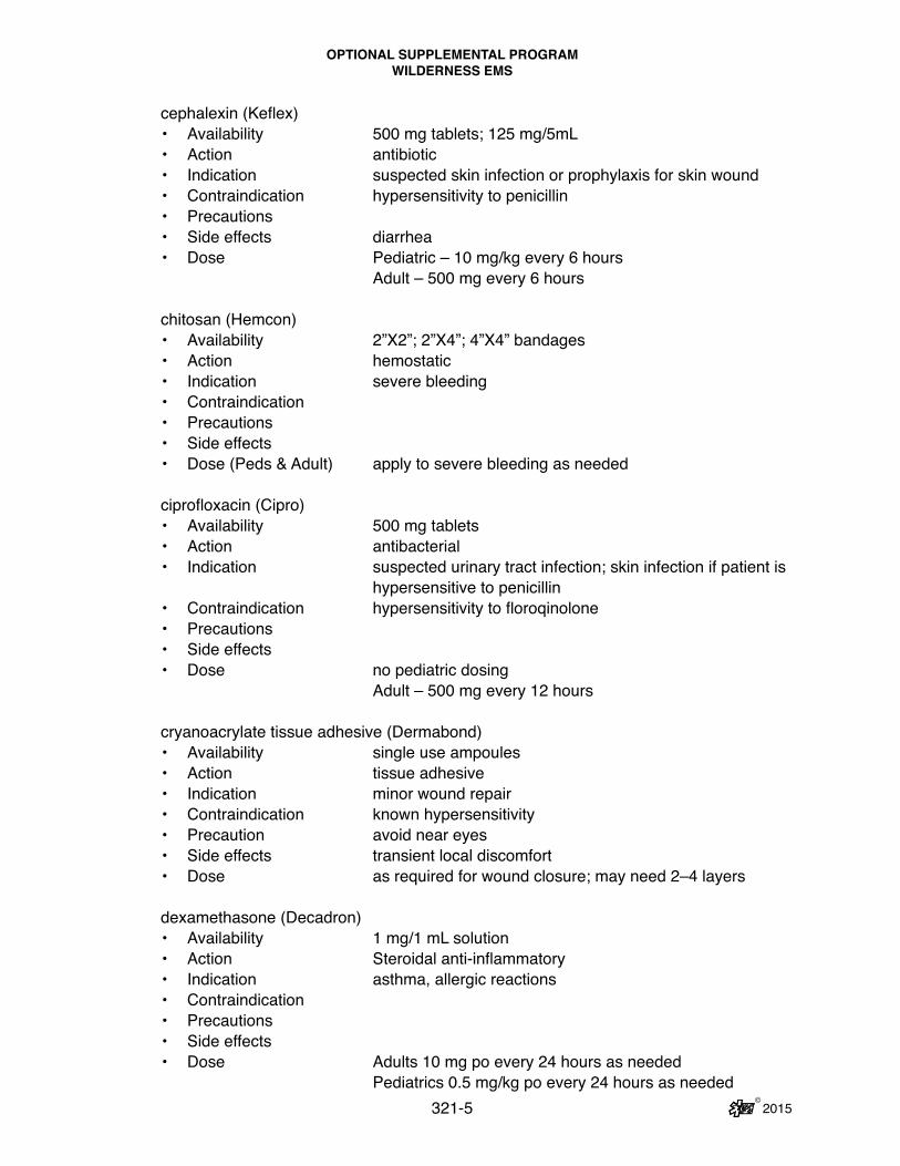
OPTIONAL SUPPLEMENTAL PROGRAMWILDERNESS EMS
© 2015 321-5
cephalexin (Keflex)• Availability 500 mg tablets; 125 mg/5mL• Action antibiotic• Indication suspected skin infection or prophylaxis for skin wound• Contraindication hypersensitivity to penicillin• Precautions • Side effects diarrhea• Dose Pediatric – 10 mg/kg every 6 hours Adult – 500 mg every 6 hours
chitosan (Hemcon)• Availability 2”X2”; 2”X4”; 4”X4” bandages• Action hemostatic• Indication severe bleeding• Contraindication • Precautions • Side effects • Dose (Peds & Adult) apply to severe bleeding as needed
ciprofloxacin (Cipro)• Availability 500 mg tablets• Action antibacterial• Indication suspected urinary tract infection; skin infection if patient is hypersensitive to penicillin• Contraindication hypersensitivity to floroqinolone• Precautions • Side effects • Dose no pediatric dosing Adult – 500 mg every 12 hours
cryanoacrylate tissue adhesive (Dermabond)• Availability single use ampoules• Action tissue adhesive• Indication minor wound repair • Contraindication known hypersensitivity• Precaution avoid near eyes• Side effects transient local discomfort• Dose as required for wound closure; may need 2–4 layers
dexamethasone (Decadron)• Availability 1 mg/1 mL solution• Action Steroidal anti-inflammatory• Indication asthma, allergic reactions• Contraindication• Precautions• Side effects• Dose Adults 10 mg po every 24 hours as needed Pediatrics 0.5 mg/kg po every 24 hours as needed

OPTIONAL SUPPLEMENTAL PROGRAMWILDERNESS EMS
© 2015 321-6
diphenhydramamine (Benadryl)• Availability 25 mg tablets; 12.5 mg/5 mL• Action antihistamine• Indication allergic reactions• Contraindication • Precautions • Side effects sedating• Dose Pediatric – 1 mg/kg to max dose 50 mg every 8 hours Adult – 25–50 mg every 8 hours as needed
doxycycline (Doxy)• Availability 100 mg tablets; 25 mg/5 mL• Action antibacterial• Indication suspected respiratory infection with contraindication to Augmentin• Contraindication • Precautions • Side effects • Dose 8–14 years old - 2.2 mg/kg every 12 hours Adults – 100 mg every 12 hours
epinephrine auto-injector (EpiPen)• Availability 0.3 mg (EpiPen); 0.15 mg (EpiPenJr) auto-injector• Action antihistamine; anti-inflammatory; vasoconstrictor• Indication moderate to severe allergic reaction • Contraindication • Precautions • Side effects tachycardia; hypertension• Dose Pediatric < 30 kg estimated weight – 0.15 mg IM > 30 kg estimated weight and adults – 0.3 mg IM
fentanyl• Availability prefilled syringe, multidose vial• Action opioid analgesic• Indication severe pain • Contraindication • Precautions • Side effects depressed level of consciousness; hypoxia; hypotension• Dose 1 mcg/kg IN/IV/IM to a max dose of 200 mcg with a repeat
dose of 1 mcg/kg to a max dose of 200 mcg every 1 hour as needed

OPTIONAL SUPPLEMENTAL PROGRAMWILDERNESS EMS
© 2015 321-7
glucagon• Availability 1 mg injector • Action facilitates release of glucose from glycogen stores in the liver• Indication suspected hypoglycemia in patient that is not able to take oral glucose• Contraindication • Precautions • Side effects • Dose Pediatric < 25 kg – 0.5 mg IM > 25 mg and adults – 1 mg IM
glucose gel (Glutose 15)• Availability 15 grams oral gel• Action raises blood glucose levels• Indication suspected hypoglycemia• Contraindication • Precautions use caution in patient with depressed level of consciousness• Side effects • Dose (Peds & Adult) give to patient by mouth in patient with depressed level of consciousness, rub the gel on the patient’s gums, but use caution
ibuprofen (Advil; Motrin)• Availability 200 mg; 400 mg; 600 mg; 40 mg/mL• Action anti-inflammatory; analgesic• Indication mild to moderate pain• Contraindication hypersensitivity; known renal disease; history of GI bleeding• Precautions • Side effects • Dose Pediatric – 10 mg/kg to max dose 600 mg every 6 hours
as needed Adult – 200 mg–600 mg every 6 hours as needed
loperamide (Immodium)• Availability 2 mg tablets• Action anti-diarrheal• Indication diarrhea• Contraindication • Precautions • Side effects constipation• Dose Pediatric – 2 mg after first watery stool, then 1 mg after each subsequent watery stool; max dose 8 mg per day Adult – 4 mg after first watery stool; then administer 2 mg after each subsequent watery stool; max dose 16 mg per day

OPTIONAL SUPPLEMENTAL PROGRAMWILDERNESS EMS
© 2015 321-8
metaclopramide (Reglan)• Availability 10 mg tablets; 5 mg/gmL• Action anti-emetic• Indication nausea and vomiting• Contraindication • Precautions • Side effects • Dose Pediatric – 0.1 mg/kg every 8 hours as needed Adult – 10 mg every 8 hours as neededmorphine• Availability 4 mg carpujet• Action opiate analgesic• Indication severe pain• Contraindication • Precautions • Side effects depressed level of consciousness; hypoxia; hypotension• Dose Pediatric – 0.1 mg/kg IM every hour as needed Adult – 4 mg IM every hour as needed
ondansetron (Zofran)• Availability 4 mg injectable solution• Action anti-emetic• Indication severe nausea and vomiting• Contraindication • Precautions • Side effects • Dose Pediatric – 0.1 mg/kg IM every 1 hour as needed up to
max dose 16 mg/day Adult – 4 mg IM every 1 hour as needed up to max dose of 32 mg/day
oxycodone• Availability 5 mg tablet• Action opiate analgesic• Indication moderate to severe pain• Contraindication • Precautions • Side effects depressed level of consciousness• Dose Pediatric – 0.05–0.15 mg/kg every 6 hours Adult – 1–2 tablets by mouth every 4 hours as needed

OPTIONAL SUPPLEMENTAL PROGRAMWILDERNESS EMS
© 2015 321-9
promethazine (Phenergan)• Availability 25 mg tablets; 6.25/5 mL• Action anti-emetic• Indication mild to moderate nausea• Contraindication • Precautions • Side effects • Dose Pediatric – 0.5 mg/kg every 8 hours as needed Adult – 25 mg every 8 hours by mouth as needed
tetracaine • Availability 0.5% ophthalmic solution• Action topical anesthetic• Indication severe eye pain; foreign body removal from the eye• Contraindication hypersensitivity • Precautions • Side effects • Dose (Peds & Adult) 2 drops to the affected eye
trimethoprim/sulfamethoxazole (Bactrim)• Availability 160 mg TMP/800 mg SMX (DS tab); 40 mg/200 mg/5 mL• Action antibiotic• Indication sinus infection, upper respiratory infection, urinary tract infection• Contraindication hypersensitivity to sulfa • Precautions• Side effects• Dose Pediatric – 5 mg/kg TMP every 12 hours Adult – 1 DS tab po bid

THIS PAGE IS INTENTIONALLY BLANK
© 2015321-10

322 © 2015
OPTIONAL SUPPLEMENTAL PROGRAMMARYLAND VACCINATION & TESTING
PROGRAM FOR PARAMEDIC PROVIDERS
Y. MARYLAND VACCINATION & TESTING PROGRAM Scope of practice for paramedic personnel has been expanded to allow select im-
munization and Purified Protein Derivative (PPD) testing by paramedic personnel. The immunizations that are allowed to be performed include Hepatitis B, Influenza, and PPD. This program is a jurisdictional option requiring the jurisdictional medical director and the jurisdiction to authorize select trained paramedic personnel to per-form these functions. There are program requirements below. Please note that you must have a written memorandum of understanding between your EMS service and the local health department before this program can be instituted. In order to become recognized and authorized to implement the immunization and testing program for paramedics, you must complete the application and submit a copy of the health department memorandum of understanding to the Office of the State EMS Medical Director. At that time you will receive a copy of the CD-ROM that has all of the pertinent documents and instructional material, along with a CDC videotape on PPD placement and interpretation. Your jurisdiction will then be rec-ognized as an authorized optional immunization and testing jurisdiction. When you are implementing this program, we strongly encourage you to advise EMS personnel at risk to seek vaccination where possible. REQUIREMENTS:1. Medical Director: Must have a jurisdictional Medical Director who is willing to
take responsibility for the program. 2. Must be under the Infection Control Program for the Jurisdiction.
3. Immunization record form with documentation of all pertinent information about vaccination or test, including the patient’s primary care practitioner.
4. Direct linkage with occupational medicine/employee health and a memoran-dum of understanding (MOU) with local public health service/department.
5. Statewide protocol approved by the EMS Board. 6. ALS resuscitation equipment (refer to The Maryland Medical Protocols for
EMS Providers) must be available on-site during vaccinations. 7. Must use the comprehensive training curriculum developed by MIEMSS In-
fection Control Committee. 8. Physician does not have to be physically present for the administration of
vaccinations or tests by the trained paramedic (Vaccination and Testing Of-ficer (VTO)).
9. Program instruction must be directed by and have participation by the juris-dictional Medical Director to select paramedics who will become the VTOs.
10. This is not for post-exposure prophylaxis (patient must be seen by occupa-tional medicine/physician for consent and treatment).
11. Only Public Safety Personnel (any career or volunteer member of a fire, res-cue, or EMS department, company, squad, or auxillary; any law enforcement officer; or the State Fire Marshal or sworn member of the State Fire Marshal’s office) are eligible to receive immunizations or testing from VTOs.

323 © 2015
OPTIONAL SUPPLEMENTAL PROGRAMMARYLAND VACCINATION & TESTING
PROGRAM FOR PARAMEDIC PROVIDERS
12. Mechanism for meeting FDA storage and refrigeration standards for vaccines and testing with the use of the Maryland Inventory Control Sheet.
13. Mechanism for follow-up a) For additional vaccinations for completion of series b) For potential complications of vaccinations or symptoms noted on adverse event form (meeting federal reporting requirements) c) Patient contact phone number for complications (e.g., bad vaccine “lot”)
14. Must have a standardized informed consent form and standardized vaccine pre-screening questionnaire form.
15. Vaccinations allowable are: a) Influenza b) Hepatitis B
16. Testing a) PPD Screening (Intradermal)
17. Recommend 30-minute observation period (to be determined by the jurisdic-tional medical director) post-immunization administration with ALS personnel and equipment available.
THE GOVERNOR’S ORIGINAL EXECUTIVE ORDER 01.01.2009.15 WAS RENEWED BY EXECUTIVE ORDER 01.01.2009.19 EXTENDING IT TO JANUARY 10, 2010. SO LONG AS THAT ORDER AND ANY RENEWAL OR REISSUE THEREOF REMAINS IN EFFECT, 18),19), AND 20) OF THIS PROTOCOL WILL BE IN EFFECT.
18. Cardiac Rescue Technicians who have been trained by the EMS Operational Program and credentialed by the Medical Director may vaccinate public safety personnel, health care providers, and members of the public with H1N1 (Swine) flu vaccine (LAIV and IM injection) after appropriate screening by a VTO, regis-tered nurse, or physician.
19. Vaccination and Testing Officers and Cardiac Rescue Technicians are permit-ted to vaccinate public safety personnel, health care providers, and members of the public with H1N1 (Swine) flu vaccine (LAIV and IM injection) at points of distribution that have been established or approved by local health departments (e.g., a clinic, occupational health site, a fire house, or other location).
20. Screening, administration, tracking, and dosage requirements for H1N1 (Swine) flu vaccine (LAIV and IM injection) shall be provided by the Maryland Depart-ment of Health and Mental Hygiene and/or local health departments.

324 © 2015
OPTIONAL SUPPLEMENTAL PROGRAMMARYLAND VACCINATION & TESTING PROGRAM FOR PARAMEDIC PROVIDERS
REFERENCE SHEET
HEPATITIS B VACCINATION Indications:
Pre-exposure: preventive
Contraindications: History of anaphylactic reaction to baker’s yeast
Adverse effects: Not clinically significant
Precautions: (1) Recipients must read and sign consent form. (2) CDC recommends antibody testing 1–2 months after the third dose to
determine immunity.
Dose: (three total, using a 3 mL syringe with 1” 25 gauge needle)
Initial 1 mL IM (deltoid) 2nd dose 4 weeks after initial; 1 mL IM (deltoid) 3rd dose 5–6 months after 2nd dose; 1 mL IM (deltoid)
INFLUENZA VACCINATION
Indications: (1) Persons who attend to patients at high risk for complications (e.g., the
elderly) (2) Persons with chronic medical conditions (3) Pregnant women who will be in the second or third trimester of pregnancy
during influenza season (4) Providers of essential community services
Contraindications: History of anaphylactic hypersensitivity to eggs
Adverse effects: (1) More common: soreness at the injection site that lasts up to 2 days (2) Less common: fever, malaise, myalgia beginning 6–12 hours after
vaccination and persisting for 1 to 2 days.
Precautions: (1) Vaccine should be delayed in the presence of acute febrile illness; admin-
ister after symptoms have abated. (2) It takes two weeks to develop adequate antibodies against the vaccine
virus strain. (3) Optimal time for organized vaccination campaigns is usually the period
from October through mid-November.

325 © 2015
OPTIONAL SUPPLEMENTAL PROGRAMMARYLAND VACCINATION & TESTING PROGRAM FOR PARAMEDIC PROVIDERS
REFERENCE SHEET
(4) Because influenza vaccine contains only noninfectious viruses, it cannot cause influenza.
(5) Recipients must read and sign consent or refusal form.
Dose: (using a 3 mL syringe with 1” 25 gauge needle)0.5–1 mL IM (deltoid)
PURIFIED PROTEIN DERIVATIVE (PPD) TEST
Indications: Yearly administration for healthcare providers
Contraindications: (1) Previous positive reaction to PPD (2) History of TB
Adverse effects: Not clinically significant
Precautions: Recipients must read and sign consent form.
Procedure (1) Injection is given intradermally and should be read 48–72 hours post
injection. (2) Feel the induration with your finger tips.
(3) Measure with approved device in millimeters (mm). (a) Less than 5 mm is negative. (b) Equal to or greater than 5 mm requires clinical correlation and evaluation
by jurisdictional medical director or other appropriate physician.
Note: Do not use erythema as margins; measure only the induration.

THIS PAGE IS INTENTIONALLY BLANK
326 © 2015

327 © 2015
EMS OPERATIONAL PROGRAM RESEARCH PROTOCOLOPTIONAL SUPPLEMENTAL PROTOCOL (RESEARCH)
SUBSTANCE ABUSE SCREENING
VI. RESEARCH PROTOCOLS Z. EMS LINKAGE TO ADDICTION TREATMENT (NEW ’15)
EMS providers will respond to requests for assistance and perform interventions in accordance with General Patient Care (GPC) of The Maryland Medical Protocols for EMS Providers. Following completion of the initial patient assessment and appropriate treatment interventions, a qualified EMS provider will assess patients for eligibility. Inclusion Criteria:1) Age greater than or equal to18 years 2) Hemodynamically stable (HR < 120 or > 60, SBP > 90, and RR < 30 or > 10) 3) Awake and oriented or person with a GCS greater than or equal to 14 Patients will be asked a series of targeted questions that screen for substance abuse:
EMS Provider: In the past 3 months:1. Has your partner or family complained about your drug or alcohol use? Y / N 2. Have you had an alcoholic drink, or used drugs, first thing in the morning to
steady your nerves or get rid of a hangover? Y / N3. Have you had black-outs when you couldn’t remember things the next day as
a result of your drug or alcohol use? Y / N4. Have you experienced withdrawal symptoms (felt sick) when you stopped
taking drugs or alcohol? Y / N

328 © 2015
5. If yes to #2, 3, or 4:a. Do these drugs include heroin? Y / N b. Do these drugs include prescription opiates (e.g., Vicodin, Percocet,
OxyContin)? Y / Ni. Where do you get these prescription opiates? ___________
(from the street, family/friends, medical provider)
If the scoring suggests no substance dependence, say: “Thanks. You’re not eligible for the study. However, if you or someone you know is interested in finding out more about addictions treatment programs, here is a card with treatment information.”
If scoring suggests substance dependence, deliver messages (see below) to bolster treatment entry and give them a study card for substance screening and HIV Volun-tary Counseling and Testing services at the study site. Messages to use include:
• “I’m really concerned about you and what your substance use is doing to your health.”
• “You must be really tired. Has anyone ever talked with you about drug treat-ment?”
• “I’ve seen many people in Baltimore who have been helped by drug treat-ment.”
• “I’m concerned about your health. Have you thought about drug treatment? It’s pretty easy to call for information about treatment slots.”
• “You look like you’re going through a lot right now. Do you think that drug treatment might help?”
• “Like with anything, the more you try to get treatment, the more likely it will work for you. It doesn’t hurt to keep trying.”
Then say: “It looks like you might be eligible for this paid study, if you are interested, call in the next week. Here is a study card. It has phone numbers to get information about the study and about treatment programs.”
EMS OPERATIONAL PROGRAM RESEARCH PROTOCOLOPTIONAL SUPPLEMENTAL PROTOCOL (RESEARCH)
SUBSTANCE ABUSE SCREENING

329 © 2015
EMS Linkage to Addiction Treatment: Referral Card
EMS OPERATIONAL PROGRAM RESEARCH PROTOCOLOPTIONAL SUPPLEMENTAL PROTOCOL (RESEARCH)
SUBSTANCE ABUSE SCREENING

THIS PAGE IS INTENTIONALLY BLANK
330 © 2015

331 © 2015
EMS OPERATIONAL PROGRAM RESEARCH PROTOCOLOPTIONAL SUPPLEMENTAL PROTOCOL (RESEARCH)
MARYLAND STATE POLICE AVIATION COMMAND
Z1. PREHOSPITAL POINT OF CARE TESTING FOR SHOCK PILOT PROGRAM (NEW ’15) 1. Prehospital Point of Care Testing for Shock Pilot Program
a. Indications i. Category A patients WITH
1) Availability of thenar eminence and site for venous cannulation in AT LEAST ONE extremity (i.e., no patients with bilateral upper extremity amputations)
ii. Category B patients WITH1) Availability of thenar eminence and site for venous cannulation
in AT LEAST ONE extremity (i.e., no patients with bilateral upper extremity amputations) AND
2) Two or more long-bone fractures AND/OR 3) Chest wall instability or deformity AND/OR 4) Open or depressed skull fractures AND/OR 5) Penetrating injuries to head/neck/torso/extremities AND/OR 6) Pelvic fracture AND/OR 7) Paralysis
iii. Category C patients WITH1) Availability of thenar eminence and site for venous cannulation
in AT LEAST ONE extremity (i.e., no patients with bilateral upper extremity amputations) AND
2) Patients in high risk auto crashes including any of the following: a. Intrusion greater than 12 inches on the occupant side b. Intrusion greater than 19 inches on the passenger side c. Ejection from the vehicle d. Death in the same passenger compartment e. Rollover without restraint f. Vehicle telemetry consistent with high risk of injury g. Auto vs. pedestrian/bicyclist thrown/run over with impact
greater than 20 MPH h. Motorcycle crash with speed greater than 25 MPH
iv. NOTE: STANDARD PATIENT CARE, AS SPECIFIED IN THE MARY-LAND MEDICAL PROTOCOLS, WILL TAKE PRIORITY AT ALL TIMES. THIS PILOT PROGRAM MUST NOT INTERRUPT STAN-DARD PATIENT CARE AT ANY TIME.

332 © 2015
EMS OPERATIONAL PROGRAM RESEARCH PROTOCOLOPTIONAL SUPPLEMENTAL PROTOCOL (RESEARCH)
MARYLAND STATE POLICE AVIATION COMMAND
b. Contraindicationsi. Pregnant patientsii. Prisonersiii. Patients under the age of 18iv. Patients dead on arrivalv. Patients who will not be transported to the Shock Trauma Center vi. Patients with upper extremity amputations, crushed, degloved, mangled,
or pulseless upper extremities that preclude application of the InSpec-tra™ tissue oximeter and/or placement of a venous catheter (note: ve-nous catheters may be placed in lower extremities)
c. Proceduresi. I-STAT device
1) A 2 mL blood sample will be obtained via a new venous cannula or closed vacutainer technique.
2) ONLY PROVIDERS WHO HAVE BEEN FORMALLY TRAINED AND SIGNED OFF ON THIS PROTOCOL, AND ARE CURRENT AS PER QUALITY CONTROL REQUIREMENTS (SEE SECTION BELOW), MAY UTILIZE THIS DEVICE.
3) ONLY THE GLUCOSE RESULT MAY BE USED FOR PATIENT CARE (IN LIEU OF GLUCOMETER TESTING AS PER THE MARYLAND MEDICAL PROTOCOLS), ALL OTHER RESULTS ARE FOR RE-SEARCH PURPOSES ONLY.
4) Turn the device ON.a. Do NOT insert the cartridge to start the device.b. Do NOT open the cartridge pouch before scanning the barcode.
5) Press “2” for I-STAT.a. If Quality Check Codes “69,” “140,” or “147” appear, STOP the test
and this pilot protocol.6) Follow prompts.7) Scan the lot number on the cartridge pouch.
a. Position barcode 3–9 inches from scanner window on the device.b. Press and hold “SCAN” to activate the scanner.c. Align the red laser light so it covers the entire barcode.d. The device will beep when it reads the barcode successfully.e. If the cartridge pouch does not have a barcode, enter the lot num-
ber manually using the numbered keys or press “ENT” to bypass this prompt.
8) Obtain the blood sample.9) Fill and seal the cartridges with the blood sample.
a. Cartridge priority: The CG4+ cartridge should be analyzed first (lactate, venous blood gas); time permitting, the EC8+ cartridge should be analyzed next (electrolytes, hemoglobin, glucose, base excess).

333 © 2015
EMS OPERATIONAL PROGRAM RESEARCH PROTOCOLOPTIONAL SUPPLEMENTAL PROTOCOL (RESEARCH)
MARYLAND STATE POLICE AVIATION COMMAND
10) Push the sealed cartridge (CG4+ first) into the device port until it clicks into place.
11) Wait for the test to complete.12) Repeat the procedure above for the next cartridge (EC8+).13) Only the glucose result may be used for patient care.14) Upon arrival at the Shock Trauma Center, the results should be print-
ed after the patient has been handed off to the trauma team. Only the glucose results may be shared with the hospital staff.a. Place the device in the cradle of an IR Link or align the IR win-
dows of the handheld and the printer.b. Turn the printer ON.c. Press “PRT” for the displayed test records on the device.
i. If the result is stored, select “Print Results” from the “Stored Results” menu. Select records to be printed by pressing the key(s) corresponding to the numbers beside the record(s). Press the “PRT” key to print the selected record(s).
ii. InSpectra™ Spot Check Tissue Oximeter1) ONLY PROVIDERS WHO HAVE BEEN FORMALLY TRAINED AND
SIGNED OFF ON THIS PROTOCOL, AND ARE CURRENT AS PER QUALITY CONTROL REQUIREMENTS (SEE SECTION BELOW), MAY UTILIZE THIS DEVICE.
2) Ensure the device is properly charged before use.3) Attach the optical cable to the Spot Check device.4) Connect the reusable StO2 clip by connecting the prongs to the clip on
the optical cable.5) Press the POWER ON button on the front of the device. a. The device should power on within 30 seconds.6) Cancel the SYSTEM CHECK button (see quality control procedures
below) by pressing the SYSTEM CHECK button again to return to the main screen.
7) Position the clip over the patient’s thenar eminence.a. The device may only be used on clean, dry skin.b. If the skin is soiled, the skin should be cleaned with an alcohol or
chlorhexidine prep.c. Ensure the clip is placed over the fleshy part of the thenar mus-
cles.8) Check THI signal strength on the main screen.
a. A THI greater than 5.0 indicates significant hemoglobin to obtain a tissue oximetry measurement.
9) Record the tissue oximetry level (%) on the study collection data form. Do not leave the clip on the thenar eminence for more than 15 min-utes.

334 © 2015
EMS OPERATIONAL PROGRAM RESEARCH PROTOCOLOPTIONAL SUPPLEMENTAL PROTOCOL (RESEARCH)
MARYLAND STATE POLICE AVIATION COMMAND
d. Quality Control Proceduresi. I-STAT Device
1) Daily proceduresa. A quality control log will be maintained and reviewed by the Medi-
cal Director for all pilot program sites.b. The device must be tested every 24 hours using the external Elec-
tronic Simulator.c. Insert the external Electronic Simulator after the LCK or “Simulator
Locked” message disappears from the display screen.d. A “PASS” must be displayed on the screen. The date and time of
this result must be logged. e. If “FAIL” is displayed on the screen, repeat the procedure with the
same Electronic Simulator. If “PASS” is displayed during the sec-ond test, the device is ready for use. If “PASS” is not displayed, the device cannot be used and the Medical Director or Primary Investigator must be notified immediately.
f. Verify that all cartridges are refrigerated and within the expiration date printed on the boxes. Cartridges cannot be left unrefrigerated for longer than the time specified on the box for the cartridges. If any cartridges are expired, set the cartridges aside, do not use them, and contact the Medical Director or Primary Investigator.
2) Monthly proceduresa. A quality control log will be maintained and reviewed by the Medi-
cal Director for all pilot program sites.b. This may also be accomplished at the Shock Trauma Center. c. Click on the “Simulator Viewer” button on the Electronic Simulator
and record the result in the quality control log. ii. InSpectra™ Spot Check Device
1) Daily proceduresa. A quality control log will be maintained and reviewed by the Medi-
cal Director for all pilot program sites.b. The device should be checked daily.c. Power on the device.d. Press the “SYSTEM CHECK” button.e. In 20 seconds or less, the device should read GREEN with a
checkmark indicating that the device is ready for use.f. If the device is out of range, the FAIL icon will remain on the
screen and the System Check screen will be RED. Do not use the device if the SYSTEM CHECK fails. Place the device out of service and contact the Medical Director or Primary Investigator.