MAXIM BIOMEDICAL, INC. - David...
24
MAXIM BIOMEDICAL, INC. HUMAN IMMUNODEFICIENCY VIRUS TYPE 1 (HIV-1) CAMBRIDGE BIOTECH HIV-1 WESTERN BLOT KIT For Detection of Antibodies to HIV-1 (Cat. No. 98002) NAME AND INTENDED USE The Cambridge Biotech HIV-1 Western Blot Kit is an in vitro qualitative assay for the detection and identification of antibodies to Human Immunodeficiency Virus Type 1 (HIV-1), contained in human serum or plasma. It is intended for use with persons of unknown risks as an additional specific test on human serum or plasma specimens found to be repeatedly reactive using a screening procedure, such as an Enzyme-Linked Immunosorbent Assay (ELISA). It is also intended for use as an additional specific test to be used with serum or plasma specimens obtained from subjects found to be reactive using rapid HIV-1 tests. SUMMARY AND EXPLANATION OF TEST The Enzyme-Linked Immunosorbent Blot Technique (“Western Blot”) 1,2 has been used to detect antibodies to Human Immunodeficiency Virus Type 1 (HIV-1), recognized as the etiologic agent of the Acquired Immunodeficiency Syndrome (AIDS). 2,3 The combination of electrophoretic separation of complex mixtures of antigens with the highly sensitive immunoblotting technique has been useful in characterizing the antigenic profile of HIV-1 and describing the immune response to this virus in exposed or infected persons. The Cambridge Biotech HIV-1 Western Blot Kit, when used as directed in this insert, will detect antibodies to HIV-1 when present in human serum or plasma. The position of bands on the nitrocellulose strips allows this antibody reactivity to be associated with specific viral antigens. Persons demonstrating antibodies to HIV-1 should be referred for medical evaluation, which may include testing by other techniques. A clinical diagnosis of AIDS can be made only if a person meets the case definition of AIDS established by the Centers for Disease Control. 4 1
Transcript of MAXIM BIOMEDICAL, INC. - David...
MAXIM BIOMEDICAL, INC.
HUMAN IMMUNODEFICIENCY VIRUS TYPE 1 (HIV-1)
CAMBRIDGE BIOTECH HIV-1 WESTERN BLOT KIT
For Detection of Antibodies to HIV-1 (Cat. No. 98002)
NAME AND INTENDED USE The Cambridge Biotech HIV-1 Western Blot Kit is an in vitro qualitative assay for the detection and identification of antibodies to Human Immunodeficiency Virus Type 1 (HIV-1), contained in human serum or plasma. It is intended for use with persons of unknown risks as an additional specific test on human serum or plasma specimens found to be repeatedly reactive using a screening procedure, such as an Enzyme-Linked Immunosorbent Assay (ELISA). It is also intended for use as an additional specific test to be used with serum or plasma specimens obtained from subjects found to be reactive using rapid HIV-1 tests. SUMMARY AND EXPLANATION OF TEST The Enzyme-Linked Immunosorbent Blot Technique (“Western Blot”)1,2 has been used to detect antibodies to Human Immunodeficiency Virus Type 1 (HIV-1), recognized as the etiologic agent of the Acquired Immunodeficiency Syndrome (AIDS).2,3 The combination of electrophoretic separation of complex mixtures of antigens with the highly sensitive immunoblotting technique has been useful in characterizing the antigenic profile of HIV-1 and describing the immune response to this virus in exposed or infected persons. The Cambridge Biotech HIV-1 Western Blot Kit, when used as directed in this insert, will detect antibodies to HIV-1 when present in human serum or plasma. The position of bands on the nitrocellulose strips allows this antibody reactivity to be associated with specific viral antigens. Persons demonstrating antibodies to HIV-1 should be referred for medical evaluation, which may include testing by other techniques. A clinical diagnosis of AIDS can be made only if a person meets the case definition of AIDS established by the Centers for Disease Control.4
1
CHEMICAL AND BIOLOGICAL PRINCIPLES OF THE PROCEDURE The Cambridge Biotech HIV-1 Western Blot Kit is manufactured by Maxim Biomedical Corporation from HIV-1 propagated in an H9/HTLV-IIIB T-Lymphocyte cell line.3 The partially purified virus is inactivated by treatment with psoralen and ultraviolet light, and detergent disruption. Specific HIV-1 proteins are fractionated according to molecular weight by electrophoresis on a polyacrylamide slab gel in the presence of sodium dodecylsulfate (SDS). The separated HIV-1 proteins are electrotransferred from the gel to a nitrocellulose membrane which is then washed, blocked (to minimize nonspecific immunoglobulin binding), and packaged. Individual nitrocellulose strips are incubated with serum or plasma specimens or controls. During incubation, if HIV-1 antibodies are present in the specimen, they will bind to the viral antigens bound to the nitrocellulose strips. The strips are washed again to remove unbound material. Visualization of the human immunoglobulins specifically bound to HIV-1 proteins is accomplished in situ using a series of reactions with goat anti-human IgG conjugated with biotin, avidin conjugated with horseradish peroxidase (HRP), and the HRP substrate, 4-chloro-1-naphthol. If antibodies to any of the major HIV-1 antigens are present in the specimen in sufficient concentration, bands corresponding to the position of one or more of the following HIV-1 proteins (p) or glycoproteins (gp) will be seen on the nitrocellulose strip: p17, p24, p31, gp41, p51, p55, p66, gp120, gp160 (number refers to the apparent molecular weight in kilodaltons). REAGENTS Reagents for the Cambridge Biotech HIV-1 Western Blot Kit procedure include: 1. NITROCELLULOSE STRIPS - Each NITROCELLULOSE STRIP contains separated, bound
antigenic proteins from partially purified inactivated HIV-1, in sufficient quantity to detect human antibodies. Bovine protein is present as a blocking agent. Strips are consecutively numbered (1 through 27).
2. NON-REACTIVE CONTROL - Normal human serum non-reactive for HIV-1 antibodies and
hepatitis B surface antigen. Contains 0.1% Sodium Azide and 0.005% Thimerosal as preservatives.
3. WEAKLY REACTIVE CONTROL - Inactivated human serum containing a low titer of antibodies
to HIV-1 antigens and non-reactive for hepatitis B surface antigens. Contains 0.1% Sodium Azide and 0.005% Thimerosal as preservatives.
4. STRONGLY REACTIVE CONTROL - Inactivated human serum containing a high titer of
antibodies to HIV-1 antigens. Non reactive for hepatitis B surface antigen. Contains 0.1% Sodium Azide and 0.005% Thimerosal as preservatives.
5. WASH BUFFER - Supplied as a 20x concentrate. When diluted, contains 0.02 M tris,
0.1 M NaCl, 0.3% Tween 20 and 0.005% Thimerosal as a preservative, at pH 7.4. 6. BLOTTING BUFFER - Supplied as a 10x concentrate. When diluted, contains 0.02 M tris,
0.1 M NaCl, Heat-inactivated normal Goat serum, and 0.01% Thimerosal as a preservative, at pH 7.4.
2
7. CONJUGATE 1 - Biotinylated Goat Anti-human IgG (heavy and light chain) antibodies. Contains 0.002% thimerosal as a preservative.
8. CONJUGATE 2 - Avidin conjugated horseradish peroxidase. Contains 0.01% thimerosal as a
preservative. 9. SUBSTRATE A - 7.8 mM solution of 4-chloro-1-naphthol in an alcohol solution. 10. SUBSTRATE B - Aqueous hydrogen peroxide solution (0.02%) in citrate buffer. 11. BLOTTING POWDER - Nonfat dry milk. CAUTIONS 1. HANDLE ASSAY SPECIMENS, STRIPS, REACTIVE, AND NON-REACTIVE CONTROLS
AS IF CAPABLE OF TRANSMITTING AN INFECTIOUS AGENT. Inactivated HIV-1 antigen has been electrophoresed and transferred onto nitrocellulose. WEAKLY and STRONGLY REACTIVE CONTROLS have been inactivated by heat treatment. In addition, plasma used to produce the CONTROLS was shown to be non-reactive for hepatitis B surface antigen. However, no known test method can offer assurance that products derived from human blood will not transmit infectious agents. Therefore, these components must be handled as if they are capable of transmitting infectious agents.
2. Do not pipet by mouth. 3. Wear disposable gloves throughout the test procedure. Dispose of gloves as biohazard
waste. Thoroughly wash hands after handling test reagents. 4. Wipe spills promptly with a 1% sodium hypochlorite solution (1:5 dilution of liquid household
bleach). Contaminated materials should be disposed of as biohazard waste. 5. Dispose of all specimens and materials used in the Cambridge Biotech HIV-1 Western Blot
Kit procedure as biohazard waste. The recommended method of disposal is autoclaving for a minimum of 1 hour at 121°C. Disposable materials may be incinerated. Mix liquid wastes with an equal volume of 5% sodium hypochlorite solution (liquid household bleach), allowing at least 60 minutes for disinfection.
6. Do not permit SUBSTRATE, especially 4-chloro-1 naphthol, to contact the skin. If contact
occurs, flush with water. 7. The CONTROLS contain sodium azide as a preservative. If these materials, either
concentrated or diluted, are disposed of through a sink or other common plumbing systems, flush with generous amounts of water to prevent accumulation of potentially explosive compounds. In addition, consult the manual guideline, “Safety Management No. CDC-22, ‘Decontamination of Laboratory Sink Drains to Remove Azide Salts’” (Centers for Disease Control, Atlanta, GA, April 30, 1976).
8. Avoid use of metal instruments in contact with SUBSTRATE B and WORKING SUBSTRATE SOLUTION, since metals can cause reduction in H2O2.
3
PRECAUTIONS WARNING: FDA has licensed this test kit for use with serum or plasma. Use of this licensed test kit with specimens other than those specifically approved for use with this test kit may result in inaccurate test results. 1. DO NOT INTERCHANGE REAGENTS BETWEEN KIT LOTS. 2. Do not use reagents beyond kit expiration dating. The date is printed on kit boxes. 3. Avoid contamination of reagents, when opening and withdrawing aliquots from primary vials.
Keep all reagents refrigerated (2-8ºC) when not in use. 4. Do not interchange vial or bottle caps and stoppers; this will lead to cross contamination of
reagents. Designate specific reservoirs for specific reagents. 5. Grossly contaminated specimens or strips may result in the development of dark spots on the
strip which should not be interpreted. Careful attention must be given to the storage of specimens and kits to prevent this problem.
6. Shield WORKING SUBSTRATE SOLUTION from sunlight during preparation and use within
30 minutes of mixing. 7. Use reagent grade water (deionized water which is bacteria-free) to dilute reagents in order
to avoid substances which may interfere with the assay. 8. Do not remove NITROCELLULOSE STRIPS from the storage tube until immediately before
use. To prevent moisture from condensing inside the strip tube, open only AFTER the strips have reached room temperature (approximately 30 minutes). Close the tube immediately after removing strips for use.
9. Allow all kit reagents and materials to reach room temperature before use (approximately 30
minutes). 10. Use only the CONTROLS supplied with the kit. 11. DO NOT CUT STRIPS. Narrower strips can lead to misinterpretation because strips may flip
over in the incubation tray, or artifacts in the reaction zones may be mistaken for possible bands or may prevent recognition of positive bands.
12. Measure all reagents. Use extreme care and calibrated pipets with good quality tips when
preparing WORKING CONJUGATE SOLUTIONS. 13. Discard bleach solution in trap prior to substrate preparation.
4

PREPARATION OF REAGENTS NOTE: Allow reagents to reach room temperature before use (approximately 30 minutes). 1. DILUTED WASH BUFFER
a. Dilute 1 volume of WASH BUFFER (20x) with 19 volumes of reagent grade water. Mix well.
b. DILUTED WASH BUFFER may be stored at room temperature for 3 months. 2. WORKING BLOTTING BUFFER
a. WORKING BLOTTING BUFFER should be prepared and used within 5 days.
b. Dilute 1 volume of BLOTTING BUFFER (10x) with 9 volumes of reagent grade water. Mix well.
c. Use 1.0 g of BLOTTING POWDER per 20 mL of the diluted BLOTTING BUFFER prepared in Step 2b above. Mix thoroughly to dissolve the powder. If the entire kit is to be used within five days, add 9.0 g to 180 mL of the diluted BLOTTING BUFFER.
Store at 2-8°C.
3. WORKING CONJUGATE 1 SOLUTION
a. Refer to the SUPPLEMENTAL INSTRUCTIONS sheet for the dilution appropriate for the CONJUGATE 1 lot supplied with the kit.
b. WORKING CONJUGATE 1 SOLUTION should be prepared fresh prior to use. 4. WORKING CONJUGATE 2 SOLUTION
a. Refer to the SUPPLEMENTAL INSTRUCTIONS sheet for the dilution appropriate for the CONJUGATE 2 lot supplied with the kit.
b. WORKING CONJUGATE 2 SOLUTION should be prepared fresh prior to use. 5. WORKING SUBSTRATE SOLUTION
a. WORKING SUBSTRATE SOLUTION should be prepared fresh prior to use.
b. Prepare WORKING SUBSTRATE SOLUTION by mixing equal volumes of SUBSTRATE A and SUBSTRATE B. Mix well.
Reagents Required (in mLs)* for Various Number of Strips NUMBER OF STRIPS 1 3 6 9 15 20 27 DILUTED WASH BUFFER 20.0 60.0 120.0 180.0 300.0 400.0 540.0 BLOTTING POWDER 0.3g 0.9g 1.8g 2.7g 4.5g 6.0g 8.1g WORKING BLOTTING BUFFER 6.0 18.0 36.0 54.0 90.0 120.0 162.0 WORKING CONJUGATE 1** 2.0 6.0 12.0 18.0 30.0 40.0 54.0 WORKING CONJUGATE 2** 2.0 6.0 12.0 18.0 30.0 40.0 54.0 SUBSTRATE A 1.0 3.0 6.0 9.0 15.0 20.0 27.0 SUBSTRATE B 1.0 3.0 6.0 9.0 15.0 20.0 27.0 *Minimum volumes. Prepare a slight excess of each solution to compensate for loss during pipetting. ** See SUPPLEMENTAL INSTRUCTIONS sheet for Dilution Calculation.
5
STORAGE INSTRUCTIONS 1. Store Cambridge Biotech HIV-1 Western Blot Kits and/or individual reagents at 2-8ºC. 2. Unused NITROCELLULOSE STRIPS should be kept dry and in the dark, in their storage
tube, at 2-8ºC. SERUM AND PLASMA SPECIMEN COLLECTION AND PREPARATION 1. The Cambridge Biotech HIV-1 Western Blot Kit may be used with human serum or plasma.
Reliability of the test results with grossly lipemic, hemolyzed, or cloudy specimens is not known.
2. Specimens should be stored at 2-8ºC for up to two weeks. For longer intervals they should
be frozen (-18ºC or lower) prior to testing. 3. Avoid multiple freeze-thaw cycles. 4. Mix specimens well. Centrifuge if necessary to remove particulate matter prior to testing. 5. If specimens are shipped, they should be shipped in accordance with requirements for
transporting etiological agents. INDICATIONS OF INSTABILITY OR DETERIORATION OF REAGENTS Changes in the physical appearance of the reagents supplied may indicate instability or deterioration of these materials. SUBSTRATE A should be colorless. If SUBSTRATE A shows color, it has become oxidized and should not be used. KNOWN INTERFERING SUBSTANCES Sodium azide interferes with horseradish peroxidase activity.
6

MATERIALS PROVIDED
Each Cambridge Biotech HIV-1 Western Blot Kit contains:
• NITROCELLULOSE STRIPS 27 Strips
• NON-REACTIVE CONTROL 1 vial (Green) (160 μL minimum/vial)
• WEAKLY REACTIVE CONTROL 1 vial (Lavender) (160 μL minimum/vial)
• STRONGLY REACTIVE CONTROL 1 vial (Red) (160 μL minimum/vial)
• WASH BUFFER (20x) 1 Bottle (60 mL minimum/bottle)
• BLOTTING BUFFER (10x) 1 Bottle (18 mL minimum/bottle)
• CONJUGATE 1 1 vial (Blue) (160 μL minimum/vial)
• CONJUGATE 2 1 vial (Black) (160 μL minimum/vial)
• SUBSTRATE A 1 Bottle (30 mL minimum/bottle)
• SUBSTRATE B 1 Bottle (30 mL minimum/bottle)
• BLOTTING POWDER 1 Package (9.0 g minimum)
• INCUBATION TRAYS 3 Trays (9 Wells/Tray)
MATERIALS REQUIRED - NOT PROVIDED
• ROCKER OR ROTARY PLATFORM
• PIPETS
• PIPETTORS AND TIPS
• ASPIRATOR WITH DISINFECTANT TRAP
• TWEEZERS OR FORCEPS
ASSAY PROCEDURE NOTE: For serum and plasma testing, the Cambridge Biotech HIV-1 Western Blot Kit provides the option of being performed in either of two (2) different assay protocols, overnight or one day. CAUTION: When handling the incubation tray supplied with the kits, take care not to splash or mix specimens. Remove the lid carefully to prevent moisture which may condense on the lid from falling into the tray. Do not handle samples or sample loaded pipet tips over uncovered incubation trays. Splashing or aerosols may lead to cross-contamination of sample wells.
7
OVERNIGHT ASSAY PROCEDURE 1. Bring all reagents to room temperature prior to use (approximately 30 minutes). 2. Add 2.0 mL of DILUTED WASH BUFFER to each well to be used. 3. Using forceps, carefully remove a NITROCELLULOSE STRIP from the vial and place numbered
side up into a well containing DILUTED WASH BUFFER. 4. Place the tray on a rocker or rotary platform for 5 to 10 minutes at room temperature, then
remove the buffer by aspiration. 5. Add 2.0 mL of WORKING BLOTTING BUFFER to each well. 6. Add 20 μL of each undiluted specimen or control to a well containing its assigned strip in
WORKING BLOTTING BUFFER. CAUTION: Use a different pipet tip for each sample. 7. Cover the tray and incubate on a rocker or rotary platform overnight (14-20 hours) at room
temperature (20 to 28ºC). 8. Carefully uncover the tray to avoid splashing or mixing of specimens. Remove condensation or
droplets on the incubation tray lid by rinsing with DILUTED WASH BUFFER or wiping with absorbent towels.
9. Aspirate the mixture from the wells into a trap containing disinfectant. Rinse aspirator tip with
DILUTED WASH BUFFER, or deionized water between samples to avoid cross-contamination. 10. To each strip, add 2.0 mL of DILUTED WASH BUFFER and rock by hand several times.
Remove buffer by aspiration. 11. Add 2.0 mL of DILUTED WASH BUFFER to each strip for a minimum of 5 minutes. Repeat a
second time, aspirating the wash buffer before and after each 5 minute wash. Perform all wash steps at room temperature on a rocking or rotary platform.
12. Add 2.0 mL of WORKING CONJUGATE 1 SOLUTION (prepared as directed in the Overnight
Assay section of the SUPPLEMENTAL INSTRUCTIONS sheet) to each well. Incubate for 60 minutes at room temperature on the rocker or rotary platform.
13. Aspirate the Conjugate from the wells. Wash each strip three times for 5 minutes as in Step 11,
above. 14. Add 2.0 mL of WORKING CONJUGATE 2 SOLUTION (prepared as directed in the Overnight
Assay section of the SUPPLEMENTAL INSTRUCTIONS sheet) to each well. Incubate for 60 minutes at room temperature on the rocker or rotary platform.
15. Aspirate the Conjugate from the wells and wash each strip three times as in Step 11, above. 16. Add 2.0 mL of WORKING SUBSTRATE SOLUTION to each well and incubate at room
temperature on the rocker or rotary platform for 10 to 15 minutes (or until the weakly reactive control exhibits p24 and gp160 bands).
17. Aspirate the Substrate and stop the reaction by rinsing the strips 2 or 3 times with at least 2mL
of reagent grade water.
8

NOTE: Some specimens may cause spots to form on the strip due to precipitation. A cotton swab dipped in reagent grade water can be used to carefully remove the spots and allow for better visualization of results. Air dry the strips between absorbent paper towels and score as described in the Interpretation of Results section. For best results and consistency, strips should be scored soon after air drying. The strips can be mounted and stored between clear plastic sheets. When mounting with tape, do not tape over developed bands. This will cause bands to fade.
18. If desired, the strips may be photographed using high resolution film. Developed strips will
retain their color if stored in the dark. Exposure to light and air will eventually cause bands to fade.
9

ONE-DAY ASSAY PROCEDURE 1. Bring all reagents to room temperature prior to use (approximately 30 minutes). 2. Add 2.0 mL of DILUTED WASH BUFFER to each well to be used. 3. Using forceps, carefully remove a NITROCELLULOSE STRIP from the vial and place numbered
side up into a well containing DILUTED WASH BUFFER. 4. Place the tray on a rocker or rotary platform for 5 to 10 minutes at room temperature, then remove
the buffer by aspiration. 5. Add 2.0 mL of WORKING BLOTTING BUFFER to each well. 6. Add 30 μL of each undiluted specimen or control to a well containing its assigned strip in
WORKING BLOTTING BUFFER. CAUTION: Use a different pipet tip for each sample. 7. Cover the tray and incubate on a rocker or rotary platform for two (2) hours at room temperature
(20 to 28ºC). 8. Carefully uncover the tray to avoid splashing or mixing of specimens. Remove condensation or
droplets on the incubation tray lid by rinsing with DILUTED WASH BUFFER or wiping with absorbent towels.
9. Aspirate the mixture from the wells into a trap containing disinfectant. Rinse aspirator tip with
DILUTED WASH BUFFER, or deionized water between samples to avoid cross-contamination. 10. To each strip, add 2.0 mL of DILUTED WASH BUFFER and rock by hand several times. Remove
buffer by aspiration. 11. Add 2.0 mL of DILUTED WASH BUFFER to each strip for a minimum of 5 minutes. Repeat a
second time, aspirating the wash buffer before and after each 5 minute wash. Perform all wash steps at room temperature on a rocking or rotary platform.
12. Add 2.0 mL of WORKING CONJUGATE 1 SOLUTION (prepared as directed in the One Day Assay
section of the SUPPLEMENTAL INSTRUCTIONS sheet) to each well. Incubate for 60 minutes at room temperature on the rocker or rotary platform.
13. Aspirate the Conjugate from the wells. Wash each strip three times for 5 minutes as in Step 11,
above. 14. Add 2.0 mL of WORKING CONJUGATE 2 SOLUTION (prepared as directed in the One Day Assay
section of the SUPPLEMENTAL INSTRUCTIONS sheet) to each well. Incubate for 60 minutes at room temperature on the rocker or rotary platform.
15. Aspirate the Conjugate from the wells and wash each strip three times as in Step 11, above. 16. Add 2.0 mL of WORKING SUBSTRATE SOLUTION to each well and incubate at room
temperature on the rocker or rotary platform for 10 to 15 minutes (or until the weakly reactive control exhibits p24 and gp160 bands).
17. Aspirate the Substrate and stop the reaction by rinsing the strips 2 or 3 times with at least 2mL of
reagent grade water.
10
NOTE: Some specimens may cause spots to form on the strip due to precipitation. A cotton swab dipped in reagent grade water can be used to carefully remove the spots and allow for better visualization of results. Air dry the strips between absorbent paper towels and score as described in the Interpretation Of Results section. For best results and consistency, strips should be scored soon after air drying. The strips can be mounted and stored between clear plastic sheets. When mounting with tape, do not tape over developed bands. This will cause bands to fade.
18. If desired, the strips may be photographed using high resolution film. Developed strips will retain
their color if stored in the dark. Exposure to light and air will eventually cause bands to fade.
11
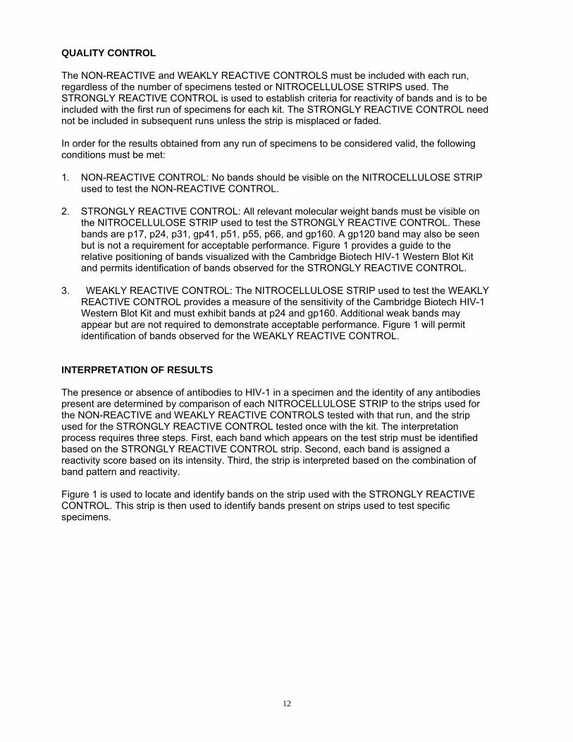
QUALITY CONTROL The NON-REACTIVE and WEAKLY REACTIVE CONTROLS must be included with each run, regardless of the number of specimens tested or NITROCELLULOSE STRIPS used. The STRONGLY REACTIVE CONTROL is used to establish criteria for reactivity of bands and is to be included with the first run of specimens for each kit. The STRONGLY REACTIVE CONTROL need not be included in subsequent runs unless the strip is misplaced or faded. In order for the results obtained from any run of specimens to be considered valid, the following conditions must be met: 1. NON-REACTIVE CONTROL: No bands should be visible on the NITROCELLULOSE STRIP
used to test the NON-REACTIVE CONTROL. 2. STRONGLY REACTIVE CONTROL: All relevant molecular weight bands must be visible on
the NITROCELLULOSE STRIP used to test the STRONGLY REACTIVE CONTROL. These bands are p17, p24, p31, gp41, p51, p55, p66, and gp160. A gp120 band may also be seen but is not a requirement for acceptable performance. Figure 1 provides a guide to the relative positioning of bands visualized with the Cambridge Biotech HIV-1 Western Blot Kit and permits identification of bands observed for the STRONGLY REACTIVE CONTROL.
3. WEAKLY REACTIVE CONTROL: The NITROCELLULOSE STRIP used to test the WEAKLY
REACTIVE CONTROL provides a measure of the sensitivity of the Cambridge Biotech HIV-1 Western Blot Kit and must exhibit bands at p24 and gp160. Additional weak bands may appear but are not required to demonstrate acceptable performance. Figure 1 will permit identification of bands observed for the WEAKLY REACTIVE CONTROL.
INTERPRETATION OF RESULTS The presence or absence of antibodies to HIV-1 in a specimen and the identity of any antibodies present are determined by comparison of each NITROCELLULOSE STRIP to the strips used for the NON-REACTIVE and WEAKLY REACTIVE CONTROLS tested with that run, and the strip used for the STRONGLY REACTIVE CONTROL tested once with the kit. The interpretation process requires three steps. First, each band which appears on the test strip must be identified based on the STRONGLY REACTIVE CONTROL strip. Second, each band is assigned a reactivity score based on its intensity. Third, the strip is interpreted based on the combination of band pattern and reactivity. Figure 1 is used to locate and identify bands on the strip used with the STRONGLY REACTIVE CONTROL. This strip is then used to identify bands present on strips used to test specific specimens.
12
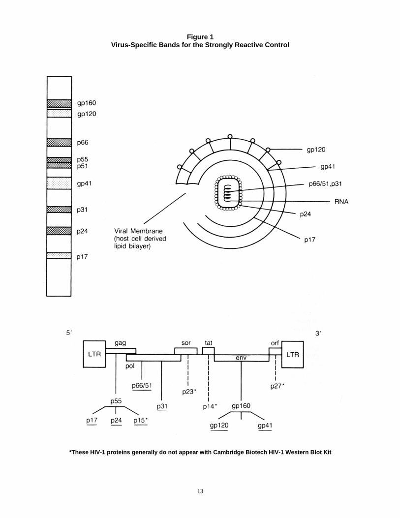
Figure 1 Virus-Specific Bands for the Strongly Reactive Control
*These HIV-1 proteins generally do not appear with Cambridge Biotech HIV-1 Western Blot Kit
13
The major HIV-1 gene products that have been identified5-10 are as follows (see Figure 1).
gp160 - Precursor of ENV glycoprotein gp120 - Outer ENV glycoprotein p66 - Reverse Transcriptase component of POL translate p55 - Precursor of GAG proteins p51 - Reverse Transcriptase component of POL translate gp41 - Transmembrane ENV glycoprotein p31 - Endonuclease component of POL translate p24 - GAG protein p17 - GAG protein
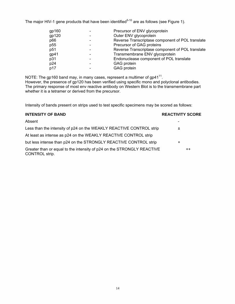
NOTE: The gp160 band may, in many cases, represent a multimer of gp4111. However, the presence of gp120 has been verified using specific mono and polyclonal antibodies. The primary response of most env reactive antibody on Western Blot is to the transmembrane part whether it is a tetramer or derived from the precursor. Intensity of bands present on strips used to test specific specimens may be scored as follows: INTENSITY OF BAND REACTIVITY SCORE
Absent -
Less than the intensity of p24 on the WEAKLY REACTIVE CONTROL strip ±
At least as intense as p24 on the WEAKLY REACTIVE CONTROL strip
but less intense than p24 on the STRONGLY REACTIVE CONTROL strip +
Greater than or equal to the intensity of p24 on the STRONGLY REACTIVE ++ CONTROL strip.
14
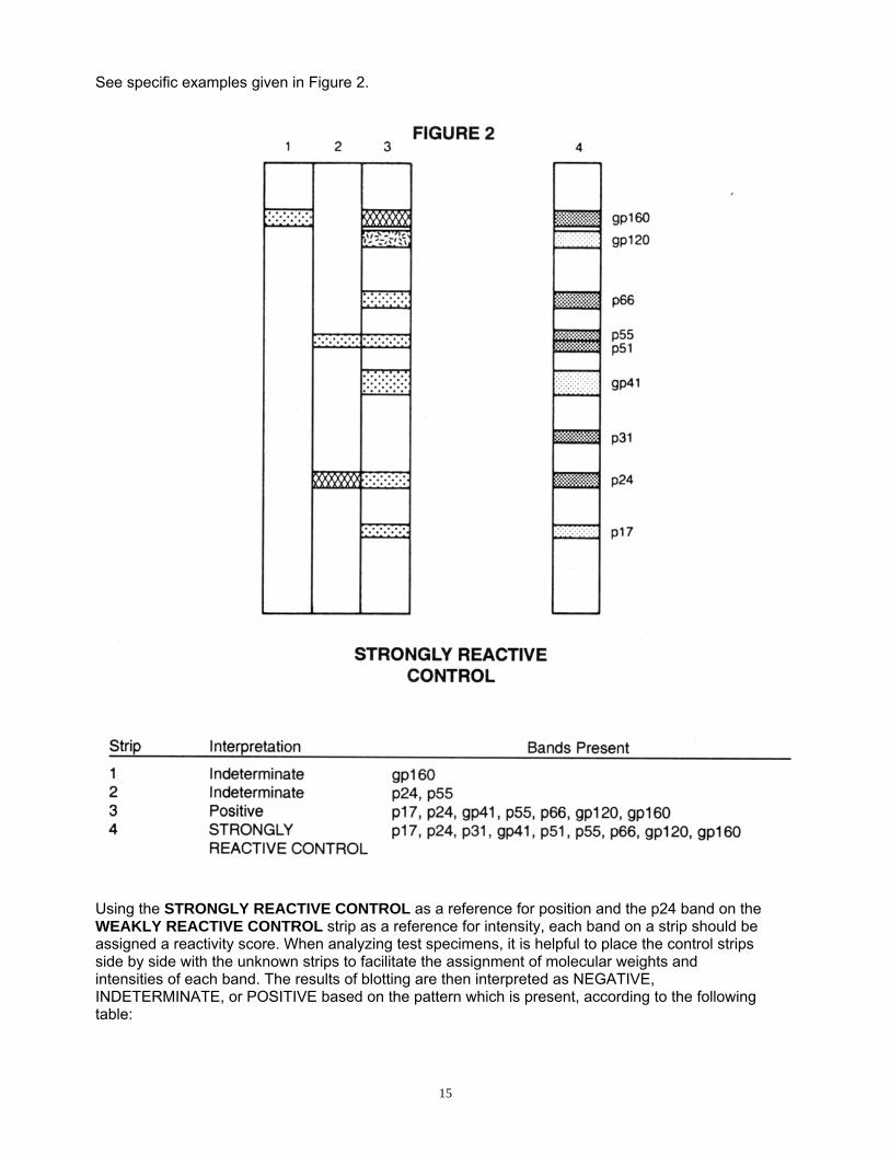
See specific examples given in Figure 2.
Using the STRONGLY REACTIVE CONTROL as a reference for position and the p24 band on the WEAKLY REACTIVE CONTROL strip as a reference for intensity, each band on a strip should be assigned a reactivity score. When analyzing test specimens, it is helpful to place the control strips side by side with the unknown strips to facilitate the assignment of molecular weights and intensities of each band. The results of blotting are then interpreted as NEGATIVE, INDETERMINATE, or POSITIVE based on the pattern which is present, according to the following table:
15

PATTERN INTERPRETATION No bands present. NEGATIVE Any bands present but pattern does not meet criteria for POSITIVE
IDETERMINATE
Any two or more of the following bands present: p24, gp41 and gp120/160. Each band has a reactivity score of + or greater. Commonly, the band at gp41 or gp160 is diffuse. Other viral bands may or may not be present.
POSITIVE
The positive criteria follow the recommendations of the Centers for Disease Control (CDC)12 and the Association of State and Territorial Public Health Laboratory Directors (ASTPHLD).13 These publications along with others14-16 have suggested that the additional requirement for p31 reactivity is unnecessary. Clinical studies with Cambridge Biotech HIV-1 Western Blot Kit have indicated that it is inappropriate to assign a POSITIVE interpretation to strips which display bands but lack any two of p24, gp41, or gp120/160 with a reactivity score of + or greater for each band present. It is known that persons who have recently seroconverted may display incomplete patterns but that they will develop increased reactivity (both number and intensity of bands) when followed for a period of four to six months. Most blots with POSITIVE results will have other virus-specific bands present including p17, p31, p51 p55, p66, and gp120. Conversely, persons at low risk for infection may have nonspecific reactions on the blot particularly in regions corresponding to p17, p24, p55 and p66, which will persist but which do not evolve into more extensive patterns over time. Although nonspecific reactivity may sometimes be attributed to autoantibodies, it is possible that in some cases the pattern may represent a cross-reaction with another human retrovirus. Persons with HIV-1 infection may also present incomplete patterns due to the natural history of AIDS or other immunodeficiency states. In particular, it has been noted that AIDS patients lose antibody reactions to p24 and p31, and that infected infants may fail to seroconvert. In addition, infants may test POSITIVE for HIV-1 due to passive transfer of maternal antibodies which may persist for several months. Also, infected patients with malignancies and patients receiving immunosuppressive drugs may fail to develop a POSITIVE pattern. Since reactivity of any degree with any of the virus-specific proteins (i.e., p24, p31, p66/51 or gp41/120/160) identified on the strip is presumptive evidence of antibodies to HIV-1, any such results (interpreted as INDETERMINATE) must be taken as suspicious and SHOULD TRIGGER REPEAT TESTING AND FOLLOW-UP TESTING. INDETERMINATE ASSAY RESULTS MUST NOT BE CONSIDERED POSITIVE OR NEGATIVE (See LIMITATION OF THE PROCEDURE
16
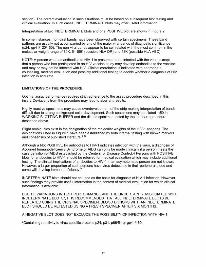
section). The correct evaluation in such situations must be based on subsequent blot testing and clinical evaluation. In such cases, INDETERMINATE blots may offer useful information. Interpretation of two INDETERMINATE blots and one POSITIVE blot are shown in Figure 2. In some instances, non-viral bands have been observed with certain specimens. These band patterns are usually not accompanied by any of the major viral bands of diagnostic significance (p24, gp41/120/160). The non-viral bands appear to be cell related with the most common in the molecular weight range of 70K, 51-55K (possible HLA DR) and 43K (possible HLA-ABC). NOTE: A person who has antibodies to HIV-1 is presumed to be infected with the virus, except that a person who has participated in an HIV vaccine study may develop antibodies to the vaccine and may or may not be infected with HIV. Clinical correlation is indicated with appropriate counseling, medical evaluation and possibly additional testing to decide whether a diagnosis of HIV infection is accurate. LIMITATIONS OF THE PROCEDURE Optimal assay performance requires strict adherence to the assay procedure described in this insert. Deviations from the procedure may lead to aberrant results. Highly reactive specimens may cause overdevelopment of the strip making interpretation of bands difficult due to strong background color development. Such specimens may be diluted 1:50 in WORKING BLOTTING BUFFER and the diluted specimen tested by the standard procedure described above. Slight ambiguities exist in the designation of the molecular weights of the HIV-1 antigens. The designations listed in Figure 1 have been established by both internal testing with known markers and consensus of published literature.5-10 Although a blot POSITIVE for antibodies to HIV-1 indicates infection with the virus, a diagnosis of Acquired Immunodeficiency Syndrome or AIDS can only be made clinically if a person meets the case definition of AIDS established by the Centers for Disease Control.4 Persons with POSITIVE blots for antibodies to HIV-1 should be referred for medical evaluation which may include additional testing. The clinical implications of antibodies to HIV-1 in an asymptomatic person are not known. However, a larger proportion of such persons have virus detectable in their peripheral blood and some will develop immunodeficiency.9-10 INDETERMINATE blots should not be used as the basis for diagnosis of HIV-1 infection. However, such findings may provide useful information in the context of medical evaluation for which clinical information is available. DUE TO VARIATIONS IN TEST PERFORMANCE AND THE UNCERTAINTY ASSOCIATED WITH INDETERMINATE BLOTS*, IT IS RECOMMENDED THAT ALL INDETERMINATE BLOTS BE REPEATED USING THE ORIGINAL SPECIMEN. BLOOD DONORS WITH AN INDETERMINATE BLOT SHOULD BE RETESTED USING A FRESH SPECIMEN AFTER SIX MONTHS. A NEGATIVE BLOT DOES NOT EXCLUDE THE POSSIBILITY OF INFECTION WITH HIV-1. *Containing reactivity to virus-specific proteins p24, p31, p66/51 or gp41/160.
17
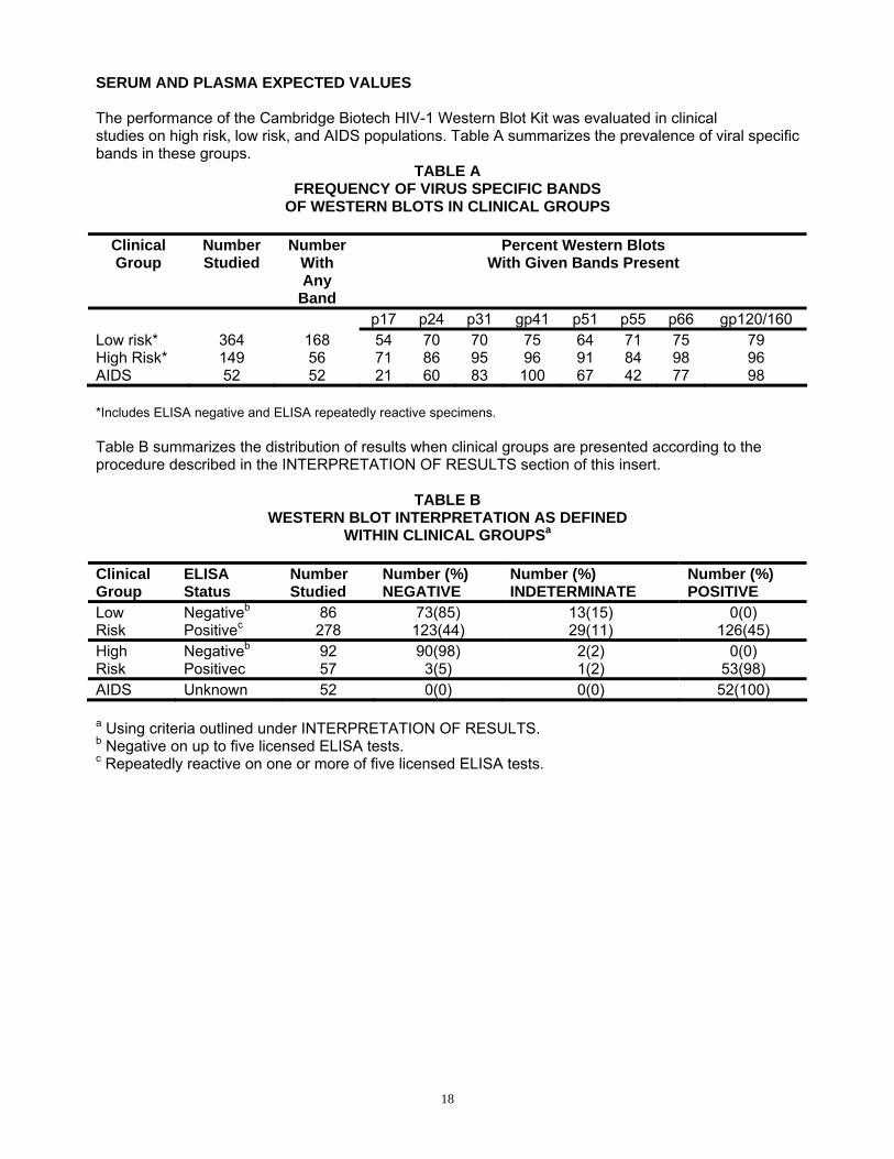
SERUM AND PLASMA EXPECTED VALUES The performance of the Cambridge Biotech HIV-1 Western Blot Kit was evaluated in clinical studies on high risk, low risk, and AIDS populations. Table A summarizes the prevalence of viral specific bands in these groups.
TABLE A FREQUENCY OF VIRUS SPECIFIC BANDS
OF WESTERN BLOTS IN CLINICAL GROUPS
Clinical Group
Number Studied
Number With Any
Band
Percent Western Blots With Given Bands Present
p17 p24 p31 gp41 p51 p55 p66 gp120/160 Low risk* High Risk* AIDS
364 149 52
168 56 52
54 71 21
70 86 60
70 95 83
75 96 100
64 91 67
71 84 42
75 98 77
79 96 98
*Includes ELISA negative and ELISA repeatedly reactive specimens. Table B summarizes the distribution of results when clinical groups are presented according to the procedure described in the INTERPRETATION OF RESULTS section of this insert.
TABLE B WESTERN BLOT INTERPRETATION AS DEFINED
WITHIN CLINICAL GROUPSa Clinical Group
ELISA Status
Number Studied
Number (%) NEGATIVE
Number (%) INDETERMINATE
Number (%) POSITIVE
Low Risk
Negativeb Positivec
86 278
73(85) 123(44)
13(15) 29(11)
0(0) 126(45)
High Risk
Negativeb Positivec
92 57
90(98) 3(5)
2(2) 1(2)
0(0) 53(98)
AIDS Unknown 52 0(0) 0(0) 52(100) a Using criteria outlined under INTERPRETATION OF RESULTS. b Negative on up to five licensed ELISA tests. c Repeatedly reactive on one or more of five licensed ELISA tests.
18
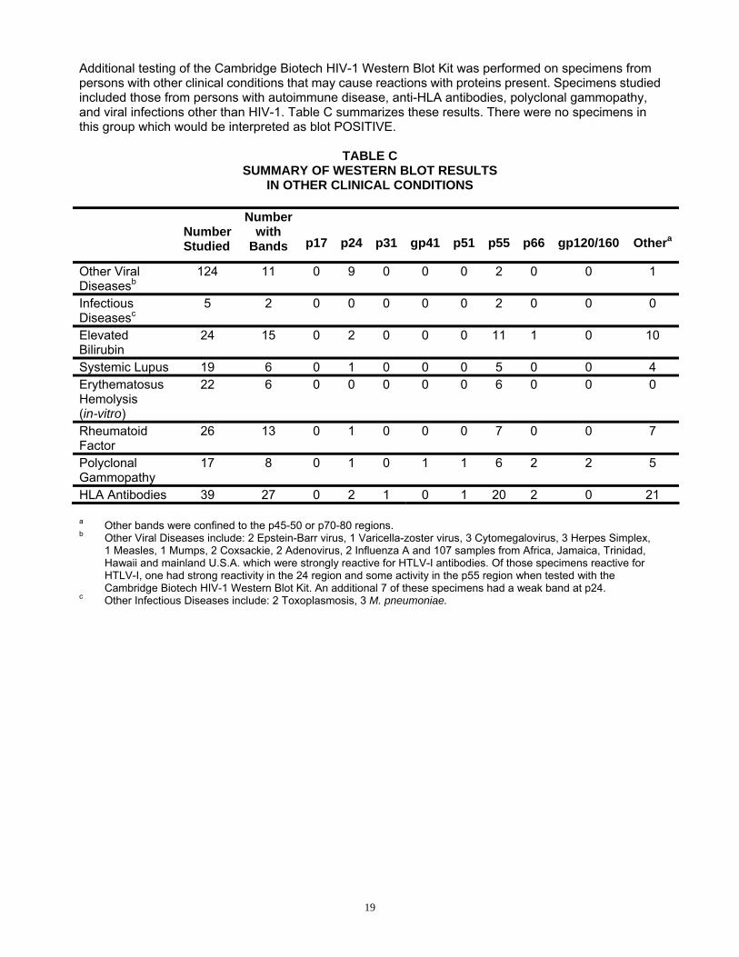
Additional testing of the Cambridge Biotech HIV-1 Western Blot Kit was performed on specimens from persons with other clinical conditions that may cause reactions with proteins present. Specimens studied included those from persons with autoimmune disease, anti-HLA antibodies, polyclonal gammopathy, and viral infections other than HIV-1. Table C summarizes these results. There were no specimens in this group which would be interpreted as blot POSITIVE.
TABLE C
SUMMARY OF WESTERN BLOT RESULTS IN OTHER CLINICAL CONDITIONS
Number Studied
Number with
Bands
p17
p24
p31
gp41
p51
p55
p66
gp120/160
Othera
Other Viral Diseasesb
124 11 0 9 0 0 0 2 0 0 1
Infectious Diseasesc
5 2 0 0 0 0 0 2 0 0 0
Elevated Bilirubin
24 15 0 2 0 0 0 11 1 0 10
Systemic Lupus 19 6 0 1 0 0 0 5 0 0 4 Erythematosus Hemolysis (in-vitro)
22 6 0 0 0 0 0 6 0 0 0
Rheumatoid Factor
26 13 0 1 0 0 0 7 0 0 7
Polyclonal Gammopathy
17 8 0 1 0 1 1 6 2 2 5
HLA Antibodies 39 27 0 2 1 0 1 20 2 0 21 a Other bands were confined to the p45-50 or p70-80 regions. b Other Viral Diseases include: 2 Epstein-Barr virus, 1 Varicella-zoster virus, 3 Cytomegalovirus, 3 Herpes Simplex,
1 Measles, 1 Mumps, 2 Coxsackie, 2 Adenovirus, 2 Influenza A and 107 samples from Africa, Jamaica, Trinidad, Hawaii and mainland U.S.A. which were strongly reactive for HTLV-I antibodies. Of those specimens reactive for HTLV-I, one had strong reactivity in the 24 region and some activity in the p55 region when tested with the Cambridge Biotech HIV-1 Western Blot Kit. An additional 7 of these specimens had a weak band at p24.
c Other Infectious Diseases include: 2 Toxoplasmosis, 3 M. pneumoniae.
19
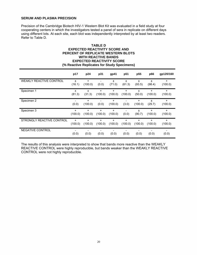
SERUM AND PLASMA PRECISION Precision of the Cambridge Biotech HIV-1 Western Blot Kit was evaluated in a field study at four cooperating centers in which the investigators tested a panel of sera in replicate on different days using different lots. At each site, each blot was independently interpreted by at least two readers. Refer to Table D.
TABLE D EXPECTED REACTIVITY SCORE AND
PERCENT OF REPLICATE WESTERN BLOTS WITH REACTIVE BANDS
EXPECTED REACTIVITY SCORE (% Reactive Replicates for Study Specimens)
p17 p24 p31 gp41 p51 p55 p66 gp120/160
WEAKLY REACTIVE CONTROL ± (16.1)
+ (100.0)
- (0.0)
± (71.0)
± (61.3)
± (93.5)
± (98.4)
+ (100.0)
Specimen 1 ± (81.3)
± (31.3)
+ (100.0)
+ (100.0)
+ (100.0)
± (50.0)
+ (100.0)
+ (100.0)
Specimen 2 - (0.0)
+ (100.0)
- (0.0)
+ (100.0)
- (3.0)
+ (100.0)
± (29.7)
+ (100.0)
Specimen 3 + (100.0)
+ (100.0)
+ (100.0)
+ (100.0)
- (0.0)
± (90.7)
+ (100.0)
+ (100.0)
STRONGLY REACTIVE CONTROL + (100.0)
+ (100.0)
+ (100.0)
+ (100.0)
+ (100.0)
+ (100.0)
+ (100.0)
+ (100.0)
NEGATIVE CONTROL - (0.0)
- (0.0)
- (0.0)
- (0.0)
- (0.0)
- (0.0)
- (0.0)
- (0.0)
The results of this analysis were interpreted to show that bands more reactive than the WEAKLY REACTIVE CONTROL were highly reproducible, but bands weaker than the WEAKLY REACTIVE CONTROL were not highly reproducible.
20
BIBLIOGRAPHY 1. Tsang, V.C.W., J.M. Peralta, and A.R. Simons, 1983. Enzyme-linked Immunoelectrotransfer
Blot Techniques (EITB) for Studying the Specificities of Antigens and Antibodies Separated by Gel Electrophoresis. Methods in Enzymology 92:377-391.
2. Sarngadharan, M.G., M. Popovic, L. Bruch, J. Schupbach, and R.C. Gallo, 1984. Antibodies
Reactive with Human T-Lymphotropic Retroviruses (HTLV-III) in the Serum of Patients with AIDS. Science 224:506-508.
3. Popovic, M., M.G. Sarngadharan, E. Read, and R.C. Gallo, 1984. Detection, Isolation, and
Continuous Production of Cytopathic Retroviruses (HTLV-III) from Patients with AIDS and Pre-AIDS. Science 224:497-500.
4. Centers for Disease Control, 1982. Update on Acquired Immune Deficiency Syndrome(AIDS) -
United States Morbidity and Mortality Weekly Report, 31:507-508. 5. Steimer, K.S., K.W. Higgins, M.A. Powers, et al., 1986. Recombinant Polypeptide from the
Endonuclease Region of the Acquired Immune Deficiency Syndrome Retrovirus Polymerase (pol) Gene Detects Serum Antibodies in Most Infected Individuals, Journal of Virology, 58:9-16.
6. Veronese, F. , A. DeVico, T. Copeland, et al., 1986. Characterization of Highly
Immunogenic p66/51 as the Reverse Transcriptase of HIV/LAV. Science 231:1289-1291. 7. Veronese, F., T. Copeland, S. Oroszlan, R.C. Gallo, M.G. Sarngadharan, 1985.
Characterization of gp41 as the Transmembrane Protein Coded by the HIV/LAV Envelope Gene. Science 229:1402-1405.
8. Allan, J.S., J.E. Coligan, F. Barin, et al., 1985. Major Glycoprotein Antigens that Induce
Antibodies in AIDS Patients Are Encoded by HIV. Science 228:291-294. 9. Sarngadharan, M.G., F. Veronese, S. Lee, R.C. Gallo, 1985. Immunological Properties of
HTLV-III Antigens Recognized by Sera of Patients with AIDS and AIDS-related Complex and of Asymptomatic Carriers of HTLV-III Infection. Cancer Research 45:4574s-4577s.
10. Groopman, J.E., F.W. Chen, J.A. Hope, et al., 1986. Serological Characterization of HIV
Infection in AIDS and Related Disorders. The Journal of Infectious Diseases 153:736-742. 11. Pinter A., W.J. Honne, S.A. Tilley, et al., 1989. Oligomeric structure of gp41, the
transmembrane protein of Human Immunodeficiency Virus Type 1. J. Virology 63:2674-9. 12. Centers for Disease Control, 1989. Interpretation and use of the Western Blot Assay for
Serodiagnosis of Human Immunodeficiency Virus Type 1 Infections. MMWR 38 (No. S-7):1-7.
13. Hausler, W.J., 1988. Report of the Third Consensus Conference of HIV Testing sponsored
by the Association of State and Territorial Public Health Laboratory Directors. Infect Control Hosp Epidemiol. 9:345-349.
14. Leitman, S.F., H.G. Klein, J.J. Melpolder, et al., 1989. Clinical Implications of Positive Tests
for Antibodies to Human Immunodeficiency Virus Type 1 in Asymptomatic Blood Donors. N Eng J Med 318:917-924.
21

15. Berenson, S., K.M. Peddecord, L.K. Hofherr, M.S. Ascher, R.N. Taylor and T.L. Hearn, 1990. Reporting the Results of Human Immunodeficiency Virus Testing. JAMA 262 No. 24:3435- 3438.
16. O’Gorman, M.R.G., D. Weber, S.E. Landis, et al., 1991. Interpretive Criteria of the Western
Blot Assay for Serodiagnosis of Human Immunodeficiency Virus Type 1 Infection. Arch Pathol Lab Med 115:26-30.
22
PATENT(S) OR PATENT APPLICATIONS(S):
USPA SN 06/602, 945, filed April 23, 1984, (USPN 4,520,113, ISSUED MAY 28, 1985) “Serological Detection of Antibodies to HTLV-III in Sera of Patients with AIDS and Pre-AIDS Conditions” USPA SN 06/602, 946, filed April 23, 1984, (USPN 4,647,773, Issued March 3, 1987) “Method of Continuous Production of Retrovirus (HTLV-III) from Patients with AIDS and Pre- AIDS” USPA SN 06/643, 729, filed August 24, 1984, (USPN 4,652,599, issued March 24, 1987) “Method of Continuous Production of Retrovirus (HTLV-III) from Patients with AIDS and Pre- AIDS Using Permissive Cells” USPA SN 06/785, 638, filed October 8, 1985, (USPN 4,708,818, issued November 24, 1987 “Human Immunodeficiency Virus Associated with Acquired Immune Deficiency Syndrome (AIDS), A Diagnostic Method for AIDS and Pre-AIDS and a Kit Therefor” USPA SN 074-117,937, filed November 5, 1987, (USPN 5,135,684 issued August 4, 1992) “Human Immunodeficiency Viruses Associated with Acquired Immune Deficiency Syndrome (AIDS), a Diagnostic Method for AIDS and Pre-AIDS and Kit Therefor”
23
Manufactured by: Maxim Biomedical, Inc. 1500 East Gude Drive
Rockville, Maryland 20850-5307 U.S. License No. 1741
Technical Services: 1-800-989-6296; 1-301-217-0619 (Outside of USA)
LN20583.06 Issue Date: 06/01/2009
24