Management of Lumbar Artery Injury Related to Pedicle ... · lumbar artery to control vascular...
4
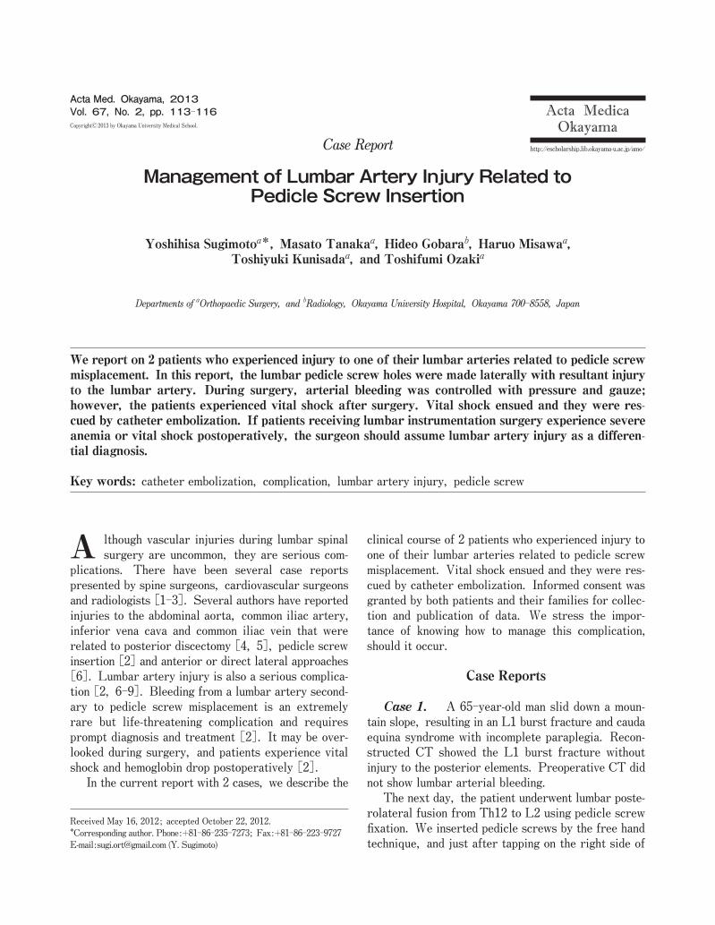
Management of Lumbar Artery Injury Related to Pedicle Screw Insertion Yoshihisa Sugimoto a* , Masato Tanaka a , Hideo Gobara b , Haruo Misawa a , Toshiyuki Kunisada a , and Toshifumi Ozaki a Departments of a Orthopaedic Surgery, and b Radiology, Okayama University Hospital, Okayama 700-8558, Japan We report on 2 patients who experienced injury to one of their lumbar arteries related to pedicle screw misplacement. In this report, the lumbar pedicle screw holes were made laterally with resultant injury to the lumbar artery. During surgery, arterial bleeding was controlled with pressure and gauze; however, the patients experienced vital shock after surgery. Vital shock ensued and they were res- cued by catheter embolization. If patients receiving lumbar instrumentation surgery experience severe anemia or vital shock postoperatively, the surgeon should assume lumbar artery injury as a differen- tial diagnosis. Key words: catheter embolization, complication, lumbar artery injury, pedicle screw lthough vascular injuries during lumbar spinal surgery are uncommon, they are serious com- plications. There have been several case reports presented by spine surgeons, cardiovascular surgeons and radiologists [1-3]. Several authors have reported injuries to the abdominal aorta, common iliac artery, inferior vena cava and common iliac vein that were related to posterior discectomy [4, 5], pedicle screw insertion [2] and anterior or direct lateral approaches [6]. Lumbar artery injury is also a serious complica - tion [2, 6-9]. Bleeding from a lumbar artery second- ary to pedicle screw misplacement is an extremely rare but life- threatening complication and requires prompt diagnosis and treatment [2]. It may be over- looked during surgery, and patients experience vital shock and hemoglobin drop postoperatively [2]. In the current report with 2 cases, we describe the clinical course of 2 patients who experienced injury to one of their lumbar arteries related to pedicle screw misplacement. Vital shock ensued and they were res- cued by catheter embolization. Informed consent was granted by both patients and their families for collec- tion and publication of data. We stress the impor- tance of knowing how to manage this complication, should it occur. Case Reports Case 1. A 65-year- old man slid down a moun- tain slope, resulting in an L1 burst fracture and cauda equina syndrome with incomplete paraplegia. Recon- structed CT showed the L1 burst fracture without injury to the posterior elements. Preoperative CT did not show lumbar arterial bleeding. The next day, the patient underwent lumbar poste - rolateral fusion from Th12 to L2 using pedicle screw fixation. We inserted pedicle screws by the free hand technique, and just after tapping on the right side of A Acta Med. Okayama, 2013 Vol. 67, No. 2, pp. 113ン116 CopyrightⒸ 2013 by Okayama University Medical School. Case Report http: // escholarship.lib.okayama-u.ac.jp / amo/ Received May 16, 2012 ; accepted October 22, 2012. * Corresponding author. Phone: + 81ン86ン235ン7273; Fax: + 81ン86ン223ン9727 E- mail:[email protected] (Y. Sugimoto)
Transcript of Management of Lumbar Artery Injury Related to Pedicle ... · lumbar artery to control vascular...
Management of Lumbar Artery Injury Related to Pedicle Screw Insertion
Yoshihisa Sugimotoa*, Masato Tanakaa, Hideo Gobarab, Haruo Misawaa, Toshiyuki Kunisadaa, and Toshifumi Ozakia
Departments of aOrthopaedic Surgery, and bRadiology, Okayama University Hospital, Okayama 700-8558, Japan
We report on 2 patients who experienced injury to one of their lumbar arteries related to pedicle screw misplacement. In this report, the lumbar pedicle screw holes were made laterally with resultant injury to the lumbar artery. During surgery, arterial bleeding was controlled with pressure and gauze; however, the patients experienced vital shock after surgery. Vital shock ensued and they were res-cued by catheter embolization. If patients receiving lumbar instrumentation surgery experience severe anemia or vital shock postoperatively, the surgeon should assume lumbar artery injury as a differen-tial diagnosis.
Key words: catheter embolization, complication, lumbar artery injury, pedicle screw
lthough vascular injuries during lumbar spinal surgery are uncommon, they are serious com-
plications. There have been several case reports presented by spine surgeons, cardiovascular surgeons and radiologists [1-3]. Several authors have reported injuries to the abdominal aorta, common iliac artery, inferior vena cava and common iliac vein that were related to posterior discectomy [4, 5], pedicle screw insertion [2] and anterior or direct lateral approaches [6]. Lumbar artery injury is also a serious complica-tion [2, 6-9]. Bleeding from a lumbar artery second-ary to pedicle screw misplacement is an extremely rare but life-threatening complication and requires prompt diagnosis and treatment [2]. It may be over-looked during surgery, and patients experience vital shock and hemoglobin drop postoperatively [2]. In the current report with 2 cases, we describe the
clinical course of 2 patients who experienced injury to one of their lumbar arteries related to pedicle screw misplacement. Vital shock ensued and they were res-cued by catheter embolization. Informed consent was granted by both patients and their families for collec-tion and publication of data. We stress the impor-tance of knowing how to manage this complication, should it occur.
Case Reports
Case 1. A 65-year-old man slid down a moun-tain slope, resulting in an L1 burst fracture and cauda equina syndrome with incomplete paraplegia. Recon-structed CT showed the L1 burst fracture without injury to the posterior elements. Preoperative CT did not show lumbar arterial bleeding. The next day, the patient underwent lumbar poste-rolateral fusion from Th12 to L2 using pedicle screw fixation. We inserted pedicle screws by the free hand technique, and just after tapping on the right side of
A
Acta Med. Okayama, 2013Vol. 67, No. 2, pp. 113ン116CopyrightⒸ 2013 by Okayama University Medical School.
Case Report http ://escholarship.lib.okayama-u.ac.jp/amo/
Received May 16, 2012 ; accepted October 22, 2012.*Corresponding author. Phone : +81ン86ン235ン7273; Fax : +81ン86ン223ン9727E-mail : [email protected] (Y. Sugimoto)
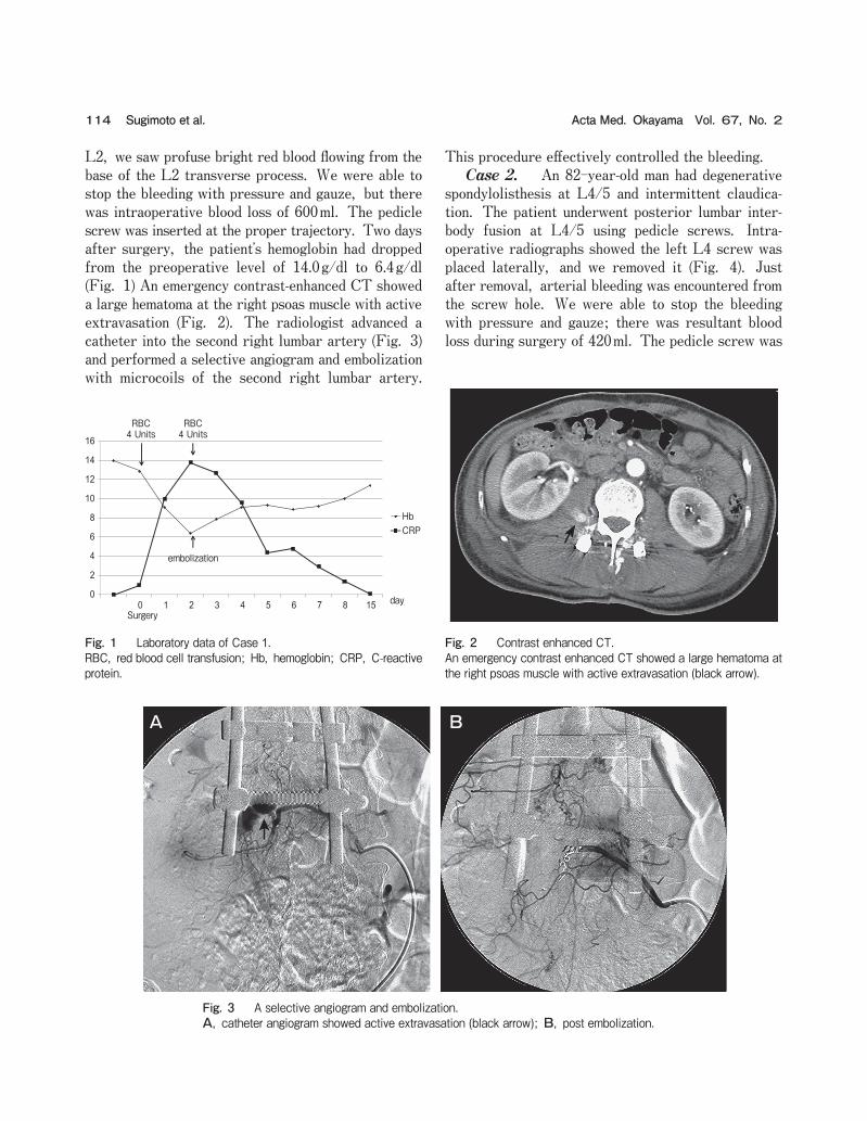
L2, we saw profuse bright red blood flowing from the base of the L2 transverse process. We were able to stop the bleeding with pressure and gauze, but there was intraoperative blood loss of 600ml. The pedicle screw was inserted at the proper trajectory. Two days after surgery, the patientʼs hemoglobin had dropped from the preoperative level of 14.0g/dl to 6.4g/dl (Fig. 1) An emergency contrast-enhanced CT showed a large hematoma at the right psoas muscle with active extravasation (Fig. 2). The radiologist advanced a catheter into the second right lumbar artery (Fig. 3) and performed a selective angiogram and embolization with microcoils of the second right lumbar artery.
This procedure effectively controlled the bleeding. Case 2. An 82-year-old man had degenerative spondylolisthesis at L4/5 and intermittent claudica-tion. The patient underwent posterior lumbar inter-body fusion at L4/5 using pedicle screws. Intra-operative radiographs showed the left L4 screw was placed laterally, and we removed it (Fig. 4). Just after removal, arterial bleeding was encountered from the screw hole. We were able to stop the bleeding with pressure and gauze; there was resultant blood loss during surgery of 420ml. The pedicle screw was
114 Acta Med. Okayama Vol. 67, No. 2Sugimoto et al.
0
2
4
6
8
10
12
14
16
0 1 2 3 4 5 6 7 8 15
HbCRP
Surgery
RBC4 Units
RBC4 Units
day
embolization
Fig. 1 Laboratory data of Case 1.RBC, red blood cell transfusion; Hb, hemoglobin; CRP, C-reactive protein.
Fig. 2 Contrast enhanced CT.An emergency contrast enhanced CT showed a large hematoma at the right psoas muscle with active extravasation (black arrow).
A B
Fig. 3 A selective angiogram and embolization.A, catheter angiogram showed active extravasation (black arrow); B, post embolization.

placed at the proper trajectory. After surgery, abdominal CT showed lumbar arterial bleeding and intramuscular hemorrhage. We did not perform cath-eter embolization because the hemoglobin drop was slow, and bleeding was controlled with continued blood infusion (Fig. 5). One week after surgery, the patient started experiencing atrial fibrillation. The cardiologist administered heparin (12,000 U per day) and the patientʼs hemoglobin dropped again. Enhanced
CT showed an enlarged hematoma at his left psoas muscle. We performed a selective angiogram and embolization with microcoils of the fourth left lumbar artery. This procedure controlled the bleeding.
Discussion
Lumbar artery injury related to surgery is an extremely rare but serious complication. Several authors have reported lumbar artery injury secondary to pedicle screw insertion and discectomy [2, 7-9]. According to a cadaveric study of the lumbar artery and its branches (dorsal branch, spinal branch and ganglionic branch), the first 4 lumbar arteries origi-nate from the iliolumbar artery or medial sacral artery [10]. Clinical manifestations of a lacerated vessel include hypotension, hemoglobin drop, and abdominal pain and distension. Enhanced CT is a use-ful diagnostic tool for detecting lumbar arterial bleed-ing and hematoma in the psoas muscle; the patient sometime is in a psoas-relaxed position. It was difficult to repair the bleeding vessel during posterior approach surgery. Traditionally, lumbar artery injury has been managed by emergent explor-atory laparotomy and direct repair of the bleeding vessel. However, this approach is associated with increased risks due to the need for a second operative site and position change [9]. Recently, selective embolization has been recommended as the first-line treatment method for iatrogenic lumbar arterial bleed-ing [2, 3, 5, 8, 9]. If the patient is hemodynami-cally stable after fluid resuscitation and transfusion, the next step should be a contrast enhanced abdominal CT and angiography to evaluate the bleeding site and treatment [2]. Karaikovic et al. reported on the suc-cessful use of coil embolization of a lumbar artery to control vascular injury during intradiscal surgery [9]. The detection time of arterial injuries in spinal surgery can be divided into 3 groups: intraoperative, immediate postoperative and late postoperative [8, 9]. In this report, the lumbar pedicle screw holes were made laterally with resultant injury to the lum-bar artery. During surgery, arterial bleeding was controlled with pressure and gauze; however, the patients experienced vital shock after surgery. Sandri et al. reported on lumbar pedicle misplacement during a scoliosis surgery [2]; 5h postoperatively, the patient suffered a hypotensive attack and her abdomen had
115Management of Lumbar Artery InjuryApril 2013
Lt
Fig. 4 Intraoperative radiograph.The left L4 screw was placed laterally (white arrow), and we removed it.
day0
2
4
6
8
10
12
14
16
18
0 1 2 3 5 7 8 9 10 11 12 14 19 23 26
HbCRP
RBC4 Units
RBC4 Units
embolization
Surgery
Fig. 5 Laboratory data of Case 2.RBC, red blood cell transfusion; Hb, hemoglobin; CRP, C-reactive protein.
become distended [2]. If we observe arterial bleeding from the screw hole, it is very important to assess the postoperative CT immediately. We should consult a radiologist while vital signs are still stable. Treatment after the patient has had severe anemia or vital shock could be too late. The clinical course of these patients shows that postoperative hemoglobin drop may occur even if intraoperative bleeding is controlled. To avoid pedicle screw lateral perforation, it could be useful to use a navigation system.
References
1. Canaud L, Hireche K, Joyeux F, DʼAnnoville T, Berthet JP, Marty-Ané C and Alric P: Endovascular Repair of Aorto-iliac Artery Injuries after Lumbar-spine Surgery. Eur J Vasc Endovasc Surg (2011) 42: 167-171.
2. Sandri A, Regis D, Marino MA, Puppini G and Bartolozzi P: Lumbar artery injury following posterior spinal instrumentation for scoliosis. Orthopedics (2011) 34: 317.
3. Lee KH, Park JH, Chung JW, Han JK, Shin SJ and Kang HS: Vascular complications in lumbar spinal surgery: percutaneous endovascular treatment. Cardiovasc Intervent Radiol (2000) 23: 65-69.
4. Akpinar B, Peynircioglu B, Cil B, Daglioglu E and Cekirge S: Iliac vascular complication after spinal surgery: immediate endovascu-lar repair following CT angiographic diagnosis. Diagn Interv Radiol (2009) 15: 303-305.
5. Skippage P, Raja J, McFarland R and Belli AM: Endovascular repair of iliac artery injury complicating lumbar disc surgery. Eur Spine J (2008) 17: S228-231.
6. Santillan A, Patsalides A and Gobin YP: Endovascular emboliza-tion of iatrogenic lumbar artery pseudoaneurysm following extreme lateral interbody fusion (XLIF). Vasc Endovascular Surg (2010) 44: 601-603.
7. Puri AS, Colen RR, Reddy AS, Groff MW, DiNobile D, Killoran T, Nikolic B and Thomas AJ: Lumbar artery pseudoaneurysm after percutaneous vertebroplasty: a unique vascular complication. J Neurosurg Spine (2011) 14: 296-299.
8. Biafora SJ, Mardjetko SM, Butler JP, McCarthy PL and Gleason TF: Arterial injury following percutaneous vertebral augmentation: a case report. Spine Phila (Pa 1976) (2006) 31: E84-87.
9. Karaikovic EE, Rattner Z, Bilimoria MM, Sener SF, McGee JP, Metrick LB, Szokol JW and Limthongkul W: Coil embolization of a lumbar artery to control vascular injury during intradiscal surgery. Spine Phila (Pa 1976) (2010) 35: E163-166.
10. Caglar S, Dolgun H, Ugur HC, Torun F, Attar A, Uz A, Tekdemir I and Elhan A: Extraforaminal lumbar arterial anatomy. Surg Neurol (2004) 61: 29-33.
116 Acta Med. Okayama Vol. 67, No. 2Sugimoto et al.