Malignant liver masses
55
Liver Malignant Lesions Dr. Anish Choudhary
-
Upload
anish-choudhary -
Category
Health & Medicine
-
view
83 -
download
0
Transcript of Malignant liver masses
Liver Malignant Lesions
Dr. Anish Choudhary

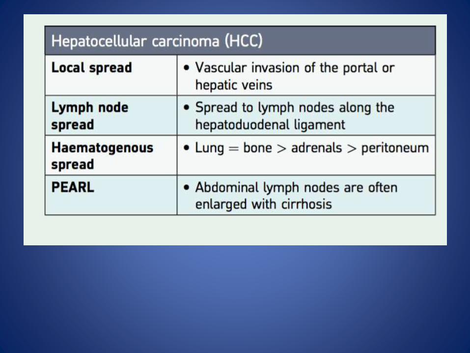
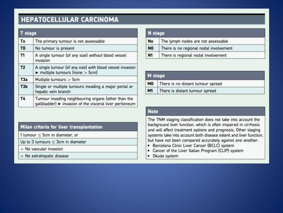
HEPATOCELLULAR CARCINOMA (HCC) / HEPATOMA
• The commonest primary malignant neoplasm of the liver, typically occurring within an abnormal (e.g . cirrhotic) liver.
• Risk factors: – direct carcinogens (e.g. aflatoxin)– chronic hepatitis B and C– cirrhosis (particularly postnecrotic cirrhosis and haemochromatosis.
• Types: – solitary– multifocal (accounting for up to 40% of cases in the Far East)– Diffuse.– It is unclear whether HCC arises from a regenerative
nodule (via a dysplastic intermediate state) or as a denovo lesion.
RADIOLOGICAL FEATUR ES
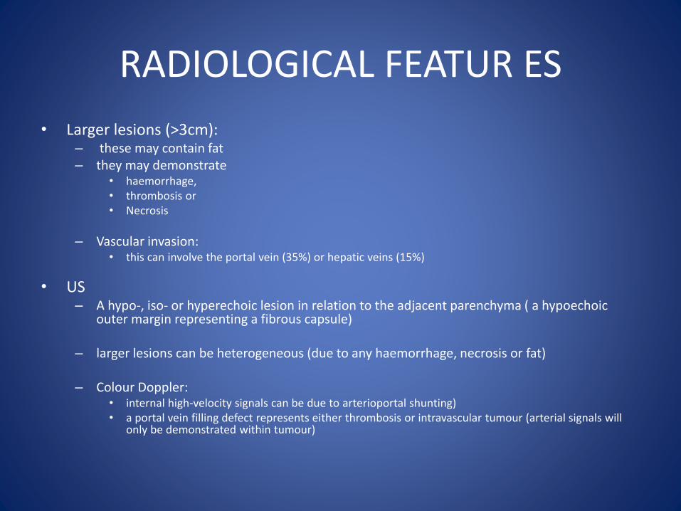
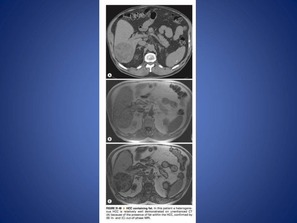
• Larger lesions (>3cm):– these may contain fat– they may demonstrate
• haemorrhage, • thrombosis or • Necrosis
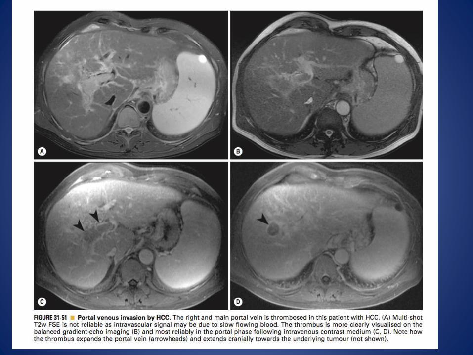
– Vascular invasion: • this can involve the portal vein (35%) or hepatic veins (15%)
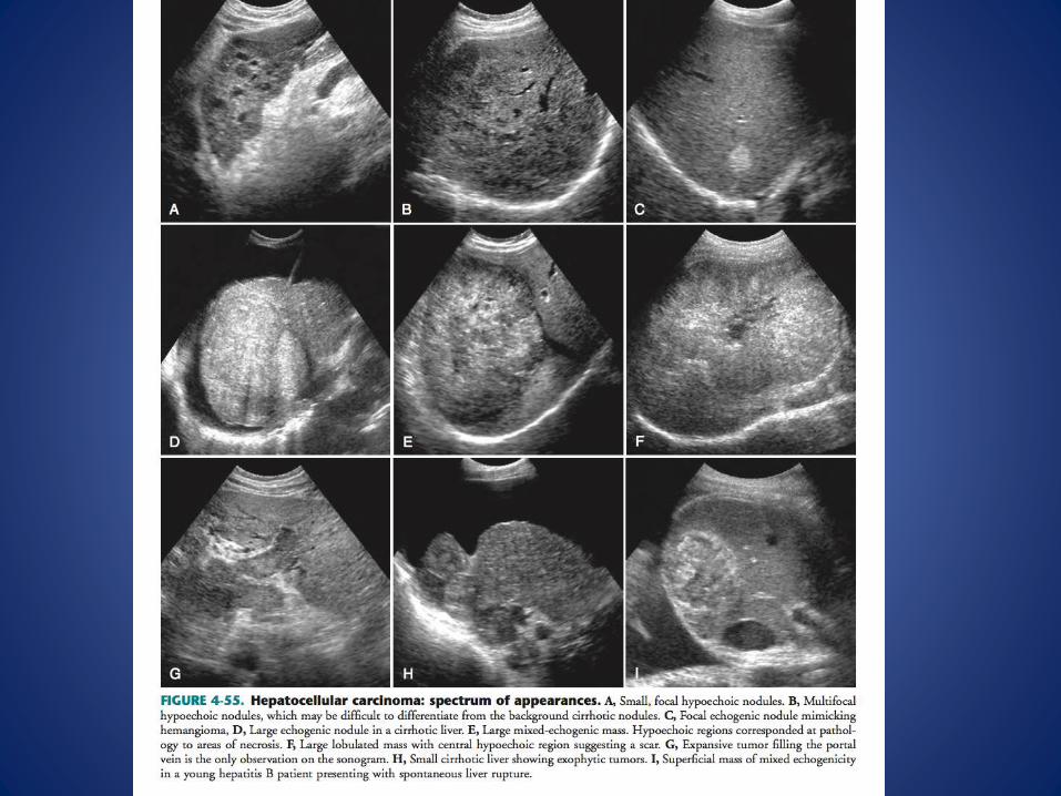
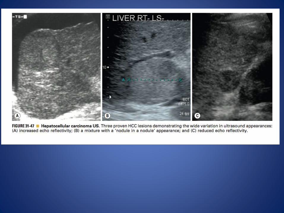
• US– A hypo-, iso- or hyperechoic lesion in relation to the adjacent parenchyma ( a hypoechoic
outer margin representing a fibrous capsule)
– larger lesions can be heterogeneous (due to any haemorrhage, necrosis or fat)
– Colour Doppler: • internal high-velocity signals can be due to arterioportal shunting)• a portal vein filling defect represents either thrombosis or intravascular tumour (arterial signals will
only be demonstrated within tumour)
CT• NECT:
– ill-defined low attenuation lesions– focal areas of internal calcification (7% of cases)– There may be a hypoattenuating capsule
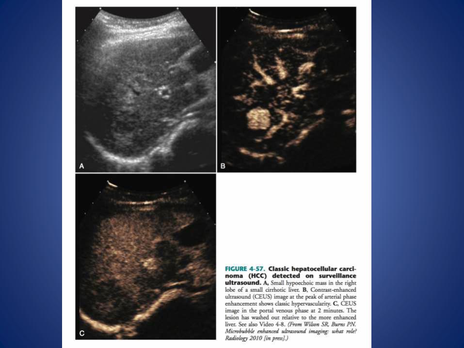
• CECT: – enhancement is seen during the arterial phase, as it is a
hypervascular tumour supplied via the hepatic artery
– it may demonstrate a mosaic enhancement pattern (with an enhancing grid-likepattern around a central lower area of attenuation) ▶
– it will become hypoattenuating to the liver parenchyma during the portal phase

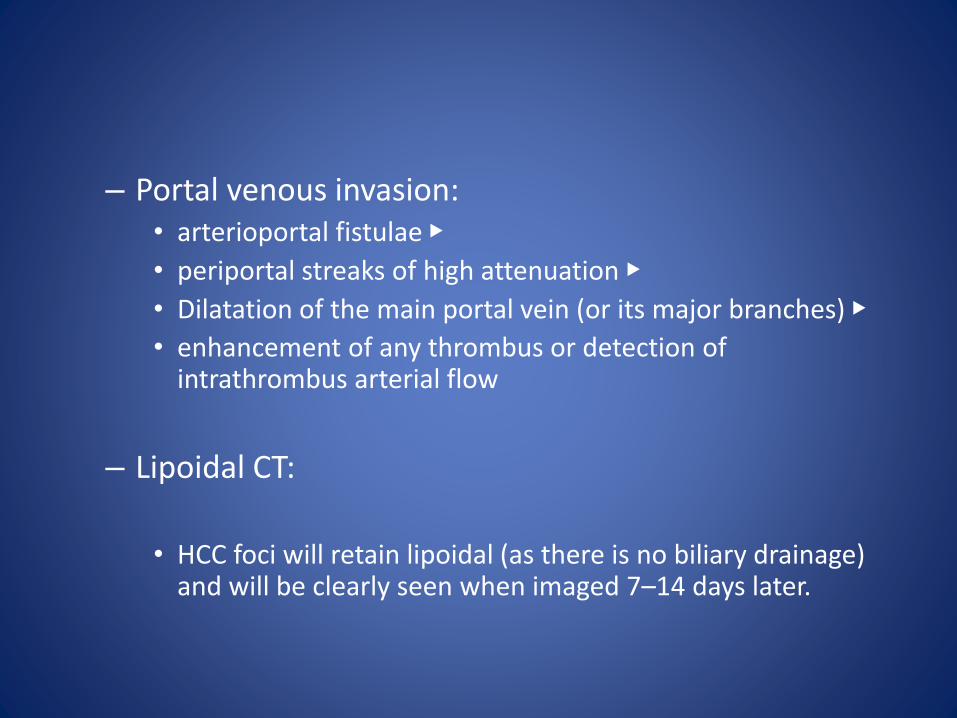
– Portal venous invasion: • arterioportal fistulae ▶
• periportal streaks of high attenuation ▶
• Dilatation of the main portal vein (or its major branches) ▶
• enhancement of any thrombus or detection of intrathrombus arterial flow
– Lipoidal CT:
• HCC foci will retain lipoidal (as there is no biliary drainage) and will be clearly seen when imaged 7–14 days later.
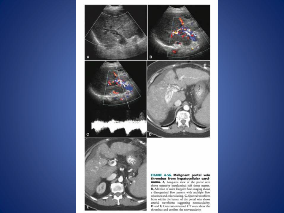
• The presence of arterial waveform within the thrombus indicates that it is neoplastic rather than bland thrombus.
• This distinction is vital because it has been shown that the presence of malignant portal vein thrombosis is the worst prognostic factor in predicting recurrence of HCC following surgicalresection or liver transplantation
MRI
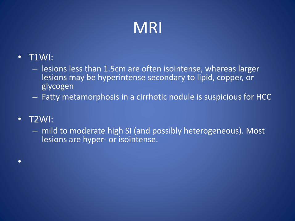
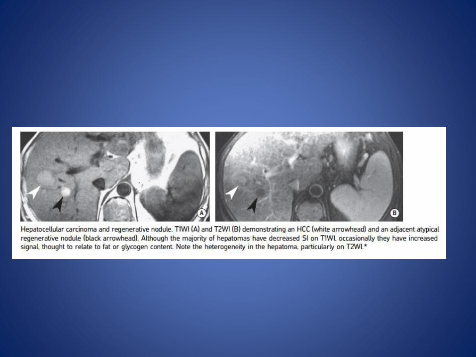
• T1WI: – lesions less than 1.5cm are often isointense, whereas larger
lesions may be hyperintense secondary to lipid, copper, or glycogen
– Fatty metamorphosis in a cirrhotic nodule is suspicious for HCC
• T2WI: – mild to moderate high SI (and possibly heterogeneous). Most
lesions are hyper- or isointense.
•
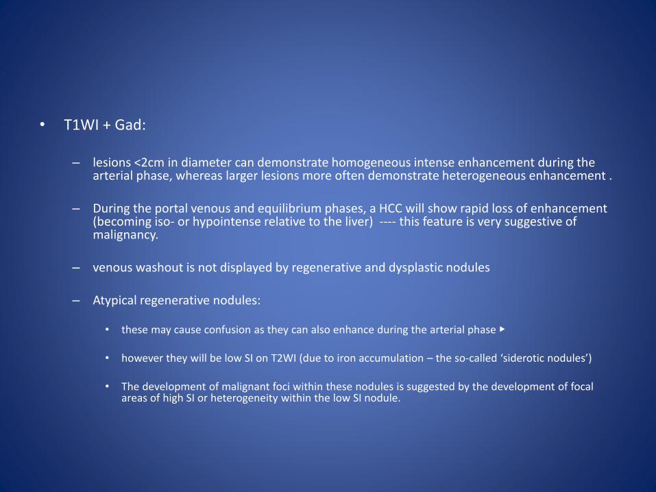
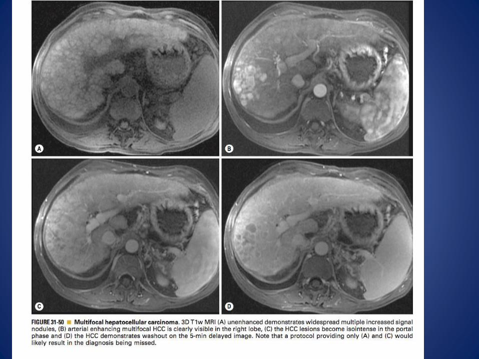
• T1WI + Gad:
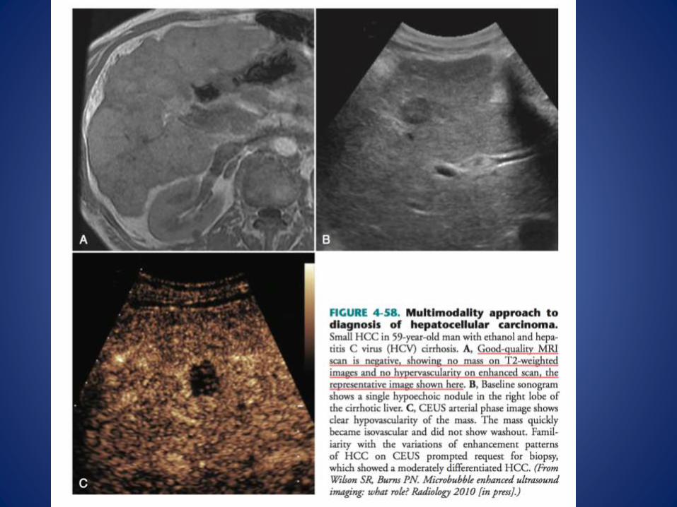
– lesions <2cm in diameter can demonstrate homogeneous intense enhancement during the arterial phase, whereas larger lesions more often demonstrate heterogeneous enhancement .
– During the portal venous and equilibrium phases, a HCC will show rapid loss of enhancement(becoming iso- or hypointense relative to the liver) ---- this feature is very suggestive of malignancy.
– venous washout is not displayed by regenerative and dysplastic nodules
– Atypical regenerative nodules:
• these may cause confusion as they can also enhance during the arterial phase ▶
• however they will be low SI on T2WI (due to iron accumulation – the so-called ‘siderotic nodules’)
• The development of malignant foci within these nodules is suggested by the development of focal areas of high SI or heterogeneity within the low SI nodule.


• DWI: – variable appearance that depends on their histologic make-up
– Well-differentiated tumors are often isointense
– Moderately to poorly differentiated tumors are more often hyperintense
• FDG PET:
– this is relatively non-specific for HCC and is not widely used
• DSA: – This is used for preoperative assessment ▶
– It defines the arterial and venous anatomy and evaluates any portal or caval involvement
– HCC is usually a vascular lesion demonstrating dilated feeding arteries, abundant abnormal vessels or arteriovenous shunting
– Portal vein invasion: a ‘threads and streaks’ appearance
Pearls
• The incidence parallels the prevalence of localpredisposing conditions (in particular chronichepatitis B and C)
• Serum a-fetoprotein (AFP) may or may not beelevated in HCC.
• AFP may also be elevated with simple cirrhosis.
• It commonly metastasizes to the lungs and bone
LIVER METASTASES
• Definition
– The liver is a common site for metastases from many primary cancers (usually due to haematogenous spread)
– GI tract tumours:
• these metastasize via the portal vein• there is evidence for blood flow separation within the portal vein as right–
sided colon cancers are more likely to spread to the right lobe (with left–sided tumours spreading to either the right or left lobes)
– Non-GI tract tumours:
• these metastasize via the hepatic artery ▶• both lobes are equally affected • Although a metastasis will derive its vascular supply from the hepatic artery it
will usually be less vascular than the adjacent liver parenchyma

Radiological features
• Metastases can be difficult to radiologically detect and characterize if they measure less than 5mm in size (particularly in distinguishing them from a biliaryhamartoma)
• FDG PET does not improve the sensitivity (as there is a relatively high normal background liver uptake) but is useful in detecting extrahepatic metastases
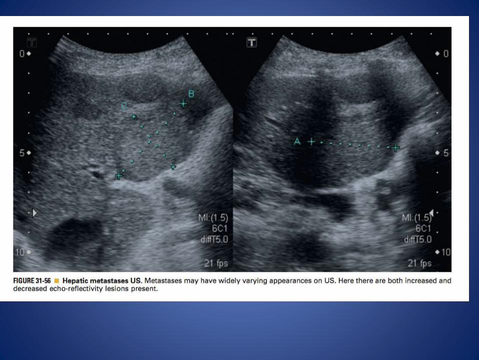
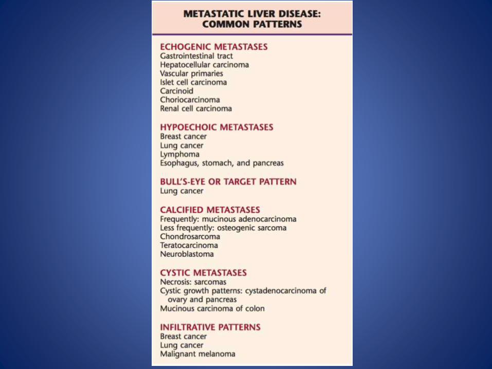
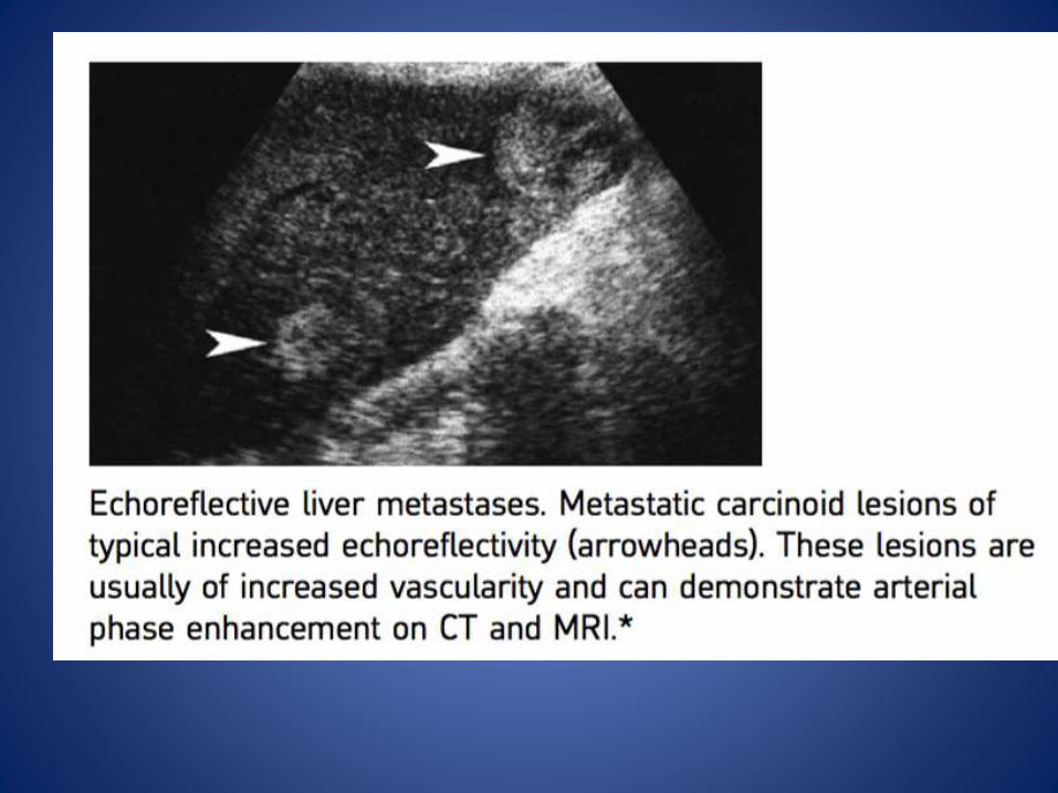
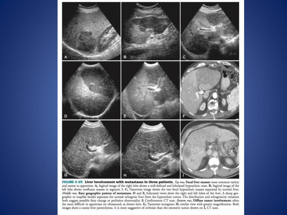
• Metastases can demonstrate a wide range of appearances but they will usually demonstrate growthon serial imaging, multiplicity, and a variation in size US Homogeneous or heterogeneous mass lesions ▶
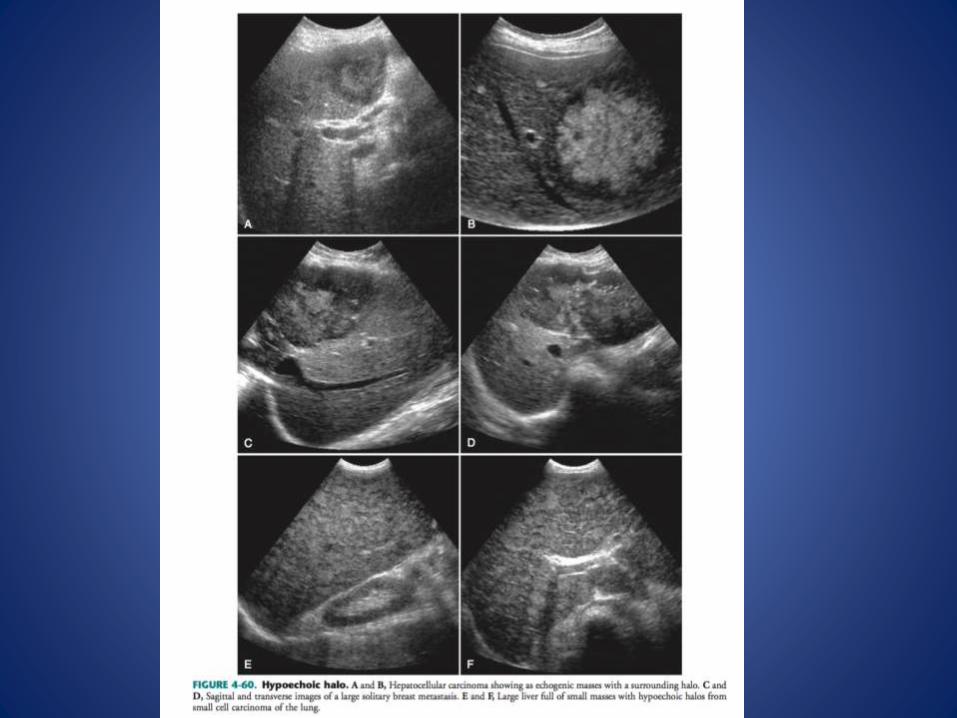
• they can be hyperechoic (mimicking a haemangioma) or hypoechoic (mimicking a simple cyst) ▶central necrosis can cause a partly cystic appearance ▶
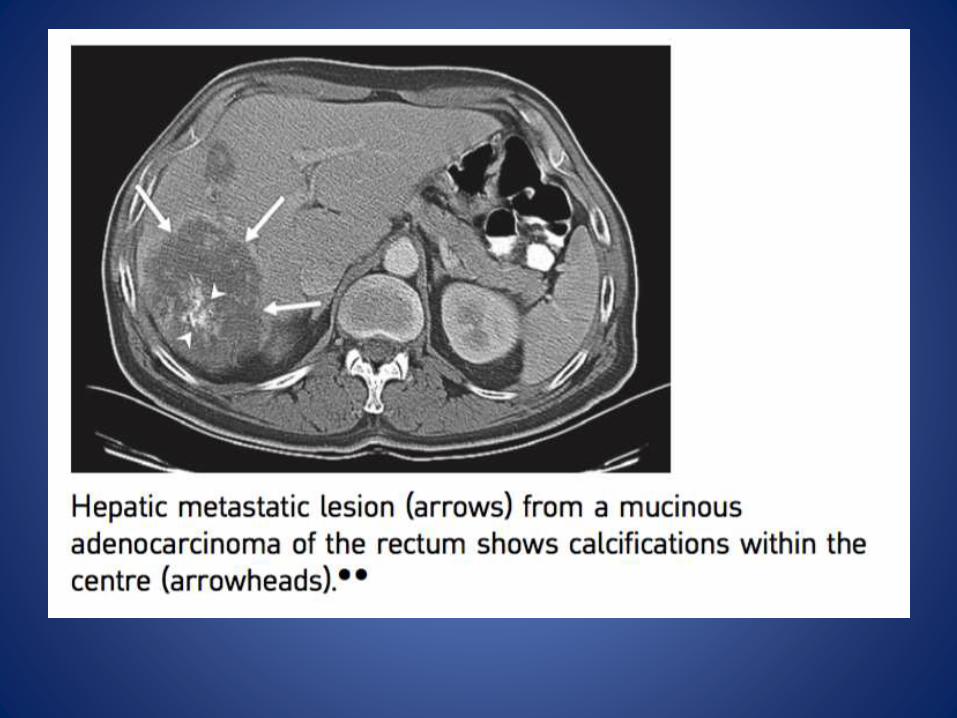
• calcification can be seen in mucin-secreting metastases from the GI tract.
• ‘Target’ appearance: there may be a surrounding rim of reduced reflectivity


CT
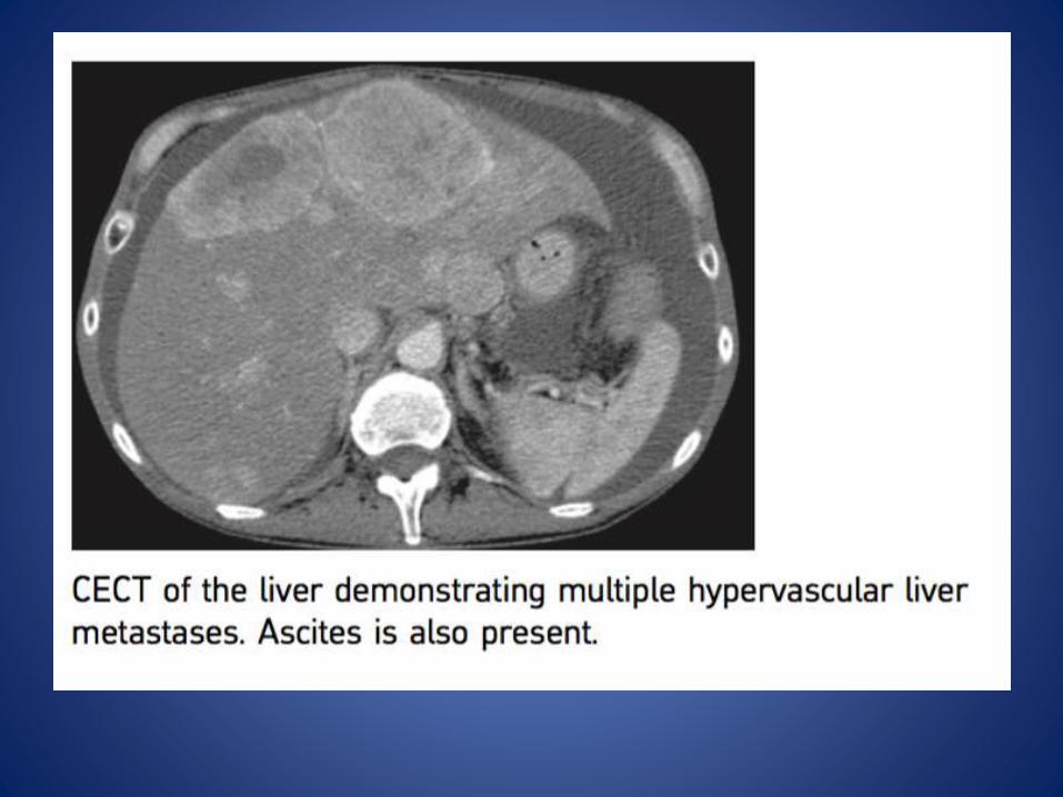
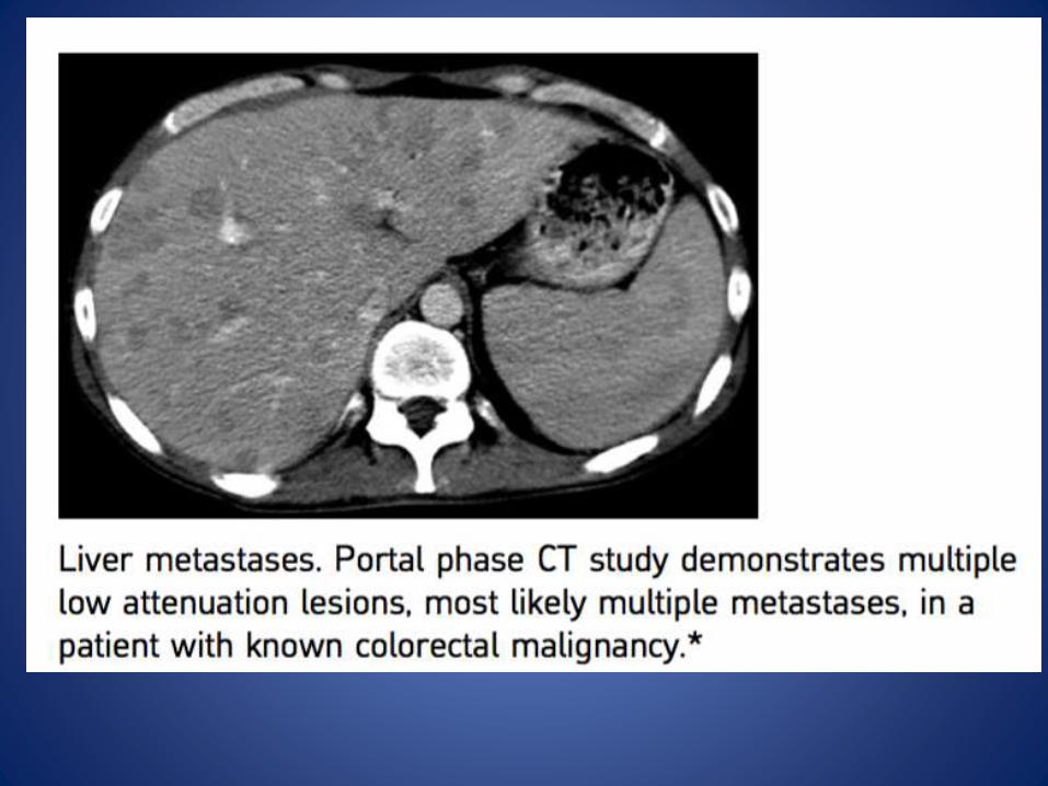
• The majority of metastases are of low attenuation onunenhanced and portal phase imaging ▶
• Hypervascular tumours may show transient arterial enhancement, becoming isoattenuating to liver during the portal phase ▶
• Central necrosis, rim enhancement and calcification (in mucinsecreting metastases of GI origin) can also be demonstrated
• A <5mm low attenuation lesion within the liver is more likely to represent a simple cyst (unless a metastasis is purely cystic it is unlikely to be of low enough attenuation to be visible at such asmall size)
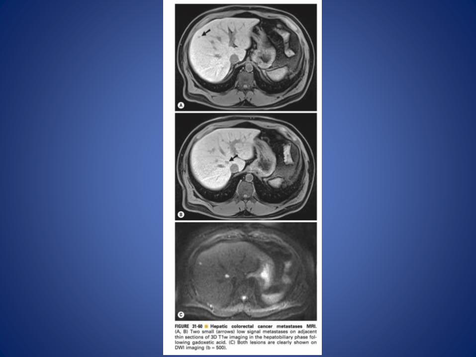
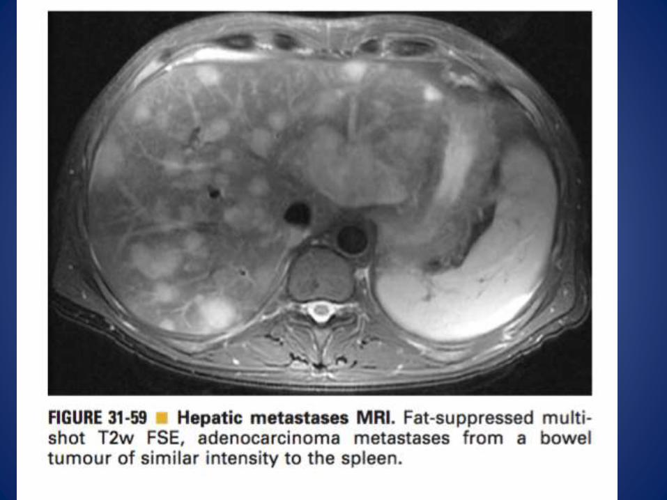
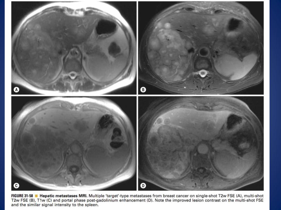
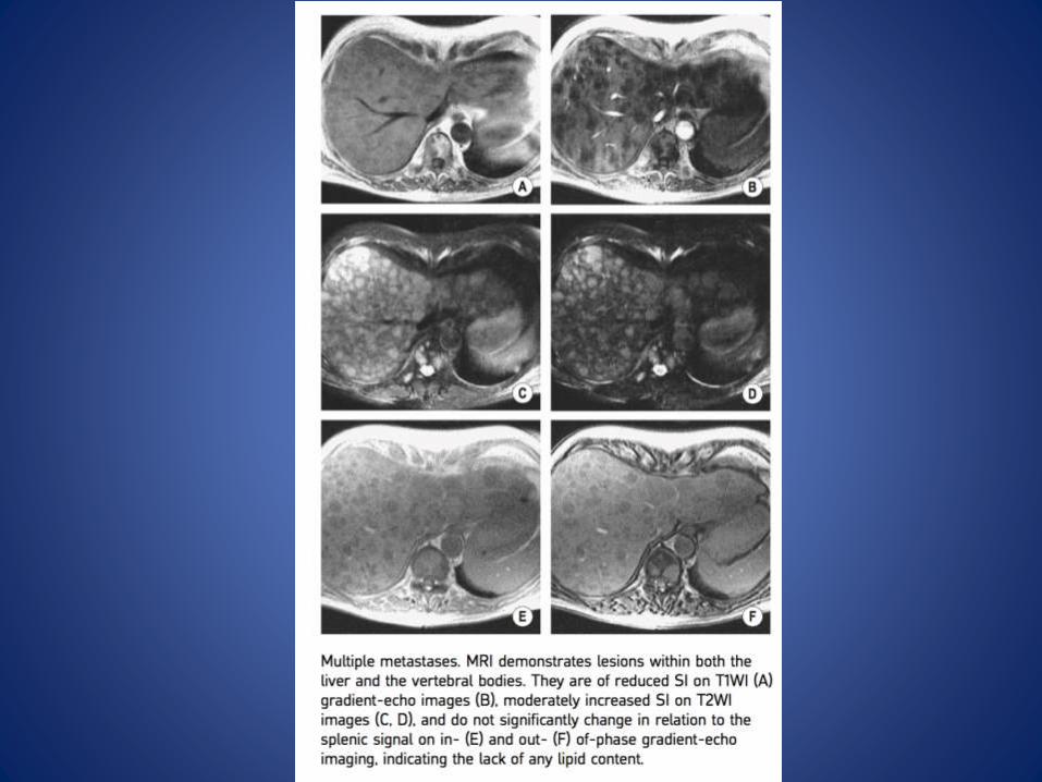
MRI
• The signal intensity of a metastasis roughly parallels that of the spleen
• T1WI: – Hypervascular metastases are moderately hypointense ▶
– haemorrhagic metastases can demonstrate hyperintensity
– Perilesional fat deposition has been specifically described with hepatic metastases from a primarypancreatic insulinoma and is thought to be related to the effects of insulin.
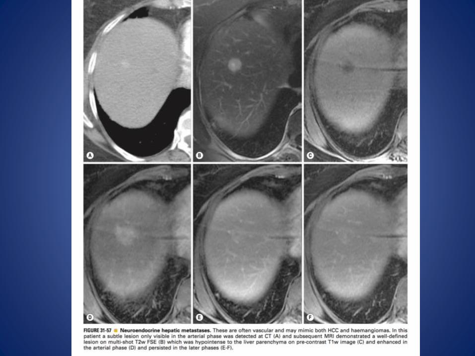
• T2WI: – hypervascular metastases are usually markedly hyperintense and may be cystic or necrotic
• T1WI + Gad: – similar enhancement characteristics as for CT
• With paramagnetic iron oxide agents the normal liver parenchyma is of low SI (due to Kupffer celluptake) – this will make a metastatic lesion more obvious
• Hyperintense Colloid scintigraphy :There is reduced activity (metastases lack Kupffer cells)
Pearls
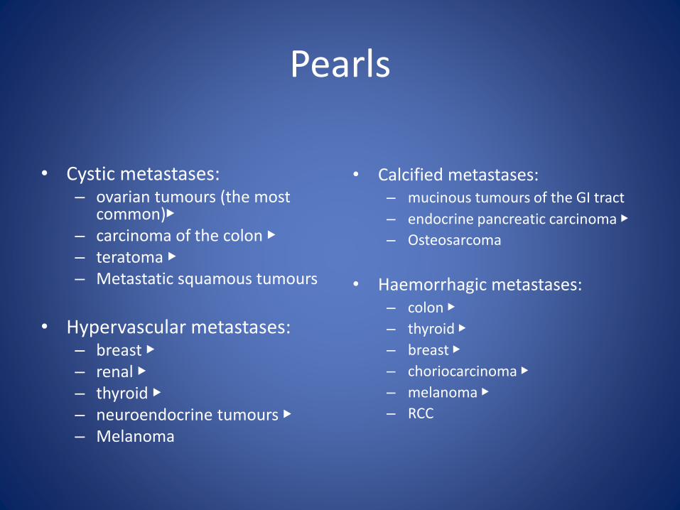
• Cystic metastases: – ovarian tumours (the most
common)▶– carcinoma of the colon ▶– teratoma ▶– Metastatic squamous tumours
• Hypervascular metastases: – breast ▶– renal ▶– thyroid ▶– neuroendocrine tumours ▶– Melanoma
• Calcified metastases: – mucinous tumours of the GI tract
– endocrine pancreatic carcinoma ▶
– Osteosarcoma
• Haemorrhagic metastases: – colon ▶
– thyroid ▶
– breast ▶
– choriocarcinoma ▶
– melanoma ▶
– RCC

• After the initiation of chemotherapy, metastases can exhibit a less aggressive enhancement pattern that can mimic a haemangioma(including early peripheral nodular enhancement and delayed retention of contrast material)
• A key distinguishing feature of chemotherapeutically treated metastases is an early, intact peripheral rim of enhancement (unlike the discontinuous peripheral enhancement seen with a haemangioma)
• Hypervascular metastases classically show marked T2 hyperintensity and restricted diffusion (compared with FNH and adenoma) ▶– they will wash out on delayed enhanced images (unlike a haemangioma)

ANGIOSARCOMA
• Definition
– A rare malignant vascular hepatic neoplasm derivedfrom the endothelial cells and which can form vascular derivatives, cavernous spaces, or solid masses
– It is associated with exposure to polyvinylchloride,arsenic and Thorotrast contrast medium
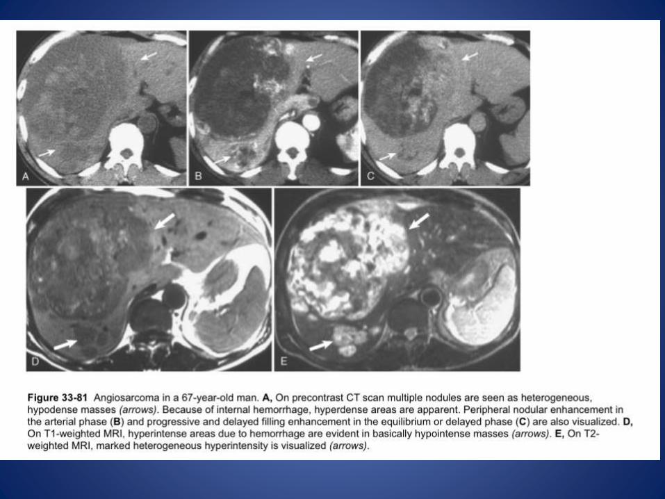
Radiological features
• CT – It can appear as an infiltrating mass demonstrating
heterogeneous enhancement ▶– it can occasionally present in a diffuse form that is not
easily detected with imaging
– Background thorotrast exposure causes heterogeneously increased attenuation within the liver, perihepatic lymph nodes and spleen
–
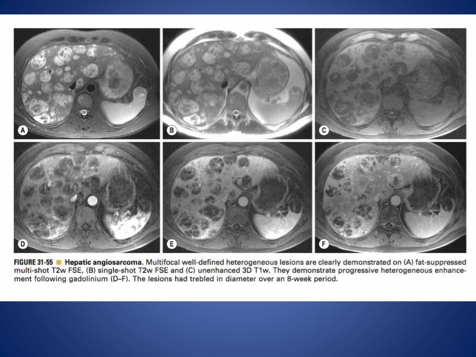
MRI
• It can present as a large mass or as multiple nodules
• T1WI: low SI ▶
• T2WI: high SI ▶
• T1WI + Gad: heterogeneous enhancement


• Thank You.