Detecting Imaging for Neoplastic Liver Masses in Hepatic ...
14
Original article 657 Detecting Imaging for Neoplastic Liver Masses in Hepatic Patients Who Received Direct Acting Antiviral Drugs Medhat M. Refaat, Osama T. Galal, Shaimaa A. Taha Abstract Background: Hepatocellular carcinoma makes up 75%-85% of all primary liver cancers and is the fourth most common cause of cancer related death worldwide. The aim of this study was to screen for hepatocellular carcinoma in hepatic patients who received direct acting antiviral drugs using different imaging modalities. Methods: This was a cross sectional study included 50 patients who were hepatic and received Direct Acting Antiviral '' DAA'' and were carried out at radiology department in Elobour Hospital for Health Insurance after obtaining written informed consent from all patients. Results: There were 31(62%) male, 19(38%) female, the mean age 52.28 years old with range (39-65), 35(70%) urban, 15(30%) rural. There were (70%) with duration of illness ≤ 5 years, (30%) with > 5 years, (100%) with DAA, (90%) easy to treat, (10%) difficult to treat. There were 35(70%) with average liver, 14(28%) with shrunken liver, 1(2%) with enlarged liver, 13(26%) with Bright pattern, 34(68%) with cirrhotic, and 3(6%) with coarse bright, the mean P.V diameter 14.68 mm (± 2.51 SD) with range (9-17 mm).There were (100%) with single focal lesion in RT lobe, the mean size was 4.47 mm with range (3.6-5.6), (33.3%) isochoric, (33.3%) with hypo echoic, (33.3%) with hyper echoic, and (66.7%) with malignancy. There were 3(100%) with typical CT enhancement, 1(33.3%) with P.V thrombosis. None were with affection on liver vascular, no one with intrahepatic biliary radicles, no one with L.N in porta hepatis. Conclusion: Based on our findings, we recommend for close imaging evaluations after DAA in cirrhotic patients. Keywords: Neoplastic; Liver; Masses; Direct Acting Antiviral; DAAs Department of Radiology, Benha faculty of medicine, Benha University, Egypt. Correspondence to: Shaimaa A. Taha, Department of Radiology, Benha faculty of medicine, Benha University, Egypt. Email: [email protected] Received: 6 February 2021 Accepted: 23 May 2021
Transcript of Detecting Imaging for Neoplastic Liver Masses in Hepatic ...
Original article
657
Detecting Imaging for Neoplastic Liver Masses in Hepatic Patients
Who Received Direct Acting Antiviral Drugs
Medhat M. Refaat, Osama T. Galal, Shaimaa A. Taha
Abstract
Background: Hepatocellular carcinoma makes up 75%-85% of all
primary liver cancers and is the fourth most common cause of cancer
related death worldwide. The aim of this study was to screen for
hepatocellular carcinoma in hepatic patients who received direct
acting antiviral drugs using different imaging modalities. Methods:
This was a cross sectional study included 50 patients who were
hepatic and received Direct Acting Antiviral '' DAA'' and were carried
out at radiology department in Elobour Hospital for Health Insurance
after obtaining written informed consent from all patients. Results:
There were 31(62%) male, 19(38%) female, the mean age 52.28 years
old with range (39-65), 35(70%) urban, 15(30%) rural. There were
(70%) with duration of illness ≤ 5 years, (30%) with > 5 years,
(100%) with DAA, (90%) easy to treat, (10%) difficult to treat. There
were 35(70%) with average liver, 14(28%) with shrunken liver, 1(2%)
with enlarged liver, 13(26%) with Bright pattern, 34(68%) with
cirrhotic, and 3(6%) with coarse bright, the mean P.V diameter 14.68
mm (± 2.51 SD) with range (9-17 mm).There were (100%) with single focal lesion in RT lobe,
the mean size was 4.47 mm with range (3.6-5.6), (33.3%) isochoric, (33.3%) with hypo echoic,
(33.3%) with hyper echoic, and (66.7%) with malignancy. There were 3(100%) with typical CT
enhancement, 1(33.3%) with P.V thrombosis. None were with affection on liver vascular, no one
with intrahepatic biliary radicles, no one with L.N in porta hepatis. Conclusion: Based on our
findings, we recommend for close imaging evaluations after DAA in cirrhotic patients.
Keywords: Neoplastic; Liver; Masses; Direct Acting Antiviral; DAAs
Department of Radiology,
Benha faculty of medicine,
Benha University, Egypt.
Correspondence to:
Shaimaa A. Taha,
Department of Radiology,
Benha faculty of medicine,
Benha University, Egypt.
Email:
Received: 6 February 2021
Accepted: 23 May 2021
Benha medical journal, vol.38, issue 2, 2021
658
Introduction
Chronic hepatitis C virus (HCV) infection is
one of the most common infections in Egypt
especially males which can be responsible
for cirrhosis, neoplastic liver masses
especially hepatocellular carcinoma (HCC),
end- stage liver diseases and other
complications such as esophageal varices
and portal hypertension (1).
Hepatocellular carcinoma (HCC) is the most
common liver cancer and the fifth most
common cancer worldwide. It results in
between 250,000 and 1 million deaths
globally per annum. The number of deaths
per year in HCC is close to that of the
incidence throughout the world, which
emphasizes the high case fatality rate of this
aggressive cancer (2).
Eighty per cent of HCC cases are associated
with chronic hepatitis B and C virus
infections. The obesity epidemic has
resulted in a growing population of patients
with non-alcoholic fatty liver disease is a
risk factor for cirrhosis and HCC in Egypt
(3).
With the rapid development of direct-acting
antivirals (DAAs), HCV treatment has
become much easier than ever because of
the short-term and well-tolerated treatment
regimens. Recent studies in real-life clinical
practice, including ours, reported that the
rates of sustained viral response (SVR)
exceeded 90% for HCV infection in
treatment with sofosbuvir (SOF) plus
ledipasvir (LDV) or SOF plus ribavirin
(RBV) (4).
Notable improvement in the biochemical
parameters of liver function and health-
related quality of life shortly after treatment.
In contrast, recent studies have highlighted
the significant impact of early HCC
recurrence However, it remains to be
clarified in a large study whether or not the
early development of malignant tumors are
associated with DAA treatment (5).
As much data as possible on this issue are
urgently needed to better manage HCC
surveillance in clinical practice. The purpose
of this study was to evaluate the short-term
risk of neoplastic masses among Egyptian
patients with chronic HCV infection who
received direct acting antiviral drugs (6).
Unlike most other cancers, HCC can be
diagnosed on imaging studies only without
tissue sampling confirmation. Currently, all
major consensus groups support the
diagnosis of HCC with contrast-enhanced
Detecting Imaging For Neoplastic Liver Masses, 2021
659
multiphasic CT. The accepted modality for
neoplastic masses screening is ultrasound.
Once malignant tumors are suspected then
CT or MRI may be used to confirm the
diagnosis. They provide data about liver
function, tumor size and number, tumor
extent, including vascular invasion and
extrahepatic spread, evidence of portal
hypertension, and clinical performance
status. Tumor proximity to large vessels and
main bile ducts (7).
This study aimed to screen for
hepatocellular carcinoma in hepatic patients
who received direct acting antiviral drugs
using different imaging modalities.
Patients and methods
This study is a cross sectional study, carried
out at radiology department in Elobour
Hospital for Health Insurance. During the
period from July 2019 till January 2020, and
included fifty patients who were hepatic and
received Direct Acting Antiviral '' DAA''
Inclusion criteria:
1. HCV RNA positive
2. patients >65 years old should undergo
cardiological assessment prior to
therapy by ECG , echocardiology and
cardiological consultation
Exclusion criteria:
1. Child's C cirrhotic patients
2. Platelet count < 50000/mm2
3. Hepatocellular carcinoma except 6
months after intervention aiming at cure
with no evidence of activity by dynamic
imaging CT or MRI
4. Extra hepatic malignancy except after
two years of disease free interval
5. In cases of lymphomas and chronic
lymphocytic leukemia treatment can be
initiated immediately after remission
based on the treating oncologist
6. Pregnancy or inability to use effective
contraception
7. Inadequately controlled diabetes
mellitus (HbA1c > 9 %)
All patients were subjected to full history
taking, complete clinical examination, and
Laboratory assessment: Complete blood
count, Liver function tests (serum bilirubin
"total, direct and indirect ", ALT, AST, total
proteins and serum albumin), Kidney
function tests (urea and creatinine),
Prothrombin time and prothrombin
concentration, Viral hepatitis markers (HBV
surface antigen, HCV antibody),
Benha medical journal, vol.38, issue 2, 2021
660
Quantitative HCV-RNA detection using
real-time polymerase chain reaction (PCR).
Radiological assessment:
1.Abdominal ultrasonography (US). Logic
P3 for all hepatic patients in the study
2.Triphasic CT for patients with hepatic
focal lesions in pelviabdominal ultrasound
using Toshiba Alexion 16: Alexion
Advanced Edition is a new mid-level 16
Slice CT System which produces high
productivity using Toshiba’s Exclusive
Feature as Industry-Leading Quantum
Detector Technology, offering a perfect
solution to middle range medical centers
and hospitals. It covers all clinical needs
of whole body examinations (except
cardiac) within a minimum time with an
optimization in anatomy visualization,
including CT Angiography. Supported by
fast accurate Automated Bone Removal,
advanced 3D, post processing applications
and the latest dose reduction technology
integrated from Premium CT Technology
for outstanding image quality and patient
safety. Using Toshiba’s unique “Double
Slice Technology”, initially developed for
the 320-row Aquilion ONE, the Alexion
Advance Edition can be configured to
generate 32 axial slices in a single rotation
using for outstanding spatial resolution
and extended flexibility.
An informed verbal consent from all
participants was taken and confidentiality of
information was assured. An official written
administrative permission letter was
obtained from dean of faculty of medicine,
Benha University hospital manager and
Elobour Hospital manager. The title and
objectives of the study were explained to
them to ensure their cooperation. Permission
from the faculty of medicine ethical
committee was also obtained, and approval
from institutional review board was taken.
Statistical analysis
Data were fed to the computer and analyzed
using IBM SPSS software package version
20.0. (Armonk, NY: IBM Corp) Qualitative
data were described using number and
percent. The Kolmogorov-Smirnov test was
used to verify the normality of distribution
Quantitative data were described using
range (minimum and maximum), mean,
standard deviation, median and interquartile
range (IQR). Significance of the obtained
results was judged at the 5% level. The used
tests were: Paired t-test: For normally
distributed quantitative variables, to
compare between two periods.
Detecting Imaging For Neoplastic Liver Masses, 2021
661
Wilcoxon signed ranks test: For abnormally
distributed quantitative variables, to
compare between two periods
Results
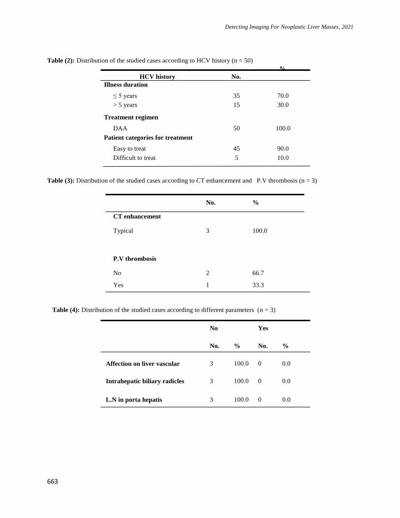
There were 31 (62%) males, 19 (38%)
females, the mean age 52.28 yrs (±6.4 SD)
with range (39-65 yrs), 35(70%) urban,
15(30%) rural, 44(88%) married, 4(8%)
widow, 2(4%) divorced, 29(58%) with
secondary education, 21(42%) with
university, 46(92%) Employee, 1(2%)
Unemployed, 3(6%) housewife, 22(44%)
with low SES, 28(56%) with middle SES,
table 1
There were 35(70%) with duration of illness
≤ 5 years, 15(30%) with > 5 years,
50(100%) with DAA, 45(90%) Easy to treat,
5(10%) Difficult to treat. table 2
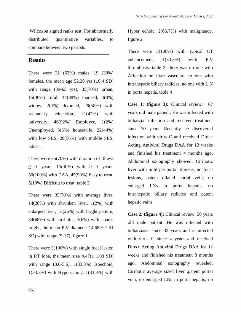
There were 35(70%) with average liver,
14(28%) with shrunken liver, 1(2%) with
enlarged liver, 13(26%) with bright pattern,
34(68%) with cirrhotic, 3(6%) with coarse
bright, the mean P.V diameter 14.68(± 2.51
SD) with range (9-17). figure 1
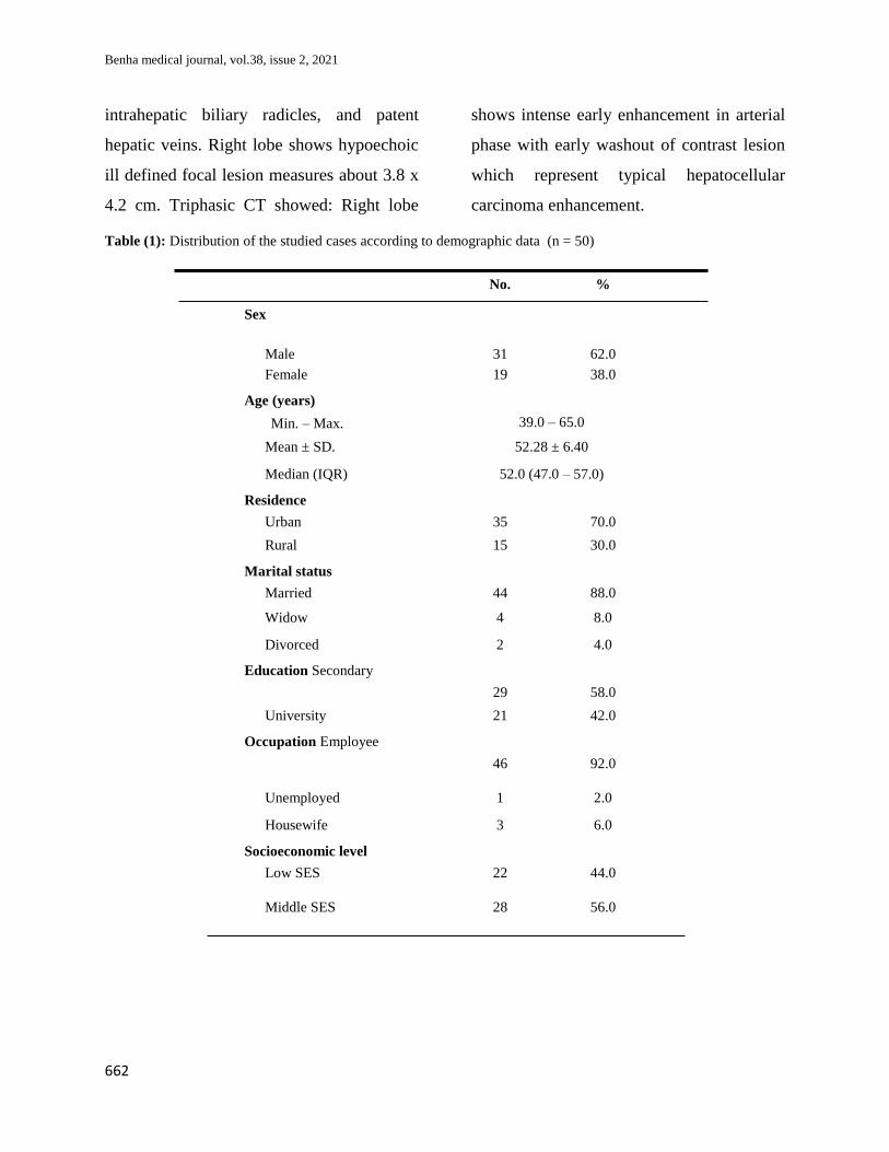
There were 3(100%) with single focal lesion
in RT lobe, the mean size 4.47(± 1.03 SD)
with range (3.6-5.6), 1(33.3%) Isoechoic,
1(33.3%) with Hypo echoic, 1(33.3%) with
Hyper echoic, 2(66.7%) with malignancy.
figure 2
There were 3(100%) with typical CT
enhancement, 1(33.3%) with P.V
thrombosis. table 3, there was no one with
Affection on liver vascular, no one with
intrahepatic biliary radicles, no one with L.N
in porta hepatis. table 4
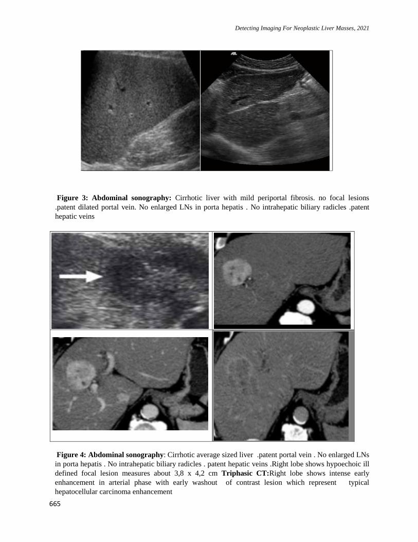
Case 1: (figure 3): Clinical review: 67
years old male patient. He was infected with
bilharzial infection and received treatment
since 30 years .Recently he discovered
infection with virus C and received Direct
Acting Antiviral Drugs DAA for 12 weeks
and finished his treatment 4 months ago.
Abdominal sonography showed: Cirrhotic
liver with mild periportal fibrosis, no focal
lesions, patent dilated portal vein, no
enlarged LNs in porta hepatis, no
intrahepatic biliary radicles and patent
hepatic veins
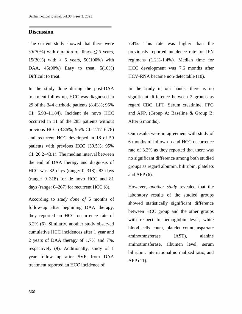
Case 2: (figure 4): Clinical review: 50 years
old male patient .He was infected with
bilharziasis since 32 years and is infected
with virus C since 4 years and received
Direct Acting Antiviral Drugs DAA for 12
weeks and finished his treatment 8 months
ago. Abdominal sonography revealed:
Cirrhotic average sized liver .patent portal
vein, no enlarged LNs in porta hepatis, no
Benha medical journal, vol.38, issue 2, 2021
662
intrahepatic biliary radicles, and patent
hepatic veins. Right lobe shows hypoechoic
ill defined focal lesion measures about 3.8 x
4.2 cm. Triphasic CT showed: Right lobe
shows intense early enhancement in arterial
phase with early washout of contrast lesion
which represent typical hepatocellular
carcinoma enhancement.
Table (1): Distribution of the studied cases according to demographic data (n = 50)
No. %
Sex
Male
31
62.0
Female 19 38.0
Age (years)
Min. – Max.
39.0 – 65.0
Mean ± SD. 52.28 ± 6.40
Median (IQR) 52.0 (47.0 – 57.0)
Residence
Urban
35
70.0
Rural 15 30.0
Marital status
Married
44
88.0
Widow 4 8.0
Divorced 2 4.0
Education Secondary
29
58.0
University 21 42.0
Occupation Employee
46
92.0
Unemployed 1 2.0
Housewife 3 6.0
Socioeconomic level
Low SES
22
44.0
Middle SES
28
56.0
Detecting Imaging For Neoplastic Liver Masses, 2021
663
Table (2): Distribution of the studied cases according to HCV history (n = 50)
HCV history No.
%
Illness duration
≤ 5 years
35
70.0
> 5 years 15 30.0
Treatment regimen
DAA
50
100.0
Patient categories for treatment
Easy to treat
45
90.0
Difficult to treat 5 10.0
Table (3): Distribution of the studied cases according to CT enhancement and P.V thrombosis (n = 3)
No. %
CT enhancement
Typical
3
100.0
P.V thrombosis
No
2
66.7
Yes 1 33.3
Table (4): Distribution of the studied cases according to different parameters (n = 3)
No
Yes
No. % No. %
Affection on liver vascular 3 100.0 0 0.0
Intrahepatic biliary radicles 3 100.0 0 0.0
L.N in porta hepatis 3 100.0 0 0.0
Benha medical journal, vol.38, issue 2, 2021
664
Fig.(2) : Distribution of the studied cases according to Echo
Fig.(1): Distribution of the studied cases according to liver size
Liver Size
Average Shrunken Enlarged
Echo
Isochoric Hypo echoic Hyper echoic
Detecting Imaging For Neoplastic Liver Masses, 2021
665
Figure 3: Abdominal sonography: Cirrhotic liver with mild periportal fibrosis. no focal lesions
.patent dilated portal vein. No enlarged LNs in porta hepatis . No intrahepatic biliary radicles .patent
hepatic veins
Figure 4: Abdominal sonography: Cirrhotic average sized liver .patent portal vein . No enlarged LNs
in porta hepatis . No intrahepatic biliary radicles . patent hepatic veins .Right lobe shows hypoechoic ill
defined focal lesion measures about 3,8 x 4,2 cm Triphasic CT:Right lobe shows intense early
enhancement in arterial phase with early washout of contrast lesion which represent typical
hepatocellular carcinoma enhancement
Benha medical journal, vol.38, issue 2, 2021
666
Discussion
The current study showed that there were
35(70%) with duration of illness ≤ 5 years,
15(30%) with > 5 years, 50(100%) with
DAA, 45(90%) Easy to treat, 5(10%)
Difficult to treat.
In the study done during the post-DAA
treatment follow-up, HCC was diagnosed in
29 of the 344 cirrhotic patients (8.43%; 95%
CI: 5.93–11.84). Incident de novo HCC
occurred in 11 of the 285 patients without
previous HCC (3.86%; 95% CI: 2.17–6.78)
and recurrent HCC developed in 18 of 59
patients with previous HCC (30.5%; 95%
CI: 20.2–43.1). The median interval between
the end of DAA therapy and diagnosis of
HCC was 82 days (range: 0–318): 83 days
(range: 0–318) for de novo HCC and 81
days (range: 0–267) for recurrent HCC (8).
According to study done of 6 months of
follow-up after beginning DAA therapy,
they reported an HCC occurrence rate of
3.2% (6). Similarly, another study observed
cumulative HCC incidences after 1 year and
2 years of DAA therapy of 1.7% and 7%,
respectively (9). Additionally, study of 1
year follow up after SVR from DAA
treatment reported an HCC incidence of
7.4%. This rate was higher than the
previously reported incidence rate for IFN
regimens (1.2%-1.4%). Median time for
HCC development was 7.6 months after
HCV-RNA became non-detectable (10).
In the study in our hands, there is no
significant difference between 2 groups as
regard CBC, LFT, Serum creatinine, FPG
and AFP. (Group A: Baseline & Group B:
After 6 months).
Our results were in agreement with study of
6 months of follow-up and HCC occurrence
rate of 3.2% as they reported that there was
no significant difference among both studied
groups as regard albumin, bilirubin, platelets
and AFP (6).
However, another study revealed that the
laboratory results of the studied groups
showed statistically significant difference
between HCC group and the other groups
with respect to hemoglobin level, white
blood cells count, platelet count, aspartate
aminotransferase (AST), alanine
aminotransferase, albumen level, serum
bilirubin, international normalized ratio, and
AFP (11).
Detecting Imaging For Neoplastic Liver Masses, 2021
667
Different groups have recently reported data
on the early occurrence and recurrence of
HCC after DAA therapy in patients with
compensated cirrhosis. In these reports, the
clinical benefits provided by HCV
eradication with DAA regimens were
hampered by the accelerated development of
HCC, occurring soon after DAA therapy end
in a significant proportion of patients. In
particular, HCC occurred early in over 3%
of patients without a prior history of liver
cancer and recurred early in about 30% of
those who had been successfully treated for
HCC, and were disease-free for different
periods of time. In addition, the emergence
of HCC seemed to be characterized by a
more aggressive and faster progression to
advanced stages (12).
Vascular invasion is a predictor of
recurrence and poor overall survival in
HCC. In particular, the presence of
microvascular invasion (MVI) is a major
risk factor for early HCC recurrence after
curative treatment (13).
The current study showed that there were
35(70%) with average liver, 14(28%) with
shrunken liver, 1(2%) with enlarged liver,
13(26%) with Bright pattern, 34(68%) with
cirrhotic, 3(6%) with coarse bright, the mean
P.V diameter 14.68(± 2.51 SD) with range
(9-17). After 6 months, there were 3(100%)
with single focal lesion in RT lobe, the mean
size 4.47(± 1.03 SD) with range (3.6-5.6),
1(33.3%) isochoric, 1(33.3%) with
hypoechoic, 1(33.3%) with hyperechoic,
2(66.7%) with malignancy. There were
3(100%) with typical CT enhancement,
1(33.3%) with P.V thrombosis, no one with
intrahepatic biliary radicles, and no one with
L.N in porta hepatis.
Our results were supported by study which
reported that at the time of HCC diagnosis,
41 neoplastic nodules were found in 29
patients: a single nodule in 22 patients, two
nodules in five, and three or more nodules in
two. Maximum nodule diameter was
between 10 and 20 mm in 27 nodules,
between 20 and 50 mm in 13, and greater
than 50 mm in one. Significant differences
were found in the presence of these
radiological signs in the HCC nodules
developed after DAA therapy in comparison
with those that occurred in the past, in the
same study population. At least two features
of MVI were present in 17/51 (33.3%)
previous HCC nodules, but in 29/41 (70.7%)
newly developed HCCs (p= 0.0007, OR:
0.21; 95% CI: 0.08–0.50). This difference
was marked even in small HCC nodules
(10– 20 mm in diameter), where MVI was
Benha medical journal, vol.38, issue 2, 2021
668
present in only 7/28 (25%) of the past HCC
nodules, but in 16/27 (59.3%) of the HCC
nodules developed after DAA therapy (p=
0.01, OR: 0.23; 95% CI: 0.07–0.72) (8).
In another study all patients with HCC
occurrence did not show any viral relapse,
achieving a SVR. The median diameter of
lesions was 33 mm (range 18–57 mm) (14).
Recent reports have raised the hypothesis
that DAA therapy may not reduce the
incidence of HCC and, paradoxically, could
accelerate the de novo occurrence or the
recurrence of HCC in patients with liver
cirrhosis (15). In addition, further
preliminary data suggest that HCC
developed after DAA therapy may present
macroscopic aggressive patterns (16).
Several risk factors have been identified that
may increase the risk of HCC recurrence
after DAA treatment , such as high liver
stiffness, antiviral treatment failure, history
of previous HCC recurrence, previous HCC
shape and stage, AFP level values, des-γ-
carbossi-prothrombin (DCP) > 40 mAU/mL,
non-curative procedures (such as TACE),
anti-HBc positivity and the time interval
between HCC complete response and DAA
initiation (17).
In a study done during the follow-up period
(median: 17 months), 46 (2.7%) patients
developed HCC. The 1-year cumulative
rates of de novo HCC were 0.4% and 4.9%
for the noncirrhosis and cirrhosis groups
respectively (log-rank test: P < 0.001) (17).
Moreover, another large prospective study
found that the 6-mo and 1-year probabilities
of HCC recurrence after DAA therapy were
12% and 26.6%, respectively. Previous
history of HCC recurrence and tumour size
were the only two independent risk factors
for early HCC recurrence. They concluded
that the probability of early recurrence in
patients who had previously been cured of
HCC remained high, despite HCV
eradication by DAA treatment. This risk was
comparable, but not higher than, the risk
reported in the literature concerning patients
who did not receive DAA treatment (18).
Conclusion
Neoplastic liver masses especially HCC may
occur after receiving of DAA but not
conditionally correlated , They seems to be
more related to cirrhotic cases more than
non cirrhotic. Repeated imaging evaluations
are needed after DAA in cirrhotic patients.
Detecting Imaging For Neoplastic Liver Masses, 2021
669
References
1. Seeff LB. Natural history of chronic hepatitis C.
Hepatology. 2002;36(5B):s35–46.
2. Torre LA, Bray F, Siegel RL, Ferlay J, Lortet‐
Tieulent J, Jemal A. Global cancer statistics,
2012. CA Cancer J Clin. 2015;65(2):87–108.
3. Bugianesi E. EASL–EASD–EASO Clinical
Practice Guidelines for the management of non-
alcoholic fatty liver disease: disease mongering
or call to action? Diabetologia.
2016;59(6):1145–7.
4. Ogawa E, Furusyo N, Nomura H, Dohmen K,
Higashi N, Takahashi K, et al. NS5A resistance-
associated variants undermine the effectiveness
of ledipasvir and sofosbuvir for cirrhotic patients
infected with HCV genotype 1b. J Gastroenterol.
2017;52(7):845–54.
5. Younossi ZM, Stepanova M, Afdhal N, Kowdley
K V, Zeuzem S, Henry L, et al. Improvement of
health-related quality of life and work
productivity in chronic hepatitis C patients with
early and advanced fibrosis treated with
ledipasvir and sofosbuvir. J Hepatol.
2015;63(2):337–45.
6. Conti F, Buonfiglioli F, Scuteri A, Crespi C,
Bolondi L, Caraceni P, et al. Early occurrence
and recurrence of hepatocellular carcinoma in
HCV-related cirrhosis treated with direct-acting
antivirals. J Hepatol. 2016;
7. Bruix J, Sherman M. Management of
hepatocellular carcinoma: an update.
Hepatology. 2011;53(3):1020–2.
8. Renzulli M, Buonfiglioli F, Conti F, Brocchi S,
Serio I, Foschi FG, et al. Imaging features of
microvascular invasion in hepatocellular
carcinoma developed after direct-acting antiviral
therapy in HCV-related cirrhosis. Eur Radiol.
2018;28(2):506–13.
9. Nakao Y, Hashimoto S, Abiru S, Komori A,
Yamasaki K, Nagaoka S, et al. Rapidly growing,
moderately differentiated HCC: A
clinicopathological characteristic of HCC
occurrence after IFN-free DAA therapy? J
Hepatol. 2018;68(4):854–5.
10. Cardoso H, Vale AM, Rodrigues S, Gonçalves
R, Albuquerque A, Pereira P, et al. High
incidence of hepatocellular carcinoma following
successful interferon-free antiviral therapy for
hepatitis C associated cirrhosis. J Hepatol.
2016;65(5):1070–1.
11. Huang AC, Mehta N, Dodge JL, Yao FY,
Terrault NA. Direct acting antivirals do not
increase the risk of hepatocellular carcinoma
recurrence after local regional therapy or liver
transplant waitlist dropout. Hepatology.
2018;68(2):449–61.
12. Reig M, Mariño Z, Perelló C, Iñarrairaegui M,
Ribeiro A, Lens S, et al. Unexpected high rate of
early tumor recurrence in patients with HCV-
related HCC undergoing interferonfree therapy. J
Hepatol. 2016;
13. Roayaie S, Blume IN, Thung SN, Guido M, Fiel
M, Hiotis S, et al. A system of classifying
microvascular invasion to predict outcome after
resection in patients with hepatocellular
carcinoma. Gastroenterology. 2009;137(3):850–
5.
14. Rinaldi L, Perrella A, Guarino M, De Luca M,
Piai G, Coppola N, et al. Incidence and risk
factors of early HCC occurrence in HCV patients
treated with direct acting antivirals: a prospective
multicentre study. J Transl Med. 2019;17(1):292.
15. Kozbial K, Moser S, Schwarzer R, Laferl H, Al-
Zoairy R, Stauber R, et al. Unexpected high
incidence of hepatocellular carcinoma in
cirrhotic patients with sustained virologic
response following interferon-free direct-acting
antiviral treatment. J Hepatol. 2016;65(4):856–8.
16. Romano A, Capra F, Piovesan S, Chemello L,
Cavalletto L, Anastassopoulos G, et al. Incidence
and pattern of" de novo" hepatocellular
carcinoma in HCV patients treated with oral
Benha medical journal, vol.38, issue 2, 2021
670
DAAs. In: Hepatology. WILEY-BLACKWELL
111 RIVER ST, HOBOKEN 070305774, NJ
USA; 2016. p. 10A-10A.
17. Ogawa E, Furusyo N, Nomura H, Dohmen K,
Higashi N, Takahashi K, et al. Short term risk of
hepatocellular carcinoma after hepatitis C virus
eradication following direct acting anti viral
treatment. Aliment Pharmacol Ther.
2018;47(1):104–13.
18. Cabibbo G, Cacciola I, Cannavò MR, Madonia
S, Calvaruso V, Petta S, et al. Risk of
hepatocellular carcinoma (HCC) recurrence in
HCV cirrhotic patients treated with Direct
Acting Antivirals (DAAs). Dig Liver Dis.
2017;49(1):e59.
To cite this article: Medhat M. Refaat, Osama T. Galal, Shaimaa A. Taha. Detecting Imaging
for Neoplastic Liver Masses in Hepatic Patients Who Received Direct Acting Antiviral Drugs.
BMFJ 2021;38(2): 657-670. DOI: 10.21608/bmfj.2021.61764.1380