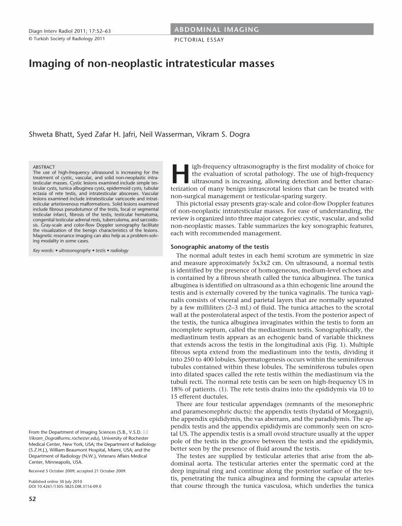
Imaging of non-neoplastic intratesticular masses - Diagnostic and
12
© Turkish Society of Radiology 2011 52 H igh-frequency ultrasonography is the first modality of choice for the evaluation of scrotal pathology. The use of high-frequency ultrasound is increasing, allowing detection and better charac- terization of many benign intrascrotal lesions that can be treated with non-surgical management or testicular-sparing surgery. This pictorial essay presents gray-scale and color-flow Doppler features of non-neoplastic intratesticular masses. For ease of understanding, the review is organized into three major categories: cystic, vascular, and solid non-neoplastic masses. Table summarizes the key sonographic features, each with recommended management. Sonographic anatomy of the testis The normal adult testes in each hemi scrotum are symmetric in size and measure approximately 5x3x2 cm. On ultrasound, a normal testis is identified by the presence of homogeneous, medium-level echoes and is contained by a fibrous sheath called the tunica albuginea. The tunica albuginea is identified on ultrasound as a thin echogenic line around the testis and is externally covered by the tunica vaginalis. The tunica vagi- nalis consists of visceral and parietal layers that are normally separated by a few milliliters (2–3 mL) of fluid. The tunica attaches to the scrotal wall at the posterolateral aspect of the testis. From the posterior aspect of the testis, the tunica albuginea invaginates within the testis to form an incomplete septum, called the mediastinum testis. Sonographically, the mediastinum testis appears as an echogenic band of variable thickness that extends across the testis in the longitudinal axis (Fig. 1). Multiple fibrous septa extend from the mediastinum into the testis, dividing it into 250 to 400 lobules. Spermatogenesis occurs within the seminiferous tubules contained within these lobules. The seminiferous tubules open into dilated spaces called the rete testis within the mediastinum via the tubuli recti. The normal rete testis can be seen on high-frequency US in 18% of patients. (1). The rete testis drains into the epididymis via 10 to 15 efferent ductules. There are four testicular appendages (remnants of the mesonephric and paramesonephric ducts): the appendix testis (hydatid of Morgagni), the appendix epididymis, the vas aberrans, and the paradidymis. The ap- pendix testis and the appendix epididymis are commonly seen on scro- tal US. The appendix testis is a small ovoid structure usually at the upper pole of the testis in the groove between the testis and the epididymis, better seen by the presence of fluid around the testis. The testes are supplied by testicular arteries that arise from the ab- dominal aorta. The testicular arteries enter the spermatic cord at the deep inguinal ring and continue along the posterior surface of the tes- tis, penetrating the tunica albuginea and forming the capsular arteries that course through the tunica vasculosa, which underlies the tunica ABDOMINAL IMAGING PICTORIAL ESSAY Imaging of non-neoplastic intratesticular masses Shweta Bhatt, Syed Zafar H. Jafri, Neil Wasserman, Vikram S. Dogra From the Department of Imaging Sciences (S.B., V.S.D. [email protected]), University of Rochester Medical Center, New York, USA; the Department of Radiology (S.Z.H.J.), William Beaumont Hospital, Miami, USA; and the Department of Radiology (N.W.), Veterans Affairs Medical Center, Minneapolis, USA. Received 5 October 2009; accepted 21 October 2009. Published online 30 July 2010 DOI 10.4261/1305-3825.DIR.3116-09.0 ABSTRACT The use of high-frequency ultrasound is increasing for the treatment of cystic, vascular, and solid non-neoplastic intra- testicular masses. Cystic lesions examined include simple tes- ticular cysts, tunica albuginea cysts, epidermoid cysts, tubular ectasia of rete testis, and intratesticular abscesses. Vascular lesions examined include intratesticular varicocele and intrat- esticular arteriovenous malformations. Solid lesions examined include fibrous pseudotumor of the testis, focal or segmental testicular infarct, fibrosis of the testis, testicular hematoma, congenital testicular adrenal rests, tuberculoma, and sarcoido- sis. Gray-scale and color-flow Doppler sonography facilitate the visualization of the benign characteristics of the lesions. Magnetic resonance imaging can also help as a problem-solv- ing modality in some cases. Key words: • ultrasonography • testis • radiology Diagn Interv Radiol 2011; 17:52–63
Transcript of Imaging of non-neoplastic intratesticular masses - Diagnostic and
© Turkish Society of Radiology 2011
52
H igh-frequency ultrasonography is the first modality of choice for the evaluation of scrotal pathology. The use of high-frequency ultrasound is increasing, allowing detection and better charac-
terization of many benign intrascrotal lesions that can be treated with non-surgical management or testicular-sparing surgery.
This pictorial essay presents gray-scale and color-flow Doppler features of non-neoplastic intratesticular masses. For ease of understanding, the review is organized into three major categories: cystic, vascular, and solid non-neoplastic masses. Table summarizes the key sonographic features, each with recommended management.
Sonographic anatomy of the testisThe normal adult testes in each hemi scrotum are symmetric in size
and measure approximately 5x3x2 cm. On ultrasound, a normal testis is identified by the presence of homogeneous, medium-level echoes and is contained by a fibrous sheath called the tunica albuginea. The tunica albuginea is identified on ultrasound as a thin echogenic line around the testis and is externally covered by the tunica vaginalis. The tunica vagi-nalis consists of visceral and parietal layers that are normally separated by a few milliliters (2–3 mL) of fluid. The tunica attaches to the scrotal wall at the posterolateral aspect of the testis. From the posterior aspect of the testis, the tunica albuginea invaginates within the testis to form an incomplete septum, called the mediastinum testis. Sonographically, the mediastinum testis appears as an echogenic band of variable thickness that extends across the testis in the longitudinal axis (Fig. 1). Multiple fibrous septa extend from the mediastinum into the testis, dividing it into 250 to 400 lobules. Spermatogenesis occurs within the seminiferous tubules contained within these lobules. The seminiferous tubules open into dilated spaces called the rete testis within the mediastinum via the tubuli recti. The normal rete testis can be seen on high-frequency US in 18% of patients. (1). The rete testis drains into the epididymis via 10 to 15 efferent ductules.
There are four testicular appendages (remnants of the mesonephric and paramesonephric ducts): the appendix testis (hydatid of Morgagni), the appendix epididymis, the vas aberrans, and the paradidymis. The ap-pendix testis and the appendix epididymis are commonly seen on scro-tal US. The appendix testis is a small ovoid structure usually at the upper pole of the testis in the groove between the testis and the epididymis, better seen by the presence of fluid around the testis.
The testes are supplied by testicular arteries that arise from the ab-dominal aorta. The testicular arteries enter the spermatic cord at the deep inguinal ring and continue along the posterior surface of the tes-tis, penetrating the tunica albuginea and forming the capsular arteries that course through the tunica vasculosa, which underlies the tunica
ABDOMINAL IMAGINGPICTORIAL ESSAY
Imaging of non-neoplastic intratesticular masses
Shweta Bhatt, Syed Zafar H. Jafri, Neil Wasserman, Vikram S. Dogra
From the Department of Imaging Sciences (S.B., V.S.D. [email protected]), University of Rochester Medical Center, New York, USA; the Department of Radiology (S.Z.H.J.), William Beaumont Hospital, Miami, USA; and the Department of Radiology (N.W.), Veterans Affairs Medical Center, Minneapolis, USA.
Received 5 October 2009; accepted 21 October 2009.
Published online 30 July 2010DOI 10.4261/1305-3825.DIR.3116-09.0
ABSTRACTThe use of high-frequency ultrasound is increasing for the treatment of cystic, vascular, and solid non-neoplastic intra-testicular masses. Cystic lesions examined include simple tes-ticular cysts, tunica albuginea cysts, epidermoid cysts, tubular ectasia of rete testis, and intratesticular abscesses. Vascular lesions examined include intratesticular varicocele and intrat-esticular arteriovenous malformations. Solid lesions examined include fibrous pseudotumor of the testis, focal or segmental testicular infarct, fibrosis of the testis, testicular hematoma, congenital testicular adrenal rests, tuberculoma, and sarcoido-sis. Gray-scale and color-flow Doppler sonography facilitate the visualization of the benign characteristics of the lesions. Magnetic resonance imaging can also help as a problem-solv-ing modality in some cases.
Key words: • ultrasonography • testis • radiology
Diagn Interv Radiol 2011; 17:52–63
Imaging of non-neoplastic intratesticular masses • 53Volume 17 • Issue 1
Figure 1. Normal testis. Longitudinal gray scale sonogram of the testis demonstrates the mediastinum testis (arrow) seen as an echogenic line traversing through the testis.
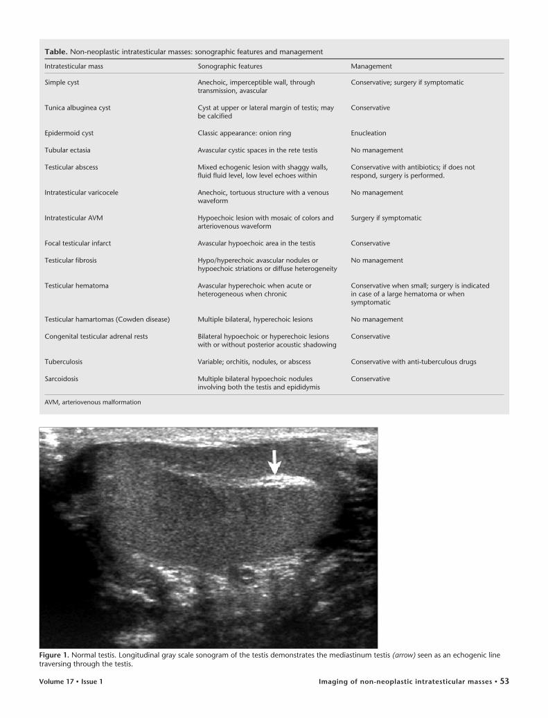
Table. Non-neoplastic intratesticular masses: sonographic features and management
Intratesticular mass Sonographic features Management
Simple cyst Anechoic, imperceptible wall, through transmission, avascular
Conservative; surgery if symptomatic
Tunica albuginea cyst Cyst at upper or lateral margin of testis; may be calcified
Conservative
Epidermoid cyst Classic appearance: onion ring Enucleation
Tubular ectasia Avascular cystic spaces in the rete testis No management
Testicular abscess Mixed echogenic lesion with shaggy walls, fluid fluid level, low level echoes within
Conservative with antibiotics; if does not respond, surgery is performed.
Intratesticular varicocele Anechoic, tortuous structure with a venous waveform
No management
Intratesticular AVM Hypoechoic lesion with mosaic of colors and arteriovenous waveform
Surgery if symptomatic
Focal testicular infarct Avascular hypoechoic area in the testis Conservative
Testicular fibrosis Hypo/hyperechoic avascular nodules or hypoechoic striations or diffuse heterogeneity
No management
Testicular hematoma Avascular hyperechoic when acute or heterogeneous when chronic
Conservative when small; surgery is indicated in case of a large hematoma or when symptomatic
Testicular hamartomas (Cowden disease) Multiple bilateral, hyperechoic lesions No management
Congenital testicular adrenal rests Bilateral hypoechoic or hyperechoic lesions with or without posterior acoustic shadowing
Conservative
Tuberculosis Variable; orchitis, nodules, or abscess Conservative with anti-tuberculous drugs
Sarcoidosis Multiple bilateral hypoechoic nodules involving both the testis and epididymis
Conservative
AVM, arteriovenous malformation

Bhatt et al.54 • March 2011 • Diagnostic and Interventional Radiology
albuginea. Branches from the capsular arteries carry blood toward the medi-astinum and divide to form the recur-rent rami that then carry blood from the mediastinum into the testis. A transmediastinal branch of the testicu-lar artery is present in approximately one-half of normal testes. It traverses through the mediastinum to supply the capsular arteries and is usually accom-panied by a large vein. The testicular veins exit from the mediastinum and drain into the pampiniform plexus, which also receives venous drainage from the epididymis and scrotal wall. These vessels join together as they pass
through the inguinal canal and form single testicular veins on each side, ul-timately draining into the vena cava on the right side and the left renal vein on the left side of the body.
Cystic lesionsSimple testicular cysts
Testicular cysts occur in approxi-mately 8% to 10% of patients (2). Benign cysts are often incidentally found and are generally not palpable (3). They usually occur near the medi-astinum testis and are associated with extratesticular spermatoceles (4, 5). On ultrasound, simple cysts have an im-
perceptible wall, an anechoic center, and through transmission, with sizes ranging from 2 mm to 2 cm in diam-eter (Fig. 2) (4).
Tunica albuginea cystsTunica albuginea cysts are benign
and arise from within the leaves of the tunica albuginea. By virtue of their lo-cation, these cysts are almost always palpable despite being very small in size, ranging from 2 to 7 mm (Fig. 3a) (4, 6). These cysts meet the criteria for a simple cyst by ultrasound but some-times may be calcified or even contain milk of calcium (Fig. 3b) (7).
Figure 3. a, b. Tunica albuginea cysts. A 40-year-old male who presented with a palpable lump in the testis (a). Longitudinal gray-scale sonogram of the testis demonstrates a cyst (arrow) in the supero-anterior part of testis within the two layers of tunica albuginea. A 38-year-old male who presented with scrotal swelling and a palpable lump in the testis (b). Longitudinal sonogram of the left scrotum demonstrates a partially calcified tunica albuginea cyst.
ba
Figure 2. Simple testicular cysts. A 45-year-old male presented with a dull ache in the left scrotum. Color flow Doppler sonogram of the left testis demonstrates multiple cystic areas (arrows) within the left testis that have an imperceptible wall, anechoic centers, posterior through transmission, and absent internal vascularity.
Imaging of non-neoplastic intratesticular masses • 55Volume 17 • Issue 1
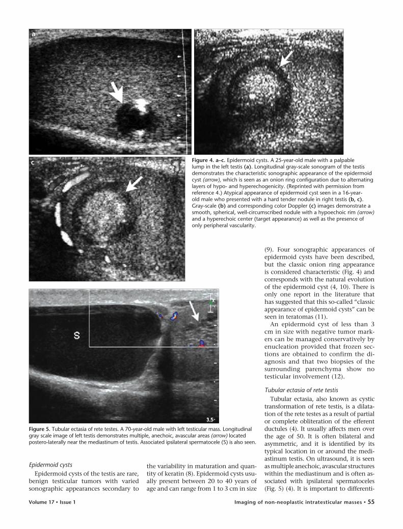
Epidermoid cystsEpidermoid cysts of the testis are rare,
benign testicular tumors with varied sonographic appearances secondary to
the variability in maturation and quan-tity of keratin (8). Epidermoid cysts usu-ally present between 20 to 40 years of age and can range from 1 to 3 cm in size
(9). Four sonographic appearances of epidermoid cysts have been described, but the classic onion ring appearance is considered characteristic (Fig. 4) and corresponds with the natural evolution of the epidermoid cyst (4, 10). There is only one report in the literature that has suggested that this so-called “classic appearance of epidermoid cysts” can be seen in teratomas (11).
An epidermoid cyst of less than 3 cm in size with negative tumor mark-ers can be managed conservatively by enucleation provided that frozen sec-tions are obtained to confirm the di-agnosis and that two biopsies of the surrounding parenchyma show no testicular involvement (12).
Tubular ectasia of rete testisTubular ectasia, also known as cystic
transformation of rete testis, is a dilata-tion of the rete testes as a result of partial or complete obliteration of the efferent ductules (4). It usually affects men over the age of 50. It is often bilateral and asymmetric, and it is identified by its typical location in or around the medi-astinum testis. On ultrasound, it is seen as multiple anechoic, avascular structures within the mediastinum and is often as-sociated with ipsilateral spermatoceles (Fig. 5) (4). It is important to differenti-
Figure 4. a–c. Epidermoid cysts. A 25-year-old male with a palpable lump in the left testis (a). Longitudinal gray-scale sonogram of the testis demonstrates the characteristic sonographic appearance of the epidermoid cyst (arrow), which is seen as an onion ring configuration due to alternating layers of hypo- and hyperechogenicity. (Reprinted with permission from reference 4.) Atypical appearance of epidermoid cyst seen in a 16-year-old male who presented with a hard tender nodule in right testis (b, c). Gray-scale (b) and corresponding color Doppler (c) images demonstrate a smooth, spherical, well-circumscribed nodule with a hypoechoic rim (arrow) and a hyperechoic center (target appearance) as well as the presence of only peripheral vascularity.
ba
c
Figure 5. Tubular ectasia of rete testes. A 70-year-old male with left testicular mass. Longitudinal gray scale image of left testis demonstrates multiple, anechoic, avascular areas (arrow) located postero-laterally near the mediastinum of testis. Associated ipsilateral spermatocele (S) is also seen.
Bhatt et al.56 • March 2011 • Diagnostic and Interventional Radiology
ate this benign entity from malignant cystic tumors of the testis and thus avoid unnecessary orchidectomy. Cystic ma-lignant tumors, most commonly the ter-atomas, can be distinguished sonograph-ically by the presence of multiple cystic areas, often surrounded by a soft tissue rind. They are almost always unilateral and are not limited to the mediastinum (13). They can be further confirmed by measuring serum tumor markers.
Intratesticular abscessAn intratesticular abscess most fre-
quently results from epididymo-orchi-tis and less commonly from a superad-ded infection in post-traumatic testicu-lar hematomas and testicular infarcts (10). Patients with an intratesticular abscess present with an acutely pain-ful scrotum and associated fever. Ultra-sonography is the imaging modality of choice and demonstrates a hypoechoic lesion within the testis marked by low-level echoes and shaggy margins. Color flow Doppler demonstrates ab-sent internal vascularity with increased peripheral hyperemia (Fig. 6) (4).
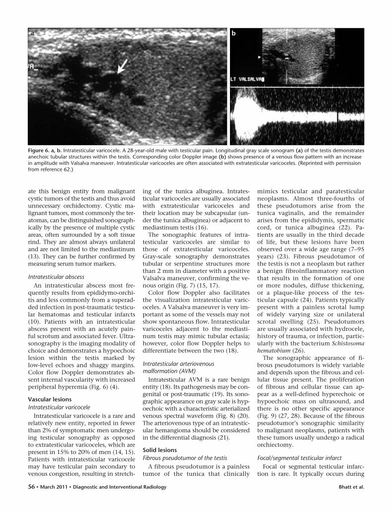
Vascular lesionsIntratesticular varicocele
Intratesticular varicocele is a rare and relatively new entity, reported in fewer than 2% of symptomatic men undergo-ing testicular sonography as opposed to extratesticular varicoceles, which are present in 15% to 20% of men (14, 15). Patients with intratesticular varicocele may have testicular pain secondary to venous congestion, resulting in stretch-
Figure 6. a, b. Intratesticular varicocele. A 28-year-old male with testicular pain. Longitudinal gray scale sonogram (a) of the testis demonstrates anechoic tubular structures within the testis. Corresponding color Doppler image (b) shows presence of a venous flow pattern with an increase in amplitude with Valsalva maneuver. Intratesticular varicoceles are often associated with extratesticular varicoceles. (Reprinted with permission from reference 62.)
ba
ing of the tunica albuginea. Intrates-ticular varicoceles are usually associated with extratesticular varicoceles and their location may be subcapsular (un-der the tunica albuginea) or adjacent to mediastinum testis (16).
The sonographic features of intra-testicular varicoceles are similar to those of extratesticular varicoceles. Gray-scale sonography demonstrates tubular or serpentine structures more than 2 mm in diameter with a positive Valsalva maneuver, confirming the ve-nous origin (Fig. 7) (15, 17).
Color flow Doppler also facilitates the visualization intratesticular varic-oceles. A Valsalva maneuver is very im-portant as some of the vessels may not show spontaneous flow. Intratesticular varicoceles adjacent to the mediasti-num testis may mimic tubular ectasia; however, color flow Doppler helps to differentiate between the two (18).
Intratesticular arteriovenous malformation (AVM)
Intratesticular AVM is a rare benign entity (18). Its pathogenesis may be con-genital or post-traumatic (19). Its sono-graphic appearance on gray scale is hyp-oechoic with a characteristic arterialized venous spectral waveform (Fig. 8) (20). The arteriovenous type of an intratestic-ular hemangioma should be considered in the differential diagnosis (21).
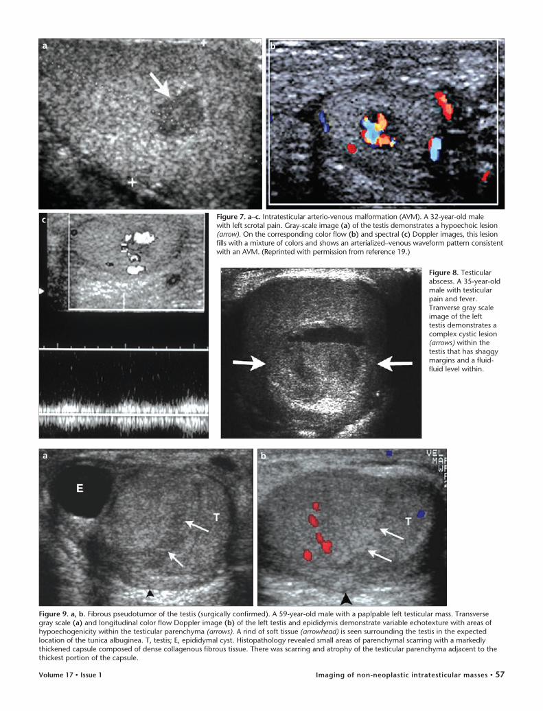
Solid lesionsFibrous pseudotumor of the testis
A fibrous pseudotumor is a painless tumor of the tunica that clinically
mimics testicular and paratesticular neoplasms. Almost three-fourths of these pseudotumors arise from the tunica vaginalis, and the remainder arises from the epididymis, spermatic cord, or tunica albuginea (22). Pa-tients are usually in the third decade of life, but these lesions have been observed over a wide age range (7–95 years) (23). Fibrous pseudotumor of the testis is not a neoplasm but rather a benign fibroinflammatory reaction that results in the formation of one or more nodules, diffuse thickening, or a plaque-like process of the tes-ticular capsule (24). Patients typically present with a painless scrotal lump of widely varying size or unilateral scrotal swelling (25). Pseudotumors are usually associated with hydrocele, history of trauma, or infection, partic-ularly with the bacterium Schistosoma hematobium (26).
The sonographic appearance of fi-brous pseudotumors is widely variable and depends upon the fibrous and cel-lular tissue present. The proliferation of fibrous and cellular tissue can ap-pear as a well-defined hyperechoic or hypoechoic mass on ultrasound, and there is no other specific appearance (Fig. 9) (27, 28). Because of the fibrous pseudotumor’s sonographic similarity to malignant neoplasms, patients with these tumors usually undergo a radical orchiectomy.
Focal/segmental testicular infarctFocal or segmental testicular infarc-
tion is rare. It typically occurs during
Imaging of non-neoplastic intratesticular masses • 57Volume 17 • Issue 1
Figure 9. a, b. Fibrous pseudotumor of the testis (surgically confirmed). A 59-year-old male with a paplpable left testicular mass. Transverse gray scale (a) and longitudinal color flow Doppler image (b) of the left testis and epididymis demonstrate variable echotexture with areas of hypoechogenicity within the testicular parenchyma (arrows). A rind of soft tissue (arrowhead) is seen surrounding the testis in the expected location of the tunica albuginea. T, testis; E, epididymal cyst. Histopathology revealed small areas of parenchymal scarring with a markedly thickened capsule composed of dense collagenous fibrous tissue. There was scarring and atrophy of the testicular parenchyma adjacent to the thickest portion of the capsule.
ba
Figure 8. Testicular abscess. A 35-year-old male with testicular pain and fever. Tranverse gray scale image of the left testis demonstrates a complex cystic lesion (arrows) within the testis that has shaggy margins and a fluid-fluid level within.
Figure 7. a–c. Intratesticular arterio-venous malformation (AVM). A 32-year-old male with left scrotal pain. Gray-scale image (a) of the testis demonstrates a hypoechoic lesion (arrow). On the corresponding color flow (b) and spectral (c) Doppler images, this lesion fills with a mixture of colors and shows an arterialized–venous waveform pattern consistent with an AVM. (Reprinted with permission from reference 19.)
ba
c
Bhatt et al.58 • March 2011 • Diagnostic and Interventional Radiology
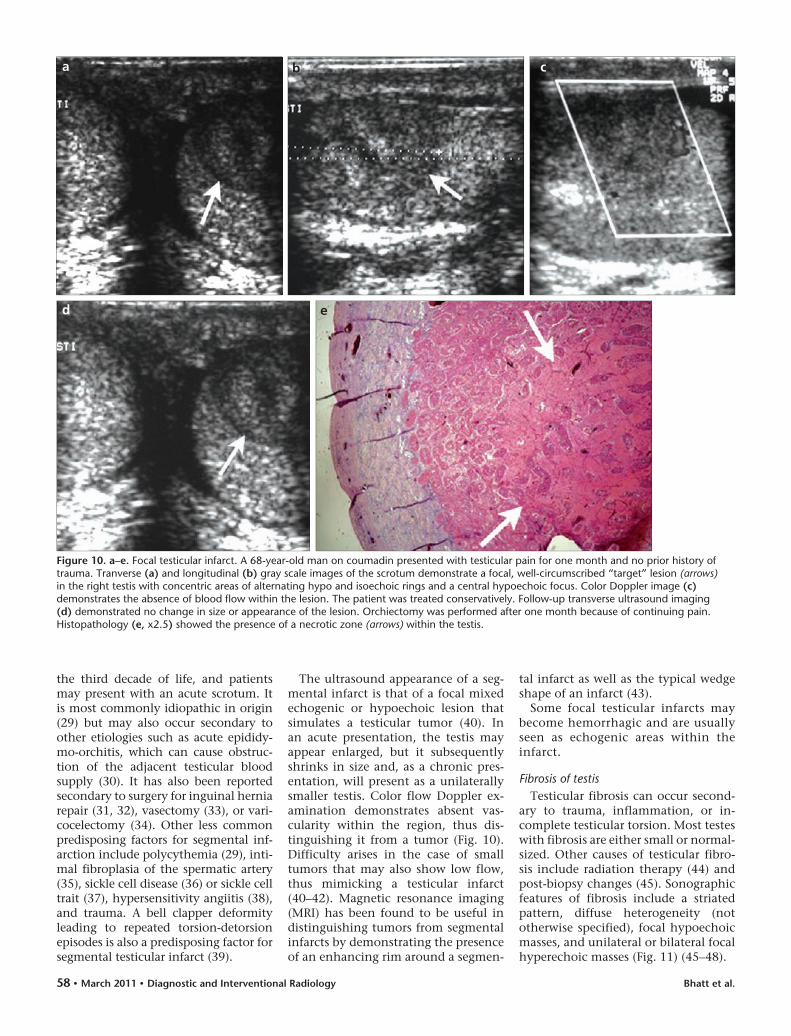
the third decade of life, and patients may present with an acute scrotum. It is most commonly idiopathic in origin (29) but may also occur secondary to other etiologies such as acute epididy-mo-orchitis, which can cause obstruc-tion of the adjacent testicular blood supply (30). It has also been reported secondary to surgery for inguinal hernia repair (31, 32), vasectomy (33), or vari-cocelectomy (34). Other less common predisposing factors for segmental inf-arction include polycythemia (29), inti-mal fibroplasia of the spermatic artery (35), sickle cell disease (36) or sickle cell trait (37), hypersensitivity angiitis (38), and trauma. A bell clapper deformity leading to repeated torsion-detorsion episodes is also a predisposing factor for segmental testicular infarct (39).
The ultrasound appearance of a seg-mental infarct is that of a focal mixed echogenic or hypoechoic lesion that simulates a testicular tumor (40). In an acute presentation, the testis may appear enlarged, but it subsequently shrinks in size and, as a chronic pres-entation, will present as a unilaterally smaller testis. Color flow Doppler ex-amination demonstrates absent vas-cularity within the region, thus dis-tinguishing it from a tumor (Fig. 10). Difficulty arises in the case of small tumors that may also show low flow, thus mimicking a testicular infarct (40–42). Magnetic resonance imaging (MRI) has been found to be useful in distinguishing tumors from segmental infarcts by demonstrating the presence of an enhancing rim around a segmen-
tal infarct as well as the typical wedge shape of an infarct (43).
Some focal testicular infarcts may become hemorrhagic and are usually seen as echogenic areas within the infarct.
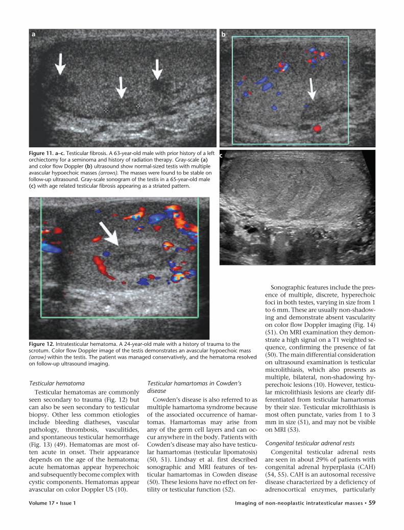
Fibrosis of testisTesticular fibrosis can occur second-
ary to trauma, inflammation, or in-complete testicular torsion. Most testes with fibrosis are either small or normal-sized. Other causes of testicular fibro-sis include radiation therapy (44) and post-biopsy changes (45). Sonographic features of fibrosis include a striated pattern, diffuse heterogeneity (not otherwise specified), focal hypoechoic masses, and unilateral or bilateral focal hyperechoic masses (Fig. 11) (45–48).
Figure 10. a–e. Focal testicular infarct. A 68-year-old man on coumadin presented with testicular pain for one month and no prior history of trauma. Tranverse (a) and longitudinal (b) gray scale images of the scrotum demonstrate a focal, well-circumscribed “target” lesion (arrows) in the right testis with concentric areas of alternating hypo and isoechoic rings and a central hypoechoic focus. Color Doppler image (c) demonstrates the absence of blood flow within the lesion. The patient was treated conservatively. Follow-up transverse ultrasound imaging (d) demonstrated no change in size or appearance of the lesion. Orchiectomy was performed after one month because of continuing pain. Histopathology (e, x2.5) showed the presence of a necrotic zone (arrows) within the testis.
ba c
ed
Imaging of non-neoplastic intratesticular masses • 59Volume 17 • Issue 1
Testicular hematomaTesticular hematomas are commonly
seen secondary to trauma (Fig. 12) but can also be seen secondary to testicular biopsy. Other less common etiologies include bleeding diatheses, vascular pathology, thrombosis, vasculitides, and spontaneous testicular hemorrhage (Fig. 13) (49). Hematomas are most of-ten acute in onset. Their appearance depends on the age of the hematoma; acute hematomas appear hyperechoic and subsequently become complex with cystic components. Hematomas appear avascular on color Doppler US (10).
Testicular hamartomas in Cowden’s disease
Cowden’s disease is also referred to as multiple hamartoma syndrome because of the associated occurrence of hamar-tomas. Hamartomas may arise from any of the germ cell layers and can oc-cur anywhere in the body. Patients with Cowden’s disease may also have testicu-lar hamartomas (testicular lipomatosis) (50, 51). Lindsay et al. first described sonographic and MRI features of tes-ticular hamartomas in Cowden disease (50). These lesions have no effect on fer-tility or testicular function (52).
Sonographic features include the pres-ence of multiple, discrete, hyperechoic foci in both testes, varying in size from 1 to 6 mm. These are usually non-shadow-ing and demonstrate absent vascularity on color flow Doppler imaging (Fig. 14) (51). On MRI examination they demon-strate a high signal on a T1 weighted se-quence, confirming the presence of fat (50). The main differential consideration on ultrasound examination is testicular microlithiasis, which also presents as multiple, bilateral, non-shadowing hy-perechoic lesions (10). However, testicu-lar microlithiasis lesions are clearly dif-ferentiated from testicular hamartomas by their size. Testicular microlithiasis is most often punctate, varies from 1 to 3 mm in size (51), and may not be visible on MRI (53).
Congenital testicular adrenal restsCongenital testicular adrenal rests
are seen in about 29% of patients with congenital adrenal hyperplasia (CAH) (54, 55). CAH is an autosomal recessive disease characterized by a deficiency of adrenocortical enzymes, particularly
Figure 11. a–c. Testicular fibrosis. A 63-year-old male with prior history of a left orchiectomy for a seminoma and history of radiation therapy. Gray-scale (a) and color flow Doppler (b) ultrasound show normal-sized testis with multiple avascular hypoechoic masses (arrows). The masses were found to be stable on follow-up ultrasound. Gray-scale sonogram of the testis in a 65-year-old male (c) with age related testicular fibrosis appearing as a striated pattern.
ba
c
Figure 12. Intratesticular hematoma. A 24-year-old male with a history of trauma to the scrotum. Color flow Doppler image of the testis demonstrates an avascular hypoechoic mass (arrow) within the testis. The patient was managed conservatively, and the hematoma resolved on follow-up ultrasound imaging.
Bhatt et al.60 • March 2011 • Diagnostic and Interventional Radiology
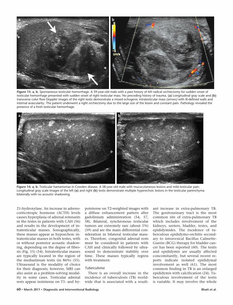
21-hydroxylase. An increase in adreno-corticotropic hormone (ACTH) levels causes hyperplasia of adrenal remnants in the testes in patients with CAH (56) and results in the development of in-tratesticular masses. Sonographically, these masses appear as hypoechoic in-tratesticular masses in both testes, with or without posterior acoustic shadow-ing, depending on the degree of fibro-sis (Fig. 15) (54). Intratesticular masses are typically located in the region of the mediastinum testis (in 86%) (55). Ultrasound is the modality of choice for their diagnosis; however, MRI can also assist as a problem-solving modal-ity in some cases. Testicular adrenal rests appear isointense on T1- and hy-
pointense on T2-weighted images with a diffuse enhancement pattern after gadolinium administration (54, 57, 58). Bilateral, synchronous testicular tumors are extremely rare (about 1%) (59) and are the main differential con-sideration in bilateral testicular mass-es. Therefore, congenital adrenal rests must be considered in patients with CAH and clinically followed by ultra-sound to demonstrate stability over time. These masses typically regress with treatment.
TuberculomaThere is an overall increase in the
incidence of tuberculosis (TB) world-wide that is associated with a result-
ant increase in extra-pulmonary TB. The genitourinary tract is the most common site of extra-pulmonary TB which includes involvement of the kidneys, ureters, bladder, testes, and epididymides. The incidence of tu-berculous epididymo-orchitis second-ary to intravesical Bacillus Calmette-Guerin (BCG) therapy for bladder can-cer has been reported (60). The testis and epididymis are usually affected concomitantly, but several recent re-ports indicate isolated epididymal involvement as well (61). The most common finding in TB is an enlarged epididymis with calcification (24). Tu-berculous involvement of the testis is variable. It may involve the whole
Figure 14. a, b. Testicular hamartomas in Cowden disease. A 38-year-old male with mucocutaneous lesions and mild testicular pain. Longitudinal gray scale images of the left (a) and right (b) testis demonstrate multiple hyperechoic lesions in the testicular parenchyma bilaterally with no acoustic shadowing.
ba
Figure 13. a, b. Spontaneous testicular hemorrhage. A 39-year-old male with a past history of left radical orchiectomy for sudden onset of testicular hemorrhage presented with sudden onset of right testicular mass. No preceding history of trauma. (a) Longitudinal gray scale and (b) transverse color flow Doppler images of the right testis demonstrate a mixed echogenic intratesticular mass (arrows) with ill-defined walls and internal avascularity. The patient underwent a right orchiectomy due to the large size of the lesion and constant pain. Pathology revealed the presence of a fresh testicular hemorrhage.
ba
Imaging of non-neoplastic intratesticular masses • 61Volume 17 • Issue 1
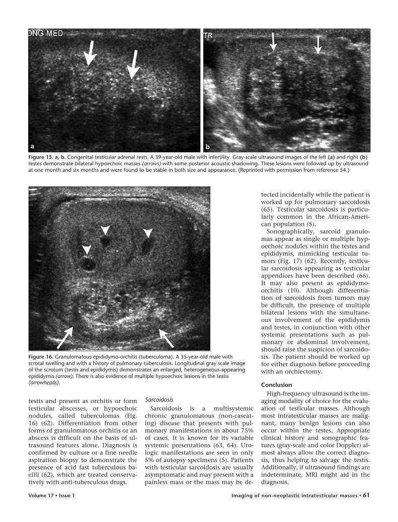
testis and present as orchitis or form testicular abscesses, or hypoechoic nodules, called tuberculomas (Fig. 16) (62). Differentiation from other forms of granulomatous orchitis or an abscess is difficult on the basis of ul-trasound features alone. Diagnosis is confirmed by culture or a fine needle aspiration biopsy to demonstrate the presence of acid fast tuberculous ba-cilli (62), which are treated conserva-tively with anti-tuberculous drugs.
SarcoidosisSarcoidosis is a multisystemic
chronic granulomatous (non-caseat-ing) disease that presents with pul-monary manifestations in about 75% of cases. It is known for its variable systemic presentations (63, 64). Uro-logic manifestations are seen in only 5% of autopsy specimens (5). Patients with testicular sarcoidosis are usually asymptomatic and may present with a painless mass or the mass may be de-
tected incidentally while the patient is worked up for pulmonary sarcoidosis (65). Testicular sarcoidosis is particu-larly common in the African-Ameri-can population (5).
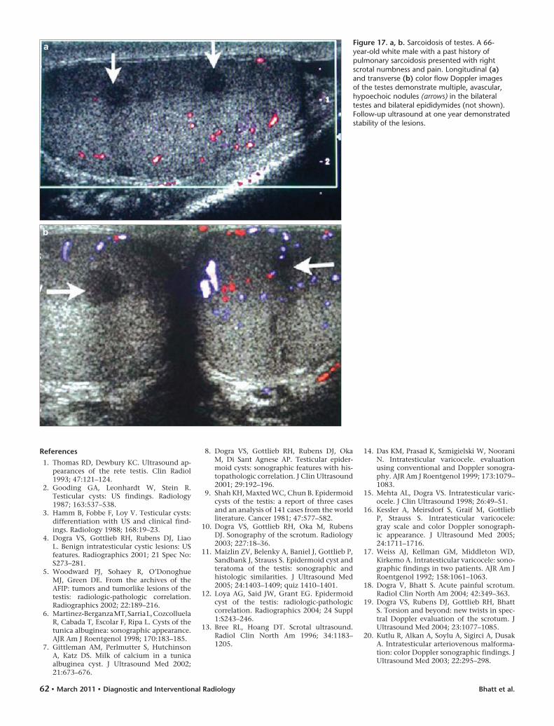
Sonographically, sarcoid granulo-mas appear as single or multiple hyp-oechoic nodules within the testes and epididymis, mimicking testicular tu-mors (Fig. 17) (62). Recently, testicu-lar sarcoidosis appearing as testicular appendices have been described (66). It may also present as epididymo-orchitis (10). Although differentia-tion of sarcoidosis from tumors may be difficult, the presence of multiple bilateral lesions with the simultane-ous involvement of the epididymis and testes, in conjunction with other systemic presentations such as pul-monary or abdominal involvement, should raise the suspicion of sarcoido-sis. The patient should be worked up for either diagnosis before proceeding with an orchiectomy.
ConclusionHigh-frequency ultrasound is the im-
aging modality of choice for the evalu-ation of testicular masses. Although most intratesticular masses are malig-nant, many benign lesions can also occur within the testes. Appropriate clinical history and sonographic fea-tures (gray-scale and color Doppler) al-most always allow the correct diagno-sis, thus helping to salvage the testis. Additionally, if ultrasound findings are indeterminate, MRI might aid in the diagnosis.
Figure 15. a, b. Congenital testicular adrenal rests. A 39-year-old male with infertility. Gray-scale ultrasound images of the left (a) and right (b) testes demonstrate bilateral hypoechoic masses (arrows) with some posterior acoustic shadowing. These lesions were followed up by ultrasound at one month and six months and were found to be stable in both size and appearance. (Reprinted with permission from reference 54.)
ba
Figure 16. Granulomatous epididymo-orchitis (tuberculoma). A 35-year-old male with scrotal swelling and with a history of pulmonary tuberculosis. Longitudinal gray scale image of the scrotum (testis and epididymis) demonstrates an enlarged, heterogeneous-appearing epididymis (arrow). There is also evidence of multiple hypoechoic lesions in the testis (arrowheads).
Bhatt et al.62 • March 2011 • Diagnostic and Interventional Radiology
References 1. Thomas RD, Dewbury KC. Ultrasound ap-
pearances of the rete testis. Clin Radiol 1993; 47:121–124.
2. Gooding GA, Leonhardt W, Stein R. Testicular cysts: US findings. Radiology 1987; 163:537–538.
3. Hamm B, Fobbe F, Loy V. Testicular cysts: differentiation with US and clinical find-ings. Radiology 1988; 168:19–23.
4. Dogra VS, Gottlieb RH, Rubens DJ, Liao L. Benign intratesticular cystic lesions: US features. Radiographics 2001; 21 Spec No:S273–281.
5. Woodward PJ, Sohaey R, O’Donoghue MJ, Green DE. From the archives of the AFIP: tumors and tumorlike lesions of the testis: radiologic-pathologic correlation. Radiographics 2002; 22:189–216.
6. Martinez-Berganza MT, Sarria L, Cozcolluela R, Cabada T, Escolar F, Ripa L. Cysts of the tunica albuginea: sonographic appearance. AJR Am J Roentgenol 1998; 170:183–185.
7. Gittleman AM, Perlmutter S, Hutchinson A, Katz DS. Milk of calcium in a tunica albuginea cyst. J Ultrasound Med 2002; 21:673–676.
8. Dogra VS, Gottlieb RH, Rubens DJ, Oka M, Di Sant Agnese AP. Testicular epider-moid cysts: sonographic features with his-topathologic correlation. J Clin Ultrasound 2001; 29:192–196.
9. Shah KH, Maxted WC, Chun B. Epidermoid cysts of the testis: a report of three cases and an analysis of 141 cases from the world literature. Cancer 1981; 47:577–582.
10. Dogra VS, Gottlieb RH, Oka M, Rubens DJ. Sonography of the scrotum. Radiology 2003; 227:18–36.
11. Maizlin ZV, Belenky A, Baniel J, Gottlieb P, Sandbank J, Strauss S. Epidermoid cyst and teratoma of the testis: sonographic and histologic similarities. J Ultrasound Med 2005; 24:1403–1409; quiz 1410–1401.
12. Loya AG, Said JW, Grant EG. Epidermoid cyst of the testis: radiologic-pathologic correlation. Radiographics 2004; 24 Suppl 1:S243–246.
13. Bree RL, Hoang DT. Scrotal ultrasound. Radiol Clin North Am 1996; 34:1183–1205.
14. Das KM, Prasad K, Szmigielski W, Noorani N. Intratesticular varicocele. evaluation using conventional and Doppler sonogra-phy. AJR Am J Roentgenol 1999; 173:1079–1083.
15. Mehta AL, Dogra VS. Intratesticular varic-ocele. J Clin Ultrasound 1998; 26:49–51.
16. Kessler A, Meirsdorf S, Graif M, Gottlieb P, Strauss S. Intratesticular varicocele: gray scale and color Doppler sonograph-ic appearance. J Ultrasound Med 2005; 24:1711–1716.
17. Weiss AJ, Kellman GM, Middleton WD, Kirkemo A. Intratesticular varicocele: sono-graphic findings in two patients. AJR Am J Roentgenol 1992; 158:1061–1063.
18. Dogra V, Bhatt S. Acute painful scrotum. Radiol Clin North Am 2004; 42:349–363.
19. Dogra VS, Rubens DJ, Gottlieb RH, Bhatt S. Torsion and beyond: new twists in spec-tral Doppler evaluation of the scrotum. J Ultrasound Med 2004; 23:1077–1085.
20. Kutlu R, Alkan A, Soylu A, Sigirci A, Dusak A. Intratesticular arteriovenous malforma-tion: color Doppler sonographic findings. J Ultrasound Med 2003; 22:295–298.
Figure 17. a, b. Sarcoidosis of testes. A 66-year-old white male with a past history of pulmonary sarcoidosis presented with right scrotal numbness and pain. Longitudinal (a) and transverse (b) color flow Doppler images of the testes demonstrate multiple, avascular, hypoechoic nodules (arrows) in the bilateral testes and bilateral epididymides (not shown). Follow-up ultrasound at one year demonstrated stability of the lesions.
b
a
Imaging of non-neoplastic intratesticular masses • 63Volume 17 • Issue 1
21. Ricci Z, Koenigsberg M, Whitney K. Sonography of an arteriovenous-type hemangioma of the testis. AJR Am J Roentgenol 2000; 174:1581–1582.
22. Akbar SA, Sayyed TA, Jafri SZ, Hasteh F, Neill JS. Multimodality imaging of parat-esticular neoplasms and their rare mimics. Radiographics 2003; 23:1461–1476.
23. Ulbright TM, Amin MB, Young RH. Miscellaneous primary tumors of the testis, adnexa, and spermatic cord. Washington, DC: Armed Forces Institute of Pathology, 1999.
24. Woodward PJ, Schwab CM, Sesterhenn IA. From the archives of the AFIP: extratestic-ular scrotal masses: radiologic-pathologic correlation. Radiographics 2003; 23:215–240.
25. Seethala RR, Tirkes AT, Weinstein S, Tomaszewski JE, Malkowicz SB, Genega EM. Diffuse fibrous pseudotumor of the testicular tunics associated with an in-flamed hydrocele. Arch Pathol Lab Med 2003; 127:742–744.
26. Parker PM, Pugliese JM, Allen RC, Jr. Benign fibrous pseudotumor of tunica vaginalis testis. Urology 2006; 68:e417–429.
27. Germaine P, Simerman LP. Fibrous pseu-dotumor of the scrotum. J Ultrasound Med 2007; 26:133–138.
28. al-Otaibi L, Whitman GJ, Chew FS. Fibrous pseudotumor of the epididymis. AJR Am J Roentgenol 1997; 168:1586.
29. Jordan GH. Segmental hemorrhagic infarct of testicle. Urology 1987; 29:60–63.
30. Bird K, Rosenfield AT. Testicular infarc-tion secondary to acute inflammatory dis-ease: demonstration by B-scan ultrasound. Radiology 1984; 152:785–788.
31. Nistal M, Palacios J, Regadera J, Paniagua R. Postsurgical focal testicular infarct. Urol Int 1986; 41:149–151.
32. Mincheff T, Bannister B, Zubel P. Focal testicular infarction from laparoscopic in-guinal hernia repair. Jsls 2002; 6:211–213.
33. Pellice C, Jr., Castella JA, Alert E, Cosme MA, Comas S. Focal infarction of the testis. Report of a case simulating a gonadal mass. Actas Urol Esp 1995; 19:716–720.
34. Secil M, Kocyigit A, Aslan G, et al. Segmental testicular infarction as a complication of varicocelectomy: sonographic findings. J Clin Ultrasound 2006; 34:143–145.
35. Brehmer-Andersson E, Andersson L, Johansson JE. Hemorrhagic infarctions of testis due to intimal fibroplasia of spermat-ic artery. Urology 1985; 25:379–382.
36. Holmes NM, Kane CJ. Testicular infarction associated with sickle cell disease. J Urol 1998; 160:130.
37. deBeck C, Ghasemian R. Acute segmen-tal testicular infarction. Int J Urol 2006; 13:651–652.
38. Baer HM, Gerber WL, Kendall AR, Locke JL, Putong PB. Segmental infarct of the tes-tis due to hypersensitivity angiitis. J Urol 1989; 142:125–127.
39. Dogra V. Bell-clapper deformity. AJR Am J Roentgenol 2003; 180:1176; author reply 1176–1177.
40. Flanagan JJ, Fowler RC. Testicular infarc-tion mimicking tumour on scrotal ul-trasound--a potential pitfall. Clin Radiol 1995; 50:49–50.
41. Kramolowsky EV, Beauchamp RA, Milby WP, 3rd. Color Doppler ultrasound for the diagnosis of segmental testicular infarc-tion. J Urol 1993; 150:972–973.
42. Han DP, Dmochowski RR, Blasser MH, Auman JR. Segmental infarction of the tes-ticle: atypical presentation of a testicular mass. J Urol 1994; 151:159–160.
43. Fernandez-Perez GC, Tardaguila FM, Velasco M, et al. Radiologic findings of segmental testicular infarction. AJR Am J Roentgenol 2005; 184:1587–1593.
44. Aguado A, Grant TH, Miller FH, Garnett J. Radiation-induced fibrosis of the spermat-ic cord: sonographic and MRI findings. AJR Am J Roentgenol 2005; 184:S102–103.
45. Yagan N. Testicular US findings after bi-opsy. Radiology 2000; 215:768–773.
46. Harris RD, Chouteau C, Partrick M, Schned A. Prevalence and significance of hetero-geneous testes revealed on sonography: ex vivo sonographic-pathologic correlation. AJR Am J Roentgenol 2000; 175:347–352.
47. Einstein DM, Paushter DM, Singer AA, Thomas AJ, Levin HS. Fibrotic lesions of the testicle: sonographic patterns mimick-ing malignancy. Urol Radiol 1992; 14:205–210.
48. Casalino DD, Kim R. Clinical importance of a unilateral striated pattern seen on sonog-raphy of the testicle. AJR Am J Roentgenol 2002; 178:927–930.
49. Sinclair J, Ferucci P, Lovell MA, Koyle MA. Spontaneous testicular hemorrhage in an adolescent. J Urol 2003; 169:303–304.
50. Lindsay C, Boardman L, Farrell M. Testicular hamartomas in Cowden disease. J Clin Ultrasound 2003; 31:481–483.
51. Woodhouse J, Ferguson MM. Multiple hy-perechoic testicular lesions are a common finding on ultrasound in Cowden disease and represent lipomatosis of the testis. Br J Radiol 2006; 79:801–803.
52. Woodhouse JB, Delahunt B, English SF, Fraser HH, Ferguson MM. Testicular lipomatosis in Cowden’s syndrome. Mod Pathol 2005; 18:1151–1156.
53. Heinemann V, Frey U, Linke J, Dieckmann KP. Testicular microlithiasis--one case and four points to note. Scand J Urol Nephrol 2003; 37:515–518.
54. Dogra V, Nathan J, Bhatt S. Sonographic appearance of testicular adrenal rest tis-sue in congenital adrenal hyperplasia. J Ultrasound Med 2004; 23:979–981.
55. Proto G, Di Donna A, Grimaldi F, Mazzolini A, Purinan A, Bertolissi F. Bilateral testicu-lar adrenal rest tissue in congenital adre-nal hyperplasia: US and MR features. J Endocrinol Invest 2001; 24:529–531.
56. Willi U, Atares M, Prader A, Zachmann M. Testicular adrenal-like tissue (TALT) in congenital adrenal hyperplasia: detection by ultrasonography. Pediatr Radiol 1991; 21:284–287.
57. Avila NA, Premkumar A, Merke DP. Testicular adrenal rest tissue in congenital adrenal hyperplasia: comparison of MR imaging and sonographic findings. AJR Am J Roentgenol 1999; 172:1003–1006.
58. Stikkelbroeck NM, Suliman HM, Otten BJ, Hermus AR, Blickman JG, Jager GJ. Testicular adrenal rest tumours in post-pubertal males with congenital adrenal hyperplasia: sonographic and MR features. Eur Radiol 2003; 13:1597–1603.
59. Dieckmann KP, Boeckmann W, Brosig W, Jonas D, Bauer HW. Bilateral testicu-lar germ cell tumors. Report of nine cases and review of the literature. Cancer 1986; 57:1254–1258.
60. Muttarak M, Lojanapiwat B, Chaiwun B, Wudhikarn S. Preoperative diagnosis of bilateral tuberculous epididymo-orchitis following intravesical Bacillus Calmette-Guerin therapy for superficial bladder carcinoma. Australas Radiol 2002; 46:183–185.
61. Viswaroop BS, Kekre N, Gopalakrishnan G. Isolated tuberculous epididymitis: a re-view of forty cases. J Postgrad Med 2005; 51:109–111, discussion 111.
62. Bhatt S, Rubens DJ, Dogra VS. Sonography of benign intrascrotal lesions. Ultrasound Q 2006; 22:121–136.
63. Hurd DS, Olsen T. Cutaneous sarcoidosis presenting as a testicular mass. Cutis 2000; 66:435–438.
64. Opal SM, Pittman DL, Hofeldt FE. Testicular sarcoidosis. Am J Med 1979; 67:147–150.
65. Naseem S, Lindley R, Mufti GR. Testicular sarcoid. Urol Int 2001; 67:100–101.
66. Obinata D, Yamaguchi K, Hirano D, Fuchinoue A, Nemoto N, Takahashi S. Intrascrotal involvement of sarcoidosis presenting like testicular appendices. Int J Urol 2007; 14:87–88.