Magnetic resonance cholangiopancreatography: … resonance cholangiopancreatography: the ABC of MRCP...
11
PICTORIAL REVIEW Magnetic resonance cholangiopancreatography: the ABC of MRCP Nyree Griffin & Geoff Charles-Edwards & Lee Alexander Grant Received: 30 May 2011 /Accepted: 9 September 2011 /Published online: 28 September 2011 # European Society of Radiology 2011 Abstract Magnetic resonance cholangiopancreatography (MRCP) is a technique that has evolved over the past two decades. It continues to have a fundamental role in the non- invasive investigation of many pancreatico-biliary disorders. The purpose of this review is to summarise the key concepts behind MRCP, the different techniques that are currently employed (including functional and secretin-stimulated MRCP), the pitfalls the reader should be aware of, and the main clinical indications for its use. Keywords MR cholangiopancreatography . Bile duct calculi . Bile duct neoplasms . Bile duct abnormalities . Pancreatic ducts Introduction It has been exactly two decades since magnetic resonance cholangiopancreatography (MRCP) was first described [1]. Over this time, the technique has evolved considerably, aided by improvements in spatial resolution and speed of acquisition. It has now an established role in the investigation of many biliary disorders, serving as a non-invasive alternative to endoscopic retrograde cholangiopancreatography (ERCP). It makes use of heavily T2-weighted pulse sequences, thus exploiting the inherent differences in the T2-weighted contrast between stationary fluid-filled structures in the abdomen (which have a long T2 relaxation time) and adjacent soft tissue (which has a much shorter T2 relaxation time). Static or slow moving fluids within the biliary tree and pancreatic duct appear of high signal intensity on MRCP, whilst surrounding tissue is of reduced signal intensity. Heavily T2-weighted images were originally achieved using a gradient-echo (GRE) balanced steady-state free precession technique [1, 2]. A fast spin-echo (FSE) pulse sequence with a long echo time (TE) was then introduced shortly after [3], with the advantages of a higher signal-to- noise ratio and contrast-to-noise ratio, and a lower sensitivity to motion and susceptibility artefacts. Modified FSE sequen- ces have been described, including rapid acquisition with rapid enhancement (RARE) [4], half-Fourier acquisition single-shot turbo spin-echo (HASTE) [5], and fast-recovery fast spin-echo (FRFSE) [6] sequences. Both breath-hold (using a single shot approach) [7] and non-breath-hold techniques (with respiratory triggering) [8] have been used, with images obtained either as a two-dimensional (2D) or three-dimensional (3D) acquisition. A 3D technique provides a higher signal to noise ratio, which is traded off for thinner contiguous slices. Acquiring images with near isotropic voxels allows improved post-processing manipulation of the images with multi-planar reconstruction, maximum intensity projection (MIP) and volume rendering. The introduction of faster gradients and a parallel acquisition technique has resulted in even greater spatial resolution and faster acquisition times. More recently, functional assessment of biliary excretion and pancreatic exocrine function has N. Griffin (*) Department of Radiology, St Thomas’ Hospital, Westminster Bridge Road, London SE1 7EH, UK e-mail: [email protected] G. Charles-Edwards Department of Medical Physics, St Thomas’ Hospital, Westminster Bridge Road, London SE1 7EH, UK L. A. Grant Department of Radiology, Royal Free Hospital, Pond Street, London NW3 2QG, UK Insights Imaging (2012) 3:11–21 DOI 10.1007/s13244-011-0129-9
Transcript of Magnetic resonance cholangiopancreatography: … resonance cholangiopancreatography: the ABC of MRCP...

PICTORIAL REVIEW
Magnetic resonance cholangiopancreatography:the ABC of MRCP
Nyree Griffin & Geoff Charles-Edwards &
Lee Alexander Grant
Received: 30 May 2011 /Accepted: 9 September 2011 /Published online: 28 September 2011# European Society of Radiology 2011
Abstract Magnetic resonance cholangiopancreatography(MRCP) is a technique that has evolved over the past twodecades. It continues to have a fundamental role in the non-invasive investigation of many pancreatico-biliary disorders.The purpose of this review is to summarise the key conceptsbehind MRCP, the different techniques that are currentlyemployed (including functional and secretin-stimulatedMRCP), the pitfalls the reader should be aware of, and themain clinical indications for its use.
Keywords MR cholangiopancreatography . Bile ductcalculi . Bile duct neoplasms . Bile duct abnormalities .
Pancreatic ducts
Introduction
It has been exactly two decades since magnetic resonancecholangiopancreatography (MRCP) was first described [1].Over this time, the technique has evolved considerably,aided by improvements in spatial resolution and speed of
acquisition. It has now an established role in the investigationof many biliary disorders, serving as a non-invasive alternativeto endoscopic retrograde cholangiopancreatography (ERCP).It makes use of heavily T2-weighted pulse sequences, thusexploiting the inherent differences in the T2-weighted contrastbetween stationary fluid-filled structures in the abdomen(which have a long T2 relaxation time) and adjacent soft tissue(which has a much shorter T2 relaxation time). Static or slowmoving fluids within the biliary tree and pancreatic duct appearof high signal intensity on MRCP, whilst surrounding tissue isof reduced signal intensity.
Heavily T2-weighted images were originally achievedusing a gradient-echo (GRE) balanced steady-state freeprecession technique [1, 2]. A fast spin-echo (FSE) pulsesequence with a long echo time (TE) was then introducedshortly after [3], with the advantages of a higher signal-to-noise ratio and contrast-to-noise ratio, and a lower sensitivityto motion and susceptibility artefacts. Modified FSE sequen-ces have been described, including rapid acquisition withrapid enhancement (RARE) [4], half-Fourier acquisitionsingle-shot turbo spin-echo (HASTE) [5], and fast-recoveryfast spin-echo (FRFSE) [6] sequences. Both breath-hold(using a single shot approach) [7] and non-breath-holdtechniques (with respiratory triggering) [8] have been used,with images obtained either as a two-dimensional (2D) orthree-dimensional (3D) acquisition. A 3D technique providesa higher signal to noise ratio, which is traded off for thinnercontiguous slices. Acquiring images with near isotropicvoxels allows improved post-processing manipulation ofthe images with multi-planar reconstruction, maximumintensity projection (MIP) and volume rendering. Theintroduction of faster gradients and a parallel acquisitiontechnique has resulted in even greater spatial resolution andfaster acquisition times. More recently, functional assessmentof biliary excretion and pancreatic exocrine function has
N. Griffin (*)Department of Radiology, St Thomas’ Hospital,Westminster Bridge Road,London SE1 7EH, UKe-mail: [email protected]
G. Charles-EdwardsDepartment of Medical Physics, St Thomas’ Hospital,Westminster Bridge Road,London SE1 7EH, UK
L. A. GrantDepartment of Radiology, Royal Free Hospital,Pond Street,London NW3 2QG, UK
Insights Imaging (2012) 3:11–21DOI 10.1007/s13244-011-0129-9

become possible with the use of hepatobiliary contrast media[9] and secretin [10] respectively.
The purpose of this pictorial review is to describe (1) theMRCP protocol used by our centre and additional/alterna-
tive sequences which can be employed, (2) the normalbiliary anatomy on MRCP, (3) the potential pitfallsassociated with this technique and (4) the main clinicalindications for its use.
Table 1 Summary of MRCP imaging parameters
Parameter T2-weighted breath-hold HASTE(liver down to ampulla)
3D T2-weighted FSEwith respiratory triggering
T2 weighted breath-hold HASTEfat-saturated thick slab
TR/TE (ms) 1,000/83 1,800/678 4,500/752
Number of averages 1 1 1
Flip angle 150 180 180
Field of view (mm) 350 × 263 380 × 380 350 × 350
Matrix size 256 × 146 384 × 380 384 × 300
Slice thickness (mm) 7 mm 1.5 mm 40 mm
Slice gap (mm) 0.7 mm 0 mm N/A
Number of slices 20 40 1
Acquisition plane Axial Coronal oblique Coronal
Half-Fourier factor 5/8 Phase-encoding: off Phase encoding: 7/8Slice-encoding: 6/8
Parallel imaging acceleration factor 2 2 2
Receiver bandwidth (Hz/pixel) 391 260 150
Turbo factor 146 127 307
Oversampling None Slice: 20% Phase: 33%
HASTE half-Fourier acquisition single shot turbo spin echo, FSE fast spin echo
Fig. 1 Example of a normalsecretin-stimulated MRCP in apatient being investigated forsphincter of Oddi dysfunction,with idiopathic dilatation of theCBD. a Thick coronal slab atbaseline shows the pancreaticduct is of normal caliber (arrow)with little secretions in theduodenum (D); b) at 3 min thereis progressive filling of theduodenum (D) due to anincrease in pancreatic secretionsand the pancreatic duct hasbecome more prominent(arrow); c) at 9 min, thepancreatic duct has returned tobaseline calibre (arrow)
12 Insights Imaging (2012) 3:11–21
MRCP protocol
Patients are fasted for 4 h prior to the study in order to reducefluid secretions within the stomach and duodenum, reducebowel peristalsis and promote gallbladder distension. We donot routinely use an anti-peristaltic agent. Some centres use anegative oral contrast agent (e.g. iron oxide or blueberry juice)to reduce the signal intensity of overlapping fluid within thestomach and duodenum, although this is not part of ourroutine protocol.
At our institution, MRCP is performed on a 1.5-T AvantoMRI system (Siemens, Erlangen, Germany), using a phased-array body coil. The protocol imaging parameters are shownin Table 1. We first perform an axial 2D breath-hold HASTEsequence. Two breath-hold acquisitions are obtained, so thatthe whole of the liver down to the duodenal ampulla isvisualised.
Following this, we perform two 3D respiratory-triggeredheavily T2-weighted FSE sequences in the coronal obliqueplane. The imaging plane is selected from the initial axialT2-weighted images, with one acquisition aligned to thecommon bile duct (CBD) in the head of the pancreas andthe second acquisition aligned to the pancreatic duct atapproximately 90 degrees to the first imaging plane. Respi-ratory triggering is achieved with the use of a navigatorsequence that employs anMRpre-pulse tomonitor respiratorymotion. The navigator is placed over the edge of thediaphragm on the coronal and sagittal localisers and imageacquisition is triggered when the position of this diaphragminterface with the lung falls within a pre-specified acceptancewindow. In this way a consistent position of the imaging sliceis obtained. The patient is asked to breathe regularlythroughout this acquisition, which takes between 3-5 min toacquire. A stack of 40 slices are obtained, which arecontiguous and each of 1.5 mm in thickness. As the imagesare heavily T2-weighted, the pancreatico-biliary tree isdisplayed as high signal intensity, whilst adjacent structuresare of reduced signal intensity. This sequence is useful indetecting small filling defects or strictures in the biliary orpancreatic ducts. From this volume of data, a MIP reformatcan be generated. This displays only the pixel with the highestsignal intensity along a ray perpendicular to the plane ofprojection. It thus highlights bile-filled and fluid-filledstructures very well. MIP reformats can be generated invarious coronal and sagittal oblique planes. We convention-
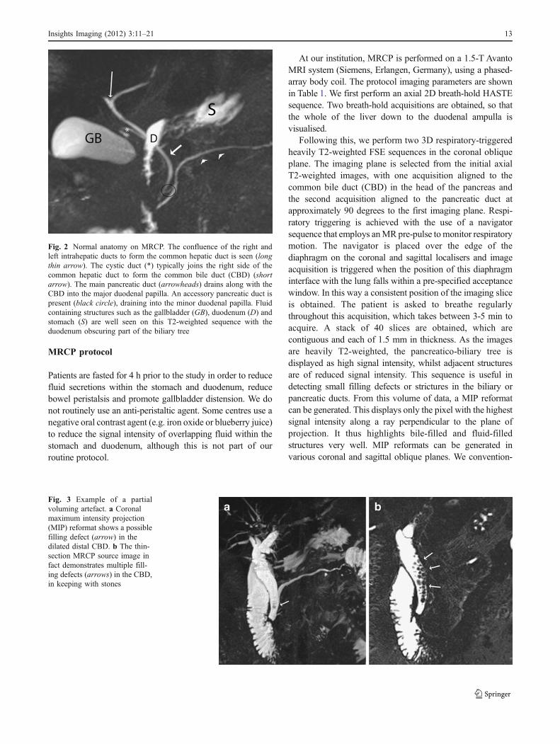
Fig. 2 Normal anatomy on MRCP. The confluence of the right andleft intrahepatic ducts to form the common hepatic duct is seen (longthin arrow). The cystic duct (*) typically joins the right side of thecommon hepatic duct to form the common bile duct (CBD) (shortarrow). The main pancreatic duct (arrowheads) drains along with theCBD into the major duodenal papilla. An accessory pancreatic duct ispresent (black circle), draining into the minor duodenal papilla. Fluidcontaining structures such as the gallbladder (GB), duodenum (D) andstomach (S) are well seen on this T2-weighted sequence with theduodenum obscuring part of the biliary tree
Fig. 3 Example of a partialvoluming artefact. a Coronalmaximum intensity projection(MIP) reformat shows a possiblefilling defect (arrow) in thedilated distal CBD. b The thin-section MRCP source image infact demonstrates multiple fill-ing defects (arrows) in the CBD,in keeping with stones
Insights Imaging (2012) 3:11–21 13

ally create 18 MIP reformats at 10-degree intervals to eachother over a radial array of 180 degrees.
In addition to, or as an alternative to the MIP reformats, athick collimation slab can be obtained in the coronal plane.This involves performing a fat saturated HASTE sequencewhere a single slab of data 4 cm in thickness is acquired in a 1-to 2-s breath-hold. It is useful in depicting the entirepancreatico-biliary tree and no post-processing is required.
In order to evaluate the duct walls, and any focalparenchymal pathology, 3D fat suppressed T1-weightedGRE sequences before and after intravenous contrast admin-istration can also be performed.
Secretin-stimulated MRCP
Secretin is an endogenous hormone normally produced bythe duodenum, which stimulates exocrine secretion of the
pancreas. When given as a synthetic agent intravenously(1 ml/10 kg body weight), it improves the visualisation ofthe pancreatic duct by increasing its calibre. Pancreaticjuice flows out of the major duodenal papilla to progres-sively fill the duodenum. We perform a thick slab MRCP inthe coronal oblique plane at baseline and then at 1, 3, 5, 7and 9 min following injection. Its effect starts almostimmediately and peaks between 2 to 5 mins. By 10 min, thecalibre of the main pancreatic duct should return to baseline(Fig. 1a-c) with persistent dilatation of >3 mm consideredabnormal. The indications for this technique include thedetection and characterisation of pancreatic ductal anoma-lies and strictures, evaluation of the integrity of thepancreatic duct, characterisation of any communicationbetween the pancreatic duct and pseudocysts/pancreaticfistulas, and the assessment of pancreatic function andsphincter of Oddi dysfunction.
Fig. 4 Example of intra-ductal factors causing potential pitfalls ininterpretation. a Axial T2-weighted MRI shows an air-fluid level in adilated proximal CBD in keeping with aerobilia (arrow), adjacent tothe duodenum (D), which also shows an air-fluid level. b Moredistally in the same patient, the cause of the obstruction is seen with adependent filling defect (arrowhead) in the distal CBD in keeping
with a stone. This should not be confused with the non-dependentaerobilia also shown at this level (arrow). c Axial T2-weighted MRI ina different patient shows a central filling defect in a dilated CBDwhich is due to flow artefact (arrow). The patient also has chroniccholecystitis with a contracted gallbladder (arrowheads)
Fig. 5 a Example of an extra-ductal factor causing a potential pitfallin interpretation. Coronal MIP reformat suggests a stricture or possiblefilling defect in the common hepatic duct (arrow) but with noupstream dilatation. Incidental note is also made of a small pseudocyst(P) associated with the main pancreatic duct due to a history of
pancreatitis. b Thin-section MRCP image more clearly shows that thisis due to extrinsic compression from the right hepatic artery whichappears as a subtle curvilinear signal void outside the duct andextending across it (arrows)
14 Insights Imaging (2012) 3:11–21

Functional MR cholangiography
This involves the use of MR lipophilic paramagnetic contrastagents, which when given intravenously, show hepato-biliaryexcretion. Contrast agents include gadobenate dimeglumine(Gd-BOPTA, Multihance; Bracco Imaging, Milan, Italy),gadolinium ethoxybenzyldiethylenetriamine penta-acetic acid(Gd-EOB-DTPA, Primovist; Bayer-Schering Pharma, Berlin,Germany) and, historically, mangafodopir trisodium (Teslascan;GE Healthcare, Oslo, Norway). Delayed imaging in the axialand coronal plane, performed between 10-120 min followingintravenous administration, normally results in hyper-intensebile on 3D T1-weighted fat-saturated GRE images. The signal-to-noise ratio is higher than conventional T2-weighted MRCP,allowing better delineation of the bile ducts. This technique canbe used for similar indications as for T2-weightedMRCP and inmost cases has a similar diagnostic accuracy. It is moreexpensive than conventional T2-weighted MRCP and only thebiliary tree is depicted. For these reasons, most centres continueto use conventional T2-weighted MRCP. However, functionalMRCP does have a number of advantages, as follows: (1) itbetter demonstrates communications between cystic lesions anddraining bile ducts in the diagnosis of congenital biliarydisorders (e.g. Caroli’s disease) [11], (2) it helps to distinguishtrue obstruction in a dilated biliary system (where delayed orno biliary excretion is demonstrated) from pseudo-obstruction[9], and (3) it can demonstrate active extravasation of contrastin suspected bile leaks [12, 13]. Another advantage is thatthese gadolinium-based hepatobiliary-specific contrast agentsinitially distribute in the extracellular fluid compartment, thusallowing for early dynamic pre-contrast and post-contrastimages in the arterial, portal venous and equilibrium phaseprior to the functional cholangiogram.
In cases where there is significant biliary obstruction orimpaired hepatocyte function, delayed images up to 24h can be performed until contrast is seen in the gallbladderand duodenum.
Normal anatomy on MRCP
Only central intra-hepatic bile ducts are normally seen onMRCP (Fig. 2), usually measuring up to 3 mm in diameter,whilst extra-hepatic bile ducts should not exceed 7 mm. Inpatients with a previous cholecystectomy, mild biliarydilatation occurs, with the CBD measuring up to 10 mmin diameter. The intra-hepatic biliary drainage systemparallels the portal venous supply. The right hepatic ducthas two major branches: the right posterior duct, which hasan almost horizontal course (draining posterior segments VIand VII), and the right anterior duct, which has a morevertical course (draining the anterior segments V and VIII).The right posterior duct usually runs posterior to the right
anterior duct and fuses with it on its left (medial) side. Theleft hepatic duct drains segments II-IV and joins the righthepatic duct to form the common hepatic duct. Segment 1drains via a separate bile duct usually into the origin of theleft or right hepatic duct.
The pancreatic duct should be no greater than 3 mm,with the main pancreatic duct of Wirsung normally draininginto the major duodenal papilla along with the CBD (91%of individuals). An accessory pancreatic duct of Santorinimay be present in 45% [14], which drains into the minorduodenal papilla. The cystic duct usually joins the extra-hepatic duct from the right lateral aspect in 50% of cases,although it may insert into its anterior or posterior aspect in30% and medial aspect in 20% of individuals.
Fig. 6 Example of a normal cystic duct variant. Coronal MIPreformat shows medial insertion of the cystic duct into the commonduct (short arrows). There is also a pseudocyst in the lesser sac due tochronic pancreatitis (long arrows)
Fig. 7 Example of variant intrahepatic bile duct branching. CoronalMIP reformat shows the commonest type, where the right posteriorsectoral duct (arrow) drains into the left hepatic duct. Note its morehorizontal orientation compared to the right anterior hepatic duct
Insights Imaging (2012) 3:11–21 15

Pitfalls on MRCP
A number of pitfalls may arise which fall into four maincategories: (1) artefacts related to technique and reconstruction;(2) normal variants mimicking pathology; (3) intra-ductalfactors; (4) extra-ductal factors.
Technique and reconstruction artefacts
A thick slab MRCP may obscure small filling defects orstrictures as the spatial resolution is degraded because ofvolume averaging effects. Partial volume effects alsodegrade spatial resolution in MIP reformats, leading to themissed filling defects (Fig. 3a, b) and over- or under-estimation of strictures. Due to respiratory motion artefact,the biliary tree may appear stenotic, dilated, disconnected orduplicated on MIP reformats. Hence it is important toalways review the original thin-section data set also.
Normal variants
A long cystic duct running parallel to the CBD maysimulate a dilated common duct, whilst a contractedcholedochal sphincter may mimic an impacted stone orstricture in the distal CBD. En face visualisation of thecystic duct insertion into the bile duct may also simulate afilling defect. Performing MRCP in multiple imagingplanes or carrying out repeat MRCP imaging will helpresolve these problems.
Intra-ductal factors
Filling defects in the bile may arise, not only from bile ductcalculi but also from the presence of gas, debris, haemor-rhage and tumour. Aerobilia is seen as a non-dependentfilling defect on the axial images (Fig. 4a, b), whilst asignal void in the central part of the bile duct is due to flow
phenomenon (Fig. 4c) and may occur in dilated ducts and atthe point of insertion of a large cystic duct. The presence ofiodinated contrast material will also reduce the signalintensity of bile.
Extra-ductal factors
Pulsatile vascular compression from adjacent vessels maymimic a stricture. The commonest site of extrinsic vascularcompression is the common hepatic duct, followed by theleft hepatic duct, both due to the right hepatic arterycrossing its posterior aspect (Fig. 5a, b). The mid portion ofthe CBD may also be narrowed due to the gastro-duodenalartery. Pseudo-obstruction is typically seen as a bandlikecompression with minimal proximal dilatation. Susceptibil-ity artefacts from metallic clips and gas may give rise todifficulties in interpretation, although titanium clips used
Fig. 8 a Coronal MIP reformat shows a triple confluence (*) of rightanterior and posterior hepatic ducts and left hepatic duct to form thecommon hepatic duct. A CBD stone is seen as a low signal fillingdefect in the distal CBD (arrow) with mild upstream dilatation. b
Axial T2-weighted MRI of a different patient shows oedematousgallbladder wall in keeping with acute cholecystitis (short arrow) withmultiple small dependent filling defects in the CBD in keeping withbile duct stones (long thin arrow)
Fig. 9 Example of a pancreas divisum. Coronal MIP reformat showsthe main pancreatic duct (P) draining into the minor duodenal papilla(arrow). The CBD (C) drains more inferiorly into the major duodenalpapilla (arrowhead). The accessory duct is not well seen on this MIP
16 Insights Imaging (2012) 3:11–21

nowadays for cholecystectomy are not magnetic. Over-lapping of the biliary tree with other stationary fluids (i.e.from adjacent bowel, cystic collections or ascites) may alsocause interpretation problems.
Clinical indications for MRCP
Identification of congenital anomalies of the cystic andhepatic ducts
A normal biliary anatomy is present in only about 60% of thepopulation. There are many cystic duct variants [15, 16],including a low insertion, a medial insertion (Fig. 6), anddrainage of the cystic duct into the right hepatic duct. Themost important variant of intrahepatic bile duct branchingclinically is the presence of an aberrant right posteriorsectoral duct draining into the common hepatic duct or cysticduct in 5% of the population. This should be recognised onMRCP (if performed), as it may be ligated or cut at the timeof cholecystectomy. The most common anatomic variant ofintrahepatic bile duct branching, occurring in 13-19% of thepopulation [15, 17] is drainage of the right anterior orposterior duct into the left hepatic duct (Fig. 7). This is
important to detect in patients undergoing left hepatectomyfor living related transplant donation as accidental ligation/resection may lead to cirrhosis of segments 5 and 8 orsegments 6 and 7 respectively. A triple confluence [15] mayalso be seen (11%) where the right posterior duct, rightanterior duct and left hepatic duct simultaneously draininto the common hepatic duct (Fig. 8a). When ERCP isused as the ‘gold standard’, MRCP has an accuracy ofbetween 90 to 95% in the diagnosis of such congenitalanomalies [16].
Post-surgical biliary anatomy and complications
ERCP is often not possible in patients with a previousbiliary-enteric anastomosis. MRCP is then useful in thedemonstration of post-surgical biliary anatomy and in thedetection of biliary complications, with a 100% sensitivityin the diagnosis of anastomotic strictures and a 90%sensitivity for choledocholithiasis [18].
Pancreas divisum
This is the most common pancreatic variant and is associatedwith an increased prevalence of acute pancreatitis. There is
Fig. 10 a Coronal MIP refor-mat shows early primarysclerosing cholangitis (PSC)with irregular dilatation andstrictures seen in the left sidedintrahepatic ducts (arrows). bCoronal MIP reformat in adifferent patient with moreadvanced PSC with multipleintrahepatic strictures andstrictures seen in the commonhepatic duct and distal CBD(arrows). There is dilatation ofthe proximal CBD
Fig. 11 a Coronal MIP reformat shows dilatation of the intrahepaticbile ducts with disconnection between the left and right sided ductsand the common duct, due to a hilar cholangiocarcinoma (arrow). The
distal CBD and pancreatic duct appear normal (arrowheads). bCorresponding axial T2-weighted image shows high signal intensitytumour at the porta hepatis (*)
Insights Imaging (2012) 3:11–21 17

failure of fusion of the dorsal and ventral pancreatic ducts. Thelarger dominant dorsal pancreatic duct drains the tail,body and superior head of the pancreas into the minorduodenal papilla (Fig. 9), whilst the smaller ventral ductdrains the inferior pancreatic head and uncinate processinto the major duodenal papilla, along with the CBD.MRCP has a 100% accuracy in the detection of thiscondition [19].
Anomalous pancreaticobiliary junction
Choledochal cysts are associated with anomalous union ofthe pancreaticobiliary duct, where the pancreatic duct andCBD unite outside the duodenal wall and form a longcommon channel greater than 15 mm in length. There arefive different types of choledochal cyst described (Todaniclassification) with three main types of anomalous pan-creaticobiliary junction. MRCP can assist detection of suchvariants when suspected clinically.
Choledocholithiasis
This is usually performed in patients presenting withobstructive liver function tests and with suspected gall-stones and/or a dilated CBD on ultrasound. It is also carriedout in patients who have persistent symptoms and abnormalliver function following cholecystectomy. A systematicreview of the literature has shown that when comparedwith ERCP, MRCP has an aggregated sensitivity, specific-ity, positive predictive value and negative predictive valueof 85%, 93%, 87%, and 82% respectively [20]. Stones (assmall as 2 mm) appear as dependent low-signal-fillingdefects within the CBD (Fig. 8a, b), surrounded by high-signal-intensity bile.
Benign biliary strictures
These usually develop following surgical injury (95%) fromprocedures such as laparoscopic cholecystectomy, hepaticresection, liver transplantation and biliary enteric anasto-mosis. Other causes include trauma, inflammation fromcholedocholithiasis, ischaemia involving the hepatic arteryand primary sclerosing cholangitis (PSC). Typically abenign stricture involves a short segment, with a regularmargin and symmetric narrowing. MRCP can demonstratethe site and extent of the stricture with a reported sensitivityof 91-100% [21].
PSC is a fibrosing inflammatory process of the bileducts, resulting in stenosis of both intrahepatic (80%) andextrahepatic ducts. Seventy percent of patients haveinflammatory bowel disease [22] and 10% of patients with
Fig. 12 Coronal thick slab MRCP shows the classical ‘double duct’sign in a patient with carcinoma at the head of pancreas with dilatationof both the CBD and pancreatic duct (arrows) and distension of thegallbladder (GB)
Fig. 13 Coronal MIP reformat shows intrahepatic bile duct dilatationand a grossly dilated CBD (arrow) with abrupt distal termination dueto a periampullary tumour. There is no pancreatic duct dilatation(arrowheads)
Fig. 14 Coronal MIP reformat in a patient with chronic pancreatitisshows dilatation of the main pancreatic duct, ectasia of the sidebranches (black arrows) and a stone in the proximal pancreatic duct(white arrow)
18 Insights Imaging (2012) 3:11–21

PSC will develop a cholangiocarcinoma. MRCP is not assensitive as ERCP in the detection of early changes, but isuseful for follow-up in established cases, in order to determineseverity and to recognise the development of complications.On MRCP, the ducts appear beaded or have a ‘pruned tree’appearance with multifocal strictures demonstrated, withintervening normal or slightly dilated ducts seen (Fig. 10a, b).
Malignant biliary strictures
Cholangiocarcinoma is a tumour arising from the bile ducts.On MRCP the periductal type is seen as a biliary stricture,
involving the CBD, common hepatic duct, biliary bifurcation(Klatskin tumour) or intrahepatic ducts. Proximal bile ductdilatation occurs (Fig. 11a). Suspicious features includeincreased wall thickness (>3 mm), increased signal intensityon T2-weighted images (Fig. 11b) and progressive enhance-ment of the bile ducts (due to the fibrotic component of thetumour) following intravenous administration of gadolinium.A malignant extrahepatic bile duct stricture is likely to belonger than a benign stricture, with an irregular margin andasymmetric narrowing.
Pancreatic adenocarcinoma usually appears as a focalmass, most often in the head of the pancreas, leading to
Fig. 15 a Coronal MIP reformat shows a small multi-septated cysticlesion (arrow) arising in the uncinate process of the pancreas withcommunication with the main pancreatic duct, in keeping with a sidebranch intraductal papillary mucinous neoplasm (IPMN). b Coronal
MIP reformat in a different patient, shows a large ill defined multiseptatedcystic lesion (arrows) arising in the region of the head/uncinate processof the pancreas separate to the gallbladder (GB) with pancreatic duct(PD) dilatation in keeping with a mixed main and side branch IPMN
Table 2 Differences in epidemiology and morphology on MRCP of cystic pancreatic tumours
Serous cystadenoma Mucinous cystic neoplasms Intraductal mucinous neoplasm (IPMN)
Demographics Typically olderwomen >60 years
Typically youngerwomen 30-50 years
Peak age 6th decade, no gender bias
Site of tumour Anywhere in the pancreas,especially the head
75% in body/tail Side branch type: usually pancreatichead/uncinate process, less frequentlyin the tail; tumour communicates withthe main pancreatic duct
Main duct type: segmental or diffuseinvolvement of the main pancreatic duct
Morphology >6 cysts (<2 cm each),thin septations, centralscar (calcification), doesnot communicate withthe pancreatic duct
Cysts >2 cm, unilocularor multilocular, doesnot communicate withthe pancreatic duct
Side branch type: macrocystic or microcysticappearances
Features of malignancy on MRdenoted by thick septations,soft tissue nodules, and/orpancreatic duct dilatation
Main duct type: diffuse duct dilatation dueto gross mucin production, micropapillarystudding, pancreatic atrophy
Average size 5 cm 6-10 cm Larger size with malignant tumours
Signalcharacteristics
Fluid signal High signal intensity on T1and T2 (mucin/blood)
High signal intensity onT1 (mucin), intermediatesignal intensity on T2
Comments Usually benign Malignant in 50% Side branch type: usuallyassociated with benign adenomas
Main duct type: malignant in 40%
Insights Imaging (2012) 3:11–21 19
encasement and obstruction of the pancreatic duct and/orCBD. Dilatation of both ducts is seen in approximately75% of cases appearing as the ‘double duct’ sign on MRCP(Fig. 12). Although contrast-enhanced CT is conventionallyused for staging local and distant spread in pancreaticcancers, MRCP may be used as an initial investigation inidentifying the level of obstruction in patients presentingwith painless obstructive jaundice.
Peri-ampullary carcinoma can lead to high-grade obstruc-tion of the CBD with abrupt termination on MRCP (Fig. 13).There is usually only mild dilatation of the pancreatic duct.
Chronic pancreatitis
Chronic inflammation of the pancreas results in parenchy-mal destruction with fibrosis, fat necrosis and dystrophiccalcification. Strictures in the main pancreatic duct mayeventually develop a ‘chain-of-lakes’ appearance withalternating stenoses and dilatation. Side branch ectasia andintraductal calculi occur (Fig. 14). In advanced cases therecan be marked dilatation of both the pancreatic duct andCBD simulating the ‘double duct’ sign seen with carcinomaof the head of pancreas. The sensitivity of MRCP in thedetection of early side branch changes can be increasedwith the use of secretin-stimulated MRCP [23].
Pseudocysts are encapsulated fluid collections seen inboth acute and chronic pancreatitis (Figs. 5, 6). These oftendevelop within the lesser sac. MRCP is more sensitive thanERCP in demonstrating these fluid collections and mayshow their connection with the pancreatic duct. With ERCP,less than 50% of pseudocysts opacify with contrast [24].
Cystic pancreatic tumours
Cystic pancreatic tumours include serous cystadenomas,mucinous cystic neoplasms and intraductal papillary mucinousneoplasm (IPMN). Often, small cystic lesions within thepancreas are detected incidentally on CT performed for otherindications. MRCP can help delineate these tumours moreclearly (Fig. 15a, b). Table 2 shows the difference inepidemiology and MRCP morphology between these cystictumours.
Biliary injuries
Following transection of the bile ducts (usually followingsurgery), bile accumulates within the sub-hepatic space.Fluid collections can be appreciated on MRCP withtransection of the affected bile duct. If functional MRCPis used, extravasation of contrast from the biliary tree isseen. A stricture may develop following accidental ligationor transection, with upstream dilatation demonstrated onMRCP.
Conclusion
The technique of MRCP has evolved considerably over thelast 2 decades, with technological advances in bothacquisition and post processing. It remains the investigationof choice for the non-invasive diagnosis of manypancreatico-biliary disorders. It is hoped that this reviewhas helped remind the reader as to the basic conceptsbehind MRCP, the different sequences that can now beemployed, the pitfalls one should be aware of, and why,even in modern day, it remains a test fit for purpose in theradiological investigation of biliary pathology.
References
1. Wallner BK, Schumacher KA, Weidenmaier W, Friedrich JM(1991) Dilated biliary tract: evaluation with MR cholangiographywith a T2-weighted contrast-enhanced fast sequence. Radiology181:805–808
2. Morimoto K, Shimoi M, Shirakawa T et al (1992) Biliaryobstruction: evaluation with three-dimensional MR cholangiography.Radiology 183:578–580
3. Outwater EK (1993) MR cholangiography with a fast spin-echosequence. J Magn Reson Imaging 3(P):131
4. Laubenberger J, Buchert M, Schneider B, Blum U, Hennig J,Langer M (1995) Breath-hold projection magnetic resonancecholangiopancreaticography (MRCP): a new method for theexamination of the bile and pancreatic ducts. Magn Reson Med33:18–23
5. Miyazaki T, Yamashita Y, Tsuchigame T, Yamamoto H, Urata J,Takahashi M (1996) MR cholangiopancreatography usingHASTE (half-Fourier acquisition single-shot turbo spin-echo)sequences. AJR Am J Roentgenol 166:1297–1303
6. Sodickson A, Mortele KJ, Barish MA, Zou KH, Thibodeau S,Tempany CMC (2006) Three-dimensional fast-recovery fast spin-echoMRCP: comparison with two-dimensional single shot fast spin echotechniques. Radiology 238:549–559
7. Takehara Y, Ichijo K, Tooyama N et al (1994) Breath-hold MRcholangiopancreatography with a long-echo-train fast spin-echosequence and a surface coil in chronic pancreatitis. Radiology192:73–78
8. Barish MA, Yucel EK, Soto JA, Chuttani R, Ferrucci JT (1995)MR Cholangiopancreatography: efficacy of three-dimensionalturbo spin-echo technique. AJR Am J Roentgenol 165:295–300
9. Fayad LM, Holland GA, Bergin D et al (2003) Functionalmagnetic resonance cholangiography (fMRC) of the gallbladderand biliary tree with contrast-enhanced magnetic resonancecholangiography. J Magn Reson Imaging 18:449–460
10. Baillie J, Kimberly J (2007) Prospective comparison of secretin-stimulated MRCP with manometry in the diagnosis of sphincter ofOddi dysfunction types II and III. Gut 56:742–744
11. Park MS, Yu JS, Lee JH, Kim KW (2007) Value of manganese-enhanced T1- and T2-weighted MR cholangiography for differ-entiating cystic parenchymal lesions from cystic abnormalitieswhich communicate with bile ducts. Yonsei Med J 48:1072–1074
12. Aduna M, Larena JA, Martin D, Martinez-Guerenu B, Aguirre I,Astigarraga E (2005) Bile duct leaks after laparoscopic cholecys-tectomy: value of contrast-enhanced MRCP. Abdom Imaging30:480–487
13. Thurley PD, Dhingsa R (2008) Laparoscopic cholecystectomy:postoperative imaging. AJR Am J Roentgenol 191:794–801
20 Insights Imaging (2012) 3:11–21

14. Vitellas KM, Keogan MT, Spritzer CE, Nelson RC (2000) MRcholangiopancreatography of bile and pancreatic duct abnormalitieswith emphasis on the single-shot fast spin echo technique. Radio-graphics 20:939–957
15. Puente SG, Bannura GC (1983) Radiological anatomy of thebiliary tract: variations and congenital abnormalities. World J Surg7:271–276
16. Taourel P, Bret PM, Reinhold C, Barkun AN, Atri M (1996)Anatomic variants of the biliary tree: diagnosis with MRcholangiopancreatography. Radiology 199:521–527
17. Gazelle GS, Lee MJ, Mueller PR (1994) Cholangiographicsegmental anatomy of the liver. Radiographics 14:1005–1013
18. Pavone P, Laghi A, Catalano C et al (1997) MR cholangiographyin the examination of patients with biliary-enteric anastomoses.AJR Am J Roentgenol 169:807–811
19. Bret PM, Reinhold C, Taourel P, Guibaud L, Atri M, Barkun AN(1996) Pancreas divisum: evaluation with MR cholangiopancreatog-raphy. Radiology 199:99–103
20. Verma D, Kapadia A, Eisen GM, Adler DG (2006) EUS vsMRCP for detection of choledocholithiasis. Gastrointest Endosc64:248–254
21. Lee MG, Lee HJ, Kim MH, Kang EM, Kim YH, Lee SG, KimPN, Ha HK, Auh YH (1997) Extrahepatic biliary diseases: 3DMR cholangiopancreatography compared with endoscopic retrogradecholangiopancreatography. Radiology 202:663–669
22. Wiesner RH, LaRusso NF (1980) Clinicopathologic features ofthe syndrome of primary sclerosing cholangitis. Gastroenterology79:200–206
23. Czako L (2007) Diagnosis of early-stage chronic pancreatitis bysecretin-enhanced magnetic resonance cholangiopancreatography.J Gastroenterol 42:113–117
24. Nealon WH, Townsend CM Jr, Thompson JC (1989) Preoperativeendoscopic retrograde cholangiopancreatography (ERCP) inpatients with pancreatic pseudocyst associated with resolvingacute and chronic pancreatitis. Ann Surg 209:532–538, discussion538-540
Insights Imaging (2012) 3:11–21 21