Macro and Microscopic. Protected by bony orbits of the skull Send information to brain directly...
24
The Eye: Structure Macro and Microscopic
-
Upload
reynard-walsh -
Category
Documents
-
view
221 -
download
0
Transcript of Macro and Microscopic. Protected by bony orbits of the skull Send information to brain directly...

The Eye: Structure
Macro and Microscopic

Eyes
Protected by bony orbits of the skull Send information to brain directly via
optic nerve.

Development
From 2 outgrowths of the brain that form the optic nerves & the optic cup: The posterior lining of the eye contains
photoreceptors In a mature eye called the retina

External features
Palpebrae: eyelids! Thin flaps of skin Controlled by
orbicular muscles Close when objects
are placed near the eye: reflex arc
Eyelashes Edges of eyelid Protect from dirt

Eye Musculature

External Structures of the Eye
1. Iris2. Lacrimal
caruncle5. Lower lid7. Pupil8. Sclera9. Upper lid

3 primary layers…
Sclera Choroid Retina

Sclera
Tough, outermost, white layer Surrounds and protects the eyeball. Its front surface, the cornea, is
transparent to let light enter the eye. Lacks blood vessels Gets nutrition through diffusion Ideal for transplants

Cornea

Choroid
The choroid coat is the middle layer of the eyeball.
Consists of the colored portion of the eye known as the iris. Iris has a hole in its center called the
pupil. Light enters through the pupil and the size
of the pupil is regulated by the iris. Constricting = parasympathetic muscles Dilating = Sympathetic muscles

Why an iris?
Too much light “bleaches” the photopigment, rhodopsin to opsin Reduces ability to see Opsin must be “reconverted” to
rhodopsin This is related to “night vision” issues
with sudden darkness…

Iris and Choroid

Lens
Transparent body Lies directly behind the pupil Held in place by ciliary muscles (run
in circular, longitudinal, radial orientation; change lens shape)
Focuses light rays of images on the retina Images inverted (both L to R and Up to
Down) Visual cortex reorients these

Lens Function
Increases amount of energy reaching photoreceptive cells
What happens when source moves closer? All light isn’t focused on retina Makes image “fuzzy” because adjacent
cells stimulated An “accommodating” lens clears image up by refocusing light

To focus image… Close image = ROUND lens (decreases
radius of curvature) Far image = FLAT lens (increases radius
of curvature) To round the lens, contract muscles in
ciliary body: Contracting a circular muscle reduces the
aperture This decreases the tension on the suspensory
ligaments, allowing lens to “round up”

Pupil, Lens, Ciliary muscle (body)

Retina Innermost layer of the eyeball. Contains microscopic structures:
Rods▪ Low-light▪ Non-color vision
Cones▪ Bright-light▪ Color vision

Vertebrate Eye
At center of retina, have fovea centralis Concentration of cone cells 1:1 cell/neuron ratio (gives good resolution)
Farther outward, mix of rods and cones, with just rods in peripheral vision Mostly b & w, low-light, low-resolution peripheral
vision Eye directs fovea centralis at objects to
maximize clarity A “blind spot” occurs where the optic
nerve/tract exits the eye

Retina through Opthalmascope
Optimal vision
Blind spot

Eye Fluids
Aqueous humor—in the anterior cavity in front of the lens Provides nutrients to cornea, lens, other
structures Vitreous humor—in the posterior
cavity behind the lens Gelatinous Holds retina to outer wall of choroid May contain “floaters”, which must be
removed surgically

Anterior and Posterior Chambers

Conjunctiva
Mucous membrane covering the front surface of the sclera and lining the eyelid Produces tears Barrier to microbes Susceptible to trauma, infections,
chemical irritation, and allergic reactions

Conjunctiva: thin, transparent epithelium covers the surface Tiny blood vessels
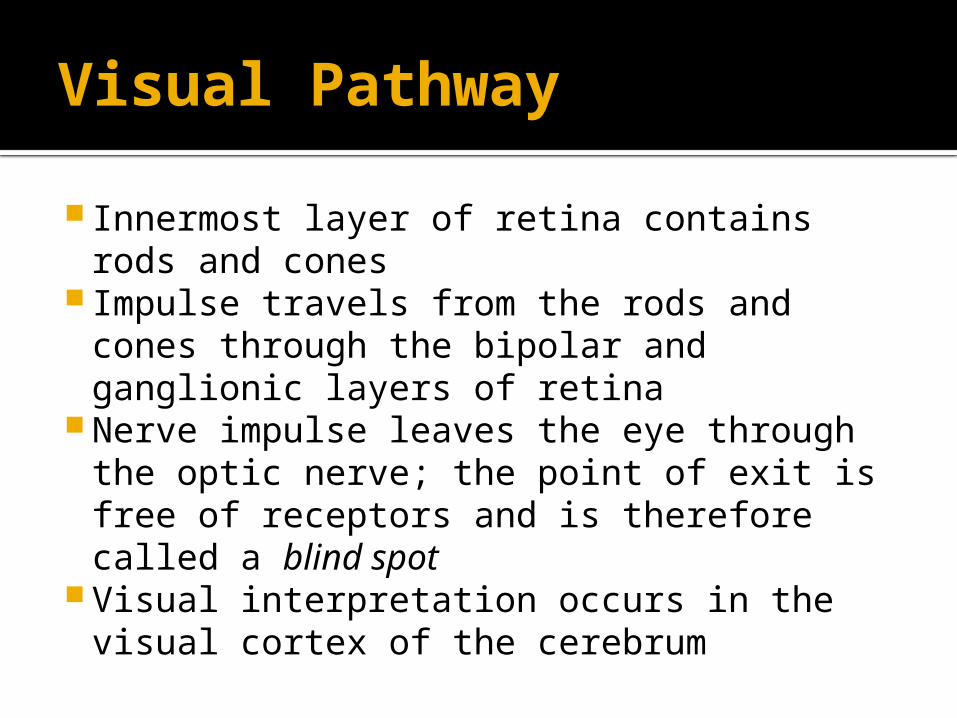
Visual Pathway
Innermost layer of retina contains rods and cones
Impulse travels from the rods and cones through the bipolar and ganglionic layers of retina
Nerve impulse leaves the eye through the optic nerve; the point of exit is free of receptors and is therefore called a blind spot
Visual interpretation occurs in the visual cortex of the cerebrum