ma - Eastern Association for the Surgery of Trauma ventilator settings in the critically-ill...
81
Easte Eas Email: m rn Asso Advanc JW Willia Corinna John J. Ga stern Associatio 633 N. Saint Chic Ph: 312-202-5 managementoffic ociation ced Prac Wo Janua Marriot Scottsd Works am Hoff, M Sicoutris, C Raquel allagher, MS David R Babak on for the Surge t Clair Street, St cago, IL 60611 5508 Fax: 312-2 c[email protected] W for the S ctitioner orkshop ary 17, 2 t Camel dale, Ari shop Facult D – Worksh CRNP – Wo l Forsythe, SN, RN, CC R. Renner, P k Sarani, M ery of Trauma te 2600 202-5064 Web: www.east Surgery rs in Tra p 013 back Inn zona ty: hop Directo orkshop Dir MD CNS, CCRN PA-C MD .org y of Trau auma n or rector N, RRT uma
Transcript of ma - Eastern Association for the Surgery of Trauma ventilator settings in the critically-ill...

Easte
Eas
Email: m
rn Asso
Advanc
JW
WilliaCorinna
John J. Ga
stern Associatio633 N. Saint
ChicPh: 312-202-5
managementoffic
ociation
ced PracWo
JanuaMarriotScottsd
Worksam Hoff, MSicoutris, C
Raquelallagher, MS
David RBabak
on for the Surget Clair Street, Stcago, IL 60611
5508 Fax: [email protected] W
for the S
ctitionerorkshop
ary 17, 2t Camel
dale, Ari
shop FacultD – Worksh
CRNP – Wol Forsythe, SN, RN, CC
R. Renner, Pk Sarani, M
ery of Trauma te 2600
202-5064 Web: www.east
Surgery
rs in Trap
013 back Innzona
ty: hop Directoorkshop DirMD
CNS, CCRNPA-C
MD
.org
y of Trau
auma
n
or rector
N, RRT
uma

Concepts and Strategies in Mechanical Ventilation
John J Gallagher MSN, RN, CCNS, CCRN, RRTTrauma Program Manager/Clinical Nurse Specialist
Division of Traumatology, Surgical Critical Care and Emergency Surgery Hospital of the University of Pennsylvania
Objectives
� Describe the standard ventilation modes as well as initial ventilator settings in the critically-ill patient.
� Describe the differences between volume and pressure targeted ventilation modes and the benefits of each.
� Describe the purpose of lung protective ventilation and alveolar recruitment in patients with ARDS.
Evolution of Mechanical Ventilators
Page 1

Case
40 y.o. male, URD involved in an MVC– One hour extrication time & hypovolemic shock
– Injuries» Multiple mesenteric bleeders
» Ruptured spleen
» Multiple liver lacerations
– Damage control laparotomy
– ICU for resuscitation
Case continued…
48 hours later…….
� Febrile
� HR 142
� Increasing oxygen requirement– P/F ratio: 80 (PaO2 80 on 1.0 FIO2)
� Rising peak inspiratory pressures
Page 2

Capillary LeakCapillary Leak
Fu Z, JAP 1992; 73:123
Acute Lung Injury
Epithelial/Endothelial Damage
Alveolar flooding with proteinacious fluid
Surfactant inactivation
Atelectasis
Shunting
ARDS Continuum
ALI ARDSSevere ARDS
Mild ARDS
Moderate ARDS
PaO2/FiO2 Ratio
Mild Moderate Severe
200 – 300 100 - 200 < 100
Page 3

ARDS Definition
� Acute onset (within 7 days)
� Bilateral opacities (CXR or CT)
� Alveolar edema is not fully explained by cardiac failure or fluid overload– Does not require normal PCWP
– Does not require absence of LA hypertension
Zone of↑ Risk
Ventilator Induced Lung Injury (VILI)“Volutrauma”
Caused by the over-expansion (over- distention)
of alveoli from ventilation with volumes in
excess of relative lung capacity
� Correlated with transalveolar pressure > 30 cmH2O
(Static “plateau” pressures of > 30 cm H2O)
Page 4

Ventilator Induced Lung InjuryVentilator Induced Lung Injury
Collapsed Alveoli
Inspiratory phase
Expiratory phase
�End-tidal collapse/shearing force
�“Milking” of surfactant from alveoli with repeat closure
Lung Protective Principles
� Maintain safe transalveolar pressures– Plateau pressure < 30 cm H20
� Prevent end-tidal alveolar collapse– PEEP
Page 5

Inspiratory Hold: PIP and Plateau pressures (VC)
PlateauPlateau
Peak PressurePeak Pressure
Inspiratory HoldInspiratory Hold
Proximal airway
pressure
Proximal airway
pressure
PIP Elevated and Plateau Normal
PlateauPlateau
Peak PressurePeak Pressure
Inspiratory HoldInspiratory Hold
Proximal airway
pressure
Proximal airway
pressure
40 cm H2O
20 cm H2O
Airway Pressures
� Peak Inspiratory Pressure High and Plat unchanged:(Greater than 10 cmH2O difference between)
– Tracheal tube obstruction
– Airway obstruction from secretions
– Acute bronchospasm
� Rx: Suctioning and Bronchodilators
Page 6

PIP Elevated and Plateau Elevated
Plateau: 36 cmH2OPlateau: 36 cmH2O
Peak Pressure: 40 cmH2OPeak Pressure: 40 cmH2O
Inspiratory HoldInspiratory Hold
Proximal airway
pressure
Proximal airway
pressure
Airway Pressures� Pip and Plat are both increased
(less than 10 cm H2O difference)
– Pneumothorax– Lobar atelectasis– Acute pulmonary edema– Worsening pneumonia– ARDS– Dynamic hyperinflation Asthma/COPD– Increased abdominal pressure (ACS)– Asynchronous breathing
Volume Control Pressure Control
PEEP
A/C
SIMV
A/C
SIMV
Support
Page 7

Volume Targeted (Control)Ventilation (VCV)
� Guaranteed tidal volume with each breath
� Pressure during the delivered breath variesbased on resistance and compliance of the lung
Ventilator Settings
� Tidal Volume (Vt) / Inspiratory pressure
� Respiratory Rate (RR)
� Inspiratory : Expiratory Ratio (I:E)
� Flowrate (Flow)
� Oxygen percentage (FiO2)
� Positive End Expiratory Pressure (PEEP)
� Mode
Respiratory Rate (frequency)
Number of breaths delivered by the ventilator in one minute
– Set to approximate the normal rate of breathing
» Adults 12 - 16 breaths per minute
Page 8
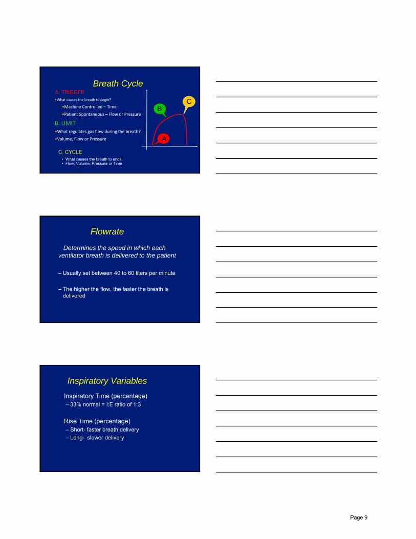
Breath Cycle
C. CYCLE• What causes the breath to end?• Flow, Volume, Pressure or Time
A
BC
A. TRIGGER•What causes the breath to begin?
•Machine Controlled – Time
•Patient Spontaneous – Flow or Pressure
B. LIMIT•What regulates gas flow during the breath?
•Volume, Flow or Pressure
Flowrate
Determines the speed in which each ventilator breath is delivered to the patient
– Usually set between 40 to 60 liters per minute
– The higher the flow, the faster the breath is delivered
Inspiratory Variables
� Inspiratory Time (percentage)– 33% normal = I:E ratio of 1:3
� Rise Time (percentage)– Short- faster breath delivery
– Long- slower delivery
Page 9

Oxygen Percentage (FiO2)
The percentage of oxygen given to the patient through the ventilator
– Range 21% to 100% 21% is what we are breathing in this room
100% is the maximum you can go
– Usually the patient is placed on 100% to start
Volume Targeted (Control) Ventilation (VCV)
Pressure
Flow
Tidal Volume
Conventional Volumes– 10 – 12 ml / kg predicted body weight (PBW)
Low Tidal Volume (LTV)– 5 - 8 ml / kg predicted body weight (PBW)
Predicted Body Weight Calculation• Male PBW in lb: 106 + [6 x (height in inches –60)]
• Female PBW in lb:105 + [5 x (height in inches –60)]
Page 10

Acute Respiratory Distress Syndrome Network ARDSNET
� Comparison of “traditional” tidal volume (12 ml/kg) versus “low” tidal volume (6 ml/kg)
861 patients at 30 centers
Low Tidal Volumes (432) Traditional Tidal Volumes (429)
25-30 cmH2O plateau 45-50 cmH2O plateau
ARDSNET (2000). Ventilation with lower tidal volumes as compared with traditional tidal volumes for acute lung injury and the acute respiratory distress syndrome. NEJM, 342(8), 1301-1308.
Acute Respiratory Distress Syndrome Network ARDSNET
Low tidal Volume Ventilation
� Lower mortality
� Lower levels of IL-6 (lung inflammation)
� Higher number of days without organ or system failure
ARDSNET (2000). Ventilation with lower tidal volumes as compared with traditional tidal volumes for acute lung injury and the acute respiratory distress syndrome. NEJM, 342(8), 1301-1308.
Positive End Expiratory Pressure (PEEP)
� Maintains the alveoli open at the end of the breath
� Reduces shear force injury to alveoli
� Displacement of lung H2O
Insp.
Exp. PEEP
Page 11

Pulmonary Pitfalls of PEEP
Overdistension of normal alveoli
Alveolar Injury Deadspace Shunt
Cardiovascular Pitfalls of PEEP
Increased Intra-thoracic Pressure
RV Afterload Venous Return (RV filling)
Cardiac OutputFalse Elevations of
CVP-PAP-PCOP
Lung Protection is Not Enough
Page 12

Recruitment
� OPEN– Overcome the Trans-alveolar Opening Pressure
� MAINTAIN– Positive End Expiratory Pressure
Recruitment Maneuver Patient Selection
� Early ARDS (<72 hrs) vs. Late ARDs
� Intra-pulmonary vs. Extra-pulmonary lung Injury
� Hemodynamic stability
� Absence of Contraindications
Alveolar Recruitment Strategies
� Recruitment Maneuvers (episodic)
� Pressure Control Ventilation
� Manipulation of I: E ratio
� Airway Pressure Release Ventilation (APRV)
� High Frequency Oscillation Ventilation (HFOV)
Page 13
Improving Oxygenation
� Manipulation of FiO2
� Manipulation of Mean Airway Pressure (Paw)
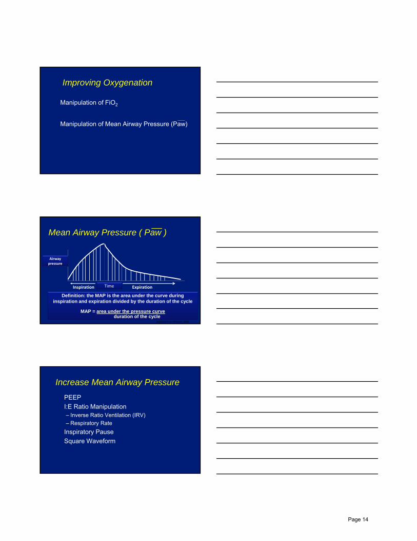
Mean Airway Pressure ( Paw )
Airway pressure
Time Inspiration Expiration
Adapted from Pilbeam, 2006
Definition: the MAP is the area under the curve during inspiration and expiration divided by the duration of the cycle
MAP = area under the pressure curveduration of the cycle
Increase Mean Airway Pressure
� PEEP
� I:E Ratio Manipulation– Inverse Ratio Ventilation (IRV)
– Respiratory Rate
� Inspiratory Pause
� Square Waveform
Page 14

Intermittent Recruitment Maneuver
Brief application of high inspiratory pressures to recruit collapsed alveoli
� Inspiratory pressure application– CPAP 40- 50 cm H2O for 30 - 40 seconds
� Optimal PEEP application to prevent collapse
� Repeat maneuver after a ventilator disconnect
Recruitment
Recruitment with PCV
50
25
5
0
I-Time E-Time I-Time
Time
Pressure
40
I-TimeE-Time
Page 15

Decremental PEEP Trial� PEEP set at 20 cm H2O� PEEP decreased 2 cmH2O every 5-20 min
– Monitoring oxygenation– Compliance
� Until Optimal PEEP is achieved– the lowest PEEP associated with the best
compliance/oxygenation
� Recruitment maneuver repeated� PEEP set 2 cm H2O above the Optimal PEEP
Recruitment Maneuver Cautions
� Pulmonary blebs, bullae, existing barotrauma
� Hemodynamic instability– Hypovolemia
– Impaired RV function
� Increased intracranial pressure (relative)
Pressure Targeted (Control) Ventilation (PCV)
An inspiratory pressure limit, rather than
a tidal volume is set by the practitioner.� Inspiratory pressure & inspiratory time*
� Airway resistance
� Lung compliance
* Practitioner controlled
Volume is variable
Page 16

Pressure Control Ventilation (PCV)
Pressure
Flow
Volume Control vs. Pressure Control Waveform
PIP
PawPaw
50
25
5
0
PCV Waveform Advantages
� Controlled, constant inspiratory pressure
� Better distribution of tidal volume to collapsed alveoli
� Optimal increase in Paw
0
5
25
Paw
Time
Pressure
Page 17

Collateral Ventilation Channels
Pressure Control Inverse Ratio (PCIRV)
50
25
5
0
I-Time E-Time I-Time
Time
Pressure
Auto-PEEP
50
25
50
I-Time E-Time I-Time
Time
Pressure
8
Set PEEPAuto- PEEP
Page 18

P
F
Exp. Flow 50 - 80% of Peak
PCV PC-IRV
Auto PEEPMeasurement
ActualPEEP
Volume Assured Pressure Modes
Pressure Limited + Minimum Volume Guarantee
Machine adjusts to changing lung mechanics to provide tidal volume within pressure limit
Also known as……
� Adaptive Pressure Control Modes
� “Dual Control” Modes
Volume Assured Pressure Modes
Page 19

Volume Assured Pressure Modes
� Combine pressure supported (limited) ventilationwith a decelerating flow pattern and a guaranteed minimum volume.
� Settings vary with ventilator
� All require selection of a “target volume”
� Spontaneous versus control modes determined by selection of settings
Volume Assured Pressure Modes
PCV + Volume Target
•Pressure Regulated Volume Control (PRVC)•Volume Support
•Volume Control Plus (VC+)•Volume Support
•Pressure Control Volume Guarantee (PCVG)
•Volume Targeted Pressure Control (VTPC)
•Adaptive Pressure Ventilation•Adaptive Support Ventilation
•Pressure Augmentation
Control Mode Settings
� Rate (fx)
� Inspiratory time (Ti)
� FiO2
� PEEP
Support Mode Settings
� Target tidal volume
� FiO2
� PEEP
Page 20
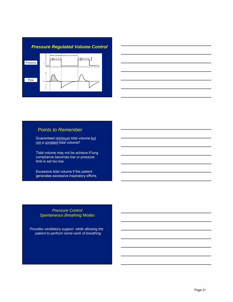
Pressure
Flow
Pressure Regulated Volume Control
Points to Remember
� Guaranteed minimum tidal volume but not a constant tidal volume!!
� Tidal volume may not be achieve if lung compliance becomes low or pressure limit is set too low
� Excessive tidal volume if the patient generates excessive inspiratory efforts
Pressure ControlSpontaneous Breathing Modes
Provides ventilatory support while allowing the patient to perform some work of breathing
Page 21

Benefits of Spontaneous Breathing
� Improved ventilation/perfusion matching
� Lower airway pressures
� Reduced hemodynamic side effects
� Improved organ perfusion
� Less need for sedation/neuromuscular blockade
� New pressure modes have exhalation valves and other technology that allow for patient interaction throughout the respiratory cycle.
Diaphragm Excursion
Awake spontaneous
Anesthetized spontaneous
Paralyzed
Active Exhalation Valve
40PCIRCcmH2O
INSP
Lmin
EXP
30
20
10
0
10
-20
80604020
020
-80
40
60
V.
0 4 8 12s2 6 10
Spontaneous Efforts Spontaneous Efforts
PCV W/O Active Valve PCV with Active Valve
Page 22

Spontaneous Breaths
P
T
PEEPHI
PEEPLO
Biphasic Ventilation
Spontaneous Breaths
•Inspiratory Pressure Limit (PEEPHI)•PEEP (PEEPLOW)•Inspiratory time (Ti)•Rate (fx)•Pressure Support
• Biphasic• Bi-level• Bi-Vent• BIPAP• Duo
PAP
APRV Characteristics� High CPAP level with a short expiratory releases at set
intervals (rate).
� APRV always implies an inverse I:E ratio
� All spontaneous breathing is done at upper pressure level
PEEPHI
PPEEPLO
T
* * ** * * *† ††
Synchronized Transition†Spontaneous Breaths*
Spontaneous Breaths
P
T
PEEPHI
PEEPLO
Spontaneous Breaths
PEEPHI
PPEEPLO
T
* * ** * * *† ††
Synchronized Transition†Spontaneous Breaths*
APRV
BiPhasic
Page 23

Alveolar Volumetric Changes
Insp.
Exp.
Conventional APRV
Insp. Exp.~~
Bi-level/APRV Considerations
� Tidal volume changes with lung compliance
� Over-sedation/NMB may reduce minute ventilation
� Caution in patients requiring longer expiratory time (COPD, Asthma)
� Elevated ICP in TBI– Hypercapnea/ Cerebral venous congestion
General Considerations
� Avoid disconnection of the circuit
� Closed system suctioning
� Transport on the ventilator
Page 24

High Frequency Ventilation
� Jet Ventilation– Up to 600 bpm
� Oscillation– 300 to 3000 bpm
Small tidal volumes
Combined applied and intrinsic (Auto-PEEP) to recruit alveoli
Oscillator
3100B Oscillator
Page 25

Alveolar Volumetric Changes During HFO
Insp.
Exp.
Conventional HFOV
Insp. Exp.~~
Volumetric Diffusive Respiration (VDR)
Images courtesy of Percussionaire Corporation
Page 26

VDR Waveform
Images courtesy of Percussionaire Corporation
So…how should you approach patient management?
� Protect the lung (low volume) and support oxygenation/ventilation
� Recruit the lung
� Know the mode you select!
Thank You
Page 27

Raquel M. Forsythe, MD FACSUniversity of Pittsburgh Medical CenterDivision of Trauma and Acute Care SurgeryEAST Advanced Practice Provider Workshop
Acute Care SurgeryCommon Procedures and Important Concepts
What is Acute Care Surgery?
• Commonly called Acute Care Surgery (ACS) or Emergency General Surgery (EGS)– Acute General Surgery problems
– Often combined with Trauma and/or an elective General Surgery Practice
• ACS is still evolving
• Minimal literature defining ACS
2
Appendicitis Cholecystitis
Vascular insufficiency of the intestine Intestinal Obstruction
Hernia Diverticulitis
Peptic Ulcer Disease Peritonitis
Pancreatitis Enteric fistula
Perianal/Perirectal abscess Skin abscess
Hematoma Gastrostomy/Jejunostomy
Tracheostomy Enterostomy/colostomy
ACS Problems
3
Page 28

• Appendectomy – open versus laparoscopic
Appendicitis
4
• Postoperatively, most patients with an uncomplicated (nonperforated) lap appendectomy can go home within 24h
• For more complicated appendicitis (perforation or gangrene) duration of hospitalization and antibiotics are variable
• Major postop risks: abscess, wound infection, stump leak, fistula
Appendicitis
5
• Laparoscopic versus open cholecystectomy
Cholecystitis
6
Page 29

• Many people with elective lap CCY (for biliary colic or biliary dyskinesia) go home the day of surgery
• For acute cholecystitis
– more common on most EGS/ACS services
– Often need to stay in the hospital postoperatively
– Follow LFTs and WBC
– If bilirubin is elevated postoperatively may represent:
• Retained stones in the CBD
• Bile leak (cystic duct, ducts of Lushka)
• Common duct injury
Postoperative cholecystectomy
7
• If there are retained stones
– ERCP is both diagnostic and therapeutic
• If there is a bile leak
– Confirm with HIDA scan and/or CT scan
– Percutaneous drain in fluid collection
– ERCP for sphincterotomy and stenting of the ampulla
– Antibiotics
• If there is a common duct injury
– Requires reoperation either a direct repair, choledochoenterostomy or hepaticoenterostomy
Postoperative cholecystectomy
8
ERCP with Cystic Duct Stump Leak
9
Page 30

• May be acute or chronic, occlusive or nonocclusive
• May be caused by thrombosis of a chronically diseased vessel or embolus from a distant site
• Most EGS contacts will be for acute mesenteric ischemia
• Most are from acute SMA embolism (50%) generally from the heart– Hx of atrial fibrillation
• May see underlying coagulation deramgements
Vascular Insufficiency of the Intestine
10
• Diagnosis– Pain out of proportion to physical
exam
– Sudden onset in embolic disease
– Lower abdominal pain with BRBPR more likely colon ischemia
– Elevated lactate (but may be absent with venous occlusion)
• Rapid diagnosis and treatment important as mortality is high
Vascular Insufficiency of the Intestine
11
• Treatment– Goal is to restore blood flow as
soon as possible
– Resuscitation, Abx, NGT
– Heparin
– Mesenteric angiogram may be diagnostic and therapeutic (possibility of embolectomy, papaverine, thrombolytics…)
– Surgery for suspicion of bowel infarction
– Resection of necrotic bowel
– Planned second look operation
– Often need an ostomy if perfusion not clearly adequate
Vascular Insufficiency of the Intestine
12
Page 31

• Generally require ICU care postop
• May develop multiple organ failure
• Acute kidney injury is common – Often have underlying chronic renal insufficiency preop
• Acute respiratory failure and pneumonia are common
• Require ongoing resuscitation and monitoring for bleeding (heparin, thrombolysis)
• Complications are frequent and ICU/hospital stay may be prolonged
Vascular Insufficiency of the Intestine - Postop
13
• Adhesions
• Malignancy
• Hernias
• Strictures
• Early postoperative obstruction
• Intussusception
• Gallstone Ileus
• SMA syndrome
Intestinal Obstruction - Causes
14
• Nasogastric tube decompression
• Long decompression tubes (Baker tube)– Rarely used at this point
• Water soluble contrast
Intestinal Obstruction - Treatment
15
Page 32

• Approximately 25% of patients “fail” nonoperativemanagement
• Triggers for surgery
– Worsening abdominal exam
– Suspicion of closed loop obstruction
– Fever
– Tachycardia
– Acidosis
Intestinal Obstruction – Surgical Treatment
16
• Exploratory laparotomy
– Goal is no find and fix the source of obstruction
• Adhesiolysis may be sufficient
• Identify hernias (internal or abdominal wall)
– Assess bowel viability
• Doppler
• Fluorescein/Wood’s Lamp
• Spy Elite system
– Resect any nonviable bowel
– Avoid enterotomy
Intestinal Obstruction – Surgical Treatment
17
• Exploratory Laparoscopy– Depends on surgeon experience with laparoscopy
– Best candidates have mild abdominal distention, more proximal obstruction, partial obstruction or suspicion of a single band
• Postoperatively– Bowel function may be significantly delayed
– Nutritional considerations
– Early mobilization
– Limit opiates
Intestinal Obstruction – Surgical Treatment
18
Page 33

• Incisional hernia
• Umbilical hernia
• Parastomal hernia
• Inguinal hernia
• Femoral hernia
• Obturator hernia
• Richter’s hernia
• Epigastric hernia
• Spigelian hernia
• Lumbar hernia
Hernia - Types
19
• Treatment of hernias is surgical
• Depending on the type of hernia the surgical treatment varies– Small hernias may be
amenable to direct suture repair
– Larger hernias require tension-free repairs
• Mesh – synthetic, biologic
• Tissue advancement flaps
– Open versus laparoscopic repairs
Hernia – Treatment
20
• Pulmonary toilet, mobilization
• Bowel function may be delayed – depending on the adhesiolysis and closure
• Abdominal compartment syndrome in component separation with loss of domain
• Binder
• No Lifting!
Hernia – Postoperative
21
Page 34

Separation of Parts (Component Separation)
22
• Hinchey Classification– I: Localized (paracolonic)
abscess
– II: Pelvic abscess
– III: Purulent Peritonitis
– IV: Fecal Peritonitis
• Up to a Hinchey III, laparoscopic washout and drainage (plus antibiotics) is acceptable
• Hinchey IV requires a Hartmann’s Procedure
Diverticulitis
23
• Sigmoid colectomy with end descending colostomy
• Postop risks: wound infection, intraabdominal abscess, rectal stump blowout
• Most have the colostomy reversed in 3-6 months
Hartmann’s Procedure
24
Page 35

• Perforation• Acute onset of pain
• Free intraperitoneal air
• Proton pump inhibitors
• Antibiotics, NGT
• Surgery
• Treatment for H. pylori
• Bleeding• Proton pump inhibitors
• EGD with possible cautery, injection of epinephrine, clipping
• Angiography
• Surgery for failure or ongoing transfusion requirment
Peptic Ulcer Disease
25
Omental (Graham) Patch
26
• Doudenotomy with oversewing of the GDA
• Often combined with an acid reducing procedure (vagotomy)
Peptic Ulcer Disease - Bleeding
27
Page 36

• Treatment for H pylori
• Nasogastric tube drainage
• Antibiotics
• Gastrograffin contrast study to assess for leak between postop day 5-7
• Ascites drain management in cirrhotic patients
Peptic Ulcer Surgery - Postop
28
• Most common causes are alcohol induced and gallstone pancreatitis
• Gallstone pancreatitis should have CCY prior to leaving the hospital
• Acute pancreatitis may require surgical treatment
– Infected necrotizing pancreatitis
– Abdominal compartment syndrome
– Pseudocysts
Pancreatitis
29
• Debride infected necrotic pancreas
• Wide drainage – often with large sump drains that allow for irrigation
• Nutritional support
• Postoperatively at risk for multiple organ failure
• Large drains in the lesser sac may erode into splenic vessels
• Pseudoaneurysms
Pancreatic necrosectomy
30
Page 37

• Generally observed for 6-8 weeks
• Endoscopic drainage
• Surgical drainage
• External (percutaneous) drainage
• Cystgastrostomy, cystduodenostomy, cystjejunostomy
Pseudocysts
31
• Pancreatic insufficiency
– Diabetes
– Malabsorption
• Splenic vein thrombosis
Pancreatitis – additional concerns
32
• Enetrocutaneous fistula
• Enteroatmospheric fistula
• Low output vs high output
• Initial management: correct fluid and electrolytes, treatment of infection, nutritional support, fistula control and skin care
• Those who fail conservative management (2/3) will require surgery
Enteric fistula
33
Page 38

• Surgery generally delayed for 3-6 months to minimize the risk of bowel injury
• Pt must be well nourished, free of infection
• Attempt to enter the abdomen in a “virgin” area
• Meticulous dissection to avoid bowel injury
• Extensive adhesiolysis usually required
• Tension free repair of well-vascularized bowel
• Must address any distal obstruction
Post op nutritional support
Enteric fistula
34
• Type I– Mixed infection caused by
aerobic and anaerobic bacteria
– Risk factors: Diabetes, PVD, immunosupression
– Mortality 21%
• Type II– Group A strep or other beta
hemolytic strep
– Mortality 14-34%
Necrotizing fasciitis
35
• Includes necrotizing soft tissue infections
• May be unimpressive on inspection of the skin with significant extension in the deeper tissues
• Treatment is surgical incision and debridement
• Must debride all devitalized tissue
Infections and Abscesses – Skin, perirectal, perianal
36
Page 39
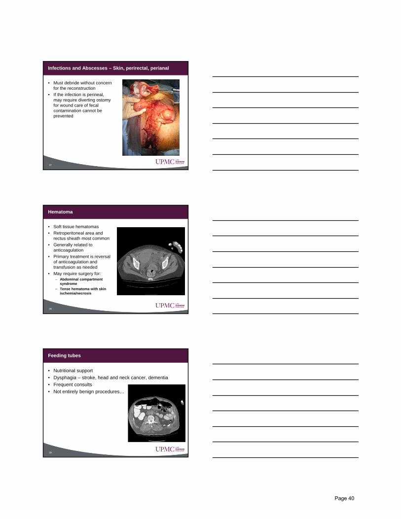
• Must debride without concern for the reconstruction
• If the infection is perineal, may require diverting ostomy for wound care of fecal contamination cannot be prevented
Infections and Abscesses – Skin, perirectal, perianal
37
• Soft tissue hematomas
• Retroperitoneal area and rectus sheath most common
• Generally related to anticoagulation
• Primary treatment is reversal of anticoagulation and transfusion as needed
• May require surgery for:– Abdominal compartment
syndrome
– Tense hematoma with skin ischemia/necrosis
Hematoma
38
• Nutritional support
• Dysphagia – stroke, head and neck cancer, dementia
• Frequent consults
• Not entirely benign procedures…
Feeding tubes
39
Page 40

• Open Gastrostomy
• Surgical gastropexy performed
• Laparoscopic Gastrostomy
• Surgical gastropexy or T-fasteners
• Percutaneous Endoscopic Gastrostomy
• Endoscopy
• Transillumination
• Needle, guidewire
• May use T-fasteners
• Relative contraindications– Prior abdominal surgery
– Malnutrition
– Ascites
– “Pullers”
Gastrostomy
40
• Tube placed into the proximal jejunum
• May be done open, laparoscopic or endoscopically (PEJ)
• The fixed point of the bowel at the abdominal wall may cause bowel obstruction (volvulus, internal hernia)
Jejunostomy
41
• Placed through the stomach for postpyloric feeding
• Benefits of jejunal feeds
• Less risk of bowel obstruction/torsion at the tube site than a jejunostomy
• Can convert a Gastrostomy to a GJ under fluoroscopy
Transgastric Jejunostomy (GJ)
42
Page 41

• Frequent consult for many EGS/ACS services
• Acute respiratory failure– Failure to wean
• Inability to maintain airway and manage secretions– Stroke
Tracheostomy
43
• Open Tracheostomy• Can be done at the bedside
(ICU) or in the OR
• Dissect down to the trachea
• Place stay sutures in the trachea
• Open the trachea, pull back tube and under direct vision place tube in the trachea
• Percutaneous Tracheostomy
• Generally done in the ICU
• Bronchoscopic guidance
• Modified Seldinger technique
• Small incision
• Dilate trachea
Tracheostomy – two common procedures
44
• Bleeding
• Concern for a sentinel bleed – indicating a possible tracheoimoninate fistula
• Trach balloon erodes through trachea into inominate artery
• Often fatal
• Tracheal Stenosis
Tracheostomy - Complications
45
Page 42

• Ileum brought out to the skin
• Generally liquid output
• May require agents to slow output to avoid dehydration
• Fluid and electrolyte management critical post op
• Usually performed as a Brooke – everted to make a “nipple” to ease pouching, minimize skin complications
• Ileostomies that are flush are often difficult to pouch
• May be loop versus end
Ileostomy
46
• Depending on site, may have liquid (right side) or solid (left side) output
• May be loop versus end
• Reversal of loop stomas are easier than end stomas
Colostomy
47
• Must be observed for function, ischemia and retraction
• Take down the bag, look at it
• Stomas may separate from the skin edges and develop an abscess
• Patients may develop a parastomal hernia– Surgical repair
Stomas - Postop
48
Page 43

• Acute C. difficile infection can become fulminant with signs of systemic toxicity
• May or may not have diarrhea
• Evidence of end-organ damage
• High clinical suspicion
• Oral or IV metronidizole
• Oral or rectal vancomycin
• Surgery– Standard therapy Subtotal
Colectomy
Fulminant Clostridium dificile colitis
49
Subtotal Colectomy
50
• Alternative surgical therapy
• Laparoscopic loop ileostomy with colonic lavage– “Zuckerbraun Procedure”
– Lavage with 8 liters of PEG and vancomycin intraoperatively
– Colonic irrigation with Vancomycin BID for 10 days
– Able to maintain the colon in 93% of patients
– Reversbile, pts are not committed to a lifetime ileostomy or ileorectal anastomosis
Fulminant Clostridium dificile colitis
51
Page 44

Approach to the Polytraumatized Patient with
Musculoskeletal Injuries
David R. Renner PA-C
Department of Orthopaedic Surgery
St. Lukes University Hospital
Why?
• In most Level 1 centers, the trauma-trained general surgeon is the leader of the multidisciplinary care team, and the orthopaedist functions as a team member
• In many community centers, a general surgeon is often in charge of the overall management of the trauma patient, but the orthopaedist may, at times, serve as leader
Why?
• Since the orthopaedist may be called upon to assume the leadership role, it is incumbent on him to be thoroughly familiar with the evaluation and management of the trauma patient, from assessment in the trauma bay through discharge and rehabilitation
Page 45

How?
• Advanced level practitioner’s are very effective in the management of these patients from admission to discharge.
• They also maximize their physician’s “time and tasks”. They can serve as a liaison to the multi-disciplinary specialists.
Associated Injuries
• Injuries due to blunt trauma, industrial accidents and falls frequently affect more than one system
• Polytraumatized patients must be evaluated with an awareness of the possibility of associated injuries and must be managed in time-relevant phases
Phases of Trauma Care
• Prehospital phase
• Hospital phase– acute
– primary
– secondary
– tertiary
• Rehabilitation phase
Page 46

Phases of Trauma Care
• Each period has its own priorities in resuscitation and injury management, as well as predictable patterns of morbidity and mortality
Mortality Patterns
• Acute and primary periods– hemodynamic complications and lethal head
injury
• Secondary period– early organ failure, particularly pulmonary
• Tertiary period– Sepsis, pulmonary failure, and delayed
organ failure
Today’s Goals
• Focus on the basic tenets of trauma care
• Focus on the evaluation of the multiply injured patient
• Focus on the benefits of early orthopaedic intervention in this setting
Page 47

Prehospital Phase
Necessary Components
• One of the goals of trauma care is to provide earlier evaluation and increasingly sophisticated prehospital care
• Optimal transport, resuscitation, stabilization, and definitive care of the multiply injured patient depend on 3 key elements
– paramedics familiar with life support protocols
– communication between paramedics and receiving hospital
– dedicated space and equipment for management of the patient
Prehospital Care
• When indicated, the patient should arrive with spinal immobilization
• Open wounds should be covered with sterile bandages
• Hemorrhage should be controlled with direct pressure
• Prefabricated splints should be used to immobilize long-bone injuries
Page 48

Acute Hospital Period
Initial 1 to 2 Hours
• A logical, systematic approach for evaluation and management must be followed
• Primary survey -->
• Acute resuscitation -->
• Secondary survey
• Some elements may be performed concurrently
Primary Survey Alphabet
• A-Airway maintenance with c-spine control
• B-Breathing and ventilation
• C-Circulation and hemorrhage control
• D-Disability evaluation(neurologic status)
• E-Exposure and environmental control(completely undress & examine, but prevent hypothermia)
Page 49

Airway
• Establishing airway is top priority
• Always must consider possibility of C-spine injury
• Need lateral of C-spine including the C7-T1 interspace
• If patient not awake and alert, negative lateral exam does not necessarily clear C-spine
Breathing and Ventilation
• Placement of an orotracheal, nasotracheal, or surgical airway is mandatory if there are mechanical factors preventing normal breathing
• If ventilation difficult, consider PTx vs.. HTx, etc.. and treat accordingly
Circulation and Hemorrhage
• Hypotension in the multiply injured patient may be due to diverse causes, including:– hemorrhage--accounts for 95% of cases
– brain injury
– quadriplegia
– hypothermia
– MI
– mediastinal shock
Page 50

Blunt Trauma and Blood Loss
• External hemorrhage-easiest to diagnose and can be treated with pressure
• Intrathoracic-usually diagnosed by PE or on CXR
• Intraperitoneal-can be evaluated by ultrasound, CT and PE
• Extraperitoneal-inferred by presence of pelvic fractures, may be life-threatening
• Long-bone fractures-identified by PE and X-rays
Disability/Neurologic Exam
• Glasgow Coma Scale is useful for quick neuro assessment– Eye opening
– Motor response
– Verbal response
– Get 3 points for showing up, 15 point max
• A more thorough exam can be made with secondary survey
Environment and Exposure
• Undress and examine
• Logroll to examine spine, posterior chest wall and flank
• Must avoid hypothermia with these maneuvers
Page 51

Resuscitation
• Concurrent with the primary assessment, the resuscitation must begin
• Large-bore IV’s with warm LR
• If no response to 2L LR, then blood is transfused
• BP, HR and urine output are decent indicators as the the adequacy of resuscitation
Mandatory Images in the Trauma Bay
• Lateral C-spine
• AP CXR
• AP Pelvis
• As indicated by PE or protocol, additional anatomy-specific radiographic studies can be obtained as part of secondary survey
Pelvis
• A major concern for trauma team is the presence of a pelvic fracture in a patient with continued hemodynamic deterioration
• If hemorrhage from the chest, belly, or external sites has been either excluded as the cause of hypotension or controlled, evaluation of the AP pelvis may reveal that a pelvic fracture is the site of hemorrhage
Page 52

Tile’s “ABC”
• Pioneering classification of pelvis fractures which offers a fairly simple description
• Type A-stable
• Type B-partially stable
• Type C-completely unstable
Tile A
• The good
• “Stable”
Tile B
• The bad
• Vertically stable
• Rotationally unstable
Page 53

Young and Burgess’ Scheme
• Pelvic fractures are divided into 4 types
– Lateral compression(LC)
– AP compression(APC)
– Vertical shear(VS)
– Combined mechanical injury
• Plane of the anterior ring disruption indicated the direction of the applied force, suggesting the nature of the posterior ring injury, and can be used to establish the risk of hemorrhage
Tile C
• The ugly
• Vertically unstable
• Rotationally unstable
Page 54

Lateral Compression Injuries
• Oblique anterior ring fracture
• Associated with:– decreasing pelvic volume
– intraperitoneal or intrathoracic hemorrhage
– high incidence of CHI
• High mortality of LC-III are usually secondary to associated injuries
Anteroposterior Compression Injuries
• Characterized by vertical pubic rami fractures
• Are associated with greatest incidence of hemorrhage
• Posterior ring injury is as follows:
– APC-I:sacrospinous & sacrotuberous ligaments
– APC-II: anterior SI ligament
– APC-III: posterior SI ligament
• Neurovascular structures adjacent to these ligaments also are disrupted
• APC-III has been associated with >20 U PRBC requirement
APC-III
Page 55

Vertical Shear Injuries
• Often associated with massive blood loss
• Typically present with initial cephalad displacement
Orthopaedic Math
• Pelvic volume = 2/3 pi r3
• Even a small increase in pelvic diameter exponentially increases volume
• As important posterior structures are sequentially torn, associated vessels may also be disrupted, resulting in hemorrhage
Unstable Pelvic Fracture and Fatal Exsanguination
• These patients are at risk due to:– the administration of IVF impairs the body’s
natural compensatory mechanism of decreasing blood flow to increase clotting
– the administration of nonclotting, often cold IVF which limits clotting ability
– movement for diagnostic and examination purposes
Page 56

Blood Loss with Pelvic Fracture
• Death due to torrential hemorrhage
• Truck rollover injury
Management of Hemorrhage
• Hemorrhage following pelvic fractures can be managed by:
– angiography and embolization
– exploration and vascular ligation
– ORIF
– CREF
– pneumatic antishock garments
• Treatment choice depends on resources of hospital and experience of personnel
Angiography and Embolization
• This technique can be used to diagnose and treat arterial hemorrhage
• It is a less than optimal treatment for the patient in extremis because the bleeding is most frequently venous
• Requires immediate availability of specialized personnel
Page 57

Angiography and Embolization
ORIF
• Mechanically the most stable form of fixation and may be done concurrently with other emergent surgery
• Must not violate retroperitoneal space, which would decompress tamponade
• Hollow viscus perforation with wound contamination is relative contraindication
ORIF
• Technically most demanding
• Usually not done acutely
• Biomechanically most stable
Page 58

External Fixation
• In many instances, CREF is method of choice for hemorrhage control
• Benefits include:– stabilization of pelvic ring
– controls pelvic volume
– minimizes dislodgement of clots
– aids in controlling cancellous bone bleeding
– facilitates early patient mobilization
External Fixation-Technique
• Pins are inserted between cortices of ilium
• Clamps attach pins into groups
• Connecting rods are loosely attached to the clamps to form a frame
• Pelvis is reduced by posterior manual compression and longitudinal traction
• Frame is locked
• Frame construct should allow further abdominal or chest diagnostic studies without releasing the reduction
External Fixation
Page 59

Pneumatic Antishock Garments
• Effective as temporary splints for the pelvis and lower extremity
• Prolonged use limits evaluation of, and access to, lower extremity trauma
• Use is contraindicated in the treatment of open fractures, and may potentiate a compartment syndrome
Acute Hospital Period--Other Orthopaedic Goals
• Reduction of major joint dislocations and fracture/dislocations should be addressed
• Orthopaedic surgeon must be aggressive in managing these injuries, particularly hip and knee dislocations
• If there is neurovascular compromise, early realignment of joint may help restore blood flow to the distal extremity, avoiding ischemia and compartment syndrome
Page 60

Acute Hospital Period:Hours 3 to 12
Re-evaluation
• Secondary survey is completed
• Because the patient is often unconscious, and unable to localize injuries, great care must be taken during this evaluation
• A patient’s overt, overwhelming injuries frequently mask other serious injuries that, if unrecognized and untreated, may cause future disability
Limb Salvage
• Decisions related to limb salvage must be considered during this time frame
• Extremities with massive injuries must be carefully evaluated for:– the degree of soft-tissue damage
– perfusion of the limb distal to the injury
– neurologic function in the distal limb
– number of levels of injury within the limb
Page 61

Limb Salvage
• Attempts to quantify the injury and outcome have not proven uniformly successful
• Indices such as MESS direct attention to the important factors such as ischemic time, hypotension and neurologic function that must be evaluated when evaluating the feasibility of salvage
• Physician experience may be the most reliable determinant of whether to salvage or amputate
Limb Salvage?
• Degloving foot injury in a 25 y/o fashion model
• This was appropriately treated with early amputation
Limb Salvage?
• Are you kidding me?
• …but notice normal foot which is sensate
Page 62

Orthopaedic Priority #1--Open Fractures
• Requires Ancef +/- Gentamicin, and tetanus booster in ER
• Wound dressed sterilely, limb splinted
• When patient stable, to OR for I + D and skeletal stabilization
• Primary closure vs. delayed primary closure?
• Early soft tissue coverage is necessary for optimal functional outcome
Open Fractures
• Injuries like this require emergent debridement and skeletal stabilization
Benefits of Early Fracture Stabilization
• Orthopaedic injuries are frequently overlooked initially in the interest of acute resuscitation of the multiply injured patient
• Many studies have shown that aggressive early management of these injuries increases long-term survival and decreases morbidity
Page 63

Benefits of Early Fracture Stabilization
• Fixation of unstable fractures of the pelvis, femur, and tibia should be done within the first 24 hours after injury if medically feasible
• The goal is stable skeletal fixation that will allow early mobilization of the patient
• Early fracture stabilization has been found to have many beneficial effects
Benefits of Early Fracture Stabilization
• Decreased musculoskeletal morbidity
– early PT decreases rehab time and reduces long-term disability
• Decreased hospital stay
– decreased ICU and hospital stays
– translates into net total hospital cost
• Other benefits
– improved nursing care
– improved patient comfort, reducing narcotic requirement
Pulmonary Complications after Trauma
• Fat Embolism Syndrome
• Adult Respiratory Distress Syndrome
• Thromboembolic Complications
Page 64

Fat Embolism Syndrome
• Following fracture, marrow fat embolizes and travels to lungs
• Cascade occurs in lungs, releasing vasoactive substances
• Clinically, FES is identified by acute hypoxemia, MS change, and interstitial infiltrate on CXR
• Incidence in isolated LBF is 0.5% to 2.0%
• In setting of multiply injured patient it approaches 10% to 15%
Fat Embolism Syndrome
• Be wary of the patient with the “isolated femur fracture”
Fat Embolism Syndrome
• Studies have demonstrated that early fracture stabilization results in a decreased incidence of FES
• In a prospective, randomized study of early versus delayed femoral nailings, Bone found no cases of FES in the early-stabilization group
Page 65

ARDS
• A devastating complication of trauma
• Characterized by refractory hypoxemia and diffuse interstitial changes on CXR
• Known to have high mortality rates
• Growing body of evidence has shown that early fracture stabilization can substantially decrease the incidence of ARDS
ARDS
• Early ARDS
• Interstitial changes and patchy infiltrates
• Early ORIF and rapid mobilization may help prevent this
Thromboembolic Complications
• DVT and PE can adversely affect patient outcome and have been reported to occur more frequently in multiply injured patient than in patients with isolated injuries
• Early fracture stabilization facilitates early patient mobilization and may decrease the incidence of thromboembolic complications
Page 66

Pulmonary Embolism
• A potentially fatal complication of DVT
• Treated by prompt diagnosis and anticoagulants
• Filters can prevent PE, not DVT
Secondary Period:Hours 13 to 72
Secondary Period
• Formulate a plan for fixation of the remaining fractures
• Must take into consideration the patient’s overall status, since changes in overall status can and will impact orthopaedic treatment plan
• Plan must be communicated with other members of the treatment team
Page 67

Secondary Period
• Continual patient reevaluation
• Possibility of compartment syndrome during this period exists as severely injured limbs are at risk whether or not fracture is present
• In the unconscious patient, the surgeons level of suspicion must be higher due to lack of symptoms
• Liberal utilization of pressure recording devices
• If present--fasciotomy is required
Secondary Period
• Traumatic wounds not closeable should undergo repeated excisional debridement
• Definitive treatment of
– spinal fractures
– pelvis & acetabulum fractures
– upper extremity fractures
– periarticular fractures
• Goal is stable fixation of fractures to allow patient mobilization
Secondary Period
• A comminuted tib/fib fracture with a contused soft-tissue envelope like this is best treated with provisional external fixation
Page 68

Secondary Period
• Poorly timed surgery can result in this, no matter how technically well-done
Tertiary Period:Beyond 3 Days
Tertiary Period
• Definitive wound management is undertaken
– STSG for healthy granulating wounds
– rotational vs. free flaps in presence of exposed bone
• Guiding principle is to obtain a closed envelope around bone as early as possible
• Consider bone grafting at time of closure for those with defects
Page 69

Tertiary Period
• Time to close, graft or flap open wounds
• Often done by trauma or plastics
Tertiary Period
• Open wounds like this need flap closure
• An intact soft tissue envelope is a prerequisite for bone healing
Tertiary Period
• 7 to 10 days post-injury is ideal time to convert provisional externally fixed limbs to ORIF
• Soft tissues should be better
• Less chance of catastrophic wound breakdown
Page 70

Rehabilitation Phase
Rehabilitation Phase
• Rehabilitation should begin early, with joint range of motion under PT/OT supervision
• Duration of rehabilitation phase is longer for polytraumatized patient than for one with isolated injuries because the cumulative effect of multiple injuries is greater than the sum of individual ones
Rehabilitation Phase
• Delayed bone reconstruction in massively injured extremity can take many forms
• In injuries at risk for nonunion, bone grafting should be undertaken early
• A healed soft-tissue envelope is desirable prior to grafting
Page 71

Rehabilitation Phase
• Deformity correction and bone transport have become more common in US since adoption of Ilizarov methods
• Although application of frames may speed recovery of the patient, techniques are demanding and require a great deal of time, not only from the surgeon but also by the patient, who must participate actively
Rehabilitation Phase
• Multitrauma patients are at increased risk of infection because of the inherent physiologic changes associated with trauma
• Nonorthopaedic infections must be managed by the trauma and critical care teams
• Orthopaedic infections must be treated by the orthopaedist in addition to other specialists
Orthopaedic Infections
• Wounds must be treated aggressively, with excisional debridement of all necrotic bone and soft tissue
• Wounds which were previously closed may now require flap coverage
• Initial plans may need to be revised
• Failure to address the issue of infection may change what may have been an excellent outcome of a difficult problem to a poor outcome and a lifetime of disability
Page 72

Orthopaedic Infections
• Antibiotics should be specific for organisms identified at the time of debridement
• Broad-spectrum antibiotics are appropriate upon presentation, but should be tailored once the specific organism and sensitivities have been identified
Orthopaedic Infections
• Nonunion may the only symptom of osteomyelitis
• Orthopaedic infections are easy to achieve, difficult to eradicate
Summary
• The orthopaedic surgeon should be involved in the care of the multiply injured patient from admission through rehabilitation
• Early orthopaedic intervention affords the benefits of decreased mortality, increased hemodynamic stability, decreased pulmonary complication rate, early mobilization, decreased complications of recumbency, decreased narcotic requirements, and a greater likelihood of an excellent outcome
Page 73

Thank You
Page 74

A New AP in a New Trauma Center
Babak Sarani, MD, FACS, FCCMAssociate Professor of Surgery
Chief, Trauma and Acute Care SurgeryGeorge Washington University
Disclosures
• None
Objectives
• The Role of the AP in a new trauma program
• Recruiting a AP to a new trauma center
• Retaining and Mentoring the AP
Page 75

House‐Officer Goals
• Learn fundamental principles of trauma care
– Junior residents: ATLS, floor‐based care
– Senior residents: Decisions regarding operative vnon‐operative management, ICU‐based care, operative maneuvers
– Fellows: Same as senior resident + understand administrative aspects of trauma center and system
The Role of the AP
• CONTINUITY
– “Trauma Provost”
• Fill the void
– “Weak Resident”
– 80‐hour resident and 16‐hour intern
• Liaison with nurses
– Identify problems early
– Feedback on new initiatives
The Role of the AP in a New Trauma Center
• New trauma system started July 1, 2012
– Dedicated trauma team
• Attending of the week
• PGY‐3 Resident
• 2 Interns
• 1 NP
– Few established protocols or guidelines
• None enforced or monitored
Page 76

• Mentor to junior residents
– Trauma Resuscitation/initial survey
– Monitor guidelines – DVT, DT, C‐spine clearance
• Mentor to nurses – especially floor RN
– Explain “why”
– Resource
The Role of the AP in a New Trauma Center
The Role of the AP in a New Trauma Center
• Hierarchy/delineation of goals
– Mentor and TPD as “Solomon”
• Logical reasoning for decisions
• No‐favorites/mutual respect
– Goal is to have the AP function at a chief/fellow level
• However, upper level residents must retain authority with the attending as the final arbiter in extreme cases
– The “turtle” wins the race
Recruiting an AP to a New Trauma Center
• The Trauma Center and the AP both win when the AP moves on and assumes a leadership position
– Clinical Excellence
– Teaching Excellence
– Research Excellence
Page 77

Recruiting an AP to a New Trauma Center
• Expectations
– Level I: Teaching and scholarship
• ATCN or other such courses
• Publications – help with clinical research
– All levels: Clinical Excellence
• With NP, teach MDs “how nurses think” and teach nurses “how doctors think”
– Even‐toned/tempered
• Critical in a new program (with old personnel)
• Realistic
– Rapid accumulation of knowledge with room for errors
– Potential for downtime/homework
• Direct report to Trauma Director
– 24‐hour access
– TPD establishes the tone of the whole program
Recruiting an AP to a New Trauma Center
• New v Experienced AP
– Patient acuity and volume
– Mentoring ability and desire
Recruiting an AP to a New Trauma Center
Page 78

Retaining the AP
• Goal: position the AP to leave for a bigger/better job in 5‐7 years
• The Trauma Center and the AP both win when the AP moves on and assumes a leadership position– Clinical Excellence
– Teaching Excellence
– Research Excellence
Retaining the AP
• Clinical Excellence
– Exposure to TB, OR, ICU, Floor
• First assist skills
• Suturing skills
• Autonomy in the TB and Floor. Cooperative with ICU team
– Med Literature knowledge base
• On‐line library of key articles
– Give AP public credit for good decisions
• Patients, nurses, other members of the team
Retaining the AP
• Teaching Excellence
– Trauma courses – local/regional
– In‐service to other departments, esp nursing
– Develop a local/regional/national reputation
Page 79

• Research/Scholarship Excellence
– Case reports initially then bigger projects
– Screen/enroll pts for projects
– Liaison for joint projects with other depts (NIH)
– Build reputation via medical literature
– Develop reputation via medical societies
Retaining the AP
Conclusion
• An AP is vital to new trauma program
• Need to ensure new program needs/expectations are in‐line with recruit
• Need to mentor/grow the recruit for longevity of person and overall program
Page 80


![[XLS]Ventilator training simulator v32 - Vanderbilt Universitypscrooke/CANVENT/Ventilator... · Web viewApproximately how often do you use pressure control ventilation, Bilevel ventilation,](https://static.fdocuments.net/doc/165x107/5aa80edf7f8b9aa7258b5a18/xlsventilator-training-simulator-v32-vanderbilt-pscrookecanventventilatorweb.jpg)