Lymphatic Vessels, Infl ammation, and Immunity in...
15
22 | CANCER DISCOVERYJANUARY 2016 www.aacrjournals.org REVIEW Lymphatic Vessels, Inflammation, and Immunity in Skin Cancer Amanda W. Lund 1,2,3,4 , Terry R. Medler 1 , Sancy A. Leachman 3,4 , and Lisa M. Coussens 1,4 1 Department of Cell, Developmental and Cancer Biology, Oregon Health and Science University, Portland, Oregon. 2 Department of Molecular Microbi- ology and Immunology, Oregon Health and Science University, Portland, Oregon. 3 Department of Dermatology, Oregon Health and Science Univer- sity, Portland, Oregon. 4 Knight Cancer Institute, Oregon Health and Science University, Portland, Oregon. Corresponding Author: Amanda W. Lund, Department of Cell, Developmental and Cancer Biology, Oregon Health and Science University, 6514 Richard Jones Hall, 3181 Sam Jackson Park Road, Mail Code: L215, Portland, OR 97239. Phone: 503-494-1095; Fax: 503-494-4253; E-mail: [email protected] doi: 10.1158/2159-8290.CD-15-0023 ©2015 American Association for Cancer Research. ABSTRACT Skin is a highly ordered immune organ that coordinates rapid responses to external insult while maintaining self-tolerance. In healthy tissue, lymphatic vessels drain fluid and coordinate local immune responses; however, environmental factors induce lymphatic vessel dysfunction, leading to lymph stasis and perturbed regional immunity. These same environmental fac- tors drive the formation of local malignancies, which are also influenced by local inflammation. Herein, we discuss clinical and experimental evidence supporting the tenet that lymphatic vessels participate in regulation of cutaneous inflammation and immunity, and are important contributors to malignancy and potential biomarkers and targets for immunotherapy. Significance: The tumor microenvironment and tumor-associated inflammation are now appreciated not only for their role in cancer progression but also for their response to therapy. The lymphatic vascu- lature is a less-appreciated component of this microenvironment that coordinates local inflammation and immunity and thereby critically shapes local responses. A mechanistic understanding of the com- plexities of lymphatic vessel function in the unique context of skin provides a model to understand how regional immune dysfunction drives cutaneous malignancies, and as such lymphatic vessels represent a biomarker of cutaneous immunity that may provide insight into cancer prognosis and effective therapy. Cancer Discov; 6(1); 22–35. ©2015 AACR. INTRODUCTION Skin is the largest organ in mammals, serving as a physical and immunologic barrier to the external environment (1). As an immune organ, skin coordinates rapid responses to external challenge through constitutive immune surveillance involving both resident and recruited leukocytes (2). Exqui- site spatiotemporal regulation of immune cells is required for homeostatic tissue maintenance—loss of function is associ- ated with a variety of dermatopathologies, including chronic infection, inflammation, autoimmunity, and cancer (1). The interplay between local inflammation, tissue remodeling, and antitumor immune responses is of particular interest given the recent success of immunotherapies, namely blockade of immune checkpoint molecules (3). Because tumor micro- environments in part regulate antitumor immunity, these also present targets for release of local immunosuppression that may synergize with checkpoint therapies (4). Although tumor-associated blood vessels are appreciated for their role in regulating leukocyte infiltration and function (5), tumor- associated lymphangiogenesis has lagged behind with respect to recognition of its role(s) in tumor-associated inflammation and immunity. Recent revelations, however, have highlighted these important roles with respect to coordination of local inflammation (6) and immunity (7–11), indicating that their deregulation may similarly influence inflammation-induced tumor progression and metastasis. In this review, we discuss clinical and experimental evidence revealing the significant inflammatory and immunomodula- tory functions of lymphatic vasculature, and suggest how tumor-associated remodeling may alter regional inflamma- tion and immunity, thereby contributing to disease progres- sion and metastasis. In addition, we suggest that lymphatic vessel morphology and function may be a biomarker of cuta- neous immune status, and discuss how lymphatic biomarkers may provide insight into diagnosis and therapy for cutaneous malignancy. INFLAMMATION AND CANCER It is now well appreciated that environmental exposures account for the occurrence of many different types of cancer, with 15% to 20% of cancer-related deaths linked to inflam- mation or infection (12). Environmental factors include on June 26, 2018. © 2016 American Association for Cancer Research. cancerdiscovery.aacrjournals.org Downloaded from Published OnlineFirst November 9, 2015; DOI: 10.1158/2159-8290.CD-15-0023
Transcript of Lymphatic Vessels, Infl ammation, and Immunity in...
22 | CANCER DISCOVERY�JANUARY 2016 www.aacrjournals.org
REVIEW
Lymphatic Vessels, Infl ammation, and Immunity in Skin Cancer Amanda W. Lund 1,2,3,4 , Terry R. Medler 1 , Sancy A. Leachman 3,4 , and Lisa M. Coussens 1,4
1 Department of Cell , Developmental and Cancer Biology, Oregon Health and Science University, Portland, Oregon. 2 Department of Molecular Microbi-ology and Immunology, Oregon Health and Science University, Portland, Oregon. 3 Department of Dermatology, Oregon Health and Science Univer-sity, Portland, Oregon. 4 Knight Cancer Institute, Oregon Health and Science University, Portland, Oregon. Corresponding Author: Amanda W. Lund, Department of Cell, Developmental and Cancer Biology, Oregon Health and Science University, 6514 Richard Jones Hall, 3181 Sam Jackson Park Road, Mail Code: L215, Portland, OR 97239. Phone: 503-494-1095; Fax: 503-494-4253; E-mail: [email protected] doi: 10.1158/2159-8290.CD-15-0023 ©2015 American Association for Cancer Research.
ABSTRACT Skin is a highly ordered immune organ that coordinates rapid responses to external insult while maintaining self-tolerance. In healthy tissue, lymphatic vessels drain
fl uid and coordinate local immune responses; however, environmental factors induce lymphatic vessel dysfunction, leading to lymph stasis and perturbed regional immunity. These same environmental fac-tors drive the formation of local malignancies, which are also infl uenced by local infl ammation. Herein, we discuss clinical and experimental evidence supporting the tenet that lymphatic vessels participate in regulation of cutaneous infl ammation and immunity, and are important contributors to malignancy and potential biomarkers and targets for immunotherapy.
Signifi cance: The tumor microenvironment and tumor-associated infl ammation are now appreciated not only for their role in cancer progression but also for their response to therapy. The lymphatic vascu-lature is a less-appreciated component of this microenvironment that coordinates local infl ammation and immunity and thereby critically shapes local responses. A mechanistic understanding of the com-plexities of lymphatic vessel function in the unique context of skin provides a model to understand how regional immune dysfunction drives cutaneous malignancies, and as such lymphatic vessels represent a biomarker of cutaneous immunity that may provide insight into cancer prognosis and effective therapy. Cancer Discov; 6(1); 22–35. ©2015 AACR.
INTRODUCTION
Skin is the largest organ in mammals, serving as a physical and immunologic barrier to the external environment ( 1 ). As an immune organ, skin coordinates rapid responses to external challenge through constitutive immune surveillance involving both resident and recruited leukocytes ( 2 ). Exqui-site spatiotemporal regulation of immune cells is required for homeostatic tissue maintenance—loss of function is associ-ated with a variety of dermatopathologies, including chronic infection, infl ammation, autoimmunity, and cancer ( 1 ). The interplay between local infl ammation, tissue remodeling, and antitumor immune responses is of particular interest given the recent success of immunotherapies, namely blockade of immune checkpoint molecules ( 3 ). Because tumor micro-environments in part regulate antitumor immunity, these
also present targets for release of local immunosuppression that may synergize with checkpoint therapies ( 4 ). Although tumor-associated blood vessels are appreciated for their role in regulating leukocyte infi ltration and function ( 5 ), tumor-associated lymphangiogenesis has lagged behind with respect to recognition of its role(s) in tumor-associated infl ammation and immunity. Recent revelations, however, have highlighted these important roles with respect to coordination of local infl ammation ( 6 ) and immunity ( 7–11 ), indicating that their deregulation may similarly infl uence infl ammation-induced tumor progression and metastasis.
In this review, we discuss clinical and experimental evidence revealing the signifi cant infl ammatory and immunomodula-tory functions of lymphatic vasculature, and suggest how tumor-associated remodeling may alter regional infl amma-tion and immunity, thereby contributing to disease progres-sion and metastasis. In addition, we suggest that lymphatic vessel morphology and function may be a biomarker of cuta-neous immune status, and discuss how lymphatic biomarkers may provide insight into diagnosis and therapy for cutaneous malignancy.
INFLAMMATION AND CANCER
It is now well appreciated that environmental exposures account for the occurrence of many different types of cancer, with 15% to 20% of cancer-related deaths linked to infl am-mation or infection ( 12 ). Environmental factors include
on June 26, 2018. © 2016 American Association for Cancer Research. cancerdiscovery.aacrjournals.org Downloaded from
Published OnlineFirst November 9, 2015; DOI: 10.1158/2159-8290.CD-15-0023
JANUARY 2016�CANCER DISCOVERY | 23
Lymphatic Vessels, Infl ammation, and Immunity in Skin Cancer REVIEW
infectious agents, physical trauma (nonhealing scars, burns), industrial toxins (e.g., tar, arsenic, radiotherapy), as well as those associated with lifestyle (tobacco, alcohol, diet, ultra-violet exposure; ref. 13 ). As one might expect, tumors whose etiology is associated with environment-induced infl amma-tion often occur in tissues with the greatest surface area, including lung, skin, and gastrointestinal tract ( 14 ). Infl am-mation is considered one of 10 hallmarks of cancer and is increasingly appreciated for its complex role in tumor initiation, promotion, malignant conversion, and metastasis ( 15 ). Furthermore, immune infi ltrates provide prognostic biomarkers ( 16 ) and potent targets for cancer therapy ( 17 ). We are just beginning to understand, however, how complex interactions between environmental agents, genetic factors, and tissue microenvironments coordinate local infl amma-tion and subsequent tumorigenesis.
SKIN INFLAMMATION AND CANCER
Skin is a stratifi ed tissue composed of epidermal, dermal, and subcutaneous adipose layers functioning in concert as a barrier to the external environment ( 1 ). Keratinocytes are the primary cell type in the epidermis and undergo continu-ous cycles of homeostatic proliferation and differentiation to maintain a cornifi ed layer protecting against water loss and microbe penetration ( 18 ). Melanocytes reside in basal layers of the epidermis. The dermis is largely composed of collagen-ous and noncollagenous (e.g., elastic fi bers and structural proteoglycans) connective tissue components that provide substratum support for blood and lymphatic vessels, nerves, mesenchymal support cells (fi broblasts), and resident immune cells. The cellular components of these layers critically regu-late immune surveillance to facilitate rapid responses to insult while, importantly, maintaining immune tolerance ( 2 , 19 )—breakdown of this balance is associated with a diverse set of immune-mediated dermatopathologies ( 1 ).
There are four major types of skin cancer: basal cell carci-noma, squamous cell carcinoma, melanoma, and nonepithe-lial skin cancers (e.g., Merkel cell carcinoma, Kaposi sarcoma, and cutaneous lymphoma). Predisposition to skin cancer is associated with chronic cutaneous infl ammation (e.g., discoid lupus erythematosus, chronic wounds, dystrophic epidermolysis bullosa), viral infections [e.g., human immu-nodefi ciency virus, human herpes virus 8, and human papil-lomavirus (HPV)], and infl ammatory environmental agents [e.g., UV radiation (UVR), viral infection, aging, diet, smoking, radiotherapy, phototherapy, physical trauma; ref. 13 ], with infl ammation playing a well-documented role in regulating progression of these cutaneous malignancies ( 20 ); however, skin cancers are also associated with chronically injured or nonhealing scars, burns, and chronic friction, with incidence of malignancy in scar tissue being low (0.1%–2.5%; ref. 13 ).
Furthermore, cells in skin secrete diverse infl amma-tory mediators, including Toll-like receptor (TLR) ligands, polypeptide growth factors, for example, TNFα, as well as chemokines that direct traffi cking and subsequent activation of recruited leukocytes, many of which have been implicated as mediators of tumor progression in skin ( 20 ). For example, mast cells foster skin carcinogenesis through the release of prosurvival and proangiogenic polypeptide growth factors
and matrix remodeling proteinases that activate proliferative programs in keratinocytes, fi broblasts, and endothelial cells ( 21 ). B cells secrete (auto)antibodies that form immune com-plexes with complement proteins—these accumulate in skin and infl uence resident and recruited myeloid cells’ activa-tion and function ( 20 ). Tumor-infi ltrating leukocytes play an important role in regulating angiogenic responses, which, in turn, regulate their entry ( 22 ), and thereby infl uence meta-static potential ( 23 ). Although much focus has been placed on blood vasculature with respect to leukocyte infi ltration and function, the lymphatic endothelium may similarly partici-pate in these complex interactions. In support of this, using a murine model of chemically induced skin carcinogenesis, it was recently reported that lymphatic vessels and their subse-quent drainage are required for induction of local leukocytic infi ltration, and consequently their absence resulted in the initiation of fewer tumors ( 6 ). Given our current understand-ing of how lymphatic vessels participate in regulating local infl ammation and immunity, as described below, it is reason-able to hypothesize that tumor-associated lymphatic remod-eling participates in shaping immune microenvironments of solid tumors.
LYMPHATIC VESSELS AND REGIONAL INFLAMMATION
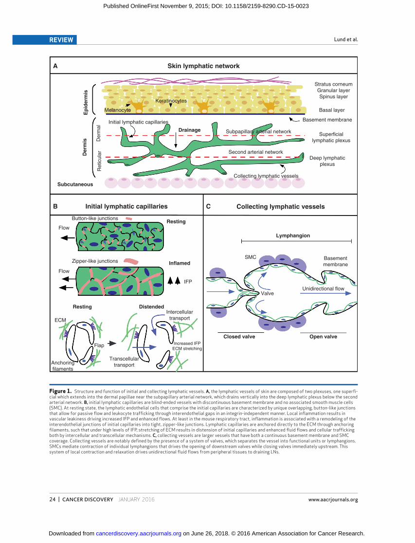
Lymphatic vessel architecture dictates local fl ow and the unidirectional transport of peripheral information to the secondary lymphoid system ( 24 ). Lymphatic vessels develop as a hierarchical, one-way drainage system functioning to transport cells and fl uid into the lymphoid system, and as a result of this architecture, lymphatic vessels lie at the interface of tissue biology and immunology ( 24 ). In skin, lymphatic ves-sels form two plexuses, one superfi cial extending into dermal papillae, and the other near the subpapillary arterial network ( Fig. 1A ). Initial lymphatic capillaries are small, blind-ended vessels with minimal basement membrane that directly attach to extracellular matrix (ECM) through anchoring fi laments, allowing for rapid response to changes in local interstitial fl uid pressure (IFP); swelling of ECM due to edema pulls on these fi laments, opening the loose, button-like interendothe-lial junctions ( Fig. 1B ; refs. 25–28 ). Lymphatic capillaries drain into collecting vessels characterized by a continuous basement membrane, smooth muscle coverage, and a system of valves that prevent retrograde fl uid fl ow ( Fig. 1C ). Lymphatic dys-function results in accumulation of protein-rich interstitial fl uid (lymphedema), leading to progressive fi brosis, adipose deposition, and infl ammation ( 29 ). Although lymphatic ves-sels are required for drainage of soluble antigen and traffi ck-ing of activated dendritic cells (DC) to local lymph nodes (LN) , their contribution to other aspects of infl ammation and immunity is just being explored. There have been several excel-lent reviews highlighting current knowledge of the contribu-tion of lymphatic vessels to infl ammation ( 30 ) and tolerance ( 31 ). Herein, we highlight their roles in immune induction and resolution or tolerance ( Fig. 2A–D ), and discuss how their dysfunction contributes to tumor progression and metastasis. Of note, as many of these studies were not performed in skin, it remains to be seen how many of these mechanisms will translate across tissue systems.
on June 26, 2018. © 2016 American Association for Cancer Research. cancerdiscovery.aacrjournals.org Downloaded from
Published OnlineFirst November 9, 2015; DOI: 10.1158/2159-8290.CD-15-0023
24 | CANCER DISCOVERY�JANUARY 2016 www.aacrjournals.org
Lund et al.REVIEW
Figure 1. Structure and function of initial and collecting lymphatic vessels. A, the lymphatic vessels of skin are composed of two plexuses, one superfi -cial which extends into the dermal papillae near the subpapillary arterial network, which drains vertically into the deep lymphatic plexus below the second arterial network. B, initial lymphatic capillaries are blind-ended vessels with discontinuous basement membrane and no associated smooth muscle cells (SMC). At resting state, the lymphatic endothelial cells that comprise the initial capillaries are characterized by unique overlapping, button-like junctions that allow for passive fl ow and leukocyte traffi cking through interendothelial gaps in an integrin-independent manner. Local infl ammation results in vascular leakiness driving increased IFP and enhanced fl ows. At least in the mouse respiratory tract, infl ammation is associated with a remodeling of the interendothelial junctions of initial capillaries into tight, zipper-like junctions. Lymphatic capillaries are anchored directly to the ECM through anchoring fi laments, such that under high levels of IFP, stretching of ECM results in distension of initial capillaries and enhanced fl uid fl ows and cellular traffi cking both by intercellular and transcellular mechanisms. C, collecting vessels are larger vessels that have both a continuous basement membrane and SMC coverage. Collecting vessels are notably defi ned by the presence of a system of valves, which separates the vessel into functional units or lymphangions. SMCs mediate contraction of individual lymphangions that drives the opening of downstream valves while closing valves immediately upstream. This system of local contraction and relaxation drives unidirectional fl uid fl ows from peripheral tissues to draining LNs.
Increased IFPECM stretching
DistendedResting
Resting
Transcellulartransport
Open valve
Unidirectional flow
Basementmembrane
SMC
Lymphangion
Stratus corneumGranular layerSpinus layer
Basement membrane
Deep lymphaticplexus
Der
mal
Ret
icul
ar
Drainage Subpapillary arterial network
Second arterial network
Collecting lymphatic vessels
Keratinocytes
Melanocyte
Initial lymphatic capillaries
Superficiallymphatic plexus
Basal layer
Closed valve
Valve
Zipper-like junctions
Button-like junctions
Inflamed
IFP
Flow
Flow
Intercellulartransport
Flap
ECM
Anchoringfilaments
Skin lymphatic network
Initial lymphatic capillaries Collecting lymphatic vessels
Subcutaneous
Der
mis
Ep
ider
mis
B
A
C
on June 26, 2018. © 2016 American Association for Cancer Research. cancerdiscovery.aacrjournals.org Downloaded from
Published OnlineFirst November 9, 2015; DOI: 10.1158/2159-8290.CD-15-0023
JANUARY 2016�CANCER DISCOVERY | 25
Lymphatic Vessels, Infl ammation, and Immunity in Skin Cancer REVIEW
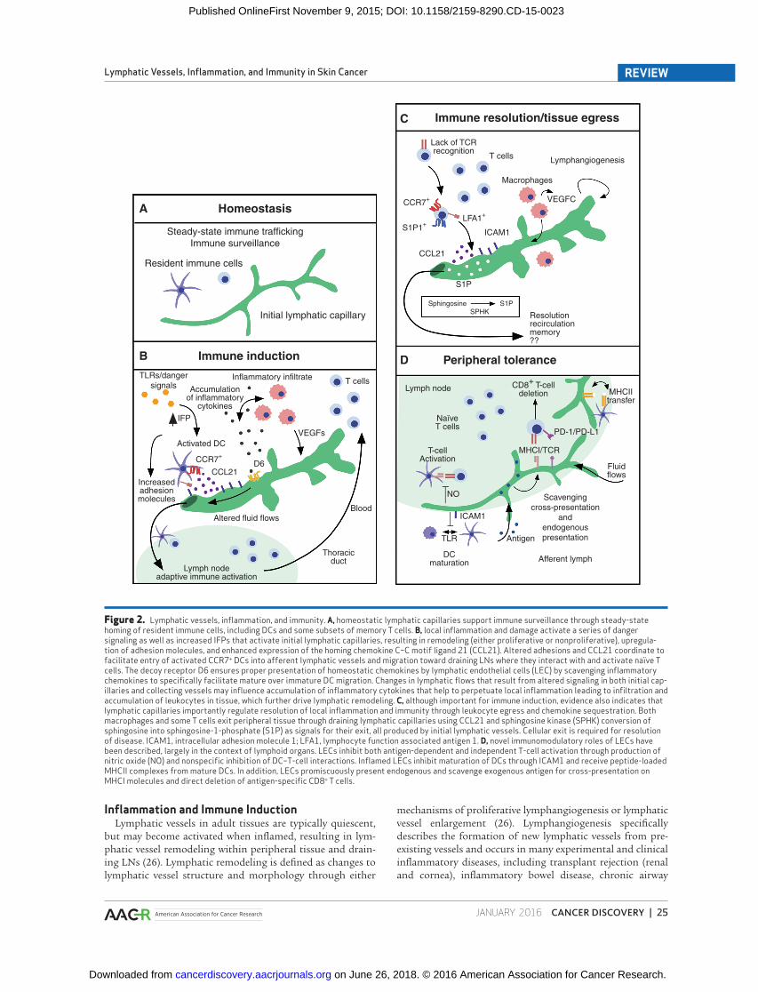
Figure 2. Lymphatic vessels, infl ammation, and immunity. A, homeostatic lymphatic capillaries support immune surveillance through steady-state homing of resident immune cells, including DCs and some subsets of memory T cells. B, local infl ammation and damage activate a series of danger signaling as well as increased IFPs that activate initial lymphatic capillaries, resulting in remodeling (either proliferative or nonproliferative), upregula-tion of adhesion molecules, and enhanced expression of the homing chemokine C–C motif ligand 21 (CCL21). Altered adhesions and CCL21 coordinate to facilitate entry of activated CCR7 + DCs into afferent lymphatic vessels and migration toward draining LNs where they interact with and activate naïve T cells. The decoy receptor D6 ensures proper presentation of homeostatic chemokines by lymphatic endothelial cells (LEC) by scavenging infl ammatory chemokines to specifi cally facilitate mature over immature DC migration. Changes in lymphatic fl ows that result from altered signaling in both initial cap-illaries and collecting vessels may infl uence accumulation of infl ammatory cytokines that help to perpetuate local infl ammation leading to infi ltration and accumulation of leukocytes in tissue, which further drive lymphatic remodeling. C, although important for immune induction, evidence also indicates that lymphatic capillaries importantly regulate resolution of local infl ammation and immunity through leukocyte egress and chemokine sequestration. Both macrophages and some T cells exit peripheral tissue through draining lymphatic capillaries using CCL21 and sphingosine kinase (SPHK) conversion of sphingosine into sphingosine-1-phosphate (S1P) as signals for their exit, all produced by initial lymphatic vessels. Cellular exit is required for resolution of disease. ICAM1, intracellular adhesion molecule 1; LFA1, lymphocyte function associated antigen 1. D, novel immunomodulatory roles of LECs have been described, largely in the context of lymphoid organs. LECs inhibit both antigen-dependent and independent T-cell activation through production of nitric oxide (NO) and nonspecifi c inhibition of DC–T-cell interactions. Infl amed LECs inhibit maturation of DCs through ICAM1 and receive peptide-loaded MHCII complexes from mature DCs. In addition, LECs promiscuously present endogenous and scavenge exogenous antigen for cross-presentation on MHCI molecules and direct deletion of antigen-specifi c CD8 + T cells.
Peripheral tolerance
Homeostasis
Steady-state immune traffickingImmune surveillance
Resident immune cells
Initial lymphatic capillary
TLRs/dangersignals
Inflammatory infiltrate T cellsAccumulation
of inflammatorycytokines
Increasedadhesionmolecules
Activated DC
IFP
CCR7+
CCL21D6
Blood
Lymph nodeadaptive immune activation
Thoracicduct
Lack of TCRrecognition
T cells Lymphangiogenesis
Macrophages
VEGFC
CCL21
ICAM1
Sphingosine S1PSPHK
S1P
Lymph node
NaïveT cells
T-cellActivation
CD8+ T-celldeletion MHCII
transfer
Fluidflows
PD-1/PD-L1
MHCI/TCR
NO
ICAM1
Antigen
Afferent lymph
TLR
DCmaturation
Scavengingcross-presentation
andendogenouspresentation
Resolutionrecirculationmemory??
LFA1+
CCR7+
S1P1+
VEGFs
Altered fluid flows
Immune induction
A
B D
C Immune resolution/tissue egress
Infl ammation and Immune Induction Lymphatic vessels in adult tissues are typically quiescent,
but may become activated when infl amed, resulting in lym-phatic vessel remodeling within peripheral tissue and drain-ing LNs ( 26 ). Lymphatic remodeling is defi ned as changes to lymphatic vessel structure and morphology through either
mechanisms of proliferative lymphangiogenesis or lymphatic vessel enlargement ( 26 ). Lymphangiogenesis specifi cally describes the formation of new lymphatic vessels from pre-existing vessels and occurs in many experimental and clinical infl ammatory diseases, including transplant rejection (renal and cornea), infl ammatory bowel disease, chronic airway
on June 26, 2018. © 2016 American Association for Cancer Research. cancerdiscovery.aacrjournals.org Downloaded from
Published OnlineFirst November 9, 2015; DOI: 10.1158/2159-8290.CD-15-0023
26 | CANCER DISCOVERY�JANUARY 2016 www.aacrjournals.org
Lund et al.REVIEW
infl ammation, and psoriasis ( 26 , 32–35 ). Although it is asso-ciated with various infl ammatory settings, it remains unclear whether lymphangiognesis is a driving force in the genera-tion of infl ammation and pathology or instead is an active attempt to resolve the infl ammatory process.
VEGFs and their receptors are largely responsible for the development, sprouting, and remodeling of lymphatic ves-sels ( 26 ). Both hematopoietic and nonhematopoietic cells in skin orchestrate lymphangiogenic responses during infl am-mation. Epithelial cells in skin and fi broblastic reticular cells in LNs support VEGFA-dependent lymphangiogenesis ( 34 , 36 ), whereas macrophages induce local lymphangiogenesis largely through secretion of VEGFC that, in turn, signals through VEGFR3, also required for developmental lymphatic vessel growth ( 37–39 ). VEGFA-producing B cells are required for infl ammatory lymphangiogenesis in draining LNs ( 40 ), but can be compensated for by neutrophils, likely through modu-lation of VEGFA bioavailability and some VEGFD production ( 41 ). In contrast, T cells can inhibit de novo lymphangiogenesis in an IFNγ-dependent manner, at least in LNs ( 42 ). Impor-tantly, at least in the respiratory tract, infl ammatory lym-phangiogenesis is characterized by remodeling of the loose, button-like interendothelial junctions into tight, zipper-like junctions, characterized by fi rm adhesion along the length of two neighboring cells ( Fig. 1A ) that may have functional consequences for mechanisms of immune cell entry and fl uid transport ( 25 , 28 ).
Mobilization of DCs toward draining afferent lymphatic vessels is dependent on expression of the C–C chemokine recep-tor 7 (CCR7), which allows for active homing toward the C–C motif ligand 21 (CCL21) and CCL19-producing lymphatic vas-culature ( 43 ). In addition to CCL21/CCL19, other chemokine signals required for cell entry into afferent lymph include chem-okine C–X–C motif chemokine ligand 12 (CXCL12), sphingo-sine-1-phosphate (S1P), CX3CL1, and the decoy receptor D6 (also known as CCBP2; ref. 44 ). Also involved are adhesion mol-ecules, including intracellular adhesion molecule 1 (ICAM1), vascular cell adhesion molecule 1 (VCAM1), L1CAM (CD171), ALCAM (CD166), C-type lectin receptor (CLEC2), CD31, sema-phorins, CD73, and the scavenger receptor CLEVER1 ( 44, 45 ). Whether integrins are absolutely required for DC transmigra-tion is debatable, but is likely a function of infl ammatory context and lymphatic endothelial cell (LEC) activation status ( 45–49 ). Cytokines, TLR ligands, and interstitial fl uid fl ows all alter expression of adhesion molecules on LECs in order to promote leukocyte migration ( 44 ), and LECs derived from various infl ammatory contexts acquire distinct transcriptional programs ( 50 ), indicating that LEC function and leukocyte traf-fi cking patterns may be infl ammation specifi c. Furthermore, the endothelial glycoprotein plasmalemma vesicle-associated protein (PLVAP) expressed by LECs controls entry of both lymphocytes and antigens into LNs ( 51 ). The PVLAP protein acts as a diaphragm spanning transendothelial channels that transect sinus-lining LECs and confer selectivity of the sinus–parenchyma barrier ( 51 ). This selectivity may provide a way for the host to segregate small, likely inert proteins that enter the LN conduit system ( 52 ) from larger agents that might cause damage if disseminated systemically.
One class of proteins that regulate or tune innate and adap-tive immunity are the chemokine decoy receptors that scav-
enge and sequester chemokines to decrease local infl ammatory signaling ( 53 ). This subfamily of “silent” chemokine receptors includes Duffy antigen receptor for chemokines (DARC), D6, and CCX-CKR (also known as CCRL1) and are strategically expressed in distinct cellular contexts (e.g., DCs and endothe-lial cells), where they regulate spatiotemporal infl ammatory β-chemokine signaling ( 53 ). D6 is predominantly expressed by LECs (skin, gut, lung, and syncytiotrophoblast layer of the placenta)—D6-null mice are unable to properly resolve local acute infl ammation following dermal challenge due to exaggerated cutaneous infl ammatory responses character-ized by an accumulation of β-infl ammatory chemokines at sites of infl ammation ( 54 ), a process that may prevent fur-ther leukocyte recruitment. Furthermore, D6, induced during infl ammation by IL6 and IFNγ, facilitates selective presenta-tion of homeostatic chemokines (e.g., CCL21) over infl am-matory chemokines to prevent inappropriate infl ammatory cell attachment to LECs and proper selection of mature over immature DCs ( 55 ). Extensive perilymphatic accumulation of leukocytes is observed in D6-null mice in both peripheral sites of infl ammation and draining LNs, resulting in lymphatic congestion and impaired transport of antigen-presenting cells and fl uid ( 56 ). Importantly, D6 is downregulated in several human malignancies, including Kaposi sarcoma, a cutaneous malignancy of lymphatic endothelial origin, where low levels of D6 are associated with disease aggressiveness and infi ltration of proangiogenic macrophages ( 57 ).
Lymphatic control of DC traffi cking through both adhe-sion molecules and chemokines is a fi nely regulated, multi-step process ( 45 , 49 ). Furthermore, given the intimate relationship peripheral afferent lymphatic vessels have with egressing leukocytes ( 45 ), it remains plausible that egressing leukocytes are educated by afferent lymphatic vessels as they exit peripheral tissue. The characterization of specialized three-dimensional structures at the interface of DC/LEC adhesions ( 58 ), intralymphatic crawling of DCs following entry into peripheral initial capillaries ( 59 ), acquisition of peptide:MHCII complexes by LECs from DCs ( 60 ), and LEC-mediated inhibition of DC maturation ( 61 ) all suggest that long-lived intercellular interactions between egressing leuko-cytes and peripheral afferent lymphatic vessels may be a novel control point for leukocyte function.
Tissue Egress and Immune Resolution In addition to providing an initial route for entry of newly
activated DCs into the afferent lymph, lymphatic vessels may also provide an important route of egress during reso-lution of acute and chronic infl ammation. Consistent with this idea, induction of lymphatic vessel growth in skin is protective against acute and chronic skin infl ammation ( 62, 63 ), and inhibition of vessel growth in experimental infl am-matory bowel disease exacerbates infl ammation and disease progression ( 35 ). In murine infl ammatory bowel disease, macrophage egress from tissue in response to VEGFC-driven lymphangiogenesis is critical for limiting local infl amma-tion ( 35 ). Similarly, in a mouse model of chronic respiratory tract infection, airway infl ammation promoted bronchial lymphedema and airway obstruction when lymphangiogen-esis was impaired ( 64 ). Furthermore, homeostatic gut leuko-cytes broadly migrate to draining LNs ( 65 ), whereas during
on June 26, 2018. © 2016 American Association for Cancer Research. cancerdiscovery.aacrjournals.org Downloaded from
Published OnlineFirst November 9, 2015; DOI: 10.1158/2159-8290.CD-15-0023
JANUARY 2016�CANCER DISCOVERY | 27
Lymphatic Vessels, Infl ammation, and Immunity in Skin Cancer REVIEW
cutaneous immunity, regulatory T cells preferentially egress from skin ( 66 ).
Taken all together, dynamic patterns of leukocyte infi ltra-tion and egress appear to contribute to disease, and control over leukocyte traffi cking and tissue egress may be a novel point of immune regulation ( 67 ). Such a strategy is used by viruses to mediate immune escape ( 67 ). IFNα production following viral infection ( 68 ) and local skin irradiation ( 69 ) drives accumulation of cells in lymphoid organs, resulting in decreased peripheral blood and thoracic duct lymphocytes and overall immune suppression. Egress from peripheral tis-sues appears tightly controlled by a combination of CCR7 and S1P signaling, although coordination of these signals is poorly understood. In models of lung and skin infec-tion, T-cell egress from acutely infl amed peripheral tissue is dependent on CCL21/CCR7 chemotactic signals ( 70, 71 ), whereas chronic infl ammation adopts CCR7-independent mechanisms for tissue exit ( 72 ). Tissue exit is likely required for recirculation of non–antigen-specifi c T cells recruited to sites of infl ammation. Consistent with this, an interplay exists between antigen recognition and egress, where T-cell receptor (TCR) stimulation results in downregulation of CCR7 and tissue retention ( 67 ). In solid tumors, at least in some cases, where antigen recognition can be limited because of physical constraints of tumor microenvironments as well as lack of potent neoepitopes for TCR recognition ( 73 ), cellular egress from tissues may correlate with poor local immunogenicity.
S1P gradients also importantly regulate tissue entry and exit throughout the body (e.g., blood to lymphoid tissue to lymph to peripheral tissue; ref. 74 ). Lymphatic vessels express sphingosine kinase and maintain high levels of S1P in lymph-generating gradients to direct tissue egress at steady state ( 75 ). Increased levels of S1P are present in infl amed peripheral tissues, and this high tissue concentration may play a role in T-cell retention ( 76 ). The S1P receptor S1P 1 inhibits migra-tion of T lymphocytes into afferent lymphatic vessels both at steady state and during infl ammation, resulting in their retention in nonlymphoid tissues at least partially through lymphocyte function associated antigen 1 (LFA1)/ICAM1 and very late antigen 4 (VLA4)/VCAM1 interactions ( 76 ). The interplay between these signals provides a novel mechanism of control over leukocyte accumulation within peripheral tis-sue and subsequent disease progression. All together, these mechanisms suggest that lymphatic vessel function infl uences accumulation and/or retention of leukocytes within infl amed tissue, thereby actively contributing to disease progression.
Tolerance Although the role of the lymphatic vasculature in cellu-
lar traffi cking is appreciated, novel functions of lymphatic vessels and endothelial cells comprising their structure are emerging and indicate a role for lymphatic vessels in local immunomodulation and tolerance ( 24 ). LECs inhibit T-cell activation and expansion through both antigen-dependent ( 7–10 ) and independent ( 77 ) mechanisms. Although central mechanisms of tolerance eliminate self-reactive T-cell clones during development in the thymus, education of the immune system is incomplete. As a consequence, tolerance must be maintained in the periphery to both environmental and self-antigens to prevent allergy and autoimmunity, respectively.
Reminiscent of thymic epithelium, which deletes autoreactive T cells in the thymus, LECs express multiple peripheral tissue antigens and directly present to autoreactive T cells, albeit by a mechanism independent of the autoimmune regula-tor AIRE ( 9 ). Direct presentation of the melanocyte-specifi c protein tyrosinase on MHCI by LN LECs mediates deletion of tyrosinase-specifi c T cells ( 10 ). T-cell tolerance, in this context, is mediated by expression of the immune checkpoint molecule programmed death ligand 1 (PD-L1) on LECs and lack of co-stimulatory molecules; loss of PD-L1 activates tyrosinase-specifi c CD8 + T-cell responses, resulting in experi-mental vitiligo ( 10 ). Although hyperactivation of melanocyte-specifi c T cells results in autoimmunity, vitiligo is prognostic for effective immunity during spontaneous regression of melanoma and following immunotherapy in patients with melanoma ( 78 ). Induction of anti-self responses, in either an autoimmune or neoplastic context, requires a break in natural mechanisms of peripheral tolerance in which lym-phatic vessels and their expression of PD-L1 play at least some role. Overexpression of PD-L1 is a hallmark of adaptive immune resistance in melanoma and a potential barrier to immunotherapy ( 79 ). Although expression of PD-L1 is largely attributed to tumor cells, a wide variety of stromal cells also express PD-L1, and it remains to be determined how these players may contribute to immune suppression in tumor microenvironments ( 17 ).
In addition to direct presentation of peripheral antigens, LECs are phagocytic, scavenge lymph-borne antigen, and cross-present to MHCI in a transport associated with antigen processing 1 (TAP1)–dependent manner ( 7, 8 ). Again, pres-entation in this context results in lack of co-stimulation and dysfunctional T-cell activation characterized by reduced IFNγ production, high levels of PD-1 expression, and enhanced apoptosis ( 7, 8 ). Furthermore, overexpression of VEGFC in murine melanomas results in immune protection against induced immunity, and specifi c cross-presentation of exog-enous, tumor-associated antigens (ovalbumin; ref. 7 ). In both the tumor context and at steady state, presentation of the CD8 epitope for ovalbumin by tumor-educated LECs results in dysfunctional activation of antigen-specifi c CD8 + T cells ( 7, 8 ). Interestingly, phagocytic activity of LN-resident LECs has recently been reported in a vaccine model, where LECs were long-term depots for vaccine antigen ( 80 ). Although in these studies the authors did not demonstrate cross-presen-tation of antigen by LECs, this antigen capture and archiving provided enhanced protection against infection ( 80 ). Under-standing the contextual cues driving LEC decision making with respect to antigen capture and presentation will be of undeniable importance moving forward with respect to vac-cination strategies and understanding the role of persisting and chronic antigen exposure during vaccination, infection, and tumorigenesis.
ENVIRONMENTAL AGENTS AND LYMPHATIC FUNCTION
Under homeostatic conditions, lymphatic vessels facilitate acute immune induction followed by appropriate resolu-tion as described above. However, environmental risk factors for malignancy (UVR, infection, and sustained trauma) also
on June 26, 2018. © 2016 American Association for Cancer Research. cancerdiscovery.aacrjournals.org Downloaded from
Published OnlineFirst November 9, 2015; DOI: 10.1158/2159-8290.CD-15-0023
28 | CANCER DISCOVERY�JANUARY 2016 www.aacrjournals.org
Lund et al.REVIEW
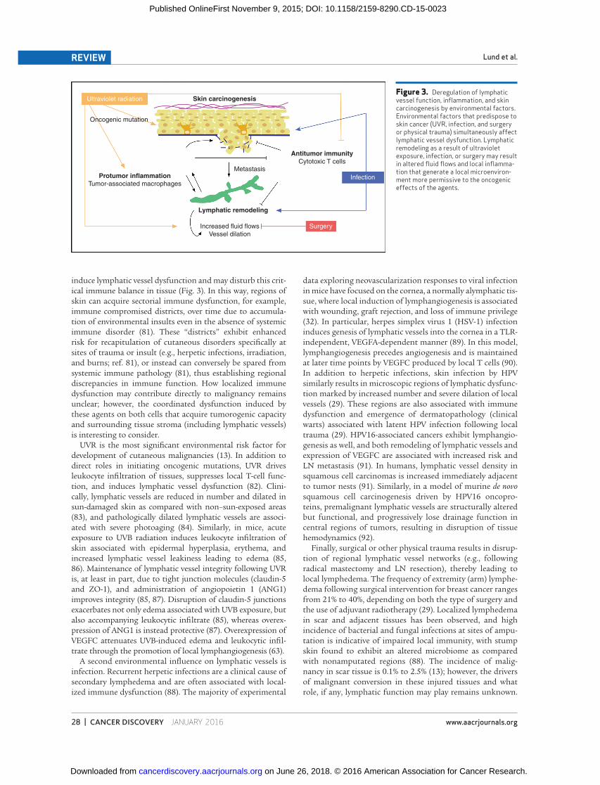
induce lymphatic vessel dysfunction and may disturb this crit-ical immune balance in tissue ( Fig. 3 ). In this way, regions of skin can acquire sectorial immune dysfunction, for example, immune compromised districts, over time due to accumula-tion of environmental insults even in the absence of systemic immune disorder ( 81 ). These “districts” exhibit enhanced risk for recapitulation of cutaneous disorders specifi cally at sites of trauma or insult (e.g., herpetic infections, irradiation, and burns; ref. 81 ), or instead can conversely be spared from systemic immune pathology ( 81 ), thus establishing regional discrepancies in immune function. How localized immune dysfunction may contribute directly to malignancy remains unclear; however, the coordinated dysfunction induced by these agents on both cells that acquire tumorogenic capacity and surrounding tissue stroma (including lymphatic vessels) is interesting to consider.
UVR is the most signifi cant environmental risk factor for development of cutaneous malignancies ( 13 ). In addition to direct roles in initiating oncogenic mutations, UVR drives leukocyte infi ltration of tissues, suppresses local T-cell func-tion, and induces lymphatic vessel dysfunction ( 82 ). Clini-cally, lymphatic vessels are reduced in number and dilated in sun-damaged skin as compared with non–sun-exposed areas ( 83 ), and pathologically dilated lymphatic vessels are associ-ated with severe photoaging ( 84 ). Similarly, in mice, acute exposure to UVB radiation induces leukocyte infi ltration of skin associated with epidermal hyperplasia, erythema, and increased lymphatic vessel leakiness leading to edema ( 85, 86 ). Maintenance of lymphatic vessel integrity following UVR is, at least in part, due to tight junction molecules (claudin-5 and ZO-1), and administration of angiopoietin 1 (ANG1) improves integrity ( 85 , 87 ). Disruption of claudin-5 junctions exacerbates not only edema associated with UVB exposure, but also accompanying leukocytic infi ltrate ( 85 ), whereas overex-pression of ANG1 is instead protective ( 87 ). Overexpression of VEGFC attenuates UVB-induced edema and leukocytic infi l-trate through the promotion of local lymphangiogenesis ( 63 ).
A second environmental infl uence on lymphatic vessels is infection. Recurrent herpetic infections are a clinical cause of secondary lymphedema and are often associated with local-ized immune dysfunction ( 88 ). The majority of experimental
data exploring neovascularization responses to viral infection in mice have focused on the cornea, a normally alymphatic tis-sue, where local induction of lymphangiogenesis is associated with wounding, graft rejection, and loss of immune privilege ( 32 ). In particular, herpes simplex virus 1 (HSV-1) infection induces genesis of lymphatic vessels into the cornea in a TLR-independent, VEGFA-dependent manner ( 89 ). In this model, lymphangiogenesis precedes angiogenesis and is maintained at later time points by VEGFC produced by local T cells ( 90 ). In addition to herpetic infections, skin infection by HPV similarly results in microscopic regions of lymphatic dysfunc-tion marked by increased number and severe dilation of local vessels ( 29 ). These regions are also associated with immune dysfunction and emergence of dermatopathology (clinical warts) associated with latent HPV infection following local trauma ( 29 ). HPV16-associated cancers exhibit lymphangio-genesis as well, and both remodeling of lymphatic vessels and expression of VEGFC are associated with increased risk and LN metastasis ( 91 ). In humans, lymphatic vessel density in squamous cell carcinomas is increased immediately adjacent to tumor nests ( 91 ). Similarly, in a model of murine de novo squamous cell carcinogenesis driven by HPV16 oncopro-teins, premalignant lymphatic vessels are structurally altered but functional, and progressively lose drainage function in central regions of tumors, resulting in disruption of tissue hemodynamics ( 92 ).
Finally, surgical or other physical trauma results in disrup-tion of regional lymphatic vessel networks (e.g., following radical mastectomy and LN resection), thereby leading to local lymphedema. The frequency of extremity (arm) lymphe-dema following surgical intervention for breast cancer ranges from 21% to 40%, depending on both the type of surgery and the use of adjuvant radiotherapy ( 29 ). Localized lymphedema in scar and adjacent tissues has been observed, and high incidence of bacterial and fungal infections at sites of ampu-tation is indicative of impaired local immunity, with stump skin found to exhibit an altered microbiome as compared with nonamputated regions ( 88 ). The incidence of malig-nancy in scar tissue is 0.1% to 2.5% ( 13 ); however, the drivers of malignant conversion in these injured tissues and what role, if any, lymphatic function may play remains unknown.
Figure 3. Deregulation of lymphatic vessel function, infl ammation, and skin carcinogenesis by environmental factors. Environmental factors that predispose to skin cancer (UVR, infection, and surgery or physical trauma) simultaneously affect lymphatic vessel dysfunction. Lymphatic remodeling as a result of ultraviolet exposure, infection, or surgery may result in altered fl uid fl ows and local infl amma-tion that generate a local microenviron-ment more permissive to the oncogenic effects of the agents.
Skin carcinogenesisUltraviolet radiation
Oncogenic mutation
MetastasisProtumor inflammation
Tumor-associated macrophages
Lymphatic remodeling
Antitumor immunityCytotoxic T cells
Increased fluid flowsVessel dilation
Surgery
Infection
on June 26, 2018. © 2016 American Association for Cancer Research. cancerdiscovery.aacrjournals.org Downloaded from
Published OnlineFirst November 9, 2015; DOI: 10.1158/2159-8290.CD-15-0023
JANUARY 2016�CANCER DISCOVERY | 29
Lymphatic Vessels, Infl ammation, and Immunity in Skin Cancer REVIEW
Interestingly, VEGFC therapy may restore lymphatic func-tion at least in some contexts to prevent surgery-associated lymphedema. Administration of growth factors along with LN transfer in pigs promoted robust growth of lymphatic vessels, helping to preserve transferred node structure and normalize lymphatic vascular anatomy ( 93 ).
LYMPHATIC VESSELS, INFLAMMATION, AND METASTASIS
Lymphatic vessels have been evaluated in the context of many solid tumors for their ability to predict locoregional, LN metastasis and overall survival ( 94 ). We limit our discussion here to the interplay between infl ammation and tumor-associ-ated lymphangiogenesis and direct the reader to recent reviews that cover mechanisms of lymphangiogenesis, lymphogenous tumor spread, and seeding in more detail ( 26, 27 ). The major molecular drivers of tumor-associated lymphangiogenesis are VEGFC and VEGFD, produced by both tumor and infi ltrat-ing myeloid cells ( 26 ). VEGFC and VEGFD exert their biologic effects through binding to VEGFR3 and VEGFR2, leading to activation of receptor-tyrosine kinase activity through autophosphorylation, which, in turn, activates aspects of LEC function and vessel formation ( 95, 96 ). VEGFC induces endothelial cell migration, permeability, proliferation, vessel enlargement, and enhanced traffi cking of neoplastic cells to LNs, largely via VEGFR3-dependent mechanisms ( 96–100 ). VEGFR3 expression correlates with lymphatic metastasis in some human tumors, and high VEGFC and VEGFD expres-sion is associated with increased lymphatic vessel density and lymphovascular invasion in both human tumors and animal models ( 97 , 99–102 ), and consequently inhibition of either VEGFR3 or its coreceptor neuropilin 2 reduces incidence of LN metastasis ( 97 , 103–105 ).
In skin cancer, the overwhelming majority of lymphatic ves-sel biomarker studies have focused on metastatic melanoma ( 94 ), where increased lymphatic vessel density correlates with VEGFC expression in tumor microenvironments, lymphatic vessel invasion, and LN metastasis ( 39 , 106 , 107 ). Although several studies have demonstrated a positive correlation between lymphatic markers and survival ( 26 , 106 ), other stud-ies have failed to demonstrate a correlation ( 108, 109 ), thus generating some controversy over the true independent prog-nostic value of lymphatic vessel density and invasion biomar-kers. In addition to melanoma, however, recent studies have reported increased lymphangiogenesis in dermis adjacent to tumor nests in squamous cell carcinoma that also exhibit enhanced levels of VEGFC produced by CD163 + CD68 + mac-rophages ( 91 ). Similarly, an increase in the absolute numbers of lymphatic capillaries was observed in Merkel cell carci-noma, likely driven again by CD163 + CD68 + tumor–associ-ated macrophages ( 110 ), and increased levels of VEGFR3 + cells (both tumor and stroma) and lymphatic (podoplanin + ) vessels correlated with disease progression in one study of Sézary syndrome, an aggressive subtype of cutaneous T-cell lymphoma ( 111 ).
In addition to remodeling of vessels in the primary tumor, signifi cant enlargement of lymph sinuses in tumor-reactive LNs is observed before the arrival of metastatic cells ( 112 ). Distal remodeling of lymphatic vessels, both structural and
functional, is mediated, at least in part, by VEGFC and neuropilin 2 ( 98 , 113 , 114 ), and these changes may play a role in establishing premetastatic niches for locoregional dis-semination. Importantly, lymphatic remodeling induced by these signals is also associated with altered fl uid fl ows, which in turn correlate with LN metastasis ( 97–100 , 115 ). Dynamic lymphoscintography that traces accumulation of radionucle-otide over time in draining LNs reveals that increased rates of lymphatic fl ow associate with increased incidence of metas-tasis and lymphatic vessel density in primary tumors ( 116 ). Moreover, VEGFC overexpression further promotes lymph fl ow ( 116 ), and exogenous application of VEGFC reduces tumor IFPs ( 117 ).
It was previously thought that functional lymphatic ves-sels were excluded from solid tumor parenchyma, thus negating their role in tumor progression. However, neither intratumoral lymphangiogenesis nor intratumoral lym-phatic vessels are required in all cases for LN metastasis ( 118, 119 ). Furthermore, an impaired capacity of lymphatic vessels to transport fl uid does not necessarily correlate with their role in transporting cells, and, instead, remodeling of peri-tumoral lymphatic vessels and activation of existing vessels is suffi cient for altered homing properties and metastatic potential ( 98 , 118 , 119 ). The mechanisms by which lym-phatic vessels actively recruit and promote seeding of tumor cells have been reviewed elsewhere ( 26 ). It is of note, however, that direct evidence for a sequential model of metastasis whereby lymphogenous spread precedes hematogenous col-onization of distal organs is lacking ( 26 , 120 ); thus, the rela-tive contribution of tumor-associated lymphatic remodeling and metastasis to overall survival and disease progression remains somewhat unclear.
What may contribute to the association of lymphatic remodeling with poor prognosis is its relationship with local infl ammation. Clinical and histopathologic studies have demonstrated a correlation between cyclooxygenase 2 (COX2) expression, lymphatic vessel density, and LN metastasis in human malignancy ( 121–124 ). Current data indicate that the prostaglandin pathway infl uences lymphangiogenesis through upregulation of VEGFC and VEGFD expression in tumor-associated macrophages ( 125 ), as well as in neo-plastic cells ( 126 ); however, the role of prostaglandins in lymphangiogenesis is not tumor-specifi c. In a subcutaneous Matrigel plug assay of granuloma formation, lymphangio-genesis was also COX2-dependent, requiring the prostag-landin E (EP) receptors 3 and 4 and macrophage production of VEGFC ( 125 ). Similarly, COX2 in the microenvironment of involuting mammary glands contributes to normal invo-lution-associated lymphangiogenesis as well as mammary tumor-associated lymphangiogenesis, tumor cell invasion into lymphatic vessels, and distal metastasis ( 127 ). Inter-estingly, it was recently reported that LECs regulate pros-taglandin degradation downstream of VEGFD–VEGFR3/2 signaling, resulting in the accumulation of tissue PGE 2 and dilation of collecting lymphatic vessels draining the tumor ( 128 ). This dilation is associated with VEGFD expression and enhanced LN metastasis in both human and mouse models ( 128 ). Specifi cally in skin, UVR stimulates COX2 expression in epidermis ( 129 )—the EP1, EP2, and EP4 receptors have been linked to UV-induced carcinogenesis ( 130 ), and notably
on June 26, 2018. © 2016 American Association for Cancer Research. cancerdiscovery.aacrjournals.org Downloaded from
Published OnlineFirst November 9, 2015; DOI: 10.1158/2159-8290.CD-15-0023
30 | CANCER DISCOVERY�JANUARY 2016 www.aacrjournals.org
Lund et al.REVIEW
inhibition of COX2 via administration of celecoxib prevents both squamous and basal cell carcinoma development in mice and humans ( 131, 132 ).
THERAPEUTIC IMPLICATIONS
Targeting Lymphangiogenesis Targeting of tumor-associated lymphangiogenesis as a
potential therapy for solid tumors has been attempted in both preclinical and clinical trials ( 27 , 94 ). It is diffi cult, how-ever, to separate effects on the lymphatic vasculature from hematogenous vessels given overlap in mechanisms required for their remodeling and growth, namely VEGFR3 ( 104 , 133 , 134 ). Either way, direct targeting of tumor vasculature in the clinic with FDA-approved drugs, such as bevacizumab, sorafenib, and sutinib, that target VEGFA and kinase activ-ity of VEGF family receptors (all of which may also interfere with signaling on tumor-associated lymphatic endothelium) provides only transient benefi t, and ultimately tumors adapt and regrow ( 133 ).
Alternatively, it has been suggested that indirect inhibition of vascular remodeling may be induced through repolariza-tion of immune microenvironments ( 135 ). Numerous studies have revealed that recruitment of myeloid cells facilitates the angiogenic switch through expression of VEGF and libera-tion of proangiogenic molecules from ECM ( 20, 21 ). Target-ing recruitment of these cells into tissue parenchyma results
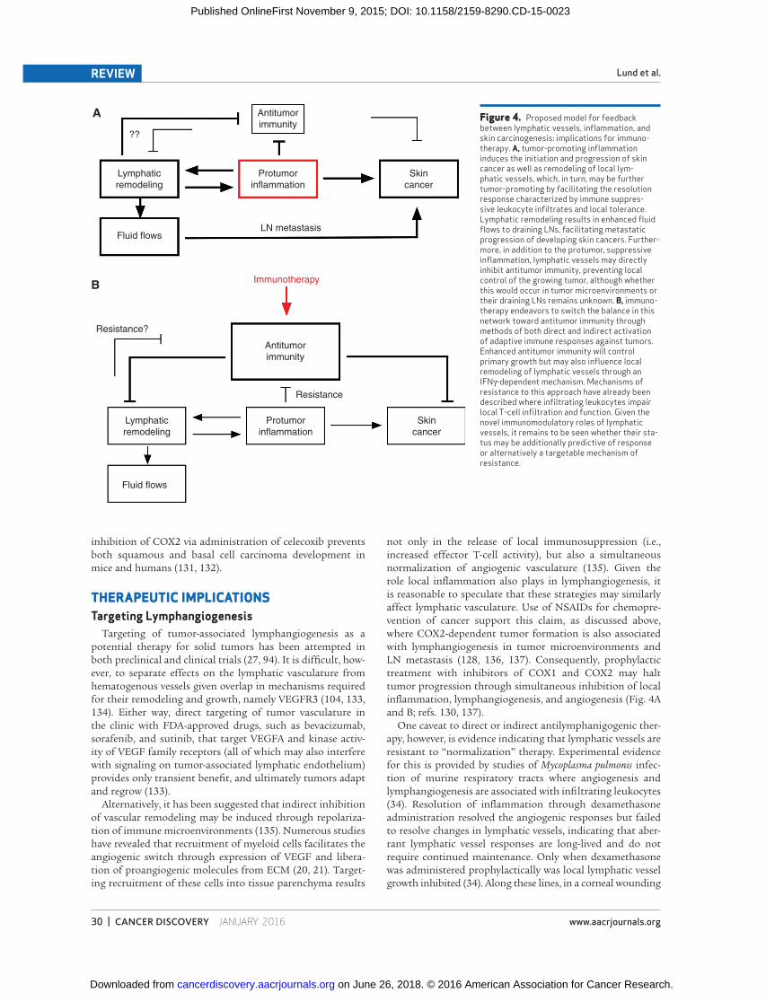
not only in the release of local immunosuppression (i.e., increased effector T-cell activity), but also a simultaneous normalization of angiogenic vasculature ( 135 ). Given the role local infl ammation also plays in lymphangiogenesis, it is reasonable to speculate that these strategies may similarly affect lymphatic vasculature. Use of NSAIDs for chemopre-vention of cancer support this claim, as discussed above, where COX2-dependent tumor formation is also associated with lymphangiogenesis in tumor microenvironments and LN metastasis ( 128 , 136 , 137 ). Consequently, prophylactic treatment with inhibitors of COX1 and COX2 may halt tumor progression through simultaneous inhibition of local infl ammation, lymphangiogenesis, and angiogenesis ( Fig. 4A and B ; refs. 130 , 137 ).
One caveat to direct or indirect antilymphanigogenic ther-apy, however, is evidence indicating that lymphatic vessels are resistant to “normalization” therapy. Experimental evidence for this is provided by studies of Mycoplasma pulmonis infec-tion of murine respiratory tracts where angiogenesis and lymphangiogenesis are associated with infi ltrating leukocytes ( 34 ). Resolution of infl ammation through dexamethasone administration resolved the angiogenic responses but failed to resolve changes in lymphatic vessels, indicating that aber-rant lymphatic vessel responses are long-lived and do not require continued maintenance. Only when dexamethasone was administered prophylactically was local lymphatic vessel growth inhibited ( 34 ). Along these lines, in a corneal wounding
Figure 4. Proposed model for feedback between lymphatic vessels, infl ammation, and skin carcinogenesis: implications for immuno-therapy. A, tumor-promoting infl ammation induces the initiation and progression of skin cancer as well as remodeling of local lym-phatic vessels, which, in turn, may be further tumor-promoting by facilitating the resolution response characterized by immune suppres-sive leukocyte infi ltrates and local tolerance. Lymphatic remodeling results in enhanced fl uid fl ows to draining LNs, facilitating metastatic progression of developing skin cancers. Further-more, in addition to the protumor, suppressive infl ammation, lymphatic vessels may directly inhibit antitumor immunity, preventing local control of the growing tumor, although whether this would occur in tumor microenvironments or their draining LNs remains unknown. B, immuno-therapy endeavors to switch the balance in this network toward antitumor immunity through methods of both direct and indirect activation of adaptive immune responses against tumors. Enhanced antitumor immunity will control primary growth but may also infl uence local remodeling of lymphatic vessels through an IFNγ-dependent mechanism. Mechanisms of resistance to this approach have already been described where infi ltrating leukocytes impair local T-cell infi ltration and function. Given the novel immunomodulatory roles of lymphatic vessels, it remains to be seen whether their sta-tus may be additionally predictive of response or alternatively a targetable mechanism of resistance.
Antitumorimmunity
??
Lymphaticremodeling
Protumorinflammation
Skincancer
LN metastasis
Antitumorimmunity
Immunotherapy
Fluid flows
A
B
Resistance?
Lymphaticremodeling
Protumorinflammation
Skincancer
Fluid flows
Resistance
on June 26, 2018. © 2016 American Association for Cancer Research. cancerdiscovery.aacrjournals.org Downloaded from
Published OnlineFirst November 9, 2015; DOI: 10.1158/2159-8290.CD-15-0023
JANUARY 2016�CANCER DISCOVERY | 31
Lymphatic Vessels, Infl ammation, and Immunity in Skin Cancer REVIEW
model, lymphatic vessel regrowth kinetics accelerated with sequential challenges, indicating potential memory responses facilitating rapid recall growth in the cornea ( 138 ). Although it remains to be seen whether similar observations can be made outside the respiratory tract and cornea, these reports present the very interesting idea that long-lived lymphatic vessel aberrancies may exist, and may predict functional con-sequences for future infl ammation, therapeutic resistance, and recurrence. Clearly, if indeed refractory to normalization strategies, lymphatic vessels present an interesting challenge to clinical therapy, particularly with respect to therapeutic timing, that should be more systematically explored.
Lymphatic Vessels as an Immune Biomarker Recent interest in the role of the immune system in tumor
progression has justifi ed a series of studies evaluating the potential for local infi ltrates to provide prognostic value for risk stratifi cation ( 16 ). Melanoma was one of the fi rst tumors to be associated with spontaneous antitumor immunity lead-ing to regression—appearance of autoimmune vitiligo ( 139 ), presence of intralesional tumor-infi ltrating lymphocytes ( 140 ), expression of lymphocyte chemotactic factors ( 141 ), and a type I IFN transcriptional profi le ( 142 ) enrich for a subset of clinical responders in this context. This idea has been formalized into a quantitative metric refl ecting immune histopathology of tumors and is a signifi cant prognostic indicator for disease-free survival and overall survival for colorectal cancer ( 16 , 143 ). Evaluation of immune contexture of tumors, for example, “Immunoscore,” includes quantifi ca-tion of the density of CD8 + and CD45RO + effector and mem-ory T cells, as a function of their location in central tumor regions as compared with invasive margins ( 143 ). The recog-nition that Immunoscore signifi cantly refl ects a prognostic biomarker for tumor progression and patient outcome has yielded a broader awareness of the importance of CD8 + T-cell infi ltration as an indicator of an immunologically reactive tumor ( 4 ). What may further inform this type of immune-based stratifi cation strategy is now an intense area of study, and efforts to include various suppressive components of tumor microenvironments might provide added value to cur-rent approaches. These efforts typically focus on infi ltrating immune cells of myeloid origin; however, the potential role for nonhematopoeitic stromal components, such as vascu-lature, both blood and lymph, deserves attention. In fact, it may be the close association between tumor-associated lym-phangiogenesis and infi ltrating myeloid cells that underlies controversy in clinical lymphatic vessel biomarker studies ( 39 , 91 ). This, together with evidence that the degree of both intratumoral and peritumoral lymphatic vessel density is associated with the presence of tumor-associated macro-phages, more advanced stage (i.e., increased tumor thickness, mitotic count, and ulceration; ref. 39 ), and decreased num-bers of infi ltrating lymphocytes ( 144 ), indicates that correla-tions between lymphatic vasculature and local immunity in clinical samples is signifi cant to consider.
Current immunohistochemical mapping of immune infi l-trates and other components of tumor microenvironments is necessarily reliant on biopsies of primary or metastatic dis-ease. Solid tumors, however, are known to be heterogeneous with respect to both distribution of intrinsic oncogenic sig-
naling and activation of microenvironmental factors ( 145 ). Consequently, biopsies may not capture the full complexity within a developing tumor. Although still the most valuable approach, identifi cation of novel tissue biomarkers must be coupled to efforts to defi ne correlative systemic biomar-ker signatures. The challenge in this approach is, however, exemplifi ed by the fact that although VEGFC is prognostic when detected in tissue by immunohistochemistry ( 101 ), few studies have demonstrated a correlation between serum VEGFA/C/D and LN metastasis ( 146 ). VEGFA/C/D pro-duced at high concentrations in local tissue may become diluted or unstable as they circulate through the lymphoid system back to the thoracic duct due to their short half-lives in circulation; as a consequence, it is not expected that serum levels will necessarily be diagnostic of LN metastases. A potential alternative source for biomarker identifi cation is tumor interstitial fl uid itself, which, unlike plasma, has a higher concentration of these proteins produced within tumor microenvironments and therefore may more reliably refl ect altered biology within the tumor itself ( 147 ). Available methods for interstitial fl uid isolation have recently been reviewed and include tissue centrifugation, tissue elution, capillary ultrafi ltration, and microdialysis ( 147 ). Enhanced levels of known cancer biomarkers, including VEGF, can be detected in interstitial fl uid when compared with plasma ( 147 ), supporting the idea that tissue interstitial fl uid, the contents of which are closer to lymph than plasma, may be a more robust source of biomarkers predicting primary tumor behavior.
CONCLUSIONS
Our understanding of the roles lymphatic vessels play in regulating tumor immune microenvironments in skin and other solid tumors is still in its infancy. The interplay between the vasculature (both hematogenous and lymphog-enous), local infl ammation, and tumor progression sets up a far more complex story than that of nutrient delivery and waste removal ( Fig. 4A and B ). The lymphatic vasculature coordinates local infl ammation and immunity, and its dys-function may contribute to deregulated local infl ammation, a hallmark of cancer as described by Hanahan and Wein-berg ( 15 ). However, where lymphatic vessels really come into play in a developing cutaneous malignancy remains unclear. Whether their early dysfunction predisposes tissue to altered immunity, thus leading to tumor immune escape, or if their predominant role comes later during metastasis and spread remains an open question. Likely, lymphatic vessels partici-pate in a continuous feedback loop, responding to microen-vironmental change in peripheral tissue to propagate a signal to LNs that then alters host responses to tissue and cyclically continues. Our continued understanding of the complexities of lymphatic vessel function in the unique contexts of normal and diseased skin will provide a model to understand how regional immune dysfunction can participate in cutaneous malignancy and potential targets for prognostic and thera-peutic strategies.
Disclosure of Potential Confl icts of Interest No potential confl icts of interest were disclosed.
on June 26, 2018. © 2016 American Association for Cancer Research. cancerdiscovery.aacrjournals.org Downloaded from
Published OnlineFirst November 9, 2015; DOI: 10.1158/2159-8290.CD-15-0023
32 | CANCER DISCOVERY�JANUARY 2016 www.aacrjournals.org
Lund et al.REVIEW
Grant Support The Lund laboratory acknowledges support from the OHSU
Knight Cancer Center (NIH P30-CA069533), the Collins Medical Trust of Oregon, the Medical Research Foundation, the Cancer Research Institute, and the Department of Defense Peer Reviewed Cancer Research Program. T.R. Medler acknowledges support from the Cathy and Jim Rudd Career Development Award for Cancer Research, the Medical Research Foundation, and the American Can-cer Society—Friends of Rob Kinas. L.M. Coussens acknowledges support from the NIH/NCI, the DOD BCRP Era of Hope Scholar Expansion Award, the Susan G. Komen Foundation, the Breast Cancer Research Foundation, Stand Up To Cancer—Lustgarten Foundation Pancreatic Cancer Convergence Dream Team Transla-tional Research Grant (SU2C-AACR-DT14-14), and the Brenden-Colson Center for Pancreatic Health. The authors also acknowledge support from the OHSU Knight Cancer Institute. Stand Up To Cancer is a program of the Entertainment Industry Foundation administered by the American Association for Cancer Research.
Received January 19, 2015; revised August 12, 2015; accepted August 19, 2015; published OnlineFirst November 9, 2015.
REFERENCES 1. Pasparakis M , Haase I , Nestle FO . Mechanisms regulating skin
immunity and infl ammation . Nat Rev Immunol 2014 ; 14 : 289 – 301 . 2. Streilein JW . Skin-associated lymphoid tissues (SALT): origins and
functions . J Invest Dermatol 1983 ; 80 ( Suppl) : 12s – 16s . 3. Topalian SL , Drake CG , Pardoll DM . Immune checkpoint blockade:
a common denominator approach to cancer therapy . Cancer Cell 2015 ; 27 : 450 – 61 .
4. Gajewski TF , Woo S-R , Zha Y , Spaapen R , Zheng Y , Corrales L , et al. Cancer immunotherapy strategies based on overcoming bar-riers within the tumor microenvironment . Curr Opin Immunol 2013 ; 25 : 268 – 76 .
5. Motz GT , Coukos G . The parallel lives of angiogenesis and immu-nosuppression: cancer and other tales . Nat Rev Immunol 2011 ; 11 : 702 – 11 .
6. Alitalo AK , Proulx ST , Karaman S , Aebischer D , Martino S , Jost M , et al. VEGF-C and VEGF-D blockade inhibits infl ammatory skin carcinogenesis . Cancer Res 2013 ; 73 : 4212 – 21 .
7. Lund AW , Duraes FV , Hirosue S , Raghavan VR , Nembrini C , Thomas SN , et al. VEGF-C promotes immune tolerance in B16 melanomas and cross-presentation of tumor antigen by lymph node lymphatics . Cell Rep 2012 ; 1 : 191 – 9 .
8. Hirosue S , Vokali E , Raghavan VR , Rincon-Restrepo M , Lund AW , Corthésy-Henrioud P , et al. Steady-state antigen scavenging, cross-presentation, and CD8 + T cell priming: a new role for lymphatic endothelial cells . J Immunol 2014 ; 192 : 5002 – 11 .
9. Cohen JN , Guidi CJ , Tewalt EF , Qiao H , Rouhani SJ , Ruddell A , et al. Lymph node-resident lymphatic endothelial cells mediate periph-eral tolerance via Aire-independent direct antigen presentation . J Exp Med 2010 ; 207 : 681 – 8 .
10. Tewalt EF , Cohen JN , Rouhani SJ , Guidi CJ , Qiao H , Fahl SP , et al. Lymphatic endothelial cells induce tolerance via PD-L1 and lack of costimulation leading to high-level PD-1 expression on CD8 T cells . Blood 2012 ; 120 : 4772 – 82 .
11. Cohen JN , Tewalt EF , Rouhani SJ , Buonomo EL , Bruce AN , Xu X , et al. Tolerogenic properties of lymphatic endothelial cells are controlled by the lymph node microenvironment . PLoS ONE 2014 ; 9 : e87740 .
12. Jemal A , Siegel R , Xu J , Ward E . Cancer statistics, 2010 . CA Cancer J Clin 2010 ; 60 : 277 – 300 .
13. Maru GB , Gandhi K , Ramchandani A , Kumar G . The role of infl am-mation in skin cancer . Adv Exp Med Biol 2014 ; 816 : 437 – 69 .
14. Loeb LA , Harris CC . Advances in chemical carcinogenesis: a histori-cal review and prospective . Cancer Res 2008 ; 68 : 6863 – 72 .
15. Hanahan D , Weinberg RA . Hallmarks of cancer: the next generation . Cell 2011 ; 144 : 646 – 74 .
16. Galon J , Mlecnik B , Bindea G , Angell HK , Berger A , Lagorce C , et al. Towards the introduction of the “Immunoscore” in the classifi ca-tion of malignant tumours . J Pathol 2014 ; 232 : 199 – 209 .
17. Coussens LM , Zitvogel L , Palucka AK . Neutralizing tumor-promot-ing chronic infl ammation: a magic bullet? Science 2013 ; 339 : 286 – 91 .
18. Proksch E , Brandner JM , Jensen J-M . The skin: an indispensable bar-rier . Exp Dermatol 2008 ; 17 : 1063 – 72 .
19. Bos JD , Zonneveld I , Das PK , Krieg SR , van der Loos CM , Kapsen-berg ML . The skin immune system (SIS): distribution and immu-nophenotype of lymphocyte subpopulations in normal human skin . J Invest Dermatol 1987 ; 88 : 569 – 73 .
20. Medler TR , Coussens LM . Duality of the immune response in cancer: lessons learned from skin . J Invest Dermatol 2014 ; 134 (e1) : E23 – 8 .
21. Coussens LM , Raymond WW , Bergers G , Laig-Webster M , Behrendt-sen O , Werb Z , et al. Infl ammatory mast cells up-regulate ang-iogenesis during squamous epithelial carcinogenesis . Genes Dev 1999 ; 13 : 1382 – 97 .
22. Stockmann C , Schadendorf D , Klose R , Helfrich I . The impact of the immune system on tumor: angiogenesis and vascular remodeling . Front Oncol 2014 ; 4 : 69 .
23. Quail DF , Joyce JA . Microenvironmental regulation of tumor pro-gression and metastasis . Nat Med 2013 ; 19 : 1423 – 37 .
24. Swartz MA , Lund AW . Lymphatic and interstitial fl ow in the tumour microenvironment: linking mechanobiology with immunity . Nat Rev Cancer 2012 ; 12 : 210 – 9 .
25. Yao L-C , Baluk P , Srinivasan RS , Oliver G , McDonald DM . Plasticity of button-like junctions in the endothelium of airway lymphatics in development and infl ammation . Am J Pathol 2012 ; 180 : 2561 – 75 .
26. Stacker SA , Williams SP , Karnezis T , Shayan R , Fox SB , Achen MG . Lymphangiogenesis and lymphatic vessel remodelling in cancer . Nat Rev Cancer 2014 ; 14 : 159 – 72 .
27. Zheng W , Aspelund A , Alitalo K . Lymphangiogenic factors, mecha-nisms, and applications . J Clin Invest 2014 ; 124 : 878 – 87 .
28. Baluk P , Fuxe J , Hashizume H , Romano T , Lashnits E , Butz S , et al. Functionally specialized junctions between endothelial cells of lym-phatic vessels . J Exp Med 2007 ; 204 : 2349 – 62 .
29. Carlson JA . Lymphedema and subclinical lymphostasis (microl-ymphedema) facilitate cutaneous infection, infl ammatory derma-toses, and neoplasia: a locus minoris resistentiae . Clin Dermatol 2014 ; 32 : 599 – 615 .
30. Kim H , Kataru RP , Koh GY . Infl ammation-associated lymphangio-genesis: a double-edged sword? J Clin Invest 2014 ; 124 : 936 – 42 .
31. Card CM , Yu SS , Swartz MA . Emerging roles of lymphatic endothe-lium in regulating adaptive immunity . J Clin Invest 2014 ; 124 : 943 – 52 .
32. Hos D , Cursiefen C . Lymphatic vessels in the development of tissue and organ rejection . Adv Anat Embryol Cell Biol 2014 ; 214 : 119 – 41 .
33. Fiedler E , Helmbold P , Marsch WC . Increased vessel density in pso-riasis: involvement of lymphatic vessels in the papillary dermis . Br J Dermatol 2008 ; 159 : 258 – 61 .
34. Yao L-C , Baluk P , Feng J , McDonald DM . Steroid-resistant lym-phatic remodeling in chronically infl amed mouse airways . Am J Pathol 2010 ; 176 : 1525 – 41 .
35. D’Alessio S , Correale C , Tacconi C , Gandelli A , Pietrogrande G , Vetrano S , et al. VEGF-C-dependent stimulation of lymphatic func-tion ameliorates experimental infl ammatory bowel disease . J Clin Invest 2014 ; 124 : 3863 – 78 .
36. Kunstfeld R , Hirakawa S , Hong Y-K , Schacht V , Lange-Asschenfeldt B , Velasco P , et al. Induction of cutaneous delayed-type hypersensi-tivity reactions in VEGF-A transgenic mice results in chronic skin infl ammation associated with persistent lymphatic hyperplasia . Blood 2004 ; 104 : 1048 – 57 .
37. Skobe M , Hamberg LM , Hawighorst T , Schirner M , Wolf GL , Alitalo K , et al. Concurrent induction of lymphangiogenesis, angiogenesis, and macrophage recruitment by vascular endothelial growth factor-C in melanoma . Am J Pathol 2001 ; 159 : 893 – 903 .
38. Schoppmann SF , Birner P , Stöckl J , Kalt R , Ullrich R , Caucig C , et al. Tumor-associated macrophages express lymphatic endothelial
on June 26, 2018. © 2016 American Association for Cancer Research. cancerdiscovery.aacrjournals.org Downloaded from
Published OnlineFirst November 9, 2015; DOI: 10.1158/2159-8290.CD-15-0023
JANUARY 2016�CANCER DISCOVERY | 33
Lymphatic Vessels, Infl ammation, and Immunity in Skin Cancer REVIEW
growth factors and are related to peritumoral lymphangiogenesis . Am J Pathol 2002 ; 161 : 947 – 56 .
39. Storr SJ , Safuan S , Mitra A , Elliott F , Walker C , Vasko MJ , et al. Objective assessment of blood and lymphatic vessel inva-sion and association with macrophage infiltration in cutaneous melanoma . Mod Pathol Off J U S Can Acad Pathol Inc 2012 ; 25 : 493 – 504 .
40. Angeli V , Ginhoux F , Llodrà J , Quemeneur L , Frenette PS , Skobe M , et al. B cell-driven lymphangiogenesis in infl amed lymph nodes enhances dendritic cell mobilization . Immunity 2006 ; 24 : 203 – 15 .
41. Tan KW , Chong SZ , Wong FHS , Evrard M , Tan SM-L , Keeble J , et al. Neutrophils contribute to infl ammatory lymphangiogenesis by increasing VEGF-A bioavailability and secreting VEGF-D . Blood 2013 ; 122 : 3666 – 77 .
42. Kataru RP , Kim H , Jang C , Choi DK , Koh BI , Kim M , et al. T lym-phocytes negatively regulate lymph node lymphatic vessel forma-tion . Immunity 2011 ; 34 : 96 – 107 .
43. Ohl L , Mohaupt M , Czeloth N , Hintzen G , Kiafard Z , Zwirner J , et al. CCR7 governs skin dendritic cell migration under infl amma-tory and steady-state conditions . Immunity 2004 ; 21 : 279 – 88 .
44. Teijeira A , Rouzaut A , Melero I . Initial afferent lymphatic vessels controlling outbound leukocyte traffi c from skin to lymph nodes . Front Immunol 2013 ; 4 : 433 .
45. Teijeira A , Russo E , Halin C . Taking the lymphatic route: dendritic cell migration to draining lymph nodes . Semin Immunopathol 2014 ; 36 : 261 – 74 .
46. Lämmermann T , Bader BL , Monkley SJ , Worbs T , Wedlich-Söldner R , Hirsch K , et al. Rapid leukocyte migration by integrin-independ-ent fl owing and squeezing . Nature 2008 ; 453 : 51 – 5 .
47. Miteva DO , Rutkowski JM , Dixon JB , Kilarski W , Shields JD , Swartz MA . Transmural fl ow modulates cell and fl uid transport functions of lymphatic endothelium . Circ Res 2010 ; 106 : 920 – 31 .
48. Becker HM , Rullo J , Chen M , Ghazarian M , Bak S , Xiao H , et al. Integrin-mediated adhesion inhibits macrophage exit from a peripheral infl ammatory lesion . J Immunol 2013 ; 190 : 4305 – 14 .
49. Johnson LA , Jackson DG . Control of dendritic cell traffi cking in lymphatics by chemokines . Angiogenesis 2014 Apr ; 17 : 335 – 45 .
50. Vigl B , Aebischer D , Nitschké M , Iolyeva M , Röthlin T , Antsiferova O , et al. Tissue infl ammation modulates gene expression of lym-phatic endothelial cells and dendritic cell migration in a stimulus-dependent manner . Blood 2011 ; 118 : 205 – 15 .
51. Rantakari P , Auvinen K , Jäppinen N , Kapraali M , Valtonen J , Kariko-ski M , et al. The endothelial protein PLVAP in lymphatics controls the entry of lymphocytes and antigens into lymph nodes . Nat Immunol 2015 ; 16 : 386 – 96 .
52. Sixt M , Kanazawa N , Selg M , Samson T , Roos G , Reinhardt DP , et al. The conduit system transports soluble antigens from the afferent lymph to resident dendritic cells in the T cell area of the lymph node . Immunity 2005 ; 22 : 19 – 29 .
53. Mantovani A , Bonecchi R , Locati M . Tuning infl ammation and immunity by chemokine sequestration: decoys and more . Nat Rev Immunol 2006 ; 6 : 907 – 18 .
54. Jamieson T , Cook DN , Nibbs RJB , Rot A , Nixon C , McLean P , et al. The chemokine receptor D6 limits the infl ammatory response in vivo . Nat Immunol 2005 ; 6 : 403 – 11 .
55. McKimmie CS , Singh MD , Hewit K , Lopez-Franco O , Le Brocq M , Rose-John S , et al. An analysis of the function and expression of D6 on lymphatic endothelial cells . Blood 2013 ; 121 : 3768 – 77 .
56. Lee KM , McKimmie CS , Gilchrist DS , Pallas KJ , Nibbs RJ , Garside P , et al. D6 facilitates cellular migration and fl uid fl ow to lymph nodes by suppressing lymphatic congestion . Blood 2011 ; 118 : 6220 – 9 .
57. Savino B , Caronni N , Anselmo A , Pasqualini F , Borroni EM , Basso G , et al. ERK-dependent downregulation of the atypical chemokine receptor D6 drives tumor aggressiveness in Kaposi sarcoma . Cancer Immunol Res 2014 ; 2 : 679 – 89 .
58. Teijeira A , Garasa S , Peláez R , Azpilikueta A , Ochoa C , Marré D , et al. Lymphatic endothelium forms integrin-engaging 3D struc-tures during DC transit across infl amed lymphatic vessels . J Invest Dermatol 2013 ; 133 : 2276 – 85 .
59. Nitschké M , Aebischer D , Abadier M , Haener S , Lucic M , Vigl B , et al. Differential requirement for ROCK in dendritic cell migra-tion within lymphatic capillaries in steady-state and infl ammation . Blood 2012 ; 120 : 2249 – 58 .
60. Dubrot J , Duraes FV , Potin L , Capotosti F , Brighouse D , Suter T , et al. Lymph node stromal cells acquire peptide-MHCII complexes from dendritic cells and induce antigen-specifi c CD4 + T cell toler-ance . J Exp Med 2014 ; 211 : 1153 – 66 .
61. Podgrabinska S , Kamalu O , Mayer L , Shimaoka M , Snoeck H , Ran-dolph GJ , et al. Infl amed lymphatic endothelium suppresses den-dritic cell maturation and function via Mac-1/ICAM-1-dependent mechanism . J Immunol 2009 ; 183 : 1767 – 79 .
62. Huggenberger R , Ullmann S , Proulx ST , Pytowski B , Alitalo K , Det-mar M . Stimulation of lymphangiogenesis via VEGFR-3 inhibits chronic skin infl ammation . J Exp Med 2010 ; 207 : 2255 – 69 .
63. Kajiya K , Sawane M , Huggenberger R , Detmar M . Activation of the VEGFR-3 pathway by VEGF-C attenuates UVB-induced edema for-mation and skin infl ammation by promoting lymphangiogenesis . J Invest Dermatol 2009 ; 129 : 1292 – 8 .
64. Baluk P , Tammela T , Ator E , Lyubynska N , Achen MG , Hicklin DJ , et al. Pathogenesis of persistent lymphatic vessel hyperplasia in chronic airway infl ammation . J Clin Invest 2005 ; 115 : 247 – 57 .
65. Morton AM , Sefi k E , Upadhyay R , Weissleder R , Benoist C , Mathis D . Endoscopic photoconversion reveals unexpectedly broad leu-kocyte traffi cking to and from the gut . Proc Natl Acad Sci U S A 2014 ; 111 : 6696 – 701 .
66. Tomura M , Honda T , Tanizaki H , Otsuka A , Egawa G , Tokura Y , et al. Activated regulatory T cells are the major T cell type emigrat-ing from the skin during a cutaneous immune response in mice . J Clin Invest 2010 ; 120 : 883 – 93 .
67. Jennrich S , Lee MH , Lynn RC , Dewberry K , Debes GF . Tissue exit: a novel control point in the accumulation of antigen-specifi c CD8 T cells in the infl uenza a virus-infected lung . J Virol 2012 ; 86 : 3436 – 45 .
68. Gresser I , Guy-Grand D , Maury C , Maunoury MT . Interferon induces peripheral lymphadenopathy in mice . J Immunol 1981 ; 127 : 1569 – 75 .
69. Chung HT , Samlowski WE , Kelsey DK , Daynes RA . Alterations in lymphocyte recirculation within ultraviolet light-irradiated mice: efferent blockade of lymphocyte egress from peripheral lymph nodes . Cell Immunol 1986 ; 102 : 335 – 45 .
70. Bromley SK , Thomas SY , Luster AD . Chemokine receptor CCR7 guides T cell exit from peripheral tissues and entry into afferent lymphatics . Nat Immunol 2005 ; 6 : 895 – 901 .
71. Debes GF , Arnold CN , Young AJ , Krautwald S , Lipp M , Hay JB , et al. Chemokine receptor CCR7 required for T lymphocyte exit from peripheral tissues . Nat Immunol 2005 ; 6 : 889 – 94 .
72. Brown MN , Fintushel SR , Lee MH , Jennrich S , Geherin SA , Hay JB , et al. Chemoattractant receptors and lymphocyte egress from extralymphoid tissue: changing requirements during the course of infl ammation . J Immunol 2010 ; 185 : 4873 – 82 .
73. Schumacher TN , Schreiber RD . Neoantigens in cancer immuno-therapy . Science 2015 ; 348 : 69 – 74 .
74. Cyster JG , Schwab SR . Sphingosine-1-phosphate and lymphocyte egress from lymphoid organs . Annu Rev Immunol 2012 ; 30 : 69 – 94 .
75. Pham THM , Baluk P , Xu Y , Grigorova I , Bankovich AJ , Pappu R , et al. Lymphatic endothelial cell sphingosine kinase activity is required for lymphocyte egress and lymphatic patterning . J Exp Med 2010 ; 207 : 17 – 27 .
76. Ledgerwood LG , Lal G , Zhang N , Garin A , Esses SJ , Ginhoux F , et al. The sphingosine 1-phosphate receptor 1 causes tissue retention by inhibiting the entry of peripheral tissue T lymphocytes into afferent lymphatics . Nat Immunol 2008 ; 9 : 42 – 53 .
77. Lukacs-Kornek V , Malhotra D , Fletcher AL , Acton SE , Elpek KG , Tayalia P , et al. Regulated release of nitric oxide by nonhematopoi-etic stroma controls expansion of the activated T cell pool in lymph nodes . Nat Immunol 2011 ; 12 : 1096 – 104 .
78. Maio M . Melanoma as a model tumour for immuno-oncology . Ann Oncol Off J Eur Soc Med Oncol ESMO 2012 ; 23 ( Suppl 8) : viii10 – 4 .
on June 26, 2018. © 2016 American Association for Cancer Research. cancerdiscovery.aacrjournals.org Downloaded from
Published OnlineFirst November 9, 2015; DOI: 10.1158/2159-8290.CD-15-0023
34 | CANCER DISCOVERY�JANUARY 2016 www.aacrjournals.org
Lund et al.REVIEW
79. Madore J , Vilain R , Menzies AM , Kakavand H , Wilmott JS , Hyman J , et al. PD-L1 expression in melanoma shows marked heterogene-ity within and between patients: implications for anti-PD-1/PD-L1 clinical trials . Pigment Cell Melanoma Res 2015 ; 28 : 245 – 53 .
80. Tamburini BA , Burchill MA , Kedl RM . Antigen capture and archiv-ing by lymphatic endothelial cells following vaccination or viral infection . Nat Commun 2014 ; 5 : 3989 .
81. Ruocco V , Ruocco E , Piccolo V , Brunetti G , Guerrera LP , Wolf R . The immunocompromised district in dermatology: a unifying patho-genic view of the regional immune dysregulation . Clin Dermatol 2014 ; 32 : 569 – 76 .
82. Nishigori C , Yarosh DB , Donawho C , Kripke ML . The immune system in ultraviolet carcinogenesis . J Investig Dermatol Symp Proc 1996 ; 1 : 143 – 6 .
83. Kajiya K , Kunstfeld R , Detmar M , Chung JH . Reduction of lym-phatic vessels in photodamaged human skin . J Dermatol Sci 2007 ; 47 : 241 – 3 .
84. Back SJ , Kim YJ , Choi DK , Lee Y , Seo YJ , Park JK , et al. Cutaneous lymphangiectasia associated with photoageing and topical corticos-teroid application . Clin Exp Dermatol 2009 ; 34 : 352 – 4 .
85. Matsumoto-Okazaki Y , Furuse M , Kajiya K . Claudin-5 haploinsuf-fi ciency exacerbates UVB-induced oedema formation by inducing lymphatic vessel leakage . Exp Dermatol 2012 ; 21 : 557 – 9 .
86. Kajiya K , Hirakawa S , Detmar M . Vascular endothelial growth factor-A mediates ultraviolet B-induced impairment of lymphatic vessel function . Am J Pathol 2006 ; 169 : 1496 – 503 .
87. Kajiya K , Kidoya H , Sawane M , Matsumoto-Okazaki Y , Yamanishi H , Furuse M , et al. Promotion of lymphatic integrity by angi-opoietin-1/Tie2 signaling during infl ammation . Am J Pathol 2012 ; 180 : 1273 – 82 .
88. Ruocco V , Brunetti G , Puca RV , Ruocco E . The immunocom-promised district: a unifying concept for lymphoedematous, her-pes-infected and otherwise damaged sites . J Eur Acad Dermatol Venereol 2009 ; 23 : 1364 – 73 .
89. Bryant-Hudson KM , Chucair-Elliott AJ , Conrady CD , Cohen A , Zheng M , Carr DJJ . HSV-1 targets lymphatic vessels in the eye and draining lymph node of mice leading to edema in the absence of a functional type I interferon response . Am J Pathol 2013 ; 183 : 1233 – 42 .
90. Conrady CD , Zheng M , Stone DU , Carr DJJ . CD8 + T cells suppress viral replication in the cornea but contribute to VEGF-C-induced lymphatic vessel genesis . J Immunol 2012 ; 189 : 425 – 32 .
91. Moussai D , Mitsui H , Pettersen JS , Pierson KC , Shah KR , Suárez-Fariñas M , et al. The human cutaneous squamous cell carcinoma microenvironment is characterized by increased lymphatic density and enhanced expression of macrophage-derived VEGF-C . J Invest Dermatol 2011 ; 131 : 229 – 36 .
92. Eichten A , Hyun WC , Coussens LM . Distinctive features of angio-genesis and lymphangiogenesis determine their functionality dur-ing de novo tumor development . Cancer Res 2007 ; 67 : 5211 – 20 .
93. Honkonen KM , Visuri MT , Tervala TV , Halonen PJ , Koivisto M , Lähteenvuo MT , et al. Lymph node transfer and perinodal lymphatic growth factor treatment for lymphedema . Ann Surg 2013 ; 257 : 961 – 7 .
94. Pasquali S , van der Ploeg APT , Mocellin S , Stretch JR , Thompson JF , Scolyer RA . Lymphatic biomarkers in primary melanomas as predictors of regional lymph node metastasis and patient outcomes . Pigment Cell Melanoma Res 2013 ; 26 : 326 – 37 .
95. Achen MG , Jeltsch M , Kukk E , Mäkinen T , Vitali A , Wilks AF , et al. Vascular endothelial growth factor D (VEGF-D) is a ligand for the tyrosine kinases VEGF receptor 2 (Flk1) and VEGF receptor 3 (Flt4) . Proc Natl Acad Sci U S A 1998 ; 95 : 548 – 53 .
96. Joukov V , Pajusola K , Kaipainen A , Chilov D , Lahtinen I , Kukk E , et al. A novel vascular endothelial growth factor, VEGF-C, is a lig-and for the Flt4 (VEGFR-3) and KDR (VEGFR-2) receptor tyrosine kinases . EMBO J 1996 ; 15 : 1751 .
97. Skobe M , Hawighorst T , Jackson DG , Prevo R , Janes L , Velasco P , et al. Induction of tumor lymphangiogenesis by VEGF-C promotes breast cancer metastasis . Nat Med 2001 ; 7 : 192 – 8 .
98. He Y , Rajantie I , Pajusola K , Jeltsch M , Holopainen T , Yla-Herttuala S , et al. Vascular endothelial cell growth factor receptor 3-mediated activation of lymphatic endothelium is crucial for tumor cell entry and spread via lymphatic vessels . Cancer Res 2005 ; 65 : 4739 – 46 .
99. Mandriota SJ , Jussila L , Jeltsch M , Compagni A , Baetens D , Prevo R , et al. Vascular endothelial growth factor-C-mediated lymphangio-genesis promotes tumour metastasis . EMBO J 2001 ; 20 : 672 – 82 .
100. Karpanen T , Egeblad M , Karkkainen MJ , Kubo H , Ylä-Herttuala S , Jäättelä M , et al. Vascular endothelial growth factor C promotes tumor lymphangiogenesis and intralymphatic tumor growth . Cancer Res 2001 ; 61 : 1786 – 90 .
101. Stacker SA , Williams RA , Achen MG . Lymphangiogenic growth fac-tors as markers of tumor metastasis . APMIS Acta Pathol Microbiol Immunol Scand 2004 ; 112 : 539 – 49 .
102. Stacker SA , Caesar C , Baldwin ME , Thornton GE , Williams RA , Prevo R , et al. VEGF-D promotes the metastatic spread of tumor cells via the lymphatics . Nat Med 2001 ; 7 : 186 – 91 .
103. Hirakawa S , Kodama S , Kunstfeld R , Kajiya K , Brown LF , Detmar M . VEGF-A induces tumor and sentinel lymph node lymphangio-genesis and promotes lymphatic metastasis . J Exp Med 2005 ; 201 : 1089 – 99 .
104. Xu Y , Yuan L , Mak J , Pardanaud L , Caunt M , Kasman I , et al. Neuropilin-2 mediates VEGF-C-induced lymphatic sprouting together with VEGFR3 . J Cell Biol 2010 ; 188 : 115 – 30 .
105. He Y , Kozaki K-I , Karpanen T , Koshikawa K , Yla-Herttuala S , Taka-hashi T , et al. Suppression of tumor lymphangiogenesis and lymph node metastasis by blocking vascular endothelial growth factor receptor 3 signaling . J Natl Cancer Inst 2002 ; 94 : 819 – 25 .
106. Dadras SS , Paul T , Bertoncini J , Brown LF , Muzikansky A , Jackson DG , et al. Tumor lymphangiogenesis: a novel prognostic indica-tor for cutaneous melanoma metastasis and survival . Am J Pathol 2003 ; 162 : 1951 – 60 .
107. Shayan R , Karnezis T , Murali R , Wilmott JS , Ashton MW , Taylor GI , et al. Lymphatic vessel density in primary melanomas predicts sentinel lymph node status and risk of metastasis . Histopathology 2012 ; 61 : 702 – 10 .
108. Wobser M , Siedel C , Schrama D , Bröcker E-B , Becker JC , Vetter-Kauczok CS . Expression pattern of the lymphatic and vascular markers VEGFR-3 and CD31 does not predict regional lymph node metastasis in cutaneous melanoma . Arch Dermatol Res 2006 ; 297 : 352 – 7 .
109. Sahni D , Robson A , Orchard G , Szydlo R , Evans AV , Russell-Jones R . The use of LYVE-1 antibody for detecting lymphatic involvement in patients with malignant melanoma of known sentinel node status . J Clin Pathol 2005 ; 58 : 715 – 21 .
110. Werchau S , Toberer F , Enk A , Dammann R , Helmbold P . Merkel cell carcinoma induces lymphatic microvessel formation . J Am Acad Dermatol 2012 ; 67 : 215 – 25 .
111. Karpova MB , Fujii K , Jenni D , Dummer R , Urosevic-Maiwald M . Evaluation of lymphangiogenic markers in Sézary syndrome . Leuk Lymphoma 2011 ; 52 : 491 – 501 .
112. Qian C-N , Berghuis B , Tsarfaty G , Bruch M , Kort EJ , Ditlev J , et al. Preparing the “soil”: the primary tumor induces vasculature reor-ganization in the sentinel lymph node before the arrival of meta-static cancer cells . Cancer Res 2006 ; 66 : 10365 – 76 .
113. Gogineni A , Caunt M , Crow A , Lee CV , Fuh G , van Bruggen N , et al. Inhibition of VEGF-C modulates distal lymphatic remodeling and secondary metastasis . PLoS ONE 2013 ; 8 : e68755 .
114. Hoshida T , Isaka N , Hagendoorn J , di Tomaso E , Chen Y-L , Pytowski B , et al. Imaging steps of lymphatic metastasis reveals that vascular endothelial growth factor-C increases metastasis by increasing deliv-ery of cancer cells to lymph nodes: therapeutic implications . Cancer Res 2006 ; 66 : 8065 – 75 .
115. Harrell MI , Iritani BM , Ruddell A . Tumor-induced sentinel lymph node lymphangiogenesis and increased lymph fl ow precede melanoma metastasis . Am J Pathol 2007 ; 170 : 774 – 86 .
116. Proulx ST , Luciani P , Derzsi S , Rinderknecht M , Mumprecht V , Leroux J-C , et al. Quantitative imaging of lymphatic function with liposomal indocyanine green . Cancer Res 2010 ; 70 : 7053 – 62 .
on June 26, 2018. © 2016 American Association for Cancer Research. cancerdiscovery.aacrjournals.org Downloaded from
Published OnlineFirst November 9, 2015; DOI: 10.1158/2159-8290.CD-15-0023
JANUARY 2016�CANCER DISCOVERY | 35
Lymphatic Vessels, Infl ammation, and Immunity in Skin Cancer REVIEW
117. Hofmann M , Pfl anzer R , Zoller NN , Bernd A , Kaufmann R , Thaci D , et al. Vascular endothelial growth factor C-induced lymphangio-genesis decreases tumor interstitial fl uid pressure and tumor . Transl Oncol 2013 ; 6 : 398 – 404 .
118. Padera TP , Kadambi A , di Tomaso E , Carreira CM , Brown EB , Boucher Y , et al. Lymphatic metastasis in the absence of functional intratumor lymphatics . Science 2002 ; 296 : 1883 – 6 .
119. Wong SY , Haack H , Crowley D , Barry M , Bronson RT , Hynes RO . Tumor-secreted vascular endothelial growth factor-C is necessary for prostate cancer lymphangiogenesis, but lymphangiogenesis is unnec-essary for lymph node metastasis . Cancer Res 2005 ; 65 : 9789 – 98 .
120. Ran S , Volk L , Hall K , Flister MJ . Lymphangiogenesis and lymphatic metastasis in breast cancer . Pathophysiology 2010 ; 17 : 229 – 51 .
121. Siironen P , Ristimäki A , Narko K , Nordling S , Louhimo J , Anders-son S , et al. VEGF-C and COX-2 expression in papillary thyroid cancer . Endocr Relat Cancer 2006 ; 13 : 465 – 73 .
122. Soumaoro LT , Uetake H , Takagi Y , Iida S , Higuchi T , Yasuno M , et al. Coexpression of VEGF-C and Cox-2 in human colorectal cancer and its association with lymph node metastasis . Dis Colon Rectum 2006 ; 49 : 392 – 8 .
123. Timoshenko AV , Chakraborty C , Wagner GF , Lala PK . COX-2-medi-ated stimulation of the lymphangiogenic factor VEGF-C in human breast cancer . Br J Cancer 2006 ; 94 : 1154 – 63 .
124. Zhang X-H , Huang D-P , Guo G-L , Chen G-R , Zhang H-X , Wan L , et al. Coexpression of VEGF-C and COX-2 and its association with lymphangiogenesis in human breast cancer . BMC Cancer 2008 ; 8 : 4 .
125. Hosono K , Suzuki T , Tamaki H , Sakagami H , Hayashi I , Narumiya S , et al. Roles of prostaglandin E2-EP3/EP4 receptor signaling in the enhancement of lymphangiogenesis during fi broblast growth factor-2-induced granulation formation . Arterioscler Thromb Vasc Biol 2011 ; 31 : 1049 – 58 .
126. Su J-L , Shih J-Y , Yen M-L , Jeng Y-M , Chang C-C , Hsieh C-Y , et al. Cyclooxygenase-2 induces EP1- and HER-2/Neu-dependent vascular endothelial growth factor-C up-regulation: a novel mechanism of lym-phangiogenesis in lung adenocarcinoma . Cancer Res 2004 ; 64 : 554 – 64 .
127. Lyons TR , Borges VF , Betts CB , Guo Q , Kapoor P , Martinson HA , et al. Cyclooxygenase-2-dependent lymphangiogenesis promotes nodal metastasis of postpartum breast cancer . J Clin Invest 2014 ; 124 : 3901 – 12 .
128. Karnezis T , Shayan R , Caesar C , Roufail S , Harris NC , Ardipradja K , et al. VEGF-D promotes tumor metastasis by regulating prostaglan-dins produced by the collecting lymphatic endothelium . Cancer Cell 2012 ; 21 : 181 – 95 .
129. Rodriguez-Burford C , Tu JH , Mercurio M , Carey D , Han R , Gordon G , et al. Selective cyclooxygenase-2 inhibition produces heterogene-ous erythema response to ultraviolet irradiation . J Invest Dermatol 2005 ; 125 : 1317 – 20 .
130. Elmets CA , Ledet JJ , Athar M . Cyclooxygenases: mediators of UV-induced skin cancer and potential targets for prevention . J Invest Dermatol 2014 ; 134 : 2497 – 502 .
131. Tang JY , Aszterbaum M , Athar M , Barsanti F , Cappola C , Estevez N , et al. Basal cell carcinoma chemoprevention with nonsteroidal anti-infl ammatory drugs in genetically predisposed PTCH1 +/− humans and mice . Cancer Prev Res Phila 2010 ; 3 : 25 – 34 .
132. Elmets CA , Viner JL , Pentland AP , Cantrell W , Lin H-Y , Bailey H , et al. Chemoprevention of nonmelanoma skin cancer with celecoxib: a randomized, double-blind, placebo-controlled trial . J Natl Cancer Inst 2010 ; 102 : 1835 – 44 .
133. Welti J , Loges S , Dimmeler S , Carmeliet P . Recent molecular discov-eries in angiogenesis and antiangiogenic therapies in cancer . J Clin Invest 2013 ; 123 : 3190 – 200 .
134. Tammela T , Zarkada G , Nurmi H , Jakobsson L , Heinolainen K , Tvorogov D , et al. VEGFR-3 controls tip to stalk conversion at vessel fusion sites by reinforcing Notch signalling . Nat Cell Biol 2011 ; 13 : 1202 – 13 .
135. Rivera LB , Bergers G . Intertwined regulation of angiogenesis and immunity by myeloid cells . Trends Immunol 2015 ; 36 : 240 – 9 .
136. Xin X , Majumder M , Girish GV , Mohindra V , Maruyama T , Lala PK . Targeting COX-2 and EP4 to control tumor growth, angiogenesis, lymphangiogenesis and metastasis to the lungs and lymph nodes in a breast cancer model . Lab Investig J Tech Methods Pathol 2012 ; 92 : 1115 – 28 .
137. Karnezis T , Shayan R , Fox S , Achen MG , Stacker SA . The connection between lymphangiogenic signalling and prostaglandin biology: a missing link in the metastatic pathway . Oncotarget 2012 ; 3 : 893 – 906 .
138. Kelley PM , Connor AL , Tempero RM . Lymphatic vessel memory stimulated by recurrent infl ammation . Am J Pathol 2013 ; 182 : 2418 – 28 .
139. Albert DM , Todes-Taylor N , Wagoner M , Nordlund JJ , Lerner AB . Vitiligo or halo nevi occurring in two patients with choroidal melanoma . Arch Dermatol 1982 ; 118 : 34 – 6 .
140. Cipponi A , Wieers G , van Baren N , Coulie PG . Tumor-infi ltrating lymphocytes: apparently good for melanoma patients. But why? Cancer Immunol Immunother CII 2011 ; 60 : 1153 – 60 .
141. Harlin H , Meng Y , Peterson AC , Zha Y , Tretiakova M , Slingluff C , et al. Chemokine expression in melanoma metastases associated with CD8 + T-cell recruitment . Cancer Res 2009 ; 69 : 3077 – 85 .
142. Fuertes MB , Kacha AK , Kline J , Woo S-R , Kranz DM , Murphy KM , et al. Host type I IFN signals are required for antitumor CD8 + T cell responses through CD8{alpha}+ dendritic cells . J Exp Med 2011 ; 208 : 2005 – 16 .
143. Galon J , Costes A , Sanchez-Cabo F , Kirilovsky A , Mlecnik B , Lagorce-Pagès C , et al. Type, density, and location of immune cells within human colorectal tumors predict clinical outcome . Science 2006 ; 313 : 1960 – 4 .
144. Azimi F , Scolyer RA , Rumcheva P , Moncrieff M , Murali R , McCarthy SW , et al. Tumor-infi ltrating lymphocyte grade is an independent predictor of sentinel lymph node status and survival in patients with cutaneous melanoma . J Clin Oncol 2012 ; 30 : 2678 – 83 .
145. Jamal-Hanjani M , Thanopoulou E , Peggs KS , Quezada SA , Swanton C . Tumour heterogeneity and immune-modulation . Curr Opin Pharmacol 2013 ; 13 : 497 – 503 .
146. Zhang Y , Meng X , Zeng H , Guan Y , Zhang Q , Guo S , et al. Serum vascular endothelial growth factor-C levels: a possible diagnostic marker for lymph node metastasis in patients with primary non-small cell lung cancer . Oncol Lett 2013 ; 6 : 545 – 9 .
147. Wagner M , Wiig H . Tumor Interstitial fl uid formation, characteriza-tion and clinical implications . Front Oncol 2015 ; 5 : 115 .
on June 26, 2018. © 2016 American Association for Cancer Research. cancerdiscovery.aacrjournals.org Downloaded from
Published OnlineFirst November 9, 2015; DOI: 10.1158/2159-8290.CD-15-0023
2016;6:22-35. Published OnlineFirst November 9, 2015.Cancer Discov Amanda W. Lund, Terry R. Medler, Sancy A. Leachman, et al. Lymphatic Vessels, Inflammation, and Immunity in Skin Cancer
Updated version
10.1158/2159-8290.CD-15-0023doi:
Access the most recent version of this article at:
Cited articles
http://cancerdiscovery.aacrjournals.org/content/6/1/22.full#ref-list-1
This article cites 147 articles, 48 of which you can access for free at:
Citing articles
http://cancerdiscovery.aacrjournals.org/content/6/1/22.full#related-urls
This article has been cited by 4 HighWire-hosted articles. Access the articles at:
E-mail alerts related to this article or journal.Sign up to receive free email-alerts
Subscriptions
Reprints and
To order reprints of this article or to subscribe to the journal, contact the AACR Publications Department at
Permissions
Rightslink site. Click on "Request Permissions" which will take you to the Copyright Clearance Center's (CCC)
.http://cancerdiscovery.aacrjournals.org/content/6/1/22To request permission to re-use all or part of this article, use this link
on June 26, 2018. © 2016 American Association for Cancer Research. cancerdiscovery.aacrjournals.org Downloaded from
Published OnlineFirst November 9, 2015; DOI: 10.1158/2159-8290.CD-15-0023