Lutzomyia longipalpis and the eco-epidemiology of American ... · Mem Inst Oswaldo Cruz, Rio de...
18
811 811 811 811 811 Mem Inst Oswaldo Cruz, Rio de Janeiro, Vol. 100(8): 811-827, December 2005 Lutzomyia longipalpis and the eco-epidemiology of American visceral leishmaniasis, with particular reference to Brazil - A Review Ralph Lainson/ + , Elizabeth F Rangel* Departamento de Parasitologia, Instituto Evandro Chagas, Av. Almirante Barroso 492, 66090-000 Belém, PA, Brasil *Departamento de Entomologia, Instituto Oswaldo Cruz-Fiocruz, Rio de Janeiro, RJ, Brasil An historical review is given of American visceral leishmaniasis (AVL), with particular reference to the eco- epidemiology of the disease in Brazil. Following the first records of AVL in this country, in 1934, the sandfly Lutzomyia longipalpis (Lutz and Neiva, 1912) was incriminated as the principal vector. It is now generally accepted, however, that there exist a number of cryptic species under the name of Lu. longipalpis s.l. and that variations in the quantity of the vasodilatory peptide maxadilan in the saliva of flies from different populations of Lu. longipalpis s.l., may account for the variable clinical manifestations of AVL seen in different geographic regions. Distribution of AVL has been shown to extend throughout most of South and Central America, with the domestic dog serving as the principal reservoir of infection for man. However, while one hypothesis suggests that the causative parasite is Leishmania infantum, imported from Europe with the Portuguese and Spanish colonists, the demonstration of a high rate of benign, inapparent infection in foxes in Amazonian Brazil raised an opposing suggestion that the parasite is indigenous to the Americas. Recent reports of similar infections in native marsupials, and possibly rodents, tend to support this view, particularly as Lu. longipalpis is primordially a silvatic sandfly. Although effective control mea- sures in foci of the disease will diminish the number of canine and human infections, the presence of such an enzootic in a variety of native animals will render the total eradication of AVL unlikely. Key words: Lutzomyia longipalpis - sandflies - American visceral leishmaniasis - eco-epidemiology - Brazil Early history: studies in the states of Sergipe, Pará, and Ceará Following the first description of the sandfly Lutzomyia longipalpis Lutz and Neiva, 1912, in an inde- terminate locality in Brazil, interest in this insect remained largely entomological until the mid-1930s. In 1934, how- ever, Henrique Penna used the viscerotome to examine liver samples from persons who were suspected to have died from yellow fever in various rural localities in Brazil, and found that 41 of these deaths were, in fact, due to visceral leishmaniasis. His results suggested the major foci of the disease to be in the northeastern states, par- ticularly in Ceará, and Carlos Chagas, at that time Director of the Instituto Oswaldo Cruz in Rio de Janeiro, sent his son Evandro Chagas to investigate the epidemiology. His first study was made in Sergipe where, in addition to giv- ing the first clinical description of a living case of Ameri- can visceral leishmaniasis (AVL) in Brazil, he made the important observation that the most frequent blood-suck- ing insect in and around the patient’s house was the phlebotomine sandfly Lu. longipalpis (Chagas 1936). Evandro Chagas was appointed head of a Commision set up in 1936 to continue his studies and, in view of the higher prevalence of AVL in the Northeast, it was there that he wished to work. Perversely, the only state govenor Financial support: Wellcome Trust, London + Corresponding author. E-mail: [email protected] Received 13 October 2005 Accepted 2 December 2005 who offered the necessary financial and logistic back-up was Da Gama Malcher of Pará in the North of Brazil, where the number of recorded cases was low, and a huge old colonial-style mansion was made available for conversion into the Commision’s laboratories, which received the imposing name of “The Institute of Experimental Pathol- ogy for the North” (IPEN). Working in the rural areas of Abaetetuba and Moju, where cases of AVL were recorded by Penna, the Com- mision uncovered more cases of the disease in both hu- mans and dogs (Chagas et al. 1938). Once more Lu. longipalpis was shown to be the principal man-biting insect in and around the houses of the infected persons, and this sandfly became the major suspect as the vector. It was concluded that the disease was essentially rural and only occurred in the close vicinity of forest or copses. For this reason it was suggested that the origin of the causative parasite, named as Leishmania chagasi by Cunha and Chagas (1937), was in some wild animal. The Commission’s hope of confirming the role of Lu. longipalpis as the vector and indicating the wild animal reservoir were dashed in 1940, however, when the tragic death of their leader Evandro Chagas in a mid-air plane collision put an abrupt end to their epidemiological stud- ies. Although the IPEN was renamed “Instituto Evandro Chagas” in his honour, his little band of dedicated work- ers never recovered fully from the loss of their brilliant and colourful leader, and research on the epidemiology of visceral leishmaniasis in Brazil went into steady decline. A rude awakening to the real importance of AVL in Brazil did not occur until 1953, when over 100 inhabitants of the small town of Sobral, Ceará, died in a severe out-
Transcript of Lutzomyia longipalpis and the eco-epidemiology of American ... · Mem Inst Oswaldo Cruz, Rio de...

811811811811811Mem Inst Oswaldo Cruz, Rio de Janeiro, Vol. 100(8): 811-827, December 2005
Lutzomyia longipalpis and the eco-epidemiology of Americanvisceral leishmaniasis, with particular reference to Brazil -
A ReviewRalph Lainson/+, Elizabeth F Rangel*
Departamento de Parasitologia, Instituto Evandro Chagas, Av. Almirante Barroso 492, 66090-000 Belém, PA, Brasil*Departamento de Entomologia, Instituto Oswaldo Cruz-Fiocruz, Rio de Janeiro, RJ, Brasil
An historical review is given of American visceral leishmaniasis (AVL), with particular reference to the eco-epidemiology of the disease in Brazil. Following the first records of AVL in this country, in 1934, the sandflyLutzomyia longipalpis (Lutz and Neiva, 1912) was incriminated as the principal vector. It is now generally accepted,however, that there exist a number of cryptic species under the name of Lu. longipalpis s.l. and that variations in thequantity of the vasodilatory peptide maxadilan in the saliva of flies from different populations of Lu. longipalpis s.l.,may account for the variable clinical manifestations of AVL seen in different geographic regions. Distribution of AVLhas been shown to extend throughout most of South and Central America, with the domestic dog serving as theprincipal reservoir of infection for man. However, while one hypothesis suggests that the causative parasite isLeishmania infantum, imported from Europe with the Portuguese and Spanish colonists, the demonstration of a highrate of benign, inapparent infection in foxes in Amazonian Brazil raised an opposing suggestion that the parasite isindigenous to the Americas. Recent reports of similar infections in native marsupials, and possibly rodents, tend tosupport this view, particularly as Lu. longipalpis is primordially a silvatic sandfly. Although effective control mea-sures in foci of the disease will diminish the number of canine and human infections, the presence of such an enzooticin a variety of native animals will render the total eradication of AVL unlikely.
Key words: Lutzomyia longipalpis - sandflies - American visceral leishmaniasis - eco-epidemiology - Brazil
Early history: studies in the states of Sergipe, Pará,and Ceará
Following the first description of the sandflyLutzomyia longipalpis Lutz and Neiva, 1912, in an inde-terminate locality in Brazil, interest in this insect remainedlargely entomological until the mid-1930s. In 1934, how-ever, Henrique Penna used the viscerotome to examineliver samples from persons who were suspected to havedied from yellow fever in various rural localities in Brazil,and found that 41 of these deaths were, in fact, due tovisceral leishmaniasis. His results suggested the majorfoci of the disease to be in the northeastern states, par-ticularly in Ceará, and Carlos Chagas, at that time Directorof the Instituto Oswaldo Cruz in Rio de Janeiro, sent hisson Evandro Chagas to investigate the epidemiology. Hisfirst study was made in Sergipe where, in addition to giv-ing the first clinical description of a living case of Ameri-can visceral leishmaniasis (AVL) in Brazil, he made theimportant observation that the most frequent blood-suck-ing insect in and around the patient’s house was thephlebotomine sandfly Lu. longipalpis (Chagas 1936).
Evandro Chagas was appointed head of a Commisionset up in 1936 to continue his studies and, in view of thehigher prevalence of AVL in the Northeast, it was therethat he wished to work. Perversely, the only state govenor
Financial support: Wellcome Trust, London+Corresponding author. E-mail: [email protected] 13 October 2005Accepted 2 December 2005
who offered the necessary financial and logistic back-upwas Da Gama Malcher of Pará in the North of Brazil, wherethe number of recorded cases was low, and a huge oldcolonial-style mansion was made available for conversioninto the Commision’s laboratories, which received theimposing name of “The Institute of Experimental Pathol-ogy for the North” (IPEN).
Working in the rural areas of Abaetetuba and Moju,where cases of AVL were recorded by Penna, the Com-mision uncovered more cases of the disease in both hu-mans and dogs (Chagas et al. 1938). Once more Lu.longipalpis was shown to be the principal man-bitinginsect in and around the houses of the infected persons,and this sandfly became the major suspect as the vector.It was concluded that the disease was essentially ruraland only occurred in the close vicinity of forest or copses.For this reason it was suggested that the origin of thecausative parasite, named as Leishmania chagasi byCunha and Chagas (1937), was in some wild animal. TheCommission’s hope of confirming the role of Lu.longipalpis as the vector and indicating the wild animalreservoir were dashed in 1940, however, when the tragicdeath of their leader Evandro Chagas in a mid-air planecollision put an abrupt end to their epidemiological stud-ies. Although the IPEN was renamed “Instituto EvandroChagas” in his honour, his little band of dedicated work-ers never recovered fully from the loss of their brilliantand colourful leader, and research on the epidemiology ofvisceral leishmaniasis in Brazil went into steady decline.
A rude awakening to the real importance of AVL inBrazil did not occur until 1953, when over 100 inhabitantsof the small town of Sobral, Ceará, died in a severe out-

812812812812812 Lu. longipalpis and the eco-epidemiology of AVL • R Lainson, EF Rangel
break which jolted the health authorities into activity.Another epidemiological enquiry was organized, involv-ing three prominent figures in Brazilian tropical medicine– JE Alencar, and the married couple LM Deane and MPDeane who had formed part of the Evandro Chagas teamin Pará. In Ceará they made two vitally important find-ings: heavy flagellate infections of what they consideredto be promastigotes of L. (L.) chagasi in wild-caughtspecimens of Lu. longipalpis (Deane & Deane 1954a),and the natural infection of foxes with that parasite (Deane& Deane 1954b). The foxes were identified as Lycalopexvetulus, but evidence exists that they were more likely tohave been Cerdocyon thous (Courtenay et al. 1996). In-fections in Lu. longipalpis were readily obtained whenthese sandflies were experimentally fed on an infected fox(Deane & Deane 1954c).
By 1955, Alencar and the Deanes had recorded nearly1000 new cases of human AVL in Ceará and neighbouringnortheastern states. They noted that these occurred inthe humid, wooded foothill valleys (boqueiros), and notin the dry lowland plains (sertões) or on the exposed slopesof the hills where the arid conditions and strong windswere unfavourable for Lu. longipalpis. Dogs suffered asbadly as man from the infection: that they were the majorreservoir of the human disease was clearly indicated bythe high rate of canine infection and the ease with whichLu. longipalpis could be infected when fed on infecteddogs. On the other hand, it was found that man was asomewhat poor source of the parasite for Lu. longipalpiswhen these were fed on infected patients (Deane 1956).Distribution of AVL
Human visceral leishmaniasis was soon shown to havea very wide distribution throughout Latin America, ex-tending from Mexico in the north to Argentina in the south.
Up to 1984 it was estimated, however, that over 90% ofthe recorded cases in the New World were from Brazil,and of a total of 8959 cases registered in this country7882 were from the Northeast and 992 from the Southeast(Deane & Grimaldi 1985). Considering inadequacy of di-agnosis and a general reluctance in permitting autopsiesin the more remote rural communities, these figures arelikely to have been considerably higher. To date, the dis-tribution of AVL in Brazil includes the states of Alagoas,Bahia, Ceará, Distrito Federal, Espírito Santo, Goiás,Maranhão, Mato Grosso, Mato Grosso do Sul, MinasGerais, Pará, Paraíba, Pernambuco, Piauí, Rio de Janeiro,Rio Grande do Norte, Roraima, Sergipe, São Paulo, andTocantins.Taxonomic position and origin of the aetiologic agentof AVL
The name Leishmania chagasi Cunha and Chagas,1937 long remained in use in spite of considerable debateregarding the origin of the parasite and its taxonomy.Thus, Lainson and Shaw (1987, 1998) gave their reasonsfor considering the parasite as indigenous to the Ameri-cas, with an origin in native wild animals, particularly foxes,whereas Killick-Kendrick (1985) and Rioux et al. (1990)favoured the view that it was in fact Leishmania infantumwhich had been imported into Latin America during the
Portuguese and Spanish colonization of that continent.Similarities in the enzyme profiles of stocks of L. chagasiand L. infantum led Lainson et al. (1981) to suggest thattaxonomic separation of the two parasites “would best beat subspecific level”, but following their studies on thegenomic diversity of members of the Leishmania donovanicomplex, Mauricio et al. (1999) considered that there wereno grounds for any separation. More recently Lainsonand Rangel (2003) and Lainson and Shaw (2005) have usedthe subspecific names of L. infantum infantum and L.infantum chagasi in consideration of previous claims thatdistinct differences exist between the two organisms basedon the kDNA fragment patterns using the restriction en-donuclease digestion technique (Jackson et al. 1982, 1984);comparison of radioiodinated surface proteins of theirpromastigotes, and monoclonal antibodies generatedagainst promastigote surfaces (Santoro et al. 1986); andcomparative radiorespirometry studies (Decker-Jackson& Tang 1982). Recently, Martinez et al. (2003) claimed todifferentiate L. infantum and L. chagasi by the RandomAmplification Polymorphic DNA technique (RAPD) and asingle 10-mers long primer.
The origin of AVL thus remains debatable.To accountfor the immense geographic distribution of the diseasefollowing the introduction of L. infantum and adaption ofthe parasite to Lu. longipalpis, one must postulate eitherthat this took place at very many points on the LatinAmerican continent, or that there was a rapid spread ofthe parasite throughout South and Central America. Sucha spread from a few isolated points of introduction byway of Lu. longipalpis seems unlikely, due to the limitedflight range of phlebotomines in general (Alexander 1987),the fairly static nature of populations of this insect andthe improbability of infected adult sandflies being unin-tentionally transported by man. Again, although spreadby way of infected dogs might take place in a given coun-try, it remains unlikely that this could account for the pres-ence of the parasite in almost the whole of the Latin Ameri-can continent in such a short time.Lu. longipalpis: the major vector of AVL
The coincidental distribution of Lu. longipalpis andAVL throughout most of Central and South America greatlystrengthened the Deanes’ conviction that this was themajor vector of the disease. Strangely enough, however,although Lu. longipalpis is perhaps the most easily colo-nized of all sandflies in the laboratory, repeated attemptsto experimentally transmit the parasite by the bite of thisinsect failed. Appropriately enough it was in the InstitutoEvandro Chagas, where so much of the early history ofAVL began, that the chain of evidence incriminating thissandfly was finally completed when five separate trans-missions to hamsters were obtained by the bites of ex-perimentally infected laboratory-bred Lu. longipalpis(Lainson et al. 1977a). The same laboratory (Lainson etal. 1984, 1985) studied a serious outbreak of AVL in theoutskirts of Santarém, Pará, where they found this sandflyto be the only species consistently present in and aroundhouses with human and canine infections. Large num-bers were captured in the back-yard of one house and fedon clean hamsters, four of which subsequently developed

813813813813813Mem Inst Oswaldo Cruz, Rio de Janeiro, Vol. 100(8), December 2005
tably with dogs, chicken houses and other animal shel-ters, are rapidly thrown up along their length, in very closeproximity to the forest edge. Lu. longipalpis females havecatholic feeding habits and quickly invade such habita-tions: thus, in an epidemiological investigation of casesof AVL along the forest-fringed Igarapé Miri-Tucuruí high-way this sandfly was found in the chicken houses of nu-merous widely separated houses only 18 months after theroad had been opened (Lainson, Shaw, Silveira & Souza,unpublished observations). Finally, even more conclu-sive evidence came from studies in the municipality ofSalvaterra, Island of Marajó, Pará, in a focus of AVL(Lainson et al. 1990). Using CDC light-traps variouslyplaced over caged chicken, a fox, and sawdust impreg-nated with the urine and faeces of a fox, attempts weremade to capture Lu. longipalpis in a pocket of residualprimary forest, the back-yard of a house some 500 m dis-tant, and neighbouring open savanna. During the dry sea-son, 80 trapping-nights in the forest produced a total of47 of these sandflies, consisting of 22 males and 25 fe-males: none were caught following 14 captures in thesavanna, and 2 captures in the back-yard of the house
fulminating infections. Dissections of the sandflies usedin this experiment indicated an infection-rate of 7%, and16 isolates were identified as L. (L.) chagasi on enzymeprofiles and by monoclonal antibodies. This transmissionby the bites of naturally infected Lu. longipalpis pro-vided the most conclusive proof possible of the role ofthis sandfly as a major vector of AVL.The ecology of Lu. longipalpis: a sylvatic origin
Most early studies on AVL in Brazil were made in thesparsely forested northeastern states, or in other parts ofthe country that have suffered considerable deforesta-tion and, as a result, there developed the tendency tothink of the disease only as one which involves the dogand Lu. longipalpis in a domestic environment. Observa-tions in the Amazon region of Brazil (Chagas et al. 1938,Lainson et al. 1986, Ryan et al. 1986c), however, indicatedthat Lu. longipalpis is primordially a sylvatic species andthat it can still be captured in remote primary forest that isfar from human habitation. In Northern Brazil this is par-ticularly evident along the length of newly opened roadsthat pass through forested areas. Primitive houses, inevi-
Figs 1-3: stages in the development of foci of leishmaniasis in Pará, Amazonian Brazil. Fig. 1: primary forest in the Serra dos Carajás, wherethe sandfly vector Lutzomyia longipalpis forms part of the phlebotomine fauna. Figs 2, 3: the margins of new roads cut though the forest(Fig. 2) are soon accupied by rustic houses (Fig. 3), with a subsequent infestion of chicken houses and other animal shelters by Lu.longipalpas coming from the adjacent forest.

814814814814814 Lu. longipalpis and the eco-epidemiology of AVL • R Lainson, EF Rangel
provided only one male and four females. During the wetseason the results were much more impressive: 32 trap-ping-nights in the forest provided 1161 (463 males and698 females); 26 captures in the savanna gave a total of 4(one male and 3 females); and 24 captures in the back-yard of the house produced a total of 1274 (572 males and702 females). From this and other studies it was clear thatthe natural savanna is an unattractive breeding-site forLu. longipalpis. On the other hand the large numbers ofthis sandfly caught in the patch of forest, and the markedassociation of males and females during both the dry andwet seasons, strongly suggested this to be an importantbreeding-site. Galati et al. (2003) recently reported thecapture of Lu. longipalpis in the forest environment inthe state of Mato Grosso do Sul, Brazil. It remains to bedetermined, however, if the peridomestic accumulation ofthis sandfly is entirely due to their migration from thesylvatic habitat or, at least in part, to the establishment ofa secondary peridomestic breeding-site.
The discovery of the latter would be a significant stepforward in the control of AVL, but to date all availableevidence suggests that the immature stages of Lu.longipalpis are thinly dispersed, and not concentratedin any particular microhabitat (Deane 1956). In Salvaterra,on the Island of Marajó, the results of an examination ofsoil removed within and around a small, heavily infestedchicken house suggested that the sandflies were notbreeding in that microhabitat, but had migrated to thechicken house from elsewhere (Dye & Quinnell, pers.commun.). It has been shown, in the laboratory, that themale produces a pheromone which attracts the female froma substantial distance (Morton & Ward 1989), leading theseauthors to suggest that the attraction of host odour andmale pheromone worked together at the same time andsynergistically. On the other hand, following their obser-vations on the progressive infestation of newly con-
structed chicken houses by Lu. longipalpis, Dye et al.(1991) and Quinnell and Dye (1994a) were led to the con-clusion that the females, accompanied by some males, areat first attracted by host odour and latterly by the phero-mone. It was noted, however, that whereas the malestended to remain longer in the chicken houses, most ofthe females did not rest there during the day.
That Lu. longipalpis females feed readily on the do-mestic chicken suggests that wild birds are likely to beamong their sylvatic hosts. This sandfly’s concentrationin chicken houses is of considerable epidemiological im-portance, because it is not customary to spray these withinsecticides during antimalarial campaigns – which stillremain the principal, indirect control of AVL.
Following experimental studies on the peridomesticdistribution of Lu. longipalpis in Salvaterra, Island ofMarajó, Quinnell and Dye (1994a,b) concluded that thissandfly tends to congregate at sites outdoors, includinganimal sheds, where leks can most easily form on abun-dant, stationary (sleeping) hosts. The flies are much lessfrequently encountered within houses and, as most dogssleep outdoors, this probably accounts for a much higherinfection-rate of AVL in dogs than in man. It was alsosuggested that humen exposure to the bites of Lu.longipalpis was greatest in poorly constructed houseswith abundant holes in the walls and the roof.The Lu. longipalpis complex
Mangabeira (1969) first drew attention to small mor-phological differences between male examples of Lu.longipalpis from Ceará, Northeast Brazil, and others fromPará, North Brazil, and Lainson and Shaw (1979) suggestedthat the presence of “….a Lu. longipalpis complex ofvery similar sandflies….may account for certain anoma-lous situations” and that “a taxonomic revision is neededof….Lutzomyia longipalpis”
Fig. 4: final stage in the formation of a focus of Amazonian visceral leismaniasis. Disorganised growth of a shanty-town, high density ofthe sandfly vector and an abundance of dogs. Outskirts of Santarém, Pará, North Brazil.

815815815815815Mem Inst Oswaldo Cruz, Rio de Janeiro, Vol. 100(8), December 2005
Ward et al. (1983) confirmed Mangabeira’s finding;namely, that the male flies from Pará had a pair of whitespots on the 4th abdominal tergite, whereas those fromCeará had two pairs of spots on the 3rd and 4th tergites.Furthermore, they showed that the two forms were sexu-ally isolated, suggesting that they represented two cryp-tic species. It was also suggested that this might accountfor epidemiological differences in AVL in the two geo-graphic areas (Ward et al. 1985).
By use of the electron-microscope, Lane et al. (1985)showed that the tergal spots were in fact the site of phero-monal glands, and further studies (Ward et al. 1988,Hamilton et al. 1996) went on to show that the differentpopulations of Lu. longipalpis produced different phero-mones and that the female flies could differentiate thecorrect one. Correct mating depended on different “songs”produced by the vibrating wings of the males.
More evidence of the existence of a species complexof Lu. longipalpis s.l. was offered by Crampton et al.(1989), who prepared a DNA probe which was specific fora Bolivian population of this sandfly, and Lanzaro et al.(1993) who compared examples from Costa Rica, Colom-bia, and Brazil by enzyme electrophoresis and cross-breed-ing experiments. They concluded that these populationswere of three distinct species, but refrained from usingany new specific names. Mutebi et al. (2002) added sup-port to this conclusion by demonstrating genetic differ-entiation of these populations and this was also demon-strated in populations of the sandfly in Venezuela(Arrivillaga et al. 2000). Further evidence for the pres-ence of cryptic species within a Lu. longipalpis complexhas been provided by numerous other workers (Dujardinet al. 1997, Lampo et al. 1999, Uribe, 1999, Yin et al. 1999,Arrivillaga & Feliciangeli 2001, Soto et al. 2001, Arrivillagaet al. 2002, 2003).
The existence of such cryptic species in Brazil wasdisputed by Mukhopadhyay et al. (1998), Mutebi et al.(1999), Azevedo et al. (2000), and Arrivillaga et al. (2002,2003) who considered that there is only a single speciesin that country, based on a study of several widely sepa-rated populations for genetic variability in biochemicalcharacters. They felt that the reasons for any epidemio-logical variations in AVL should be sought elsewhere. Infavour of this view, a recent study of Lu. longipalpispopulations from six locations in a transect across east-ern Brazil by mitochondrial cytochrome b gene sequenceanalysis suggested that sequence divergence also didnot adequately indicate cryptic species (Hodgkinson etal. 2003). On the other hand, comparison of the court-ship “songs” has detected differences of song patternbetween Brazilian populations of Lu. longipalpis, andthese were consistent with the level of molecular diver-gence, found at the cacophony locus, among the differ-ent populations (Bottechia et al. 2004, Souza et al. 2004).These groups of workers suggest that their findings, to-gether with other evidence, does suggest the existence ofa cryptic species-complex under the name of Lu.longipalpis in Brazil, with as many as four sibling species(Souza et al. 2004). Among the additional evidence, forexample, populations of this sandfly from Jacobina (Ba-hia), Lapinha (Minas Gerais), and Natal (Rio Grande do
Norte) had been differentiated on genetic grounds (Bauzeret al. 2002), and Maingon et al. (2003) produced geneticevidence of the existence of two sibling species of Lu.longipalpis, that produce distinct male sex pheromonesin Sobral, Ceará, Northeast Brazil. Finally, Watts et al.(2005) investigated the phylogeographic pattern of varia-tion at microsatellite loci among 11 populations from Bra-zil and Venezuela, related to their male pheromone. Theyconcluded that “Temporal genetic differentiation wasmostly not significant at the same site. Spatial geneticdifferentiation was, however, strong, although there wasonly a weak relationship between genetic differentiationand the geographic distance separating the samples….Geographic separation explained a much greater…. per-centage of the genetic differences among populationswhen samples with the same pheromone type were ana-lyzed separately.” A cluster analysis showed 5 groups:Lu. cruzi (Brazil) and Lu. pseudolongipalpis (Venezuela)as separate species, two Venezuelan and Brazilian groups,and a very distinct cluster of Brazilian cembrene popula-tions.
Most authors have cautiously refrained from givingnames to examples of “cryptic species”, and it has rightlybeen asked if these different populations might not sim-ply indicate the initiation of a speciation process ratherthan the existence of valid species (Bottecchia et al. 2004).It has also been questioned as to whether Lu. longipalpis“…..is a highly polymorphic and geographically variablespecies, but not a species complex” (Bauzer et al. 2002).
Arrivillaga and Feliciangeli (2001), however, gave thename of Lutzomyia pseudolongipalpis to a sandfly inVenezuela. The adults are apparently morphologically in-distinguishable from those of Lu. longipalpis, but thelarvae are morphologically distinct. In addition, the adultfly’s biting activity was shown to be continuous through-out the night, unlike that of two populations of Venezu-elan Lu. longipalpis which was found to be at its great-est before 23.00 h and to steadily decrease from that timeonwards (Feliciangeli et al. 2004). Arrivillaga et al. (2003)made phylogenetic analyses of thirty-one populations ofLu. longipalpis s.l. originating throughout this species’geographic range, using seven isozyme loci and genes inthe mitochondrial genome. The analyses revealed fourdistinct clades which, it was considered, supported theexistence of four species. These had distinct geographicranges, defined as (1) Brazil (Lu. longipalpis sensustricto); (2) Laran (Northwestern Venezuela populations);(3) cis-Andean-Colombia; and (4) trans-Andean-CentralAmerican populations. The Brazilian clade was repre-sented by 11 populations sampled throughout this coun-try, including the areas in which Lu. longipalpis was origi-nally described; the sandfly of the Laran clade = Lu.pseudolongipalpis from Northwest Venezuela; the cis-Andean clade consisted of Colombian populations inBucaramanga, Palo Gordo, Neiva, Durania, and a popula-tion from Pacaraima, North Brazil (a mountainous area inRoraima, on the borders of Venezuela and Guyana); thetrans-Andean clade included 11 populations from vari-ous parts of Central America. The authors have pro-posed to prepare descriptions and new specific names forthe sandflies of the latter two clades.

816816816816816 Lu. longipalpis and the eco-epidemiology of AVL • R Lainson, EF Rangel
Differing opinion will doubtless continue concerningthe criteria needed before a considered “cryptic species”can be given specific rank, but intrinsic reproductive iso-lation, as demonstrated by cross-breeding experiments,must surely be high on the list. In this connection, thework on the sandfly’s mating “song” is particularly inter-esting, as the vocalization of the males of a number ofinsects appears to be the most important barrier isolatingthe different species (Imms 1964, Perdeck 1957).
The existence of a complex of cryptic species, underthe name of Lu. longipalpis s.l., helps considerably inexplaining why very different clinical manifestations ofAVL exist in Latin America, especially when this is con-sidered in the light of studies on the nature of the salivaof Lu. longipalpis s.l. from widely separated geographi-cal areas.The influence of the saliva of Lu. longipalpis s.l. oninfection of man with L. infantum chagasi
Although infection with L. i. chagasi is predominatelyassociated with a visceral disease, the same parasite hasbeen shown to produce only non-ulcerative cutaneouslesions in Costa Rica (Zeledón et al. 1989), while in Hon-duras it may cause both visceral and cutaneous leish-maniasis in the same focus (Ponce et al. 1991). The salivaof Lu. longipalpis contains a potent vasodilatory pep-tide, ‘maxadilan’ (Lerner et al. 1991). In experiments in-vestigating the possible influence of the sandfly’s salivaon the course of human infection with L. i. chagasi,Warburg et al. (1994) fed Lu. longipalpis s.l. of Brazilian,Colombian and Costa Rican origin on the arms of volun-teers. They found that the measurements of the resultingerythemas at the sites of the bites correlated well with thelevels of maxadilan in the sandflies from the three geo-graphical areas. Saliva from the Brazilian colony was themost potent, while that from the Colombian flies was lessso. Saliva from the Costa Rican specimens had very littlemaxadilan, a very low vasodilatory activity and producednegligible erythema: when mixed with promastigotes ofLeishmania major and inoculated into the foot-pads ofmice it strongly enhanced proliferation of cutaneous le-sions. On the other hand, similar inoculations of mixturesof promastigotes and saliva from Colombian and BrazilianLu. longipalpis exacerbated the development of cutane-ous lesions to a lesser degree. It was suggested that someof the variability in the clinical presentations of L. i.chagasi infections may be due to the different composi-tion of the saliva of the sandfly, presumably accountingfor the manifestation of L. i. chagasi infection in man aseither a visceral or a cutaneous disease. The significanceof these findings regarding the nature of infections inwild or domestic reservoir hosts in foci of human cutane-ous and/or visceral leishmaniasis due to this parasite re-mains to be studied. A cutaneous lesion due to L. i.chagasi has been reported in a patient from the state ofRio de Janeiro, Brazil (Oliveira et al. 1986). Unlike thosedescribed in Costa Rica and Honduras, however, the le-sion was ulcerative, and cutaneous manifestations of in-fection with this parasite in Brazil would appear to be ararity.
Other possible vectors of L. i. chagasi in Brazil andneighbouring countries
The assumption that Lu. longipalpis s.l. was the solesandfly vector of L. i. chagasi throughout the whole geo-graphical range of AVL was to persist for over 50 years.Suspicions were raised, however, that other species ofsandflies might be involved in Venezuela when cases ofthe disease were recorded in the apparent absence of thissandfly.
Thus, Potenza and Anduze (1942) were unable to findLu. longipalpis in two districts of the state of Bolivar,where two cases of infantile visceral leishmaniasis hadbeen diagnosed, and Pifano and Romero (1964) suggestedthat Lu. evansi (Nuñez-Tovar) might be an alternativevector in a focus of AVL in the Turmiquire hills, state ofSucre, Venezuela, where Lu. longipalpis was seeminglyabsent. A further 26 years were to elapse, however, be-fore this suspicion was substantiated, when Travi et al.(1990) showed that 87% of the sandflies captured in afocus of AVL in the Córdoba Department of Colombia wereLu. evansi and that one of these flies was infected with L.i. chagasi, as identified by isolation of the parasite and itscharacterization by isoenzyme electrophoresis. In fur-ther studies in north Colombia, promastigotes were foundin nine more specimens of Lu. evansi and the parasiteagain identified as L. i. chagasi on two occasions (Traviet al. 1996). This, the presence of Lu. evansi in peridomesticand intradomestic habitats throughout the year, and theapparent absence of Lu. longipalpis, led to the conclu-sion that Lu. evansi is the principal vector of AVL in thatregion of Colombia, although elsewhere the vector hasbeen shown to be Lu. longipalpis (Ferro et al. 1995). Re-cently, in Carabobo state, Venezuela, Aguilar et al. (1998)recorded the presence of promastigotes in a single speci-men of Lu. evansi captured in an area endemic for AVL,and among 1757 sandflies caught in and around houses72.9% were Lu. evansi and only 1.3% Lu. longipalpis.Finally, Feliciangeli et al. (1999) used k-DNA restrictionanalysis to show high homologies between the cultureforms of the parasite from Lu. evansi and a standard stockof L. i. chagasi. These findings regarding Lu. evansi raisetwo major questions: firstly, whether or not it may be analternative vector of AVL in other parts of this sandfly’sgeographical range, and secondly if there exist other al-ternative vectors. In addition to Colombia and Venezuela,Lu. evansi has been recorded in Costa Rica, Honduras,Nicaragua, El Salvador, Guatemala (Young & Duncan1994), and Mexico (Ibáñez-Bernal et al. 2004). Evidencesuggests that adaptation of L. i. chagasi to Lu. evansi isa relatively recent event which is still in progress. Thus,Montoya-Lerma et al. (2003) made a study of the infec-tion-rates and development of L. i. chagasi in Lu.longipalpis and Lu. evansi in natural and experimentalconditions. Experimental infection-rates and the cycle ofL. i. chagasi in the two flies have shown that parasitecolonization, differentiation, attachment to the gut epi-thelium and migration to the fore-gut were all more fre-quent and uniform in Lu. longipalpis than they were inLu. evansi.

817817817817817Mem Inst Oswaldo Cruz, Rio de Janeiro, Vol. 100(8), December 2005
As far as we can ascertain, Lu. evansi has not beenfound in Brazil, but speculations have been made regard-ing the possible role of a variety of other sandfly speciesin the transmission of L. i. chagasi. Oliveira et al. (1959)failed to find Lu. longipalpis in a village in Minas Geraiswhere there was a high incidence of AVL, and suspicionfell principally on Lu. intermedia and Lu. whitmani.Coelho et al. (1965) were also unable to capture Lu.longipalpis in a focus of the disease in southwest Goiás,where the most common sandflies were Lu. intermedia,Lu. whitmani, Lu. shannoni, and Lu. (Psychodopygus)davisi. Ryan et al. (1984) recorded heavy promastigoteinfections in Lu. antunesi captured in a focus of AVL onthe Island of Marajó, Pará. Although the organism re-mained unidentified, its suprapylarian development in thesandfly raised the question as to whether or not it was L.i. chagasi. The same authors (unpublished observations)found heavy infestations of Lu. furcata in pigsties in anarea near Belém, Pará, where isolations of L. i. chagasihad been made from foxes but where Lu. longipalpis couldnot be found. Lu. furcata is not anthropophilic but at-tacks a variety of wild and domestic animals. It couldpossibly represent, therefore, an alternative vector amongsuch reservoir hosts of L. i. chagasi as dogs and foxes:experimentally, it has been shown to be capable of trans-mitting another species of Leishmania, L. (L.) ama-zonensis (Ryan et al. 1986a).
The female of the sandfly Lu. cruzi is considered to bemorphologically indistinguishable from that of Lu.longipalpis (Martins et al. 1984), and the two species canonly be reliably separated by small differences when com-paring the males. To add to the confusion, a distributionoverlap makes it difficult to incriminate either species asthe vector of AVL in areas where the two are found to-gether. Santos et al. (1998) dissected a large number ofsandflies captured in CDC light-traps around houses in afocus of AVL in Corumbá and Ladário, Mato Grosso doSul, and found promastigotes in 14 female specimens, allwith the morphology of Lu. longipalpis/Lu. cruzi. Theparasite was identified as L. i. chagasi by monoclonalantibodies and, in virtue of the apparent absence of malesof Lu. longipalpis in their captures, these authors con-cluded that all the infected flies were Lu. cruzi and that“......L. cruzi is the vector of Leishmania chagasi in thearea of Mato Grosso do Sul, Brazil”. Although the evi-dence for this supposition is strong, it is not yet conclu-sive, and in a more recent publication Santos et al. (2003)have, in fact, confirmed the presence of Lu. longipalpisin the Corumbá area of study. Until infected females canbe conclusively identified as Lu. cruzi – by way of bio-chemical methods (Ryan et al. 1986b), by DNA probes(Ready et al. 1991), or after the production of adults byraising them from the eggs of infected flies (Ryan et al.1987), the role of Lu. cruzi as a vector of L. i. chagasi mustremain doubtful. Little information exists on the distribu-tion of Lu. cruzi. Young and Duncan (1994) suggest thatin Brazil this sandfly is restricted to the state of MatoGrosso do Sul. Santos et al. (1998) suggest that the epi-demiology of AVL in the area of Bolivia bordering MatoGrosso do Sul “....certainly should be the same....” :namely, that Lu. cruzi also occurs in Bolivia. The limited
distribution of both Lu. cruzi and Lu. evansi comparedwith that of Lu. longipalpis leaves no doubt regardingthe overwhelming importance of the latter as the principalsandfly host of L. i. chagasi.
Among other possible “alternative” vectors, Lu.intermedia and Lu. whitmani must be included. Lu.intermedia, highly suspected as a vector of L. (V.)braziliensis in southeast Brazil, shares a similar habitatto that of Lu. longipalpis, is highly anthropophilic andalso known to feed on dogs: in addition, it has been ex-perimentally infected with L. i. chagasi (Chagas 1940,Paraense & Chagas 1940). On the other hand, Lu.intermedia has not been recorded further north thanParaíba, in Alagoa Grande and Areia, and part ofPernambuco in Lagoa dos Gatos, Nazaré, Quipapá,Timbaúba and Vitória de Santo Antão (Martins et al. 1978,Young & Duncan 1994). Consequently, it cannot be in-volved as a secondary vector of AVL in the highly en-demic areas in Ceará and Piauí, or in the states of Maranhãoand Pará. Lu. whitmani sensu stricto is a confirmed vec-tor of L. (V.) braziliensis in Northeastern Brazil (Rangel &Lainson 2003) and, as mentioned above, has been sus-pected as a vector of AVL in Minas Gerais and Goiás. It ishighly anthropophilic and frequently found, together withLu. longipalpis, in chicken houses and human dwellingplaces. Regarding transmission in the sylvatic habitat innorth Brazil, Lu. whitmani sensu lato might function as avector among foxes, but its rarity near houses and itsnon-anthropophilic habits militates against it being a vec-tor of L. i. chagasi to man.
In the Amazon region, suspicion must fall on Lu.flaviscutellata as a conceivable alternative vector of L. i.chagasi. It is better known as the major silvatic vector ofL. (L.) amazonensis among a variety of rodents and mar-supials, but this parasite has been isolated from a fox inPará (Lainson & Shaw 1987), indicating that this sandflydoes include foxes among its hosts, and these animals arenatural hosts of L. i. chagasi. Lu. flaviscutellata is occa-sionally found invading the peridomestic habitat in areaswhere isolated cases of Amazonian AVL have been diag-nosed (Lainson et al. 1994). It is not greatly attracted toman, however, so its role as a secondary vector, if indeedit exists, would be of minor importance. Souza et al. (2003)were unable to find Lu. longipalpis in 6 of 18 foci of AVLin the municipality of Rio de Janeiro and suggested theparticipation of other species of sandflies such as Lu.migonei and Lu. firmatoi. When considering the appar-ent absence of Lu. longipalpis in such foci of AVL, how-ever, it must be remembered that with the change of rainyto dry season the population density of this sandfly mayfall to such an extent that no examples can be found untiladvent of the next wet season.Amazonian AVL: indigenous or introduced?
While there remains little doubt that peridomestic/intradomestic infestations by Lu. longipalpis and/or Lu.evansi originate(d) from sylvatic populations of thesesandflies, the origin of L. i. chagasi has remained contro-versial, particularly in the more remote forested areas.
It has been argued that in Brazil the parasite was intro-duced into the Amazon region by way of infected dogs

818818818818818 Lu. longipalpis and the eco-epidemiology of AVL • R Lainson, EF Rangel
accompanying immigrants coming from major foci of AVLin the northeastern states, such as Ceará and Piauí. Atthe time of Penna’s discovery of human visceral leishma-niasis in Pará in 1934, however, there were no roads link-ing that state with Northeastern or Southern Brazil. Ac-cess was possible only by boat or small aircraft and, atthat time, this would have severely limited any migrationof families and their dogs to northern Brazil from thoseregions. Moreover it is highly likely that previous casesof AVL in Pará must have gone undiagnosed long beforePenna’s chance discovery, and at a time, therefore, whenimportation of the disease into the forested north of Bra-zil, in this way, was even more unlikely. Relatively recenteco-epidemiological studies in some areas of northern Paráhave shown cases to be sporadic, widely separated andnot significantly associated with immigrant families. Fur-thermore, infections registered in men sleeping in lumbercamps in or near forest, and far from fixed habitations,suggested a feral source of the parasite (Lainson, Shaw,Silveira, and Souza, unpublished observations).
In the Old World it has been suggested that the originof human visceral leishmaniasis, due to parasites of the L.(L.) donovani complex, was a rural enzootic in wild canidssuch as foxes, jackals and wolves, and that it later spreadto dogs (Lysenko 1971). Wild canids have been presentin South America since the Pleistocene era some 2-3 mil-lion years ago, and a similar origin from such animals hasbeen postulated (Lainson 1989). In support of this is thepredominantly benign infection commonly found in foxes,which does suggest an ancient well balanced host-para-site relationship, as opposed to the usually virulent na-ture of infection in the dog. In Venezuela, and doubtlessimpressed by the Deanes’ record of infected foxes inCeará, Torrealba and Torrealba (1964) inoculated a speci-men of Cerdocyon thous with L. i. chagasi. The animalshowed no signs of infection, but amastigotes were en-countered in its bone marrow seven months later. In Ama-zonian Brazil, workers of the Instituto Evandro Chagasexamined 23 C. thous from agricultural land close to bothprimary and secondary forest on the outskirts of Belém,Pará, and isolated L. i. chagasi from three of them (Lainsonet al. 1969, 1987).
None of the animals showed outward signs of infec-tion, and neither canine nor human visceral leishmaniasishad ever been recorded in that locality, which was verysparsely inhabited. Turning their attention to foci of AVLin rural areas of the Island of Marajó, Pará, the same labo-ratory (Silveira et al. 1982, Lainson et al. 1987) isolated theparasite from 11 of 26 C. thous (42.3%) by the inocula-tion of hamsters with triturates of liver and spleen fromthese animals. In addition, it was shown that 22 otherspecimens (54.6%) were serologically positive by the in-direct fluorescent antibody test (IFAT): none of the para-sitologically or serologically positive animals showedsigns of infection. A similar occult infection of C. thoushas also been recorded in a focus of AVL in Corumbá, inthe state of Mato Grosso do Sul, Brazil (Mello et al. 1988).
Although the Deanes (1954c) had shown that Lu.longipalpis could readily be infected when fed on a foxsuffering from an acute infection with L. i. chagasi, itremained to show that apparently healthy foxes with an
occult infection could also serve as a source of infectionfor these sandflies. Lainson et al. (1990) infected labora-tory-bred Lu. longipalpis with a fox strain of the parasiteby feeding them on a blood-suspended triturate of heavilyinfected hamster spleen, through a chick-skin membrane.On the sixth day post-infection, and following oviposi-tion, only four of the sandflies remained alive, and at-tempts were made to feed these on a young fox which wasserologically negative at the time of the experiment andfor seven weeks previously. Only two of the flies fed onthe animal, but subsequent dissection showed them bothto be heavily infected. Five weeks later the IFAT titre ofthe fox was 1: 1280, and when 60 clean Lu. longipalpiswere fed on the animal after a further 10 weeks, four of the54 surviving flies (7.4%) showed promastigote infections.Infection of the fox was confirmed by isolation of the para-site from skin, spleen, liver and bone marrow.Other wild animal hosts of L. i. chagasi
Sherlock et al. (1984, 1988) isolated L. i. chagasi fromtwo opossums, Didelphis albiventris, captured in a fo-cus of AVL in Jacobina, Bahia, but considered that it wasunlikely that this animal represented an important reser-voir of the parasite because of the very low infection-rate(two of 84 examined).
Workers in Colombia (Corredor et al. 1989a,b, Travi etal. 1994) registered the isolation of the parasite from thecommon opossum Didelphis marsupialis following thein vitro culture of spleen, liver and skin in various mediaand the intraperitoneal inoculation of hamsters. In onefocus of AVL the infection-rate of the opossums was ashigh as 12/37 (32%), and it was concluded that this animalis an important reservoir of L. i. chagasi. Travi et al. (1998a)followed up these findings by experimentally infecting D.marsupialis with both amastigotes and promastigotes ofL. i. chagasi (dog strain). No parasites could be detectedby culture of the opossums’ blood and only very few Lu.longipalpis were infected when fed on these animals.They nevertheless considered that xenodiagnosis withthe sandfly Lu. longipalpis was a more sensitive methodfor detecting infection than was the polymerase chain re-action (PCR). Travi et al. (1998b) then studied a variety ofsmall mammals captured in both undisturbed and de-graded, dry forest in northern Colombia, using the PCRand dot-blot hybridization techniques: they made no at-tempt to isolate the parasite. Positive PCR/hybridizationresults for L. i. chagasi DNA were obtained for 3/21 (14.3%)D. marsupialis caught in undisturbed forest and 13/137(9.5%) of this animal from the degraded forest. Positiveresults were also recorded for 3/34 specimens of the ro-dent Proechimys canicollis from undisturbed forest andin 2/4 from degraded forest, and the authors consideredthese results to indicate active infections of these rodentswith L. i. chagasi. No foxes were examined in these sur-veys, although the authors state that C. thous was presentin the study areas and “…might contribute to the mainte-nance of L. chagasi”. The very high percentage of C.thous infected in foci of AVL in North Brazil and experi-mental infection of Lu. longipalpis fed on an infectedfox, together suggest this to be highly likely.
819819819819819Mem Inst Oswaldo Cruz, Rio de Janeiro, Vol. 100(8), December 2005
As a result of the positive PCR-hybridization tests forL. i. chagasi in wild-caught Proechimys canicollis, theColombian workers investigated the susceptibility of an-other spiny rat, Proechimys semispinosus, to experimen-tal infection with L. i. chagasi by the intracardial and in-tradermal inoculation of promastigotes (Travi et al. 2002).No parasites could be isolated from these spiny rats bythe periodic culture of liver aspirates, but at autopsy theywere isolated in cultures of splenic material from 5/10 ofthe animals. No parasites could be found in stained spleensmears, however, and repeated xenodiagnosis (Lu.longipalpis) failed to reveal parasites. Finally, PCR-hy-bridization examination of skin (ears) were all negative.
The authors concluded that “The inability to infect P.semispinosus experimentally with L. chagasi indicates thatit is not highly susceptible to this Leishmania species….”;that “…. L. chagasi infection in Proechimys semispinosusis contained and compartmentalized.”; and that“Proechimys canicollis, which is naturally infected withL. chagasi in Northern Colombia, may be a more capablereservoir host than P. semispinosus”
During studies on leishmaniasis in the Amazon regionof north Brazil by workers in the Instituto Evandro Chagas,a total of 2637 wild animals were examined for leishmanialinfection, including rodents, marsupials, procyonids,canids and edentates (Lainson et al. 1987): this list in-
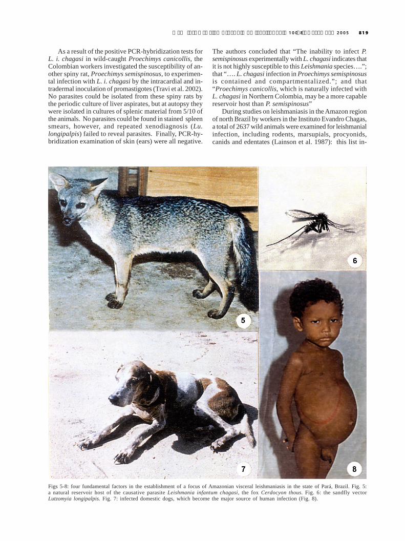
Figs 5-8: four fundamental factors in the establishment of a focus of Amazonian visceral leishmaniasis in the state of Pará, Brazil. Fig. 5:a natural reservoir host of the causative parasite Leishmania infantum chagasi, the fox Cerdocyon thous. Fig. 6: the sandfly vectorLutzomyia longipalpis. Fig. 7: infected domestic dogs, which become the major source of human infection (Fig. 8).

820820820820820 Lu. longipalpis and the eco-epidemiology of AVL • R Lainson, EF Rangel
cluded large numbers of the opossum D. marsupialis andthe spiny rat P. guyannensis, many of which were cap-tured near the houses of patients with AVL and, in thecase of opossums, frequently in the backyards of suchhouses. No infections with L. i. chagasi were detected inany animal other than the fox C. thous, following the cul-ture of spleen and liver tissue and the inoculation of thismaterial intraperitineally into hamsters. At the time of thesestudies the PCR/hybridization technique had not beendeveloped and, in view of the finding of Travi et al. (1998b)that tissues of wild-caught P. canicollis gave a positivePCR to L. i. chagasi DNA in Colombia, Lainson et al.(2002) examined the susceptibility of laboratory-bred P.guyannensis to experimental infection with a canine strainof L. i. chagasi from north Brazil. The animal proved tobe totally resistant to infection by way of promastigotesand amastigotes after massive intraperitoneal inocula-tion of the parasite, and subsequent PCR/hybridizationtests made on liver and spleen tissue were negative. This,failure in attempts to feed laboratory-bred Lu. longipalpison P. guyannensis or to capture this sandfly in trapsbaited with the rodent and placed in or near houses in-fested by Lu. longipalpis, led to the conclusion that thisspecies of spiny rat plays no part in the eco-epidemiol-ogy of AVL in north Brazil.
At the 3rd World Congress on Leishmaniasis in April,2005, workers at the Adolfo Lutz Institute, São Paulo, Bra-zil, presented the results of an examination of wild ani-mals for evidence of Leishmania infections in two locali-ties of endemic cutaneous leishmaniasis in the state ofSão Paulo. Each animal was examined by “….one or moreof the following methods: detection of rK39 antibody inwhole blood; intradermal inoculation of hamsters withskin biopsies from lesions and/or hipocromic spots orculture and/or DNA extraction for PCR and RFLP tests”.Among the positive results were “L. (L.) chagasi in 1Akodon sp. and 2 D. marsupialis”. Unfortunately, thepublished abstract of the presentation (Tolezano et al.2005) does not indicate by which method these resultswere obtained or, more importantly, if the parasite wasisolated from these three animals.
The finding of a benign infection of L. i. chagasi inmarsupials and rodents similar to that commonly found infoxes, does tend to support the hypothesis that the para-site is indigenous to the Americas, in spite of genomicevidence to the contrary (Mauricio et al. 1999). Of greaterimportance, however, is the fact that it raises the questionas to what extent these animals may act as reservoirs ofinfection for the sandfly vector and thus play a role in theepidemiology of human AVL.
Fig. 9: suggested eco-epidemiology of American visceral leishmaniasis in the state of Pará, North Brazil. The parasite L. i. chagasi,originating from a silvatic enzootic in foxes and possibly other wild animals (1), is maintained by a silvatic population of the sandflyLutzomyia longipalpis. Invasion of dwelling places on the edge of the forest by this sandfly enables the establishement of canine and humaninfection (2, 3), and the domestic dog now becames the major source of the parasite. Unbroken lines indicate definite routes oftransmission. Broken lines represent possible transmission with other wild animals, and possibly man himself, serving as a source ofinfection for sandflies (Modified from Lainson, 1989).

821821821821821Mem Inst Oswaldo Cruz, Rio de Janeiro, Vol. 100(8), December 2005
An effective reservoir-host of any parasite is one whichcan participate in the maintenance and dissemination ofthat parasite in nature, and when parasites are dependanton haematophagous vectors for their transmission it isclearly necessary to show that these can be infected whenfed on the host in question. Until this is done the infectedanimal is best referred to as a potential reservoir. The iso-lation of L. i chagasi from numerous specimens of the foxC. thous and the opossum D. marsupialis, and the experi-mental infection of Lu. longipalpis fed on these animals,places them firmly in the category of natural reservoirs ofL. i. chagasi (Lainson et al. 1990, Travi et al. 1998a). Onthe other hand, although positive results of PCR tests onthe tissues of some wild rodents do suggest that thesemay also represent reservoirs, isolation of the parasiteand experimental infection of Lu. longipalpis fed on theinfected animals are needed to confirm this.Control
In the areas of high endemicity, such as in the north-eastern states of Brazil, past attempts to control visceralleishmaniasis had the form of a three-pronged attack, withthe treatment of patients, destruction of infected dogsand regular insecticide spraying of houses (Deane 1956).Although this effectively reduced the number of humancases of AVL, the system was costly and could rarely bemaintained for a sufficiently long period to totally elimi-nate the disease and, with recent criticism of culling in-fected dogs, the control of AVL still presents a seriousproblem for the health authorities throughout the vastgeographic range of the causative parasite.
Destruction of serologically and parasitologicallypositive dogs - Although this may temporarily affect thecumulative incidence of seroconversion in these animalsand may also diminish the incidence of human cases ofAVL (Ashford et al. 1998), it inevitably meets with opposi-tion on the part of dog owners, who understandably findit difficult to see why their apparently healthy (but sero-logically positive) dogs are condemned to death. For thisreason, dogs tend to be hidden during control measures;there is the constant problem of innumerable strays, andthere remains the potential danger of new or revitalizedfoci of AVL from the wild animal enzootic. Mathematicalmodels regarding the three methods of control (Dye 1996)suggest that the destruction of serologically positive dogsis far less likely to solve the problem of AVL than insecti-cide spraying or (if and when available) the vaccinationof these animals (Tesh 1995).
Vaccination of dogs - An efficient vaccine would beof immense help in the control of AVL and its applicationcould coincide with anti-rabies vaccination, thus cuttingthe costs considerably. Preliminary experiments with avaccine prepared from L. (V.) braziliensis combined withBCG gave promise (Mayrink et al. 1996), but the Phase IIItrials led to the conclusion that the vaccine “did not ap-pear to protect the dogs against visceral leishmaniasis”(Genaro et al. 1996). The “Fucose-Mannose ligand” (FML),a complex glycoproteic fraction isolated from an aqueousextract of L. (L.) donovani, has been used in conjunctionwith a saponin adjuvant in attempts to vaccinate dogs
against L. i. chagasi (Silva et al. 2000). The authors re-garded the vaccine as a promising tool in the control ofcanine visceral leishmaniasis in endemic areas of AVL.However, the Brazilian Ministry of Health is still not usingthis vaccine in the National Programme for Control of Vis-ceral Leishmaniasis.
Use of insecticides - The development of chemical in-secticides in the 1940’s resulted in extensive use of DDTagainst mosquito vectors of malaria and this, at the sametime, had a fortuitous and dramatic effect on theperidomestic sandfly vectors of visceral leishmaniasis, inparticular in the endemic areas of kala-azar due to L. (L.)donovani in India. In Brazil it was first used in the 1950sto spray the internal and external walls of houses in thestate of Ceará, specifically to combat AVL, and it resultedin a considerable reduction in the number of cases in somelocalities (Deane et al. 1955, Deane 1958, Alencar 1961,1962). In others, however, there were perplexingly poorresults which were probably due to a failure to apply theinsecticide at the right time of year (Alencar 1961).
In spite of evidence militating against the use of DDT,due to both environmental side effects and human healthhazards, this insecticide was still recommended as the oneof choice by the World Health Organization up to 1990,because of its low cost, high efficacy and long residualaction (WHO 1990). It is still used to this day in Brazil,but attention has now been focused on the syntheticpyrethroids. Although some have described the resultsas rather inconsistent (Silans et al. 1998), Le Pont et al.(1989), and Marcondes and Nascimento (1993) found thespraying of deltamethrin (DM) to be effective against Lu.longipalpis in Bolivia and Brazil, respectively.
A novel approach to the control of canine visceralleishmaniasis due to L. i. infantum in the Cévennes, South-ern France, utilized PVC plastic dog collars impregnatedwith deltamethrin (Killick-Kendrick et al. 1997). The col-lars protected dogs from 96% of the bites of sandflies(laboratory-bred Phlebotomus perniciosus) and this ac-tivity was maintained for up to 34 weeks. It was concludedthat “at least in the Mediterranean subregion, this insec-ticidal collar would protect a dog from the majority ofsandfly bites and retain a killing effect for a completesandfly season. Moreover, it seems likely that the use ofthe collars on all dogs in a focus of L. infantum wouldreduce contact between sandfly vectors and canine res-ervoir hosts sufficiently to diminish the risk of infectionin humans as well as dogs”. DM treatment of dogs byway of an aqueous bath has been found to be highlyeffective in protecting the animals against sandfly bitesin China (Xiong et al. 1994, 1995). DM impregnated collarshave been used in the same country (Chen et al. 2001),Italy (Maroli et al. 2001), and Brazil (David et al. 2001,Reithinger et al. 2004). Among the great advantages ofDM is its repellant and killing action of long duration Noside effects have been detected, and it is considered tooffer no human or canine health risk (WHO 1967). Fol-lowing the results of field trials with DM impregnated dogcollars in Brazil, it would seem that an extensive and gov-ernmentally managed programme for the control of ca-nine AVL in Latin America might well utilize either this

822822822822822 Lu. longipalpis and the eco-epidemiology of AVL • R Lainson, EF Rangel
method or the periodic bathing of dogs in aqueous solu-tions of the insecticide.Biological control
In addition to Leishmania, Trypanosoma, and Endo-trypanum spp., and possibly other trypanosomatids,neotropical sandflies may harbour other parasites includ-ing microsporidians (Lainson et al. 1977b), gregarines(Adler & Mayrink 1961, Ayala 1971, Brazil et al. 1996, Lewiset al. 1970), some Plasmodium spp. of lizards (Klein et al.1987), and even nematodes (Brazil & Ryan 1984). There islittle information on the pathological effects these para-sites may produce in their sandfly hosts, but the gregarineAscogregarina chagasi is known to reduce longevity andegg production and can effectively destroy a laboratorycolony of Lu. longipalpis (Dougherty & Ward 1991):microsporidial infection may sometimes be extremelyheavy, and these parasites are known to be highly patho-genic in some insects (Kudo 1960). To what extent A.chagasi and microsporidian spp., could be employed inbiological control of wild populations of sandflies, par-ticularly Lu. longipalpis, is questionable. One seriousobstacle to such control is that the precise locality of thebreeding sites of sandfly vectors of both visceral andcutaneous leishmaniasis are poorly known. When theseare in the forest, application of biological control is likelyto be particularly difficult and possibly of environmentalrisk.Epilogue
The present-day enzootology and epidemiology ofAVL as seen in the more remote parts of Latin Americapossibly represents a recapitulation of what transpiredwhen the early colonists first commenced their ecologicalupheavals. Native animals harbour L. i. chagasi as anenzootic maintained by sylvatic sandflies (Lu. longipalpisand/or Lu. evansi) and the parasite inevitably gains en-trance into newly established human settlements. Thismay be by way of infected sandflies that migrate therefrom the nearby sylvatic enzootic, or when infected scav-engers such as foxes or opossums invade human habita-tions and are fed on by the sandflies from peridomesticpopulations. Transmission of the parasite to dogs or manis then only a question of time and a single infected dog,often with a vast supply of amastigotes for these sandfliesin its skin, sets the scene for a small focus of canine orhuman visceral leishmaniasis. When human habitationsbecome overcrowded , with conditions of poor hygieneand an abundance of domestic animals, particularly dogs,the concentration of the vector Lu. longipalpis (and/orLu. evansi in some parts of Latin America) may reach avery high level and there is the risk of a serious outbreakof canine and human disease. At this stage of events thedog becomes the major reservoir of infection for man, andthe wild animal host may, in fact, no longer be present inthe immediate area. Control measures in such foci of in-fection will certainly reduce the number of human casesof AVL but, unfortunately, total elimination of the diseaseis unlikely, due to a persistent source of L. i. chagasi inthe wild animal enzootic - a problem equally difficult toresolve in control of the cutaneous leishmaniases.
REFERENCES
Adler S, Mayrink W 1961. A gregarine, Monocystis chagasin.sp., of Phlebotomus longipalpis. Remarks on the acces-sory glands of P. longipalpis. Rev Inst Med Trop São Paulo3: 230-238.
Aguilar CM, Fernández E, Cannova DC, Ferrer E, Cabrera Z,Souza WJS, Coutinho SG 1998. Urban visceral leishma-niasis in Venezuela. Mem Inst Oswaldo Cruz 93: 15-16.
Alencar JE 1961. Profilaxia do calazar no Ceará, Brasil. RevInst Med Trop São Paulo 3: 175-180.
Alencar JE 1962. Kala-azar in Brazil. Sci Rep Inst Sup Sanitá2: 116-123.
Alexander JB 1987. Dispersal of phlebotomine sandflies in aColombian coffee plantation. J Med Entomol 24: 552-558.
Arrivillaga JC, Feliciangeli MD 2001. Lutzomyia pseudo-longipalpis, the first new species within the longipalpis(Diptera: Psychodidae: Phlebotominae) complex fromRinconada, Curarigua, Lara State, Venezuela. J Med Entomol38: 783-790.
Arrivillaga JC, Rangel Y, Oviedo M, Feliciangeli MD 2000.Genetic diversity among Venezuelan populations ofLutzomyia longipalpis (Diptera:Psychodidae: Phleboto-minae). J Med Entomol 37: 325-330.
Arrivillaga JC, Norris DE, Feliciangeli MD, Lanzaro GC 2002.Phylogeography of the neotropical sand fly Lutzomyialongipalpis inferred from mitochondrial DNA sequences.Inf Gen Evol 2: 83-95.
Arrivillaga J, Mutebi JP, Pinango H, Norris D, Alexander B,Feliciangeli MD, Lanzaro GC 2003. The taxonomic statusof genetically divergent populations of Lutzomyialongipalpis (Diptera: Psychodidae) based on the distribu-tion of mitochondrial and isozyme variation. J Med Entomol40: 615-627.
Ashford DA, David JR, Freire M, David R, Sherlock I, EulalioM, Sampaio DP, Badaró R 1998. Studies on control of vis-ceral leishmaniasis: impact of dog control of canine andhuman visceral leishmaniasis in Jacobina, Bahia, Brazil.Am J Trop Med Hyg 59: 53-57.
Ayala SC 1971. Gregarine infections in the Californian sandfly,Lutzomyia vexatrix occidentis. J Invert Path 17: 440-441.
Azevedo ACR, Monteiro FA, Cabello PH, Souza NA, Rosa-Freitas MG, Rangel EF 2000. Studies on populations ofLutzomyia longipalpis (Lutz & Neiva, 1912) (Diptera: Psy-chodidae: Phlebotominae) in Brazil. Mem Inst OswaldoCruz 95: 305-322.
Bauzer LGSR, Souza NA, Ward RD, Kyriacou CP, Peixoto AA2002. The period gene and genetic differentiation betweenthree Brazilian populations of Lutzomyia longipalpis. In-sect Mol Biol 11: 315-323.
Bottecchia M, Oliveira SG, Bauzer LGSR, Souza NA, WardRD, Garner KJ, Kyriacou CP, Peixoto AA 2004. Geneticdivergence in the cacophony IVS6 Intron among five Brazil-ian populations of Lutzomyia longipalpis. J Mol Evol 58:754-761.
Brazil RP, Ryan L 1984. Nota sobre a infecção de Lutzomyiaevandroi (Diptera: Psychodidae) por Ascocystis chagasi(Adler & Mayrink, 1961) no Estado do Maranhão. MemInst Oswaldo Cruz 79: 375-376.

823823823823823Mem Inst Oswaldo Cruz, Rio de Janeiro, Vol. 100(8), December 2005
Brazil RP, Oliveira SMP, Dias CMG, Andrade JD, Falcão AL1996. Natural infection of New World phlebotominesandflies (Diptera:Psychodidae) with Ascogregarina. MemInst Oswaldo Cruz 91 (Suppl.): 116.
Chagas AW 1940. Criação de flebótomos e transmissão experi-mental da leishmaniose visceral americana. Mem InstOswaldo Cruz 35: 327-333.
Chagas E 1936. Primeira verificação em individuo vivo, daleishmaniose visceral no Brasil. Bras-Méd 50: 221-222.
Chagas E, Cunha AM, Ferreira LC, Deane L, Deane G, GuimarãesFN, Paumgartten MJ, Sá B 1938. Leishmaniose visceralamericana (Relatório dos trabalhos realizados pelaCommissão Encarregada do Estudo da Leishmaniose Vis-ceral Americana em 1937). Mem Inst Oswaldo Cruz 33: 89-229.
Chen S, Li F, He J, Chen X, Wang D, Wei L, Yang H, Guan L2001. Experimental study on prevention of dog-sand flycontact by Deltamethrin collar. End Dis Bull 16: 17-19.
Coelho MV, Cunha AS, Falcão AR 1965. Notas sobre um focode calazar no sudoeste do Estado de Goiás. Rev Bras MalariolDoenças Trop 17: 143-148.
Corredor A, Gallego JF, Tesh RB, Peláez D, Diaz A, MontillaM, Paláu MT 1989a. Didelphis marsupialis, an apparentwild reservoir of Leishmania donovani chagasi in Colom-bia, South America. Trans R Soc Trop Med Hyg 83: 195.
Corredor A, Gallego JF, Tesh RB, Morales A, Ferro deCarrasquilla C, DG, Kreutzer RD, Boshell J, Paláu MT,Caceres E, Peláez D 1989b. Epidemiology of visceral leish-maniasis in Colombia. Am J Trop Med Hyg 40: 480-486.
Courtenay O, Santana EW, Johnson PJ, Vasconcelos IAB,Vasconcelos AW 1996. Visceral leishmaniasis in the hoaryzorro Dusicyon vetulus: a case of mistaken identity. TransR Soc Trop Med Hyg 90: 498-502.
Crampton J, Knapp T, Ward RD 1989. DNA probes for vectortaxonomy. In DDT Hart, Leishmaniasis. The CurrentStatus and New Strategies for Control, NATO ASI SeriesA: Life Sci 163 Plenum Press & NATO Scientific AffairsDivision, New York and London, p. 957-964.
Cunha AM, Chagas E 1937. Nova espécie de protozoário dogênero Leishmania patogênico para o homem. Leishmaniachagasi n.sp. Nota Prévia. Hospital (Rio de Janeiro) 11:3-9.
David JR, Stamm LM, Bezerra HS, Souza RN, Killick-KendrickR, Lima JWO 2001. Deltamethrin-impregnated dog collarshave a potent anti-feeding and insecticidal effect onLutzomyia longipalpis and Lutzomyia migonei. Mem InstOswaldo Cruz 96: 839-847.
Deane LM 1956. Leishmaniose Visceral no Brasil. ServiçoNacional de Educação Sanitária, Rio de Janeiro, p. 1-162.
Deane LM 1958. Epidemiologia e profilaxia do calazar ameri-cano. Rev Brás Malariol 10: 431-450.
Deane MP, Deane LM 1954a. Infecção natural do Phlebotomuslongipalpis por leptomonas, provavelmente de Leishmaniadonovani, em foco de calazar, no Ceará. Hosptital (Rio deJaneiro) 45: 697-702.
Deane LM, Deane MP 1954b. Encontro de leishmanias nasvísceras e na pele de uma raposa, em zona endêmica decalazar, nos arredores de Sobral, Ceará. Hospital (Rio deJaneiro) 45: 419-421.
Deane MP, Deane LM. 1954c. Infecção experimental do Phle-botomus longipalpis em raposa (Lycalopex vetulus) natu-ralmente parasitada pela Leishmania donovani. Hospital(Rio de Janeiro) 46: 651-653.
Deane LM, Grimaldi G 1985. Leishmaniasis in Brazil. In KPChang, RS Bray (eds), Leishmaniasis, Elsevier, Amsterdam,p. 247-281.
Deane LM, Deane MP, Alencar JE 1955. Observações sobre ocombate ao Phlebotomus longipalpis pela dedetização emárea endêmica de calazar no Ceará. Rev Bras Malariol 7:131-141.
Decker-Jackson FE, Tang DB 1982. Identification of Leishma-nia spp. By radiorespirometry. II. A statistical method ofdata analysis to evaluate the reproductibility and sensitiv-ity of the technique. In ML Chance, BC Walton (eds),Biochemical Characterization of Leishmania, proceedingsof a workshop held at the PanAmerican Health Organiza-tion, Geneva, p. 205-245.
Dougherty MJ, Ward RD 1991. Methods of reducing Asco-gregarina chagasi parasitaemia in laboratory colonies ofLutzomyia longipalpis. In Proceedings of the First Interna-tional Symposium on Phlebotomine Sandflies, Rome.Parassitologia (Roma) 33: 185-191.
Dujardin JP, Torrez EM, Le Pont F, Hervas D, Sossa D 1997.Isozymic and metric variation in the Lutzomyia longipalpiscomplex. Med Vet Entomol 11: 394-400.
Dye C 1996. The logic of visceral leishmaniasis control. Am JTrop Med Hyg 55: 125-130.
Dye C, Davies CR, Lainson R 1991. Communication amongphlebotomine sandflies: a field study of domesticatedLutzomyia longipalpis populations in Amazonian Brazil.Animal Behaviour 42: 183-192.
Feliciangeli MD, Arrivillaga JC, Bravo A, Arias F 2004. Activ-ity of Lutzomyia pseudolongipalpis and L. longipalpis s.l.(Diptera: Psychodidae) in Venezuela. Parasite 11: 273-278.
Feliciangeli MD, Rodriguez N, De Guglielmo Z, Rodriguez A1999. The re-emergence of American visceral leishmaniasisin an old focus in Venezuela. II. Vectors and parasites. Para-site 6: 113-120.
Ferro C, Morrison AC, Torres M, Pardo R, Wilson ML, TeshRB 1995. Age structure, blood-feeding behaviour, and Leish-mania chagasi infection in Lutzomyia longipalpis (Diptera:Psychodidae) at an endemic focus of visceral leishmaniasisin Colombia. J Med Ent 32: 618-629.
Galati EAB, Nunes VLB, Cristaldo G, Rocha HC 2003.Aspectos do comportamento da fauna flebotomínea(Diptera:Psychodidae) em foco de leishmaniose visceral etegumentar na Serra da Bodoquena e área adjacente, Estadode Mato Grosso do Sul, Brasil. Rev Pat Trop 32: 235-261.
Genaro O, Pinto JA, Costa CA, França-Silva JC, Costa RT,Silva JC, Sanguinetti LSR,Vieira EP, Toledo VPCP, MayrinkW 1996. Phase III randomized double blind clinical trial onthe efficacy of a vaccine against canine visceral leishmania-sis in urban area of Montes Claros, MG, Brazil. Mem InstOswaldo Cruz 91 (Suppl.): 166.
Hamilton JGC, Ward RD, Dougherty R, Maingon C, Ponce E,Noyes H, Zelodón R 1996. Comparison of the sex-phero-mone components of Lutzomyia longipalpis from areas ofvisceral and atypical cutaneous leishmaniasis in Hondurasand Costa Rica. Ann Trop Med Parasitol 90: 533-541.

824824824824824 Lu. longipalpis and the eco-epidemiology of AVL • R Lainson, EF Rangel
Hodgkinson VH, Birungi J, Quintana M, Deitze R, MunstermannLE 2003. Mitochondrial cytochrome B variation in popu-lations of the visceral leishmaniasis vector Lutzomyialongipalpis across eastern Brazil. Am J Trop Med Hyg 69:386-392.
Ibáñez-Bernal S, Rodríguez-Domínguez G, Gómez-HernándezCH, Ricardez-Esquinc JR 2004. First record of Lutzomyiaevansi (Nuñez-Tovar 1924) in México (Diptera: Psychod-idae: Phlebotominae). Mem Inst Oswaldo Cruz 99: 127-129.
Imms AD 1964. A General Textbook of Entomology, Methuen& Co Ltd, London, EP Dutton & Co Inc., New York, 886pp.
Jackson PR, Wohlhieter J, Hockmeyer WT 1982. Leishmaniacharacterization by restriction endonuclease digestion ofkinetoplastic DNA. In Abstr Vth Internat Congr ParasitolToronto, Canada, p. 342.
Jackson PR, Stiteler JM, Wohlhieter JA, Reed SG, Badaró R,Inverso JA, Jackson JE 1984. Characterization of Leish-mania responsible for visceral disease in Brazil by restric-tion endonuclease digestion and hybridization of kineto-plast DNA. In Proc 11th Internat Congr Trop Med MalarCalgary, Canada, p. 68.
Killick-Kendrick R 1985. Some epidemiological consequencesof the evolutionary fit between leishmaniae and theirphlebotomine vectors. Bull Soc Path Exot 78: 747-755.
Killick-Kendrick R, Killick-Kendrick M, Focheux C, DereureJ, Puech M-P, Cadiergues MC 1997. Protection of dogsfrom the bites of phlebotomine sandflies by deltamethrincollars for control of canine leishmaniasis. Med Vet Ent 11:105-111.
Klein TA, Young DG, Telford SR, Kimsey R 1987. Experimen-tal transmission of Plasmodium mexicanum by bites of in-fected Lutzomyia vexator (Diptera: Psychodidae). J AmMosq Contr Assoc 3: 154-164.
Kudo RR 1960. Protozoology, Charles C Thomas, Springfield,Illinois, 966 pp.
Lainson R 1989. Demographic changes and their influence onthe epidemiology of the American leishmaniases. In M Ser-vice, Demography and Vector Borne Diseases, CRC Press,Boca Raton, FL, p. 85-106.
Lainson R, Rangel EF 2003. Ecologia das leishmanioses:Lutzomyia longipalpis e a eco-epidemiologia da leishmaniosevisceral americana (LVA) no Brasil. In EF Rangel, R Lainson(eds), Flebotomíneos do Brasil, Fiocruz, Rio de Janeiro,p. 311-336.
Lainson R, Shaw JJ 1979. The role of animals in the epidemi-ology of South American leishmaniasis. In WHR Lumsden,DA Evans (eds), Biology of the Kinetoplastida, Vol. 2, Aca-demic Press, London, p. 1-116.
Lainson R, Shaw JJ 1987. Evolution, classification and geo-graphic distribution. In W Peters, R Killick-Kendrick (eds),The Leishmaniases in Biology and Medicine, Vol. 1, Aca-demic Press, London, p. 1-120.
Lainson R, Shaw JJ 1998. New World leishmaniasis – theneotropical Leishmania species. In L Collier, A Balows, MSussman (eds), Topley & Wilson’s Microbiology and Micro-bial Infections, Vol. 5, Parasitology. Arnold, London, p.241-266.
Lainson R, Shaw JJ 2005. Leishmaniasis in the New World. InL Collier, A Balows, M Sussman (eds), Topley & Wilson’sMicrobiology and Microbial Infections, 10th ed., Vol 5, Para-sitology, Arnold, London, p. 313-349.
Lainson R, Ishikawa EAY, Silveira FT 2002. American visceralleishmaniasis: wild animal hosts. Trans R Soc Trop MedHyg 96: 630-631.
Lainson R, Miles MA, Shaw JJ 1981. On the identification ofviscerotropic leishmanias. Ann Trop Med Parasitol 75: 251-253.
Lainson R, Shaw JJ, Lins CZ 1969. Leishmaniasis in Brazil, IV.The fox, Cerdocyon thous (L.) as a reservoir of Leishmaniadovovani in Pará State, Brazil. Trans R Soc Trop Med Hyg63: 741-745.
Lainson R, Ward RD, Shaw JJ 1977a. Experimental trans-mission of Leishmani chagasi causative agent of neotropicalvisceral leishmaniasis, by the sandfly Lutzomyia longipalpis.Nature (London) 266: 628-630.
Lainson R, Killick-Kendrick R, Canning EU, Shaw JJ, WardRD, Leaney AJ Nicholas JP 1977b. Microsporidia of Bra-zilian sandflies. Trans R Soc Trop Méd Hyg 71: 381.
Lainson R, Shaw JJ, Ryan L, Ribeiro RSM, Silveira FT 1984.Presente situação da leishmaniose visceral na Amazônia,com especial referência a um novo surto da doença ocorridoem Santarém, Estado do Pará, Brasil. Bol Epidemiol F.SESP,Rio de Janeiro (número especial): 1-8.
Lainson R, Shaw JJ, Ryan L, Silveira FT 1985. Leishmaniasisin Brazil XXI. Visceral leishmaniasis in the Amazon Re-gion and further observations on the role of Lutzomyialongipalpis (Lutz & Neiva, 1912) as the vector. Trans RSoc Trop Med Hyg 79: 223-226.
Lainson R, Shaw JJ, Silveira FT, Braga RR 1987. Americanvisceral leishmaniasis: on the origin of Leishmania (Leish-mania) chagasi. Trans R Soc Trop Med Hyg 81: 517.
Lainson R, Shaw JJ, Silveira FT, Braga RR, Ryan L, PovoaMM, Ishikawa EAY 1986. A Leishmania e as leishmani-oses. In Instituto Evandro Chagas: 50 Anos de Contribuiçãoàs Ciências Biológicas e à Medicina Tropical, Vol. I, Serviçosde Saúde Pública, Belém, Pará, p. 83-124.
Lainson R, Dye C, Shaw JJ, MacDonald DW, Courtenay O,Souza AAA, Silveira FT 1990. Amazonian visceral leish-maniasis – Distribution of the vector Lutzomyia longipalpis(Lutz & Neiva) in relation to the fox Cerdocyon thous (Linn)and the efficiency of this reservoir host as a source of infec-tion. Mem Inst Oswaldo Cruz 85: 135-137.
Lainson R, Shaw JJ, Silveira FT, Souza AAA, Braga RR,Ishikawa EAY 1994. The dermal leishmaniases of Brazil,with special reference to the eco-epidemiology of the dis-ease in Amazonia. Mem Inst Oswaldo Cruz 89: 435-443.
Lampo M, Torgerson D, Márquez LM, Rinaldi M, Garcia CZ,Arab A 1999. Occurrence of sibling species of Lutzomyialongipalpis (Diptera: Psychodidae) in Venezuela: first evi-dence from reproductively isolated sympatric populations.Am J Trop Med Hyg 61: 1004-1009.
Lane R, Phillips A, Molyneux DH, Procter G, Ward RD 1985.Chemical analysis of the abdominal glands of two forms ofLutzomyia longipalpis: site of a possible sex pheromone?Ann Trop Med Parasitol 79: 225-229.
Lanzaro GC, Ostrovska K, Herrero MV, Lawyer PG, Warburg

825825825825825Mem Inst Oswaldo Cruz, Rio de Janeiro, Vol. 100(8), December 2005
A 1993. Lutzomyia longipalpis is a species complex: ge-netic divergence and interspecific hybrid sterility amongthree populations. Am J Trop Med Hyg 48: 839-847.
Le Pont F, Padilla J, Dejeux P, Richard A, Mouchet J 1989.Impact de pulverizations de deltametrine dans un foyer deleishmaniose de Bolivie. Ann Soc Belge Méd Trop 69: 223-232.
Lerner EA, Ribeiro JMC, Nelson RJ, Lerner M 1991. Isola-tion of Maxadilan, a potent vasodilatory peptide from thesalivary glands of the sandfly Lutzomyia longipalpis. J BiolChem 266: 11234-11236.
Lewis DJ, Lainson R, Shaw JJ 1970. Determination of parousrates in phlebotomine sandflies, with special reference toAmazon species. Bull Ent Res 60: 209-219.
Lutz A, Neiva A 1912. Contribuição para o conhecimento dasespécies do gênero Phlebotomus existentes no Brasil. MemInst Oswaldo Cruz 4: 84-95.
Lysenko AJ 1971. Distribution of leishmaniasis in the Old World.Bull WHO 44: 515-520.
Maingon RD, Ward RD, Hamilton JG, Noyes HA, Souza N,Kemp SJ, Watts PC 2003. Genetic identification of twosibling species of Lutzomyia longipalpis (Diptera: Psychod-idae) that produce distinct male sex pheromones in Sobral,Ceara State, Brazil. Mol Ecol 12: 1879-1894.
Mangabeira O 1969. Sobre a sistemática e biologia dos Phle-botomus do Ceará. Rev Bras Malariol Doenças Trop 21: 3-26.
Marcondes CB, Nascimento JA 1993. Avaliação de eficiênciade deltametrina (K-Othrine CE) no controle de Lutzomyialongipalpis (Diptera: Psychodidae), no município de SantaRita, Paraíba, Brasil. Rev Soc Bras Med Trop 26: 15-18.
Maroli M, Mizzoni V, Siragusa C, D’Orazi A, Gradoni L 2001.Evidence for an impact on the incidence of canine leishma-niasis by the mass use of deltamethrin-impregnated dogcollars in southern Italy. Med Vet Ent 15: 358-363.
Martinez E, Alonso V, Quispe A, Thomas MC, Alonso R, PiñeroJE, González AC, Ortega A, Valladares B 2003. RAPDmethod useful for distinguishing Leishmania species: de-sign of specific primers for L. braziliensis. Parasitology127: 513-517.
Martins AV, Williams P, Falcão AL 1978. American Sand Flies,Acad Bras Ciências, Rio de Janeiro, 195 pp.
Martins AV, Falcão AL, Silva JE, Dias ES 1984. Nota sobreLutzomyia (Lutzomyia) cruzi (Mangabeira, 1938), com adescrição da fêmea (Diptera, Psychodidae, Phlebotominae).Mem Inst Oswaldo Cruz 79: 439-442.
Mauricio IL, Howard MK, Stothard JR, Miles MA 1999. Ge-nomic diversity in the Leishmania donovani complex. Para-sitology 119: 237-246.
Mayrink W, Genero O, Silva JCF, Costa RT, Tafuri WL, To-ledo VPCP, Silva AR, Reis AB, Williams P, Costa CA1996. Phase I and II open clinical trials of a vaccine againstLeishmania chagasi infection in dogs. Mem Inst OswaldoCruz 91: 695-697.
Mello DA, Rego FA, Oshozo E, Nunes VLB 1988. Cerdocyonthous (L.) (Carnivora: Canidae) naturally infected with Leish-mania chagasi (Cunha & Chagas, 1937) in Corumbá (MataGrosso do Sul State, Brazil). Mem Inst Oswaldo Cruz 83:259.
Montoya-Lerma J, Cadena H, Oveido M, Ready PD, BarazarteR, Travi BL, Lane RP 2003. Comparative vectorial effi-ciency of Lutzomyia evansi and L. longipalpis for transmit-ting Leishmania chagasi. Acta Trop 85: 19-29.
Morton I, Ward RD 1989. Laboratory responses of femaleLutzomyia longipalpis sandflies to a host and male phero-mone source over distance. Med Vet Ent 3: 219-223.
Mukhopadhyay J, Ghosh K, Rangel EF, Munstermann LE 1998.Genetic variability in biochemical characters of Brazilianfield populations of the Leishmania vector, Lutzomyialongipalpis (Diptera: Psychodidae). Am J Trop Med Hyg59: 893-901.
Mutebi JP, Alexander B, Sherlock I, Wellington J, Souza AA,Shaw J, Rangel EF, Lanzaro GC 1999. Breeding structureof the sand fly Lutzomyia longipalpis (Lutz & Neiva) inBrazil. Am J Trop Med Hyg 61: 149-157.
Mutebi JP, Tripet F, Alexander JB, Lanzaro GC 2002. Geneticdifferentiation among populations of Lutzomyia longipalpis(Diptera: Psychodidae) in Central and South America. AnnEntomol Soc Amer 95: 740:752.
Oliveira AC, Batista SM, Falcão AL 1959. Calazar em MinasGerais. Revisão dos dados epidemiológicos obtidos até 1958.Hospital (Rio de Janeiro) 56: 625-643.
Oliveira MP, Grimaldi G, Momen H, Pacheco RS, MarzochiMCA, McMahon-Pratt D 1986. Active cutaneous leish-maniasis in Brazil, induced by Leishmania donovani chagasi.Mem Inst Oswaldo Cruz 81: 303-309.
Paraense L, Chagas AW 1940. Transmissão experimental daleishmaniose visceral americana pelo Phlebotomus interme-dius. Nota prévia. Bras Med 54: 179-180.
Penna HA 1934. Leishmaniose visceral no Brasil. Bras Med48: 949-950.
Perdeck AC 1957. The isolating value of specific song patternsin two sibling species of grasshoppers (Chorthippusbrunneus Thumb and C. biguttulus L.). Behaviour 12: 1-75.
Pífano CF, Romero MJ 1964. Comprobación de um nuevo focode leishmaniose em Venezuela, Valle de Cumanacoa, Edo.Sucre. Gac Méd Caracas 72: 473-479.
Ponce C, Ponce E, Morrison A, Cruz A, Kreutzer R, McMahon-Pratt D, Neva F 1991. Leishmania donovani chagasi: newclinical variant of cutaneous leishmaniasis in Honduras.Lancet 337: 67-70.
Potenza L, Anduze J 1942. Kala-azar en el Estado de Bolívar,Venezuela. Rev Policlín (Caracas) 11: 312-317.
Quinnell RJ, Dye C 1994a. An experimental study of theperidomestic distribution of Lutzomyia longipalpis(Diptera: Psychodidae). Bull Ent Res 84: 379-382.
Quinnell RJ, Dye C 1994b. Correlates of the domestic abun-dance of Lutzomyia longipalpis (Diptera: Psychodidae) inAmazonian Brazil. Med Vet Ent 8: 219-224.
Rangel EF, Lainson R 2003. Ecologia das leishmanioses.Transmissores de leishmaniose tegumentar Americana. InEF Rangel, R Lainson (eds), Flebotomíneos do Brasil,Fiocruz, Rio de Janeiro, p. 291-309.
Ready PD, Lainson R, Shaw JJ, Souza AA 1991. DNA probesfor distinguishing Psychodopygus wellcomei from

826826826826826 Lu. longipalpis and the eco-epidemiology of AVL • R Lainson, EF Rangel
Psychodopygus complexus (Diptera: Psychodidae). MemInst Oswaldo Cruz 86: 41-49.
Reithinger R, Coleman PG, Alexander B, Vieira EP, Assis G,Davies CR 2004. Are insecticide-impregnated dog collarsa feasible alternative to dog culling as a strategy for control-ling visceral leishmaniasis in Brazil? Int J Parasitol 34:55-62.
Rioux JA, Lanotte G, Serres E, Pratlong F, Bastien P, PerieresJ 1990. Taxonomy of Leishmania. Use of isoenzymes.Suggestions for a new classification. Ann Parasitol HumComp 65: 111-125.
Ryan L, Lainson R, Shaw JJ 1986a. The experimental trans-mission of Leishmania mexicana amazonensis Lainson &Shaw, between hamsters by the bite of Lutzomyia furcata(Mangabeira). Trans R Soc Trop Med Hyg 80: 164-165.
Ryan L, Lainson R, Shaw JJ 1987. Leishmaniasis in Brazil.XXIV. Natural flagellate infections of sandflies (Diptera:Psychodidae) in Pará State, with particular reference to therole of Psychodopygus wellcomei as the vector of Leishma-nia brazilienisis braziliensis in the Serra dos Carajás. TransR Soc Trop Med Hyg 81: 353-359.
Ryan L, Silveira FT, Lainson R, Shaw JJ 1984. Leishmanialinfections in Lutzomyia longipalpis and Lu. antunesi(Diptera: Psychodidae) on the island of Marajó, Pará State,Brazil. Trans R Soc Trop Med Hyg 78: 547-548.
Ryan L, Lainson R, Shaw JJ, Fraiha H 1986c. Ecologia deflebotomíneos (Diptera: Psychodidae: Phlebotominae) naRegião Amazônica. In Instituto Evandro Chagas: 50 Anosde Contribuição às Ciências Biológicas e à Medicina Tropi-cal, Vol. I, Serviços de Saúde Pública, Belém, Pará, p. 307-320.
Ryan L, Phillips RA, Milligan P, Lainson R, Molyneux DH,Shaw JJ 1986b. Separation of female Psychodopyguswellcomei and Ps. complexus (Diptera: Psychodidae) bycuticular hydrocarbon analysis. Acta Trop 43: 85-89.
Santoro F, Lemesre JL, Rizvi FS, Afchain D, Sadogirsky M1986. Spécificité au niveau de protéines de surface despromastigotes de Leishmania donovani (Laveran et Mesnil,1903), Leishmania infantum Nicolle, 1908 et Leishmaniachagasi Cunha et Chagas, 1937. In JA Rioux (2-6 July,1984). Leishmania. Taxonomie et Phylogenése. Aplicationseco-Épidemiologiques. Montpellier, France: ColloqueInternat, IMMEE, p. 71-76.
Santos SO, Arias J, Ribeiro AA, Hoffmann MP, Freitas RA,Malacco MAF 1998. Incrimination of Lutzomyia cruzi asa vector of American visceral leishmaniasis. Med Vet Ent 12:315-317.
Santos SO, Arias JR, Hoffmann MP, Furlan MBG, FerreiraWF, Pereira C, Ferreira L 2003. The presence of Lutzomyialongipalpis in a focus of American visceral leishmaniasiswhere the only proven vector is Lutzomyia cruzi, Corumbá,Mato Grosso do Sul State. Rev Soc Bras Med Trop 36:633-634.
Sherlock IA, Miranda JC, Sadigursky M, Grimaldi G 1984.Natural infection of the opossum Didelphis albiventris(Marsupialia: Didelphidae) with Leishmania donovani inBrazil. Mem Inst Oswaldo Cruz 79: 511.
Sherlock IA, Miranda JC, Sadigursky M, Grimaldi G 1988.Observações sobre calazar em Jacobina, Bahia. VI.Investigações sobre reservatórios silvestres e comensais.
Rev Soc Bras Med Trop 21: 23-27.
Silans LNMP, Dedet J-P, Arias JR 1998. Field monitoring ofcypermethrin residua effect on the mortality rates of thephlebotomine sand fly Lutzomy longipalpis in the state ofParaíba, Brazil. Mem Inst Oswaldo Cruz 93: 339-344.
Silva VO, Borja-Cabrera GP, Correia Pontes NN, Souza EP,Luz KG, Palatinik M, Palatinik de Souza CD 2000. APhase III trial of efficacy of the FML-vaccine against ca-nine calazar in an endemic area of Brasil (São Gonçalo doAmarante, RN). Vaccine 19: 1082-1092.
Silveira FT, Lainson R, Shaw JJ, Póvoa MM 1982. Leishma-niasis in Brazil: XVIII. Further evidence incriminating thefox Cerdocyon thous (L.) as a reservoir of Amazonian vis-ceral leishmaniasis. Trans R Soc Trop Med Hyg 76: 830-832.
Soto SI, Lehmann T, Rowton ED, Velez BID, Porter CH 2001.Speciation and population structure in the morphospeciesLutzomyia longipalpis (Lutz and Neiva) as derived from themitochondrial ND4 gene. Mol Phylogenet Evol 18: 84-93.
Souza MB, Marzochi MCA, Carvalho RW, Ribeiro PC, Pon-tes CS, Caetano JM, Meira AM 2003. Ausência daLutzomyia longipalpis em algumas áreas de ocorrência deleishmaniose visceral no Município do Rio de Janeiro. CadSaúde Púb 19: 1881-1885.
Souza NA, Vigoder FM, Araki AS, Ward RD, Kyriacou CP,Peixoto AA 2004. Analysis of the copulatory courtshipsongs of Lutzomyia longipalpis in six populations fromBrazil. J Med Entomol 41: 906-913.
Tesh RB 1995. Control of zoonotic visceral leishmaniasis: is ittime to change strategies? Am J Trop Med Hyg 52: 287-292.
Tolezano JE, Taniguchi HH, Araújo MF, Cunha EA,ChioccolaVLP, Pirmez C, Shaw JJ 2005. Evidence of dif-ferent Leishmania enzootic cycles, especially L. (Viannia)braziliensis, in two endemic American cutaneous leishma-niasis (ACL) regions of São Paulo State, Brazil. AbstractBook, Third World Congress on Leishmaniasis,10-15April, 2005. Palermo-Terrasini, Sicily, Italy, p. 213.
Torrealba JW, Torrealba JF 1964. Infección experimental deCerdocyon thous (zorro común) com Leishmania donovani.Gac Méd Caracas 72: 117-118.
Travi BL, Arteaga LT, Leon AP, Adler GH 2002. Susceptibil-ity of spiny rats (Proechimys semispinosus) to Leishmania(Viannia) panamensis and Leishmania (Leishmania)chagasi. Mem Inst Oswaldo Cruz 97: 887-892.
Travi BL, Jaramillo C, Montoya J, Segura I, Zea A, GoncalvesA, Vélez ID 1994. Didelphis marsupialis, an importantreservoir of Trypanosoma cruzi and Leishmania (Leishma-nia) chagasi in Colombia. Am J Trop Med Hyg 50: 557-565.
Travi BL, Montoya J, Gallego J, Jaramillo C, Llano R, VélezID 1996. Bionomics of Lutzomyia evansi (Diptera: Psy-chodidae), vector of visceral leishmaniasis in Northern Co-lombia. J Med Entomol 33: 278-285.
Travi BL, Osorio Y, Becerra MT, Adler GH 1998b. Dynamicsof Leishmania chagasi infection in small mammals of theundisturbed and degraded tropical dry forests of northernColombia. Trans R Soc Trop Med Hyg 92: 275-278.
Travi BL, Osório Y, Guarin N, Cadena H 1998a. Leishmania(Leishmania) chagasi: clinical and parasitological observa-

827827827827827Mem Inst Oswaldo Cruz, Rio de Janeiro, Vol. 100(8), December 2005
tions in experimentally infected Didelphis marsupialis, res-ervoir of New World visceral leishmaniasis. Exp Parasitol88: 73-75.
Travi BL, Vélez ID, Brutus L, Segura I, Jaramillo C, MontoyaJ 1990. Lutzomyia evansi, an alternative vector of Leish-mania chagasi in a Colombian focus of visceral leishmania-sis. Trans R Soc Trop Med Hyg 84: 676-677.
Uribe S 1999. The status of the Lutzomyia longipalpis speciescomplex and possible implications for Leishmania trans-mission. Mem Inst Oswaldo Cruz 94: 729-734.
Warburg A, Saraiva E, Lanzaro GC, Titus RG, Neva F 1994.Saliva of Lutzomyia longipalpis sibling species differs in itscomposition and capacity to enhance leishmaniasis. PhilTrans R Soc Lond Serie B 345: 223-230.
Ward RD, Ribeiro AL, Ready PD, Murtagh A 1983. Reproduc-tive isolation between different forms of Lutzomyialongipalpis (Lutz & Neiva) (Diptera: Psychodidae), thevector of Leishmania donovani chagasi Cunha & Chagasand its significance to kala-azar distribution in SouthAmerica. Mem Inst Oswaldo Cruz 78: 269-280.
Ward RD, Phillips A, Burnet B, Marcondes CB 1988. TheLutzomyia longipalpis complex: reproduction and distribu-tion. In MW Service, Biosystematics of HaematophagusInsects, Clarendon Press, Oxford, p. 257-269.
Ward RD, Ribeiro AL, Ryan L, Falcão AL, Rangel EF 1985.The distribution of two morphological forms of Lutzomyialongipalpis (Lutz & Neiva) (Diptera: Psychodidae). MemInst Oswaldo Cruz 80: 145-148.
Watts PC, Hamilton JG, Ward RD, Noyes HA, Souza NA,Kemp SJ, Feliciangeli MD, Brazil R, Maingon RD 2005.Male sex pheromones and the phylogeographic structure ofthe Lutzomyia longipalpis species complex (Diptera: Psy-chodidae) from Brazil and Venezuela. Am J Trop Med Hyg73: 734-743.
WHO-World Health Organization 1967. Safe use of pesticidesin Public Health. Tech Rep Series 356, Geneva.
WHO-World Health Organization 1990. Control of Leishma-niasis. Tech Rep Series 793, Geneva, 158 pp.
Xiong G, Jin C, Cheng X, Su Z 1994. Deltamethrin bath ofdomestic dog in the prevention of sandfly bite. End DisBull 9: 32-34.
Xiong G, Jin C, Hong Y, Su Z 1995. Studies on the deltamethrin-medicated bath of domestic dogs for interrupting visceralleishmaniasis transmission. Chinese J Parasitol & ParasitDis 13: 178-181.
Yin H, Mutebi JP, Marriott S, Lanzaro GC 1999. Metaphasekaryotypes and G-banding in sand flies of the Lutzomyialongipalpis complex. Med Vet Entomol 13: 72-77.
Young DG, Duncan MA 1994. Guide to the identification andgeographic distribution of Lutzomyia sandflies in Mexico,the West Indies, Central and South America (Diptera: Psy-chodidae). Mem Am Entomol Inst 54: 881 pp.
Zeledón R, Hidalgo H, Viquez A, Urbina A 1989. Atypical cuta-neous leishmaniasis in a semi-arid region of north-west CostaRica. Trans R Soc Trop Med Hyg 83: 789.