Lupus Academy Middle East Summit Conference · Lupus Academy Middle East Summit Conference Report...
25
Lupus Academy Middle East Summit Conference Report Page 1 of 25 Lupus Academy Middle East Summit Conference Crowne Plaza Hotel, Dubai, United Arab Emirates 9−10th December 2016 Introduction ..................................................................................................................... 2 Meeting Objectives .......................................................................................................... 2 Plenary Session I............................................................................................................... 3 Biomarkers in SLE: How useful are they? – David Isenberg (UK)........................................................ 3 Guidelines: Use of steroids and hydroxychloroquine in SLE – Guillermo Ruiz-Irastorza (Spain)........ 6 Plenary Session II............................................................................................................ 10 Clinical manifestations and evaluation of cutaneous lupus – Annegret Kuhn (Germany) ............... 10 Decreasing morbidity and mortality and improving outcomes in SLE – Ian Bruce (UK)................... 14 Lupus Nephritis: Update on modern management – Liz Lightstone (UK) ........................................ 18 Treat-to-target in SLE – Andrea Doria (Italy) .................................................................................... 22
Transcript of Lupus Academy Middle East Summit Conference · Lupus Academy Middle East Summit Conference Report...

Lupus Academy Middle East Summit Conference Report
Page 1 of 25
Lupus Academy Middle East Summit Conference
Crowne Plaza Hotel,
Dubai, United Arab Emirates
9−10th December 2016
Introduction ..................................................................................................................... 2
Meeting Objectives .......................................................................................................... 2
Plenary Session I ............................................................................................................... 3
Biomarkers in SLE: How useful are they? – David Isenberg (UK) ........................................................ 3
Guidelines: Use of steroids and hydroxychloroquine in SLE – Guillermo Ruiz-Irastorza (Spain) ........ 6
Plenary Session II ............................................................................................................ 10
Clinical manifestations and evaluation of cutaneous lupus – Annegret Kuhn (Germany) ............... 10
Decreasing morbidity and mortality and improving outcomes in SLE – Ian Bruce (UK) ................... 14
Lupus Nephritis: Update on modern management – Liz Lightstone (UK) ........................................ 18
Treat-to-target in SLE – Andrea Doria (Italy) .................................................................................... 22

Lupus Academy Middle East Summit Conference Report
Page 2 of 25
Introduction The Lupus Academy is a long-term initiative committed to improving patient outcomes in systemic
lupus erythematosus and allied diseases. By providing a highly interactive educational forum, the Lupus
Academy is dedicated to sharing best clinical practice through the dissemination and discussion of
cutting edge scientific and clinical research.
During the past 6 years the Lupus Academy has built a solid reputation for providing high quality
educational meetings, which stimulate discussion, provide clinical practice insight and support
improved patient outcomes.
The Lupus Academy Middle East Summit Conference was held in Dubai in December 2016, with the aim
of reviewing and discussing insights in research and clinical practice in lupus and associated diseases.
This two day meeting brought together 17 international and regional faculty and >150 clinicians and
scientists, with a specialist interest in lupus, from across the Middle East. The meeting was CME
accredited and was designated for a maximum of 6 European CME credits.
The scientific programme, developed by members of the Lupus Academy Steering Committee,
provided a highly interactive forum through which information and experiences about the management
of lupus was exchanged.
This report highlights key content from the main meeting sessions, excluding interactive workshops.
Meeting Objectives To facilitate improvement in clinical practice and patient outcomes by enabling clinicians to:
• Better diagnose and manage lupus through improved understanding of biomarkers in SLE, early
lupus characteristics and fundamentals of the SLE treat-to-target approach.
• Implement best practice when using steroids and hydroxychloroquine in SLE.
• Identify clinical challenges and use cutting edge methods to improve the management of APS.
• Improve their clinical practice by better identifying and more effectively managing difficult clinical
cases of lupus, including the febrile lupus patient, haematological challenges, pregnant patients
and those with CNS manifestations of lupus.
• More readily recognise and effectively evaluate patients with cutaneous manifestations of lupus.
• Develop their clinical practice skills by understanding and implementing the latest techniques for
managing lupus nephritis.
• Work to more effectively understand and manage individual patients with lupus, through the
principle of identifying the treatment target (eg. remission, low disease activity) and applying
treatment goals.

Lupus Academy Middle East Summit Conference Report
Page 3 of 25
Plenary Session I
Biomarkers in SLE: How useful are they? – David Isenberg (UK)
Professor Isenberg reviewed the diverse roles of biomarkers in the diagnosis and management of
systemic lupus erythematosus (SLE) and ongoing attempts to develop a variety of newer and better
biomarkers for the future management of SLE.
Professor Isenberg opened his presentation with a summary of the value of biomarkers in SLE,
including their role in diagnosis and classification of SLE, identification of disease subsets and activity,
understanding end-organ effects and disease prediction, and noting that anti dsDNA antibodies,
antiphospholipid antibodies and anti-Sm antibodies are all used in the ACR/SLICC classification criteria.
Although >100 autoantibodies have been identified, only a few biomarkers are present in >25% of SLE
patients, these are however beneficial in identifying disease subsets (Table).
Table. Biomarkers and disease subtypes.
Biomarker Disease type
anti dsDNA Renal disease
Anti-phospholipid Blood clots / Miscarriages
anti Ro Photosensitivity/ Neonatal lupus (also anti-La)
anti RNP Overlap features
Professor Isenberg continued with a review of anti-RNP and the history of mixed connective tissue
disease (MCTD), highlighting the overlap features leading to what became known as MCTD, as
identified by Sharp et al in 1972.1 In 1980, Nimelstein et al revisited these patients and their
observations and found very different results, leading them to conclude that certain features of the
patients that had originally been thought to make them clinically distinct had not held true over time.2
There has been little agreement about MCTD over the past 35 years, as shown by the many conflicting
studies about the distinctiveness of MCTD, its serology, its consequences and outcomes. Redefining
MCTD as a Muddled Concept To be Discarded appears to be an appropriate response to this short
review of MCTD.
Professor Isenberg emphasised that biomarkers can only be useful if they effect disease activity, and
be linked to end organ function. In the 1950s to 1980s, 60 disease activity assessment systems were
reported, but none were shown to be valid or reliable. In the 1980s, new reliable indices were devised
and further developed. The (computerised) classic British Isles Lupus Assessment Group (BILAG)
divided lupus into eight different systems or organs, and is based on the physician’s intent-to-treat.3,4
This was then changed (removal of damage items; addition of gastrointestinal, ophthalmic sections;
redistribution of vasculitis section; change to renal system and changes to allow organ/system score,
with improvement to go AB C rather AC) to reflect things learnt during use.

Lupus Academy Middle East Summit Conference Report
Page 4 of 25
Antibodies are an established biomarker, but there are some new(ish) kids on the block (Table).
Table. Biomarkers in lupus: New(ish) kids on the block.
Renal CNS
TWEAK (Tumour necrosis factor-like inducer of
apoptosis)
in urine of SLE with nephritis vs non-renal
Anti-NMDA receptors
CSF IFN /β
MCP-1 (chemokine)
in urine of SLE with nephritis
IFN inducible chemokines e.g. MIP-3B (CCL 19)
(anti-ribosomal P abs)
Anti-Clq antibodies
A study by Orbai et al (2015), compared 308 SLE patients with 389 patients with other rheumatologic
diseases, anti C1q in combination with anti dsDNA and low complement was the strongest serological
association with renal involvement.5 Another study by Morrow et al (1983) looked at 4 patients with
SLE; 43 patients with RA, were assayed for C3d, breakdown product of the third component of
complement (C3).6 Although C3d values increased with worsening clinical condition in SLE, it was not
sufficiently clear to be useful and was not deemed to provide any advantage over the existing C3 assay.
Focusing on cytokines/chemokine abnormalities linked to SLE, Professor Isenberg highlighted a study
noting the discovery of a role for BLyS in patients with autoimmune disease. Zhang et al (2001), found
elevated levels of BLyS in patients with SLE (n=40) and RA (n=110), which suggested that BLyS may be
a useful marker for early activation of an autoimmune diathesis and likely plays a critical role in
triggering activation of self-Ag-driven autoimmune B cells in SLE, making BLyS a viable therapeutic
target. A decade later Phase III studies of belimumab showed that the BLyS inhibitor belimumab, plus
standard therapy significantly improved SRI response rate, reduced disease activity and severe flares
and was well tolerated in patients with SLE. In addition, he described the role of interferon in the
immunopathogenesis of SLE, presenting evidence for increased IFN⍺ in SLE sera and increased gene
signature in lesional skin and synovial tissue in SLE patients and highlighting the correlation between
type 1 IFN gene signature and several autoantibodies eg,. anti dsDNA, Ro, Sm, and RNP. However,
there are conflicting data about correlation between type 1 IFN gene signature score and disease
activity, with key clinical data coming from sifalumumab7 and anifrolumab8 studies.
Concluding, Professor Isenberg spoke of disease prediction; presenting data on the onset and
progression of autoantibody development before clinical diagnosis. Evidence shows that
autoantibodies present years before SLE diagnosis and their appearance follows as predictable course,
with accumulation of autoantibodies before any SLE symptoms appear. Genome Wide Association
Studies have also reported >40 loci associated with an increased risk of SLE development.
Lupus Academy Middle East Summit Conference Report
Page 5 of 25
SLE is complicated! The ‘classic’ anti dsDNA, anti Sm, ↓ C3 are still good for classification/sub-
setting/prediction, but in limited numbers of patients; a host of new potential markers e.g.
cytokines/epigenetic markers/genetic loci have been identified. The key problems are to show the
relevance of biomarkers to disease and the costs involved to develop affordable assays.
References 1. Sharp GC, Irvin WS, Tan EM, Gould RG, Holman HR. Mixed connective tissue disease--an apparently
distinct rheumatic disease syndrome associated with a specific antibody to an extractable nuclear antigen (ENA). Am J Med 1972; 52(2): 148-59.
2. Nimelstein SH, Brody S, McShane D, Holman HR. Mixed connective tissue disease: a subsequent evaluation of the original 25 patients. Medicine (Baltimore) 1980; 59(4): 239-48.
3. Bencivelli W, Vitali C, Isenberg DA, et al. Disease activity in systemic lupus erythematosus: report of the Consensus Study Group of the European Workshop for Rheumatology Research. III. Development of a computerised clinical chart and its application to the comparison of different indices of disease activity. The European Consensus Study Group for Disease Activity in SLE. Clin Exp Rheumatol 1992; 10(5): 549-54.
4. Isenberg DA, Rahman A, Allen E, et al. BILAG 2004. Development and initial validation of an updated version of the British Isles Lupus Assessment Group's disease activity index for patients with systemic lupus erythematosus. Rheumatology (Oxford) 2005; 44(7): 902-6.
5. Orbai AM, Truedsson L, Sturfelt G, et al. Anti-C1q antibodies in systemic lupus erythematosus. Lupus 2015; 24(1): 42-9.
6. Morrow WJ, Williams DJ, Ferec C, et al. The use of C3d as a means of monitoring clinical activity in systemic lupus erythematosus and rheumatoid arthritis. Ann Rheum Dis. 1983;42(6):668-71.
7. Khamashta M, Merrill JT, Werth VP, et al. Sifalimumab, an anti-interferon-alpha monoclonal antibody, in
moderate to severe systemic lupus erythematosus: a randomised, double-blind, placebo-controlled study. Ann Rheum Dis. 2016;75(11):1909-16.
8. Furie R, Khamashta M, Merrill JT, et al. Anifrolumab, an Anti-Interferon-alpha Receptor Monoclonal
Antibody, in Moderate-to-Severe Systemic Lupus Erythematosus. Arthritis Rheumatol. 2017;69(2):376-86.

Lupus Academy Middle East Summit Conference Report
Page 6 of 25
Guidelines: Use of steroids and hydroxychloroquine in SLE – Guillermo Ruiz-Irastorza (Spain)
Professor Ruiz-Irastorza reviewed the use of hydroxychloroquine and steroids for the management of
SLE, highlighting that while they are both effective treatments, their toxicity, damage accrual and
effect of survival differ and support a need to change clinical practice.
Professor Ruiz-Irastorza opened his presentation highlighting that antimalarials and glucocorticoids
are old, cheap, widely available and effective and, thus, have been long established as the basis for
SLE treatment. Whilst both treatments share good efficacy profiles, their safety profiles differ.
Professor Ruiz-Irastorza presented several studies demonstrating the effectiveness of
hydroxychloroquine (HCQ) in controlling SLE disease activity, reducing damage accrual and improving
survival in patients with SLE. A 6 month randomised study of the effect of withdrawing HCQ sulfate in
47 patients with SLE, by The Canadian Hydroxychloroquine Study Group (1991), demonstrated that
disease flares were 2.5 times more likely in patients taking placebo versus HCQ, moreover, time to
flare-up was shorter in patients receiving placebo.1 The study concluded that patients with quiescent
SLE and taking HCQ are less likely to have a clinical flare-up if they are maintained on the drug. In
2006, Ruiz-Irastorza et al. investigated the effect of antimalarials in protecting against thrombosis and
improving survival in 232 SLE patients.2 The study showed that antimalarials were protective against
thrombosis, while aPL-positivity and history of thrombosis increased the risk of thrombosis. HCQ has
also been shown to be independently associated with a reduced risk of damage accrual in a study of
>500 patients with SLE and no accrued damage when starting treatment.3 Thus, it is not surprising
that studies in Europe, Latin America and the USA have all shown that HCQ has a protective effect on
survival.2, 4, 5 So, HCQ is effective, what about toxicity? Adverse events with antimalarials are usually
mild. A systematic review of 95 articles found that antimalarials were not only effective in preventing
flares and increasing survival, but their toxicity was infrequent, mild and reversible.6 Retinal toxicity
with HCQ is rare and easy to detect at early stages with regular eye monitoring (annual screening
following 5 years of use), which should include high-sensitivity techniques such as optical coherence
tomography.7
Professor Ruiz-Irastorza, went on to review the guideline-recommended use of glucocorticoids and
the dose-dependent (eg. >30 mg/day) damage they cause. In 2004, Buttgereit et al. published an
update on the mechanism of action of glucocorticoids in the treatment of rheumatic diseases.8 This
article highlighted that the important anti-inflammatory and immunomodulatory effects of
glucocorticoids are mediated predominantly by genomic mechanisms, with membrane-bound
glucocorticoids receptors’ (GCRs) saturation occurring in a dose-dependent manner. Thus, intensifying
therapeutically relevant genomic actions and that with increasing dosages, the initiation of additional
and qualitatively-different nonspecific non-genomic actions of GCs (Table).

Lupus Academy Middle East Summit Conference Report
Page 7 of 25
Table. Current knowledge on the relationship between clinical dosing and cellular actions of
glucocorticoids.
Terminology* Genomic actions (receptor saturation)§
Nongenomic actions§
Non-specific cGCR-
mediated
Low dose (≤7.5 mg/day)
+ (<50%) – ?
Medium dose (>7.5 to ≤30 mg/day)
++ (>50 to <100%) (+) (+)
High dose (>30 to ≤100 mg/day)
++(+) (almost 100%) + +
Very high dose (>100 mg/day)
+++ (almost 100%) ++ +(+?)
Pulse therapy (≥250 mg for 1 or a few days)
+++ (100%) +++ +(++?)
* Values represent mg of prednisone equivalent per day. § cGCR = cytosolic glucocorticoid receptor; ? = unknown; – = not relevant; (+) _ perhaps relevant, but of minor importance; + relevant; +(+?) + relevant or perhaps even very relevant; +(++?) = relevant or perhaps even very or most relevant; ++ = very relevant; ++(+) = very relevant to most relevant; +++ = most relevant.
This explains whilst steroid doses below 5–7.5 mg/d have a good safety profile, those doses >30 mg/d are associated with a sharp increase in the frequency of side effects9 and several studies provide evidence of the damage glucocorticoids cause in patients with SLE.9-14 Professor Ruiz-Irastorza presented results from the RELES cohort, which showed that first month prednisone dose predicts prednisone burden during the following 11 months. 15 Patients taking medium or high dose prednisolone were more likely to be treated with prednisone >7.5 mg/d compared with patients on no prednisone; conversely, patients receiving low doses in the first month were not. Moreover, there is evidence that low dose prednisone works well,16-19 so why start high?
Given this, there is a strong reasoning for considering antimalarials as a main treatment for SLE. Yet, a community-based cohort revealed that HCQ use was suboptimal and recommended that those not using HCQ, those with longer disease duration and those who see non-rheumatologists should be targeted for improvement in treatment. Other studies have shown a marked increase in the number of patients taking antimalarials between 2003 and 2016.20-24
Professor Ruiz-Irastorza concluded his presentation with three principles of action and a short ‘recipe’ for effective management of SLE (Table).
1. Hydroxychloroquine is for the treatment of SLE—glucocorticoids and immunosuppressive drugs are for the treatment of some SLE manifestations.
2. Give steroid pulses and combine drugs to allow lower starting steroid doses and faster steroid tapering.
3. Do not use doses of prednisolone >5 mg/d for maintenance treatment.

Lupus Academy Middle East Summit Conference Report
Page 8 of 25
Table: Treatment recommendations.
Standard treatment HCQ
Mild-moderate flares Prednisone 2.5‒7.5 mg/d Methylprednisolone pulses, 125‒250 mg x3
Severe flares Methylprednisolone pulses, 250‒500 mg x3 Prednisone up to 20‒30 mg/d Rapid tapering and maintenance 2.5‒5 mg/d
Severe activity Need for long-term prednisone >5 mg/d
Add immunosuppressive
Refractory cases Biologic
References
1. A randomized study of the effect of withdrawing hydroxychloroquine sulfate in systemic lupus
erythematosus. N Engl J Med. 1991;324(3):150-4.
2. Ruiz-Irastorza G, Egurbide MV, Pijoan JI, et al. Effect of antimalarials on thrombosis and survival in
patients with systemic lupus erythematosus. Lupus. 2006;15(9):577-83.
3. Fessler BJ, Alarcon GS, McGwin G, Jr., et al. Systemic lupus erythematosus in three ethnic groups: XVI.
Association of hydroxychloroquine use with reduced risk of damage accrual. Arthritis Rheum. 2005;52(5):1473-80.
4. Alarcon GS, McGwin G, Bertoli AM, et al. Effect of hydroxychloroquine on the survival of patients with
systemic lupus erythematosus: data from LUMINA, a multiethnic US cohort (LUMINA L). Ann Rheum Dis. 2007;66(9):1168-72.
5. Shinjo SK, Bonfa E, Wojdyla D, et al. Antimalarial treatment may have a time-dependent effect on lupus
survival: data from a multinational Latin American inception cohort. Arthritis Rheum. 2010;62(3):855-62.
6. Ruiz-Irastorza G, Ramos-Casals M, Brito-Zeron P, Khamashta MA. Clinical efficacy and side effects of
antimalarials in systemic lupus erythematosus: a systematic review. Ann Rheum Dis. 2010;69(1):20-8.
7. Marmor MF, Kellner U, Lai TY, Melles RB, Mieler WF. Recommendations on Screening for Chloroquine
and Hydroxychloroquine Retinopathy (2016 Revision). Ophthalmology. 2016;123(6):1386-94.
8. Buttgereit F, Straub RH, Wehling M, Burmester GR. Glucocorticoids in the treatment of rheumatic
diseases: an update on the mechanisms of action. Arthritis Rheum. 2004;50(11):3408-17.
9. Ruiz-Arruza I, Ugarte A, Cabezas-Rodriguez I, et al. Glucocorticoids and irreversible damage in patients
with systemic lupus erythematosus. Rheumatology (Oxford). 2014;53(8):1470-6.
10. Thamer M, Hernan MA, Zhang Y, Cotter D, Petri M. Prednisone, lupus activity, and permanent organ
damage. J Rheumatol. 2009;36(3):560-4.
11. Conti F, Ceccarelli F, Perricone C, et al. The chronic damage in systemic lupus erythematosus is driven
by flares, glucocorticoids and antiphospholipid antibodies: results from a monocentric cohort. Lupus. 2016;25(7):719-26.
12. Gladman DD, Urowitz MB, Rahman P, Ibanez D, Tam LS. Accrual of organ damage over time in patients
with systemic lupus erythematosus. J Rheumatol. 2003;30(9):1955-9.
13. Zonana-Nacach A, Barr SG, Magder LS, Petri M. Damage in systemic lupus erythematosus and its
association with corticosteroids. Arthritis Rheum. 2000;43(8):1801-8.
14. Bruce IN, O'Keeffe AG, Farewell V, et al. Factors associated with damage accrual in patients with systemic
lupus erythematosus: results from the Systemic Lupus International Collaborating Clinics (SLICC) Inception Cohort. Ann Rheum Dis. 2015;74(9):1706-13.
15. Ruiz-Irastorza G, Garcia M, Espinosa G, et al. First month prednisone dose predicts prednisone burden
during the following 11 months: an observational study from the RELES cohort. Lupus Sci Med.
2016;3(1):e000153. 16. Ruiz-Irastorza G, Danza A, Perales I, et al. Prednisone in lupus nephritis: how much is enough?
Autoimmun Rev. 2014;13(2):206-14.
17. Fischer-Betz R, Chehab G, Sander O, et al. Renal outcome in patients with lupus nephritis using a steroid-
free regimen of monthly intravenous cyclophosphamide: a prospective observational study. J Rheumatol. 2012;39(11):2111-7.
18. Ruiz-Arruza I, Barbosa C, Ugarte A, Ruiz-Irastorza G. Comparison of high versus low-medium prednisone
doses for the treatment of systemic lupus erythematosus patients with high activity at diagnosis. Autoimmun Rev. 2015;14(10):875-9.
19. Zeher M, Doria A, Lan J, et al. Efficacy and safety of enteric-coated mycophenolate sodium in combination
with two glucocorticoid regimens for the treatment of active lupus nephritis. Lupus. 2011;20(14):1484-93.

Lupus Academy Middle East Summit Conference Report
Page 9 of 25
20. Cervera R, Khamashta MA, Font J, et al. Morbidity and mortality in systemic lupus erythematosus during
a 10-year period: a comparison of early and late manifestations in a cohort of 1,000 patients. Medicine (Baltimore). 2003;82(5):299-308.
21. Bernatsky S, Peschken C, Fortin PR, et al. Medication use in systemic lupus erythematosus. J Rheumatol.
2011;38(2):271-4. 22. Parker B, Urowitz MB, Gladman DD, et al. Impact of early disease factors on metabolic syndrome in
systemic lupus erythematosus: data from an international inception cohort. Ann Rheum Dis.
2015;74(8):1530-6. 23. Pego-Reigosa JM, Rua-Figueroa I, Lopez-Longo FJ, et al. Analysis of disease activity and response to
treatment in a large Spanish cohort of patients with systemic lupus erythematosus. Lupus.
2015;24(7):720-9. 24. Ruiz-Irastorza G, Garcia M, Espinosa G, et al. Patterns of drug therapy in newly diagnosed Spanish
patients with systemic lupus erythematosus. Clin Exp Rheumatol. 2016;34(3):466-72.

Lupus Academy Middle East Summit Conference Report
Page 10 of 25
Plenary Session II
Clinical manifestations and evaluation of cutaneous lupus – Annegret Kuhn (Germany)
Professor Kuhn reviewed the various clinical manifestations of cutaneous lupus erythematosus and
provided an update on their assessment and clinical management, as well as the growing importance
of identifying biomarkers and using sunscreen for prevention of lesions.
Despite their frequency and high burden of disease, the treatment of cutaneous lupus erythematosus
(CLE) is often met with many challenges. Professor Kuhn began her presentation with a case study of
a young man with SLE and a bullous eruption along with systemic flares, to highlight the challenges
and unmet needs of classifying skin lesions in SLE.1 There is also a great need to support development
of treatment strategies that focus on CLE underpinned by well-designed trials using validated skin
scores.2
The classification system developed by Gilliam and Sontheimer divided cutaneous manifestations into
SLE into LE-specific (CLE) and LE-non-specific cutaneous manifestations.3 The 2004 Duesseldorf
Classification breaks CLE into 4 subtypes (Table).
Table. CLE-specific disease subtypes.4
1. Acute cutaneous lupus erythematosus (ACLE)
2. Subacute cutaneous lupus erythematosus (SCLE)
3. Chronic cutaneous lupus erythematosus (CCLE)
a. Discoid lupus erythematosus (DLE)
b. Chilblain lupus erythematosus (CHLE)
c. Lupus erythematosus profundus/panniculitis (LEP)
4. Intermittent cutaneous lupus erythematosus (ICLE)
Lupus erythematosus tumidus (LET)
1. ACLE is associated with 20–60% of SLE cases.4 The localised form of ACLE is characterised by a
malar rash on the central portion of the face and the generalized form is more widespread on
other areas of the face and body. These rashes are frequently accompanied by oral mucosal
lesions and LE-non-specific manifestations. Diffuse thinning of the hair (“lupus hair” or “wooly
hair”) is also evident in ACLE. Approximately 40–90% of ACLE patients have anti-dsDNA
antibodies and 10–30% have anti-Sm antibodies.4
2. SCLE presents in annular and/or papulosquamous forms with polycyclic confluence of lesions.
It is non-scarring, with vitiligo-like depigmentation. Patients have high photosensitivity, with
70–90% having anti-Ro/SSA antibodies and 30–50% having anti-La/SSB antibodies.There can
be a genetic predisposition for SCLE: HLA-A1, -B8, -DR3. More than 50% present with ≥ 4 ACR
criteria for the classification of SLE (Table), yet severe disease is only present in 10–15% (less
renal and CNS disease).4
3. CCLE manifests as discoid LE (DLE), chilblain LE (CHLE) and LE panniculitis/profunus (LEP). DLE
presents mainly as a localised (80%) but also as a disseminated (20%) form and with discoid
erythematous plaques with follicular hyperkeratosis, central scarring with hypopigmentation
and active erythematous border. There is frequent involvement of the scalp (scarring
Lupus Academy Middle East Summit Conference Report
Page 11 of 25
alopecia) and mucous membranes moreover, DLE can be provoked by irritative stimuli
(“Köbner” or isomorphic phenomenon). High ANA titre is only evident in <5%.4
4. ICLE is characterised by erythematous, urticaria-like, succulent plaques without epidermal
involvement. It is fairly localised, affecting sun-exposed areas like the face, upper trunk,
extensor aspects of the arms. High photosensitivity, with positive photoprovocation test, is
evident in >70% of patients and ANA is present in 10–30% of patients and anti-Ro/SSA and/or
anti-La/SSB antibodies in 5–10% of patients with variable good prognosis and in some cases
spontaneous remission.4
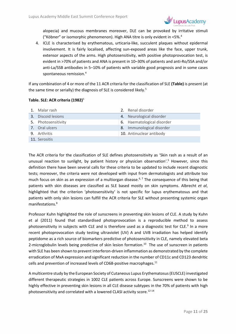
If any combination of 4 or more of the 11 ACR criteria for the classification of SLE (Table) is present (at
the same time or serially) the diagnosis of SLE is considered likely.5
Table. SLE: ACR criteria (1982)5
1. Malar rash 2. Renal disorder
3. Discoid lesions 4. Neurological disorder
5. Photosensitivity 6. Haematological disorder
7. Oral ulcers 8. Immunological disorder
9. Arthritis 10. Antinuclear antibody
11. Serositis
The ACR criteria for the classification of SLE defines photosensitivity as ‘Skin rash as a result of an
unusual reaction to sunlight, by patient history or physician observation‘.5 However, since this
definition there have been several calls for these criteria to be updated to include recent diagnostic
tests; moreover, the criteria were not developed with input from dermatologists and attribute too
much focus on skin as an expression of a multiorgan disease.6, 7 The consequence of this being that
patients with skin diseases are classified as SLE based mostly on skin symptoms. Albrecht et al,
highlighted that the criterion ‘photosensitivity‘ is not specific for lupus erythematosus and that
patients with only skin lesions can fulfill the ACR criteria for SLE without presenting systemic organ
manifestations.8
Professor Kuhn highlighted the role of sunscreens in preventing skin lesions of CLE. A study by Kuhn
et al (2011) found that standardised photoprovocation is a reproducible method to assess
photosensitivity in subjects with CLE and is therefore used as a diagnostic test for CLE.9 In a more
recent photoprovocation study testing ultraviolet (UV) A and UVB irradiation has helped identify
peptidome as a rich source of biomarkers predictive of photosensitivity in CLE, namely elevated beta
2-microglobulin levels being predictive of skin lesion formation.10 The use of sunscreen in patients
with SLE has been shown to prevent interferon-driven inflammation as demonstrated by the complete
erradication of MxA expression and significant reduction in the number of CD11c and CD123 dendritic
cells and prevention of increased levels of CD68-positive macrophages.11
A multicentre study by the European Society of Cutaneous Lupus Erythematosus (EUSCLE) investigated
different therapeutic strategies in 1002 CLE patients across Europe. Sunscreens were shown to be
highly effective in preventing skin lesions in all CLE disease subtypes in the 70% of patients with high
photosensitivity and correlated with a lowered CLASI activity score.12-14

Lupus Academy Middle East Summit Conference Report
Page 12 of 25
In 2005, the first validated score with specific evaluation of skin lesions was published as “Cutaneous
Lupus Erythematosus Disease Area and Severity Index” (CLASI).8 This score evaluates “activity” and
“damage” of the heterogeneous skin lesions of CLE by taking into account both anatomical regions
and morphological aspects. In 2010, the CLASI score was revised and modified by including additional
aspects of the mucocutaneous spectrum of the disease. Reliability analysis supported the validity and
applicability of the “revised CLASI” (RCLASI).15 Due to its detailed and comprehensive structure, the
RCLASI may be applied to support the diagnosis of the various CLE subtypes and to evaluate the
efficacy of treatment (Figure). Moreover, it is a valuable instrument for monitoring the disease on
different sites of involvement, not only in routine clinical practice but also in long-term clinical trials.
Figure. RCLASI.15
References
1. Bacman D, Ostendorf B, Megahed M, et al. [Bullous systemic lupus erythematosus]. Hautarzt.
2004;55(4):392-5.
2. Kuhn A, Landmann A, Bonsmann G. The skin in autoimmune diseases-Unmet needs. Autoimmun Rev.
2016;15(10):948-54.
3. Gilliam JN, Sontheimer RD. Distinctive cutaneous subsets in the spectrum of lupus erythematosus. J Am
Acad Dermatol. 1981;4(4):471-5.
4. Kuhn A, Landmann A. The classification and diagnosis of cutaneous lupus erythematosus. J Autoimmun.
2014;48-49:14-9.
5. Tan EM, Cohen AS, Fries JF, et al. The 1982 revised criteria for the classification of systemic lupus
erythematosus. Arthritis Rheum. 1982;25(11):1271-7.
6. Hochberg MC. Updating the American College of Rheumatology revised criteria for the classification of
systemic lupus erythematosus. Arthritis Rheum. 1997;40(9):1725.
7. Albrecht J, Berlin JA, Braverman IM, et al. Dermatology position paper on the revision of the 1982 ACR
criteria for systemic lupus erythematosus. Lupus. 2004;13(11):839-49.
8. Albrecht J, Taylor L, Berlin JA, et al. The CLASI (Cutaneous Lupus Erythematosus Disease Area and
Severity Index): an outcome instrument for cutaneous lupus erythematosus. J Invest Dermatol. 2005;125(5):889-94.

Lupus Academy Middle East Summit Conference Report
Page 13 of 25
9. Kuhn A, Wozniacka A, Szepietowski JC, et al. Photoprovocation in cutaneous lupus erythematosus: a
multicenter study evaluating a standardized protocol. J Invest Dermatol. 2011;131(8):1622-30.
10. Calderon C, Zucht HD, Kuhn A, et al. A multicenter photoprovocation study to identify potential biomarkers
by global peptide profiling in cutaneous lupus erythematosus. Lupus. 2015;24(13):1406-20.
11. Zahn S, Graef M, Patsinakidis N, et al. Ultraviolet light protection by a sunscreen prevents interferon-
driven skin inflammation in cutaneous lupus erythematosus. Exp Dermatol. 2014;23(7):516-8.
12. Biazar C, Sigges J, Patsinakidis N, et al. Cutaneous lupus erythematosus: first multicenter database
analysis of 1002 patients from the European Society of Cutaneous Lupus Erythematosus (EUSCLE). Autoimmun Rev. 2013;12(3):444-54.
13. Sigges J, Biazar C, Landmann A, et al. Therapeutic strategies evaluated by the European Society of
Cutaneous Lupus Erythematosus (EUSCLE) Core Set Questionnaire in more than 1000 patients with cutaneous lupus erythematosus. Autoimmun Rev. 2013;12(7):694-702.
14. Kuhn A, Sigges J, Biazar C, et al. Influence of smoking on disease severity and antimalarial therapy in
cutaneous lupus erythematosus: analysis of 1002 patients from the EUSCLE database. Br J Dermatol. 2014;171(3):571-9.
15. Kuhn A, Meuth AM, Bein D, et al. Revised Cutaneous Lupus Erythematosus Disease Area and Severity
Index (RCLASI): a modified outcome instrument for cutaneous lupus erythematosus. Br J Dermatol.
2010;163(1):83-92.

Lupus Academy Middle East Summit Conference Report
Page 14 of 25
Decreasing morbidity and mortality and improving outcomes in SLE – Ian Bruce (UK)
Professor Bruce presented the role of SDI scoring in the assessment and management of organ
damage. Highlighting that pre-existing damage is an important predictor of damage accrual and
mortality, Professor Bruce presented fixed risk factors (ie. age, race/ethnicity and male gender) factors
that increase risk (ie. asteroids, hypertension, high disease activity) of damage accrual and also
strategies to reduce this risk (ie. antimalarials, targeted therapies)
Professor Bruce began his presentation by highlighting that mortality associated with systemic lupus
erythematosus (SLE) has improved dramatically over the past 60 years, with the 1950s1 seeing a 50%
5-year mortality rate compared with a 10-year survival rate of >90% seen in more recent decades.2 In
addition, a meta-analysis of 12 studies by Yurkovich et al has also shown that mortality in SLE is
improving compared with the general population with the standardised mortality ratio falling from
>10.0 in the 1970s to 2.53.5 in the first decade of the new millennium.3,4
Professor Bruce highlighted data from long-term studies around the world, which identified the
leading causes of death in SLE to include cardiovascular disease, infections and SLE complications.5-8
Allthesame, with improvement in survival over the past 60 years, there is now increased focus on
outcomes, including irreversible SLE-associated organ damage.9 It is essential to understand damage
in SLE as higher levels of damage are associated with greater accumulation of damage in the future as
well as increased mortality.10-12 The development of the ACR/SLICC Damage Index (SDI) includes 12
organ systems and allows the assessment of overall long-term morbidity associated with SLE; it has
proved to be a consistent and reliable measure of damage over time allowing the prediction of both
further damage and also mortality.13 Professor Bruce reviewed several studies that assessed organ
damage in patients with SLE. A study by Nived et al highlighted the clinical relevance of the SDI for
survival in SLE. Nived found that SDI scores after 5 years have a high predictive value for survival during
the following median observation time of 7 years.11 Cardoso et al also found that baseline and accrued
organ damage, as measured by SDI, was useful in predicting increased risk of mortality over a 6.3 year
follow-up in Brazilian SLE patients.12 Similar results have also been found in Nordic patients with SLE,
with liner damage accrual occurring in 54% over a 10 year period.14 A systematic literature review of
50 articles by Sutton et al shows a clear pattern of damage increase overtime and that this is a
predictor of mortality.15
Having presented clear evidence of the association between damage accrual and mortality, Professor
Bruce reviewed the factors associated with damage accrual and poor prognosis in SLE, including older
age, race/ethnicity (including African ancestry), male gender, low socioeconomic status, active
disease/flares, renal involvement and steroids. In 1996 Stoll et al proved the validity of the SDI as a
predictor of severe outcome and indicator of morbidity in different ethnic groups, which showed that
Afro-Caribbeans and Asians had higher damage scores than Caucasians.16 A more recent study by
Alarcon et al (2004) found that key predictors of damage accrual in Hispanic, African-Americans and
Caucasians, included previous damage, age, disease activity and use of corticosteroids.17 It is clear
damage in SLE begets damage in SLE, and this is exactly what the SLICC inception cohort confirmed,
showing that patients with damage at baseline were more likely to have worsening SDI than those
without damage at baseline (Figure).3

Lupus Academy Middle East Summit Conference Report
Page 15 of 25
Figure. Time to SDI Worsening in SLICC Cohort.
Whole group Patients with and without initial damage
Reproduced from Ann Rheum Dis. 2015;74(9):1706-13.
Disease flares also result in future damage. A study of Chinese patients by Mok et al 2003, showed SDI scores (notably, renal, musculoskeletal and gonadal) to significantly increase over a 3 year period, with severe disease flares and cyclophosphamide being key predictors of new damage.18 Similarly, a Latin American cohort demonstrated that the greater the number of flares, regardless of severity and other risk factors, the higher the risk of damage accrual.19 Conversely, a study of Caucasian patients found that 37% of patients achieved a prolonged remission, which was associated with a better outcome in terms of damage accrual.20
As mentioned, steroids also beget damage.17 In a study of 73 patients, a significant proportion of damage (58%) was attributed to steroid use and this damage accumulated over time with 80% of damage being caused by steroids at Year 10.21 Steroid dose is also a significantly predictive factor for the rate of damage accrual, according to a study by Petri et al (2012).22 Conversely, hydroxychloroquine (HCQ) is associated with a reduced risk of damage accrual over time in patients who have not yet accrued damage at the time of treatment initiation.23
Reviewing the SLICC inception cohort, Professor Bruce highlighted the SDI transition rates before
presenting new approaches to reducing damage accrual in SLE.3 Presenting pooled data from the
BLISS-52 and BLISS-76 studies, Professor Bruce highlighted that long-term treatment with belimumab
(and standard of care) resulted in a low incidence of organ damage accrual, with the majority of
patients (85.1%) having no change in SDI at 5–6 years versus baseline.24 Moreover, the median time
to first worsening was 679 days in patients with no organ damage at baseline and 677 days in patients
with organ damage at baseline.
Concluding, Professor Bruce reiterated the main strategies for damage prevention and improving
outcomes, including better disease control and managing individual risk factors such as hypertension

Lupus Academy Middle East Summit Conference Report
Page 16 of 25
and hyperlipidaemia, introducing antimalarials, minimising steroid use, and recommending
vaccinations, cancer screening, and smoking cessation programmes.
References
1. Merrell M, Shulman LE. Determination of prognosis in chronic disease, illustrated by systemic lupus
erythematosus. J Chronic Dis. 1955;1(1):12-32.
2. Urowitz MB, Gladman DD, Abu-Shakra M, Farewell VT. Mortality studies in systemic lupus
erythematosus. Results from a single center. III. Improved survival over 24 years. J Rheumatol.
1997;24(6):1061-5.
3. Bruce IN, O'Keeffe AG, Farewell V, et al. Factors associated with damage accrual in patients with systemic
lupus erythematosus: results from the Systemic Lupus International Collaborating Clinics (SLICC) Inception Cohort. Ann Rheum Dis. 2015;74(9):1706-13.
4. Yurkovich M, Vostretsova K, Chen W, Avina-Zubieta JA. Overall and cause-specific mortality in patients
with systemic lupus erythematosus: a meta-analysis of observational studies. Arthritis Care Res (Hoboken). 2014;66(4):608-16.
5. Abu-Shakra M, Urowitz MB, Gladman DD, Gough J. Mortality studies in systemic lupus erythematosus.
Results from a single center. I. Causes of death. J Rheumatol. 1995;22(7):1259-64.
6. Cervera R, Khamashta MA, Font J, et al. Morbidity and mortality in systemic lupus erythematosus during
a 10-year period: a comparison of early and late manifestations in a cohort of 1,000 patients. Medicine (Baltimore). 2003;82(5):299-308.
7. Chambers SA, Allen E, Rahman A, Isenberg D. Damage and mortality in a group of British patients with
systemic lupus erythematosus followed up for over 10 years. Rheumatology (Oxford). 2009;48(6):673-5.
8. Wang Z, Wang Y, Zhu R, et al. Long-term survival and death causes of systemic lupus erythematosus in
China: a systemic review of observational studies. Medicine (Baltimore). 2015;94(17):e794.
9. Strand V, Gladman D, Isenberg D, et al. Outcome measures to be used in clinical trials in systemic lupus
erythematosus. J Rheumatol. 1999;26(2):490-7.
10. Rahman P, Gladman DD, Urowitz MB, Hallett D, Tam LS. Early damage as measured by the SLICC/ACR
damage index is a predictor of mortality in systemic lupus erythematosus. Lupus. 2001;10(2):93-6.
11. Nived O, Jonsen A, Bengtsson AA, Bengtsson C, Sturfelt G. High predictive value of the Systemic Lupus
International Collaborating Clinics/American College of Rheumatology damage index for survival in systemic lupus erythematosus. J Rheumatol. 2002;29(7):1398-400.
12. Cardoso CR, Signorelli FV, Papi JA, Salles GF. Initial and accrued damage as predictors of mortality in
Brazilian patients with systemic lupus erythematosus: a cohort study. Lupus. 2008;17(11):1042-8.
13. Gladman DD, Urowitz MB, Goldsmith CH, et al. The reliability of the Systemic Lupus International
Collaborating Clinics/American College of Rheumatology Damage Index in patients with systemic lupus erythematosus. Arthritis Rheum. 1997;40(5):809-13.
14. Becker-Merok A, Nossent HC. Damage accumulation in systemic lupus erythematosus and its relation to
disease activity and mortality. J Rheumatol. 2006;33(8):1570-7.
15. Sutton EJ, Davidson JE, Bruce IN. The systemic lupus international collaborating clinics (SLICC) damage
index: a systematic literature review. Semin Arthritis Rheum. 2013;43(3):352-61.
16. Stoll T, Seifert B, Isenberg DA. SLICC/ACR Damage Index is valid, and renal and pulmonary organ scores
are predictors of severe outcome in patients with systemic lupus erythematosus. Br J Rheumatol.
1996;35(3):248-54.
17. Alarcon GS, Roseman JM, McGwin G, Jr., et al. Systemic lupus erythematosus in three ethnic groups.
XX. Damage as a predictor of further damage. Rheumatology (Oxford). 2004;43(2):202-5.
18. Mok CC, Ho CT, Wong RW, Lau CS. Damage accrual in southern Chinese patients with systemic lupus
erythematosus. J Rheumatol. 2003;30(7):1513-9.
19. Ugarte-Gil MF, Acevedo-Vasquez E, Alarcon GS, et al. The number of flares patients experience impacts
on damage accrual in systemic lupus erythematosus: data from a multiethnic Latin American cohort. Ann Rheum Dis. 2015;74(6):1019-23.
20. Zen M, Iaccarino L, Gatto M, et al. Prolonged remission in Caucasian patients with SLE: prevalence and
outcomes. Ann Rheum Dis. 2015;74(12):2117-22.
21. Gladman DD, Urowitz MB, Rahman P, Ibanez D, Tam LS. Accrual of organ damage over time in patients
with systemic lupus erythematosus. J Rheumatol. 2003;30(9):1955-9.
22. Petri M, Purvey S, Fang H, Magder LS. Predictors of organ damage in systemic lupus erythematosus: the
Hopkins Lupus Cohort. Arthritis Rheum. 2012;64(12):4021-8.

Lupus Academy Middle East Summit Conference Report
Page 17 of 25
23. Fessler BJ, Alarcon GS, McGwin G, Jr., et al. Systemic lupus erythematosus in three ethnic groups: XVI.
Association of hydroxychloroquine use with reduced risk of damage accrual. Arthritis Rheum. 2005;52(5):1473-80.
24. Bruce IN, Urowitz M, van Vollenhoven R, et al. Long-term organ damage accrual and safety in patients
with SLE treated with belimumab plus standard of care. Lupus. 2016;25(7):699-709.
Lupus Academy Middle East Summit Conference Report
Page 18 of 25
Lupus Nephritis: Update on modern management – Liz Lightstone (UK)
Professor Lightstone reviewed the evolving management of lupus nephritis, presenting both the
standard of care and importance of focusing on steroid-sparing regimens when treating and
developing new treatments for lupus nephritis.
Lupus nephritis is one of the most serious complications of systemic lupus erythematosus (SLE), with
overt kidney disease present in 40-60% of patients at the time of diagnosis. Lupus nephritis affects
those from non-Caucasian backgrounds more severely than those from Caucasian backgrounds.
Induction of remission improves patient survival, with ≥50% reduction in proteinuria at 6 months being
linked to a significant improvement in 15 year survival in this group.1
Professor Lightstone presented a case study to asking the question when is biopsy is necessary and
when is it not? She highlighted the importance of preserving renal function and life, quality of life (QoL)
and fertility requires (1) rational targeted therapy related to pathophysiology, which induces remission
and prevents flares; (2) minimising treatment-related toxicities and improving QoL and survival by
getting rid of steroids, improving adherence and facilitating healthy pregnancies.
Lupus Nephritis Standard of Care
Focusing on treatment, the only current licenced medications for the treatment of lupus nephritis are
aspirin, steroids and hydroxychloroquine. None of the other treatments (cyclophosphamide [CyP],
mycophenolate mofetil [MMF], azathioprine, tacrolimus, rituximab—any biologic) regularly used are
licensed for treating SLE. Standard of care for induction is defined by the Euro-Lupus2, 3 and ALMS4, 5
trials, yet the unmet needs present themselves as low rates of complete remission as shown by the
ALMS,6 LUNAR,7 BELONG8 and ACCESS9 trials, while an analysis of studies in developed countries
between 2000–2006 by Tektonidou MG et al (2016) shows that a high percentage of patients still reach
end stage renal failure (ESRF) within 15 years of diagnosis.10 Although complete remission is judged
at 6/12 months in most trials, what matters to patients is long-term outcomes (ie. will my kidneys still
work in 10–20 years?). Recent studies looking at predictors of long-term outcomes may change our
notion of what we are trying to achieve in the short term. The Euro-Lupus trial found that low-dose CyP
was as effective as and less toxic than high-dose CyP and at 10 years there was no difference between
two groups for death, doubling serum creatinine/ESRF. Moreover, a significant fall in proteinuria at 3
and 6 months predicted good renal outcome.3 Dall’Era M et al (2015) also showed that proteinuria levels
of <0.8g/day at 12 months was the best predictor of good long-term renal function in the Euro-Lupus
Nephritis Trial.11 In addition, Professor Lightstone highlighted that long-term ALMS outcomes suggest
CyP induction is associated with less failure, with non-Hispanic patients more likely to achieve complete
remission. Factors associated with treatment failure included lack of treatment with hydroxychloroquine,
failure to reduce anti-dsDNA during the first 8 weeks of induction, failure to reduce UP/C during the first
8 weeks of induction and anti-dsDNA positivity at the end of induction.12 There is also growing evidence
demonstrating that multitarget therapy provides superior efficacy compared with intravenous CyP as
induction therapy for lupus nephritis. In a 26-centre TCT by Liu Z et al (2015), more patients in the
multitarget group (45.9%) achieved complete remission than in the intravenous cyclophosphamide
group (25.6%). Moreover, the multitarget group also achieved higher overall response rates and a
shorter time to overall response compared with CyP.13 Professor Lightstone highlighted the situations
when CyP should be used, these included:
• Severe extra renal disease (cerebral, myocarditis).
• RPGN picture – with steroids, ± PEX, ± rituximab.
• Commonest – 2nd line; when MMF has failed.
• When adherence is a predictable issue.

Lupus Academy Middle East Summit Conference Report
Page 19 of 25
Steroids and Steroid Avoidance
Professor Lightstone then highlighted toxicity issues with steroids. Having been introduced in 1951,
their inclusion in the lupus nephritis treatment paradigm was considered level 1a evidence. Most current
treatment protocols for LN involve oral steroids and often high dose. However, there has been no
systematic attempt to define correct dose of steroids, duration of steroids and how to wean steroids.
The general consensus being steroids on their own are not efficacious but contribute to infectious
complications and cause damage; moreover relapsing/remitting lupus nephritis means long-term
treatment with oral steroids is often necessary, with such chronic exposure leading to significant
morbidity and mortlity.14-18 In addition there is a dose-response relationship with steroids and the
adverse effects they produce.19 There is a clear need to improve response rates with new treatments
and reduce toxicity through steroid sparing regimens. Rovin BH et al (2012) assessed the efficacy and
safety of rituximab in patients with lupus nephritis treated with concomitant MMF and steroids
(LUNAR).7 Although rituximab therapy led to more responders and greater reductions in anti-dsDNA
and C3/C4 levels, it did not improve clinical outcomes after 1 year of treatment. This said, the primary
endpoint aimed to prove a 20% delta in complete and partial remission with major weighting towards
complete remission. Moreover, in addition to the positive secondary endpoints, several exploratory
endpoints at Week 78 favoured the rituximab regimen, including steroid sparing. It appears less is more
when it comes to improving outcomes with steroid sparing regimens. Zeher M et al (2011) found that
enteric-coated mycophenolate sodium may facilitate steroid reduction without loss of efficacy and fewer
infections.20 In addition, Ruiz-Irastorza G et al (2014) found that a combination of medium-dose
prednisone, methylprednisolone pulses, cyclophosphamide and hydroxychloroquine was at least as
effective as regimes containing high-dose prednisone in inducing remission and caused less toxicity.21
Despite rituximab and steroid sparing, there are currently no positive trials for the use of any biologic
for lupus nephritis. Experience from renal transplantation does however show that steroid avoidance
and introduction of an anti-IL-2R antibody and methyl prednisolone induction, one week of steroids and
then tacrolimus plus MMF resulted in a 20% reduction in rejection, >35% reduction in new onset
diabetes and 5 kg reduction in post-transplant weight gain over one year.22-24
Changing the Paradigm
Having presented the evidence for steroid sparing, Professor Lightstone focused on presenting the
rationale for changing the current treatment paradigm to steroid avoidance by replacing with biologic
monotherapy. The Rituxilup protocol is a single centre cohort study that evaluated the effectiveness of
treating lupus nephritis with rituximab and mycophenolate mofetil but no oral steroids.25 Of the 50
patients enrolled, 42 have reached 5 years follow-up, the majority of whom (88%) achieved complete
or partial remission and the majority of whom (77%) are steroid free. Relapses were only in patients
with Class IV or V disease, the majority responded to retreatment, again with no oral steroids. Given
these results, the open label, randomised, multi-centre, controlled Rituxilup trial (NCT01773616) will
now aim to demonstrate the combination of rituximab and no oral steroids to be as effective as MMF
and prednisolone in inducing renal remission.
Sustained disease remission and steroid reduction in lupus nephritis has also been demonstrated in
the RituxiRescue cohort with the RituxiRescue regimen (2 x 1g rituximab ± 125-500mg
methylprednisolone on Days 1 and 15, maintenance mycophenolate mofetil + no increase in b/line
steroid dose).26 At 1 year 62% of patients were in remission, with this increasing to 91% by Year 5
(Figure). Over 50% of patients were completely weaned off steroids by Year 5 with average steroid
doses falling from 14.9 mg/day at baseline to 5.8 mg/day by Year 5, with low rates of relapse.

Lupus Academy Middle East Summit Conference Report
Page 20 of 25
Figure. RituxiRescue cohort (n=37) 5 year remission rates
The calcineurin inhibitor, voclosporin (23.7 mg or 39.5 mg), in combination with MMF and steroids
improves rates of remission patients with active lupus nephritis, as demonstrated in the global
multicenter AURA trial.27 At Week 24 both low and high dose voclosporin were statistically superior to
placebo in partial remission and time to complete and partial remission. Voclosporin continues to be
studied in Phase III trials of lupus nephritis (NCT03021499).
Summarising, Professor Lightstone reinforced that it is time to change the treatment paradigm in lupus
nephritis and that there are potentially more effective and less toxic alternatives to steroids. Trials like
AURA are setting new benchmarks for steroid dosing. Steroid sparing regimens should be implemented
from the start of treatment and steroid minimisation a primary goal of all lupus trials.
References
1. Korbet SM, Lewis EJ. Severe lupus nephritis: the predictive value of a >/= 50% reduction in proteinuria at
6 months. Nephrol Dial Transplant. 2013;28(9):2313-8.
2. Houssiau FA, Vasconcelos C, D'Cruz D, et al. Immunosuppressive therapy in lupus nephritis: the Euro-
Lupus Nephritis Trial, a randomized trial of low-dose versus high-dose intravenous cyclophosphamide. Arthritis Rheum. 2002;46(8):2121-31.
3. Houssiau FA, Vasconcelos C, D'Cruz D, et al. The 10-year follow-up data of the Euro-Lupus Nephritis
Trial comparing low-dose and high-dose intravenous cyclophosphamide. Ann Rheum Dis. 2010;69(1):61-
4.
4. Appel GB, Contreras G, Dooley MA, et al. Mycophenolate mofetil versus cyclophosphamide for induction
treatment of lupus nephritis. J Am Soc Nephrol. 2009;20(5):1103-12.
5. Radhakrishnan J, Moutzouris DA, Ginzler EM, et al. Mycophenolate mofetil and intravenous
cyclophosphamide are similar as induction therapy for class V lupus nephritis. Kidney Int. 2010;77(2):152-
60.
6. Dooley MA, Jayne D, Ginzler EM, et al. Mycophenolate versus azathioprine as maintenance therapy for
lupus nephritis. N Engl J Med. 2011;365(20):1886-95.
7. Rovin BH, Furie R, Latinis K, et al. Efficacy and safety of rituximab in patients with active proliferative
lupus nephritis: the Lupus Nephritis Assessment with Rituximab study. Arthritis Rheum. 2012;64(4):1215-26.
8. Mysler EF, Spindler AJ, Guzman R, et al. Efficacy and safety of ocrelizumab in active proliferative lupus
nephritis: results from a randomized, double-blind, phase III study. Arthritis Rheum. 2013;65(9):2368-79.
9. Treatment of lupus nephritis with abatacept: the Abatacept and Cyclophosphamide Combination Efficacy
and Safety Study. Arthritis Rheumatol. 2014;66(11):3096-104.

Lupus Academy Middle East Summit Conference Report
Page 21 of 25
10. Tektonidou MG, Dasgupta A, Ward MM. Risk of End-Stage Renal Disease in Patients With Lupus
Nephritis, 1971-2015: A Systematic Review and Bayesian Meta-Analysis. Arthritis Rheumatol. 2016;68(6):1432-41.
11. Dall'Era M, Cisternas MG, Smilek DE, et al. Predictors of long-term renal outcome in lupus nephritis trials:
lessons learned from the Euro-Lupus Nephritis cohort. Arthritis Rheumatol. 2015;67(5):1305-13.
12. Dall'Era M, Levesque V, Solomons N, Truman M, Wofsy D. Identification of clinical and serological factors
during induction treatment of lupus nephritis that are associated with renal outcome. Lupus Sci Med. 2015;2(1):e000089.
13. Liu Z, Zhang H, Liu Z, et al. Multitarget therapy for induction treatment of lupus nephritis: a randomized
trial. Ann Intern Med. 2015;162(1):18-26.
14. Urowitz MB, Bookman AA, Koehler BE, et al. The bimodal mortality pattern of systemic lupus
erythematosus. Am J Med. 1976;60(2):221-5.
15. Petri M, Lakatta C, Magder L, Goldman D. Effect of prednisone and hydroxychloroquine on coronary
artery disease risk factors in systemic lupus erythematosus: a longitudinal data analysis. Am J Med. 1994;96(3):254-9.
16. Petri M. Detection of coronary artery disease and the role of traditional risk factors in the Hopkins Lupus
Cohort. Lupus. 2000;9(3):170-5.
17. Gladman DD, Urowitz MB, Rahman P, Ibanez D, Tam LS. Accrual of organ damage over time in patients
with systemic lupus erythematosus. J Rheumatol. 2003;30(9):1955-9.
18. Nossent J, Cikes N, Kiss E, et al. Current causes of death in systemic lupus erythematosus in Europe,
2000--2004: relation to disease activity and damage accrual. Lupus. 2007;16(5):309-17.
19. Ruiz-Irastorza G, Danza A, Khamashta M. Glucocorticoid use and abuse in SLE. Rheumatology (Oxford).
2012;51(7):1145-53.
20. Zeher M, Doria A, Lan J, et al. Efficacy and safety of enteric-coated mycophenolate sodium in combination
with two glucocorticoid regimens for the treatment of active lupus nephritis. Lupus. 2011;20(14):1484-93.
21. Ruiz-Irastorza G, Danza A, Perales I, et al. Prednisone in lupus nephritis: how much is enough?
Autoimmun Rev. 2014;13(2):206-14.
22. Borrows R, Loucaidou M, Van Tromp J, et al. Steroid sparing with tacrolimus and mycophenolate mofetil
in renal transplantation. Am J Transplant. 2004;4(11):1845-51.
23. Borrows R, Chan K, Loucaidou M, et al. Five years of steroid sparing in renal transplantation with
tacrolimus and mycophenolate mofetil. Transplantation. 2006;81(1):125-8.
24. Vincenti F. Is complete avoidance of calcineurin inhibitors or steroids now possible? Importance of patient
selection and choice of regimen. Transplant Proc. 2001;33(4 Suppl):11s-8s.
25. Condon MB, Ashby D, Pepper RJ, et al. Prospective observational single-centre cohort study to evaluate
the effectiveness of treating lupus nephritis with rituximab and mycophenolate mofetil but no oral steroids. Ann Rheum Dis. 2013;72(8):1280-6.
26. Pillay C, Griffith M, Levy J, Cairns T, Lightstone L. Treating Lupus Nephritis with Rituximab and
Mycophenolate Mofetil (RituxiRescue Regimen) without Increasing Maintenance Oral Steroids Leads to Sustained Disease Remission and Steroid Reduction. J Am Soc Nephrol. 2016;27:FR-PO597.
27. Pendergraft W, Tumlin J, Rovin B, et al. Successful Treatment of Active Lupus Nephritis with Voclosporin.
J Am Soc Nephrol. 2016;27:HI-OR05.

Lupus Academy Middle East Summit Conference Report
Page 22 of 25
Treat-to-target in SLE – Andrea Doria (Italy)
Professor Doria presented the concepts of T2T and LLDAS in SLE, highlighting the importance of
achieving remission and low disease activity state in minimising damage accrual and improving patient
outcomes in SLE.
Professor Doria began his presentation with a reminder of the poor long-term prognoses in SLE,
highlighting that key risk factors for long term complications in SLE include persistence of active
disease despite standard of care and side effects of treatments such as corticosteroids and
immunosuppressants.1, 2 In 1976 a long-term systematic analysis of 81 SLE patients over 5 years at the
University of Toronto Rheumatic Disease Unit revealed that 11 patients died, 6 of these within the
first year following diagnosis and 5 patients an average of 8.6 years after diagnosis.3 It was found that
those who died early in the disease with active lupus were taking high doses of steroids, and that those
who died late in the disease had been taking steroids long-term and there was a high incidence of
myocardial infarction. Thirty year later, Doria et al (2006) demonstrated an improvement in survival
in patients with SLE, however he noted that long-term prognosis is still poor in patients with severe
SLE manifestations.1 Treating-to-target (T2T) involves defined treatment goals in order to improve the
patient’s prognosis by addressing important unmet needs, such as treating persistent disease activity
manifesting and chronic active disease and relapsing-remitting disease and avoiding drug-related side
effects, especially those resulting from corticosteroid use.4 The principle of T2T is targeting a specific
value, not the symptoms, with the aim being to improve long-term outcomes such as organ damage.
T2T has already proven successful in the management of diabetes,5 hyperlipidaemia6 and
hypertension.7 T2T involves identifying a target for the individual patient, making a therapeutic
intervention, reassessing the patient after an established period of time and, if the target is not met,
modifying the intervention. Recently published recommendations for SLE suggest looking at clinical
remission or low disease activity (LDA) due the detrimental effects of persistent disease activity and
protracted corticosteroid therapy on patient outcomes.
T2T: Remission
Professor Doria focused on the application of T2T principles in SLE, highlighting that the treatment
target in SLE should be disease remission, or when this cannot be achieved, low disease activity should
be the therapeutic goals.8 There are however a number of different ad hoc definitions of remission
that have been used in clinical trials and observational studies.9 Such existence of so many definitions
highlights the need for amalgamation of SLE research whereby we can better understand the
treatment and outcomes for SLE patients. A recent publication by van Vollenhoven et al (2016)
outlines consensus (DORIS) findings on definitions of remission in SLE.10 The definitions of remission
in SLE would be worded as follows: “remission in SLE is a durable state characterized by … (reference
to symptoms, signs, routine labs). To define remission, a validated index must be used, e.g., clinical-
SLEDAI = 0, BILAG 2004 D/E only, clinical ECLAM =0; with routine laboratory assessments included,
and supplemented with Physician Global Assessment. Moreover, there must be distinction between
remission off therapy and remission on therapy, where remission-off-therapy requires the patient to
be on no other treatment for SLE than maintenance antimalarials; and remission-on-therapy allows
patients to be treated with maintenance antimalarials, stable low-dose glucocorticoids (prednisone
≤5 mg/d), stable maintenance immunosuppressives and/or stable maintenance biologics. The task
force also agreed that the most appropriate outcomes (dependent variables) for testing the prognostic
value (construct validity) of potential remission definitions are: Death, Damage, Flares, and measures

Lupus Academy Middle East Summit Conference Report
Page 23 of 25
of Health-related quality of life.”10 Professor Doria also presented work by Zen et al (2015), which
assess the prevalence effect of prolonged remission (≥5 consecutive years) on outcomes in Caucasian
patients with SLE (DORIA).11 The study defined three levels of remission using the SLE Disease Activity
Index (SLEDAI-2K):
1. Complete remission: no disease activity in steroid-free and immunosuppressant-free patients 2. Clinical remission off steroids 3. Clinical remission on steroids
Damage accrual was measured using the SLICC/American College of Rheumatology Damage Index (SDI). There are some notable similarities between the DORIS and DORIA definitions of remission (Table).8,
11 Table. Comparison of DORIA and DORIS definitions of remission.
DORIA1 DORIS2 Clinical Complete Clinical Complete on Cs off CS
on TX
off Tx
on TX
off Tx
cSLEDAI=0 ✓ ✓ ✓ ✓ ✓ ✓ ✓
PGA<0.5 - - - ✓ ✓ ✓ ✓
Anti-dsDNA/low C3/C4 ✓ ✓ - ✓ ✓ - -
HCQ/CQ ✓ ✓ ✓ ✓ ✓ ✓ ✓
Prednisone ≤5 mg/d ✓ - - ✓ - ✓ -
Immunosuppressants ✓ ✓ - ✓ - ✓ -
Biologics ✓ ✓ - ✓ - ✓ -
Of the 224 patients included in the study, at 5 years 7.1% achieved complete remission, 14.7%
achieved clinical remission of steroids and 15.6% achieved prolonged remission on steroids. Key
predictors of organ damage included patients with unremittent disease and those taking
corticosteroids.11
Professor Doria then considered what the shortest duration of remission was that would yield a
decrease in damage progression, presenting data from a 7-year follow-up study by Zen et al (2017),12
using the same classification criteria and the aforementioned study by Zen et al (2015). Prevalence of
remission ≥5 years was achieved in >38% of the cohort (n=293) (Figure).

Lupus Academy Middle East Summit Conference Report
Page 24 of 25
Figure. Prevalence of different durations (consecutive years) of remission in 293 patients during the
7-year follow-up.
Damage was similar, irrespective of the level of remission achieved in those achieving <4 years
remission, however damage was higher in patients in clinical remission for ≥5 years and taking
steroids. The study concluded that two consecutive years is the shortest duration of remission
associated with decrease in damage progression. Another study by Wilhelm et al (2016) used DORIS
to estimate rates and predictors of remission in a cohort of 2307 patients with SLE and found that high
baseline treatment was the major predictor of a longer time to remission, followed by high baseline
activity.13
T2T: LLDAS
When remission cannot be achieved we look towards a lupus low disease activity state (LLDAS). Low
disease activity has been defined and validated using LLDAS attainment, in a single-centre SLE cohort,
to predict non-accrual of irreversible organ damage, measured using the SDI (Table).14
Table. LLDAS definition.
1. SLEDAI-2K ≤4, with no activity in major organ systems (renal, CNS, cardiopulmonary, vasculitis, fever) and no haemolytic anaemia or gastrointestinal activity
2. No new features of lupus disease activity compared with the previous assessment
3. SELENA-SLEDAI physician global assessment (PGA, scale 0–3) ≤1
4. Current prednisolone (or equivalent) dose ≤7.5 mg daily
5. Well tolerated standard maintenance doses of immunosuppressive drugs and approved biological agents, excluding investigational drugs
Achievement of LLDAS was determined in 191 patients over a mean of 3.9 years. Patients spending
>50% in LLDAS had significantly reduced organ damage accrual compared with patients spending
<50% (p=0.0007); they were also significantly less likely to have an increase in SDI of ≥1 (p=0.005)
during the follow-up period or have severe flares at subsequent visits (p<0.0001). Another study by
Tsang et al (2016) sought to identify predictors of organ damage and specifically the relationship
38.6
11.69.6
16.8
15.48.9
5 years
unremitted
1 year
2 years
3 years
4 years

Lupus Academy Middle East Summit Conference Report
Page 25 of 25
between prolonged disease remission or low disease activity and damage accrual in a longitudinal
cohort of 183 SLE patients followed up over a mean of 5 years.15 They found that the most significant
predictors of damage included one major flare, mean daily prednisone dose during follow-up and
nephrological manifestations at baseline. Both prolonged remission and LLDAS were associated with
improved outcomes.
Professor Doria concluded by highlighting that, in everyday practice, patients should be brought to
the lowest level of disease activity, ensuring a significant benefit over a persistently active disease,
this being either clinical remission or LDA.16 However, complete remission, clinical remission or LDA
should not be seen as different treatment goals, rather they should be read as different scores of the
same lupus target, aiming to the highest degree of disease quiescence that can be applied to any
patient.
References
1. Doria A, Iaccarino L, Ghirardello A, et al. Long-term prognosis and causes of death in systemic lupus
erythematosus. Am J Med. 2006;119(8):700-6.
2. Hoes JN, Jacobs JW, Boers M, et al. EULAR evidence-based recommendations on the management of
systemic glucocorticoid therapy in rheumatic diseases. Ann Rheum Dis. 2007;66(12):1560-7.
3. Urowitz MB, Bookman AA, Koehler BE, et al. The bimodal mortality pattern of systemic lupus
erythematosus. Am J Med. 1976;60(2):221-5.
4. Doria A, Gatto M, Zen M, Iaccarino L, Punzi L. Optimizing outcome in SLE: treating-to-target and definition
of treatment goals. Autoimmun Rev. 2014;13(7):770-7.
5. Rachmani R, Slavacheski I, Berla M, Frommer-Shapira R, Ravid M. Treatment of high-risk patients with
diabetes: motivation and teaching intervention: a randomized, prospective 8-year follow-up study. J Am Soc Nephrol. 2005;16 Suppl 1:S22-6.
6. Egan BM, Lackland DT, Cutler NE. Awareness, knowledge, and attitudes of older americans about high
blood pressure: implications for health care policy, education, and research. Arch Intern Med. 2003;163(6):681-7.
7. Pearson TA, Ballantyne CM, Veltri E, et al. Pooled analyses of effects on C-reactive protein and low
density lipoprotein cholesterol in placebo-controlled trials of ezetimibe monotherapy or ezetimibe added to baseline statin therapy. Am J Cardiol. 2009;103(3):369-74.
8. van Vollenhoven RF, Mosca M, Bertsias G, et al. Treat-to-target in systemic lupus erythematosus:
recommendations from an international task force. Ann Rheum Dis. 2014;73(6):958-67.
9. van Vollenhoven RF, Voskuyl A, Morand E, Aranow C. Remission in SLE: closing in on the target. Ann
Rheum Dis. 2015;74(12):2103-6.
10. van Vollenhoven R, Voskuyl A, Bertsias G, et al. A framework for remission in SLE: consensus findings
from a large international task force on definitions of remission in SLE (DORIS). Ann Rheum Dis. 2016.
11. Zen M, Iaccarino L, Gatto M, et al. Prolonged remission in Caucasian patients with SLE: prevalence and
outcomes. Ann Rheum Dis. 2015;74(12):2117-22.
12. Zen M, Iaccarino L, Gatto M, et al. The effect of different durations of remission on damage accrual: results
from a prospective monocentric cohort of Caucasian patients. Ann Rheum Dis. 2017;76(3):562-5.
13. Wilhelm TR, Magder LS, Petri M. Remission in systemic lupus erythematosus: durable remission is rare.
Ann Rheum Dis. 2016.
14. Franklyn K, Lau CS, Navarra SV, et al. Definition and initial validation of a Lupus Low Disease Activity
State (LLDAS). Ann Rheum Dis. 2016;75(9):1615-21.
15. Tsang ASMW, Bultink IE, Heslinga M, Voskuyl AE. Both prolonged remission and Lupus Low Disease
Activity State are associated with reduced damage accrual in systemic lupus erythematosus. Rheumatology (Oxford). 2016.
16. Doria A, Gatto M, Iaccarino L, Punzi L. Value and goals of treat-to-target in systemic lupus erythematosus:
knowledge and foresight. Lupus. 2015;24(4-5):507-15.