
Lupus and the Skin - Netfirmslfaga.netfirms.com/lupus_and _the _skin.pdf · Lupus and the Skin 2015...
52
Lupus and the Skin 2015 Georgia Lupus Summit Presented by Cynthia Lawrence-Elliott, MD Member, LFAGA Medical Advisory Board Prepared by Frank Santoro, MD Assistant Professor of Dermatology at the University of Connecticut Board member, CT LFA chapter
Transcript of Lupus and the Skin - Netfirmslfaga.netfirms.com/lupus_and _the _skin.pdf · Lupus and the Skin 2015...

Lupus and the Skin
2015 Georgia Lupus Summit
Presented by Cynthia Lawrence-Elliott, MD
Member, LFAGA Medical Advisory Board
Prepared by Frank Santoro, MD
Assistant Professor of Dermatology at the University of Connecticut
Board member, CT LFA chapter

Lupus and the Skin Outline
• Discuss the importance of skin care in lupus
• Review the different types of cutaneous lupus
• Stress the role of sun protection and avoidance
• Provide an overview of newer therapies for cutaneous lupus

Why is the skin important in lupus?
• Patients with cutaneous lupus have a lower quality of life, particularly from an emotional standpoint
– No ethnic differences in impact on quality of life
– The emotional impact is similar to patients diagnosed with diabetes, high blood pressure, and heart attacks
• A flare of cutaneous lupus can lead to a systemic flare of lupus, including nephritis (kidney involvement)

Lupus and the Skin Outline
• Discuss the importance of skin care in lupus
• Review the different types of cutaneous lupus
• Stress the role of sun protection and avoidance
• Provide an overview of newer therapies for cutaneous lupus

Forms of cutaneous lupus
• Acute cutaneous lupus erythematosus (malar erythema, photodistributed eruption)most associated with systemic lupus erythematosus
• Chronic cutaneous LE
– Discoid lupus (DLE)
– Tumid lupus
– Lupus panniculitis
– Chilblain lupus
• Subacute cutaneous LE (SCLE)


Butterfly rashes of Acute Cutaneous LE
Patchy
Scaling Edematous

Post-inflammatory hyperpigmentation from lupus

Photodistribution of lupus (in sun-exposed areas)

Hands: Acute cutaneous lupus erythematosus

Oral ulcers of LE: both painless or painful
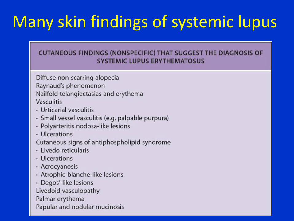
Many skin findings of systemic lupus

Flares of lupus cause non-scarring alopecia (hair loss)

Raynaud’s phenomenon

Raynaud’s phenomenon -3 colors: White, red, blue -severe cases can ulcerate

Primary (not worrisome) vs. secondary (due to an underlying problem)
Raynaud’s phenomenon

12/20/2011
6
Nailfold Capillary Changes
• Can be assessed with dermatoscope
• Look for…
– Dilated capillary loops
– Hemorrhage
– Capillary drop out/avascular areas
– Neoangiogenesis
Secondary Raynaud’s Phenomenon
• Systemic sclerosis (90 to 95% of patients)
• Mixed connective tissue disease (85%)
• Undifferentiated connective tissue disease
• Systemic lupus erythematosus (10 to 45%)
• Dermatomyositis (20%)
• Primary Sjogren’s syndrome (33%)
Lambova, S. N. and U. Muller-Ladner. The role of capillaroscopy in differentiation of primary and secondary Raynaud's phenomenon in rheumatic diseases: a
review of the literature and two case reports. Rheumatol Int. 2009. 29(11): 1263-71.
Take home messages thus far …
• “Scleroderma” now …
– Morphea = “localized scleroderma”
– Limited cutaneous systemic sclerosis (previously CREST)
– Diffuse cutaneous systemic sclerosis
– Systemic sclerosis sine scleroderma
• Raynaud’s phenomenon with nailfold capillary changes is VERY SUGGESTIVE of connective tissue disease

What other conditions can cause a red face?

Seborrheic dermatitis can resemble malar erythema
Seborrheic dermatitis can involve the scalp,
forehead, eyebrows, ears, beard, chest

Seborrheic dermatitis can resemble malar erythema

Rosacea can resemble malar erythema
Examination more likely rosacea if:
-Presence of red bumps and pus
-Redness crosses nasolabial folds

Another example of rosacea

Rhinophymatous rosacea

Sometimes rosacea vs. malar erythema is very difficult to differentiate
• Timing: Acute or chronic?
• Triggers: Stress, heat, alcohol, spicy foods?
• Any pimples ever?
• Other rashes?
• Photosensitivity (can be both)
• Associated symptoms: joint aches, fatigue more than usual, eye symptoms
• Labs: Role of ANA?

Forms of cutaneous lupus
• Acute cutaneous lupus erythematosus (malar erythema, photodistributed eruption)most associated with systemic lupus erythematosus
• Chronic cutaneous LE
– Discoid lupus (DLE)
– Lupus panniculitis
– Chilblain lupus
• Subacute cutaneous LE (SCLE)

Discoid lupus is in the criteria for systemic lupus, however, a patient can have discoid lesions commonly
without systemic lupus

Various clinical presentations of discoid lupus erythematosus (DLE)
Vitiligo-like lesions Active purple border with hypopigmentation and scar

DLE can be destructive
Conchal bowls in the ear
are a common site for DLE

Scarring alopecia due to DLE

Discoid lupus erythematosus (DLE)

Clinical tips about DLE
• Rarely progresses to systemic lupus erythematosus (SLE) --only 5-15%
– Though patients with SLE can have DLE lesions
– More likely to progress to SLE if widespread
• Patients can have arthralgias (joint aches)
• 24% can have lesions in the mouth
• Can develop squamous cell carcinomas (cancers) in chronic lesions

Lupus panniculitis—a form of cutaneous lupus in the fat

Chilblain lupus—a form of lupus that can appear on the toes, ears, nose

Forms of cutaneous lupus
• Acute cutaneous lupus erythematosus (malar erythema, photodistributed eruption)most associated with systemic lupus erythematosus
• Chronic cutaneous LE
– Discoid lupus (DLE)
– Lupus panniculitis
– Chilblain lupus
• Subacute cutaneous LE (SCLE)

SCLE (Subacute cutaneous lupus erythematosus)
• Two variants
– Annular (round)
– Psoriasiform (looks like psoriasis)
• Photodistributed, nonscarring
• Photosensitivity: 50%
• Labs: Often ANA + (80%), SSA +
• Some patients will meet SLE criteria
– 75% with arthritis or arthralgias
– 20% with leukopenia
• Recently, many of these patients have an associated medication causing their disease

Annular variant of SCLE

Psoriasiform SCLE

Lupus and the Skin Outline
• Discuss the importance of skin care in lupus
• Review the different types of cutaneous lupus
• Stress the role of sun protection and avoidance
• Provide an overview of newer therapies for cutaneous lupus

Sunscreens
• Suggest broad-spectrum UVA and UVB SPF 50 and above sunscreen.
– Apply every 2 hours while outside and more often if still symptoms
– Apply 15 minutes prior to sun exposure
– One ounce of sunscreen to exposed areas
– Best ingredients (physical sunblocks): titanium dioxide, zinc oxide
• Tend to not absorb that well, but better

There is proof that broad spectrum sunscreen works in lupus
In A-D: The skin was irradiated with UVA and UVB. Left box (no sunscreen) and right box (with sunscreen)
Kuhn Experimental Derm 2012

Sun avoidance
• It’s important to also avoid the sun
– Prefer to stay in the shade
– Avoid sun at peak hours between 11am-3pm
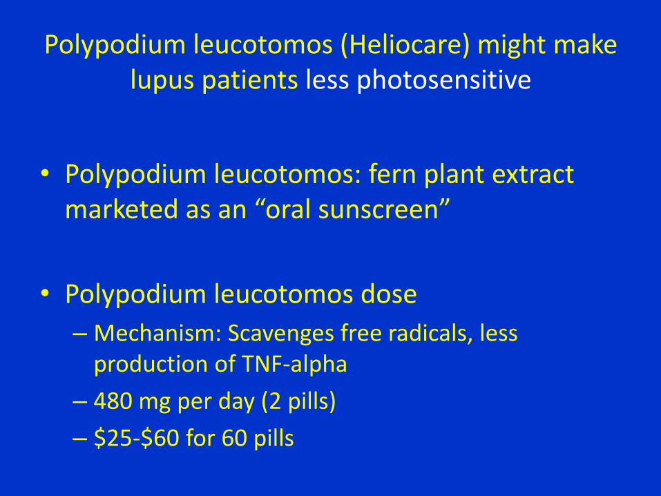
Polypodium leucotomos (Heliocare) might make lupus patients less photosensitive
• Polypodium leucotomos: fern plant extract marketed as an “oral sunscreen”
• Polypodium leucotomos dose
– Mechanism: Scavenges free radicals, less production of TNF-alpha
– 480 mg per day (2 pills)
– $25-$60 for 60 pills

Sun protective clothing
• Regular clothing does not protect well against UV light
• Increasingly there is sun-protective clothing (available online) that can block 98% of UV radiation
• Additive into the laundry: Can use a powder (can search for “Sung Guard) in the laundry to protect against UV light

Lupus and the Skin Outline
• Discuss the importance of skin care in lupus
• Review the different types of cutaneous lupus
• Stress the role of sun protection and avoidance
• Provide an overview of newer therapies for cutaneous lupus

Smoking cessation
• There is a link between smoking cigarettes and worse skin in lupus
– Patients also tend to be less responsive to antimalarial treatments when they smoke cigarettes
• Need to make attempts to quit smoking

Traditional therapies for cutaneous lupus
• Topical
– Corticosteroids
– Calcineurin inhibitors: tacrolimus (protopic), pimecrolimus (Elidel)
• Systemic
– Anti-malarials • Hydroxychloroquine
(Plaquenil)
• Chloroquine
• Quinacrine
– Corticosteroids
– Methotrexate
– Dapsone
– Azathioprine
– Mycophenolate

Melanocyte-keratinocyte transplantation to improve DLE hypopigmentation

Pulsed dye laser to treat active DLE lesions
6 years later

Belimumab (Benlysta) is a new treatment for SLE
• Mechanism
– Antibody to B lymphocyte stimulator (BLyS) involved in B cell activation. Depletes activated and naïve B cells and plasma cells.
– IV infusion: 0, 14, 28 and every 28 days
• Indications: For patients with active, autoantibody positive SLE without renal or CNS disease who have failed conventional therapy or have contraindications
• Some benefit in controlling disease. Skin disease responds slowly
• Unknown answers:
– What is the role of combination therapy?
– Does it have any effect as a first-line treatment?
– Will it help renal or CNS disease?
Still unanswered questions
• What’s the extent of the impact of the following on cutaneous lupus?
– Hormones/Gender?
– Genetics?
– Environmental exposures?
– Viruses/infections?

QUESTIONS?