Longitudinal Image Registration with Non-Uniform Appearance Change
8
Longitudinal Image Registration with Non-Uniform Appearance Change Istvan Csapo 1 , Brad Davis 3 , Yundi Shi 1 , Mar Sanchez 4 , Martin Styner 1 , and Marc Niethammer 1,2 1 University of North Carolina at Chapel Hill, NC 2 Biomedical Research Imaging Center, UNC Chapel Hill, NC 3 Kitware, Inc., Carrboro, NC 4 Emory University, Atlanta, GA [email protected] Abstract. Longitudinal imaging studies are frequently used to inves- tigate temporal changes in brain morphology. Image intensity may also change over time, for example when studying brain maturation. How- ever, such intensity changes are not accounted for in image similarity measures for standard image registration methods. Hence, (i) local sim- ilarity measures, (ii) methods estimating intensity transformations be- tween images, and (iii) metamorphosis approaches have been developed to either achieve robustness with respect to intensity changes or to si- multaneously capture spatial and intensity changes. For these methods, longitudinal intensity changes are not explicitly modeled and images are treated as independent static samples. Here, we propose a model-based image similarity measure for longitudinal image registration in the pres- ence of spatially non-uniform intensity change. 1 Introduction 0.5 3 6 12 18 Fig. 1. Brain slices and magnifications for a monkey at ages 2 weeks, 3, 6, 12, and 18 months. White matter appearance changes locally as ax- ons are myelinated during brain development. To study changes that occur dur- ing brain development, neurode- generation, or disease progres- sion in general, longitudinal imag- ing studies are important. Spatial correspondences almost always need to be established between images for longitudinal analysis through image registration. Most image registration methods have been developed to align images that are similar in appearance or structure. If such similarity is not given (e.g., in case of pathologies or for pre- and post-surgery images) cost function masking is typically used to discard image regions without correspondence from the reg- istration. Such strict exclusion is not always desirable. When investigating brain
Transcript of Longitudinal Image Registration with Non-Uniform Appearance Change

Longitudinal Image Registration withNon-Uniform Appearance Change
Istvan Csapo1, Brad Davis3, Yundi Shi1, Mar Sanchez4, Martin Styner1, andMarc Niethammer1,2
1 University of North Carolina at Chapel Hill, NC2 Biomedical Research Imaging Center, UNC Chapel Hill, NC
3 Kitware, Inc., Carrboro, NC4 Emory University, Atlanta, GA
Abstract. Longitudinal imaging studies are frequently used to inves-tigate temporal changes in brain morphology. Image intensity may alsochange over time, for example when studying brain maturation. How-ever, such intensity changes are not accounted for in image similaritymeasures for standard image registration methods. Hence, (i) local sim-ilarity measures, (ii) methods estimating intensity transformations be-tween images, and (iii) metamorphosis approaches have been developedto either achieve robustness with respect to intensity changes or to si-multaneously capture spatial and intensity changes. For these methods,longitudinal intensity changes are not explicitly modeled and images aretreated as independent static samples. Here, we propose a model-basedimage similarity measure for longitudinal image registration in the pres-ence of spatially non-uniform intensity change.
1 Introduction
0.5 3 6 12 18
0.5 3 6 12 180.5 3 6 12 180.5 3 6 12 180.5 3 6 12 180.5 3 6 12 18
Fig. 1. Brain slices and magnifications for amonkey at ages 2 weeks, 3, 6, 12, and 18 months.White matter appearance changes locally as ax-ons are myelinated during brain development.
To study changes that occur dur-ing brain development, neurode-generation, or disease progres-sion in general, longitudinal imag-ing studies are important. Spatialcorrespondences almost alwaysneed to be established betweenimages for longitudinal analysisthrough image registration. Mostimage registration methods havebeen developed to align imagesthat are similar in appearance or structure. If such similarity is not given (e.g.,in case of pathologies or for pre- and post-surgery images) cost function maskingis typically used to discard image regions without correspondence from the reg-istration. Such strict exclusion is not always desirable. When investigating brain

maturation for example (our target application in this paper) valid correspon-dences for the complete brain are expected to exist. However, brain appearancechanges continuously over time due to biological tissue changes (here, myelina-tion of white matter [1,12]) and adversely affects image registration results [9].
The effect of appearance change on the result of an image registration de-pends on the chosen transformation model and the chosen image similarity mea-sure. Generally, transformation models with few degrees of freedom (such as rigidor affine transformations) are affected less by local changes in image appearancethan transformation models which can capture localized spatial changes, suchas elastic or fluid models. In particular, we have previously shown that affinemethods perform well even in the presence of strong non-uniform appearancechange, while deformable methods introduce erroneous local deformations in or-der to resolve inconsistencies in appearance [3]. However, transformation modelswhich can capture local deformations are desirable for many longitudinal studiesas changes in morphology tends to be spatially non-uniform.
For longitudinal registration, temporal regularization of the transformationmodel has been explored recently. This is motivated by the assumption thatunrealistic local changes can be avoided by enforcing temporal smoothness ofa transformation [5,7]. In this paper we instead focus on the complementaryproblem of determining an appropriate image similarity measure for longitudinalregistration in the presence of temporal changes in image intensity.
Approaches which address non-uniform intensity changes have mainly ad-dressed registration for image-pairs so far and either rely on local image uni-formities [9,13] or try to estimate image appearance changes jointly with animage transform [8,11,10]. Often (e.g., for bias field compensation in magneticresonance imaging), image intensity changes are assumed to be smooth. Ourproposed approach in contrast, estimates local longitudinal models of intensitychange using all available images. Our approach alternates between parameterestimation for the local models of intensity change and estimation of the spa-tial transformation. Image similarities are computed relative to the estimatedintensity models, hence accounting for local changes in image intensities.
Section 2 introduces the model-based image similarity measure (mSM). Sec-tion 3 discusses parameter estimation. Section 4 describes the performed exper-iments and discusses results. The paper concludes with a summary and outlookon future work.
2 Model-Based Similarity Measure
Assume we have an image intensity model I(x, t; p) which for a parameterization,p, describes the expected intensity values for a given point x at a time t. Thismodel is defined in a spatially fixed target image. Then, instead of registeringa measured image Ii at ti to a fixed target image IT we can register it to itscorresponding intensity-adjusted image I(x, ti; p), effectively removing temporalintensity changes for a good model and a good parameterization, p. Hence,
Sim(Ii Φi, IT ) is replaced by Sim(Ii Φi, I(x, ti; p)),

where Sim(·, ·) is any chosen similarity measure (e.g., sum of squared differences(SSD), normalized cross correlation, or mutual information), and Φi is the mapfrom image Ii to the spatially fixed target space. Since our method aims tocreate an intensity adjusted model I that matches the appearance of the sourceimage, we use SSD in this paper. We call the intensity-adjusted SSD similaritymeasure a sum of squared residual (SSR) model, where the residual is definedas the difference between the predicted and the measured intensity value.
2.1 General Local Intensity Model Estimation for SSD
Since SSR is a local image similarity measure, for a given set of N measurementimages Ii at times ti we can write the full longitudinal similarity measureas the sum over the individual SSRs, i.e.,
SSR(Ii; p) =
N−1∑i=0
∫Ω
(Ii Φi(x)− I(x, ti; p))2 dx,
where Ω is the image domain of the fixed image. For given spatial transforms Φithis is simply a least-squares parameter estimation problem given the measure-ments IiΦi(x) and the predicted model values I(x, ti; p). We use alternatingoptimization with respect to the intensity model parameters, p, and the spatialtransformations Φi to convergence (see Sec. 3).
2.2 Logistic Intensity Model with Elastic Deformation
t
i
2w 3m 6m 12m
α
Fig. 2. Logistic intensitymodel.
SSR can be combined with any model for intensitychange, ranging from a given constant target im-age (the trivial model), linear models, and splines tomodels more closely adapted to the myelination pro-cess we are interested in capturing during neurode-velopment. Since the myelination process exhibits arapid increase during early brain development fol-lowed by a gradual leveling off [4], nonlinear appear-ance models are justified. In this paper we investigate the logistic model
I(x, t;α(x), β(x), k(x)) =α(x)
1 + β(x)e−k(x)t, (1)
which is often used in growth studies [6]. Here, α, β, and k are spatially varyingmodel parameters with biological meaning, k being the maximum rate of inten-sity change, α the maximum increase of white matter intensity during myelina-tion, and β is related to the onset time of myelination (see Fig. 2). Assumingthat both unmyelinated and fully myelinated white matter intensities are spa-tially uniform we keep α constant as the difference between myelinated (upperasymptote) and unmyelinated (lower asymptote) white matter intensities. Thisis a simplifying, but reasonable assumption since intensity inhomogeneities inunmyelinated or myelinated white matter are small compared to the white mat-ter intensity change due to the myelination process itself [1].

3 Parameter Estimation
Once the parameters for the local intensity models are known, SSR can be usedto replace the image similarity measure in any longitudinal registration method.Here, we use an elastic deformation model (Sec. 3.1) and jointly estimate theparameters for the intensity model (Sec. 3.2).
3.1 Registration model
The growth process of the brain not only includes appearance change but com-plex morphological changes as well, hence the need for a deformable transfor-mation model. To single out plausible deformations, we use (for simplicity) anelastic regularizer [2] defined on the displacement field u as
S[u] =
∫Ω
µ
4
d∑j,k=1
(∂xjuk + ∂xk
uj)2︸ ︷︷ ︸
rigidity
+λ
2(div u)2︸ ︷︷ ︸
volume change
dx,
where µ (=1) and λ (=0) are the Lame constants that control elastic behavior,and the div is the divergence operator defined as ∇ · u, where ∇ is the gradi-ent operator. Registrations over time then decouple into pairwise registrationbetween the intensity-adjusted target image and a given source image Ii. Thisis sufficient for our test of the longitudinal image similarity measure, but couldeasily be combined with a spatio-temporal regularizer which would then directlycouple the transformations between the images of a time-series (instead of onlyhaving an indirect coupling through the model-based similarity measure).
3.2 Model Parameter Estimation
We estimate the intensity model parameters only within the white matter (seg-mentation was obtained at the last time-point with an atlas-based segmentationmethod and propagated to earlier time-points [14]) where image appearancechanges non-uniformly over time; for simplicity, gray matter intensity was as-sumed to stay constant.
Note that overestimating the white matter results in fitting the model to thesurrounding gray matter voxels. This has negligible effect on the registration,since the model can capture the constant gray matter intensities. Underestimat-ing the white matter, on the other hand, can lead to uncorrected intensities nearthe white-gray matter boundary and introduce erroneous local deformations.
Instead of estimating the parameters independently for each voxel, spatialregularization was achieved by estimating the parameters from overlapping local3× 3× 3 neighborhoods using robust statistics (median of the parameters).

The algorithm is defined as follows
0) Initialize model I parameters to p = p0.1) Affinely pre-register images Ii to I.2) Estimate the appearance of I at times ti, giving I(ti).3) Estimate displacement fields ui by registering images Ii to I(ti).4) Estimate model parameters p from the registered images Ii ui.5) Repeat from step 2 until convergence.
The algorithm terminates once the registration energy decreases by less thana given tolerance between subsequent iterations. In all our experiments onlyfew iterations (typically less than 5) were required. A more in-depth numericalconvergence analysis should be part of future work. If desired, a prior modeldefined in the target image (a form of intensity model parameter atlas) couldeasily be integrated into this framework.
4 ExperimentalResults
12mo 6mo 3mo 2wk
Fig. 3. Corresponding target (cyan) and source (red)landmarks for a single subject.
We compared the model-based similarity measureto mutual information(MI) on sets of longitu-dinal magnetic resonanceimages of 9 monkeys, each with 4 time-points. Each set was affinely pre-registeredand intensity normalized so that the gray matter intensity distributions matchedafter normalization (gray matter intensity generally stays constant over time).
We registered 3D images of the three early time-points I2wk, I3mo, I6mo to thetarget image I12mo with an elastic registration method. Since the ground truthdeformations were not known, manually selected landmarks (Fig. 3) identifiedcorresponding regions of the brain at the different time-points (10-20 landmarksin a single slice for each of the 4 time-points in all 9 subjects; the landmarkswere picked based on geometric considerations). The distance between trans-formed and target landmarks yielded registration accuracy. Figure 4 shows theexperimental setup.
Note that for the model-based method the target image is not I12mo butthe model I12mo estimated at time t of the source image. Here, we estimate theintensity change of I12mo as white matter segmentation is easily obtained giventhe good gray matter white matter contrast, but other time-points could beused.
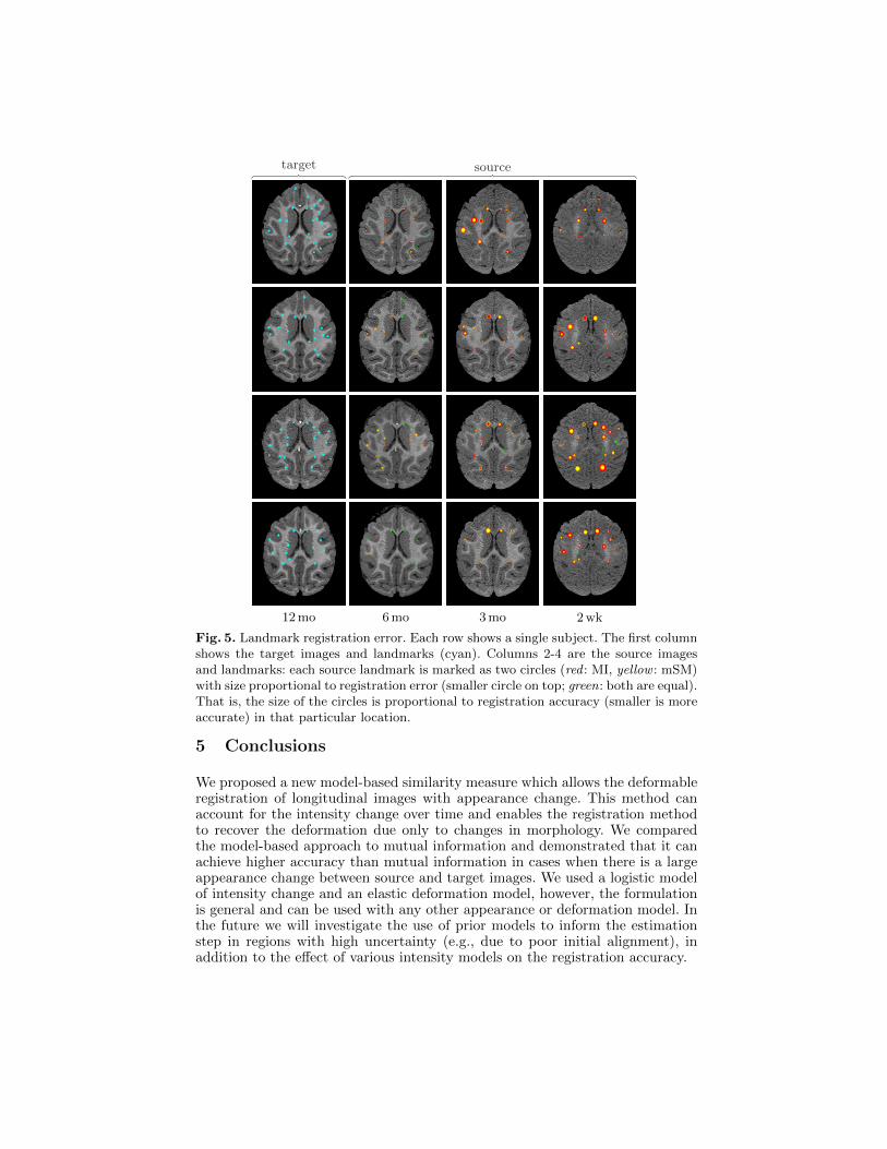
With MI, the registration method accounts for both the non-uniform whitematter appearance change and the morphological changes due to growth throughlarge local deformations. This is especially apparent for registrations betweenI2wk (and to a lesser extent I3mo) and the target I12mo and suggests large lo-cal morphological changes contradictory to normal brain development [4]. Thelandmark mismatch results (Fig. 5) show that both mutual information and

1
3
5
7
source model
original
mSM MI
Fig. 4. Experimental setup and results for a single subject. To test MI, the sourceimages (blue; from bottom: I2wk, I3mo, I6mo) are registered to the latest time-pointI12mo (green). The resulting deformation field and the magnitude of the deformations(in pixels) is shown in the right panel. For mSM, the source images are registered tothe model (red) that estimates the appearance of I12mo at the corresponding time ofeach source image (results in middle panel).
the model-based approach perform well in the absence of large intensity non-uniformity, however, mSM consistently introduces smaller erroneous deforma-tions than MI.
Table 1 shows the aggregate results of the landmark mismatch calculationsfor both methods. The model-based approach can account for appearance changeby adjusting the intensity of the model image (see the estimated model images inFig. 4) and therefore is most beneficial when the change in appearance betweenthe source and target image is large (I2wk, I3mo).
We also compared 1st and 2nd degree polynomial intensity models to thelogistic model and found no significant difference. This is expected with only4 time-points as even the 1st degree model can reasonably estimate the localappearance changes. However, we expect the logistic model to outperform thesimpler models in larger studies with more time-points or when the model ap-pearance needs to be extrapolated (e.g., if new images are acquired later in alongitudinal study after the previous time-points have been aligned). This willbe investigated as part of future work.
12 mo 6 mo 3 mo 2 wk
target source
Fig. 5. Landmark registration error. Each row shows a single subject. The first columnshows the target images and landmarks (cyan). Columns 2-4 are the source imagesand landmarks: each source landmark is marked as two circles (red : MI, yellow : mSM)with size proportional to registration error (smaller circle on top; green: both are equal).That is, the size of the circles is proportional to registration accuracy (smaller is moreaccurate) in that particular location.
5 Conclusions
We proposed a new model-based similarity measure which allows the deformableregistration of longitudinal images with appearance change. This method canaccount for the intensity change over time and enables the registration methodto recover the deformation due only to changes in morphology. We comparedthe model-based approach to mutual information and demonstrated that it canachieve higher accuracy than mutual information in cases when there is a largeappearance change between source and target images. We used a logistic modelof intensity change and an elastic deformation model, however, the formulationis general and can be used with any other appearance or deformation model. Inthe future we will investigate the use of prior models to inform the estimationstep in regions with high uncertainty (e.g., due to poor initial alignment), inaddition to the effect of various intensity models on the registration accuracy.

Table 1. Landmark registration error (in voxels) between target I12mo and source im-ages I2wk, I3mo, I6mo (significance level is α = 0.05; significant results are highlighted).
I2wk I3mo I6mo
mean std 50th 90th mean std 50th 90th mean std 50th 90th
MI 1.76 1.09 1.60 3.22 1.07 0.77 0.84 2.04 0.66 0.46 0.56 1.22mSM 1.15 0.84 0.93 2.24 0.74 0.57 0.54 1.66 0.61 0.39 0.50 1.18
Acknowledgments. This work was supported by NSF EECS-1148870, NSFEECS-0925875, NIH NIHM 5R01MH091645-02, NIH NIBIB 5P41EB002025-28, U54EB005149, P50 MH078105-01A2S1, P50 MH078105-01.
References
1. Barkovich, A.J., Kjos, B.O., Jackson, D.E., Norman, D.: Normal maturation of theneonatal and infant brain: MR imaging at 1.5T. Radiology 166, 173–180 (1988)
2. Broit, C.: Optimal registration of deformed images. Ph.D. thesis, University ofPennsylvania (1981)
3. Csapo, I., Davis, B., Shi, Y., Sanchez, M., Styner, M., Niethammer, M.:Temporally-dependent image similarity measure for longitudinal analysis. In: InPress, Proceedings of WBIR (2012)
4. Dobbing, J., Sands, J.: Quantitative growth and development of human brain.Archives of Disease in Childhood 48(10), 757–767 (1973)
5. Durrleman, S., Pennec, X., Trouve, A., Gerig, G., Ayache, N.: Spatiotemporalatlas estimation for developmental delay detection in longitudinal datasets. In:Proceedings of MICCAI. pp. 297–304. Springer (2009)
6. Fekedulegn, D., Siurtain, M.P.M., Colbert, J.J.: Parameter estimation of nonlineargrowth models in forestry. Silva Fennica 33(4), 327–336 (1999)
7. Fishbaugh, J., Durrleman, S., Gerig, G.: Estimation of smooth growth trajecto-ries with controlled acceleration from time series shape data. In: Proceedings ofMICCAI. pp. 401–408. Springer (2011)
8. Friston, K., Ashburner, J., Frith, C., Poline, J., Heather, J.D., Frackowiak, R.:Spatial registration and normalization of images. Human Brain Mapping 2, 165–189 (1995)
9. Loeckx, D., Slagmolen, P., Maes, F., Vandermeulen, D., Suetens, P.: Nonrigid imageregistration using conditional mutual information. IEEE Transactions on MedicalImaging 29(1), 19–29 (2010)
10. Miller, M.I., Younes, L.: Group actions, homeomorphisms, and matching: A generalframework. International Journal of Computer Vision 41(1/2), 61–84 (2001)
11. Roche, A., Guimond, A., Ayache, N., Meunier, J.: Multimodal elastic matching ofbrain images. In: Proceedings of ECCV. pp. 511–527. Springer (2000)
12. Sampaio, R.C., Truwit, C.L.: Myelination in the developing brain. In: Handbookof developmental cognitive neuroscience, pp. 35–44. MIT Press (2001)
13. Studholme, C., Drapaca, C., Iordanova, B., Cardenas, V.: Deformation-based map-ping of volume change from serial brain mri in the presence of local tissue contrastchange. IEEE Transactions on Medical Imaging 25(5), 626–639 (2006)
14. Styner, M., Knickmeyer, R., Coe, C., Short, S.J., Gilmore, J.: Automatic regionalanalysis of dti properties in the developmental macaque brain. Society of Photo-Optical Instrumentation Engineers (SPIE) Conference Series, vol. 6914 (2008)